


Abstract
The protective effect of physical activity on breast cancer recurrence may be mediated changes in by sex hormone levels. In this study, we examined the association between habitual physical activity and estrogen and androgen plasma levels in postmenopausal women with localised breast cancer. We conducted a cross-sectional study among 47 postmenopausal women who were breast cancer survivors with estrogen receptor-positive tumours (enrolled at the Medical Oncology Department of University Hospital Dr. Peset, Valencia, Spain). Habitual physical activity was assessed using the International Physical Activity Questionnaire (IPAQ), and a weighted estimate of total physical activity per week (MET∙min∙wk−1) was calculated. Total plasma levels of estrone, 17β-estradiol, progesterone, androstenedione, testosterone, and dehydroepiandrosterone-sulphate (DHEA-sulphate) were measured. Bivariate analyses by the Spearman correlation test were done between physical activity and each hormone concentration. Multivariate analyses (linear regression) using concentration of each hormone as the dependent variable and physical activity, age, marital status, BMI, Charlson Comorbidity Index, tumour stage, previous radiotherapy, or previous chemotherapy as predictor variables. Estrone concentration was positively and significantly correlated with BMI (ρ = 0.332, p = 0.022), but no other correlations were found between BMI and the other hormone concentrations, nor were concentrations of any hormone associated with age or Charlson Comorbidity Index (p > 0.05 in all cases). Physical activity was significantly and inversely correlated with estrone concentration (ρ = −0.308; p = 0.035). Linear regression analysis confirmed a statistically significant association between estrone concentration and BMI and physical activity, after adjusting for all potential confounders (for BMI: standardised β coefficient = 0.407; non-standardised β coefficient = 1.054; t = 2.898; p = 0.006; 95% CI for non-standardised beta: 0.318- to 1.790; for physical activity: standardised β coefficient = −0.300; non-standardised β coefficient = −0.005; t = −2.135; p = 0.039; 95% CI for non-standardised beta: −0.010- to 0.000). The relationship between estrone concentration and physical activity may be further explored as a biomarker for evaluating the protective effect of physical activity against breast cancer recurrence in women receiving anti-estrogen therapies.
Keywords:
survivorships; breast cancer; menopause; estrogens; testosterone; chemotherapy; comorbidities 1. Introduction
Breast cancer is the most common malignancy among women worldwide and constitutes one of the leading causes of female mortality. Despite increases in survival rates, it remains the leading cause of cancer-related death in women [1,2,3,4]. Approximately 70–80% of newly diagnosed breast cancers are hormone receptor-positive [5,6,7]. The role of estrogens is also crucial in breast cancer recurrence in women with estrogen receptor–positive breast cancer. In fact, lowering estrogen concentrations using aromatase inhibitors as adjuvant endocrine therapy in high-risk tumours is currently recommended for 5–10 years to prevent cancer relapse in postmenopausal women [5,8,9].
While estradiol is the most potent and predominant estrogen during reproductive life, estrone becomes the main circulating form after menopause [10,11,12,13]. These estrogens are primarily synthesised by peripheral conversion of androgens produced by the postmenopausal ovaries and adrenal glands [11,12,14]. Epidemiological evidence has linked elevated estrogen and androgen concentrations to an increased risk of estrogen receptor–positive breast cancer [15,16,17,18,19]. Androgens can directly stimulate cellular proliferation through androgen-responsive pathways, and may influence immune regulation and the microenvironment surrounding premalignant cells [20,21,22]. The adrenal hormone dehydroepiandrosterone sulphate (DHEA-sulphate) concentrations are generally increased in breast tumour tissue of women with breast cancer. DHEA-sulphate activity in breast cancer may be due to further metabolism to androstenedione, testosterone, or estradiol [19,23].
Strong evidence supports physical activity as a protective factor in preventing cancer and reducing its recurrence [24,25], including breast cancer [26,27,28,29]. In breast cancer specifically, physical activity is associated with reduced recurrence [30,31,32] and mortality [24,29,31]. Reports indicate that women who engage in regular physical activity have a 25% lower average risk than those who are inactive [33,34]. The strongest associations were observed for recreational activity, lifetime or postmenopausal activity, and regular, moderate-to-vigorous physical activity. The beneficial effects of physical activity have been attributed to reductions in body fat, modulation of estrogen metabolism, and regulation of inflammatory biomarkers [35,36,37,38,39].
Physical activity in postmenopausal women is associated with lower serum estrogen-to-androgen ratios, particularly reducing estradiol, while having a limited impact on overall androgen metabolite levels. While higher activity correlates with decreased estradiol and testosterone, studies often focus on total estrogen levels, leaving specific metabolic distinctions between estrone and estradiol in this demographic relatively understudied [40,41,42]. This distinction is important because estrone appears to play a more critical role than estradiol in postmenopausal women, including effects on skeletal muscle physiology, which may influence physical activity performance [43]. Additionally, low-intensity physical activities, such as walking, may exert differential effects on the hormonal profile, representing a topic of considerable interest for primary breast cancer prevention strategies [44,45,46]. Studies suggest that extended sedentary behaviour and low levels of physical activity may increase estrogen concentrations by enhancing the conversion of androgens through aromatisation [27,41,47,48,49].
The present study aimed to investigate the association between plasma estrogens, progestagens, and androgens (estrone, 17β-estradiol, progesterone, testosterone, androstenedione, and DHEA-sulphate) and physical activity in postmenopausal women with estrogen receptor-positive breast cancer. The hypothesis of this study is that the amount of physical activity is associated with the concentration of circulating estrogens and/or androgen levels in post-menopausal breast cancer survivors. The study specifically sought to explore potential associations between hormone concentrations and physical activity in breast cancer survivors. We also evaluated the contribution of clinically relevant variables, including age, Charlson Comorbidity Index, body mass index (BMI), tumour stage, and previous chemotherapy and radiotherapy, to the association between hormone concentrations and physical activity.
2. Results
2.1. Clinical and Sociodemographic Characteristics of the Sample
The clinical and sociodemographic characteristics of this cohort have been previously published in a longitudinal study conducted by García Sánchez et al. [50], which used the same participant sample. All women had hormone-dependent tumours with positive expression of estrogen and/or progesterone receptors, and were classified according to the TNM system as stage I (2.1%), stage II (72.1%), or stage III (25.5%). Detailed data are presented in Table 1. Regarding body mass index (BMI), and according to standard BMI categories [51], 10 women (21.3%) were of normal weight, 21 (44.7%) were overweight, and 16 (34.0%) were obese. All women enrolled in the study were evaluated prior to the start of treatment with aromatase inhibitors as an adjuvant therapy to prevent cancer recurrence. Patients who received adjuvant radiation therapy were evaluated an average of 2–3 months after breast cancer diagnosis, following surgery. In patients who received chemotherapy, the evaluation was performed after the completion of chemotherapy and prior to the start of aromatase inhibitor therapy, which occurred an average of 8 months after breast cancer diagnosis.
Table 1.
Sociodemographic and Clinical Data.
2.2. Baseline Characteristics of Hormone Levels and Physical Activity
Plasma hormone concentrations (mean ± standard deviation) were as follows: estrone 31.33 ± 13.57 pg/mL (range: 16.6–74.1 pg/mL), 17β-estradiol 7.51 ± 5.42 pg/mL (range: 5–34 pg/mL), progesterone 0.077 ± 0.036 ng/mL (range: 0.05–0.20), DHEA-sulphate 92.31 ± 55.95 µg/dL (range: 16.4–200.7 µg/dL), testosterone 0.27 ± 0.17 ng/mL (range: 0.1–1.0 ng/mL), and androstenedione 108.47 ± 61.63 pg/mL.
The mean daily moderate physical activity at baseline was 12.88 ± 3.77 min per day (range: 0–90). The average daily walking duration was 55.26 ± 5.73 min per day (range: 0–180). The total weekly physical activity score reached a mean value of 286.4 ± 233.8 min per week (range: 0–900). This corresponds to a mean MET score of 993.42 ± 810.65 MET/week (range 0–2970), based on the sum of the different reported activity intensities. No participants engaged in vigorous physical activity. Regarding the minimum recommended 150 min of physical activity per week, 30 women (66.7%) did not meet the recommendation, whereas 17 (33.3%) did.
2.3. Relationship Between Physical Activity and Hormone Levels
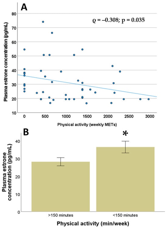
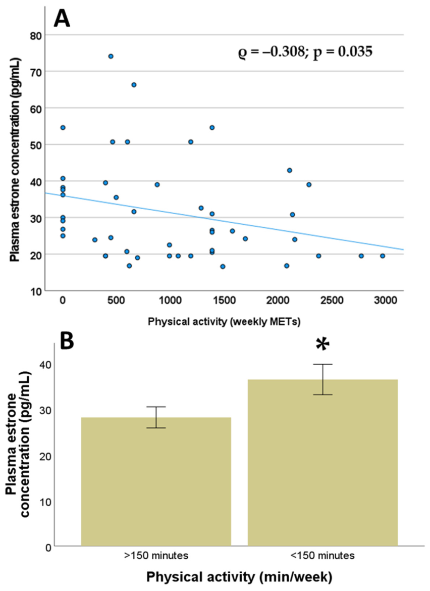
A significant negative correlation was observed between baseline physical activity and plasma estrone concentration (ρ = −0.308, p = 0.035), as illustrated in Figure 1A. In contrast, no significant correlation (p > 0.05) was found between baseline physical activity and the other hormone concentrations (Table 2).
Figure 1.
Physical activity and estrone concentration in plasma. (A) Correlation between plasma estrone concentration and weekly physical activity (MET/week) significant values (correlation coefficient and p value) are reported; (B) estrone concentration (expressed as mean ± standard deviation) between women who met or who did not meet the recommendation of 150 min of physical activity per week asterisk indicates a significant difference (p < 0.05) [52].
Table 2.
Associations between physical activity type and hormone concentration. Table indicated correlation coefficients and p values for the correlation analyses between weekly total physical activity (MET × min/week) and plasma concentrations of estrone, estradiol, progesterone, testosterone, androstenedione, and DHEA-sulphate. Asterisks indicate significant correlations (p < 0.05).
The sample was composed of a majority of sedentary women, with 66.7% not meeting the recommended 150 min per week of moderate-intensity aerobic activity. When we analysed the difference in estrone concentration between women who met or did not meet the recommendation, we found it to be significantly higher (p = 0.037) in those who did not meet it compared with those who did (Figure 1B). Estrone concentration was positively and significantly correlated with BMI (ρ = 0.332, p = 0.022), but no other correlations were found between BMI and the other hormone concentrations (p > 0.05 in all cases). When categorising women into normal-weight and overweight-obese groups, women in the latter group had significantly higher estrone concentrations compared with those of normal weight (33.03 ± 2.27 vs. 25.03 ± 3.50, p = 0.025, Mann–Whitney test). No differences were observed for the other hormones between normal-weight and overweight/obese women (p > 0.05, in all cases, Mann–Whitney test). No significant correlations or associations (p > 0.05) were observed in bivariate analyses between hormone concentrations and age, marital status, Charlson Comorbidity Index, time (months) since breast cancer diagnosis, tumour stage, previous radiotherapy, or previous chemotherapy.
2.4. Multivariate Analyses
Multivariate analyses (linear regression) using concentration of each hormone as the dependent variable and physical activity, age, marital status, BMI, Charlson Comorbidity Index, tumour stage, previous radiotherapy, or previous chemotherapy as predictor variables.
Linear regression analysis confirmed a statistically significant association between estrone concentration and BMI and physical activity, after adjusting for all potential confounders (R squared: 0.303; for BMI: standardised β coefficient = 0.407; t = 2.898; p = 0.006; 95% CI: 0.318- to 1.790; for physical activity: standardised β coefficient = −0.300; t = −2.135; p = 0.039; 95% CI: −0.010- to 0.000). None of the other variables were statistically significant (p > 0.05 in all cases). The results of the linear regression model are presented in Table 3. When considering as a dependent variable the concentration of 17β-estradiol, progesterone, androstenedione, testosterone, or DHEA-sulphate, physical activity was not significantly associated with the concentration of these hormones (p > 0.05, in all cases).
Table 3.
Linear regression analysis with weekly physical activity (MET/week) as the dependent variable and predictors. Predictors included in the regression model were plasma concentrations of estrogens, progesterone, and androgens, age, BMI, and relevant clinical data.
3. Discussion
Among postmenopausal breast cancer survivors with low-to-moderate physical activity, total physical activity (time or MET/week) was negatively associated with plasma estrone concentrations. This finding aligns with the previously demonstrated associations in postmenopausal women without breast cancer [53,54,55]. The amount and intensity of physical activity have long been recognised as independent predictors of serum estrone concentrations; for example, more active postmenopausal women have lower estrone levels [56]. Active postmenopausal women have approximately 5–20% lower estrone levels than non-physically active women [48,57]. A recent study reported an inverse relationship between estrone concentrations and non-recreational (occupational or household) physical activity, indicating that higher levels of daily movement or manual labour are associated with lower estrone levels [41]. In contrast to other studies, we did not find any association between plasma estradiol concentration and physical activity [47,52]. This is likely because such associations are more pronounced in non-obese women and in those engaging in high-intensity exercise [46,58]. In our sample, the majority of women were overweight or obese, and none engaged in high-intensity exercise. Similarly, a study conducted within the Prospect-EPIC project in postmenopausal women did not report any association between estrone concentrations and physical activity, assessed using a four-level classification that included time spent cycling, playing sports, and occupational activity [59]. However, these apparent discrepancies among studies could be explained by recent evidence supporting a non-linear relationship between physical activity and hormone concentrations in blood [52,60]. Compared with women reporting moderate activity, estrogen and androgen levels were generally lower among the most and least active women, indicating a U-shaped relationship [60].
We found that breast cancer survivors who were overweight or obese, as estimated by BMI, had higher plasma estrone concentrations. This confirms previous findings in postmenopausal women without cancer, in which concentrations of estradiol and estrone increased significantly with increasing BMI [61] and other measures of adiposity, such as waist and hip circumference [57,62]. McTiernan and colleagues [54] analysed the effects of a 12-month moderate-intensity exercise intervention on blood estrogen concentrations in sedentary, overweight postmenopausal women, compared with a control group who did not receive such an intervention. After three months, the women in the exercise group showed decreases in estrone and estradiol concentrations of approximately 4% and 8%, respectively, compared with no change or increases in the control group. The effect was limited to women who lost body fat during the intervention, as assessed by dual-energy X-ray absorptiometry. Interestingly, the variables we found to be associated with estrone concentration, namely BMI and physical activity, are also linked to postmenopausal breast cancer risk [34]. In multivariate analysis, the association between estrone concentration and physical activity remained significant after adjusting for BMI as a confounder, suggesting that factors beyond adiposity contribute to this relationship. In light of the findings regarding the association between estrone concentration and BMI, a limitation of this study is the potential for overadjustment bias regarding the role of BMI. While included as a covariate, BMI likely functions as both a confounder and a mediator in the pathway between physical activity and estrone levels [57,63]. By adjusting for BMI, our model may primarily capture the direct effect of physical activity, potentially masking the indirect effect mediated through adiposity reduction in non-overweight women. Furthermore, this relationship may be further conditioned by unmeasured dietary factors, which could act as residual confounders. While a subgroup analysis would offer a clearer interpretation of these interactions, our current sample size precluded such stratification. Consequently, our findings may provide a conservative estimate of the total impact of physical activity on estrone concentrations, and further studies would need to analyse the contribution of fat and lean composition and dietary factors.
Physical activity may influence estrogen levels through several pathways, including increased hepatic metabolism of estrogens [64], alterations in insulin sensitivity [65], and higher circulating levels of sex hormone–binding globulin, which reduces the bioavailability of circulating estrogens [66,67]. These mechanisms may partly explain why more physically active postmenopausal women tend to have lower circulating estrogen concentrations, independent of body fatness [47]. Physical activity typically has little to no direct impact on postmenopausal women’s already extremely low plasma progesterone levels. Consistent with other findings [68], plasma progesterone concentrations were not associated with physical activity in these patients.
Higher levels of habitual physical activity among postmenopausal women have been associated with lower endogenous testosterone in some studies [52,59]. Self-reported physical activity was associated with somewhat lower concentrations of total and calculated free testosterone (postmenopausal 5% and 11% difference, respectively, in the most versus least active group), whereas the opposite was true for self-reported sedentary time [69]. Other studies did not report any association between testosterone and physical activity [70]. To our knowledge, the association between physical activity and androgens has not been previously studied in postmenopausal breast cancer survivors. In our study, we did not observe any significant association between habitual physical activity and either testosterone or androstenedione, consistent with other reports [56,68]. The concentration of DHEA-sulphate, a weak androgen produced primarily by the adrenal glands, which acts as a circulating reservoir that can be converted into stronger androgens and estrogens, was not associated with physical activity [23,71]. Another study in postmenopausal women without breast cancer reported that high levels of physical activity are associated with higher levels of DHEA-sulphate [72]; however, in our sample, very few women engaged in high-intensity physical activity.
The present study has several limitations, including a small sample size which was calculated a priori based on a moderate correlation in bivariate analyses; however, when calculating the sample size for multiple regression analysis with eight predictors, the number should have been about 60–65 women, which would have reduced the generalizability of the present results. The cross-sectional design of the study prevents the determination of causality about the significant associations that emerged in this exploratory study. Multiple testing adjustment was not applied in this study because the analyses were exploratory and based on a limited number of pre-specified hypotheses focusing on the association between plasma hormone concentration and physical activity measures [73,74]. Given the exploratory nature of examining potentially complex biological relationships, applying strict corrections such as the False Discovery Rate could increase the risk of Type II errors and obscure biologically meaningful associations [75]. Therefore, results are presented without adjustment but interpreted with caution, and the need for replication in independent cohorts. Physical activity was measured using a well-validated self-reported method; however, the results should be interpreted with caution. In breast cancer patients, IPAQ-measured moderate and vigorous physical activity has been shown to overestimate activity by sevenfold and tenfold, respectively, compared with the accelerometer measurements [76]. The absence of participants engaging in vigorous activity, combined with the high proportion of not physically active women, resulted in restricted variability in activity levels. The sample was composed primarily of not physically active women, as 66.7% did not meet the minimum recommendation of 150 min of moderate-intensity aerobic activity per week [57], limiting the generalisability of the association between estrone and physical activity to women engaging in vigorous physical activity. This limited variability in physical activity in the study sample may have reduced the ability to detect potential associations with other hormones or a more powerful association with estrone concentration.
4. Materials and Methods
4.1. Design and Study Population
An analytical, cross-sectional study was conducted. The study enrolled postmenopausal women with non-metastatic hormone-dependent breast cancer (stages I–III) who had undergone surgery, radiation, or chemotherapy prior to starting anti-estrogen therapy with aromatase inhibitors. The Medical Oncology Department of Doctor Peset University Hospital in Valencia, Spain, conducted recruitment and assessments between Q4 2019 and Q1 2020. Exclusion criteria included the inability to understand Spanish, severe neuropsychiatric disorders, and physical impairments or mobility problems preventing them from taking part in regular and independent physical activity. A total of 47 patients agreed to participate and signed informed consent. The study was approved by the Clinical Research Ethics Committee of Doctor Peset University Hospital (protocol code 57/18; approved on 5 July 2018).
4.2. Clinical Data
Age, marital status, body mass index (BMI), Charlson Comorbidity Index [77], tumour stage and histology, and previous chemotherapy and/or radiotherapy were retrieved from medical records to evaluate potential modulating effects on the association between hormone concentrations and physical activity.
4.3. Physical Activity Assessment
Self-reported physical activity was assessed using the International Physical Activity Questionnaire (IPAQ), which evaluates hours of exercise per week and has been validated in Spanish [78]. The primary focus of the IPAQ is the quantity of physical activity performed during the past seven days. The IPAQ asks participants to report the time spent walking and performing moderate- and vigorous-intensity activities. Participants are asked to consider all types of physical activity within these categories. The reported minutes per week in each category were weighted by a MET (metabolic equivalent of task) value to calculate total physical activity over seven days. A weighted estimate of total physical activity per week (MET∙min∙wk−1) was calculated by multiplying duration (minutes), frequency (days), and MET intensity (walking = 3.3, moderate = 4, vigorous = 8), and adding together the results across the three domains [78].
4.4. Hormone Concentration Measurements
Blood samples were collected from fasting participants between 09:00 and 10:00 a.m. After collection, blood samples were centrifuged at 1500 rpm at room temperature for 5 min. Within three days of collection, the supernatant was frozen at −20 °C in 1 mL aliquots for subsequent analysis. Total plasma levels of 17β-estradiol, progesterone, testosterone, and DHEA-sulphate were measured using the chemiluminescent microparticle immunoassay (CMIA) on the Abbott Alinity platform. ELISA kits (Cloud-Clone Corp., Houston, TX, USA), reference cod CC-IEA456Ge; Arbor Assays (Ann Arbor, MI, USA), reference code AA-K031-H5) were used to measure androstenedione and estrone levels according to manufacturers’ instructions. Hormone levels were extrapolated using a standard calibration curve.
4.5. Statistical Analysis
The sample size was calculated based on the number of patients with breast cancer who started hormonal adjuvant treatment over a one-year period in the Oncology Department of Doctor Peset University Hospital, Valencia, with a total of 77 patients in 2021. The study sample size was calculated and estimated using two-tailed correlation tests with G*Power 3.1.9.2 software (G*Power©, Dusseldorf, Germany). A moderate correlation coefficient of r = 0.4, a two-tailed hypothesis, α = 0.05 (corresponding to a 95% confidence interval), β = 20%, and power (1 − β) = 0.80 were considered. As correlation coefficients between 0.3 and 0.7 are considered moderate [79], a value of 0.4 was assumed for the correlation coefficient between physical activity and hormone concentrations for sample size calculation. A sample size of at least 44 participants was therefore considered a priori appropriate for this study.
Descriptive statistics included frequency distributions for qualitative variables. For quantitative (discrete) variables, measures of central tendency (arithmetic mean), dispersion (standard deviation, SD), and range were calculated. Non-parametric tests were performed using the Mann–Whitney U-test. Spearman’s correlation was used to analyse the relationship between two quantitative (discrete) variables. The statistical relationship between two categorical variables was analysed using Pearson’s chi-square test. Linear regression analysis was carried out to determine associations and develop a predictive model for the dependent variable, e.g., each hormone concentration, and physical activity, adjusting for potential confounding variables (age, BMI, Charlson Comorbidity Index, tumour stage, and previous radiotherapy or chemotherapy). Collinearity among predictors in the regression models was assessed by calculating the variance inflation factor (VIF) and its reciprocal tolerance. These measures are based on the R2 value obtained by regressing each predictor on all other predictors in the regression analysis. A VIF of 1 indicates no correlation between a predictor and the remaining predictors, and therefore, the variance of the estimated regression coefficient is not inflated. The confidence level used for all analyses was 95%, with statistical significance set at p < 0.05. Data were analysed using IBM SPSS Statistics version 28.
5. Conclusions
The inverse association between physical activity and estrone concentration in breast cancer survivors may contribute to the reported inverse relationship between physical activity as a protective factor and breast cancer recurrence. Our study in postmenopausal women with estrogen receptor-positive breast cancer and prior starting adjuvant treatment with aromatase inhibitors suggests, taking into account the limitations of this study, that physical activity, by modulating estrone levels (directly and/or indirectly with BMI), may contribute to the effect of adjuvant aromatase inhibitor therapy in reducing the risk of breast cancer recurrence. However, this hypothesis should be tested in larger samples and in longitudinal studies during aromatase inhibitor treatment.
Author Contributions
Conceptualization: M.A.M.-E., J.G.S., G.C.-G., L.O.-P.d.V., M.D.T. and O.C.; methodology: M.A.M.-E., J.G.S., G.C.-G., L.O.-P.d.V., M.D.T. and O.C.; formal analysis: M.A.M.-E. and O.C.; investigation: M.A.M.-E., J.G.S., M.D.T. and O.C.; data curation: M.A.M.-E. and J.G.S.; writing—original draft preparation: M.A.M.-E., J.G.S., G.C.-G., L.O.-P.d.V., M.D.T. and O.C.; writing (review and editing): G.C.-G., L.O.-P.d.V., M.D.T. and O.C. All authors have read and agreed to the published version of the manuscript.
Funding
This research received no external funding.
Institutional Review Board Statement
The study was conducted in accordance with the Declaration of Helsinki, and approved by the Clinical Research Ethics Committee of Doctor Peset University Hospital in Valencia, Spain (protocol code 57/18; approved on 5 July 2018).
Informed Consent Statement
Informed consent was obtained from all subjects involved in the study.
Data Availability Statement
The data presented in this study are available from the corresponding author on reasonable request.
Conflicts of Interest
The authors declare no conflicts of interest.
References
- Siegel, R.L.; Kratzer, T.B.; Giaquinto, A.N.; Sung, H.; Jemal, A. Cancer statistics, 2025. CA Cancer J. Clin. 2025, 75, 10–45. [Google Scholar] [CrossRef] [Scilit]
- Baeyens-Fernández, J.A.; Molina-Portillo, E.; Pollán, M.; Rodríguez-Barranco, M.; Del Moral, R.; Arribas-Mir, L.; Sánchez-Cantalejo Ramírez, E.; Sánchez, M.-J. Trends in incidence, mortality and survival in women with breast cancer from 1985 to 2012 in Granada, Spain: A population-based study. BMC Cancer 2018, 18, 721. [Google Scholar] [CrossRef] [Scilit]
- Ghoncheh, M.; Pournamdar, Z.; Salehiniya, H. Incidence and Mortality and Epidemiology of Breast Cancer in the World. Asian Pac. J. Cancer Prev. 2016, 17, 43–46. [Google Scholar] [CrossRef] [Scilit]
- Bray, F.; Laversanne, M.; Sung, H.; Ferlay, J.; Siegel, R.L.; Soerjomataram, I.; Jemal, A. Global cancer statistics 2022: GLOBOCAN estimates of incidence and mortality worldwide for 36 cancers in 185 countries. CA Cancer J. Clin. 2024, 74, 229–263. [Google Scholar] [CrossRef] [Scilit]
- Dimauro, I.; Grazioli, E.; Antinozzi, C.; Duranti, G.; Arminio, A.; Mancini, A.; Greco, E.A.; Caporossi, D.; Parisi, A.; Luigi, L. Di Estrogen-Receptor-Positive Breast Cancer in Postmenopausal Women: The Role of Body Composition and Physical Exercise. Int. J. Environ. Res. Public Health 2021, 18, 9834. [Google Scholar] [CrossRef] [Scilit]
- Roulot, A.; Héquet, D.; Guinebretière, J.M.; Vincent-Salomon, A.; Lerebours, F.; Dubot, C.; Rouzier, R. Tumoral heterogeneity of breast cancer. Ann. Biol. Clin. 2016, 74, 653–660. [Google Scholar] [CrossRef] [Scilit] [PubMed]
- Park, S.; Koo, J.S.; Kim, M.S.; Park, H.S.; Lee, J.S.; Lee, J.S.; Kim, S.I.; Park, B.W. Characteristics and outcomes according to molecular subtypes of breast cancer as classified by a panel of four biomarkers using immunohistochemistry. Breast 2012, 21, 50–57. [Google Scholar] [CrossRef] [Scilit] [PubMed]
- Hadji, P.; Body, J.J.; Aapro, M.S.; Brufsky, A.; Coleman, R.E.; Guise, T.; Lipton, A.; Tubiana-Hulin, M. Practical guidance for the management of aromatase inhibitor-associated bone loss. Ann. Oncol. 2008, 19, 1407–1416. [Google Scholar] [CrossRef] [Scilit] [PubMed]
- Burstein, H.J.; Temin, S.; Anderson, H.; Buchholz, T.A.; Davidson, N.E.; Gelmon, K.E.; Giordano, S.H.; Hudis, C.A.; Rowden, D.; Solky, A.J.; et al. Adjuvant endocrine therapy for women with hormone receptor-positive breast cancer: American society of clinical oncology clinical practice guideline focused update. J. Clin. Oncol. 2014, 32, 2255–2269. [Google Scholar] [CrossRef] [Scilit]
- Bancroft, J.; Cawood, E.H.H. Androgens and the menopause; a study of 40-60-year-old women. Clin. Endocrinol. 1996, 45, 577–587. [Google Scholar] [CrossRef] [Scilit]
- Bulun, S.E.; Zeitoun, K.; Sasano, H.; Simpson, E.R. Aromatase in aging women. Semin. Reprod. Endocrinol. 1999, 17, 349–358. [Google Scholar] [CrossRef] [Scilit]
- Nelson, L.R.; Bulun, S.E. Estrogen production and action. J. Am. Acad. Dermatol. 2001, 45, S116–S124. [Google Scholar] [CrossRef] [Scilit]
- Madigan, M.P.; Troisi, R.; Potischman, N.; Dorgan, J.F.; Brinton, L.A.; Hoover, R.N. Serum hormone levels in relation to reproductive and lifestyle factors in postmenopausal women (United States). Cancer Causes Control 1998, 9, 199–207. [Google Scholar] [CrossRef] [Scilit] [PubMed]
- Masamura, S.; Adlercreutz, H.; Harvey, H.; Lipton, A.; Demers, L.M.; Santen, R.J.; Santner, S.J. Aromatase inhibitor development for treatment of breast cancer. Breast Cancer Res. Treat. 1995, 33, 19–26. [Google Scholar] [CrossRef] [Scilit] [PubMed]
- Muti, P. The role of endogenous hormones in the etiology and prevention of breast cancer: The epidemiological evidence. Recent Results Cancer Res. 2005, 166, 245–256. [Google Scholar] [CrossRef] [Scilit]
- Zhang, X.; Tworoger, S.S.; Eliassen, A.H.; Hankinson, S.E. Postmenopausal plasma sex hormone levels and breast cancer risk over 20 years of follow-up. Breast Cancer Res. Treat. 2013, 137, 883–892. [Google Scholar] [CrossRef] [Scilit]
- Kenemans, P.; Van Der Mooren, M.J. Androgens and breast cancer risk. Gynecol. Endocrinol. 2012, 28, 46–49. [Google Scholar] [CrossRef] [Scilit] [PubMed]
- Chiodo, C.; Morelli, C.; Cavaliere, F.; Sisci, D.; Lanzino, M. The Other Side of the Coin: May Androgens Have a Role in Breast Cancer Risk? Int. J. Mol. Sci. 2021, 23, 424. [Google Scholar] [CrossRef] [Scilit]
- Labrie, F.; Luu-The, V.; Labrie, C.; Simard, J. DHEA and its transformation into androgens and estrogens in peripheral target tissues: Intracrinology. Front. Neuroendocrinol. 2001, 22, 185–212. [Google Scholar] [CrossRef] [Scilit]
- Asirvatham, A.J.; Schmidt, M.; Gao, B.; Chaudhary, J. Androgens regulate the immune/inflammatory response and cell survival pathways in rat ventral prostate epithelial cells. Endocrinology 2006, 147, 257–271. [Google Scholar] [CrossRef] [Scilit]
- Ben-Batalla, I.; Vargas-Delgado, M.E.; von Amsberg, G.; Janning, M.; Loges, S. Influence of Androgens on Immunity to Self and Foreign: Effects on Immunity and Cancer. Front. Immunol. 2020, 11, 1184. [Google Scholar] [CrossRef] [Scilit]
- Hu, Y.-M.; Zhao, F.; Graff, J.N.; Chen, C.; Zhao, X.; Thomas, G.V.; Wu, H.; Kardosh, A.; Mills, G.B.; Alumkal, J.J.; et al. Androgen receptor activity inversely correlates with immune cell infiltration and immunotherapy response across multiple cancer lineages. bioRxiv 2024. [Google Scholar] [CrossRef] [Scilit]
- Chatterton, R.T. Dehydroepiandrosterone (DHEA) in relation to breast cancer. Vitam. Horm. 2025, 129, 61–97. [Google Scholar] [CrossRef] [Scilit] [PubMed]
- Mctiernan, A.; Friedenreich, C.M.; Katzmarzyk, P.T.; Powell, K.E.; Macko, R.; Buchner, D.; Pescatello, L.S.; Bloodgood, B.; Tennant, B.; Vaux-Bjerke, A.; et al. Physical Activity in Cancer Prevention and Survival: A Systematic Review. Med. Sci. Sports Exerc. 2019, 51, 1252–1261. [Google Scholar] [CrossRef] [Scilit] [PubMed]
- Brown, J.C.; Winters-Stone, K.; Lee, A.; Schmitz, K.H. Cancer, physical activity, and exercise. Compr. Physiol. 2012, 2, 2775–2809. [Google Scholar] [CrossRef] [Scilit] [PubMed]
- Fortner, R.T.; Brantley, K.D.; Tworoger, S.S.; Tamimi, R.M.; Rosner, B.; Holmes, M.D.; Willett, W.C.; Eliassen, A.H. Recreational physical activity and breast cancer risk by menopausal status and tumor hormone receptor status: Results from the Nurses’ Health Studies. Breast Cancer Res. Treat. 2024, 206, 77–90. [Google Scholar] [CrossRef] [Scilit]
- Smith, A.J.; Phipps, W.R.; Thomas, W.; Schmitz, K.H.; Kurzer, M.S. The Effects of Aerobic Exercise on Estrogen Metabolism in Healthy Premenopausal Women. Cancer Epidemiol. Biomark. Prev. 2013, 22, 756–764. [Google Scholar] [CrossRef] [Scilit]
- Guo, W.; Fensom, G.K.; Reeves, G.K.; Key, T.J. Physical activity and breast cancer risk: Results from the UK Biobank prospective cohort. Br. J. Cancer 2020, 122, 726–732. [Google Scholar] [CrossRef] [Scilit]
- Monninkhof, E.M.; Elias, S.G.; Vlems, F.A.; Van Der Tweel, I.; Schuit, A.J.; Voskuil, D.W.; Van Leeuwen, F.E. Physical activity and breast cancer: A systematic review. Epidemiology 2007, 18, 137–157. [Google Scholar] [CrossRef] [Scilit]
- Biganzoli, E.; Desmedt, C.; Demicheli, R. Does Physical Activity Have an Impact on Recurrence Dynamics in Early Breast Cancer Patients? J. Clin. Med. 2021, 10, 831. [Google Scholar] [CrossRef] [Scilit]
- Filis, P.; Markozannes, G.; Chan, D.S.; Mauri, D.; Foukakis, T.; Matikas, A.; Droufakou, S.; Pentheroudakis, G.; Tsilidis, K. Grading the evidence for physical activity and any outcome in cancer survivors: An Umbrella review of 740 meta-analytic associations. Crit. Rev. Oncol. Hematol. 2025, 207, 104602. [Google Scholar] [CrossRef] [Scilit]
- Zagalaz-Anula, N.; Mora-Rubio, M.J.; Obrero-Gaitán, E.; Del-Pino-Casado, R. Recreational physical activity reduces breast cancer recurrence in female survivors of breast cancer: A meta-analysis. Eur. J. Oncol. Nurs. 2022, 59, 102162. [Google Scholar] [CrossRef] [Scilit]
- Hildebrand, J.S.; Gapstur, S.M.; Campbell, P.T.; Gaudet, M.M.; Patel, A.V. Recreational physical activity and leisure-time sitting in relation to postmenopausal breast cancer risk. Cancer Epidemiol. Biomark. Prev. 2013, 22, 1906–1912. [Google Scholar] [CrossRef] [Scilit]
- Neilson, H.K.; Friedenreich, C.M.; Brockton, N.T.; Millikan, R.C. Physical activity and postmenopausal breast cancer: Proposed biologic mechanisms and areas for future research. Cancer Epidemiol. Biomark. Prev. 2009, 18, 11–27. [Google Scholar] [CrossRef] [Scilit]
- Mills, R.C. Breast Cancer Survivors, Common Markers of Inflammation, and Exercise: A Narrative Review. Breast Cancer Basic Clin. Res. 2017, 11, 1178223417743976. [Google Scholar] [CrossRef] [Scilit]
- Haley, J.S.; Hibler, E.A.; Zhou, S.; Schmitz, K.H.; Sturgeon, K.M. Dose-dependent effect of aerobic exercise on inflammatory biomarkers in a randomized controlled trial of women at high risk of breast cancer. Cancer 2020, 126, 329–336. [Google Scholar] [CrossRef] [Scilit] [PubMed]
- Jones, S.B.; Thomas, G.A.; Hesselsweet, S.D.; Alvarez-Reeves, M.; Yu, H.; Irwin, M.L. Effect of exercise on markers of inflammation in breast cancer survivors: The yale exercise and survivorship study. Cancer Prev. Res. 2013, 6, 109–118. [Google Scholar] [CrossRef] [Scilit] [PubMed]
- Marlin, S.; Goepp, M.; Desiderio, A.; Rougé, S.; Aldekwer, S.; Le Guennec, D.; Goncalves-Mendes, N.; Talvas, J.; Farges, M.C.; Rossary, A. Long-Term High-Fat Diet Limits the Protective Effect of Spontaneous Physical Activity on Mammary Carcinogenesis. Int. J. Mol. Sci. 2024, 25, 6221. [Google Scholar] [CrossRef] [Scilit] [PubMed]
- Michou, V.; Zervoudis, S.; Eskitzis, P.; Tsamos, G.; Vasdeki, D.; Vouxinou, A.; Markja, A.; Iatrakis, G. Exercise Interventions in Breast Cancer: Molecular Mechanisms, Physical Benefits, and Practical Recommendations. Medicina 2025, 61, 1167. [Google Scholar] [CrossRef] [Scilit]
- Schmitz, K.H.; Lin, H.; Sammel, M.D.; Gracia, C.R.; Nelson, D.B.; Kapoor, S.; DeBlasis, T.L.; Freeman, E.W. Association of physical activity with reproductive hormones: The penn ovarian aging study. Cancer Epidemiol. Biomark. Prev. 2007, 16, 2042–2047. [Google Scholar] [CrossRef] [Scilit]
- Oh, H.; Saquib, N.; Ochs-Balcom, H.M.; Pfeiffer, R.M.; Richey, P.A.; Shadyab, A.H.; Wild, R.A.; Underland, L.; Anderson, G.L.; Xu, X.; et al. Recreational Physical Activity, Sitting, and Androgen Metabolism among Postmenopausal Women in the Women’s Health Initiative Observational Study. Cancer Epidemiol. Biomark. Prev. 2022, 31, 97–107. [Google Scholar] [CrossRef] [Scilit] [PubMed]
- Shahid, W.; Noor, R.; Bashir, M.S. Effects of exercise on sex steroid hormones (estrogen, progesterone, testosterone) in eumenorrheic females: A systematic to review and meta-analysis. BMC Women’s Health 2024, 24, 354. [Google Scholar] [CrossRef] [Scilit] [PubMed]
- Enns, D.L.; Tiidus, P.M. The influence of estrogen on skeletal muscle: Sex matters. Sports Med. 2010, 40, 41–58. [Google Scholar] [CrossRef] [Scilit]
- Eliassen, A.H.; Hankinson, S.E.; Rosner, B.; Holmes, M.D.; Willett, W.C. Physical activity and risk of breast cancer among postmenopausal women. Arch. Intern. Med. 2010, 170, 1758–1764. [Google Scholar] [CrossRef] [Scilit] [PubMed]
- Hojman, P.; Gehl, J.; Christensen, J.F.; Pedersen, B.K. Molecular Mechanisms Linking Exercise to Cancer Prevention and Treatment. Cell Metab. 2018, 27, 10–21. [Google Scholar] [CrossRef] [Scilit]
- Ortega, M.A.; Fraile-Martínez, O.; García-Montero, C.; Pekarek, L.; Guijarro, L.G.; Castellanos, A.J.; Sanchez-Trujillo, L.; García-Honduvilla, N.; Álvarez-Mon, M.; Buján, J.; et al. Physical Activity as an Imperative Support in Breast Cancer Management. Cancers 2020, 13, 55. [Google Scholar] [CrossRef] [Scilit]
- Ennour-Idrissi, K.; Maunsell, E.; Diorio, C. Effect of physical activity on sex hormones in women: A systematic review and meta-analysis of randomized controlled trials. Breast Cancer Res. 2015, 17, 139. [Google Scholar] [CrossRef] [Scilit]
- Dallal, C.M.; Brinton, L.A.; Matthews, C.E.; Pfeiffer, R.M.; Hartman, T.J.; Lissowska, J.; Falk, R.T.; Garcia-Closas, M.; Xu, X.; Veenstra, T.D.; et al. Association of Active and Sedentary Behaviors with Postmenopausal Estrogen Metabolism. Med. Sci. Sports Exerc. 2016, 48, 439–448. [Google Scholar] [CrossRef] [Scilit]
- Oh, H.; Arem, H.; Matthews, C.E.; Wentzensen, N.; Reding, K.W.; Brinton, L.A.; Anderson, G.L.; Coburn, S.B.; Cauley, J.A.; Chen, C.; et al. Sitting, physical activity, and serum oestrogen metabolism in postmenopausal women: The Women’s Health Initiative Observational Study. Br. J. Cancer 2017, 117, 1070–1078. [Google Scholar] [CrossRef] [Scilit] [PubMed]
- García-Sánchez, J.; Mafla-España, M.A.; Tejedor-Cabrera, C.; Avellán-Castillo, O.; Torregrosa, M.D.; Cauli, O. Plasma Aromatase Activity Index, Gonadotropins and Estrone Are Associated with Frailty Syndrome in Post-Menopausal Women with Breast Cancer. Curr. Oncol. 2022, 29, 1744–1760. [Google Scholar] [CrossRef] [Scilit] [PubMed]
- Adult BMI Categories|BMI|CDC. Available online: https://www.cdc.gov/bmi/adult-calculator/bmi-categories.html (accessed on 6 March 2026).
- Choudhury, F.; Bernstein, L.; Hodis, H.N.; Stanczyk, F.Z.; Mack, W.J. Physical activity and sex hormone levels in estradiol- and placebo-treated postmenopausal women. Menopause 2011, 18, 1079–1086. [Google Scholar] [CrossRef] [Scilit]
- Cauley, J.A.; Gutai, J.P.; Kuller, L.H.; Ledonne, D.; Powell, J.G. The epidemiology of serum sex hormones in postmenopausal women. Am. J. Epidemiol. 1989, 129, 1120–1131. [Google Scholar] [CrossRef] [Scilit]
- McTiernan, A.; Tworoger, S.S.; Ulrich, C.M.; Yasui, Y.; Irwin, M.L.; Rajan, K.B.; Sorensen, B.; Rudolph, R.E.; Bowen, D.; Stanczyk, F.Z.; et al. Effect of exercise on serum estrogens in postmenopausal women: A 12-month randomized clinical trial. Cancer Res. 2004, 64, 2923–2928. [Google Scholar] [CrossRef] [Scilit]
- Matthews, C.E.; Sampson, J.N.; Brenner, D.R.; Moore, S.C.; Courneya, K.S.; Ziegler, R.G.; Friedenreich, C.M. Effects of Exercise and Cardiorespiratory Fitness on Estrogen Metabolism in Postmenopausal Women. Cancer Epidemiol. Biomark. Prev. 2018, 27, 1480–1482. [Google Scholar] [CrossRef] [Scilit] [PubMed]
- Liedtke, S.; Schmidt, M.E.; Becker, S.; Kaaks, R.; Zaineddin, A.K.; Buck, K.; Flesch-Janys, D.; Wahrendorf, J.; Chang-Claude, J.; Steindorf, K. Physical activity and endogenous sex hormones in postmenopausal women: To what extent are observed associations confounded or modified by BMI? Cancer Causes Control 2011, 22, 81–89. [Google Scholar] [CrossRef] [Scilit]
- McTiernan, A.; Wu, L.L.; Chen, C.; Chlebowski, R.; Mossavar-Rahmani, Y.; Modugno, F.; Perri, M.G.; Stanczyk, F.Z.; Van Horn, L.; Wang, C.Y. Relation of BMI and physical activity to sex hormones in postmenopausal women. Obesity 2006, 14, 1662–1677. [Google Scholar] [CrossRef] [Scilit] [PubMed]
- Friedenreich, C.M.; Woolcott, C.G.; McTiernan, A.; Ballard-Barbash, R.; Brant, R.F.; Stanczyk, F.Z.; Terry, T.; Boyd, N.F.; Yaffe, M.J.; Irwin, M.L.; et al. Alberta physical activity and breast cancer prevention trial: Sex hormone changes in a year-long exercise intervention among postmenopausal women. J. Clin. Oncol. 2010, 28, 1458–1466. [Google Scholar] [CrossRef] [Scilit]
- Chan, M.F.; Dowsett, M.; Folkerd, E.; Bingham, S.; Wareham, N.; Luben, R.; Welch, A.; Khaw, K.T. Usual physical activity and endogenous sex hormones in postmenopausal women: The European prospective investigation into cancer-norfolk population study. Cancer Epidemiol. Biomark. Prev. 2007, 16, 900–905. [Google Scholar] [CrossRef] [Scilit] [PubMed]
- Bertone-Johnson, E.R.; Tworoger, S.S.; Hankinson, S.E. Recreational physical activity and steroid hormone levels in postmenopausal women. Am. J. Epidemiol. 2009, 170, 1095–1104. [Google Scholar] [CrossRef] [Scilit]
- Key, T.J.; Appleby, P.N.; Reeves, G.K.; Roddam, A.; Dorgan, J.F.; Longcope, C.; Stanczyk, F.Z.; Stephenson, H.E.; Falk, R.T.; Miller, R.; et al. Body mass index, serum sex hormones, and breast cancer risk in postmenopausal women. J. Natl. Cancer Inst. 2003, 95, 1218–1226. [Google Scholar] [CrossRef] [Scilit] [PubMed]
- Key, T.J.; Appleby, P.; Barnes, I.; Reeves, G.; Dorgan, J.F.; Longcope, C.; Franz, C.; Stanczyk, F.Z.; Chang, L.C.; Stephenson, H.E.; et al. Endogenous Sex Hormones and Breast Cancer in Postmenopausal Women: Reanalysis of Nine Prospective Studies. J. Natl. Cancer Inst. 2002, 94, 606–616. [Google Scholar] [CrossRef] [Scilit]
- Arensberg, M.B.; Gahche, J.; Clapes, R.; Kerr, K.W.; Merkel, J.; Dwyer, J.T. Research is still limited on nutrition and quality of life among older adults. Front. Med. 2023, 10, 1225689. [Google Scholar] [CrossRef] [Scilit]
- Lavoie, J.M.; Pighon, A. NAFLD, Estrogens, and Physical Exercise: The Animal Model. J. Nutr. Metab. 2012, 2012, 914938. [Google Scholar] [CrossRef] [Scilit]
- Bird, S.R.; Hawley, J.A. Update on the effects of physical activity on insulin sensitivity in humans. BMJ Open Sport Exerc. Med. 2017, 2, e000143. [Google Scholar] [CrossRef] [Scilit]
- Kim, J.W.; Kim, D.Y. Effects of Aerobic Exercise Training on Serum Sex Hormone Binding Globulin, Body Fat Index, and Metabolic Syndrome Factors in Obese Postmenopausal Women. Metab. Syndr. Relat. Disord. 2012, 10, 452–457. [Google Scholar] [CrossRef] [Scilit]
- Atmaca, L.; Chauhan, S.; Najaf, S.S.; Elmadani, M.; Al-Jawarneh, M.; Soloh, A.; Varga, Z.; Vitrai, J.; Prémusz, V. Effectiveness of exercise interventions on androgen and sex hormone-binding globulin levels in women with polycystic ovary syndrome: A systematic review and meta-analysis. Front. Sport. Act. Living 2026, 7, 1686566. [Google Scholar] [CrossRef] [Scilit] [PubMed]
- Rinaldi, S.; Kaaks, R.; Friedenreich, C.M.; Key, T.J.; Travis, R.; Biessy, C.; Slimani, N.; Overvad, K.; Østergaard, J.N.; Tjønneland, A.; et al. Physical activity, sex steroid, and growth factor concentrations in pre- and post-menopausal women: A cross-sectional study within the EPIC cohort. Cancer Causes Control 2014, 25, 111–124. [Google Scholar] [CrossRef] [Scilit] [PubMed]
- Tin Tin, S.; Reeves, G.K.; Key, T.J. Body size and composition, physical activity and sedentary time in relation to endogenous hormones in premenopausal and postmenopausal women: Findings from the UK Biobank. Int. J. Cancer 2020, 147, 2101–2115. [Google Scholar] [CrossRef] [Scilit] [PubMed]
- Hawkins, M.; Tobias, D.K.; Alessa, H.B.; Chomistek, A.K.; Barnett, J.B.; Willett, W.C.; Hankinson, S.E. Objective and Self-Reported Measures of Physical Activity and Sex Hormones: Women’s Lifestyle Validation Study. J. Phys. Act. Health 2019, 16, 355–361. [Google Scholar] [CrossRef] [Scilit]
- Burger, H.G. The endocrinology of the menopause. Maturitas 1996, 23, 129–136. [Google Scholar] [CrossRef] [Scilit]
- Van Gils, C.H.; Peeters, P.H.M.; Schoenmakers, M.C.J.; Nijmeijer, R.M.; Onland-Moret, N.C.; Van Der Schouw, Y.T.; Monninkhof, E.M. Physical activity and endogenous sex hormone levels in postmenopausal women: A cross-sectional study in the Prospect-EPIC Cohort. Cancer Epidemiol. Biomark. Prev. 2009, 18, 377–383. [Google Scholar] [CrossRef] [Scilit] [PubMed]
- Ranganathan, P.; Pramesh, C.S.; Buyse, M. Common pitfalls in statistical analysis: The perils of multiple testing. Perspect. Clin. Res. 2016, 7, 106. [Google Scholar] [CrossRef] [Scilit] [PubMed]
- Streiner, D.L.; Norman, G.R. Correction for multiple testing: Is there a resolution? Chest 2011, 140, 16–18. [Google Scholar] [CrossRef] [Scilit] [PubMed]
- García-Pérez, M.A. Use and misuse of corrections for multiple testing. Methods Psychol. 2023, 8, 100120. [Google Scholar] [CrossRef] [Scilit]
- Biskup, M.; Macek, P.; Terek-Derszniak, M.; Zak, M.; Krol, H.; Falana, K.; Gozdz, S. Agreement between Accelerometer-Assessed and Self-Reported Physical Activity and Sedentary Behavior in Female Breast Cancer Survivors. Diagnostics 2023, 13, 3447. [Google Scholar] [CrossRef] [Scilit]
- Charlson, M.E.; Carrozzino, D.; Guidi, J.; Patierno, C. Charlson Comorbidity Index: A Critical Review of Clinimetric Properties. Psychother. Psychosom. 2022, 91, 8–35. [Google Scholar] [CrossRef] [Scilit] [PubMed]
- Sember, V.; Meh, K.; Sorić, M.; Jurak, G.; Starc, G.; Rocha, P. Validity and Reliability of International Physical Activity Questionnaires for Adults across EU Countries: Systematic Review and Meta Analysis. Int. J. Environ. Res. Public Health 2020, 17, 7161. [Google Scholar] [CrossRef] [Scilit]
- Kraemer, H.C. Correlation coefficients in medical research: From product moment correlation to the odds ratio. Stat. Methods Med. Res. 2006, 15, 525–545. [Google Scholar] [CrossRef] [Scilit]

Disclaimer/Publisher’s Note: The statements, opinions and data contained in all publications are solely those of the individual author(s) and contributor(s) and not of MDPI and/or the editor(s). MDPI and/or the editor(s) disclaim responsibility for any injury to people or property resulting from any ideas, methods, instructions or products referred to in the content. |
© 2026 by the authors. Licensee MDPI, Basel, Switzerland. This article is an open access article distributed under the terms and conditions of the Creative Commons Attribution (CC BY) license.