


Abstract
This study reports the fabrication of curcuminoid-loaded nanofibrillated cellulose (NFC)–alginate scaffolds via extrusion-based 3D printing, integrating supercritical CO2 extraction with biofabrication. NFC–alginate inks were pre-crosslinked with CaCl2 (1 to 10 mM) to tune structure and properties. Rheological analysis confirmed shear-thinning behavior suitable for extrusion printing. Mechanical testing revealed a non-linear dependence on crosslinking: optimal performance was achieved by Ink 2 (1 mM CaCl2 with curcuminoid extract), with tensile strength increasing from ~0.60 to ~0.80 MPa and Young’s modulus from ~1.5 to ~3.0 MPa relative (Ink 1, 10 mM CaCl2, without extract), reflecting the combined effect of extract incorporation and ionic pre-crosslinking rather than crosslinker concentration alone; higher crosslinking reduced stiffness (~1.15 MPa). SEM revealed porous architectures (Ink 1: 542 ± 63 μm; Ink 4: 398 ± 71 μm) with increasing structural heterogeneity upon curcuminoid incorporation. In vitro release exhibited biphasic, diffusion-dominated behavior, reaching ~50 to 60% in ethanol-containing media; PBS inclusion as a physiological reference confirmed minimal release (<5%), consistent with the known hydrophobicity and pH-dependent instability of curcuminoids and defining the physicochemical delivery boundaries of the system. The highest release (~372 ng/mL) was achieved at intermediate loading (10×). Kinetic modeling confirmed Higuchi-type diffusion as the dominant mechanism (R2 ≈ 0.90 to 0.99). These results establish a clear structure–property–release relationship and position the scaffolds as a tunable, diffusion-controlled delivery platform for hydrophobic bioactives in topical or formulation-assisted applications employing co-solvents or solubilizing excipients.
1. Introduction
The development of functional, bioactive scaffolds that combine structural integrity, tunable mechanical properties, and controlled release capabilities remains a central challenge in tissue engineering (TE) and biomedical materials science. Among various material platforms, polysaccharide-based systems have attracted significant attention due to their biocompatibility, biodegradability, and structural similarity to native extracellular matrices. In particular, nanofibrillated cellulose (NFC) has emerged as a promising building block owing to its high aspect ratio, mechanical robustness, and ability to form percolating fibrillar networks through hydrogen bonding and physical entanglement [1,2]. These features make NFC-based materials especially suitable for the fabrication of porous scaffolds with tailored mechanical and transport properties. Alginate (Alg), a naturally derived anionic polysaccharide, is widely used in combination with NFC to enhance processability and enable ionic crosslinking via divalent cations such as Ca2+. The resulting hybrid systems benefit from the shear-thinning behavior required for extrusion-based 3D printing while maintaining structural fidelity after deposition. Direct ink writing (DIW) has therefore become a powerful technique for fabricating architecturally controlled scaffolds with predefined geometries and hierarchical porosity [3,4]. However, achieving a balance between printability, mechanical stability, and functional performance remains challenging, particularly when incorporating bioactive compounds that may interfere with crosslinking mechanisms or alter the microstructure of the printed network.
Curcumin, a natural polyphenolic compound extracted from Curcuma longa L., has attracted considerable interest due to its well-documented antioxidant, anti-inflammatory, and antimicrobial properties [5,6,7]. Despite its therapeutic potential, its practical application is limited by poor aqueous solubility, low bioavailability, and chemical instability under physiological conditions [8,9]. Advanced encapsulation and delivery strategies are therefore required to stabilize curcumin and enable controlled release. Supercritical CO2 extraction represents an attractive, environmentally friendly method for obtaining curcuminoid-rich extracts without the use of toxic solvents, while preserving bioactivity [10,11]. Integrating such extracts into printable polysaccharide-based systems offers a promising route toward multifunctional scaffolds capable of both structural support and controlled bioactive release. Translation to therapeutic applications will, however, require comprehensive biological validation, including cytotoxicity assessment, cell compatibility studies, degradation profiling, and swelling characterization under physiological conditions, none of which were conducted in the present study. Recent studies have demonstrated that the incorporation of bioactive molecules into nanocellulose-based inks can significantly influence rheological behavior, crosslinking dynamics, and final scaffold performance [3,4]. In particular, interactions between curcuminoids and divalent ions may compete with Alg crosslinking sites, leading to changes in network homogeneity and mechanical response [5,6]. At the same time, controlled ionic crosslinking has been shown to enhance inter-fibrillar cohesion and improve mechanical strength, although excessive crosslinking may induce structural heterogeneity and brittleness [12,13]. These competing effects highlight the need for systematic investigation of formulation parameters to achieve optimal scaffold performance.
Although our group previously reported NFC–alginate systems containing supercritical CO2-derived curcuminoid extracts [3], the present work extends that earlier study by systematically examining CaCl2 pre-crosslinking as an independent formulation parameter and quantifying its influence on rheological behavior, thixotropic recovery, printability, scaffold morphology, tensile properties, and diffusion-controlled release kinetics. By varying both ionic pre-crosslinking concentration and extract loading, this study establishes the structure–property–release relationships that govern scaffold performance and elucidates the interplay between composition, processing, and functional outcome [14,15].
2. Materials and Methods
2.1. Material
Turmeric was sourced from Alfred Galke GmbH (Samtgemeinde Bad Grund, Germany), ground, and dried for processing. Ethanol (≥99.9%) was used as a solvent for ABTS+• analysis, while methanol (≥99.9%) and DPPH• (≥97.0%) were used for DPPH• assays (all from Sigma-Aldrich, St. Louis, MO, USA ). CO2 (99.99%) for supercritical extraction was obtained from MESSER (Ruše, Slovenia). Analytical standards—curcumin (≥98.0%), bisdemethoxycurcumin (≥95.0%), demethoxycurcumin (≥95.0%), along with ABTS+• salt, potassium persulfate, Trolox, Folin–Ciocalteu’s reagent, Na2CO3, FeSO4·7H2O, acetonitrile, formic acid, and solvents used for curcuminoid analysis were all purchased from Sigma-Aldrich (St. Louis, MO, USA). Anhydrous sodium acetate, glacial acetic acid, phosphate-buffered saline (PBS) tablets, and Tween20 (Sigma-Aldrich, St. Louis, MO, USA) were used for in vitro release study. Nanofibrillated cellulose suspension (3 wt%) was provided by Sappi Europe (Brussels, Belgium); alginate and CaCl2·2H2O by Sigma-Aldrich (Sigma-Aldrich, St. Louis, MO, USA).
2.2. Extraction Method
The protocol for the extraction of curcuminoid-rich extracts via supercritical fluid extraction (SFE) has already been described previously [10]. A lab-scale, high pressure autoclave made of stainless steel with a volume of 0.5 L was used for the extraction. A high-pressure autoclave filled with 25.00 g of turmeric powder was immersed in a water bath at three different temperatures of 40, 50, and 60 °C and pressures of 25, 30, and 35 MPa for a period of 180 min. The extraction conditions were determined based on previous reports [10,11] and previous extraction experience. The supercritical CO2 in the autoclave was pumped from the gas cylinder using a high-pressure pump with flow regulator (type 765.0700–18, SITEC-Sieber Engineering AG, Zurich, Switzerland). The constant flow of CO2 through the autoclave was maintained at approximately 3.5 mL/min throughout the experiment. The soluble components were dissolved by the supercritical CO2 flowing through the material and precipitated in separator (a pre-weighed glass vial) where the separation was performed at 0.1 MPa and at temperature of 0 °C. The extraction procedure was repeated in triplicate, and the extracts were stored at −18 °C until further use for ink preparation. The yield was calculated using Equation (1) based on the mass obtained after each extraction.
2.3. Extract Formulation
Ink Preparation with Pre-Crosslinking
The following procedure used for the preparation of NFC-Alg-curcuminoid inks has been described in detail previously, with some modifications [3,4]. In brief, 20.00 g of NFC (3.0 w%) was taken in a 50 mL falcon tube and stirred with laboratory mechanical stirrer (IKA EUROSTAR 20, IKA, Staufen, Germany) equipped with an in-house 3D printed stirrer at 1000–2000 rpm until no NFC fibers were seen (~15 min). After the initial mixing step, CaCl2·2H2O in a concentration range of 1 mM to 10 mM was added into the NFC for the pre-crosslinking, followed by an additional mixing cycle at 2000 rpm for 5 min. Subsequently, 1.33 g of Alg was added to the pre-crosslinked NFC, followed by an additional mixing step at 2000 rpm for 10 min. This resulted in a slightly homogenous ink, which was stored in a refrigerator at 2 to 8 °C or directly used for printing. This three-component system is referred to as Ink 1. For comparison, Inks 2–4 were prepared using the same procedure, but with the addition of the curcuminoid extract (0.4 mL) obtained at 40 °C and 35 MPa.
2.4. Direct-Ink-Writing (3D Printing) and Post-Ionic Crosslinking
3D printing of the ink was performed by using a BioScaffolder 3.2 (GeSim, Radeberg, Germany). Cubic shaped scaffolds (radius 8 mm, height 2.5–3.5 mm, number of corners: 4) were built in a layer-by-layer fashion. These dimensions were created using the GeSiM Robotics BioScaffolder 3.2 software. A 10 mL dark polyethylene-based plastic syringe (Nordson Limited, Dunstable, UK) with an inner nozzle (Nordson Limited, Dunstable, UK) diameter of 250 μm was used to dispense the ink onto a glass plate (dimension 75 mm × 25 mm). The syringe was tightly packed with ink and stored at 8 °C or used directly for printing. The scaffolds were printed with an extrusion pressure of 60 to 110 kPa and a printing speed of 15 mm/s. The spacing between strands was maintained at 1.0 mm, and the strand dimensions were 0.21 mm in height and 0.25 mm in width. The layers were deposited in alternating orientations, with each successive layer rotated by 90°.
To obtain more stable structure, post-crosslinking of the printed structures was carried out using a 1% CaCl2 solution. Following printing, 200 to 300 µL of the CaCl2 solution was applied to the surface of each scaffold. After allowing the solution to react for 5 min, the scaffolds were rotated 180° along the horizontal axis, and the cross-linking process was repeated on the opposite side.
2.5. Attenuated Total Reflectance Infrared (ATR-IR) Spectroscopy
ATR-IR spectra were recorded using an Agilent Cary 630 FTIR spectrometer (Agilent Technologies, Santa Clara, CA, USA) with the diamond ATR module at a scan range of 4000 to 650 cm−1 with a step of 1 cm−1 [16]. The scans were performed in three different places in 8 repetitions directly on as-prepared scaffolds. For this analysis, the crosslinked samples were air dried and used.
2.6. Rheology
The rheological properties of inks with different pre-crosslinker concentrations (1, 5, and 10 mM) were evaluated with a modular compact rheometer MCR 502 (Anton Paar, Graz, Austria) at 25 °C. The shear viscosity was measured at shear rates from 0.1 s–1 to 100 s–1 with 21 measuring points per 30 s for a total duration of 630 s. Oscillation amplitude sweeps were performed with a shear stress from 1 to 2000 Pa at a frequency of 1 Hz with a constant gap size of 0.5 mm and a constant compressive force set to 0.37 N. A three-interval thixotropic recovery test was performed under oscillatory shear, where the material was probed at a low strain amplitude (1st interval: γ = 0.1%, ω = 10 rad/s) for 600 s, subjected to structural breakdown at high strain amplitude (2nd interval: γ = 50%, ω = 10 rad/s) for 300 s, and subsequently monitored for structural recovery at the initial low strain amplitude (3rd interval) for 600 s. All rheological measurements were performed in at least four independent replicates.
The shear-thinning behavior of the NFC–alginate inks was described using the power-law fluid model with Equation (2):
where η is the dynamic viscosity, is the shear rate, K is the flow consistency index (Pa·sn), and n is the flow behavior index (dimensionless). Values of n < 1 confirm pseudoplastic, shear-thinning behavior. Model parameters were obtained by least-squares fitting of the experimental flow curves measured over a shear rate range of 0.1–100 s−1 with a parallel-plate geometry (gap: 0.5 mm). The fitted parameters for all four ink formulations are reported in Table 1.
Table 1.
Ink component composition and fitted power-law fluid parameters.
2.7. Tensile Testing
For the tensile tests, dog bone specimens (thickness: 1.00 to 2.50 mm) were punched out from 50 mm × 8.5 mm 3D printed specimen according to DIN 53504 S3A. The maximum stress (MPa), the elongation (%), and Young’s modulus (MPa) were measured using Shimadzu AGS-X (Shimadzu, Kyoto, Japan) universal mechanical testing machine with a speed of 50 mm min−1 and standard clamps. At least four specimens were tested for each condition.
2.8. Scanning Electron Microscopy (SEM)
The surface morphology of freeze-dried scaffolds was analyzed using a Quanta 200 3D (FEI, Hillsboro, OR, USA) electron microscope (SEM). The freeze-dried scaffolds were prepared according to the protocol published elsewhere [17,18]. The images were recorded under low vacuum conditions of 60 Pa with an acceleration voltage of 10 kV. The samples were mounted on sample holders using a double-sided carbon adhesive tape (SPI, West Chester, PA, USA), and a thin gold layer was sputtered onto the surface prior to analysis to enhance conductivity. A large field detector was employed to capture high-resolution micrographs at various magnifications from 100× to 5000×, allowing detailed observation of structural features and surface texture at a working distance of approximately 10 mm. All SEM analysis was limited to top-surface and exterior scaffold views; cross-sectional imaging was not performed in this study, and the depth of CaCl2 post-crosslinking penetration into the scaffold interior therefore could not be assessed directly. The pore size and struts or filament uniformity were quantified by analyzing the SEM images using ImageJ 1.47 software. The printed filament widths were measured at 10 random locations per construct and compared with the 25 G nozzle with diameter of 0.25 mm. Filament uniformity was assessed using the coefficient of variation (CV%), calculated as the standard deviation divided by the mean filament width.
2.9. Release Study
A 708-DS diffusion apparatus (Agilent Technologies, Santa Clara, CA, USA) was used to evaluate the release kinetics of curcuminoids from the post-crosslinked scaffolds. The apparatus consists of covered glass vessels immersed in a thermostated bath, provided with a motor, a metallic stirrer and a cylindrical metallic basket. The weighed amount of curcuminoid scaffold (~1.0 g) was placed in the cylindrical basket, immersed in the 1 L buffer solution and left under a stirring rate of 100 rpm for 96 h. For the release study, scaffolds made from Ink 4 were prepared with three different curcuminoid extract concentrations (5×, 10×, and 15×) to evaluate the effect of curcuminoid loading on release behavior. To investigate the influence of different release environments, four release media were used: (i) sodium acetate buffer with pH 5.5 (JBT20), (ii) sodium acetate buffer with methanol (70:30 v/v; pH 5.5) (JB30MT20), (iii) sodium acetate buffer with ethanol (70:30 v/v; pH 5.5) (JB30ET20), and (iv) sodium acetate buffer with pH 4.4 (ABT20) all with the addition of Tween20, and (v) PBS (pH 7.4) without Tween20, included as a physiological reference medium to assess scaffold behavior under aqueous conditions and to benchmark biological relevance. The Tween20-containing acetate-based media were selected to reflect the solubilization conditions of topical formulation vehicles in which ethanol/Tween20 co-solvents are clinically established; PBS was deliberately included without Tween20 to serve as a stringent aqueous reference condition, allowing the system’s release behavior under purely physiological solvent conditions to be characterized and compared against the solubilizing media.
Aliquots (2 mL) of dissolution media were withdrawn manually using a 2 mL syringe at selected time intervals and passed through 0.20 μm membrane prior to analysis. The removed dissolution medium was replaced by the same amount of fresh buffer solution. All the samples were analyzed by LC-MS/MS using Agilent 1200 HPLC and Agilent 6460 JetStream triple quadrupole mass spectrometer (Agilent Technologies, Santa Clara, CA, USA). The method used in this work has been described in detail previously [3,10]. Cumulative amounts released (in percentage of the initial amounts) were plotted vs. time. Each release study was repeated at least four times. All experiments were conducted in at least four independent replicates.
2.10. Statistical Analysis
Statistical analysis was performed using Prism 10.1 (GraphPad, San Diego, CA, USA). The One-Way ANOVA (Tukey’s multiple comparison test) or Student’s t-test was used for statistical comparisons of independent group means. p-values < 0.05, 0.01, 0.001 were considered statistically significant.
3. Results and Discussion
3.1. Supercritical Extraction
SFE using supercritical CO2 was performed according to the protocol established and validated in our previous studies [3,10]. Briefly, the dried and milled turmeric powder was extracted under different pressure and temperature conditions, followed by solvent separation upon depressurization. In this study, attention was placed on the extract obtained at 35 MPa and 40 °C, which was selected for further use in scaffold preparation based on the comparative evaluation previously reported in [3]. In that study, nine pressure–temperature combinations were assessed for extraction yield, antioxidant activity, and total phenolic content (TPC). The condition of 35 MPa and 40 °C was identified as the most suitable compromise between extraction efficiency, bioactive compound recovery, and mild processing conditions. Under these conditions, an extraction yield of 4.2 wt.% was achieved, indicating efficient extraction of curcuminoid-rich compounds while maintaining mild processing conditions. The extract showed an intense yellow color, suggesting a high content of curcuminoids. The chemical and functional properties of the extract were analyzed using various analytical methods. Liquid chromatography coupled with tandem mass spectrometry (LC-MS/MS) analysis confirmed the presence of the main curcuminoids, including curcumin, demethoxycurcumin, and bisdemethoxycurcumin. Analyses of antioxidant activity and TPC were performed using a UV/Vis spectrophotometer. Antioxidant activity was assessed using DPPH• and ABTS+• assays, yielding 23.02% inhibition and 52.81 mg Trolox/g DW, respectively, indicating significant radical scavenging capacity. Furthermore, the TPC reached 230.77 mg GA/100 g material, confirming a high abundance of phenolic compounds.
3.2. Rheological Analysis
The rheological response of the NFC–Alg inks, with and without curcuminoid extract and at varying CaCl2 concentrations, is presented in Figure 1. All formulations exhibited strong shear-thinning behavior across the investigated shear-rate range (0.1–100 s−1), as evidenced by a continuous decrease in viscosity with increasing shear rate, a property essential for efficient nozzle extrusion and rapid structural recovery upon deposition. At low shear rates, all inks remained highly viscous, supporting shape retention prior to printing, while the marked viscosity reduction under shear facilitated flow during extrusion. Quantitative fitting of the experimental flow curves using the power-law model yielded consistency index K and flow behavior index n values confirming the non-Newtonian character of all formulations [19]. Ink 1 (10 mM CaCl2, no extract) exhibited the highest consistency (K = 433.5 Pa·sn, n = 0.168), followed by Ink 4 (K = 436.3 Pa·sn, n = 0.162), while Inks 2 and 3 showed moderately lower K values (323.4 and 331.4 Pa·sn, respectively) and slightly higher n values (0.203 and 0.202, respectively), as summarized in Table 1. The higher K values of Inks 1 and 4 reflect greater resistance to flow at low shear, consistent with denser ionic crosslinking and the absence of curcuminoid-mediated network disruption. The marginally higher n values of Inks 2 and 3 indicate a somewhat less pronounced shear-thinning response, attributable to the combined effect of curcuminoid incorporation and reduced CaCl2 concentration on network organization. Across all formulations, n values between 0.162 and 0.203 confirm strongly pseudoplastic behavior, ensuring sufficient viscosity reduction during extrusion while enabling rapid structural rebuilding upon deposition, a prerequisite for shape fidelity in layer-by-layer DIW printing. At shear rates representative of the extrusion process (10–100 s−1), the apparent viscosities of all four formulations ranged from 10 to 80 Pa·s (Figure 1a), falling well within the window commonly reported for NFC–Alg-based inks processed by extrusion-based 3D printing [19], and confirming that all formulations can be reliably extruded through a 250 μm nozzle under the applied pressures of 60–110 kPa used in this study.
Figure 1.
Rheological behavior of NFC–Alg inks with different CaCl2 concentrations and curcuminoid extract loading: (a) viscosity as a function of shear rate; (b) storage modulus G′ and loss modulus G″ as a function of shear strain; (c,d) thixotropic recovery of G′ and G″ over time. Ink 1: 10 mM CaCl2, without extract; Ink 2: 1 mM CaCl2 + extract; Ink 3: 5 mM CaCl2 + extract; Ink 4: 10 mM CaCl2 + extract. Data are presented as mean values from at least four independent replicates (n = 4).
As shown in Figure 1b, all formulations exhibit a typical viscoelastic behavior characterized by a dominant storage modulus (G′) over the loss modulus (G″) at low shear strain, indicating the formation of a structured, solid-like network governed by nanocellulose fibrillar entanglement and polymer–polymer interactions [3,4,20,21]. For the ink 1 (without extract), G′ is significantly higher than G″ over the entire strain range, confirming a stable and elastic network [22,23]. The gradual decrease in both moduli with increasing shear strain reflects progressive structural breakdown. The crossover point, where G′ equals G″, occurs at relatively high strain (~40,000 Pa), indicating a strong network with high resistance to deformation [24]. The incorporation of curcuminoid extract combined with low CaCl2 concentration (Ink 2, 1 mM) results in a noticeable shift in rheological behavior. Although G′ remains initially higher than G″, the crossover occurs at a much lower strain (~13,000 Pa), indicating earlier yielding of the network. A similar trend is observed for Ink 3 (5 mM CaCl2), where the crossover also occurs around ~13,000 Pa. While moderate ionic crosslinking enhances initial stiffness compared to Ink 2, the network still exhibits reduced structural stability under deformation [25]. This behavior is consistent with the possible presence of heterogeneous crosslinking domains, where localized ionic interactions between the Alg and CaCl2 may increase stiffness and reduce the ability of the network to dissipate stress uniformly [26]; however, this interpretation is inferred from bulk rheological response and should be regarded as a proposed mechanistic explanation rather than a confirmed structural characterization. In contrast, at higher CaCl2 concentration (Ink 4, 10 mM), the viscoelastic response shifts again toward a more elastic-dominated behavior. The crossover point increases to ~40,000 Pa, similar to the extract-free system. This suggests that higher ionic crosslinking density compensates for the disruptive effect of curcuminoids, leading to the formation of a more interconnected and mechanically robust network [27,28]. However, the steeper decline in G′ at higher strains indicates that the network may contain rigid, highly crosslinked regions that are prone to abrupt structural breakdown [25,28].
The thixotropic recovery tests in Figure 1c,d reveal the structural rebuilding capacity of each ink following high-shear deformation. During the high-strain interval, both G′ and G″ drop sharply across all four inks, confirming disruption of the viscoelastic network. Upon return to low-strain conditions, all inks exhibit partial recovery of their viscoelastic moduli, although the extent and rate of recovery differ among the inks. Ink 1 and Ink 4 recover to comparatively higher G′ and G″ values, indicating that their denser networks, stabilized by stronger fibrillar entanglement (Ink 1) and higher ionic crosslinking density (Ink 4), are more capable of rebuilding structural integrity after deformation. In contrast, Ink 2 and Ink 3 recover to noticeably lower moduli, suggesting that the combined presence of curcuminoid extract and low-to-moderate CaCl2 concentrations (1 and 5 mM, respectively) impairs complete network re-formation, likely due to disrupted polymer–polymer interactions at the network junctions. Notably, the recovery remains incomplete within the measured timeframe for all formulations, indicating a degree of irreversible structural breakdown under large-amplitude deformation. These results reinforce the conclusion that higher crosslinking density is critical not only for initial network stiffness but also for structural resilience during and after the shear conditions encountered in extrusion-based printing.
Overall, these results demonstrate a clear interplay between ionic crosslinking and extract incorporation. While low to moderate CaCl2 concentrations (1 to 5 mM) promote initial network formation, they are insufficient to counterbalance the structural perturbations introduced by curcuminoids, resulting in earlier yielding. In contrast, higher crosslinker concentration (10 mM) restores network integrity and enhances resistance to deformation, albeit at the expense of increased structural heterogeneity. From an application perspective, all inks exhibit shear-thinning and yield behavior, which are essential for extrusion-based processing. However, the balance between printability and structural stability is governed by crosslinking density, with moderate concentrations providing optimal processability and higher concentrations favoring mechanical robustness [29,30,31].
3.3. 3D Printing, Crosslinking, Morphological, and Mechanical Analysis
3.3.1. Printability and Structural Integrity of 3D Printed Scaffolds
Unlike our previous publication [3], which focused on extract incorporation and preliminary printing feasibility, the present study provides a comparison of the effects of CaCl2 concentration on shear-thinning behavior, yield stress response, thixotropic recovery, and post-print structural stability. The printed scaffolds obtained from the four ink formulations (Ink 1–4) are shown in Figure 2, illustrating the effect of curcuminoid incorporation and CaCl2 concentration on print fidelity and structural stability before and after post-crosslinking. All inks enabled the fabrication of well-defined lattice structures with a characteristic grid geometry, confirming their suitability for extrusion-based printing. The scaffold printed from Ink 1 (without extract) exhibits a highly uniform architecture with clearly defined filaments and open, regular pores. The strands appear continuous and well-resolved, indicating optimal rheological behavior to maintain shape fidelity after deposition. This behavior is consistent with a stable nanocellulose–alginate network dominated by fibrillar entanglement and homogeneous ionic interactions [32,33].
Figure 2.
Optical images of 3D printed nanocellulose–alginate scaffolds fabricated using different ink formulations before (a) and after (b) ionic post-crosslinking with CaCl2. From left to right: Ink 1 (10 mM CaCl2, without curcuminoid extract), Ink 2 (1 mM CaCl2 + extract), Ink 3 (5 mM CaCl2 + extract), and Ink 4 (10 mM CaCl2 + extract). The dimensions of all printed scaffolds are 1.0 × 1.0 cm in the x–y plane, with a height of 2.5 to 3.5 mm.
With the incorporation of curcuminoid extract (Inks 2–4), noticeable changes in scaffold appearance and structural definition are observed. The scaffolds exhibit a yellow coloration due to the presence of curcuminoids, confirming successful incorporation of the bioactive extract. However, differences in strand uniformity and pore regularity become evident depending on CaCl2 concentration. At low CaCl2 concentration (Ink 2, 1 mM), the printed structures show slightly reduced shape fidelity, with minor filament spreading and less sharply defined pore edges. This suggests that the combined effect of curcuminoid incorporation and insufficient ionic crosslinking weakens the network cohesion, leading to partial deformation after extrusion. At intermediate CaCl2 concentration (Ink 3, 5 mM), the scaffolds display improved structural definition compared to Ink 2, with more uniform filaments and better retention of the designed geometry. This indicates that moderate ionic crosslinking enhances inter-fibrillar interactions and partially compensates for the disruptive effects of curcuminoids. The resulting balance between viscosity and elasticity enables stable extrusion and adequate shape retention. At higher CaCl2 concentration (Ink 4, 10 mM), the scaffolds exhibit the most robust and well-defined structures among the extract-containing samples. The filaments are more compact, and the pore geometry is clearly preserved after printing and post-crosslinking. This behavior suggests that increased ionic crosslinking density reinforces the network sufficiently to overcome the interference caused by curcuminoid molecules. However, slight irregularities and local thickening of strands can be observed, consistent with the possible presence of heterogeneous crosslinking domains, an inference drawn from optical and mechanical observations but not directly confirmed at the microstructural level (see the below sections).
Comparison between the upper (pre-crosslinked, Figure 2a) and lower (post-crosslinked, Figure 2b) scaffolds further highlights the importance of ionic post-crosslinking. After post-treatment with CaCl2, all scaffolds show improved structural stability, reduced filament spreading, and enhanced shape fidelity. This secondary crosslinking step strengthens the outer layers, contributing to macroscopic shape retention and observable improvements in filament definition. As cross-sectional SEM analysis was not performed in this study, the extent of CaCl2 solution penetration into the scaffold interior cannot be directly assessed from the current data; the observed improvements in shape fidelity are therefore attributed to surface-layer reinforcement. Overall, the results demonstrate that scaffold printability and structural integrity depend on the balance between NFC network formation, ionic crosslinking, and curcuminoid incorporation. While the extract introduces structural changes, appropriate tuning of CaCl2 concentration enables the recovery of print fidelity and mechanical stability. Among the tested Inks, moderate to high crosslinking (5–10 mM CaCl2) provides the optimal balance between processability and structural robustness.
3.3.2. Scanning Electron Microscopy (SEM)
The microstructural characteristics of the freeze-dried NFC–alginate scaffolds fabricated from Ink 1 and Ink 4 were investigated by SEM at multiple magnifications (Figure 3). At low magnification, scaffolds prepared from Ink 1 (Figure 3a) exhibited a well-preserved 3D architecture with regularly distributed and well-defined square pores. Quantitative pore size analysis was performed using ImageJ software based on SEM micrographs (n = 15 pores per condition); this count reflects the number of well-resolved, unobstructed pores available per SEM micrograph at the magnification employed, meaning that pores that were partially occluded, overlapping, or located at image edges were excluded to ensure measurement accuracy, which precluded achieving the n ≥ 20. Note that this n refers exclusively to the number of pores measured per condition and is distinct from the number of scaffold specimens (n = 4) used for the morphometric data shown in Figure 3c. The effective mean pore size of Ink 1 scaffolds was determined to be 542 ± 63 μm, indicating excellent shape fidelity following both printing and freeze-drying. All reported values reflect post-processing measurements and are accordingly smaller than the nominal dimensions defined in the slicing software, owing to shrinkage associated with ice sublimation during freeze-drying.
Figure 3.
SEM images of the freeze-dried curcuminoid scaffolds printed with (a) Ink 1 (without curcuminoid extract), and (b) Ink 4 (with curcuminoid extract). The image on the right below, the red dotted circle highlights the presence of curcumin extract on the strut surfaces of the Ink 4. (c) Quantitative morphometric analysis: (i) pore size, (ii) filament width, and (iii) filament uniformity for Ink 1 (gray) and Ink 4 (orange). Data are presented as mean ± SD (n = 4 scaffold specimens; pore-size measurements based on n = 15 well-resolved pores per condition from SEM micrographs); Statistical comparisons were performed using an unpaired Student’s t-test; values of * p < 0.05 were considered statistically significant.
The printed filaments appeared continuous, smooth, and relatively uniform in thickness, suggesting stable extrusion behavior and effective ionic stabilization during scaffold fabrication. Filament/strut uniformity analysis revealed a low coefficient of variation in strand diameter, confirming homogeneous deposition throughout the scaffold structure. At higher magnifications, the filament surfaces displayed a dense and interconnected fibrillar network in which NFC formed entangled and partially aligned fibril bundles embedded within the alginate matrix. This homogeneous microstructure indicates good compatibility among NFC, alginate, and CaCl2 and further confirms that the pre-crosslinking strategy enhanced network cohesion and structural integrity. Only minor roughness was observed, which can be attributed to ice-crystal templating during freeze-drying, a characteristic feature of cryogenically processed hydrogel-based scaffolds [34,35]. In contrast, scaffolds fabricated from Ink 4 (Figure 3b), containing the curcuminoid extract, exhibited a more heterogeneous morphology. Although the overall grid architecture remained intact after processing, the pores appeared less regular and more constricted, with a significantly reduced effective mean pore size of 398 ± 71 μm. This corresponds to an approximately 27% decrease in pore size compared with Ink 1, indicating that incorporation of the curcuminoid extract influenced filament deposition and scaffold contraction during drying. Quantitative analysis further revealed increased variability in filament diameter, indicating reduced filament uniformity relative to Ink 1.
At intermediate and high magnifications, the filament surfaces displayed irregular protrusions, attached particulate domains, and localized discontinuities. These features are indicative of curcuminoid-rich aggregates or phase-separated regions formed during gelation, printing, or freeze-drying. Such microstructural heterogeneities likely arise from interactions between the hydrophobic curcuminoid compounds and the hydrophilic nanocellulose–alginate matrix, resulting in localized variations in polymer packing density and crosslinking distribution.
Overall, quantitative image analysis demonstrated clear architectural differences between the two scaffold formulations. Ink 1 generated a highly open and morphologically uniform porous network characterized by larger pore dimensions and superior filament consistency. In contrast, Ink 4 produced a denser scaffold structure with reduced pore size, increased filament thickness, and greater structural variability. These observations suggest that incorporation of the curcuminoid extract altered the balance between strand compaction and structural regularity, most likely through combined effects on rheological behavior, ionic crosslinking efficiency, and drying-induced shrinkage. Despite the increased heterogeneity, the modified formulation successfully maintained the overall three-dimensional architecture while producing a more compact scaffold structure, which may influence subsequent drug-release and mass-transport properties. The observed differences suggest that curcuminoid incorporation modifies the microstructural organization of the scaffold. Because curcuminoids are lipophilic and only sparingly soluble in aqueous media, they may accumulate at fiber–matrix interfaces and disturb the uniformity of the polymer network. In addition, the β-diketone and phenolic hydroxyl groups of curcuminoids are known to chelate divalent metal ions, which may compete with Alg carboxylate groups for Ca2+ coordination architecture [5,6,7,36]. One plausible explanation is that such competitive interactions may locally perturb ionic crosslinking density, suggesting a possible contribution to the heterogeneous strut morphology and less uniform pore geometry observed; however, this remains an indirect inference based on SEM morphology alone and should not be interpreted as a confirmed mechanism. Overall, the SEM analysis reveals a transition from a smooth, well-integrated fibrillar network in Ink 1 to a rougher and more heterogeneous architecture in Ink 4, while still preserving the printed macroporous scaffold design.
3.3.3. Attenuated Total Reflectance Infrared (ATR-IR) Spectroscopy
The infrared spectra of neat Alg, CaCl2-crosslinked Alg, Ink 1, and Ink 4 are presented in Figure 4. The spectra were analyzed to evaluate the effects of ionic crosslinking, incorporation of NFC, and addition of curcuminoid extract on the chemical structure of the scaffold system. Neat Alg exhibits a broad O–H stretching band in the range of ~3370 to 3240 cm−1, associated with hydrogen-bonded hydroxyl groups, and a weak C–H stretching band near ~2923 cm−1. The characteristic carboxylate bands appear at ~1595 cm−1 (asymmetric –COO− stretching) and ~1406 cm−1 (symmetric –COO− stretching), while the region between ~1290 and ~1020 cm−1 corresponds to C–O, C–O–C, and glycosidic vibrations of the polysaccharide backbone. The Alg crosslinked with CaCl2 retains its primary features but shows a noticeable shift in the symmetric carboxylate band from ~1406 cm−1 to ~1425 cm−1, while the asymmetric band at ~1595 cm−1 remains largely unchanged. This shift is consistent with coordination of Ca2+ ions to Alg carboxylate groups and formation of ionic junction zones, in agreement with the well-established “egg-box” model [32,37,38]. The persistence of the broad O–H band indicates that hydrogen bonding and bound water remain significant in the freeze-dried network.
Figure 4.
ATR-FTIR spectra of neat Alg, Alg-CaCl2, Ink 1, and Ink 4 containing curcuminoid extract.
Ink 1 shows similar spectral features to Ca2+-crosslinked Alg, confirming that the ionic network is preserved after 3D printing and post-crosslinking. The O–H stretching band appears broader and slightly shifted (~3306 cm−1), reflecting the combined contribution of Alg and NFC hydroxyl groups. In addition, the band near ~1053 cm−1 becomes more pronounced, which can be attributed to overlapping C–O and C–O–C vibrations from both Alg and cellulose. These observations support the formation of a hybrid NFC–Alg network stabilized by a combination of ionic interactions and hydrogen bonding. Ink 4 retains the characteristic Alg carboxylate bands (~1595 and ~1425 cm−1), indicating that Ca2+-mediated crosslinking is maintained in the presence of curcuminoid extract. Minor differences are observed in the C–H stretching region (~2925 and ~2851 cm−1) and as a weak band at ~1736 cm−1. The latter peak is absent in neat Alg and can be attributed to carbonyl stretching from curcuminoid-associated keto/enol structures or other extract-derived components [39,40]. These features indicate successful incorporation of the turmeric-derived bioactive fraction into the scaffold matrix. Importantly, the relative broadening and reduced definition of bands in the 1200–900 cm−1 region for Ink 4 suggest a more heterogeneous local environment compared with Ink 1. This observation is consistent with the SEM and rheological results, which indicate that curcuminoid incorporation perturbs the uniformity of the network. Because curcuminoids contain β-diketone and phenolic hydroxyl functionalities, they may interact with Ca2+ ions and partially compete with Alg carboxylates for ionic coordination. One plausible explanation is that curcuminoids interact with Ca2+ and locally perturb the alginate crosslinking density, which would be consistent with the structural heterogeneity observed in the extract-containing scaffold; however, this remains a hypothesis based on indirect spectroscopic evidence and should not be presented as a confirmed mechanism.
Overall, ATR-FTIR analysis confirms (i) successful Ca2+-mediated crosslinking of Alg, (ii) formation of a hydrogen-bonded NFC–Alg hybrid network in Ink 1, and (iii) retention of the crosslinked structure in Ink 4 with indications of increased molecular heterogeneity. These findings are consistent with the structural and rheological behavior discussed in subsequent sections.
3.4. Mechanical Properties
The tensile behavior of the 3D printed NFC–Alg scaffolds is shown in Figure 5, including representative stress–strain curves (Figure 5a) and the derived parameters of maximum stress, elongation at break, and Young’s modulus (Figure 5b–d). The results demonstrate that the mechanical response is strongly influenced by both the degree of ionic pre-crosslinking and the incorporation of curcuminoid extract, confirming that the scaffold network can be tuned over a broad mechanical window by adjusting composition and crosslinking conditions. Ink 1, which contains NFC and Alg without extract, exhibits a typical ductile hydrogel-like response with a maximum stress of approximately 0.60 MPa, elongation at break of about 43%, and a Young’s modulus of ~1.5 MPa. This behavior is characteristic of physically entangled NFC–Alg systems, where load transfer is mainly governed by hydrogen bonding, fibrillar entanglement, and moderate Ca2+-mediated junction formation. Such values fall within the range commonly reported for 3D printed nanocellulose/polysaccharide scaffolds and other ionically crosslinked cellulose-based hydrogels, which generally show strengths in the sub-MPa range and moduli from roughly 1 to a few MPa, depending on formulation and printing architecture [4,13,41]. A marked enhancement is observed for Ink 2, where addition of curcuminoid extract together with low CaCl2 concentration (1 mM) increases the maximum stress to ~0.80 MPa and the Young’s modulus to ~3.0 MPa, while maintaining substantial extensibility (~39%). This ink/formulation therefore provides the best overall combination of strength and stiffness. The improvement suggests that low level ionic crosslinking reinforces the fibrillar network, possibly by facilitating Ca2+-mediated bridging between Alg chains and NFC surfaces, which is consistent with enhanced interfacial cohesion. However, this proposed molecular-level mechanism is inferred from bulk mechanical data and has not been directly confirmed. Similar reinforcement effects have been described for cellulose nanofibril and Alg-based hydrogels, where moderate ionic or multivalent crosslinking enhances stiffness and tensile resistance without sacrificing deformability [12,42].
Figure 5.
Mechanical properties ((a) stress-strain curves, (b) tensile stress, (c) elongation, (d) Youngs modulus) of scaffolds printed with four different inks (1–4). Error bars represent the standard deviations from four independent measurements for each ink (n = 4). Statistical significance for panels (b–d) was analyzed using one-way ANOVA followed by Tukey’s multiple-comparisons test. * p < 0.05, ** p < 0.01, *** p < 0.001 were considered statistically significant; no significant differences were observed in elongation among the inks.
At intermediate crosslinking (Ink 3, 5 mM CaCl2), the tensile strength remains relatively high (~0.72 MPa), but the modulus decreases to ~2.0 MPa and the mechanical response becomes less optimal than for Ink 2. Although the scaffold still retains good extensibility (~41%), the decrease in stiffness suggests the onset of structural heterogeneity. This interpretation is consistent with the rheological results, where the extract containing formulations at intermediate CaCl2 concentrations showed earlier yielding, and with SEM observations indicating emerging irregularities in filament morphology. Such behavior is widely reported for polysaccharide hydrogels, where increasing crosslink density beyond an optimal threshold leads to non-uniform crosslinking domains, less efficient stress redistribution, and reduced mechanical efficiency [13,43,44]. A further increase in CaCl2 concentration to 10 mM (Ink 4) results in a decline in both maximum stress (~0.58 MPa) and Young’s modulus (~1.15 MPa), despite the highest elongation at break (~48%). This indicates a more deformable but mechanically less effective network. The corresponding stress–strain curve is shallower and extends to higher strain before failure, suggesting that excessive ionic crosslinking no longer improves load-bearing capacity but instead promotes heterogeneous domains and weak interfaces. In the present system, this effect may be further influenced by the presence of curcuminoids, whose β-diketone and phenolic hydroxyl groups can potentially compete with alginate carboxylate moieties for Ca2+ coordination. Such interactions may locally interfere with the formation of uniform alginate “egg-box” junctions, a hypothesis that is supported by indirect evidence from SEM and ATR-FTIR analyses but cannot be conclusively confirmed based on the current data. Consequently, the scaffold may exhibit regions with heterogeneous crosslinking density and localized stress concentrations, which could limit the efficiency of mechanical reinforcement. Comparable non-monotonic trends have been reported in highly crosslinked alginate and cellulose-reinforced hydrogels, where increased crosslink density initially improves mechanics but eventually induces brittleness, heterogeneity, or inefficient deformation mechanisms [42,43,44].
Overall, the mechanical results reveal a clear non-linear structure–property relationship. Moderate ionic crosslinking, as represented by Ink 2, provides the optimal balance between network cohesion, stiffness, and extensibility, whereas higher CaCl2 concentrations lead to reduced structural homogeneity and lower mechanical efficiency. This trend is fully consistent with the combined FTIR, SEM, and rheological analyses, which collectively indicate that mechanical performance is governed not simply by the amount of crosslinker but by the spatial uniformity of the hybrid NFC–Alg–curcuminoid network. Compared with literature, the present scaffolds show competitive tensile properties relative to reported 3D printed nanocellulose-based scaffolds, cellulose-reinforced hydrogels, and ionically crosslinked alginate systems, particularly in the case of Ink 2, which reaches the upper range typically reported for such physically and ionically crosslinked soft biomaterials [4,41,42,44].
3.5. In Vitro Release Study
The release behavior of curcuminoids from NFC–Alg scaffolds was evaluated at elevated extract loadings (5×, 10×, and 15×; Table 2) compared to structural/mechanical testing formulations (0.4 mL extract; Table 1) to specifically probe loading-dependent kinetics, achieve detectable release quantities for LC-MS/MS analysis (~372 ng/mL maximum at 10×), and investigate matrix saturation effects in diffusion-controlled systems. The release behavior of curcuminoids from NFC–Alg scaffolds, evaluated at different extract loadings (5×, 10×, and 15×) and release media, exhibits a well-defined biphasic kinetic profile consisting of an initial burst phase (10–25 h) followed by a sustained diffusion-controlled regime extending up to 96 h (Figure 6a–i). The initial burst release is attributed to the rapid desorption and diffusion of surface-associated and weakly bound curcuminoids, as evidenced by pronounced peaks in the first-derivative plots (Figure 6g–i). This is followed by a slower, sustained phase governed by matrix-controlled transport through the hydrated nanofibrillar network, where diffusion pathways are defined by pore structure and crosslinking density. The extent of release is strongly dependent on the release medium. Ethanol-containing systems (JB30ET20) achieve the highest cumulative release (~50–60%), whereas negligible release (<5%) is observed in PBS, highlighting the combined effects of curcuminoid instability, poor aqueous solubility, and absence of solubilizing agents at physiological pH [45,46,47]. This behavior is consistent with the known rapid hydrolytic degradation of curcuminoids under neutral conditions. This low PBS release does not indicate a structural deficiency of the scaffold; rather, it reflects the inherent physicochemical constraints of the curcuminoid extract and defines the most appropriate application context as topical or formulation-assisted delivery employing co-solvents, surfactants, or equivalent solubilization strategies. A detailed physicochemical interpretation and definition of intended application scope are provided in Section 3.5.4.
Table 2.
Ink component composition for release study.
Figure 6.
In vitro release profiles of curcuminoids from NFC–Alg scaffolds at 5× (a,d,g), 10× (b,e,h), and 15× (c,f,i) extract loadings across different media: cumulative % release (a–c) and curcuminoid concentration (ng mL−1) (d–f); first-derivative plots showing burst phase (g–i). Media: JBT20 (●), JB30MT20 (■), JB30ET20 (▲), ABT20 (▼). All release experiments were performed in quadruplicate (n = 4).
It should be noted that the present release study exclusively employed Ink 4 (10 mM CaCl2) and therefore does not examine the influence of ionic crosslinking density on curcuminoid release kinetics. Given that the preceding mechanical and morphological characterization demonstrated that crosslinking density substantially influences network density and structural homogeneity, both directly relevant to diffusive transport, which represents a notable limitation. A systematic investigation of the interplay between ionic crosslinking density and release kinetics across the full CaCl2 concentration range employed (1–10 mM) is planned as a primary direction for future research.
3.5.1. Medium and Solvent Effects
Different release media containing Tween20 and ethanol were strategically employed to evaluate curcuminoid release under physiologically relevant, solubilizing, and matrix-interacting conditions, addressing its extreme hydrophobicity (<1 μg/mL), rapid alkaline degradation, and pH-dependent behavior. Tween20 (0.5–1% w/v) was included to prevent aggregation and stabilize colloidal dispersions, enabling accurate LC-MS/MS quantification under sink conditions, whereas its absence typically leads to artificially low release due to irreversible precipitation. Ethanol (30% v/v) further enhanced release by improving curcuminoid solubility and promoting matrix swelling, thereby accelerating Fickian diffusion through the hydrated NFC–Alg network.
The presence of organic co-solvents plays a decisive role in governing release efficiency. Ethanol-containing media enhance release by up to ~69% compared to methanol-based systems at higher loadings, reflecting superior solvation of hydrophobic curcuminoids and enhanced diffusion through the hydrogel matrix. In addition, organic solvents promote matrix swelling and polymer chain relaxation, thereby reducing diffusion resistance and facilitating transport. This aligns with the observed hierarchy: JB30ET20 > JB30MT20 > JBT20 ≈ ABT20, reflecting solvent-induced matrix swelling that facilitates polymer relaxation and Fickian diffusion [48]. The PBS reference condition resulted in minimal curcuminoid release (<5%), highlighting the importance of co-solvents and surfactants for maintaining curcuminoid solubility and sink conditions. However, it should be noted that Tween 20 was included in all release media except PBS, introducing a confounding factor. Consequently, the substantially lower release observed in PBS cannot be attributed solely to differences in pH or ionic composition, as the simultaneous absence of a surfactant likely further limited curcuminoid solubilization. This combined effect of pH, ionic composition, and surfactant absence prevents the independent assessment of any single parameter and limits the interpretation of the PBS condition as a physiologically relevant control. To better distinguish the contributions of surfactant-mediated solubilization from those arising from pH and ionic strength, future studies will include a surfactant-supplemented PBS medium (e.g., PBS, pH 7.4, containing 0.5% v/v Tween 20 or Tween 80) as an additional control condition [49].
3.5.2. Effect of Curcuminoid Loading
Extract loading exhibits a pronounced non-linear influence on both the magnitude and kinetics of release. At 5× loading, a moderate release (~20 to 40%) is observed, followed by early plateauing, indicating diffusion-limited transport within a relatively undersaturated and structurally open matrix. Increasing the loading to 10× results in optimal performance, characterized by near-linear sustained release over 24–96 h and the highest cumulative concentration (~372 ng/mL), reflecting a favorable balance between drug availability and network permeability. In contrast, further increasing the loading to 15× leads to a pronounced initial burst but reduced long-term release efficiency, suggesting the onset of matrix saturation, curcuminoid aggregation, and formation of heterogeneous domains [46,50]. These structural constraints increase diffusion resistance and limit sustained transport, as further supported by kinetic model analysis.
3.5.3. Kinetic Model Analysis and Mechanistic Insight
Kinetic modeling is crucial for identifying the dominant release mechanism (Fickian diffusion vs. erosion), establishing structure–property relationships, and enabling predictive design of scaffolds for controlled bioactive release (note: no biological validation was performed in this study; translation to targeted therapeutic delivery requires cytotoxicity, cell compatibility, degradation, and swelling studies as future work) [45,46,47,51]. Therefore, we used five different kinetic models to quantify the release properties of the curcuminoid extract from the scaffold. Quantitative fitting of the release data using zero-order, first-order, Higuchi, and Hixson–Crowell models [52,53] (Table 3) reveals that the Higuchi model provides the best overall correlation (R2 ≈ 0.90 to 0.99) across all formulations and media. This confirms that curcuminoid release is predominantly governed by Fickian diffusion from a porous, hydrated hydrogel matrix, where release is proportional to the square root of time. The Hixson–Crowell model (R2 ≈ 0.74 to 0.98) also shows good agreement, particularly in solvent-containing systems, indicating that matrix swelling and partial erosion contribute as secondary mechanisms, especially under conditions that promote polymer relaxation. In contrast, zero-order (R2 ≈ 0.73 to 0.95) and first-order (R2 ≈ 0.75 to 0.98) models show comparatively lower correlation, confirming that the system does not exhibit constant-rate release and that concentration-dependent effects play only a secondary role. The Korsmeyer–Peppas model was additionally tested across all formulation–medium combinations; however, poor overall fits were consistently obtained (R2 = 0.4–0.6), attributed to the biphasic burst-then-diffusion release profile, which violates the model’s assumption of a single diffusion-dominated mechanism operative across the full release time course. The model was therefore omitted from the primary kinetic analysis; this omission is reported here for transparency.
Table 3.
R2 values obtained from fitting the curcumin release profiles of 3D printed samples at pH 7.4 and different curcumin concentrations to various kinetic release models. All experiments were conducted in quadruplicate (n = 4).
Mechanistically, the system can be described as diffusion-dominated transport modulated by structural gradients, where deviations from ideal Higuchi behavior at higher loading arise from heterogeneous domain formation and restricted diffusion pathways (Table 4). This interpretation is consistent with SEM observations of phase-separated regions and ATR-FTIR indications suggesting a possible competition between curcuminoids and Ca2+ crosslinking sites, which may locally perturb network density and transport pathways. Consequently, moderate loading (10×) preserves network homogeneity and enables efficient sustained release, whereas excessive loading (15×) introduces diffusion barriers that limit release efficiency. Overall, these findings demonstrate that curcuminoid release from NFC–alginate scaffolds is governed by a coupled interplay between solvent-mediated solubilization and structure-controlled diffusion, providing a tunable platform for controlled delivery of hydrophobic bioactives.
Table 4.
Release medium-dependent kinetic hierarchy (quantitative ranking) based on R2-values.
3.5.4. Physicochemical Boundaries of Release, Physiological Reference Data, and Targeted Application Scope
The PBS release data (<5% cumulative release at pH 7.4) define the operating boundaries of the scaffold system under aqueous physiological conditions. Three well-established properties of curcuminoids underlie this outcome: (i) extreme hydrophobicity (<1 μg/mL aqueous solubility), which severely restricts diffusion into an aqueous sink without solubilizing agents; (ii) rapid hydrolytic degradation at neutral pH, which reduces available cargo before diffusion can occur; and (iii) the absence of solubilizing excipients in PBS, representing the most stringent test condition. The contrast between ~50–60% cumulative release in ethanol-containing media and <5% in PBS accordingly characterizes the solvent-dependency of curcuminoid release—a result that is mechanistically informative and directly relevant to formulation design, rather than reflective of a scaffold limitation.
To extend the translational relevance of the platform, three formulation strategies are proposed for future development: (i) secondary lipid nanoencapsulation prior to scaffold incorporation—via solid lipid nanoparticles or nanostructured lipid carriers—to protect cargo from alkaline hydrolysis and present curcuminoids in a pre-solubilized, nanodispersed form capable of aqueous release; (ii) incorporation of biocompatible polysorbate-based excipients (e.g., polysorbate 80) directly into the scaffold matrix or release medium at levels accepted in topical and transdermal formulations, improving sink conditions without reliance on organic co-solvents; and (iii) prodrug or surface-modification approaches, including curcumin derivatives with improved aqueous stability or scaffold surface functionalization to reduce crystalline domain formation. Each strategy would require independent validation within the NFC–Alg system.
Based on the physicochemical evidence presented, the scaffolds represent promising candidates for topical dermal applications, including wound dressings, dermal patches, and local anti-inflammatory systems, where ethanol- and polysorbate-based vehicles are standard excipients in approved pharmaceutical formulations. It must be emphasized, however, that no biological validation was conducted in this study: cytotoxicity screening, cell culture studies, degradation profiling, and swelling behavior under physiological conditions are all absent, and biocompatibility therefore remains unconfirmed. These studies constitute mandatory prerequisites before any wound-healing or tissue engineering application can be substantiated, even for topical use. Any application involving direct contact with biological fluids at physiological pH, including implantable scaffolds, wound bed interfaces under aqueous exudate conditions, or systemic delivery, would additionally require dedicated formulation development to ensure physiologically meaningful curcuminoid release. All claims regarding tissue engineering and wound-healing applications have been moderated throughout the manuscript to reflect these physicochemical and biological constraints.
4. Conclusions
This study integrated supercritical CO2-extracted curcuminoids into extrusion-printed NFC–alginate scaffolds and systematically investigated the effect of CaCl2 pre-crosslinking concentration and curcuminoid loading on rheological behavior, scaffold morphology, mechanical performance, and diffusion-controlled release kinetics. CaCl2-dependent structure–property–release relationships were established, demonstrating that formulation composition governs network homogeneity, tensile response, and release selectivity in a non-linear and coupled manner. The combination of curcuminoid extract incorporation with low-level Ca2+ pre-crosslinking (1 mM CaCl2), reflecting the combined effect of extract incorporation and ionic pre-crosslinking rather than crosslinker concentration alone, together with intermediate loading (10×), was identified as the condition yielding optimal tensile performance and sustained curcuminoid release in ethanol-containing media, with Higuchi-type diffusion confirmed as the dominant release mechanism (R2 ≈ 0.90 to 0.99). Based on the physicochemical evidence presented, and pending the biological validation explicitly acknowledged below (cytotoxicity, cell compatibility, degradation profiling, and swelling characterization), the scaffolds show physicochemical promise as candidates for further exploration in topical dermal applications, including wound dressings and local anti-inflammatory delivery systems, where ethanol and polysorbate-based vehicles are clinically established. Critically, curcuminoid release in PBS remained below 5% across all conditions, confirming that solubilization by co-solvents or surfactants is a prerequisite for measurable release from this system and defining the physicochemical boundaries within which these scaffolds can function as delivery platforms. The microstructural and mechanical differences observed between Ink 1 and Ink 4 are consistent with, but do not conclusively prove, competitive interactions between curcuminoids and Ca2+ at alginate binding sites; this mechanistic interpretation is based on indirect SEM and ATR-FTIR evidence and should be considered a working hypothesis pending direct experimental verification.
Three principal limitations of the present study must be explicitly acknowledged. First, curcuminoid release in PBS was negligible (<5%), which demonstrates that the unmodified scaffold system is not suitable for delivery into purely aqueous biological environments without further formulation development; this defines the applicable delivery scope as topical or co-solvent-assisted systems and is not a contingent finding but a fundamental physicochemical constraint of the curcuminoid cargo. Second, the study entirely lacks biological validation: cytotoxicity screening, in vitro cell culture, swelling behaviour under physiological conditions, and degradation profiling were not conducted, and the bio-compatibility and biological efficacy of the scaffolds therefore remain unconfirmed; these studies are mandatory prerequisites before any wound-healing or tissue engineering application can be substantiated. Third, the experimental design was partially decoupled: the release study was conducted exclusively using Ink 4 (10 mM CaCl2), meaning that crosslinking density was not systematically varied across the release experiments; this limits full establishment of the quantitative crosslinking–release relationship that underpins the proposed structure–function model and represents a gap that future work should directly address.
Four concrete directions for future research arise directly from the observations and limitations of the present work. Cross-sectional SEM characterization of both freeze-dried and hydrated scaffold sections would provide essential insight into internal pore architecture and curcuminoid domain distribution, which were inaccessible in the present surface-focused analysis. Extension of the release study to the full CaCl2 concentration range (1–10 mM) employed in this work would enable a systematic quantitative cross-linking–release relationship to be established, directly addressing the decoupled design limitation identified above. To overcome the physiological release constraint, secondary nanoencapsulation of curcuminoids prior to scaffold incorporation—for example via solid lipid nanoparticles or nanostructured lipid carriers—is the most mechanistically justified reformulation strategy, as it would protect the cargo from alkaline hydrolysis and enable meaningful aqueous release by presenting curcuminoids in a pre-solubilized, nanodispersed form. Finally, in vitro cytotoxicity assays and cell culture studies using relevant cell lines are indispensable prerequisites for establishing the biocompatibility and biological efficacy of this platform, and constitute essential steps toward any further translational development.
Author Contributions
Conceptualization, G.S. and T.M.; methodology, G.S.; formal analysis, P.K. and S.H.; investigation, G.S.; resources, Ž.K. and K.S.K.; data curation, G.S.; writing—original draft preparation, G.S. and T.M.; writing—review and editing, P.K., Ž.K., M.K.M., S.H. and K.S.K.; visualization, G.S. and T.M.; supervision, Ž.K., M.K.M. and K.S.K.; funding acquisition, Ž.K. All authors have read and agreed to the published version of the manuscript.
Funding
This research was founded by the Slovenian Research and Innovation Agency program, grant numbers P2-0046 (Separation processes and production design).
Data Availability Statement
The raw data supporting the conclusions of this article will be made available by the authors on request.
Acknowledgments
The authors would like to acknowledge the financial support provided by the Slovenian Research and Innovation Agency for the research program P2–0046: Separation processes and production design, as well as the scholarship awarded to G.S. They also acknowledge the use of the SITEC-Sieber Engineering AG pump and flow regulator for CO2, procured within the project Upgrading national research infrastructures—RIUM, which was co-financed by the Republic of Slovenia, the Ministry of Higher Education, Science and Innovation, and the European Union from the European Regional Development Fund. The authors further acknowledge the support of the Erasmus+ traineeship programme and express their gratitude to Graz University of Technology, Institute of Chemistry and Technology of Biobased Systems (iBioSys), for providing research facilities and academic support.
Conflicts of Interest
The authors declare that this study received pump and flow regulator as support from SITEC-Sieber Engineering AG. The company was not involved in the study design, collection, analysis, interpretation of data, the writing of this article or the decision to submit it for publication.
Abbreviations
The following abbreviations are used in this manuscript:
| ABTS | 2,2-azinobis(3-ethylbenzothiazoline-6-sulfonic acid) |
| ALG | Alginate |
| ATR-IR | Attenuated total reflectance infrared spectroscopy |
| DIW | Direct ink writing |
| DPPH | 2,2-diphenyl-1-picrylhydrazyl |
| DW | Dry weight |
| GA | Gallic acid |
| LC-MS/MS | Liquid chromatography coupled with tandem mass spectrometry |
| NFC | Nanofibrillated cellulose |
| PBS | Phosphate-buffered saline |
| SEM | Scanning electron microscopy |
| SFE | Supercritical fluid extraction |
| TE | Tissue engineering |
| TPC | Total phenolic content |
References
- Moon, R.J.; Martini, A.; Nairn, J.; Simonsen, J.; Youngblood, J. Cellulose Nanomaterials Review: Structure, Properties and Nanocomposites. Chem. Soc. Rev. 2011, 40, 3941–3994. [Google Scholar] [CrossRef] [Scilit] [PubMed]
- Aulin, C.; Netrval, J.; Wågberg, L.; Lindström, T. Aerogels from Nanofibrillated Cellulose with Tunable Oleophobicity. Soft Matter 2010, 6, 3298–3305. [Google Scholar] [CrossRef] [Scilit]
- Slaček, G.; Mohan, T.; Kotnik, P.; Steindorfer, T.; Beaumont, M.; Knez, Ž.; Marevci, M.K.; Kleinschek, K.S. Macromolecular Inks of Nanocellulose-Alginate for Direct-Ink-Writing: Functional Scaffolds Enriched with Curcumin Extracts. Int. J. Biol. Macromol. 2026, 335, 149261. [Google Scholar] [CrossRef] [Scilit] [PubMed]
- Lackner, F.; Knechtl, I.; Novak, M.; Nagaraj, C.; Dobaj Štiglic, A.; Kargl, R.; Olschewski, A.; Stana Kleinschek, K.; Mohan, T. 3D-Printed Anisotropic Nanofiber Composites with Gradual Mechanical Properties. Adv. Mater. Technol. 2023, 8, 2201708. [Google Scholar] [CrossRef] [Scilit]
- Ahmed, K.; Li, Y.; McClements, D.J.; Xiao, H. Nanoemulsion- and Emulsion-Based Delivery Systems for Curcumin: Encapsulation and Release Properties. Food Chem. 2012, 132, 799–807. [Google Scholar] [CrossRef] [Scilit]
- Refat, M.S. Synthesis and Characterization of Ligational Behavior of Curcumin Drug towards Some Transition Metal Ions: Chelation Effect on Their Thermal Stability and Biological Activity. Spectrochim. Acta Part A Mol. Biomol. Spectrosc. 2013, 105, 326–337. [Google Scholar] [CrossRef] [Scilit] [PubMed]
- Priyadarsini, K.I. Photophysics, Photochemistry and Photobiology of Curcumin: Studies from Organic Solutions, Bio-Mimetics and Living Cells. J. Photochem. Photobiol. C Photochem. Rev. 2009, 10, 81–95. [Google Scholar] [CrossRef] [Scilit]
- Kharat, M.; Du, Z.; Zhang, G.; McClements, D.J. Physical and Chemical Stability of Curcumin in Aqueous Solutions and Emulsions: Impact of pH, Temperature, and Molecular Environment. J. Agric. Food Chem. 2017, 65, 1525–1532. [Google Scholar] [CrossRef] [Scilit] [PubMed]
- Zheng, B.; Zhang, Z.; Chen, F.; Luo, X.; McClements, D.J. Impact of Delivery System Type on Curcumin Stability: Comparison of Curcumin Degradation in Aqueous Solutions, Emulsions, and Hydrogel Beads. Food Hydrocoll. 2017, 71, 187–197. [Google Scholar] [CrossRef] [Scilit]
- Slaček, G.; Kotnik, P.; Osmić, A.; Postružnik, V.; Knez, Ž.; Finšgar, M.; Knez Marevci, M. The Extraction Process, Separation, and Identification of Curcuminoids from Turmeric Curcuma longa. Foods 2023, 12, 4000. [Google Scholar] [CrossRef] [Scilit] [PubMed]
- Widmann, A.-K.; Wahl, M.A.; Kammerer, D.R.; Daniels, R. Supercritical Fluid Extraction with CO2 of Curcuma longa L. in Comparison to Conventional Solvent Extraction. Pharmaceutics 2022, 14, 1943. [Google Scholar] [CrossRef] [Scilit] [PubMed]
- Kuo, C.K.; Ma, P.X. Maintaining Dimensions and Mechanical Properties of Ionically Crosslinked Alginate Hydrogel Scaffolds in Vitro. J. Biomed. Mater. Res. Part A 2008, 84, 899–907. [Google Scholar] [CrossRef] [Scilit] [PubMed]
- Yang, J.; Xu, F.; Han, C.-R. Metal Ion Mediated Cellulose Nanofibrils Transient Network in Covalently Cross-Linked Hydrogels: Mechanistic Insight into Morphology and Dynamics. Biomacromolecules 2017, 18, 1019–1028. [Google Scholar] [CrossRef] [Scilit] [PubMed]
- Wdowiak, K.; Tajber, L.; Miklaszewski, A.; Cielecka-Piontek, J. Application of the Box–Behnken Design in the Development of Amorphous PVP K30–Phosphatidylcholine Dispersions for the Co-Delivery of Curcumin and Hesperetin Prepared by Hot-Melt Extrusion. Pharmaceutics 2025, 17, 26. [Google Scholar] [CrossRef] [Scilit] [PubMed]
- Wdowiak, K.; Pietrzak, R.; Tykarska, E.; Cielecka-Piontek, J. Hot-Melt Extrusion as an Effective Technique for Obtaining an Amorphous System of Curcumin and Piperine with Improved Properties Essential for Their Better Biological Activities. Molecules 2023, 28, 3848. [Google Scholar] [CrossRef] [Scilit] [PubMed]
- Finšgar, M.; Uzunalić, A.P.; Stergar, J.; Gradišnik, L.; Maver, U. Novel Chitosan/Diclofenac Coatings on Medical Grade Stainless Steel for Hip Replacement Applications. Sci. Rep. 2016, 6, 26653. [Google Scholar] [CrossRef] [Scilit] [PubMed]
- Štiglic, A.D.; Gürer, F.; Lackner, F.; Bračič, D.; Winter, A.; Gradišnik, L.; Makuc, D.; Kargl, R.; Duarte, I.; Plavec, J.; et al. Organic Acid Cross-Linked 3D Printed Cellulose Nanocomposite Bioscaffolds with Controlled Porosity, Mechanical Strength, and Biocompatibility. iScience 2022, 25, 104263. [Google Scholar] [CrossRef] [Scilit] [PubMed]
- Mohan, T.; Bračič, M.; Bračič, D.; Lackner, F.; Nagaraj, C.; Štiglic, A.D.; Kargl, R.; Kleinschek, K.S. Protocol for the Fabrication of Self-Standing (Nano)Cellulose-Based 3D Scaffolds for Tissue Engineering. STAR Protoc. 2025, 6, 103583. [Google Scholar] [CrossRef] [Scilit] [PubMed]
- Lan, X.; Ma, Z.; Szojka, A.R.A.; Kunze, M.; Mulet-Sierra, A.; Vyhlidal, M.J.; Boluk, Y.; Adesida, A.B. TEMPO-Oxidized Cellulose Nanofiber-Alginate Hydrogel as a Bioink for Human Meniscus Tissue Engineering. Front. Bioeng. Biotechnol. 2021, 9, 766399. [Google Scholar] [CrossRef] [Scilit] [PubMed]
- Peharda, A.; Lackner, F.; Steindorfer, T.; Fink, J.; Kotzbeck, P.; Kargl, R.; Kleinscheck, K.S.; Mohan, T. Tailoring Mechanical and Functional Properties of 3D-Printed Nanocellulose-Alginate Scaffolds via Alternative Ionic Crosslinkers. Carbohydr. Polym. Technol. Appl. 2026, 13, 101084. [Google Scholar] [CrossRef] [Scilit]
- Lackner, F.; Šurina, P.; Fink, J.; Kotzbeck, P.; Kolb, D.; Stana, J.; Grab, M.; Hagl, C.; Tsilimparis, N.; Mohan, T.; et al. 4-Axis 3D-Printed Tubular Biomaterials Imitating the Anisotropic Nanofiber Orientation of Porcine Aortae. Adv. Healthc. Mater. 2024, 13, 2302348. [Google Scholar] [CrossRef] [Scilit] [PubMed]
- Gong, Z.; Zhang, G.; Zeng, X.; Li, J.; Li, G.; Huang, W.; Sun, R.; Wong, C. High-Strength, Tough, Fatigue Resistant, and Self-Healing Hydrogel Based on Dual Physically Cross-Linked Network. ACS Appl. Mater. Interfaces 2016, 8, 24030–24037. [Google Scholar] [CrossRef] [Scilit] [PubMed]
- Piao, Y.; Chen, B. Synthesis and Mechanical Properties of Double Cross-Linked Gelatin-Graphene Oxide Hydrogels. Int. J. Biol. Macromol. 2017, 101, 791–798. [Google Scholar] [CrossRef] [Scilit] [PubMed]
- Zhai, Y.; Ghaffar, S.; Zhao, Y.; Zhang, Y.; Kong, L.; Li, J.; Lei, Y.; Jiang, Y. Hydrogen-Bond-Network-Driven Biodegradable PLA Elastomer with High Strength and Toughness. ACS Appl. Polym. Mater. 2025, 7, 5684–5695. [Google Scholar] [CrossRef] [Scilit]
- Hashemnejad, S.M.; Kundu, S. Rheological Properties and Failure of Alginate Hydrogels with Ionic and Covalent Crosslinks. Soft Matter 2019, 15, 7852–7862. [Google Scholar] [CrossRef] [Scilit] [PubMed]
- Girón-Hernández, J.; Gentile, P.; Benlloch-Tinoco, M. Impact of Heterogeneously Crosslinked Calcium Alginate Networks on the Encapsulation of β-Carotene-Loaded Beads. Carbohydr. Polym. 2021, 271, 118429. [Google Scholar] [CrossRef] [Scilit] [PubMed]
- Matyash, M.; Despang, F.; Ikonomidou, C.; Gelinsky, M. Swelling and Mechanical Properties of Alginate Hydrogels with Respect to Promotion of Neural Growth. Tissue Eng. Part C Methods 2014, 20, 401–411. [Google Scholar] [CrossRef] [Scilit] [PubMed]
- Qu, R.; Wang, Y.; Li, D.; Wang, L. Rheological Behavior of Nanocellulose Gels at Various Calcium Chloride Concentrations. Carbohydr. Polym. 2021, 274, 118660. [Google Scholar] [CrossRef] [Scilit] [PubMed]
- Schwab, A.; Levato, R.; D’Este, M.; Piluso, S.; Eglin, D.; Malda, J. Printability and Shape Fidelity of Bioinks in 3D Bioprinting. Chem. Rev. 2020, 120, 11028–11055. [Google Scholar] [CrossRef] [Scilit] [PubMed]
- Amorim, P.A.; D’Ávila, M.A.; Anand, R.; Moldenaers, P.; Van Puyvelde, P.; Bloemen, V. Insights on Shear Rheology of Inks for Extrusion-Based 3D Bioprinting. Bioprinting 2021, 22, e00129. [Google Scholar] [CrossRef] [Scilit]
- Grunberg, N.; Mcmeeking, A.; Dieckmann, E.; Cheeseman, C. Development of Printable Bacterial Nanocellulose Bioinks for Bioprinting Applications. Biotechnol. Sustain. Mater. 2024, 1, 14. [Google Scholar] [CrossRef] [Scilit]
- Lee, K.Y.; Mooney, D.J. Alginate: Properties and Biomedical Applications. Prog. Polym. Sci. 2012, 37, 106–126. [Google Scholar] [CrossRef] [Scilit] [PubMed]
- Markstedt, K.; Mantas, A.; Tournier, I.; Martínez Ávila, H.; Hägg, D.; Gatenholm, P. 3D Bioprinting Human Chondrocytes with Nanocellulose–Alginate Bioink for Cartilage Tissue Engineering Applications. Biomacromolecules 2015, 16, 1489–1496. [Google Scholar] [CrossRef] [Scilit] [PubMed]
- Grenier, J.; Duval, H.; Lv, P.; Barou, F.; Le Guilcher, C.; Aid, R.; David, B.; Letourneur, D. Interplay between Crosslinking and Ice Nucleation Controls the Porous Structure of Freeze-Dried Hydrogel Scaffolds. Biomater. Adv. 2022, 139, 212973. [Google Scholar] [CrossRef] [Scilit] [PubMed]
- Odziomek, K.; Drabczyk, A.K.; Kościelniak, P.; Konieczny, P.; Barczewski, M.; Bialik-Wąs, K. The Role of Freeze-Drying as a Multifunctional Process in Improving the Properties of Hydrogels for Medical Use. Pharmaceuticals 2024, 17, 1512. [Google Scholar] [CrossRef] [Scilit] [PubMed]
- Abd-alhamed, H.; Keshe, M.; Alabaas, R. The Chemical Organic Reactions of β-Diketones to Prepare Different β-Diketone Derivatives, Their Properties and Its Applications: A Review. Results Chem. 2024, 12, 101860. [Google Scholar] [CrossRef] [Scilit]
- Braccini, I.; Pérez, S. Molecular Basis of Ca2+-Induced Gelation in Alginates and Pectins: The Egg-Box Model Revisited. Biomacromolecules 2001, 2, 1089–1096. [Google Scholar] [CrossRef] [Scilit] [PubMed]
- Makarova, A.O.; Derkach, S.R.; Khair, T.; Kazantseva, M.A.; Zuev, Y.F.; Zueva, O.S. Ion-Induced Polysaccharide Gelation: Peculiarities of Alginate Egg-Box Association with Different Divalent Cations. Polymers 2023, 15, 1243. [Google Scholar] [CrossRef] [Scilit] [PubMed]
- Lupu, A.; Bercea, M.; Avadanei, M.; Gradinaru, L.M.; Nita, L.E.; Gradinaru, V.R. Temperature Sensitive Pluronic F127-Based Gels Incorporating Natural Therapeutic Agents. Macromol. Mater. Eng. 2025, 310, 2400341. [Google Scholar] [CrossRef] [Scilit]
- Perera, K.D.C.; Weragoda, G.K.; Haputhanthri, R.; Rodrigo, S.K. Study of Concentration Dependent Curcumin Interaction with Serum Biomolecules Using ATR-FTIR Spectroscopy Combined with Principal Component Analysis (PCA) and Partial Least Square Regression (PLS-R). Vib. Spectrosc. 2021, 116, 103288. [Google Scholar] [CrossRef] [Scilit]
- Maier, A.S.; Lackner, F.; Fink, J.; Steindorfer, T.; Hofmann, E.; Stana Kleinschek, K.; Rieger, B. High-Fidelity Direct Ink Writing of Poly(Vinylphosphonate)-Reinforced Polysaccharide Inks with Tunable Properties. ACS Appl. Polym. Mater. 2025, 7, 1752–1762. [Google Scholar] [CrossRef] [Scilit]
- Ji, D.; Park, J.M.; Oh, M.S.; Nguyen, T.L.; Shin, H.; Kim, J.S.; Kim, D.; Park, H.S.; Kim, J. Superstrong, Superstiff, and Conductive Alginate Hydrogels. Nat. Commun. 2022, 13, 3019. [Google Scholar] [CrossRef] [Scilit] [PubMed]
- Zhao, X. Multi-Scale Multi-Mechanism Design of Tough Hydrogels: Building Dissipation into Stretchy Networks. Soft Matter 2014, 10, 672–687. [Google Scholar] [CrossRef] [Scilit] [PubMed]
- Yang, J.; Han, C.-R.; Zhang, X.-M.; Xu, F.; Sun, R.-C. Cellulose Nanocrystals Mechanical Reinforcement in Composite Hydrogels with Multiple Cross-Links: Correlations between Dissipation Properties and Deformation Mechanisms. Macromolecules 2014, 47, 4077–4086. [Google Scholar] [CrossRef] [Scilit]
- Inthanusorn, W.; Boonthip, C.; Paenkaew, S.; Wichai, U.; Channei, D.; Rutnakornpituk, B.; Rutnakornpituk, M. Highly Flexible Thermoresponsive Alginate-Containing Hydrogels: Curcuminoid Encapsulation and Its Release Kinetics and Mechanism. Int. J. Biol. Macromol. 2025, 318, 145183. [Google Scholar] [CrossRef] [Scilit] [PubMed]
- Peram, M.R.; Jalalpure, S.S.; Palkar, M.B.; Diwan, P.V. Stability Studies of Pure and Mixture Form of Curcuminoids by Reverse Phase-HPLC Method under Various Experimental Stress Conditions. Food Sci. Biotechnol. 2017, 26, 591–602. [Google Scholar] [CrossRef] [Scilit] [PubMed]
- Mohan, T.; Ajdnik, U.; Nagaraj, C.; Lackner, F.; Dobaj Štiglic, A.; Palani, T.; Amornkitbamrung, L.; Gradišnik, L.; Maver, U.; Kargl, R.; et al. One-Step Fabrication of Hollow Spherical Cellulose Beads: Application in pH-Responsive Therapeutic Delivery. ACS Appl. Mater. Interfaces 2022, 14, 3726–3739. [Google Scholar] [CrossRef] [Scilit] [PubMed]
- Siepmann, J.; Peppas, N.A. Higuchi Equation: Derivation, Applications, Use and Misuse. Int. J. Pharm. 2011, 418, 6–12. [Google Scholar] [CrossRef] [Scilit] [PubMed]
- Saka, O.M.; Aygüler, C.İ.; Özdemir, N.S.; Sürücü, B.; Çakırlı, E.; Nemutlu, E.; Demirbolat, G.M. An Experimental Design Approach for Producing Curcumin-Loaded Solid Lipid Nanoparticles. Pharmaceuticals 2025, 18, 470. [Google Scholar] [CrossRef] [Scilit] [PubMed]
- Madech, P.; Sitthichai, N.; Phetdee, C.; Prangkio, P.; Punyodom, W.; Manokruang, K. Release Kinetics and Binding Interactions of Curcumin-Encapsulated Nanoparticles Released from Chitosan-Graft-(Methoxy Poly(Ethylene Glycol)-Block-Polycaprolactone) Injectable Hydrogels. Polym. Int. 2023, 72, 693–703. [Google Scholar] [CrossRef] [Scilit]
- Maver, T.; Mohan, T.; Gradišnik, L.; Finšgar, M.; Stana Kleinschek, K.; Maver, U. Polysaccharide Thin Solid Films for Analgesic Drug Delivery and Growth of Human Skin Cells. Front. Chem. 2019, 7, 217. [Google Scholar] [CrossRef] [Scilit] [PubMed]
- Askarizadeh, M.; Esfandiari, N.; Honarvar, B.; Sajadian, S.A.; Azdarpour, A. Kinetic Modeling to Explain the Release of Medicine from Drug Delivery Systems. ChemBioEng Rev. 2023, 10, 1006–1049. [Google Scholar] [CrossRef] [Scilit]
- Pourtalebi Jahromi, L.; Ghazali, M.; Ashrafi, H.; Azadi, A. A Comparison of Models for the Analysis of the Kinetics of Drug Release from PLGA-Based Nanoparticles. Heliyon 2020, 6, e03451. [Google Scholar] [CrossRef] [Scilit] [PubMed]
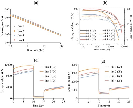
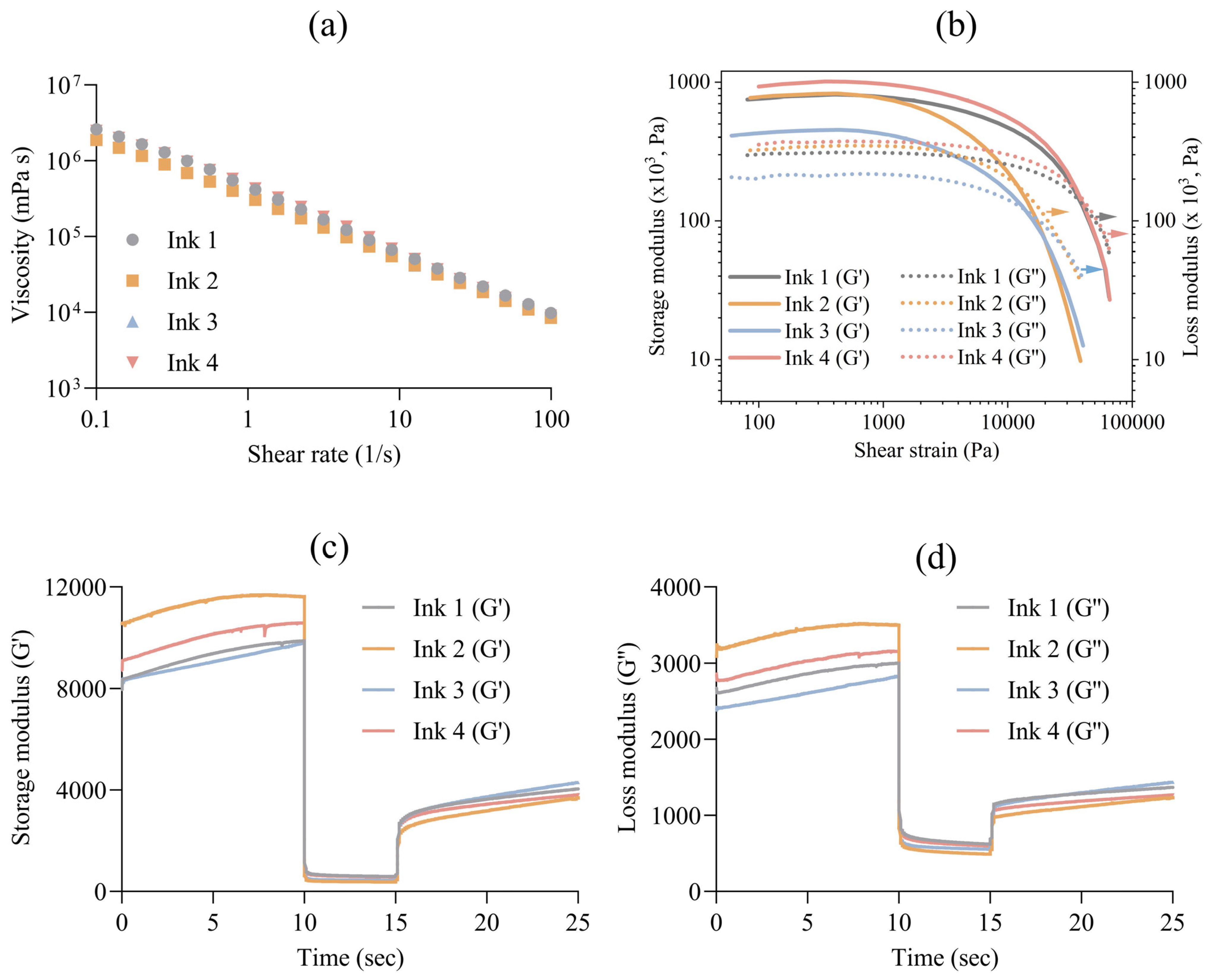
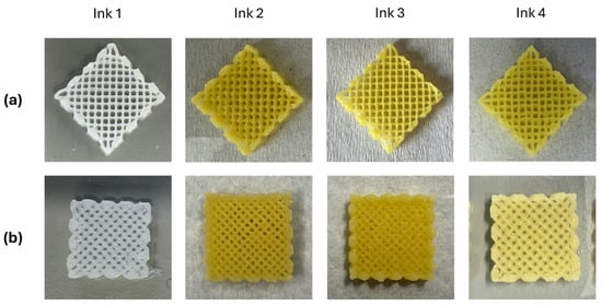
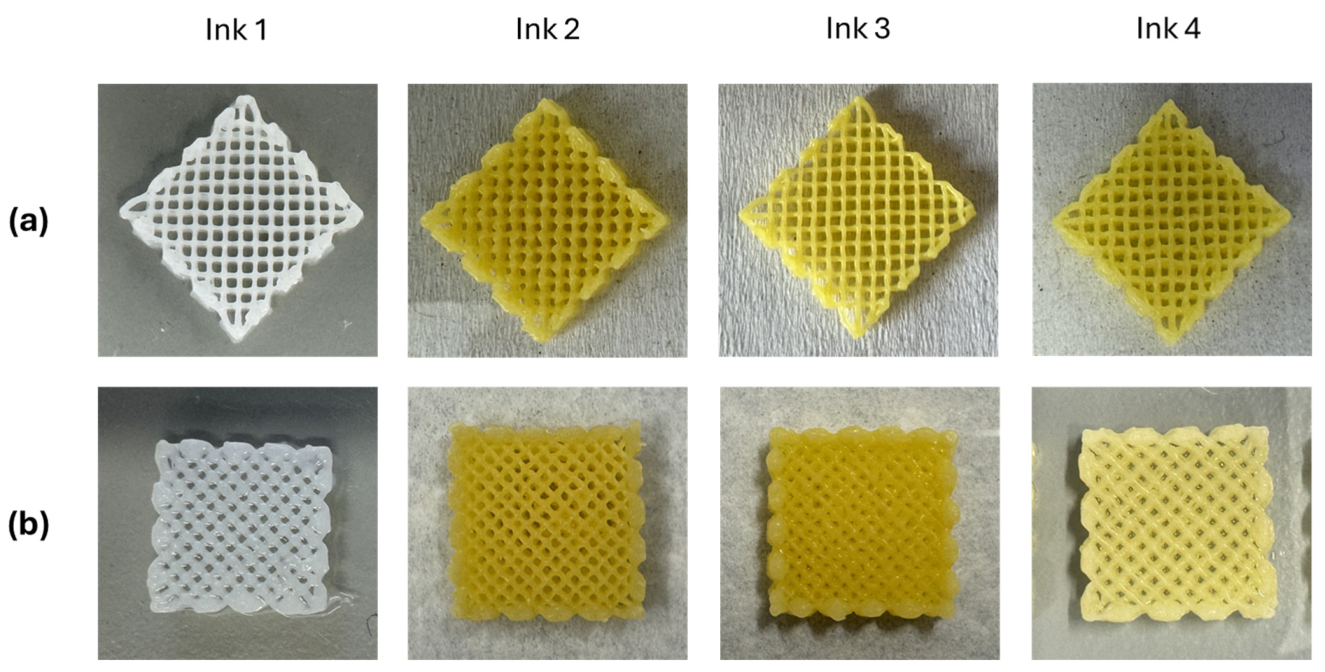
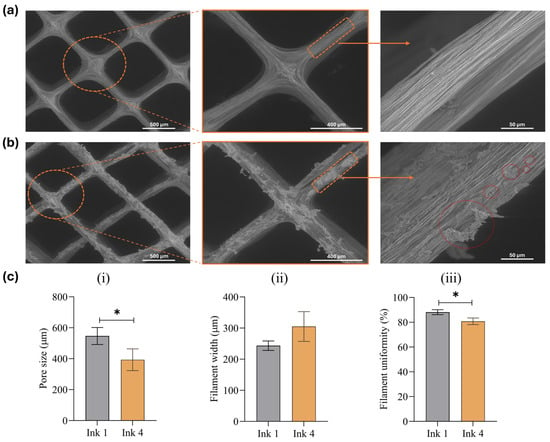
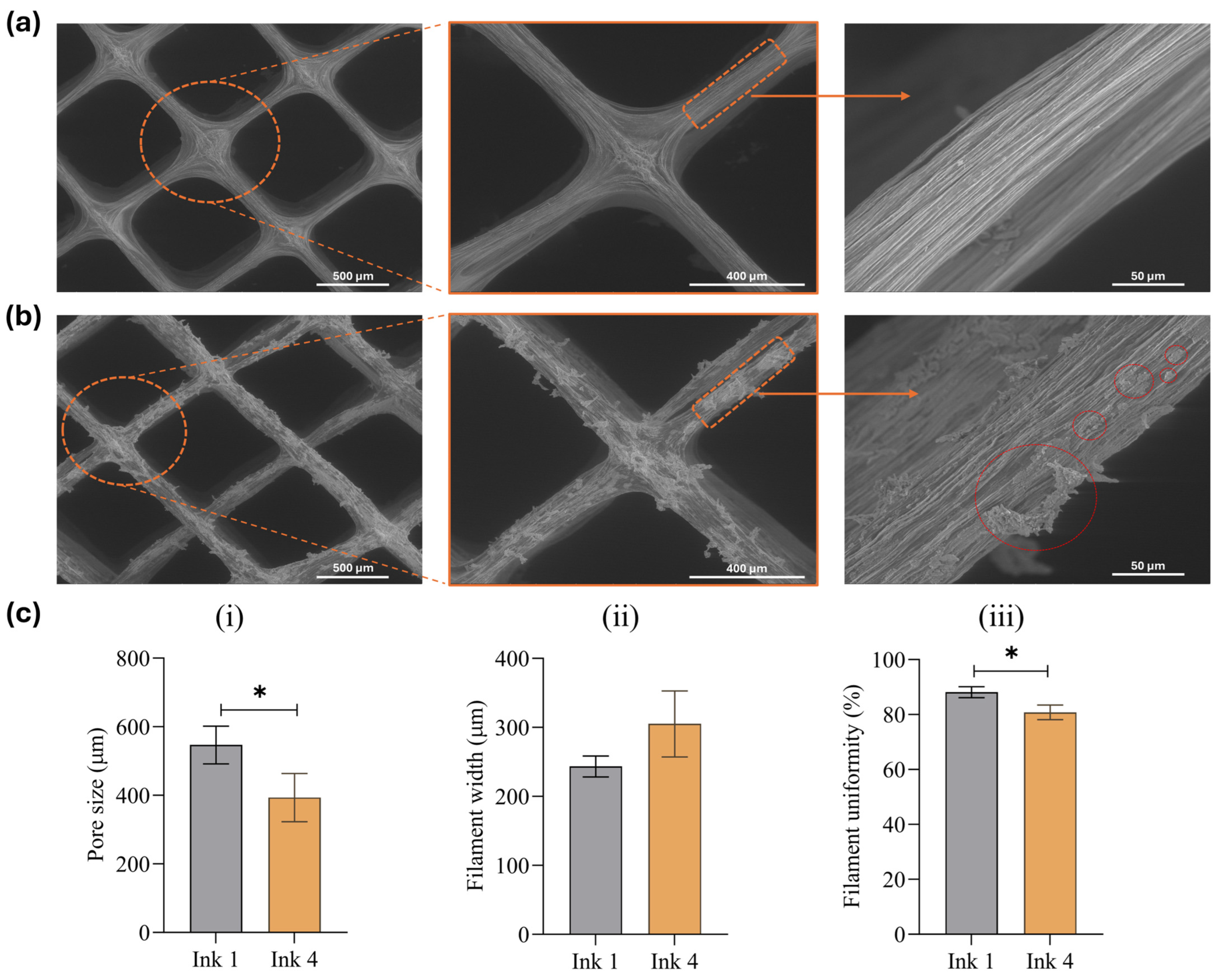
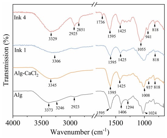
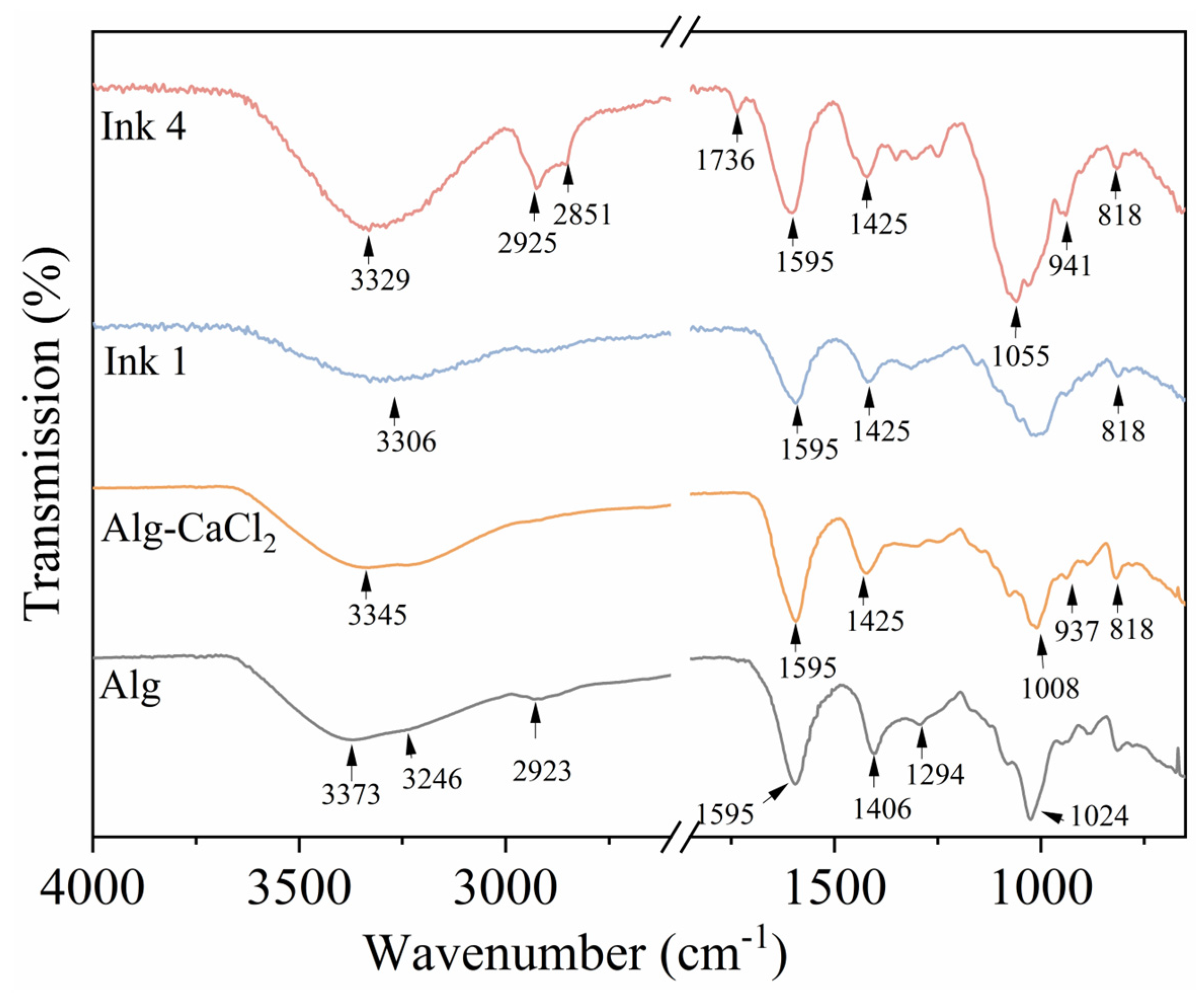
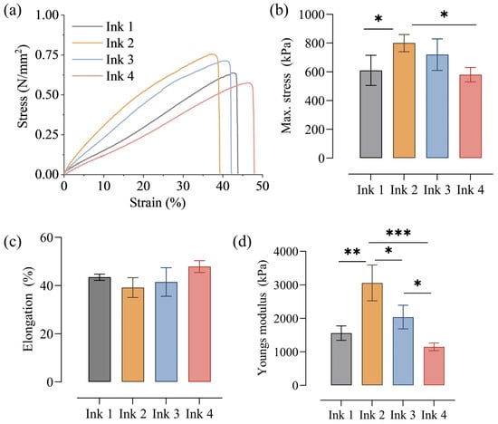
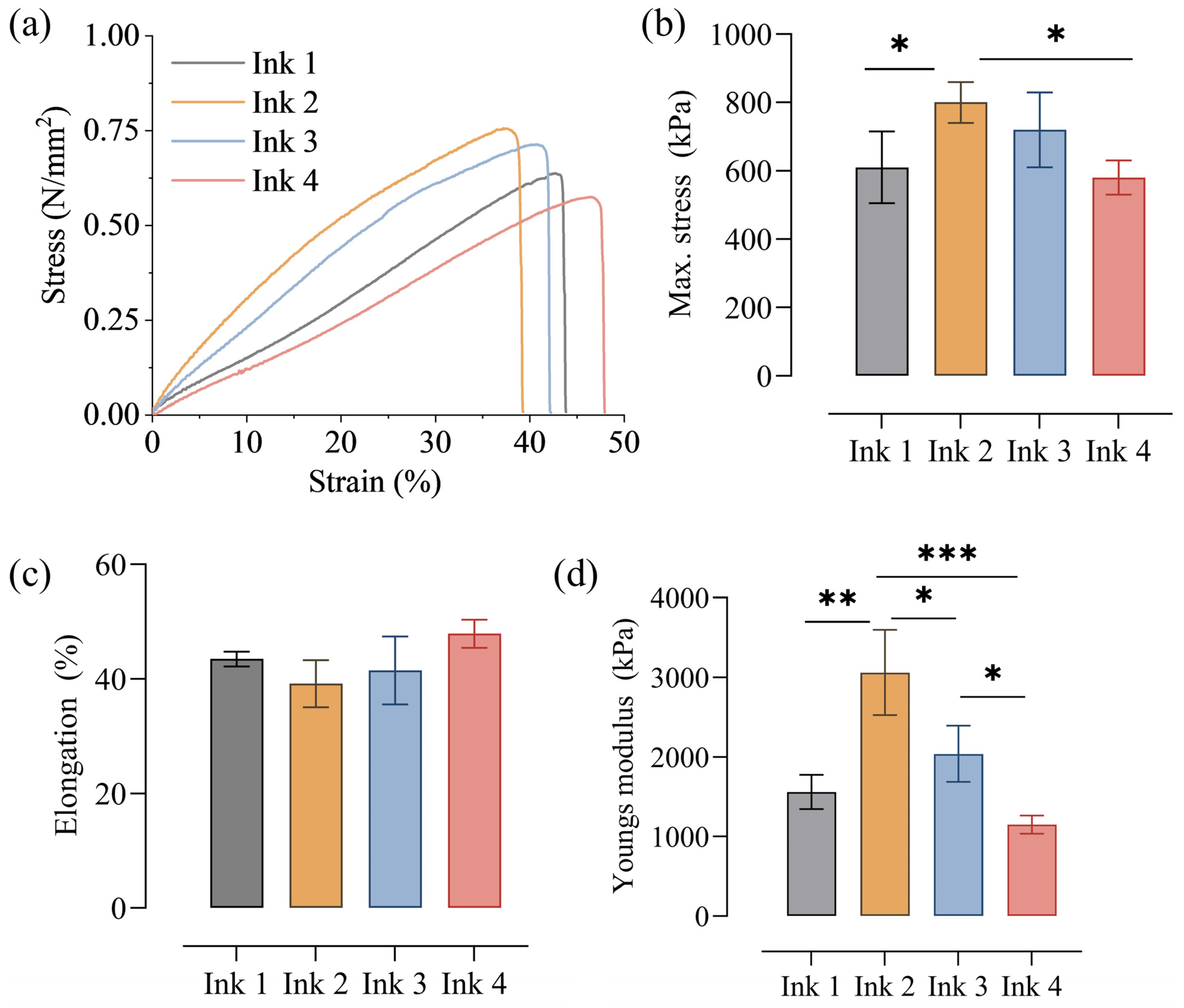
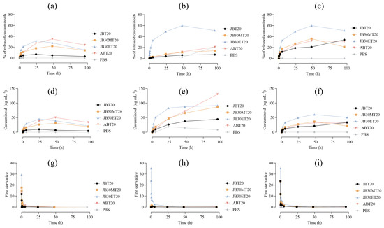
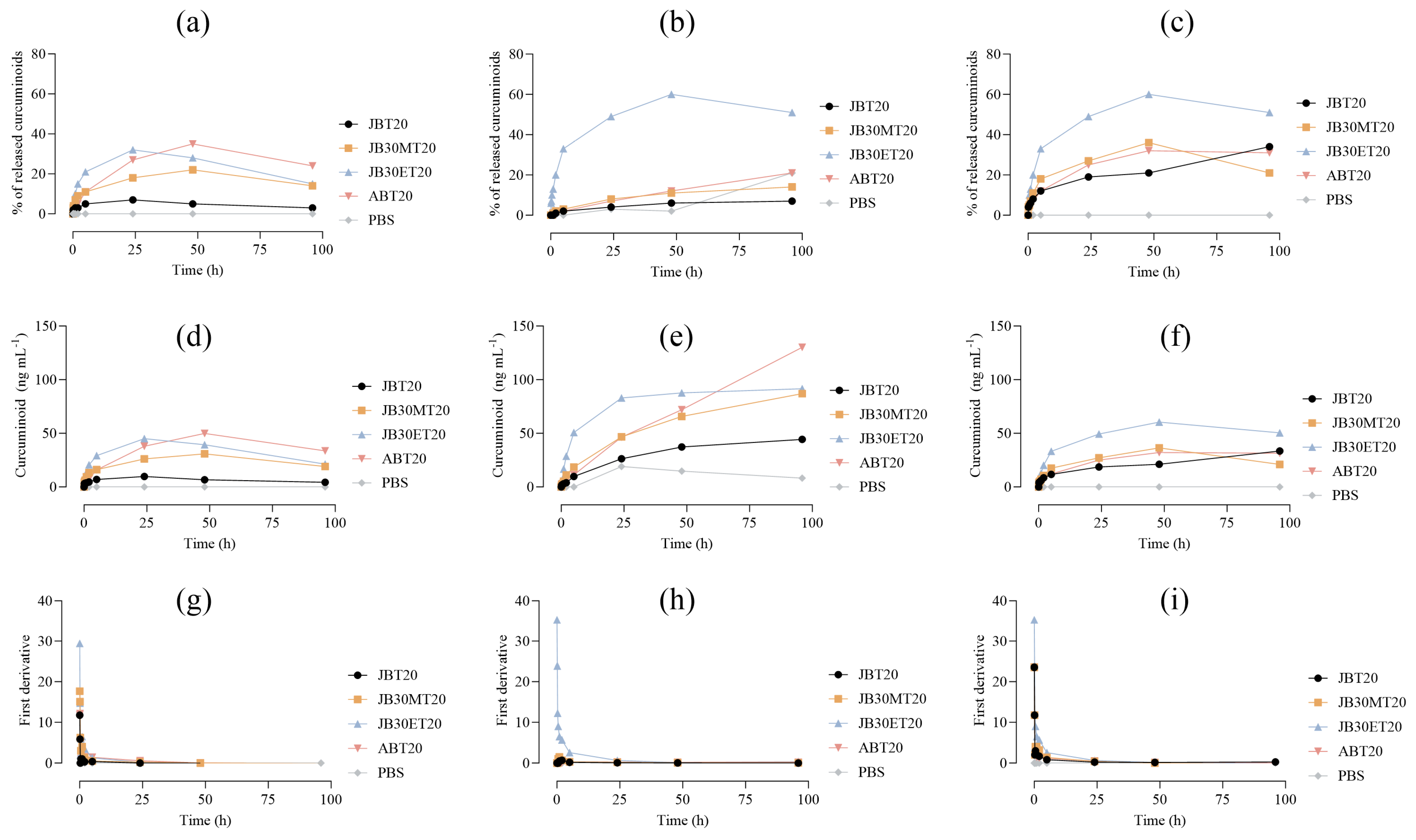

Disclaimer/Publisher’s Note: The statements, opinions and data contained in all publications are solely those of the individual author(s) and contributor(s) and not of MDPI and/or the editor(s). MDPI and/or the editor(s) disclaim responsibility for any injury to people or property resulting from any ideas, methods, instructions or products referred to in the content. |
© 2026 by the authors. Licensee MDPI, Basel, Switzerland. This article is an open access article distributed under the terms and conditions of the Creative Commons Attribution (CC BY) license.