


Abstract
In recent years, advancements in gene therapy have highlighted the important role of adeno-associated viruses (AAVs) due to their favorable characteristics, such as low immunogenicity compared to other viral vectors, e.g., lentivirus and HSV, and the ability to maintain gene expression in a variety of tissues. However, the production of recombinant AAVs in biological systems can lead to variability in the biophysical properties of viral capsid proteins, primarily due to post-translational modifications (PTMs) and cleavage events during downstream processing and storage. A critical quality attribute of AAV capsids is charge variant heterogeneity, which is significantly influenced by PTMs like deamidation, phosphorylation, acetylation and glycosylation. These modifications can impact the safety and efficacy of the viral vectors. The traditional imaged capillary isoelectric focusing (iCIEF) method, which uses absorbance or fluorescence detection, has been the primary choice for characterizing charge variants. However, it often lacks the sensitivity and resolution needed for AAVs’ charge variants. We introduce an optimized, highly sensitive capillary-based Western method to measure the apparent isoelectric point (pI) and detect charge heterogeneity at the individual VP protein level under reduced denatured conditions. This approach involves generating and purifying polyclonal antibodies to detect charge variants specific to the VP1, VP2, and VP3 proteins across different AAV serotypes, including AAV1, AAV8, and AAV5. This method is a valuable tool for the characterization of AAV capsids and can be utilized for the stability assessment and analysis of in-process and purified samples during process optimization from a charge heterogeneity perspective.
1. Introduction
Adeno-associated viruses (AAVs) were first identified nearly sixty years ago as contaminants in adenovirus preparations [1]. The European Medical Agency (EMA) approved the first AAV product (Glybera) in 2012. Subsequently, in 2017, the U.S. Food and Drug Administration (FDA) approved Luxturna, which is the first AAV-based gene therapy in the United States [2,3,4]. To date, seven AAV-based therapies have received FDA approval, and many others are in clinical and preclinical stages, targeting diseases previously considered incurable [2,5]. Gene therapy can be broadly defined as drugs that repair or modify genomes using viruses that deliver genes to specific locations in the body. Viral vectors, such as retrovirus, lentivirus, herpes simplex virus (HSV), AAV, and adenovirus (Ad) [2], have been explored for in vivo applications. However, AAV vectors have emerged as promising candidates for treating a wide range of genetic diseases due to their low immunogenicity compared to other viral vectors, long-term gene expression, and efficient cellular entry [6].
AAVs are composed of a protein shell, or capsid, consisting of 60 copies of viral proteins VP1, VP2, and VP3 in a 1:1:10 ratio, respectively, which encapsidate a single-stranded DNA approximately 4.7 kilobases [2,5,7,8,9] long. The VP proteins share a structural core (VP3) which constitutes the majority of VP1 and VP2. The unique N-terminal regions of VP1 and VP2 contribute to specific viral functions, such as intracellular trafficking. However, the structural core primarily governs aspects like tropism and serotype specificity through its interactions with cell surface receptors and sugar-binding sites [2,10,11]. For example, AAV8 exhibits enhanced tropism for liver cells, while AAV1 and AAV9 are more effective for targeting and transducing cardiac and skeletal muscle cells for gene delivery. This highlights the importance of serotype selection based on therapeutic goals [5,12,13]. The VP sequence differences contribute to tissue-specific transduction efficiency, with amino acid sequence homology ranging from ~55% to 99% between serotypes. X-ray crystallography studies have identified nine hypervariable regions (HVRs) in VP3 monomers as primary sources of sequence homology differences [14,15].
Recombinant AAVs (rAAVs) are utilized for gene therapy. In this process, the rep and cap genes of the wild-type AAV are removed to make them replication-deficient. These genes are then replaced with a therapeutic gene of interest, which is included in a separate plasmid during the AAV manufacturing process [4,16]. Further, the inserted transgene is flanked by inverted terminal repeats (ITRs) that aid in the efficient packaging and replication of the viral genome. The effectiveness of AAV-based gene therapy to meet clinical needs is highly dependent on the advancements in the manufacturability of rAAVs. As a result, there is a growing demand for large-scale rAAV production platforms because of the rapidly expanding clinical pipelines of AAV-based gene therapies. However, producing AAVs in eukaryotic cells poses significant challenges [17]. Factors like cell culture conditions and intracellular environments can introduce heterogeneity in the final product, and this can potentially affect capsid protein composition, genome packaging, or post-translational modifications (PTMs), thereby influencing the specificity, potency, and immunogenicity of rAAVs. Common PTMs include phosphorylation, methionine oxidation, acetylation, ubiquitination, and deamidation [18]. Previous studies by Giles et al. [14] and Zhong Li et al. [19] have demonstrated that deamidation and phosphorylation affect AAV capsid potency, and this emphasizes the need to monitor PTM changes in capsid proteins.
With up to 75 PTMs identified in capsid proteins, comprehensive characterization using various biophysical methods is crucial to ensure batch-to-batch consistency and achieve desired therapeutic outcomes [8,18]. Capsid proteins are susceptible to a wide range of PTMs, and several, such as deamidation, phosphorylation, and O-linked glycosylation, can induce charge heterogeneity [8]. While quantitative peptide mapping is a powerful method for analyzing post-translational modifications such as deamidation, its reliance on mass spectrometry makes it a complex and challenging method to implement in quality control laboratory settings. Over the past decade, native imaged capillary isoelectric focusing (iCIEF) has been widely used to characterize charge heterogeneity in monoclonal antibodies, viruses, and fusion proteins [20]. However, this method has poor peak resolution which masks localized modifications within the AAV capsid that contribute to charge heterogeneity. Understanding the heterogeneity of specific VPs is crucial. It was previously reported that VP1 and VP2 play an important role in endosomal trafficking, endosomal escape, nuclear trafficking, and genome release, while VP3 contributes to structural integrity and the determination of viral tropism [21]. Previously, we published a novel high-throughput reduced denatured iCIEF–Western method to specifically characterize the charge heterogeneity of antibody heavy and light chains [22]. This method selectively characterizes the HC and LC charge variants using detection antibodies specific to the HC or LC. We showed that a capillary-coupled immunodetection assay utilizing specific antibodies (iCIEF–Western) offers 100 times more sensitivity than standard UV or fluorescence-based iCIEF methods and is suitable for both crude (e.g., in-process samples) and purified samples [22]. This technology has shown that performing charge analysis by iCIEF–Western under reduced denatured conditions is advantageous, as it enables monitoring the charge heterogeneity of individual capsid proteins [8,23]. A critical consideration for immunodetection-based assays is the selection of sensitive antibodies specific to individual capsid proteins across different serotypes. While native iCIEF provides an overall charge profile of intact AAV capsids, it does not resolve charge heterogeneity at the individual VP protein level because the signal reflects the combined contribution of VP1, VP2, and VP3. MS-based approaches can provide detailed modification information, but they are less practical for high-throughput routine process monitoring and can be complicated by overlapping VP sequences. Therefore, the reduced denatured iCIEF–Western approach provides a sensitive, VP-resolved, and high-throughput practical orthogonal method for monitoring capsid protein charge heterogeneity in purified, stressed, and in-process AAV samples. Most commercial antibodies lack specificity for individual capsid proteins, as they are typically generated against the C-terminus of the VPs. Additionally, given the rapidly expanding application of various serotypes, it is essential to have detection antibodies available for the capsid proteins of different serotypes of interest.
In this study, we assessed the effects of additional reagents, such as urea, formamide, and alternative carrier solutions, like hydroxy propyl methyl cellulose (HPMC), on the charge variant profiles of AAV capsid proteins using a reduced denatured iCIEF–Western charge variant analysis method. Additionally, we developed a strategy for generating and purifying polyclonal antibodies (pAbs) specific to individual capsid proteins and demonstrated their application in iCIEF–Western analysis under both native and reduced denaturing conditions. The developed method conditions described in this study highlighted the importance of the urea-to-formamide ratio in facilitating the characterization of charge variants corresponding to empty, partial, and full AAV capsid proteins. This serves as a valuable tool for monitoring capsid proteins in either in-process or purified samples during purification process optimization or as a stability monitoring tool from a charge variant analysis perspective.
2. Results
2.1. Generation of Anti-VP Polyclonal Antibodies for AAV8, AAV1 and AAV5
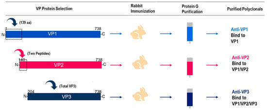
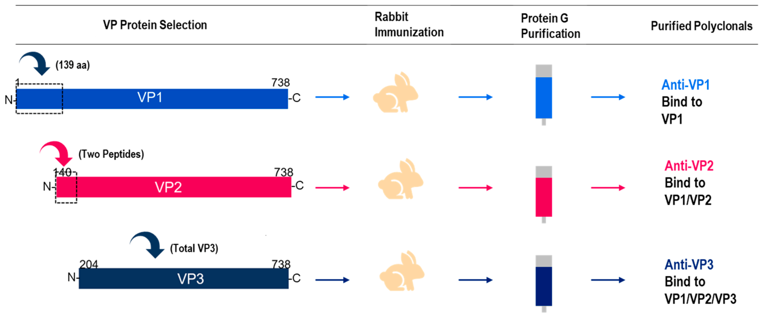
Creating specific antibodies for each individual VP protein is difficult because of the shared sequences at the C-terminus of the VP proteins. To overcome this challenge, we utilized the unique, non-overlapping sequence of 139 amino acids between VP1 and VP2 for immunization to produce anti-VP1 pAbs. To generate anti-VP2-specific pAbs, we designed two peptides based on the non-overlapping sequences of 64 amino acids between VP2 and VP3 (Figure 1) of AAV8 and used these peptides for immunization. Finally, the entire VP3, consisting of 534 amino acids, was used to generate anti-VP3 pAbs. The designed VP1, VP2, and VP3 peptides/proteins, highlighted in boxes (Figure 1), were injected into rabbits, and after a few weeks, antisera were collected and purified using a Protein G column to enrich the desired antibodies specific for each VP protein (Figure 1). Because these detection reagents were rabbit polyclonal antibodies, the purified preparations were expected to contain heterogeneous immunoglobulin populations recognizing multiple epitopes within the target antigen regions. Therefore, the primary characterization focused on functional specificity toward the intended VP proteins rather than additional purity assessment. This specificity was evaluated by traditional Western blotting and CE-Western analysis, as described in Section 2.2.
Figure 1.
Illustrative representation of polyclonal antibodies targeting AAV8 capsid proteins VP1, VP2, and VP3.
The anti-VP1 pAbs are expected to be specific and bind exclusively to the VP1 protein. In contrast, anti-VP2 pAbs can bind to both the VP1 and VP2 proteins, while anti-VP3 pAbs can bind to all three proteins due to a shared N-terminal sequence.
2.2. Evaluation of Anti-VP Polyclonal Antibody Specificity
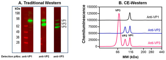
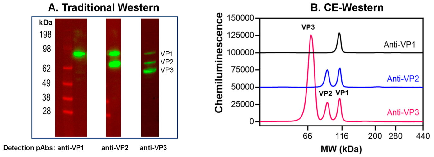
The specificity of the purified antibodies was assessed using traditional Western blotting and the CE–Western platform for protein size analysis. In the Western blot analysis of AAV8 samples, the anti-VP3 polyclonal antibodies detected all three VP protein bands at approximately ~80 kDa, ~70 kDa, and ~60 kDa, which correspond to VP1, VP2, and VP3, respectively (Figure 2A). This detection is expected since the polyclonal antibodies target the shared common C-terminus region of the VPs. Additionally, the VP protein bands showed relatively lower intensities for the VP1 and VP2 bands compared to VP3, and this is consistent with the expected 1:1:10 ratio of VP1:VP2:VP3. Similarly, using anti-VP2 polyclonal antibodies resulted in the detection of bands for both the VP1 and VP2 proteins, as these antibodies were generated using two short peptides sharing a common region between VP1 and VP2. Finally, the anti-VP1 polyclonal antibodies specifically detected only the VP1 protein, as they were generated for the unique N-terminal region of VP1. Similarly, the specificity of the anti-VP polyclonal antibodies was analyzed on the CE–Western size platform (Figure 2B). The results were consistent with the traditional western data. The anti-VP1 polyclonal antibodies specifically detected a peak at 80 kDa corresponding to VP1. The anti-VP2 polyclonal antibodies detected both VP1 and VP2 at their respective predicted molecular weights of 80 kDa and 70 kDa. The anti-VP3 polyclonal antibodies detected three peaks at the expected molecular weights of 80 kDa, 70 kDa, and 60 kDa, corresponding to VP1, VP2, and VP3, respectively.
Figure 2.
Specificity evaluation of anti-VP1, anti-VP2, and anti-VP3 polyclonal antibodies using traditional Western blotting (A) and CE–Western platform (B). AAV8 samples were first heat denatured at 80 °C for 10 min, and then capsid proteins were detected separately with rabbit anti-VP1, anti-VP2 and anti-VP3 polyclonal antibodies.
2.3. AAV8 Charge Variant Analysis by Native iCIEF–Western Method
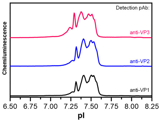
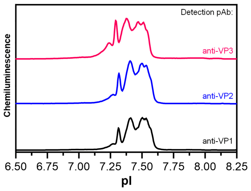
The application of native iCIEF–Western for the charge variant analysis of intact AAVs presents certain limitations due to the inherent size and complexity of AAVs. While this method is commonly used for analyzing mAb charge variants, its sensitivity to local changes within the AAV structure is limited. This is primarily because the large size of AAVs can obscure subtle alterations in charge distribution that might occur at a more localized level. Furthermore, when compared to mAbs, the native iCIEF profiles for intact AAV capsids are broad. This broadness in the profiles poses a significant challenge in detecting minor differences in the charge isoforms of the AAVs (Figure 3) and makes it difficult to discern subtle variations that could be critical for understanding the charge heterogeneity within the AAV population. Additionally, the intact AAV capsids, which contain all the viral proteins (VPs) together, limit the specificity of individual polyclonal antibodies to provide additional discriminatory information for each VP protein. As a result, the observed profiles tend to have similar peak patterns across all detections (Figure 3). In light of these challenges, we explored the development of a charge variant analysis method for AAVs under reduced denatured conditions.
Figure 3.
iCIEF–Western analysis of native charge variants of intact AAV8 samples using anti-VP1-, anti-VP2-, and anti-VP3-specific rabbit polyclonal antibodies for detection.
2.4. AAV8 Charge Variant Analysis by Reduced Denatured iCIEF–Western
In contrast to native iCIEF–Western, where the charge profiles represent a combination of all viral proteins (VPs), the reduced denatured charge variant analysis enables the identification and quantification of charge isoforms specific to each individual VP. Previously, we published a similar study on monoclonal antibodies (mAbs) under reduced denatured conditions to examine charge heterogeneity specific to antibody heavy and light chains [22]. However, the denaturation and electrophoretic conditions used in that study were not directly applicable to AAV analysis because of the distinct characteristics of AAV capsid proteins compared to mAbs. Therefore, both the denaturing and electrophoretic conditions needed to be specifically tailored for AAVs. The sample preparation conditions were adapted from a ProteinSimple application note related to AAV analysis on the Maurice iCIEF platform with a modification to the reducing agent and denaturation temperature to account for the melting points of different AAV serotypes [24]. Meanwhile, the electrophoretic conditions were optimized by leveraging our prior experience with mAb analysis.
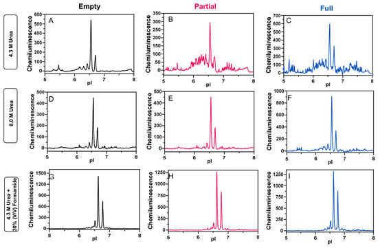
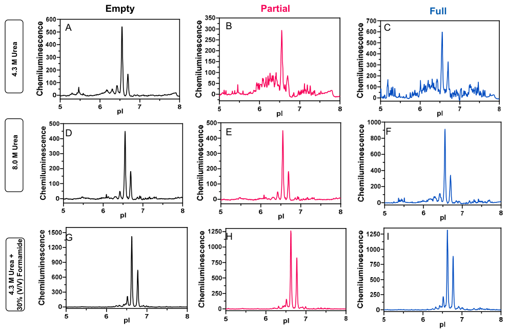
For mAbs, an essential factor in achieving the optimal separation of charge variants under reducing and denaturing conditions is the concentration of urea used during sample preparation and electrophoresis [22]. Initially, various urea concentrations ranging from 4.3 M to 8.0 M were evaluated to establish the minimum amount necessary to stabilize the reduced and denatured capsid proteins. Following this, urea concentrations of 4.3 M and 8.0 M were compared using empty, partial, and full capsids to evaluate the impact of the presence or absence of DNA on baseline noise (Figure 4). The data indicated that a higher urea concentration of 8.0 M significantly improved the baseline, especially for capsids containing partial or full DNA. However, the baselines for partial and full capsids remained noisier compared to empty capsids. To further address this issue, formamide, which is a common alternative to urea, was introduced in concentrations ranging from 0 to 30% during the electrophoretic step to assess its effect on baseline noise [25]. A matrix of urea concentrations (4.3–8.0 M) and formamide concentrations (0–30%) was tested to identify conditions that yield reproducible data with minimal baseline noise. The optimized results demonstrated that using 4.3 M urea combined with 30% formamide produced the most consistent and stable baseline across empty, partial, and full capsids. This combination of urea and formamide successfully minimized baseline noise and achieved clearer and more reliable data for analysis (Figure 4).
Figure 4.
Reduced denatured charge variant profiles for empty (A,D,G), partial (B,E,H) and full (C,F,I) capsids in the presence of either 4.3 M urea, 8.0 M urea or 4.3 M urea + 30% (v/v) formamide. The addition of 30% formamide significantly reduced the baseline noises.
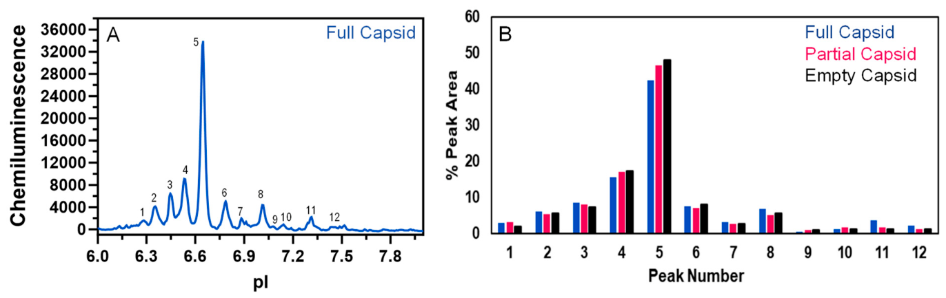
The sensitivity of the optimized method was further enhanced by incorporating 10× concentrated luminol–peroxide solution and was assessed for its ability to detect low-abundant charge isoforms within the empty, partial, and full capsids using anti-VP3 polyclonal detection antibodies (Figure 5). The analysis revealed 12 distinct comparable peak profiles, areas and baseline noise levels across all three sample types, each containing varying amounts of DNA. This consistency indicates that the method is robust and versatile, and it is suitable for analyzing AAV8 samples irrespective of the presence or absence of nucleic acid material. It is worth noting that the observed differences in the charge variant distribution of the current samples, compared to the data presented in Figure 4, are attributed to variations in the manufacturing process.
Figure 5.
Using the optimized reduced denatured iCIEF–Western method, AAV8 samples containing varying levels of encapsulated DNAs (full, partial, and empty capsids) were characterized. The results demonstrated that all samples exhibited very similar charge variant profiles. Panel (A) displays a representative profile for the AAV8 full capsid. Panel (B) provides the percentage peak area quantitation for each charge variant in full, empty, and partial capsids.
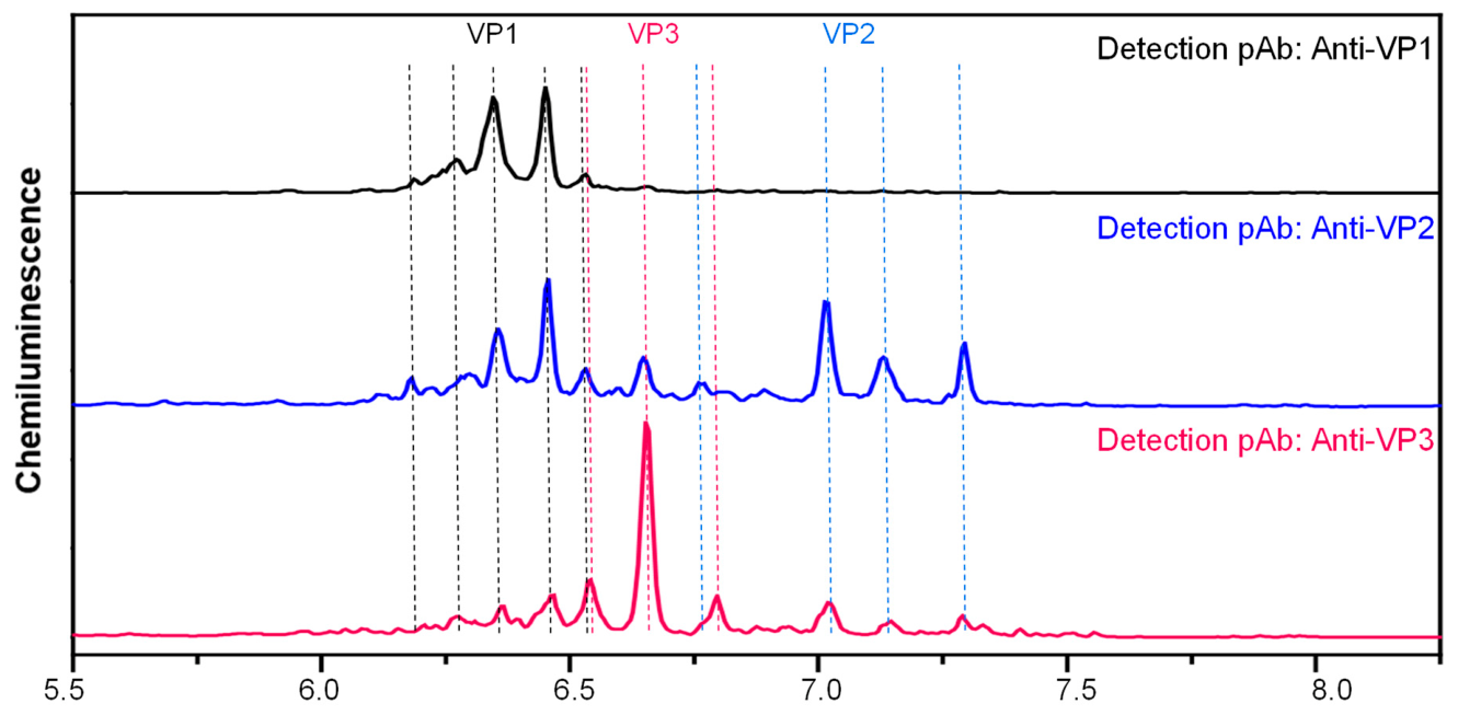
The developed method was employed to evaluate the specificity of anti-VP polyclonal antibodies to individual VPs under reduced denatured conditions (Figure 6). Reduced denatured AAV8 samples were probed with three different anti-VP polyclonal antibodies in separate capillaries. Anti-VP1 polyclonal antibodies exclusively detected charge variants corresponding to VP1, with two major peaks centered at pIs between 6.3 and 6.4. When the reduced denatured AAV8 sample was probed with anti-VP2 antibodies, additional charge variant peaks were observed compared to those detected by anti-VP1 antibodies. These peaks correspond to VP2, with the major charge variant centered at a pI of around 7.1. Probing the sample with anti-VP3 polyclonal antibodies revealed charge variants corresponding to VP3 proteins, with a major peak at a pI of approximately 6.7, while also identifying peaks corresponding to both VP1 and VP2. The intensity of the charge variant peaks for VP3 was significantly higher compared to those for VP1 and VP2, consistent with the relative abundance of the individual VP proteins in AAV capsids (1:1:10). The observed peak positions closely matched the theoretical pI values of 6.1, 6.9, and 6.3 for VP1, VP2, and VP3, respectively. Due to the potential overlap of charge variant peaks among VP proteins, individual isoform-level assignments remain challenging. Therefore, the observed profiles are interpreted as VP-associated charge variant regions based on antibody-specific detection patterns and consistency with the theoretical pI values, rather than as definitive assignments of every individual charge isoform.
Figure 6.
The detection of individual capsid proteins of AAV8 using the specific anti-VP detection polyclonal antibodies under reduced denatured conditions by iCIEF–Western. Samples: Thermally denatured AAV8 at 95 °C for 10 min. The major charge variants unique to VP1, VP2, and VP3 are represented by black, blue, and red dashed lines, respectively.
2.5. AAV1 and AAV5 Charge Variant Analysis by Reduced Denatured iCIEF–Western
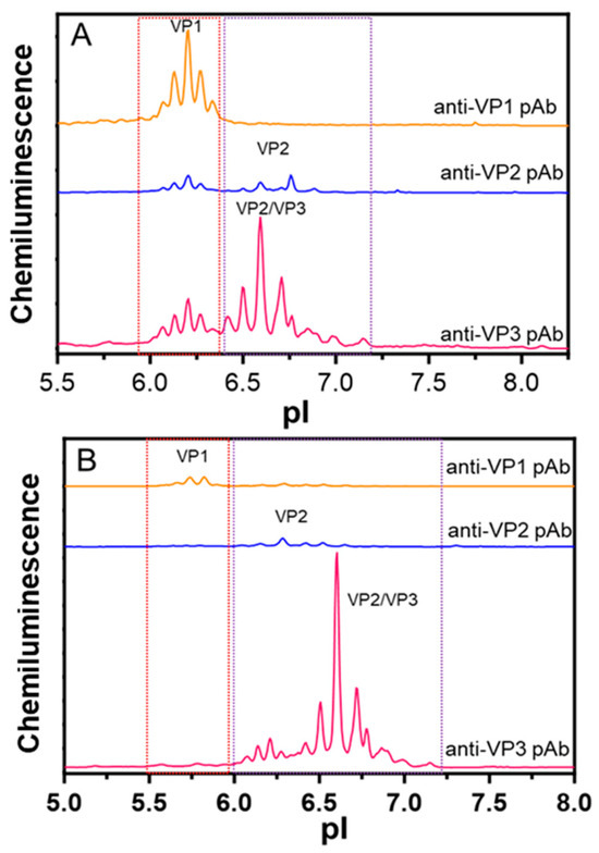
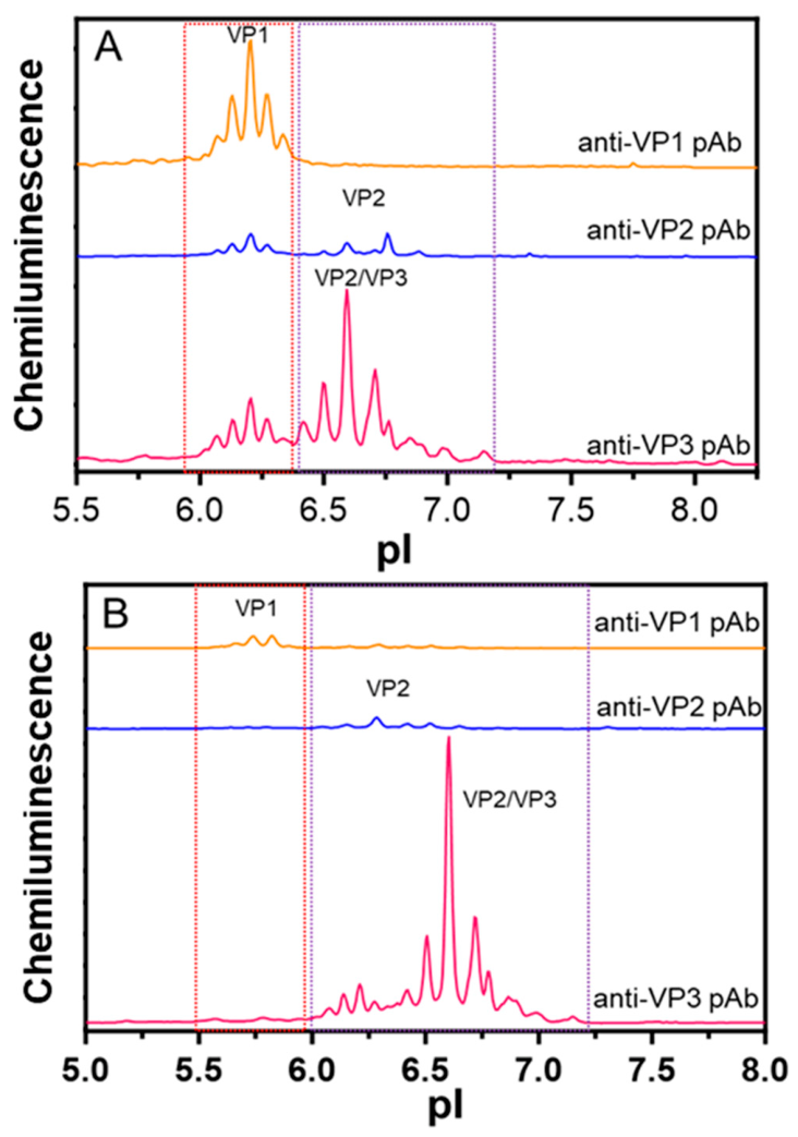
The reduced denatured charge variant method, originally developed for the AAV8 serotype, was systematically evaluated across other AAV serotypes to assess its broader applicability. Each AAV serotype possesses a distinct capsid structure which affects its tropism through interactions with specific cell surface receptors. In this study, the AAV1 and AAV5 serotypes were chosen due to their high sequence homology of 84% and low sequence homology of 55% with AAV8, respectively [10]. The polyclonal antibodies developed against individual anti-AAV8 VPs were used to assess their effectiveness in identifying charge variants associated with AAV1 and AAV5. For AAV1, anti-AAV8 VP1 polyclonal antibodies successfully detected charge variants associated with the VP1 protein with good sensitivity, and anti-AAV8 VP3 polyclonal antibodies also effectively detected charge variants for all three viral proteins (Figure 7A). Conversely, anti-AAV8 VP2 polyclonal antibodies showed lower sensitivity in detecting the VP2 charge variants of AAV1 compared to those of AAV8. Despite having the same theoretical pI at 6.23, the distribution and relative intensity of the VP2 and VP3 charge variants in AAV1 are different when comparing the traces detected by anti-AAV8 VP2 and anti-AAV8 VP3 polyclonal antibodies. For AAV5, the sensitivity of anti-AAV8 VP1 and VP2 polyclonal antibodies demonstrated even lower sensitivity, likely due to the lower sequence homology to AAV8 (Figure 7B). VP3 detection showed relatively better intensity, possibly because more polyclonal antibodies were generated against the longer sequence of the VP3 protein during immunization (Figure 1). Although overall sequence homology provides useful context, the differential anti-VP2 detection sensitivity is likely more directly influenced by conservation within the VP2 immunogen region used to generate the anti-AAV8 VP2 polyclonal antibodies. Because these antibodies may recognize multiple epitopes within this region, exact epitope-level assignments were not made; however, reduced regional conservation is expected to contribute to the lower VP2 signal observed across serotypes. The further alignment of the VP2 regions from AAV1 and AAV5 against AAV8 showed that the regional sequence similarity values were consistent with the overall sequence homology observed among these serotypes.
Figure 7.
The detection of AAV1 (Panel (A)) and AAV5 (Panel (B)) VP1, VP2 and VP3 protein charge variants was achieved using anti-AAV8 VP-specific polyclonal antibodies. The charge variant regions specific to VP1 and VP2/VP3 are highlighted in red and purple dashed boxes respectively.
2.6. Stressed Sample Characterization by Reduced Denatured iCIEF–Western
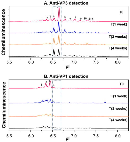
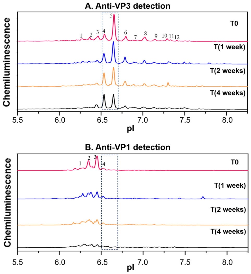
Thermal stress can trigger a range of PTMs in proteins, including oxidation and deamination, which can produce distinct charge variants with varying isoelectric points (pIs). With the capsid composed of 60 copies of VPs, it is essential to identify which capsid proteins undergo modifications. The developed reduced denatured iCIEF–Western method was employed to characterize the stressed AAV8 samples to evaluate whether the method has the sensitivity to detect modifications specific to each VP protein. AAV samples were subjected to thermal stress at 40 °C for durations of 1 week, 2 weeks, and 4 weeks. These stressed samples, along with a non-stressed control, were analyzed using reduced denatured iCIEF–Western with anti-VP3 polyclonal antibodies, which can detect all three VP proteins. The analysis revealed that the intensity of Peak 4 increased with prolonged stress exposure, while the intensity of the main Peak 5 decreased (Figure 8A). The change in the relative ratio was further confirmed by the change in the relative peak area ratio of Peak 5 to Peak 4 from ~2.8 to 0.9 from T0 to 4-week samples. Since Peak 4 is within the overlapping isoelectric point (pI) range of VP1 and VP3 (Figure 8A,B dashed rectangle), it is challenging to ascertain whether the post-translational modification occurred on VP1 or VP3.
Figure 8.
The identity of an increased charge variant Peak 4 in thermally stressed AAV8 samples was confirmed using the detection of both anti-VP3 (A) and anti-VP1 (B) polyclonal antibodies.
To confirm the identity of Peak 4, anti-VP1 polyclonal antibodies were employed to analyze all non-stressed and stressed samples. The absence of an increase in VP1 charge variant peaks within the Peak 4 pI range suggests that the modification associated with Peak 4 occurred in VP3 rather than VP1 (Figure 8B). In addition to VP3, notable changes were observed with the VP1 charge variant profile in terms of both signal loss and the relative distribution of the charge isoforms. The differences could be due to thermal stress-induced PTMs which create extra-low-abundance acidic species which are not detected by the anti-VP1 pAb.
2.7. In-Process Sample Analysis by Reduced Denatured iCIEF–Western to Support Manufacturing Process Hold Time Studies
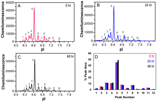
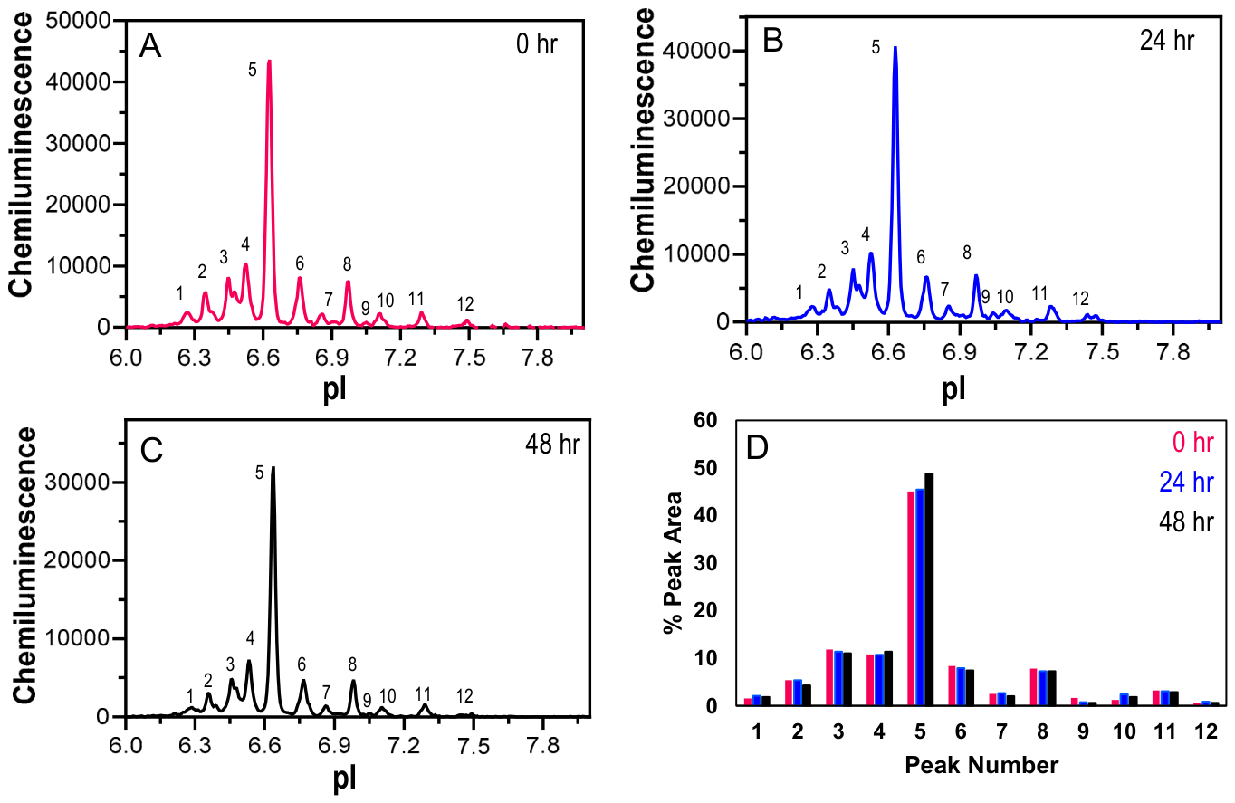
By utilizing specific antibody detection, iCIEF–Western is well-suited for analyzing non-purified or crude samples. This capability presents substantial advantages for supporting in-process sample analysis, as it allows for the direct assessment of complex mixtures without the need for extensive purification steps. The developed method was used to support manufacturing hold time stability studies, which simulate potential delays or storage periods within the manufacturing process. Charge variant profiles were compared for anion exchange column-purified samples held for extended durations of up to 48 h at room temperature (Figure 9). A total of 12 peaks were identified using anti-VP3 polyclonal antibodies across all five different hold time samples, including four acidic and seven basic peaks. The major peaks in both lots were concentrated at a pI of approximately 6.7, aligning with the theoretical pI of the VP3 protein. Additionally, the peak positions were consistent across all detected peaks. The percent peak area quantitation for the major peak across the lots was observed to be within 45–48%. Similarly, the quantitation of percent peak areas for both the acidic and basic species was analyzed (Figure 9D). Overall, the data indicate that the profiles remain both qualitatively and quantitatively comparable when samples are held for different durations.
Figure 9.
Three samples from a manufacturing process with different hold times at room temperature ((A): 0 h; (B): 24 h; (C): 48 h) were analyzed using reduced denatured iCIEF–Western. The analysis showed that the peak profiles and the quantification of each peak area (D) were comparable across the samples.
3. Discussion
During the recombinant AAV production process in biological systems, capsid proteins undergo various PTMs such as deamidation, phosphorylation, and oxidation. These modifications can significantly affect the stability and efficacy of therapeutic molecules. It is essential to quantify and monitor these critical quality attributes throughout the product development process. Charge heterogeneity characterization is typically conducted using methods based on either surface charge or intrinsic charge. Generally, these methods are performed under native conditions, where the therapeutic molecule remains in its intact conformation. Consequently, for large biomolecules like AAVs, localized changes within the molecule may be obscured in the overall broad charge variant profile. Our study aimed to address the challenges associated with the characterization of AAV capsid protein charge heterogeneity, which is crucial for ensuring the consistency and efficacy of AAV-based therapies.
The development of polyclonal antibodies specific to the individual VP proteins of AAV presents a significant challenge, which mainly arises from the common sequence at the C-terminus. By targeting unique, non-overlapping sequences between individual VP proteins, we were able to generate pAbs with high specificity for VP1, VP2 and VP3. This specificity was confirmed through both traditional Western blotting and CE–Western analysis, which demonstrated that our polyclonal antibodies reliably detected individual VP proteins at their expected molecular weights, with a ratio close to 1:1:10. The ability to distinguish between the VP1, VP2, and VP3 proteins is critical, as it allows for the precise monitoring of capsid protein modifications, which may impact the therapeutic efficacy and safety of AAV vectors.
After purifying and verifying the specificity of pAbs to individual VPs, we began identifying charge variants specific to each VP using experimental conditions previously established for analyzing charge heterogeneity in antibody heavy and light chains under reduced denatured conditions. However, the charge variant profiles for AAV capsid proteins showed a noisy baseline, particularly in full capsids. This baseline noise difference between empty and full capsids is possibly due to nucleic acid interference during either denaturation or electrophoretic steps. Formamide was explored as an additional excipient, and a combination of urea and formamide was able to achieve comparable baseline noise and reproducible signals across empty, partial, and full AAV capsids without the need to remove the encapsulated nucleic acid before electrophoretic separation. Qualitative and quantitative analyses under optimized conditions confirmed comparability across full, partial and empty capsids.
The anti-AAV8 VP-specific polyclonal antibodies were further characterized for their ability to detect AAV1 and AAV5 charge variants to determine whether their application could be broadened to additional serotypes. The in-house-generated rabbit polyclonal antibodies for the AAV8 serotype offer a practical advantage when suitable commercial VP-specific antibodies are limited or unavailable. Furthermore, their recognition of multiple epitopes can improve the likelihood of detecting VP-associated charge variants and support robust signal generation. However, this heterogeneous epitope recognition limits precise epitope-level interpretation and may reduce cross-serotype sensitivity when the antibody-targeted regions are less conserved. Consistent with this limitation, the AAV8-derived antibodies detected charge variants in both AAV1 and AAV5, but reduced sensitivity was observed for VP1 and VP2, particularly VP2, likely due to sequence divergence from AAV8. This effect was the most pronounced for AAV5, which exhibits lower sequence homology in the targeted immunogen regions. In addition, the anti-VP2 antibody was generated using a short peptide sequence located within the non-overlapping VP2 region, which may further contribute to reduced cross-reactivity across serotypes.
Sensitivity to the VP1 and VP2 proteins can be improved by increasing the concentration of either the sample or the primary antibody to achieve a better signal-to-noise ratio. Alternatively, serotype-specific anti-VP1 and anti-VP2 polyclonal antibodies could be developed for AAV5 and AAV1 using a method similar to that employed for creating AAV8 VP polyclonal antibodies (Figure 1). Using serotype-specific anti-VP2 polyclonal antibodies, the denatured iCIEF–Western signals for VP2 charge variants can be further enhanced, and this approach can be effectively applied to all other serotypes. The ability to detect charge variants specific to each VP protein across multiple serotypes underscores the robustness and versatility of this analytical approach.
The application of the reduced denatured iCIEF–Western method for characterizing stressed AAV8 samples and in-process manufacturing samples provided insights into the stability and integrity of AAV capsid proteins under various conditions. Thermal stress can induce a variety of PTMs in proteins, leading to changes in charge variants with distinct isoelectric points [26]. Our study demonstrated that the use of anti-VP1 polyclonal antibody detection helped us to confirm that the modifications associated with a major charge variant change occurred in the VP3 protein rather than VP1. Further, anti-VP1 pAb-specific detection also indicated that there are changes to the charge variant profile in terms of both relative proportions and a decrease in signal intensity, which was consistent with previously reported studies [23]. These differences in VP1 could be due to the additional modifications, such as deamidation due to thermal stress and stability under thermal stress, leading to either protein truncation or precipitation during sample preparation. This specificity is crucial for understanding the impact of stress on individual VP proteins and can guide strategies to enhance the stability of AAV vectors.
The ability of the reduced denatured iCIEF–Western method to analyze non-purified or crude samples offers substantial advantages for in-process sample analysis. By allowing for the direct assessment of complex mixtures, this method supports manufacturing hold time stability studies, which are critical for simulating potential delays or storage periods within the manufacturing workflow. The consistency in charge variant profiles across different hold times of anion exchange pool samples indicates that the AAV capsid proteins remain stable over extended durations.
In summary, we developed an automated, high-throughput, and highly sensitive method for characterizing AAV capsid protein charge variants by using specific antibody detection on the Simple Western platform. This includes a strategy for producing and purifying in-house antibodies targeting each specific viral protein. The developed method is applicable to studying stressed samples and lot-to-lot comparability and the characterization of crude AAV samples, which allow for analysis at various stages of the manufacturing process to observe how different conditions affect PTMs. Additionally, this method serves as a method orthogonal to traditional direct UV or fluorescence-based iCIEF methods and provides the added advantage of specific antibody-based detection, which can provide valuable identity information.
4. Materials and Methods
4.1. Materials
All AAV samples (AAV8, AAV1 and AAV5) used in this study were manufactured at Regeneron Pharmaceuticals, Inc. (Tarrytown, NY, USA). Briefly, HEK293 suspension cell-derived AAVs were purified through AAVX resin affinity capture followed by an ion exchange polishing step. Subsequently, the purified AAVs were buffer-exchanged into the appropriate buffer prior to being stored at −80 °C. Buffer components and chemical denaturants urea and formamide were purchased from Sigma-Aldrich (St. Louis, MO, USA), and DMSO and HPMC (H7509) were purchased from VWR (Radnor, PA, USA). Bond-Breaker TCEP solution and formamide were purchased from ThermoFisher ScientificTM (Waltham, MA, USA). Pharmalytes 3–10 and pharmalytes 5–8 were obtained from Cytiva (Marlborough, MA, USA), while iCIEF–Western components including 1% methylcellulose, premix-G2 (pH 3–10), Bicine/CHAPS lysis buffer, pI ladder, antibody diluent, Simple Sol Protein Solubilizer, Wash Buffer, Catholyte, and Anolyte buffer were obtained from ProteinSimple (San Jose, CA, USA). HRP-conjugated anti-rabbit H + L detection antibodies were purchased from Southern Biotech (Birmingham, AL, USA).
4.2. Immunogen Generation
For AAV8 anti-VP antibody generation, four immunogens were prepared. Recombinant VP1 and VP3 protein fragments were produced by GenScript (Piscataway, NJ, USA) using an Escherichia coli expression system and the pET-30a(+) expression vector. The VP1 immunogen consisted of the non-overlapping N-terminal region (amino acids 1–139) and was used to generate VP1-specific polyclonal antibodies. The VP3 immunogen comprised amino acids 204–738 and was used to generate polyclonal antibodies recognizing a conserved region shared among all three VP proteins. Recombinant VP1 (amino acids 1–139) and VP3 proteins were purified by GenScript and confirmed to have a purity greater than 80% based on SDS-PAGE analysis prior to immunization.
In addition, two synthetic peptides, GKKRPVEPSPQRSPC and CSSTGIGKKGQQPAR, were synthesized by GenScript and conjugated to keyhole limpet hemocyanin (KLH). Peptide immunogens, GKKRPVEPSPQRSPC-KLH and KLH-CSSTGIGKKGQQPAR, were administered together to generate VP1/VP2-specific polyclonal antibodies.
For each immunization, two rabbits were immunized according to GenScript’s standard polyclonal antibody production protocol. Three production bleeds were collected from each rabbit during the immunization campaign. Rabbit antisera were evaluated by ELISA, and endpoint titers for the anti-VP1, anti-VP1/VP2, and anti-VP1/VP2/VP3 antibody responses were all greater than 1:512,000. Polyclonal antibodies were subsequently purified from the production bleeds and used for antibody characterization and VP protein detection studies described herein.
4.3. Anti-VP Polyclonal Antibody Purification
The affinity-based purification of anti-VP polyclonal antibodies was performed using the AKTA Pure 25 Fast Protein Liquid Chromatography (FPLC) system. Rabbit sera were diluted in a 1:1 ratio with 2× Dulbecco’s Phosphate-Buffered Saline (DPBS) prior to being applied on a HiTrap Protein G HP column (Cytvia, cat#17040503). Following sample loading, the column was washed with 1× DPBS and eluted using Pierce™ IgG Elution Buffer (cat#21009). The purified pAbs were buffer-exchanged into 1× DPBS, pH 7.2.
4.4. Traditional Western Blot Analysis
AAV samples at ~8.9 × 10¹³ capsids/mL were diluted to 1:10 in the SDS buffer prior to denaturation at 10 min at 80 °C and subsequently run on 4–12% Bis-Tris gel for 65 min at 200 V using 1× MES Running buffer. The See Blue Plus marker was used as a ladder for this analysis. After electrophoresis, the resolved proteins were transferred onto PVDF membranes using the iBlot2 system. The membranes were blocked using 5% w/v non-fat dry milk powder in TBST with gentle shaking at room temperature for an hour. The blocked membranes were separately probed with either anti-VP1 pAb, anti-VP2 pAb or anti-VP3 pAbs in concentrations ranging from 5 to 10 µg/mL and were incubated for 1 h at room temperature. The blots were then washed twice with TBST with gentle shaking at room temperature for 10 min prior to incubation with HRP-conjugated Goat anti-rabbit IgG secondary Ab at ~500 µg/mL in blocking buffer, with gentle shaking for 1 h at room temperature. Finally, after a gentle TBST wash, the blots were developed using Amersham ECL Detection Reagents A and B in a 1:1 proportion, and the signal was detected on the ChemiDoc MP Imaging System from Bio-Rad.
4.5. CE–Western Size Assay
Samples for the reduced and denatured CE–Western analysis were prepared by incubating the AAV sample at ~8.9 × 10¹¹ capsids/mL for 10 min at 80 °C according to the manufacturer instructions provided for the 66–440 kDa kit.
The reduced AAV samples were analyzed by CE–Western (ProteinSimple, JessTM, San Jose, CA, USA) to characterize the size heterogeneity inherent to the specific AAV capsid proteins. Prepared samples were loaded into a 66–440 kDa pre-filled plate and automatically injected into the 25-capillary cartridge. Samples were electrophoresed by applying 475 Volts for 30 min. In each capillary, separated protein size variants were immobilized onto the capillary wall, and the separation matrix was washed away. The immobilized proteins were then blocked for 5 min with antibody diluent. Subsequently, anti-VP1-, anti-VP2- and anti-VP3-specific rabbit polyclonal antibodies were injected at a concentration of ~100 µg/mL separately into three different capillaries to detect the size variants of the VP1, VP2 and VP3 capsid proteins, respectively. Capillaries probed with rabbit anti-VP-specific antibodies were subsequently detected with anti-rabbit-HRP at ~10 µg/mL. Both the rabbit anti-VP antibodies and anti-rabbit-HRP were incubated for 30 min, and each of these steps was followed by a wash step. Finally, luminol–peroxide was injected into the capillaries for chemiluminescence detection. The resulting electropherograms were analyzed using Compass® software (version 6.2.0).
4.6. Native and Reduced Denatured iCIEF–Western
Samples for the reduced and denatured iCIEF–Western analyses were prepared by incubating the AAV samples at ~1 × 10¹¹–1 × 10¹³ capsids/mL for 10 min at 95 °C in the presence of 40% (v/v) DMSO and 15 mM TCEP. A master mix was prepared, containing 86% (v/v) of 1% (w/v) HPMC in 4–8 M urea solution, 5.0% (v/v) pH 3–10 and 5.0% (v/v) pH 5–8 pharmalytes, and 4.0% (v/v) pI ladder 1. The denatured samples were mixed with 50% (v/v) master mix and 0–30% (v/v) formamide prior to iCIEF–Western analysis. The composition of the urea-to-formamide ratio was achieved by adjusting the volume of formamide added into the solution and by incorporating an additional volume of Bicine/CHAPS lysis buffer containing 12 M urea as needed. For the native charge variant analysis, either neat or diluted AAV samples were combined with the master mix containing 43% (v/v) of 1% methylcellulose, 2.5% (v/v) pH 3–10 and 2.5% (v/v) pH 5–8 pharmalytes, 2.0% (v/v) pI ladder 1, and 48% (v/v) Protein Solubilizer.
The samples prepared under native and denatured conditions were analyzed by iCIEF–Western (ProteinSimple, Peggy SueTM, Minneapolis, MN, USA) to characterize the charge heterogeneity inherent to the specific AAV capsid proteins. Prepared AAV samples were loaded into a 384-well plate and automatically injected into capillaries that were subsequently electrophoresed by applying 21,000 µWatts for 40 min. In each capillary, separated protein charge variants were immobilized onto the capillary wall, and the separation matrix was washed away. The immobilized proteins were then blocked for 30 min. Subsequently, anti-VP1-, anti-VP2- and anti-VP3-specific rabbit polyclonal antibodies were injected at an average concentration of 100 µg/mL separately into three different capillaries as primary antibodies to detect the charge variants of the VP1, VP2 and VP3 capsid proteins respectively. Capillaries probed with rabbit anti-VP-specific antibodies were subsequently detected with anti-rabbit-HRP. The primary antibody incubation step and anti-rabbit-HRP binding step incorporated an incubation of 60 min and 120 min, respectively, and each of these steps was followed by a wash step. Finally, luminol–peroxide was injected into the capillaries for chemiluminescence detection.
4.7. Data Analysis
All the experiments were conducted as part of extended drug characterization, including lot-to-lot comparability, hold time studies during purification development, and early research in drug development. Statistical analysis is not typically required for these applications. However, to ensure reproducibility, the data was collected in duplicate and showed high comparability. To ensure lot-to-lot comparability, all the testing was performed side by side within the same experimental setup for direct comparison. The results were analyzed using Compass® software (version 5.0.1), and the percentage peak area was calculated as the ratio of the peak area of each individual peak to the total peak area multiplied by 100 and reported as percentage peak area.
Author Contributions
Conceptualization, K.L. and N.P.; formal analysis, G.D.; investigation, G.D., R.S., C.T., S.P., S.D., B.C.K., B.J. and K.M.; resources, E.A.P.; writing—original draft preparation, G.D.; writing—review and editing, K.L., N.P. and E.A.P.; visualization, G.D.; supervision, K.L. All authors have read and agreed to the published version of the manuscript.
Funding
This research received no external funding.
Data Availability Statement
Data is contained within the article.
Conflicts of Interest
Authors are employed by the company Regeneron Pharmaceuticals, Inc. The authors declare that the research was conducted in the absence of any commercial or financial relationships that could be construed as a potential conflict of interest.
References
- Weitzman, M.D.; Linden, R.M. Adeno-associated virus biology. Methods Mol. Biol. 2011, 807, 1–23. [Google Scholar] [CrossRef] [Scilit]
- Wang, J.-H.; Gessler, D.J.; Zhan, W.; Gallagher, T.L.; Gao, G. Adeno-associated virus as a delivery vector for gene therapy of human diseases. Signal Transduct. Target. Ther. 2024, 9, 78. [Google Scholar] [CrossRef] [Scilit] [PubMed]
- Puri, S.; Mazza, M.; Roy, G.; England, R.M.; Zhou, L.; Nourian, S.; Subramony, J.A. Evolution of nanomedicine formulations for targeted delivery and controlled release. Adv. Drug Deliv. Rev. 2023, 200, 114962. [Google Scholar] [CrossRef] [Scilit] [PubMed]
- Liu, D.; Li, T.; Liu, L.; Che, X.; Li, X.; Liu, C.; Wu, G. Adeno-associated virus therapies: Pioneering solutions for human genetic diseases. Cytokine Growth Factor Rev. 2024, 80, 109–120. [Google Scholar] [CrossRef] [Scilit] [PubMed]
- Issa, S.S.; Shaimardanova, A.A.; Solovyeva, V.V.; Rizvanov, A.A. Various AAV Serotypes and Their Applications in Gene Therapy: An Overview. Cells 2023, 12, 785. [Google Scholar] [CrossRef] [Scilit] [PubMed]
- Verdera, H.C.; Kuranda, K.; Mingozzi, F. AAV Vector Immunogenicity in Humans: A Long Journey to Successful Gene Transfer. Mol. Ther. 2020, 28, 723–746. [Google Scholar] [CrossRef] [Scilit] [PubMed]
- Li, C.; Samulski, R.J. Engineering adeno-associated virus vectors for gene therapy. Nat. Rev. Genet. 2020, 21, 255–272. [Google Scholar] [CrossRef] [Scilit] [PubMed]
- He, X.Z.; Powers, T.W.; Huang, S.; Liu, Z.; Shi, H.; Orlet, J.D.; Mo, J.J.; Srinivasan, S.; Jacobs, S.; Zhang, K.; et al. Development of an icIEF assay for monitoring AAV capsid proteins and application to gene therapy products. Mol. Ther.-Methods Clin. Dev. 2023, 29, 133–144. [Google Scholar] [CrossRef] [Scilit] [PubMed]
- Dobrowsky, T.; Gianni, D.; Pieracci, J.; Suh, J. AAV manufacturing for clinical use: Insights on current challenges from the upstream process perspective. Curr. Opin. Biomed. Eng. 2021, 20, 100353. [Google Scholar] [CrossRef] [Scilit]
- Fu, Y.; Choudhary, D.; Liu, N.; Moon, Y.; Abdubek, P.; Sweezy, L.; Rosconi, M.; Palackal, N.; Pyles, E. Comprehensive Biophysical Characterization of AAV-AAVR Interaction Uncovers Serotype- and pH-Dependent Interaction. J. Pharm. Chem. 2023, 234, 115562. [Google Scholar] [CrossRef] [Scilit] [PubMed]
- Pillay, S.; Zou, W.; Cheng, F.; Puschnik, A.S.; Meyer, N.L.; Ganaie, S.S.; Deng, X.; Wosen, J.E.; Davulcu, O.; Yan, Z.; et al. Adeno-associated Virus (AAV) Serotypes Have Distinctive Interactions with Domains of the Cellular AAV Receptor. J. Virol. 2017, 91, e00391-17. [Google Scholar] [CrossRef] [Scilit] [PubMed]
- Keng, C.T.; Guo, K.; Liu, Y.-C.; Shen, K.Y.; Lim, D.S.; Lovatt, M.; Ang, H.P.; Mehta, J.S.; Chew, W.L. Multiplex viral tropism assay in complex cell populations with single-cell resolution. Gene Ther. 2022, 29, 555–565. [Google Scholar] [CrossRef] [Scilit] [PubMed]
- Naso, M.F.; Tomkowicz, B.; Perry, W.L.; Strohl, W.R. Adeno-Associated Virus (AAV) as a Vector for Gene Therapy. Biodrugs 2017, 31, 317–334. [Google Scholar] [CrossRef] [Scilit] [PubMed]
- Giles, A.R.; Sims, J.J.; Turner, K.B.; Govindasamy, L.; Alvira, M.R.; Lock, M.; Wilson, J.M. Deamidation of Amino Acids on the Surface of Adeno-Associated Virus Capsids Leads to Charge Heterogeneity and Altered Vector Function. Mol. Ther. 2018, 26, 2848–2862. [Google Scholar] [CrossRef] [Scilit] [PubMed]
- Mietzsch, M.; Jose, A.; Chipman, P.; Bhattacharya, N.; Daneshparvar, N.; McKenna, R.; Agbandje-McKenna, M. Completion of the AAV Structural Atlas: Serotype Capsid Structures Reveals Clade-Specific Features. Viruses 2021, 13, 101. [Google Scholar] [CrossRef] [Scilit] [PubMed]
- Jiang, Z.; Dalby, P.A. Challenges in scaling up AAV-based gene therapy manufacturing. Trends Biotechnol. 2023, 41, 1268–1281. [Google Scholar] [CrossRef] [Scilit] [PubMed]
- Kimura, T.; Ferran, B.; Tsukahara, Y.; Shang, Q.; Desai, S.; Fedoce, A.; Pimentel, D.R.; Luptak, I.; Adachi, T.; Ido, Y.; et al. Production of adeno-associated virus vectors for in vitro and in vivo applications. Sci. Rep. 2019, 9, 13601. [Google Scholar] [CrossRef] [Scilit] [PubMed]
- Mary, B.; Maurya, S.; Arumugam, S.; Kumar, V.; Jayandharan, G.R. Post-translational modifications in capsid proteins of recombinant adeno-associated virus (AAV) 1-rh10 serotypes. FEBS J. 2019, 286, 4964–4981. [Google Scholar] [CrossRef] [Scilit] [PubMed]
- Zhong, L.; Li, B.; Jayandharan, G.; Mah, C.S.; Govindasamy, L.; Agbandje-McKenna, M.; Herzog, R.W.; Aken, K.A.W.-V.; Hobbs, J.A.; Zolotukhin, S.; et al. Tyrosine-phosphorylation of AAV2 vectors and its consequences on viral intracellular trafficking and transgene expression. Virology 2008, 381, 194–202. [Google Scholar] [CrossRef] [Scilit] [PubMed]
- Wu, J.; McElroy, W.; Pawliszyn, J.; Heger, C.D. Imaged capillary isoelectric focusing: Applications in the pharmaceutical industry and recent innovations of the technology. TrAC Trends Anal. Chem. 2022, 150, 116567. [Google Scholar] [CrossRef] [Scilit]
- Drouin, L.M.; Agbandje-McKenna, M. Adeno-associated virus structural biology as a tool in vector development. Future Virol. 2013, 8, 1183–1199. [Google Scholar] [CrossRef] [Scilit] [PubMed] [PubMed Central]
- Dhulipala, G.; Broszeit, A.; Lu, K.; Palackal, N.; Pyles, E. Development of a novel, high-throughput imaged capillary isoelectric focusing-Western method to characterize charge heterogeneity of monoclonal antibody heavy and light chains. mAbs 2024, 16, 2429414. [Google Scholar] [CrossRef] [Scilit] [PubMed]
- Ramírez, F.; Wu, J.; Haitjema, C.; Heger, C. Development of a highly sensitive imaged cIEF immunoassay for studying AAV capsid protein charge heterogeneity. Electrophoresis 2023, 44, 1258–1266. [Google Scholar] [CrossRef] [Scilit] [PubMed]
- Bennett, A.; Patel, S.; Mietzsch, M.; Jose, A.; Lins-Austin, B.; Yu, J.C.; Bothner, B.; McKenna, R.; Agbandje-McKenna, M. Thermal Stability as a Determinant of AAV Serotype Identity. Mol. Ther. Methods Clin. Dev. 2017, 6, 171–182. [Google Scholar] [CrossRef] [Scilit] [PubMed]
- Zhang, X.; Voronov, S.; Mussa, N.; Li, Z. A novel reagent significantly improved assay robustness in imaged capillary isoelectric focusing. Anal. Biochem. 2017, 521, 1–7. [Google Scholar] [CrossRef] [Scilit] [PubMed]
- Bhanuramanand, K.; Ahmad, S.; Rao, N.M. Engineering deamidation-susceptible asparagines leads to improved stability to thermal cycling in a lipase. Protein Sci. 2014, 23, 1479–1490. [Google Scholar] [CrossRef] [Scilit] [PubMed]
Disclaimer/Publisher’s Note: The statements, opinions and data contained in all publications are solely those of the individual author(s) and contributor(s) and not of MDPI and/or the editor(s). MDPI and/or the editor(s) disclaim responsibility for any injury to people or property resulting from any ideas, methods, instructions or products referred to in the content. |
© 2026 by the authors. Licensee MDPI, Basel, Switzerland. This article is an open access article distributed under the terms and conditions of the Creative Commons Attribution (CC BY) license.