


Abstract
Cynodon dactylon (Bermuda grass) is a perennial medicinal grass widely distributed across tropical and subtropical regions and known for its antioxidant and anti-inflammatory properties. The present study aimed to identify bioactive metabolites from the leaves of C. dactylon and evaluate their potential interaction with PTEN-induced kinase 1 (PINK1), a crucial regulator of mitochondrial quality control implicated in neurodegenerative disorders, particularly Parkinson’s disease. GC–MS analysis identified a total of 95 phytochemicals, of which the top 20 metabolites were selected based on retention time and area percentage. These metabolites were subjected to virtual screening using PyRx, with ATP employed as the reference ligand. Among the screened metabolites, 5,8,11-eicosatrienoic acid was the high-affinity compound which predicted a binding affinity of −5.9 kcal/mol and forming two hydrogen bond interactions within the PINK1 active site. The docked complexes were further evaluated through a 100 ns molecular dynamics simulation in replicates that showed stable binding of the protein–ligand complex, as reflected by RMSD values, reduced residue fluctuations and stable radius of gyration and solvent-accessible surface area. These findings suggest that 5,8,11-eicosatrienoic acid from C. dactylon may act as a potential PINK1 modulator for Parkinson’s disease.
1. Introduction
Bermuda grass (Cynodon dactylon) is a well-known medicinal plant widely used in traditional systems of medicine for its antioxidant, anti-inflammatory and protective properties. Recent scientific studies suggest that medicinal plants rich in antioxidant compounds may play an important role in managing neurodegenerative diseases such as Alzheimer’s and Parkinson’s disease. Neurodegenerative disorders are mainly associated with oxidative stress, inflammation, mitochondrial dysfunction, and progressive neuronal damage [1]. Experimental studies have reported that extracts of C. dactylon can reduce oxidative stress in the brain, enhance antioxidant enzyme activity, and protect against memory impairment in animal models [2,3].These neuroprotective effects are particularly relevant to Parkinson’s disease, where oxidative damage and mitochondrial failure play a central role in dopaminergic neuronal loss. Though Bermuda grass has no direct relevance to neurodegenerative disease targets, indirect action by modulation of the protein target can exert network-based action. GC–MS analysis of C. dactylon has revealed the presence of multiple bioactive phytochemicals, with studies reporting the identification of numerous compounds contributing to its antioxidant and therapeutic properties [4,5].
Mitochondrial quality control is critically regulated by PTEN-induced putative kinase 1 (PINK1), a key protein involved in identifying and removing damaged mitochondria through the PINK1–Parkin-mediated mitophagy pathway. Dysfunction or mutation of PINK1 leads to the accumulation of defective mitochondria, increased oxidative stress, and neuronal degeneration, which are hallmark features of Parkinson’s disease [6]. Pharmacological activation of PINK1 has been shown to improve mitochondrial function and reduce Parkinson’s disease pathology, highlighting its importance as a disease-modifying therapeutic target [7].
Recent cryo-electron microscopy studies have revealed key structural states underlying PINK1 activation, enabling targeted drug design strategies [8]. Beyond mitophagy, PINK1 also regulates apoptosis, synaptic plasticity, immune responses, and cellular metabolism, highlighting its multifunctional nature [9,10]. Environmental factors such as manganese toxicity and metabolic pathways like insulin–AMPK signaling further modulate PINK1 activity, connecting it to broader physiological and pathological contexts [11,12]. Therapeutic advances have identified small molecules such as BL-918 that enhance PINK1 activity and mitophagy in preclinical models of neurodegeneration [13]. Despite these advances, challenges remain in translating biochemical activation into effective cellular and clinical outcomes, necessitating further research into PINK1 regulatory mechanisms and drug development.
Recent studies also suggest that phytochemicals may act as potential modulators of PINK1 activity, offering a promising and safer therapeutic strategy for Parkinson’s disease management [14]. In this context, herbal medicines such as C. dactylon, with strong antioxidant and neuroprotective properties, are increasingly explored as supportive therapeutic agents that may help regulate oxidative stress and mitochondrial pathways associated with PINK1 signaling. Although current evidence is largely preclinical, these findings support further investigation into the role of C. dactylon and PINK1-targeted approaches in Parkinson’s disease. Preclinical studies indicate that these phytoconstituents can modulate the oxidative stress and inflammatory signaling that act as key pathological drivers of neurodegenerative diseases. Also, it suggests potential indirect neuroprotective effects even in the absence of efficient blood–brain barrier penetration [15,16]. Emerging molecular evidence highlights interactions with mitochondrial quality-control proteins such as PINK1, a kinase ubiquitously expressed in brain cells and critically implicated in Parkinson’s disease, supporting the rationale for exploring C. dactylon-derived compounds as multi-target regulators in neurodegenerative disease pathways [17]. Metabolic profiling is greatly influenced by the extraction solvent used, plant source and experimental conditions. In this study, GC–MS analysis of methanolic leaf extract of Cynodon dactylon was collected from TNAU and subjected to molecular docking and simulations with the PINK1 protein to evaluate their binding efficiency and explore potential regulatory interactions with this key mitochondrial kinase implicated in neurodegenerative disease pathways.
2. Materials and Methods
2.1. GC-MS Analysis
Bermuda grass leaf (Cynodon dactylon) samples were collected from Tamil Nadu Agricultural University, Coimbatore. The samples were finely ground, and methanolic extracts were prepared for analysis. The extracts were derivatized and subjected to GC–MS analysis. The analysis was performed using a Shimadzu GCMS-TQ8040 NX triple quadrupole gas chromatograph–mass spectrometer equipped with an SH-Rxi-5Sil MS column.
2.2. ADME and Toxicity Prediction
Compounds identified through GC–MS analysis were screened, and the top 20 compounds were selected based on their retention time and relative peak area. The three-dimensional (3D) structures and canonical SMILES representations of these compounds were retrieved from the PubChem database (https://pubchem.ncbi.nlm.nih.gov/, accessed on 15 February 2026) [18]. The pharmacokinetic properties and drug likeness of the selected phytochemicals were predicted using the SwissADME web server (https://www.swissadme.ch/) (accessed on 10 March 2026) [19], while toxicity profiling was carried out using the Deep-PK tool (https://biosig.lab.uq.edu.au/deeppk/) (accessed on 15 March 2026) [20].
2.3. Target Class Prediction Analysis
The SMILES representations of the top 20 compounds screened based on area percentage and retention time from GC-MS analysis were used as input for target prediction analysis. The prediction of potential biological targets was performed using the SwissTargetPrediction web server (https://www.swisstargetprediction.ch/, accessed on 15 February 2026) [21]. The results obtained for all compounds were combined, and the top 15 most abundant targets were selected for target class distribution analysis.
2.4. Molecular Docking
The top 20 metabolites from GC-MS analysis were docked into the ATP-binding site (Ala217, Lys219, Glu240) of the PINK1 protein, as reported in our previous study [13]. The three-dimensional structure of the human PINK1 kinase domain (145–581 aa) was retrieved from the AlphaFold database using the UniProt ID Q9BXM7. Structural validation with experimentally resolved homologous kinase structures were previously performed in our earlier work [14], confirming the reliability of the model for downstream computational analysis. In addition, validation of the protein model was performed using MolProbity (v4.4) [22], evaluating parameters such as MolProbity score, clash score, and Ramachandran plot statistics. The model showed high structural quality, with a MolProbity score of 1.23 and a low clash score of 0.89, indicating minimal steric clashes and good geometric reliability.
All crystallographic water molecules and heteroatoms were removed to avoid non-specific interactions, and polar hydrogens were added to ensure proper protonation states and accurate hydrogen-bonding interactions. Protein preparation and conversion from PDB to PDBQT format were performed using PyRx (https://pyrx.sourceforge.io/, accessed on 15 February 2026; version 0.8) [23]. Docking was carried out using a grid box centered at x = 67.0165, y = 10.1648, and z = 27.2264, with dimensions of 60.4929 Å × 48.0767 Å × 52.6859 Å, covering the ATP-binding region. The exhaustiveness parameter was set to 8 to achieve a balance between computational efficiency and search precision. Based on the docking results, complexes exhibiting higher binding affinity and significant hydrogen bonding interactions were selected for further analysis through molecular dynamics simulations.
2.5. Molecular Dynamics Simulation
Molecular dynamics simulations were performed using GROMACS version 2024.4 (https://www.gromacs.org/) [24] with the CHARMM36 force field on the selected ligand–protein complexes, along with the ATP–protein complex and the apo protein. Each system was simulated in duplicate to ensure reproducibility for 100 ns. Ligand topology parameters were generated via the CGenFF server (https://cgenff.com/) [25]. All systems were solvated in an SPC water model within a periodic boundary condition box and appropriate counter ions were added to maintain charge neutrality. Energy minimization was carried out to eliminate steric clashes, followed by equilibration under NVT and NPT ensembles to stabilize temperature and pressure conditions. Post-molecular dynamics simulation analysis was performed on 100 ns production simulations for each system to evaluate their stability, conformational dynamics, and interaction behavior.
2.6. MMPBSA Analysis
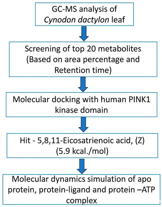
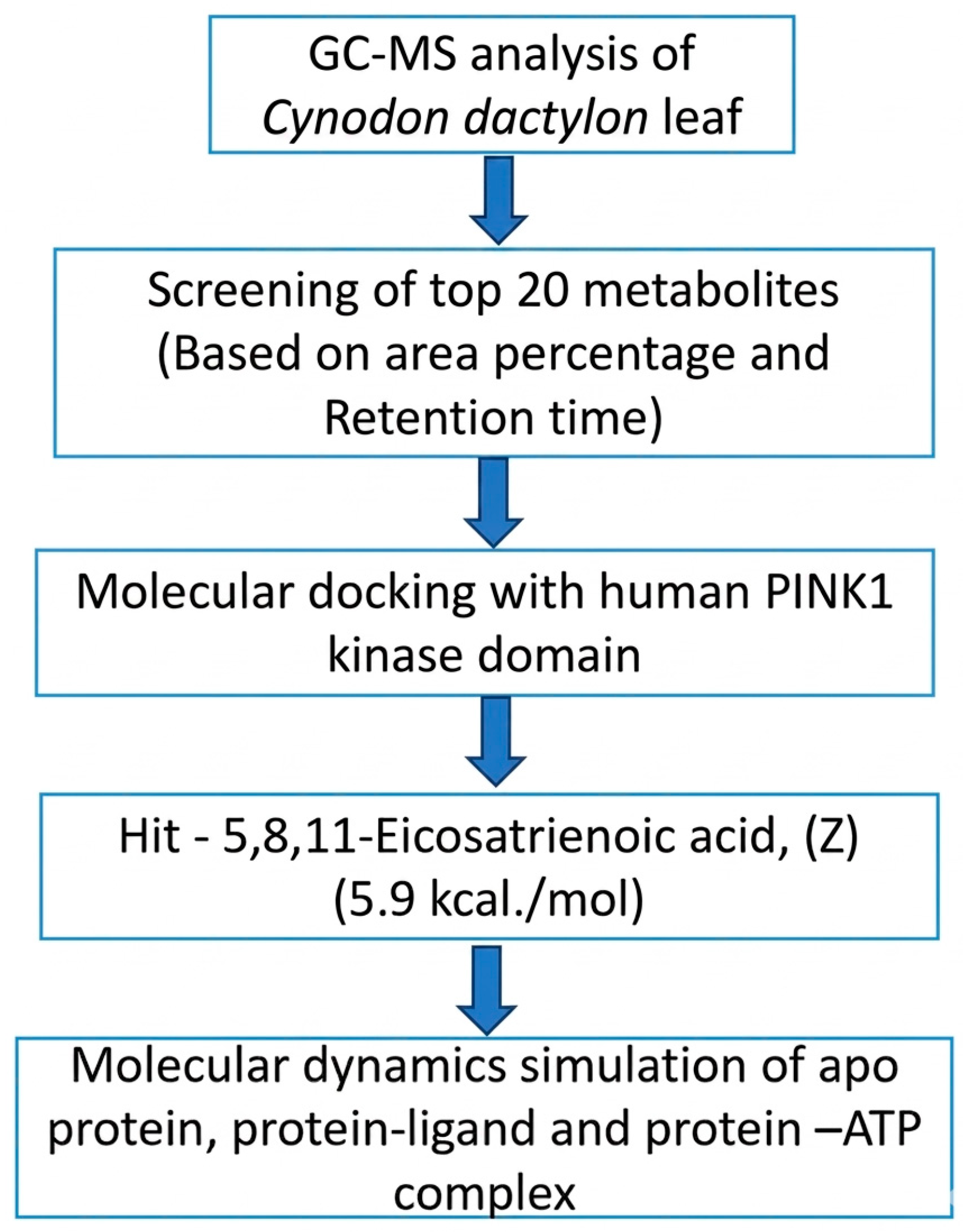
The binding affinity of protein–ligand complexes was estimated using the MM-PBSA approach based on molecular dynamics trajectories obtained from a 100 ns simulation. The binding free energy (ΔG_bind) was calculated as the difference between the free energy of the protein–ligand complex and the combined free energies of the unbound protein and ligand. These calculations were performed using gmx_MMPBSA (version 1.6.4; https://rashmikumari.github.io/g_mmpbsa/) (accessed on 15 March 2026) [26] and the results were further analyzed using gmx_MMPBSA_. The complete workflow is presented in Figure 1.
Figure 1.
Workflow of GC–MS metabolite profiling and computational screening of Cynodon dactylon leaf metabolites targeting human PINK1.
3. Results
3.1. GC-MS Studies
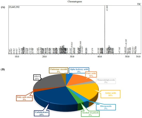
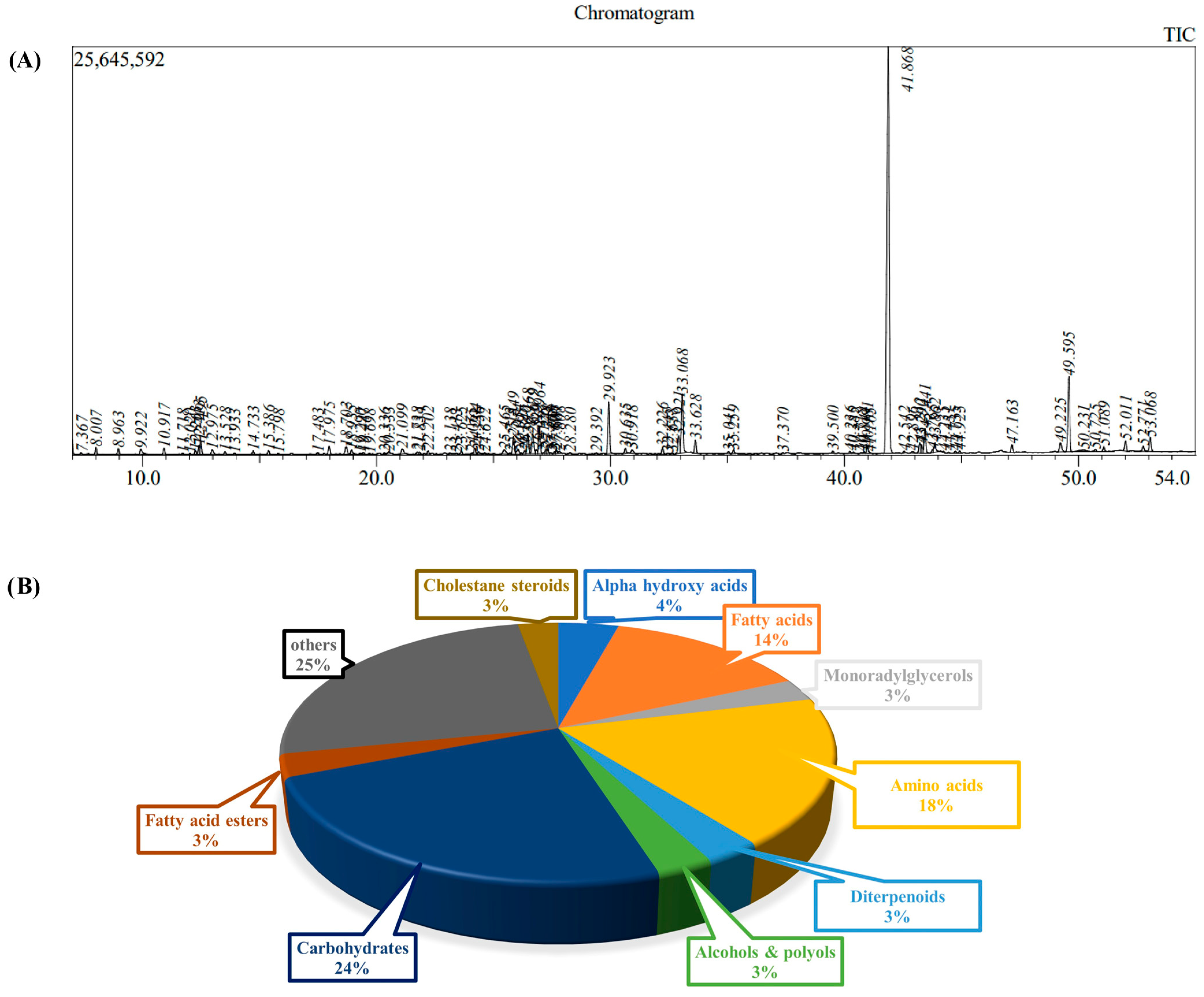
GC-MS analysis of Bermuda grass leaves identified a total of 95 phytochemicals (Table 1 and Figure 2A). These phytochemicals were distributed across various classes, with carbohydrates accounting for 24% of the total compounds, while amino acids (18%) and fatty acids (14%) were also prominently represented. Alpha-hydroxy acids accounted for 4%, whereas cholestane steroids, fatty acids esters, diterpenoids, monoradylglycerols, alcohol and polyols each contributed 3% to the total phytochemical distributions. The remaining 25% consists of a diverse set of phytochemicals, including linoleic acids, fatty aldehydes, non-metal phosphates, phosphate esters, beta hydroxy acids, medium-chain hydroxy acids, gamma butyrolactones, benzenediols, urea, purine nucleosides, phenethylamines, oxanes, phenethylamines, carbonyl compounds, dicarboxylic acids, organosilicon compounds, fatty alcohols, and glycerophosphates (Figure 2B).
Table 1.
GC-MS phytochemical profiling of Bermuda grass leaves.
Figure 2.
GC–MS chromatogram illustrating the identified phytochemicals across retention times (A). Pie chart illustrating the relative distribution of major phytochemical classes identified in Bermuda grass leaves (B).
3.2. ADME Studies
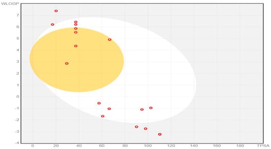
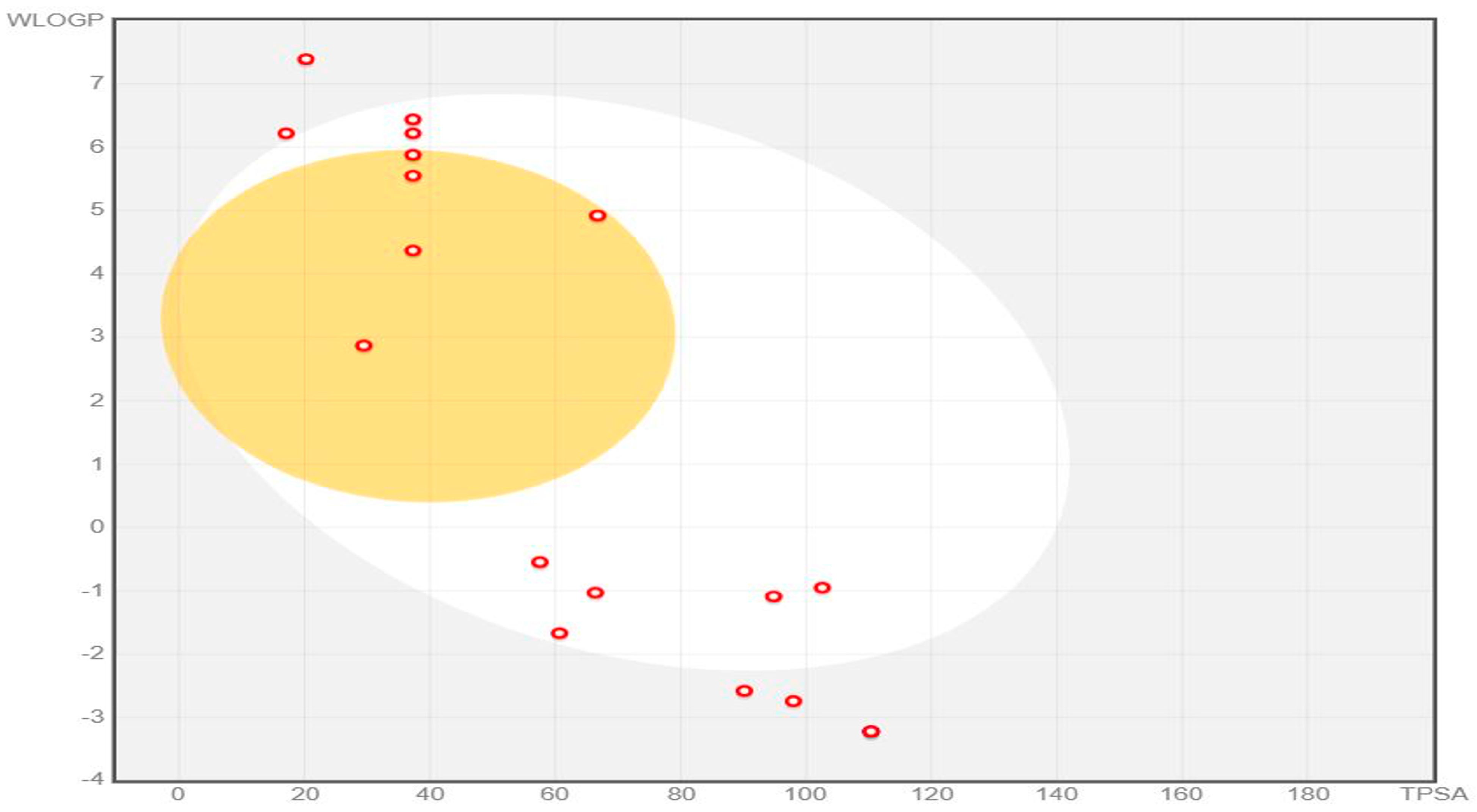
The BOILED-Egg analysis from SwissADME shows the gastrointestinal (GI) absorption and blood–brain barrier (BBB) permeability of the top 20 phytochemicals identified through GC–MS analysis, based on their lipophilicity (WLOGP) and topological polar surface area (TPSA).
In this model, the white region indicates molecules with a high probability of passive GI absorption, while the yellow yolk represents those predicted to possess BBB permeability. Seven phytochemicals were distributed within the white region, indicating good oral absorption (Figure 3). Five phytochemicals were located within the yellow yolk, suggesting their potential to cross the blood–brain barrier. However, these BBB predictions should be interpreted carefully, as the BOILED-Egg model is based only on WLOGP and TPSA.
Figure 3.
BOILED-Egg plot showing GI absorption and BBB permeability of the top 20 phytochemicals based on WLOGP and TPSA. The white region indicates high GI absorption, while the yellow region represents BBB-permeable phytochemicals. Red dots represents the individual phytochemicals.
Some phytochemicals were positioned outside the egg regions or near the periphery, reflecting higher polarity or suboptimal lipophilicity, which may limit passive membrane diffusion and reduce bioavailability. In addition, three phytochemicals were found to be outside the BOILED-Egg plot range due to extreme physicochemical properties and were therefore not displayed within the diagram. CYP inhibition analysis showed that four phytochemicals inhibited CYP1A2 and four phytochemicals inhibited CYP2C9, while no phytochemicals inhibited CYP2C19 or CYP3A4. In addition, only one phytochemical inhibited CYP2D6, indicating generally low interaction with major drug-metabolizing enzymes.
The toxicity profiles of the top 20 phytochemicals were evaluated using the Deep-PK tool, and the results are summarized in Table 2. The majority of the compounds were predicted to be non-toxic across key toxicity endpoints, including AMES mutagenicity, carcinogenicity, drug-induced liver injury (DILI), hERG inhibition, nuclear receptor (NR-AhR), and stress response pathway (SR-p53). A significant proportion of the compounds showed no toxicity across all evaluated parameters, indicating a favorable safety profile.
Table 2.
In silico toxicity prediction of the top 20 phytochemicals using the Deep-PK tool.
3.3. Target Class Prediction
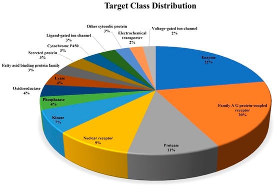
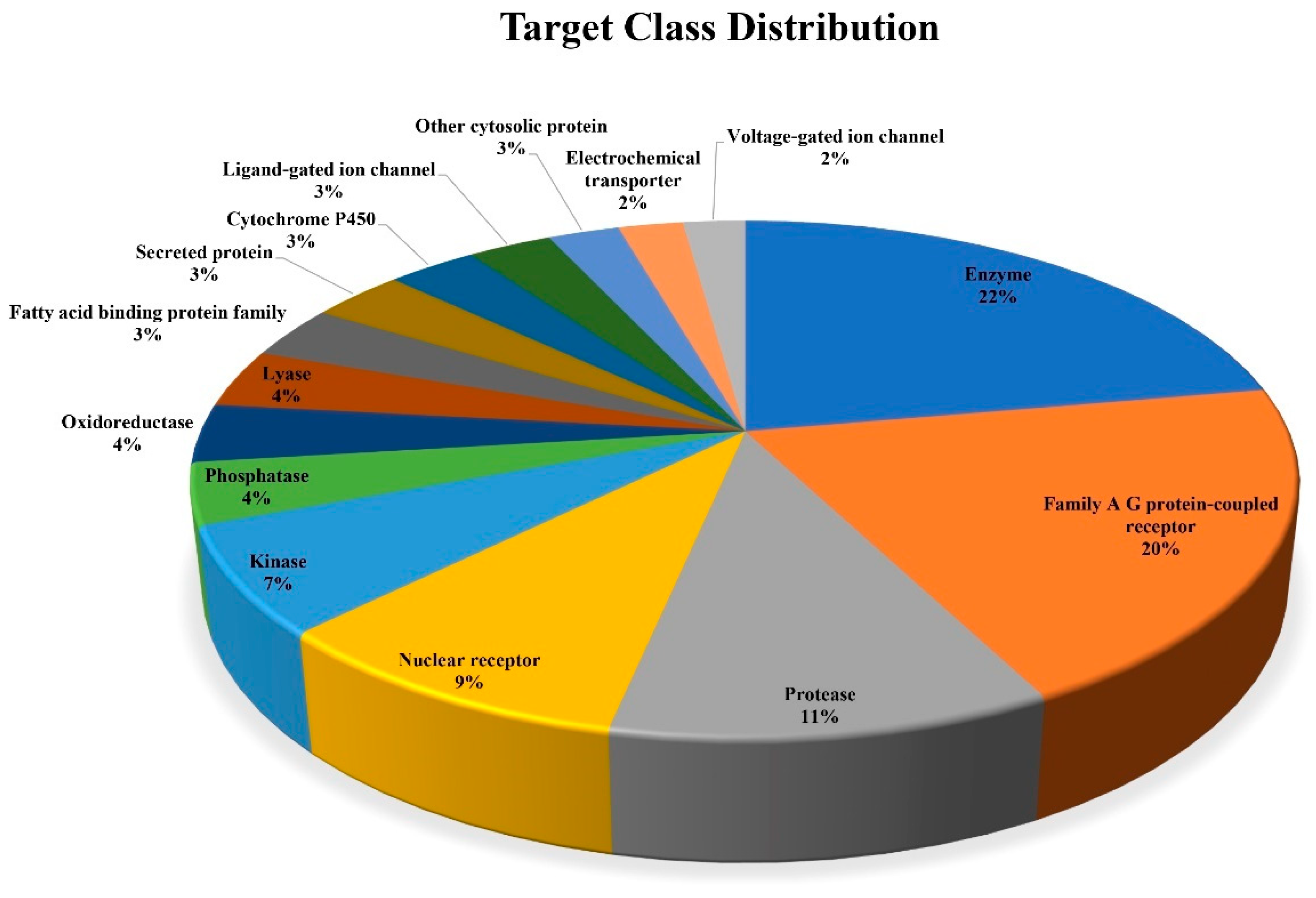
Target prediction of the top 20 compounds was performed using the SwissTargetPrediction web server. The predicted targets were combined, and the top 15 most abundant targets were selected for distribution analysis (Figure 4). The target class distribution analysis showed that enzymes were the most dominant class, accounting for 22%, followed by Family A G protein-coupled receptors at 20% and proteases at 11%. Nuclear receptors and kinases contributed 9% and 7%, respectively. Phosphatases, oxidoreductases, and lyases exhibited moderate presence at 4% each, whereas other target classes, including cytochrome P450, ligand-gated ion channels, and fatty acid-binding proteins, were observed at lower frequencies ranging from 2% to 3%.
Figure 4.
Target class distribution of predicted protein targets for the top 20 phytochemicals.
3.4. Molecular Docking Studies
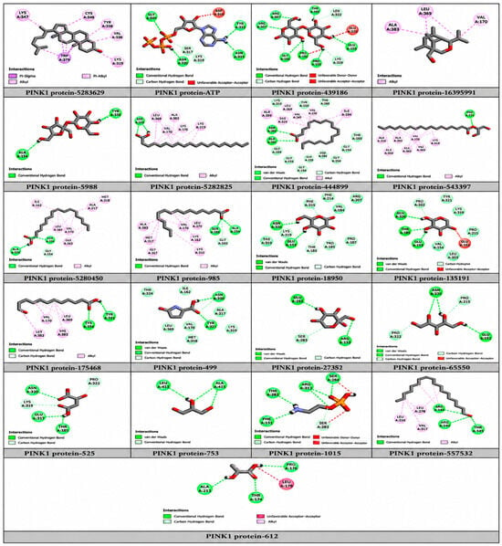
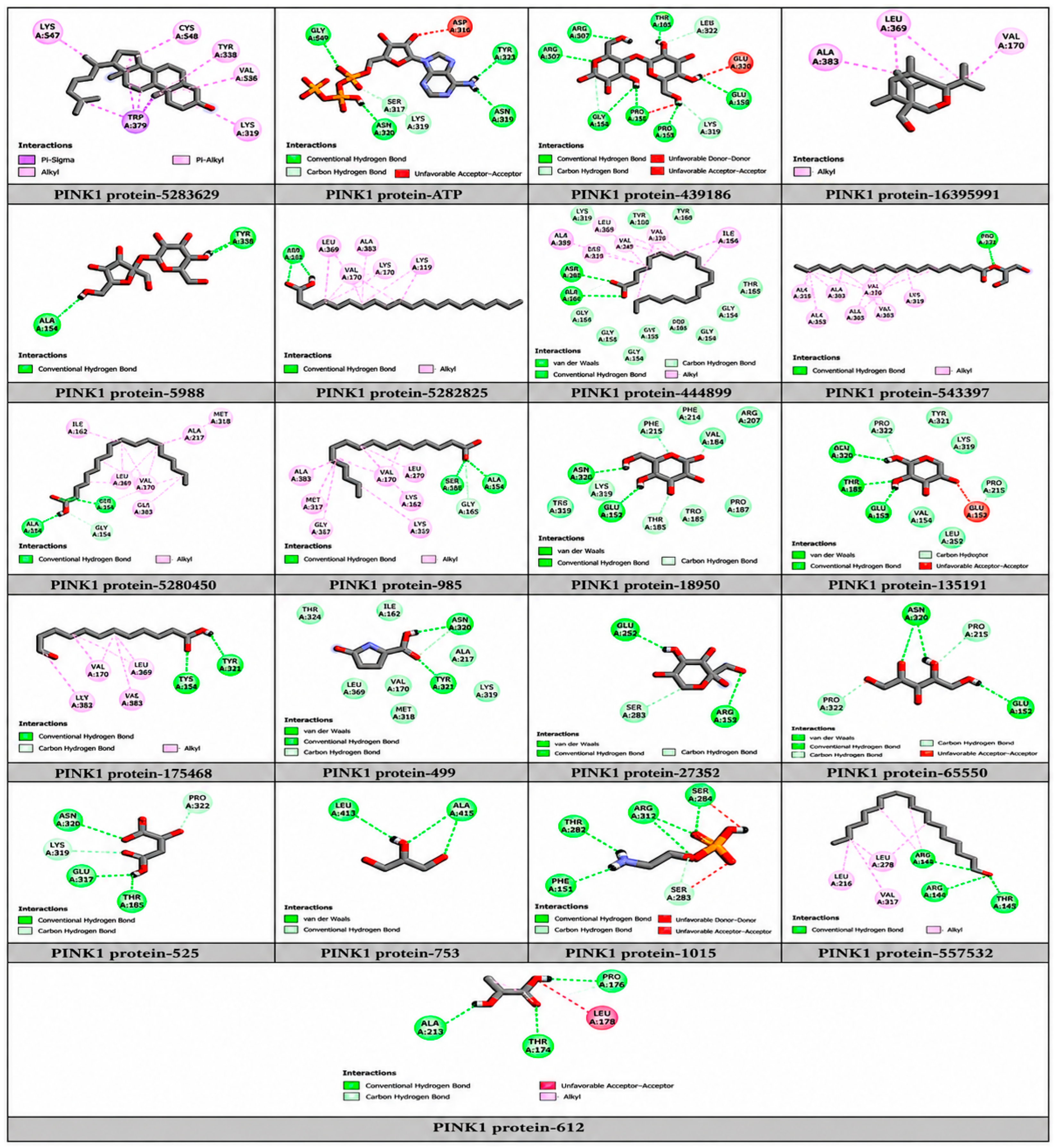
Molecular docking analysis was performed to evaluate the binding affinity of the top 20 phytochemicals identified from C. dactylon against the ATP-binding pocket of the PINK1 protein. The top 20 compounds identified from GC–MS analysis were selected based on their retention time and area percentage to ensure inclusion of the major detected constituents present in the extract. This approach enables representative screening of predominant phytochemicals in Bermuda grass. Although compound abundance does not necessarily correlate with biological activity, the present study aims to perform an initial broad in silico screening to identify potential bioactive candidates. As no promising drug compounds have been reported for PINK1 protein, a comparison of apo protein, PINK1-ATP and PINK1-ligand was performed. ATP was used as a reference ligand to compare the binding behavior of the phytochemicals within the active site. The binding affinities and interacting residues are shown in Table 3, and the 2-D interactions of the PINK1 protein with ATP and the top 20 phytochemicals in Figure 5. The reference ligand ATP exhibited a binding affinity of −7.3 kcal/mol and formed hydrogen bond interactions with Gly165, Asn320, Asn367, and Tyr321, indicating stable binding within the ATP-binding pocket.
Table 3.
Molecular docking results of the top 20 phytochemicals and ATP against PINK1 protein.
Figure 5.
Two-dimensional interactions of PINK1 protein with ATP and selected phytochemicals, showing key hydrogen bonds and hydrophobic interactions within the ATP-binding site.
Among the screened phytochemicals, Cholest-5-en-3-ol (PubChem ID: 5283629) exhibited the highest binding affinity toward PINK1, with a docking score of −7.5 kcal/mol, which was slightly stronger than that of ATP. The diterpenoid compound ((1R,4S,5R,8S,9R)-4-isopropyl-6,8,9-trimethyl-3-oxabicyclo[3.3.1]non-6-en-1-yl) methanol showed a binding affinity of −6.5 kcal/mol. However, no significant hydrogen bond interactions were observed, indicating that the binding is driven primarily by hydrophobic interactions.
Maltose (PubChem ID: 439186) showed a binding affinity of −6.9 kcal/mol and formed multiple hydrogen bond interactions with Phe214, Arg207, Glu172, Asn320, Pro215, Glu183, and Thr185, indicating extensive polar interactions within the binding pocket. Similarly, sucrose-8TMS exhibited a binding affinity of −6.2 kcal/mol and formed hydrogen bonds with Ala168 and Tyr321 residues positioned within the ATP-binding region of the kinase domain. Other carbohydrates and small organic acids, including mannose, D-(+)-xylose, fructose, malic acid and 5-oxoproline, exhibited moderate binding affinities and comparatively weaker interactions with the PINK1 active site. Fatty acid derivatives also showed favorable binding within the PINK1 ATP-binding pocket. Among them, 5,8,11-Eicosatrienoic acid (PubChem ID: 5282825) exhibited a binding affinity of −5.9 kcal/mol and formed a hydrogen bond interaction with Tyr321, which is positioned within the predicted ATP-binding region. Other fatty acids, including arachidonic acid, linoleic acid, and palmitic acid, displayed binding energies ranging from −5.7 to −5.5 kcal/mol and interacted mainly with residues such as Lys219, Ala168, Ser167, and Asp384.
Although sucrose and maltose (highlighted in blue fonts in Table 3) exhibited good docking scores and hydrogen bonding interactions, they may have limited membrane permeability and oral bioavailability according to Lipinski-based drug-likeness principles [27,28]. Based on binding affinity, interaction profile, and biological relevance, 5,8,11-Eicosatrienoic acid (Table 3) was selected for further molecular dynamics simulation analysis to evaluate the stability and dynamic behavior of the protein–ligand complex.
3.5. Molecular Simulation Studies
3.5.1. Hydrogen Bond Analysis
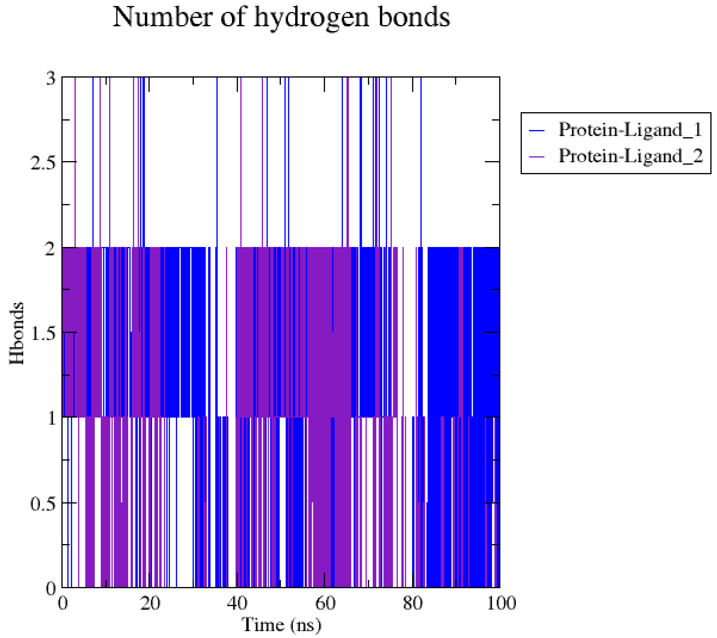
The hydrogen bond analysis shows that the protein–5,8,11-Eicosatrienoic acid complex forms 1–2 hydrogen bonds consistently throughout the 100 ns simulation and up to 3 hydrogen bonds at certain time scales in both the replicates (Figure 6). The presence of hydrogen bonds throughout the simulation indicates stable interactions between the ligand and the ATP-binding site of PINK1. Although the ligand is a fatty acid with limited hydrogen bond donors, the observed interactions suggest that hydrogen bonding along with hydrophobic interactions contribute to binding stability.
Figure 6.
Hydrogen bond analysis of the PINK1 complexed with 5,8,11-eicosatrienoic acid simulated for 100 ns.
3.5.2. Solvent Accessible Surface Area (SASA)
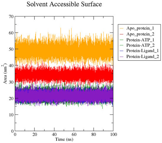
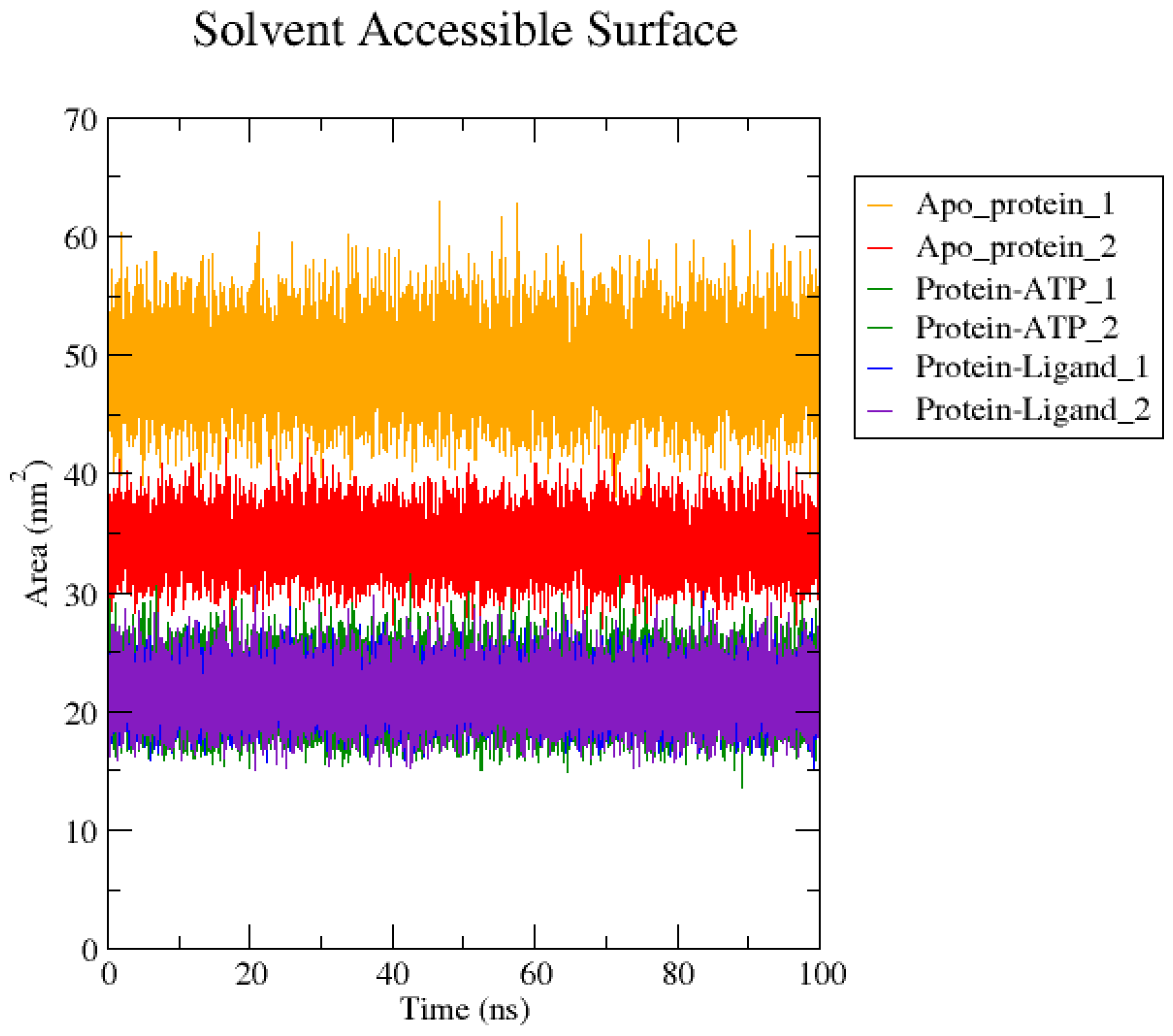
The solvent-accessible surface area (SASA) plot (Figure 7) shows the variation in surface exposure of the apo protein, PINK1–ATP complex and protein–ligand complex over the 100 ns simulation period, including replicates.
Figure 7.
SASA analysis of apo PINK1, PINK1–ATP, and PINK1–5,8,11-eicosatrienoic acid complexes in replicates over 100 ns simulation.
The apo protein exhibits the highest SASA values, ranging between 45 and 60 nm2, indicating a more exposed and less compact structure in the absence of any bound molecule. In comparison, the PINK1–ATP complex shows reduced SASA values in the range of 30–40 nm2, suggesting that ATP binding induces partial compaction and stabilization of the protein structure.
The PINK1–5,8,11-eicosatrienoic acid complex displays the lowest SASA values, ranging between 18 and 28 nm2, and forms compact conformation with reduced solvent exposure. This reduction suggests effective binding of the ligand within the active site and the burial of hydrophobic regions.
All systems exhibit consistent SASA with minimal fluctuations over time, which shows their stability and the reproducibility of the protein–ligand complex during simulations in replicates.
3.5.3. Radius of Gyration (Rg)
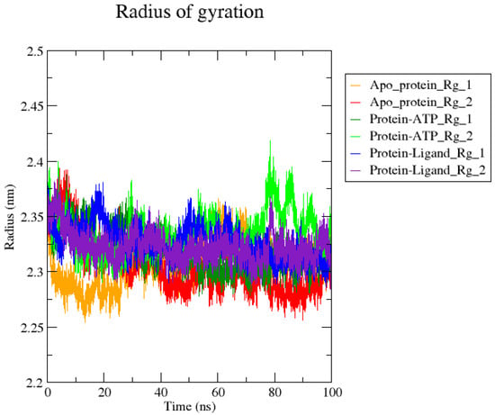
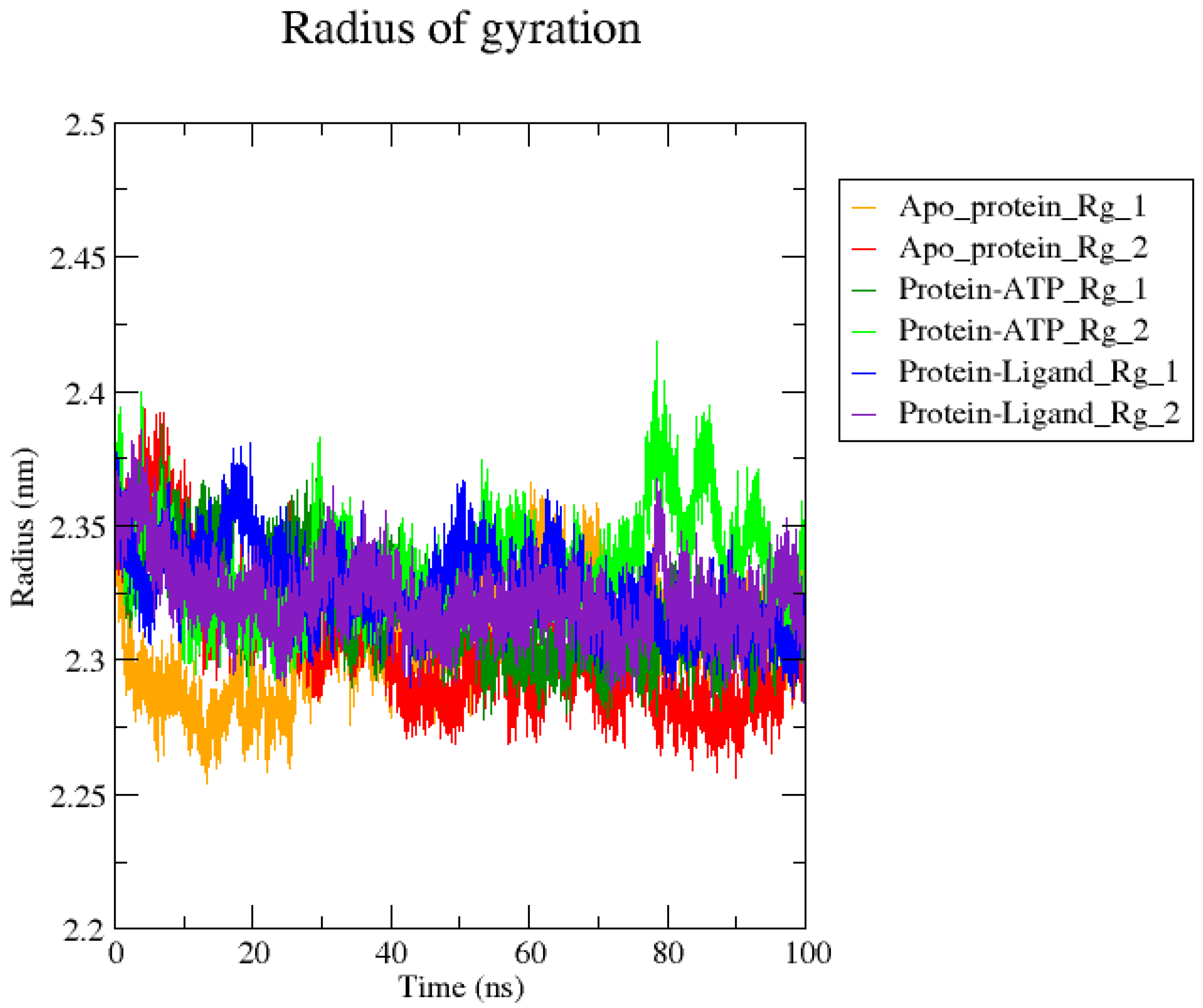
The radius of gyration analysis reflects the overall compactness of the protein (Figure 8). In this study, the Rg values for all systems remain within the range of 2.33–2.40 nm, where the protein maintains a stable folded structure throughout the simulation.
Figure 8.
Radius of gyration analysis of apo PINK1, PINK1-ATP, and PINK1-5,8,11-eicosatrienoic acid complexes over the 100 ns simulation with replicates.
The protein–ligand complex with 5,8,11-eicosatrienoic acid shows consistent Rg values with minimal fluctuations, suggesting that ligand binding promotes structural stability and compactness, while the ATP-bound system exhibits slightly higher fluctuations, increasing up to 2.40 nm with minor conformational changes. Across replicates, the trends are consistent, confirming the reliability of the results, which shows that the protein does not undergo major structural unfolding.
3.5.4. Root Mean Square Fluctuation (RMSF)
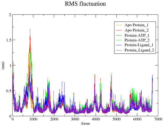
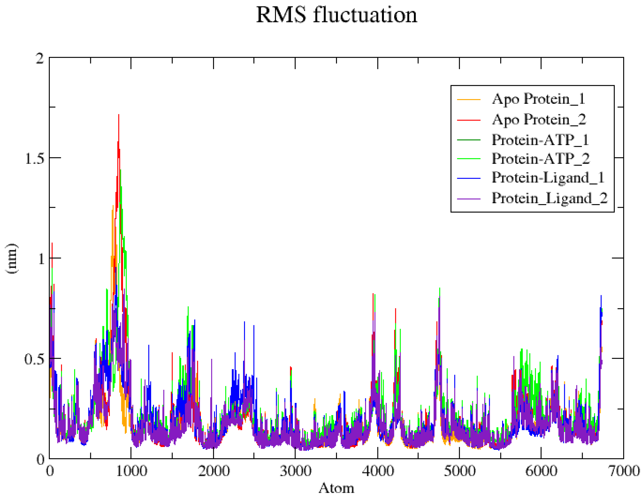
The RMSF plot (Figure 9) illustrates residue-level flexibility of the apo protein, PINK1–ATP complex, and protein–ligand complex across the simulation. The residues mostly exhibit low fluctuations in the range of 0.1–0.3 nm, indicating a stable protein backbone.
Figure 9.
RMSF analysis of apo PINK1, PINK1–ATP, and PINK1–5,8,11-eicosatrienoic acid complexes over the 100 ns simulations with replicates.
The apo protein shows comparatively higher fluctuations, particularly in the region around 800–1000 atoms, where peaks reach values in the range of 1.5–1.7 nm, exhibiting increased flexibility in the absence of ligand binding. Both the ATP-bound and 5,8,11-eicosatrienoic acid-bound complex systems show reduced fluctuations in this region (range of 1.0–1.4 nm), which confers stabilization upon binding.
Moderate fluctuations in the range of 0.3–0.8 nm are observed, primarily corresponding to loop regions. The protein–ligand complex with 5,8,11-eicosatrienoic acid shows RMSF values comparable to the ATP-bound state in both duplicates.
3.5.5. Root Mean Square Deviation (RMSD)
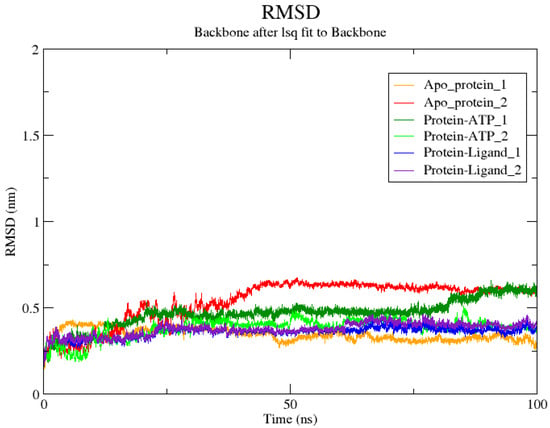
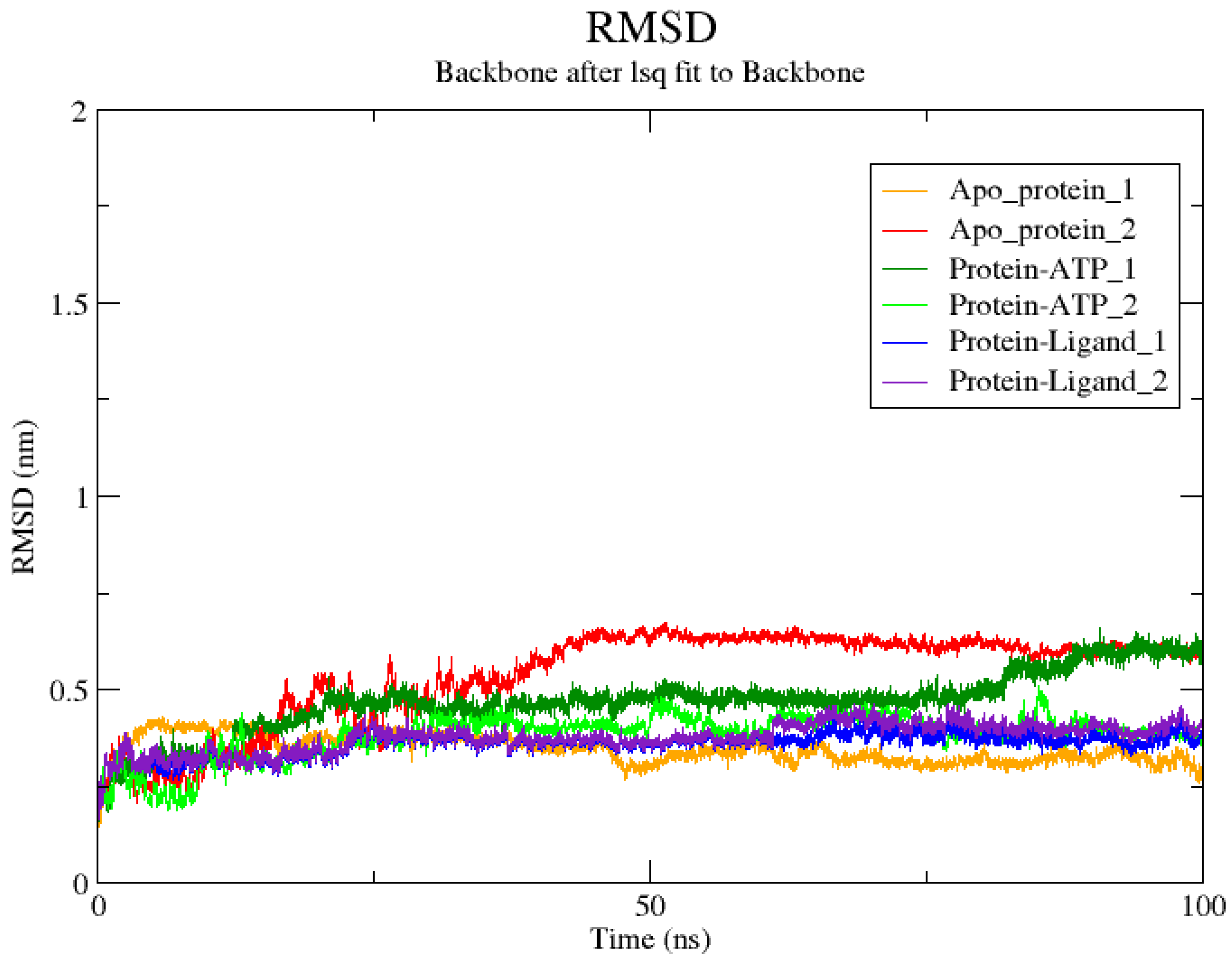
The RMSD plot (Figure 10) represents the backbone stability of the apo protein, PINK1–ATP complex and protein–ligand complex with 5,8,11-eicosatrienoic acid over the 100 ns simulation, including replicates.
Figure 10.
RMSD analysis of apo PINK1, PINK1–ATP, and PINK1–5,8,11-eicosatrienoic acid complexes over the 100 ns simulation time period with replicates.
The apo protein shows comparatively lower RMSD values in the range of 0.25–0.40 nm, with a stable but more flexible conformation in the absence of binding partners. Among the replicates, apo protein_2 exhibits slightly higher deviations, reaching up to 0.60–0.65 nm, showing minor conformational adjustments during the simulation.
The PINK1–ATP complex displays RMSD values in the range of 0.35–0.60 nm, showing the structural deviation upon ATP binding. One replicate stabilizes around 0.45–0.50 nm, while the other shows a gradual increase up to 0.60–0.65 nm, reflecting conformational adaptation associated with ligand binding.
The protein–ligand complex with 5,8,11-eicosatrienoic acid shows relatively stable RMSD values in the range of 0.30–0.45 nm across both replicates. The trajectories stabilize after the initial equilibration phase (around 10–20 ns), exhibiting stable binding throughout the simulation.
3.5.6. MMPBSA Analysis
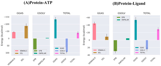
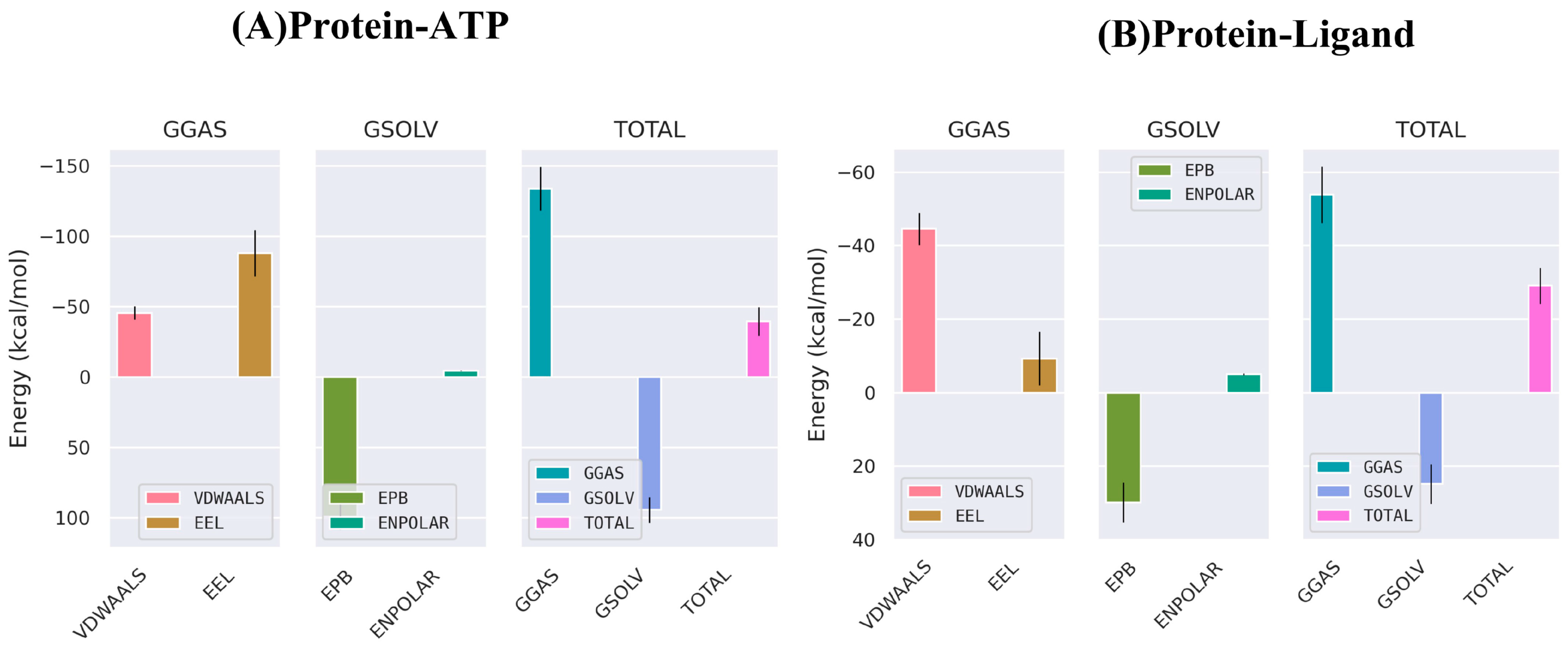
The binding free energy of the complexes was calculated using the MM-PBSA method, and the results are presented in Table 4 and Figure 11. The protein–ATP complex exhibited a total binding free energy of −39.69 ± 10.11 kcal/mol, indicating higher binding affinity. The interaction energy includes van der Waals (VDWAALS) (−45.68 ± 4.71 kcal/mol) and electrostatic interactions (EEL) (−88.22 ± 16.5 kcal/mol), resulting in a highly favorable gas-phase energy (GGAS) (−133.89 ± 15.45 kcal/mol). However, a large polar solvation energy (EPB) (98.88 ± 9.32 kcal/mol) leads to an overall solvation energy (GSOLV) of 94.2 ± 9.33 kcal/mol. The protein–ligand complex showed a total binding free energy of −29.12 ± 4.91 kcal/mol, suggesting a stable interaction. The binding was mainly governed by van der Waals interactions (−44.6 ± 4.41 kcal/mol), with a smaller contribution from electrostatic energy (−9.32 ± 7.26 kcal/mol). The gas-phase energy was −53.92 ± 7.66 kcal/mol, while the polar solvation energy (29.83 ± 5.41 kcal/mol) and non-polar solvation energy (ENPOLAR) (−5.03 ± 0.27 kcal/mol) resulted in a total solvation energy (GSOLV) of 24.8 ± 5.36 kcal/mol. Although both complexes experienced unfavorable polar solvation effects, the strong gas-phase interactions resulted in negative binding free energies. The relatively lower standard deviation observed for the protein–ligand complex indicates more consistent binding behavior compared to the protein–ATP complex. Overall, both ATP and the ligand form stable complexes with the protein, with ATP showing relatively stronger binding affinity.
Table 4.
MM-PBSA binding free energies for protein–ATP and protein–ligand complexes.
Figure 11.
MM-PBSA binding free energy profiles of (A) protein–ATP and (B) protein–ligand complexes, illustrating contributions from van der Waals, electrostatic, polar solvation, non-polar solvation, and total binding energies.
4. Discussion
Oxidative stress and mitochondrial dysfunction are major contributors to the pathogenesis of Parkinson’s disease. Mitochondria function both as a primary source and a target of reactive oxygen species (ROS), and impairment of mitochondrial respiratory complexes (complex I) results in excessive ROS production. This increased oxidative stress disrupts mitochondrial function and accelerates neuronal damage. In this context, the protein PINK1 plays a crucial role in maintaining mitochondrial quality control, especially under conditions of cellular stress. PINK1 is involved in several key signaling pathways, including mitophagy, apoptosis, and microglial inflammatory responses, and its expression is induced by oxidative stress [29,30]. Loss-of-function mutations in PINK1 disrupt mitochondrial quality control, leading to impaired mitophagy, increased oxidative stress, and increased vulnerability of neurons to cell death [31]. Structural studies report that the ATP-binding region of PINK1 is essential for its kinase activation and downstream phosphorylation events, making it a potential site for therapeutic modulation [6].
Cynodon dactylon (Bermuda grass), widely known as Durva in traditional Indian medicine, has been extensively used in Ayurveda, Siddha, Unani, and African folk medicine for managing inflammatory conditions, wounds, fever, convulsions, and disorders linked to oxidative stress, indicating its long-standing therapeutic relevance [32]. Phytochemical studies have identified a rich profile of bioactive constituents, including flavonoids, triterpenoids, phenolic acids and polysaccharides, which are known to exert antioxidant and anti-inflammatory effects, which are the two key pathological drivers underlying neurodegenerative diseases such as Parkinson’s and Alzheimer’s disorders [33]. Emerging preclinical evidence suggests that extracts of C. dactylon significantly suppress nitric oxide production and free-radical generation, supporting its potential to modulate neuroinflammation and mitochondrial dysfunction through indirect mechanisms [34].
In the present study, GC–MS identified several bioactive metabolites from C. dactylon, and molecular docking revealed stable interaction of 5,8,11-Eicosatrienoic acid within the ATP-binding pocket of PINK1. Previous GC–MS studies on C. dactylon have reported metabolites such as phytol, hexadecanoic acid, linoleic acid, oleic acid, squalene, and β-sitosterol, which are mainly fatty acids, terpenoids, and sterols [4,5]. In this study, a wider range of metabolites including sugars (sucrose, fructose, mannose), amino acids (alanine, serine, glutamic acid), organic acids (malic acid, lactic acid), and fatty acids (palmitic acid, linoleic acid, arachidonic acid) were identified. Target prediction reported the enzyme targets as major components for the C. dactylon leaf compounds. Based on the provided research and computational findings, the interaction between 5,8,11-eicosatrienoic acid and the PINK1 kinase represents a significant pharmacological lead for Parkinson’s disease therapy. Derived from C. dactylon, this fatty acid shows a higher binding affinity of −5.9 kcal/mol, outperforming other common lipids such as arachidonic and palmitic acid. Central to this efficacy is its specific targeting of the Tyr321 residue located within the critical ATP-binding region of the PINK1 catalytic core. In kinase biology, this “hinge region” is essential for orienting ATP; by forming a hydrogen bond with Tyr321. 5,8,11-eicosatrienoic acid acts as a molecular stabilizer by lowering the activation energy required for PINK1 to achieve its functional state, overcoming oxidative stress [35]. It should be noted that ATP retained a favorable binding affinity (−8.3 kcal/mol) even in the presence of 5,8,11-eicosatrienoic acid within the active site upon docking, indicating that ligand binding does not completely hinder ATP interaction. However, slight variations in the interaction profile suggest minor alterations in the binding environment, which supports modulation activity rather than complete inhibition.
Molecular dynamics simulations further supported complex stability, interactions and lower binding free energy, as is evident from the RMSD values, RMSF fluctuations, stable radius of gyration, SASA profiles and MMPBSA analysis. Unlike sucrose or maltose, which have poor membrane permeability, the lipophilic 5,8,11-eicosatrienoic acid adheres to Lipinski’s Rule of Five, suggesting favorable oral bioavailability and cellular uptake [28,36]. The MD data shows consistent RMSD and SASA profiles, along with reduced RMSF fluctuations at key residues. This indicates that the fatty acid effectively interacts with the enzymatic loop, creating a rigid, efficient environment that facilitates the downstream phosphorylation of Parkin [37]. Such stabilization is vital in PD, as it promotes healthy mitophagy, the process required to clear the toxic mitochondrial debris that otherwise leads to dopaminergic neuron death [35]. Even if direct blood–brain barrier penetration is still under investigation, the compound’s role in “network-level” regulation makes it a prime candidate for managing neurodegeneration through systemic and cellular mitochondrial quality control [31]. Furthermore, its lipophilic nature ensures a favorable bioavailability profile consistent with medicinal chemistry standards for oral drugs. Ultimately, its neuroprotective potential lies in its ability to stabilize PINK1, thereby facilitating the phosphorylation of Parkin and promoting the clearance of dysfunctional mitochondria.
This study indicates that peripherally acting natural compounds can influence neurodegenerative disease progression by regulating systemic inflammation, redox balance and interconnected protein signaling networks [38].
5. Conclusions
In conclusion, the present study provides molecular insights into the interaction of Cynodon dactylon-derived phytochemicals with the PINK1 protein, a key regulator of mitochondrial quality control in Parkinson’s disease. Among the screened metabolites, 5,8,11-eicosatrienoic acid showed stable binding within the ATP-binding pocket of PINK1, as supported by molecular docking and molecular dynamics analyses. We report that 5,8,11-eicosatrienoic acid modulates the activity of PINK1 protein, which in turn has an impact on Parkinson’s disease. Furthermore, MM-PBSA analyses indicate favorable binding energetics and stable conformational states of the protein–ligand complex.
However, the findings are based solely on in silico approaches, and the lack of experimental validation remains a limitation of the study. Therefore, further in vitro and in vivo studies are necessary to confirm the modulatory potential of 5,8,11-eicosatrienoic acid on PINK1 activity and its relevance in Parkinson’s disease therapeutics.
Author Contributions
Conceptualization, S.N. and R.P.; methodology, S.N.; software, S.N.; validation, S.N., R.B.M.Y. and N.V.; formal analysis, S.N., R.B.M.Y. and N.V.; investigation, S.N.; resources, S.N.; writing—original draft preparation, S.N.; writing—review and editing, S.N. and R.P.; supervision, S.N. and R.P. All authors have read and agreed to the published version of the manuscript.
Funding
This research was supported by Biotechnology Information System (BTIS) program, (BT/PR40235/BTIS/137/39/2022) of the Department of Biotechnology (DBT), Government of India, New Delhi, awarded to SN and Memorial University Dean of Science Startup funds, awarded to RP.
Institutional Review Board Statement
Not applicable.
Informed Consent Statement
Not applicable.
Data Availability Statement
Computational data will be available upon special request to the corresponding authors.
Conflicts of Interest
The authors declare no conflicts of interest.
Abbreviations
The following abbreviations are used in this manuscript:
| PINK1 | PTEN-induced kinase 1 |
| GC-MS | Gas chromatography–mass spectrometry |
| PDB | Protein Data Bank |
| TMS | Trimethylsilyl |
| RMSD | Root mean square deviation |
| RMSF | Root mean square fluctuation |
| SASA | Solvent accessible surface area |
| Rg | Radius of gyration |
| ATP | Adenosine triphosphate |
| VDWAALS | Van der Waals interactions |
| EEL | Electrostatic energy |
| EPB | Electrostatic Poisson–Boltzmann energy |
| ENPOLAR | Non-polar solvation energy |
| GGAS | Gas phase free energy |
| GSOLV | Solvation free energy |
| MM-PBSA | Molecular Mechanics Poisson–Boltzmann Surface Area |
References
- Kumar, P.; Chaudhary, R.; Ashawat, M.S. Exploring the Efficacy of Herbal Medications in the Treatment of Parkinsonism: A Mini Review. Curr. Drug Ther. 2025, 20, 126–134. [Google Scholar] [CrossRef] [Scilit]
- Poojary, R.; Kumar, N.A.; Kumarchandra, R.; Vinodini, N.; Bhagyalakshmi, K.; Sanjeev, G. Cynodon dactylon Extract Ameliorates Cognitive Functions and Cerebellar Oxidative Stress in Whole Body Irradiated Mice. Asian Pac. J. Trop. Biomed. 2019, 9, 278–283. [Google Scholar] [CrossRef] [Scilit]
- Kothari, S.; Sahu, M. Effect of Aqueous Extract of Cynodon dactylon (Doob Grass) on Normal and Impaired Memory in Mice. Asian J. Pharm. Clin. Res. 2022, 15, 130–133. [Google Scholar] [CrossRef] [Scilit]
- Mozafari, A.A.; Vafaee, Y.; Shahyad, M. Phytochemical Composition and in Vitro Antioxidant Potential of Cynodon dactylon Leaf and Rhizome Extracts as Affected by Drying Methods and Temperatures. J. Food Sci. Technol. 2018, 55, 2220–2229. [Google Scholar] [CrossRef] [Scilit]
- Chauhan, R.P.; Garg, A.; Baksh, Z.; Pandey, L.; Sandhu, S.S. Metabolomics Profiling of Cynodon dactylon Using Gas Chromatography–Mass Spectrometry and Cytotoxicity Study. Int. J. Technol. Emerg. Res. 2025, 1, 195–211. [Google Scholar]
- Trempe, J.F.; Gehring, K. Structural Mechanisms of Mitochondrial Quality Control Mediated by Pink1 and Parkin. J. Mol. Biol. 2023, 435, 168090. [Google Scholar] [CrossRef] [Scilit]
- Chin, R.M.; Rakhit, R.; Ditsworth, D.; Wang, C.; Bartholomeus, J.; Liu, S.; Mody, A.; Laishu, A.; Eastes, A.; Tai, C.; et al. Pharmacological Pink1 Activation Ameliorates Pathology in Parkinson’s Disease Models. bioRxiv 2023. [Google Scholar] [CrossRef] [Scilit] [PubMed]
- Gan, Z.Y.; Callegari, S.; Cobbold, S.A.; Cotton, T.R.; Mlodzianoski, M.J.; Schubert, A.F.; Geoghegan, N.D.; Rogers, K.L.; Leis, A.; Dewson, G.; et al. Activation mechanism of PINK1. Nature 2022, 602, 328–335. [Google Scholar] [CrossRef] [Scilit]
- Arena, G.; Gelmetti, V.; Torosantucci, L.; Vignone, D.; Lamorte, G.; De Rosa, P.; Cilia, E.; Jonas, E.A.; Valente, E.M. PINK1 protects against cell death induced by mitochondrial depolarization, by phosphorylating Bcl-xL and impairing its pro-apoptotic cleavage. Cell Death Differ. 2013, 20, 920–930. [Google Scholar] [CrossRef] [Scilit] [PubMed]
- Steer, E.K.; Dail, M.K.; Chu, C.T. Beyond mitophagy: Cytosolic PINK1 as a messenger of mitochondrial health. Antioxid. Redox Signal. 2015, 22, 1047–1059. [Google Scholar] [CrossRef] [Scilit]
- Liu, K.; Liu, Z.; Liu, Z.; Ma, Z.; Deng, Y.; Liu, W.; Xu, B. Manganese induces S-nitrosylation of PINK1 leading to nerve cell damage by repressing PINK1/Parkin-mediated mitophagy. Sci. Total Environ. 2022, 834, 155358. [Google Scholar] [CrossRef] [Scilit]
- Hees, J.T.; Wanderoy, S.; Lindner, J.; Helms, M.; Murali Mahadevan, H.; Harbauer, A.B. Insulin signalling regulates Pink1 mRNA localization via modulation of AMPK activity to support PINK1 function in neurons. Nat. Metab. 2024, 6, 514–530. [Google Scholar] [CrossRef] [Scilit]
- Wang, Y.; Luo, S.; Su, H.; Wang, Z.; Chu, L.; Zhang, C. BL-918 activates PINK1/Parkin signaling pathway to ameliorate the progression of Parkinson’s disease. J. Biol. Chem. 2024, 300, 107543. [Google Scholar] [CrossRef] [Scilit]
- Nallusamy, S.; Selvamani, S.; Sathyaseelan, C.; Selvakumar, D.; Panigrahi, R. Targeting Pink1 Using Phytochemicals: Exploring Molecular Insights into Parkinson’s Disease Therapeutics. Biochem. Cell Biol. 2025, 103, 1–55. [Google Scholar] [CrossRef] [Scilit]
- Uttara, B.; Singh, A.V.; Zamboni, P.; Mahajan, R.T. Oxidative Stress and Neurodegenerative Diseases: A Review of Upstream and Downstream Antioxidant Therapeutic Options. Curr. Neuropharmacol. 2009, 7, 65–74. [Google Scholar] [CrossRef] [Scilit]
- Lee, E.H.; Lim, S.S.; Yuen, K.H.; Lee, C.Y. Curcumin and a Hemi-Analogue with Improved Blood-Brain Barrier Permeability Protect Against Amyloid-Beta Toxicity in Caenorhabditis Elegans via Skn-1/Nrf Activation. J. Pharm. Pharmacol. 2019, 71, 860–868. [Google Scholar] [CrossRef] [Scilit] [PubMed]
- d’Amora, M.; Angelini, C.; Marcoli, M.; Cervetto, C.; Kitada, T.; Vallarino, M. Expression of Pink1 in the Brain, Eye and Ear of Mouse During Embryonic Development. J. Chem. Neuroanat. 2011, 41, 73–85, Erratum in: J. Chem. Neuroanat. 2011, 42, 99–100. [Google Scholar] [CrossRef] [Scilit] [PubMed]
- Kim, S.; Chen, J.; Cheng, T.; Gindulyte, A.; He, J.; He, S.; Li, Q.; Shoemaker, B.A.; Thiessen, P.A.; Yu, B.; et al. PubChem 2025 Update. Nucleic Acids Res. 2025, 53, D1516–D1525. [Google Scholar] [CrossRef] [Scilit]
- Daina, A.; Michielin, O.; Zoete, V. SwissADME: A Free Web Tool to Evaluate Pharmacokinetics, Drug-Likeness and Medicinal Chemistry Friendliness of Small Molecules. Sci. Rep. 2017, 7, 42717. [Google Scholar] [CrossRef] [Scilit] [PubMed]
- Myung, Y.; de Sá, A.G.; Ascher, D.B. Deep-PK: Deep Learning for Small Molecule Pharmacokinetic and Toxicity Prediction. Nucleic Acids Res. 2024, 52, W469–W475. [Google Scholar] [CrossRef] [Scilit]
- Gfeller, D.; Grosdidier, A.; Wirth, M.; Daina, A.; Michielin, O.; Zoete, V. SwissTargetPrediction: A Web Server for Target Prediction of Bioactive Small Molecules. Nucleic Acids Res. 2014, 42, W32–W38. [Google Scholar] [CrossRef] [Scilit]
- Chen, V.B.; Arendall, W.B.; Headd, J.J.; Keedy, D.A.; Immormino, R.M.; Kapral, G.J.; Murray, L.W.; Richardson, J.S.; Richardson, D.C. MolProbity: All-Atom Structure Validation for Macromolecular Crystallography. Acta Crystallogr. Sect. D 2010, 66, 12–21. [Google Scholar] [CrossRef] [Scilit]
- Dallakyan, S.; Olson, A.J. Small-Molecule Library Screening by Docking with PyRx. Methods Mol. Biol. 2015, 1263, 243–250. [Google Scholar] [CrossRef] [Scilit] [PubMed]
- Van Der Spoel, D.; Lindahl, E.; Hess, B.; Groenhof, G.; Mark, A.E.; Berendsen, H.J. GROMACS: Fast, Flexible, and Free. J. Comput. Chem. 2005, 26, 1701–1718. [Google Scholar] [CrossRef] [Scilit] [PubMed]
- Vanommeslaeghe, K.; MacKerell, A.D., Jr. Automation of the CHARMM General Force Field (CGenFF) I: Bond Perception and Atom Typing. J. Chem. Inf. Model. 2012, 52, 3144–3154. [Google Scholar] [CrossRef] [Scilit]
- Valdés-Tresanco, M.S.; Valdés-Tresanco, M.E.; Valiente, P.A.; Moreno, E. gmx_MMPBSA: A New Tool to Perform End-State Free Energy Calculations with GROMACS. J. Chem. Theory Comput. 2021, 17, 6281–6291. [Google Scholar] [CrossRef] [Scilit] [PubMed]
- Duchowicz, P.R.; Talevi, A.; Bellera, C.; Bruno-Blanch, L.E.; Castro, E.A. Application of Descriptors Based on Lipinski’s Rules in the QSPR Study of Aqueous Solubilities. Bioorg. Med. Chem. 2007, 15, 3711–3719. [Google Scholar] [CrossRef] [Scilit]
- Lipinski, C.A.; Lombardo, F.; Dominy, B.W.; Feeney, P.J. Experimental and Computational Approaches to Estimate Solubility and Permeability in Drug Discovery and Development Settings. Adv. Drug Deliv. Rev. 2001, 46, 3–26. [Google Scholar] [CrossRef] [Scilit]
- Gaki, G.S.; Papavassiliou, A.G. Oxidative Stress-Induced Signaling Pathways Implicated in the Pathogenesis of Parkinson’s Disease. Neuromol. Med. 2014, 16, 217–230. [Google Scholar] [CrossRef] [Scilit]
- Chang, K.-H.; Chen, C.-M. The Role of Oxidative Stress in Parkinson’s Disease. Antioxidants 2020, 9, 597. [Google Scholar] [CrossRef] [Scilit]
- Exner, N.; Lutz, A.K.; Haass, C.; Winklhofer, K.F. Mitochondrial Dysfunction in Parkinson’s Disease: Molecular Mechanisms and Pathophysiological Consequences. EMBO J. 2012, 31, 3038–3062. [Google Scholar] [CrossRef] [Scilit]
- Raman, N.; Radha, A.; Balasubramanian, K.; Raghunathan, R.; Priyadarshini, R. Bioactive Molecules from Cynodon dactylon of Indian Biodiversity. In Biodiversity: Biomolecular Aspects of Biodiversity and Innovative Utilization; Şener, B., Ed.; Springer: Boston, MA, USA, 2002; pp. 253–256. [Google Scholar] [CrossRef] [Scilit]
- Parihar, S.; Sharma, D. Cynodon dactylon: A Review of Pharmacological Activities. Sch. Acad. J. Pharm. 2021, 10, 183–189. [Google Scholar] [CrossRef] [Scilit]
- Nagendra Prasad, M.N.; Sreeramulu, K.; Sekhar, K.B.C. In Vitro Antioxidant and Anti-Inflammatory Activity of Cynodon dactylon. J. Ethnopharmacol. 2008, 114, 416–422. [Google Scholar]
- Pickrell, A.M.; Youle, R.J. The Roles of PINK1, Parkin, and Mitochondrial Fidelity in Parkinson’s Disease. Neuron 2015, 85, 257–273. [Google Scholar] [CrossRef] [Scilit]
- Veber, D.F.; Johnson, S.R.; Cheng, H.Y.; Smith, B.R.; Ward, K.W.; Kopple, K.D. Molecular Properties That Influence the Oral Bioavailability of Drug Candidates. J. Med. Chem. 2002, 45, 2615–2623. [Google Scholar] [CrossRef] [Scilit] [PubMed]
- Kondapalli, C.; Kazlauskaite, A.; Zhang, N.; Woodroof, H.I.; Campbell, D.G.; Gourlay, R.; Burchell, L.; Walden, H.; Macartney, T.J.; Deak, M.; et al. PINK1 is activated by mitochondrial membrane potential depolarization and stimulates Parkin E3 ligase activity by phosphorylating Serine 65. Open Biol. 2012, 2, 120080. [Google Scholar] [CrossRef] [Scilit] [PubMed]
- Gil-Martínez, A.L.; Cuesta, A.; Molina, J.A.; Martín-Jiménez, C.A.; Blesa, J. Natural Bioactive Molecules as Neuromedicines for the Treatment/Prevention of Neurodegenerative Diseases. ACS Omega 2023, 8, 12347–12374. [Google Scholar] [CrossRef] [Scilit]

Disclaimer/Publisher’s Note: The statements, opinions and data contained in all publications are solely those of the individual author(s) and contributor(s) and not of MDPI and/or the editor(s). MDPI and/or the editor(s) disclaim responsibility for any injury to people or property resulting from any ideas, methods, instructions or products referred to in the content. |
© 2026 by the authors. Licensee MDPI, Basel, Switzerland. This article is an open access article distributed under the terms and conditions of the Creative Commons Attribution (CC BY) license.