


Abstract
This paper presents a comprehensive comparative analysis of recent bioengineering advances in smart bone prosthetics. It covers the current research gap in the limitations of conventional implants and graft materials, as well as the impact of nanomaterials and their advancements. Emphasis is placed on integrating embedded sensors, adaptive control systems, and wireless monitoring into metallic, carbon-based, and bioceramic materials. The evaluation of essential characteristics of mechanical strength, durability, and biocompatibility is combined with the integration of smart functionality. The key mechanical properties, such as tensile strength, Young’s modulus, and fatigue life, are reviewed to assess how each material supports long-term prosthetic performance. Concurrently, biocompatibility factors, tissue integration and inflammatory response are examined to ensure safe and effective clinical application. The integrative approach can help clinicians and biomedical engineers to fine-tune the selection of the optimal material-smart system and provide individually tailored combinations to specific patient needs and surgical-operative contexts.
1. Introduction
1.1. Contextual Background
Orthopedic and bone prosthetics have been documented since antiquity. They have significantly evolved over the past few decades. Industry 4.0 is described as “physical, digital and biological convergence” [1]. In bone prosthetics and osseous implants, this is reflected in the introduction of new materials with specific properties and in digitalization. This review focuses on the cross-sectional aspects of the feasibility of implementing smart prosthetics. It takes into account that a collaborative team is composed of specialists from different areas of domain knowledge. It brings together aspects of bioengineering, materials science, biosensors, biomolecular science, clinical research, and practical medicine. A deeper understanding of other domains helps to address challenges more effectively and find optimal solutions for the patients.
Historically, materials used for prosthetics were mostly metals, wood, and leather, with little attention to biocompatibility, durability or wear resistance. The introduction of lightweight metals such as titanium in the mid-20th century, bioceramics [2], and carbon fiber composites [3] offered bone prosthetics with higher strength-to-weight ratios. AI-enabled design opens up possibilities for the complex integration of other elements, such as sensors and actuators [4]. Advancements in prosthetic sensing capabilities added a dynamic informational dimension. Modern bone prosthetics and osseous implants can incorporate a variety of embedded sensors that measure force, motion, position, chemical properties, and electromyographic signals, enabling real-time monitoring and adaptive control [5]. Smart materials with sensor-like properties, such as piezoelectric polymers, shape-memory alloys [6], and carbon nanotube- or polymer-based composites with unique properties [7], can also be incorporated. Active responses to environmental stimuli like pressure, temperature, or electrical signals enable nuanced follow-up and necessary intervention. Continuous flow of data from sensors can be analyzed by AI applications for adaptation, personalization and optimization of prosthetic function, often in real time. Reinforcement learning techniques allow smart prosthetic devices to self-improve and enhance mobility and user experience. AI and ML tools are also actively used for material and structural design, including 4D printing [8], and sensor data is important in this context.
1.2. Problem Statement
Research into bone prostheses and osseous implants has achieved considerable advances in the development of metallic, carbon-based and bioceramic materials. Significant improvements have been made in prosthetics engineering, mechanical properties and clinical performance. Metallic alloys, such as titanium, cobalt-chromium, and zirconium, offer exceptional strength and durability compared to stainless steel, making them appropriate for high-load applications, while magnesium and zinc alloys are suitable for biodegradable applications [9]. Carbon-based composites, such as carbon fiber-reinforced polymers, provide lightweight alternatives that enhance patient mobility [10]. Bioceramic prosthetics show unique strength, high biocompatibility and osseointegration [11]. A significant number of studies also reported on bioglass biocompatibility and clinical outcomes [12]. Metallic alloys, bioceramics, bioglasses and carbon-based composites exhibit exceptional mechanical strength, durability, and weight efficiency. However, their distinct contributions and limitations within the rapidly evolving domain of smart prosthetic systems remain only partially elucidated, with nuanced interactions yet to be fully explored [13].
However, integrating new materials into smart prosthetic systems, incorporating advanced features such as embedded sensors, adaptive actuators, and real-time biomechanical feedback loops, introduces complex challenges that must be fully addressed. A comprehensive comparative analysis integrating materials science, smart functionality, and clinical applicability is essential for further development of bone prosthetics and osseous implants. This nuanced approach has to elucidate the trade-offs among metallic alloys, bioceramics, and carbon-based composites.
1.3. Comprehensive Review Aims
This review seeks to:
- Analyze and align data on material properties of metallic alloys, bioceramics, and carbon-based composites in smart orthopedic prosthetics. Comparative evaluation includes key characteristics such as tensile strength, Young’s modulus, fatigue life, density, and corrosion resistance.
- Analyze the application of these materials in smart prosthetic designs for osseous implants and bone prosthetics engineering, to assess their impact on functional performance. The focus is on integrating advanced functionalities: real-time biomechanical feedback, adaptive joint control, and sensor systems.
- Evaluate the biocompatibility of these materials within intelligent prosthetic systems by assessing aseptic inflammatory responses, immune reactions, and other potential adverse reactions.
- Identify and compare the clinical contexts in which metallic alloys, bioceramics, and carbon-based composites are most effective in smart prosthetics. Patient-specific factors such as activity level, body weight, anatomical requirements, and lifestyle are considered to guide the development of potential personalized prosthetic solutions.
- Identify possible gaps in the current state of intelligent prosthetic systems and propose directions for future studies to enhance the functionality and patient-centered outcomes.
2. Biological Basis of Bone Tissue
Bone is a specialized load-bearing organ that supports body weight, enables movement, and protects internal organs from mechanical damage. Bone as an organ and tissue also participates in calcium and phosphate metabolism and serves as a hematopoietic structure. It actively interacts with the immune system through cytokines. Interleukin-6 (IL-6) and Tumor Necrosis Factor alpha (TNF-α) regulate inflammatory processes and bone remodeling. Both are produced by osteoblasts and other cell types [14].
Bones are classified anatomically by shape into five categories, each associated with characteristic cortical-to-trabecular ratios and mechanical roles. Long bones, such as the femur, tibia, and humerus, are cortical-bone-dominant and primarily load-bearing. Short bones, such as carpals and tarsals, are largely trabecular and provide stability with limited motion. Flat bones, such as the sternum, cranium, and scapula, combine two cortical layers around a trabecular core (diploë) and function primarily in protection. Irregular bones, for instance the vertebrae and pelvis, have complex geometries balancing load-bearing and articulation. The last, sesamoid bones, such as the patella, are embedded within tendons to reduce friction and redirect muscular forces. This classification underlies the trabecular-dominant bone types (short, flat, irregular) modeled in Figure 1 and is a key consideration in prosthetic and graft material selection, since the cortical-to-trabecular ratio at a given anatomical site determines the appropriate mechanical stiffness target discussed throughout Section 3.
Figure 1.
Trabecular-dominant bone models using the Gibson–Ashby cellular solid framework plotted against relative density. The blue-shaded envelope represents the full range of Gibson–Ashby model predictions across plausible structural geometries; the green-shaded region represents the experimentally reported stiffness range for each type.
Bone is a composite tissue consisting of osteoid, a mineralized extracellular matrix (30% collagen type I, 60% hydroxyapatite [15]), and an interspersed cellular network, providing mechanical strength and resistance to deformation while preserving structural integrity.
Cortical (compact) bone, comprising ~80% of skeletal mass, forms the dense outer layer and provides mechanical rigidity, compressive strength, and torsional resistance responsible for primary load-bearing capacity [16]. Trabecular, cancellous (spongy) bone has a porous, three-dimensional lattice structure. It reduces bone weight and provides shock absorption through optimized energy dissipation. The porous structure facilitates metabolic exchange. Trabecular bone has a higher surface area-to-volume ratio. It facilitates an enhanced bone remodeling rate, efficient oxygen and nutrient transport, and robust hematopoietic activity [17].
Because cortical bone is a two-phase composite of collagen and hydroxyapatite, its stiffness can be estimated using classical composite bounds before turning to the geometry-specific model needed for porous trabecular bone. The Voigt, Reuss, and Hill relations below (Equations (1)–(3)) provide, respectively, an upper bound, a lower bound, and an average estimate of composite stiffness based only on the volume fraction and modulus of each phase, independent of their spatial arrangement. The volume fraction of the mineral phase is 33–43% due to its compactness. The Voigt bound for the cortical bond assumes uniform strain across phases, yielding an upper bound on stiffness. Vi reflects a volume fraction of phase i, Ei is the elastic modulus of phase i, and N is the total number of phases:
The Reuss bound assumes uniform stress across phases and provides a lower bound for stiffness, where Vi/Ei is the compliance contribution:
Hill’s average is a simple form:
Taking representative phase moduli of E_collagen ≈ 1.5 GPa and E_HA ≈ 114 GPa [cite HA modulus source] with a mineral volume fraction of ~40%, the Voigt bound gives E_V ≈ 46.5 GPa, the Reuss bound gives E_R ≈ 2.5 GPa, and the Hill average gives E_H ≈ 24.5 GPa. The wide gap between these bounds reflects the fact that neither the perfectly parallel (Voigt) nor perfectly series (Reuss) arrangement captures the hierarchical microstructure of real bone. The measured cortical bone modulus (15–20 GPa [18]) falls within this range, closer to the lower/average estimate, motivating the need for the geometry-aware Gibson–Ashby model used below for the more strongly porous trabecular bone. The mechanical behavior of cellular solids, such as trabecular bone, is better described by the Gibson–Ashby power-law model. The model envelope captures the observed stiffness variation, underscoring relative density as the primary determinant of trabecular mechanical behavior.
Figure 1 demonstrates models of trabecular-dominant bones, such as short bones (carpals), flat bones (sternum), and irregular bones (vertebrae), using the Gibson–Ashby cellular solid framework plotted against relative density (ρ/ρs). The blue shaded envelope represents the full range of Gibson–Ashby model predictions across plausible structural geometries [19]. The green-shaded region indicates the experimentally reported stiffness range for each bone type, consistent with the density-dependent mechanical data reported by Keaveny and Hayes [20].
Across the three trabecular-dominant bone types examined (short, flat, and irregular bones), the experimentally reported stiffness ranges substantially overlap with, though do not fully coincide with, the Gibson–Ashby model predictions based on relative density. This supports relative density as an important determinant of trabecular stiffness within these bone types. We note that this analysis does not include comparative data isolating the effect of anatomical shape independent of microarchitecture and therefore does not permit a conclusion about the relative contribution of microarchitecture versus anatomical form to skeletal mechanical behavior; such a comparison would require shape-matched experimental data not available in the present dataset.
Physico-mechanical parameters of synthetic materials are essential metrics for the functionality of bone implants. The sustainability of mechanical loads and mechanical stresses influences structural integration and usability. Tensile strength is the maximum stress a material can sustain when subjected to elongation. It dictates the load-bearing capacity of long bones under bending or at tendon insertion sites. Compressive strength reflects the ability to withstand compressive loads and is important in weight-bearing sites such as the vertebrae, femoral condyles, and tibial plateau. Fatigue life is the number of cycles of repeated load that the material can withstand before failure [21].
The mechanical behavior of bone exhibits significant variation between its cortical and trabecular compartments, reflecting their specialized structural and functional roles (Table 1). Cortical bone, constituting the dense outer layer, demonstrates remarkable compressive strength ranging from 75.9 to 136.6 MPa [22] and tensile strength from roughly 100 to 150 MPa [23]. Young’s modulus (E) quantifies material stiffness as the ratio of stress to strain, E = σ/ε. Stress, σ = F/A (measured in Pascals), is the applied force (F) divided by the cross-sectional area (A) over which it acts; strain, ε = ΔL/L0, is the resulting fractional length change, where ΔL is the change in length and L0 is the original length. For cortical bone, E is approximately 15–20 GPa [18].
Table 1.
Key mechanical properties of bone and cartilage tissue [17,24,25].
Cortical bone tissue has the capacity to withstand deformation under substantial mechanical loads [22]. In contrast, trabecular bone tissue is characterized by its porous, lattice-like architecture. It exhibits a considerably lower tensile strength of 2–12 MPa (up to 30 MPa) and a Young’s modulus as low as 0.1–2 GPa (up to 8.8 GPa) [18]. It is an effective shock absorber. Cartilage, a compliant and viscoelastic tissue, possesses an intermediate tensile strength of 10–25 MPa, depending on hydration levels, and a markedly lower Young’s modulus of 0.5–1 MPa [24,25]. Collagen and HA fractions create a clear correlation with cortical and transverse Young’s modulus.
The anisotropic property of cortical bone, with the Haversian system running in the longitudinal or transverse direction, can influence the direction of fraction lines.
Cortical bone has a fracture toughness of 2–7 MPa·m1/2 [26]. Trabecular bone, however, displays reduced toughness of 0.5–3 MPa·m1/2, making it more susceptible to microfractures under load. This property is enhanced by collagen-mediated energy dissipation, which can mitigate crack propagation [27]. Cartilage exhibits minimal fracture resistance, with its integrity heavily reliant on the viscoelastic properties of its extracellular matrix. The matrix effectively dissipates energy but offers little inherent toughness [28]. Cortical bone endures cyclic loading through gradual microdamage accumulation, while trabecular bone fatigues more rapidly. Cartilage relies entirely on viscoelastic deformation for energy dissipation rather than on conventional fatigue resistance.
Bone stress–strain behavior comprises elastic deformation, yield, and plastic flow prior to fracture. This biomechanical profile provides the basis for considerations in prosthetic design. Ideally, it should replicate the native bone stiffness of approximately 20 GPa to prevent stress shielding. Stress shielding is a well-known phenomenon in which overly rigid implants redistribute loads, often inducing localized bone resorption due to reduced stress. Prosthetic materials must possess sufficient toughness to ensure long-term durability. Alignment of implant mechanical properties and biochemical compatibility with native tissue is essential in preventing loosening, microdamage, and early failure [29].
Bone tissue condition is dynamic. It is influenced by age, mechanical load, nutritional habits, and acquired or inherited disease. Aging is reflected in bone mass decline, progressing to osteopenia or osteoporosis, due to reduced osteoblast activity and increased resorption by still-active osteoclasts. Gravity and physical activity maintain structural integrity, while lower gravity or reduced activity also lead to bone tissue decline. Nutritional deficiencies, particularly of calcium and vitamin D, can significantly impair mineralization. Inherited or acquired disorders, hormonal imbalances, chronic inflammation, medications, or metabolic diseases can significantly alter bone density and architecture. Repeated microtrauma can induce cumulative structural and cellular changes in bone tissue condition [28].
3. Material Classification and Property Comparison
Osseointegration of the bone implant is influenced by surface texture, structure, chemistry, and bioactivity. Rough or porous surfaces enhance osteoblast adhesion and matrix deposition. Some alloys, such as titanium Ti-6Al-4V, are coated with hydroxyapatite for cementless implantation [30]. Rapid cell adhesion and proliferation within 7–14 days is desirable. Bioactive coatings can also be used to promote immune acceptance and minimize potential aseptic inflammation. Inadequate biocompatibility can lead to fibrous tissue formation, encapsulation, hindered integration, loosening, or fracture [31].
There are other potential incompatibility issues, such as differences in the mechanical properties of the bone and implant, as shown in Figure 2. Material with the most closed parameters is the most suitable for bone replacement or material integration. The mismatch, especially in high-stiffness synthetic implants, can cause stress shielding. Figure 3 demonstrates that metallic alloys (316LVM stainless steel, Ti-6Al-4V, Co-Cr-Mo) exhibit strengths of 300–1000 MPa, far exceeding cortical bone (5–150 MPa), while UHMWPE (Ultra-High-Molecular-Weight Polyethylene) and cancellous bone show comparatively low strength. This pronounced mismatch contributes to uneven load transfer and stress shielding at the implant–bone interface. Most of the load is taken by the implant, and reduced bone tissue load leads to resorption [32]. Young’s modulus of the material ideally has to be matched to that of the bone. It is also important for bone prostheses to distribute loads evenly. Energy absorption with 10–20% energy dissipation is acceptable. Fatigue resistance, for example, in a femoral implant, must exceed 106 cycles.
Figure 2.
Young’s modulus of common prosthetic materials compared with cortical and cancellous bone. Metallic implants (stainless steel, cobalt–chromium alloy, titanium alloy) and bioceramics (alumina, hydroxyapatite) exhibit stiffness values far exceeding those of native bone (1–19 GPa), underscoring the mechanical mismatch at the bone–implant interface.
Figure 3.
Compressive (left) and flexural (right) strength of selected material classes compared with bone tissues. Materials are grouped by family: metals (blue shades), ceramics (red), polymers (orange shades), natural bone (green shades).
Prosthetic design has to match these mechanical and biological criteria to ensure durability, functional performance, and patient-specific compatibility [33,34]. There are a number of biocompatibility standards applicable to synthetic implants and grafts (Table 2). Materials are tested for local genotoxicity, carcinogenicity, reproductive toxicity, hemocompatibility, cytotoxicity, and implantation histology (ISO 10993-3, -4, -5, -6 [35,36,37,38]), toxicokinetics (ISO 10993-13, -14, -15, -16 [39,40,41,42]), chemical characterization (ISO 10993-17, -18 [43,44]) and possible immunotoxicity [45]. In turn, contact between biological liquids and tissues and metallic implants and graft materials can lead to material corrosion, so ASTM standards apply.
Table 2.
Main biocompatibility standards for synthetic implants and grafts.
3.1. Material Properties of Metallic Alloys, Bioceramics, and Carbon-Based Composites
Bone prostheses and osseous implants have specific physical and chemical properties that depend on the materials used (Table 3). Strength, weight, wear resistance, biocompatibility, and osseointegration can be tailored to different types of bone replacement. Main prosthetic functionality must be accompanied by the ability to integrate with smart systems or to demonstrate specific properties that can be utilized in smart bone prostheses [46].
Table 3.
Properties of materials used for bone prostheses.
Metals and metallic alloys possess high tensile strength, ductility, fatigue resistance, moderate to high weight, moderate to high wear resistance, and generally good sensor-integration potential. Titanium and cobalt-chromium alloys are widely used due to their mechanical robustness and corrosion resistance, although long-term ion leaching remains a concern [47].
Bioceramics and bioglasses, such as alumina, ZrO2, and hydroxyapatite, have high compressive strength but are brittle in tension. Their weight is moderate, and wear resistance is high. These materials exhibit excellent biocompatibility and osseointegration, though their insulating nature limits smart integration capabilities [48].
Carbon-based composites, such as carbon fiber (CF), carbon nanotubes (CNT), and graphene, have exceptional strength-to-weight ratios. They are ultra-lightweight with tunable wear resistance. Most demonstrate good biocompatibility and are ideal for sensor integration due to their electrical conductivity and inherent smart properties [46].
Every type and subtype of material has particular mechanical and physical properties that make it more compatible with certain bone tissues. Cortical bones are usually grafted with metallic alloys for load-bearing due to their high tensile strength and fatigue resistance [47]. Specific subchondral bone areas can be mapped with bioceramics due to compressive forces and wear stresses at joints. These materials, such as alumina and zirconia, offer high biocompatibility and wear resistance but are brittle under tension [49]. Trabecular bones match with carbon-based materials, which are lightweight, potentially porous, and offer excellent strength-to-weight ratios and conductivity for smart integration [46].
Composite and hybrid implants and grafts often include several materials. Coatings and significant inclusions are used in composites, while hybrids are mixed on the molecular or nanoscale. Metamaterials are usually designed by combining smart properties and sensor functions.
There are additional types of bone tissue (Table 4) that can influence the feasibility of synthetic materials’ application. Woven bone is usually found in areas of rapid development, such as a fractured callus or the embryonic skeleton. Other forms of bone tissue are pathological, including osteoporotic, osteomalacic, rickets bone, or Paget’s disease bone. Due to low density and poor organization, there is little to no possibility of synthetic implants and grafts.
Table 4.
Normal and pathological types of bone tissue compatible with synthetic implants and graft material.
Polymer materials such as CF-PMMA (Carbon-Fiber-Reinforced Polymethyl Methacrylate) and CFR-PEEK (Carbon-Fiber-Reinforced Polyetheretherketone) can be used as fixators in cases of fracture, mostly as plates or rods, with limited potential for macro-scaffolding or full implants or grafts. Recent studies show that nanofiber-coated CF-PEEK composites improve hydrophilicity and osteogenesis, making them promising for orthopedic applications despite their bioinert base [50]. Calcium Phosphate Cement (CPC), based on hydroxyapatite or brushite, can be used as an osteoinductive filler but has limited mechanical abilities and is often used for osteoplasty and filling in non-load-bearing areas [51].
3.2. Physical and Mechanical Properties of Synthetic Implant and Graft Materials
Tensile, compressive, and fatigue properties, along with Young’s modulus, were defined in Section 2 (Equations (1)–(3) and Table 1). Here, we consider how these same properties govern material suitability for specific implant sites and loading conditions. Excessive material stiffness leads to stress shielding (Figure 4), resulting in bone tissue resorption. Insufficient stiffness may lead to mechanical collapse or implant loosening [52]. Fracture toughness, which was not previously introduced, is a metric of resistance to material crack initiation and crack propagation.
Figure 4.
Simplified comparative stress–strain behavior of bone tissues and prosthetic materials. Metals (titanium), ceramics (alumina), composites (carbon fiber), cortical bone, and trabecular bone are shown for comparison. Ceramics fail abruptly at ~0.3% strain; metals yield near ~1% and fracture around ~3%, while composites sustain the highest strains (~5%), most closely resembling trabecular bone. The pronounced stiffness mismatch between metallic implants and bone highlights the importance of modulus matching to mitigate stress shielding and subsequent bone resorption. Line style (solid, dashed) is used only to visually distinguish different materials.
The weight of the material is related to density. Lightweight materials reduce musculoskeletal energy expenditure. They enhance mobility and improve usability while maintaining safety. Wear and abrasion resistance are important in joint replacements. Insufficiently smooth surfaces may exacerbate cartilage degradation and joint loosening, leading to further mechanical damage. Microscopic wear particles can trigger an aseptic inflammatory response, leading to subsequent implant loosening.
Simplified stress–strain curves for bone tissue and the main types of prosthetic materials are shown in Figure 4.
3.2.1. Metallic Alloys
Metallic alloy substrates provide high thermal and electromagnetic conductivity. Titanium alloys, such as Ti-6Al-4V, have a conductivity of 1.7 × 10⁶ S/m [53], while 316SL steel exhibits 1.4 × 10⁶ S/m [54]. They support rapid heat spreading and efficient DC and low-frequency AC conduction. However, alternating fields can induce surface currents in the implant. The current density decays rapidly with depth from the metal surface, and the decay rate depends on frequency, conductivity and magnetic permeability [55]. Metallic substrates can produce RF coupling, localized heating and variable signal attenuation. Frequency, Environment, Form and Surface (FEFS) determine these effects. Frequency sets induced current distribution and alloy surface depth. The environment, such as surrounding tissue permittivity and conductivity, controls EM absorption. Form, such as implant geometry, length and leads, creates antenna and resonance behavior. Surface properties and coatings govern current confinement and contact losses [56]. The substrate is intended to be suitable for smart sensors and actuators, especially for the integration of smart alloys, but is limited by these effects.
Some alloys are adaptive, such as shape-memory alloys (e.g., NiTi with transformation temperatures of 0–100 °C) and magnetostrictive materials (e.g., Terfenol-D or Galfenol with magnetostriction of 1000–2000 ppm). They can be integrated directly into metallic matrices or applied as functional coatings 10–100 μm thick [57,58]. These systems can provide temperature-responsive stiffness adaptation with elastic modulus changes from 28 GPa (austenite) to 70 GPa (martensite), magnetic field-controlled actuation with strain rates up to 104 s−1, or stress-responsive mechanical property modulation [59]. The metallic substrate provides structural stability while the smart component delivers responsive functionality. Smart components can be integrated as capsules and films, protected from direct contact with biological tissues and liquids due to biocompatibility and corrosion concerns.
Magnesium alloys are biodegradable with average corrosion rates of 0.1–1 mm/year. The biodegradability can be controlled through alloying and coatings [60]. Smart sensors embedded in biodegradable magnesium matrices can provide temporary monitoring capabilities with functional lifetimes of 3–12 months. At this time, the implant is dissolved and replaced by natural tissue [61]. This approach helps to avoid responses against long-term foreign body presence and can provide additional data [62].
However, there are identified challenges related to both the structural integrity of the prostheses and their sensor functionality. Sensor integration involves structural modification of prosthetic implants. Pin-socket sensor systems require drilling numerous holes in the implant surface to mount sensors, which may cause structural damage to the implant itself [63]. A sensor cavity of diameter d in an implant wall of width W acts as a stress-raiser. Its severity is captured by the classic hole-in-a-plate stress concentration factor (Peterson):
Kt = 3 − 3.14(d/W) + 3.667(d/W)2 − 1.527(d/W)3
Because fatigue life falls off steeply with stress (Basquin’s law, exponent b ≈ −0.10 for Ti 6Al 4V), even modest increases in Kt cause large drops in cycles-to-failure. As an example, Table 5 below demonstrates how sensor cavity affects fatigue in a Ti 6Al 4V stem with W = 12 mm.
Table 5.
Dependency between sensors’ cavities and their effects.
Embedded sensor cavities in load-bearing metallic sections should be limited to d/W ≤ ~0.2, with center-to-center spacing between cavities of ≥3d to avoid overlapping stress fields [64]. Beyond this, sensor integration measurably trades off against fatigue safety margin, particularly in high-cycle-loaded components (106–107 cycles).
This mechanical alteration can potentially compromise the load-bearing capacity and long-term durability of the prosthesis. It raises questions about the trade-off and balance between monitoring capabilities and the structural integrity of smart metallic prostheses.
Beyond structural concerns, sensors themselves can undergo unwanted degradation in the biological environment. Sensors attached to standard implant surfaces experience debonding from the underlying substrate within a few weeks of implantation. Furthermore, sensors attached to porous tissue ingrowth surfaces can lose their original accuracy as tissue ingrowth progresses [63]. This degradation is a serious limitation for long-term monitoring. Sensor readings can become unreliable precisely when longitudinal data would be most valuable.
Material compatibility also poses significant issues, as direct bonding of sensing elements can be a biological irritant or hazard [65]. For long-term biocompatible implantation, common ferromagnetic materials such as cobalt and nickel, used in some sensor designs, are cytotoxic. They require careful attention to packaging quality, encapsulation and cover, as well as lifetime performance monitoring [66]. Mechanical tightening methods may damage the sensor patches themselves. A significant technical restriction is the size of the sensors and telemetry systems. They have to be sufficiently miniaturized to fit within standard bone grafts or prostheses without requiring significant structural modifications [63]. These changes can jeopardize implant performance and safety. Miniaturization also reduces the capabilities of sensors to function, to collect reliable information and to have the necessary range of parameters.
Metallic alloys are extensively used in load-bearing bone prosthetic replacements. They possess high tensile strength, ductility, and fatigue resistance (Figure 5).
Figure 5.
Stress–strain behavior of prosthetic metallic alloys. Stainless steel 316L, Ti-6Al-4V, Co-Cr-Mo, Zn-0.8Li-0.4Mg, and a Mg alloy are shown with their yield points, tensile strengths, and fracture strains. Co-Cr-Mo exhibits the highest strength (~2100 MPa) but limited ductility, whereas the Mg alloy reaches the greatest fracture strain (~12%), illustrating the strength–ductility trade-off among implant metals.
For example, stainless steel 316L is non-magnetic, with high corrosion resistance [67]. It has less than 0.03% carbon, 16–18% chromium, 10–14% nickel, and 2–3% molybdenum and less than 2% each of manganese, silicon, phosphorus, sulfur, and nitrogen. Chromium forms a passive Cr2O3 oxide layer, which makes 316L steel corrosion-resistant [68]. Nickel stabilizes the austenitic 12-atom face-centered cubic (FCC) structure at room temperature.
Any main alloy atom can be in every position due to close atomic radii sizes: Fe ≈ 1.26 Å, Cr ≈ 1.28 Å, Ni ≈ 124 pm = 1.24 Å, Mo ≈ 139 pm = 1.39 Å. Molybdenum enhances pitting and crevice corrosion resistance, especially in chloride environments, which is important for tissue compatibility [69]. High strength and fatigue life make the material useful for long bone replacement. However, relatively high weight (density) and moderate wear resistance make it a suboptimal choice as a prosthetic alloy. The high Young’s modulus typical of most prosthetic alloys can cause stress shielding [70].
Titanium alloys, such as Ti-6Al-4V, are comparatively light and have relatively high strength, Young’s modulus and fatigue life. Ti-6Al-4V has alpha and beta phases. The proportion of the two phases in the alloy depends on the thermomechanical treatment [71].
The alpha phase is hexagonal close-packed (HCP) and is stabilized at room temperature by aluminum. Atoms in HCP are also packed in layered ABAB triangles, when A-level atoms occupy positions dissimilar to level B atoms. The beta phase is body-centered cubic (BCC) and is stabilized at room temperature by vanadium. Atoms are packed in a more complex ABC way, which is also characteristic of FCC. Another alpha–beta implant titanium alloy is Ti-6Al-7Nb. Nitinol, a nickel-titanium alloy, is used in monolithic and foam form and possesses superelastic and shape-memory effects [72].
Cobalt–Chromium–Molybdenum (Co–Cr–Mo) alloys are dense and hard. Due to these qualities, Co–Cr–Mo alloys are used for articulating surfaces in joint replacements [68]. Wear particles generated from these surfaces can elicit inflammatory responses and contribute to implant loosening [69].
3.2.2. Bioceramics
Bioceramics possess extremely high compressive strength but can be brittle under tensile stress, with a relatively low fatigue life of 103–104 cycles [73].
The hardness, structural stability, and high wear resistance make them effective for joint resurfacing, dental implants, and coating materials. Alumina (Al2O3), or corundum, has a fine-grained polycrystalline structure in which a hexagonal close-packed oxygen lattice with Al3+ ions in octahedral sites is formed. It has trigonal symmetry [74]. Strong ionic-covalent Al–O bonds are responsible for the properties of the material. These make Alumina suitable for compression bearing, for example, in articulating surfaces and in joint prostheses, for wear resistance [75]. Usually Alumina is not used alone and is combined with other materials, such as alloys. The high Young’s modulus can create stress shielding.
Zirconia (ZrO2) is monoclinic at room temperature and has a stabilized tetragonal FCC structure with Y3+ doping [76]. It has strong ionic and covalent Zr–O bonds. Cations (Zr4+ and Y3+) form a face-centered cubic (FCC) lattice. Anions (O2−) occupy all the tetrahedral sites with possible vacancies. The monoclinic transformation under stress increases fracture toughness.
Hydroxyapatite (HA, Ca10(PO4)6(OH)2) has ionic Ca–O and phosphate covalent bonds, which are weak compared to metals, and forms crystals with a hexagonal lattice. It can be porous or dense polycrystalline. Highly brittle, it is bioactive via the Ca–P framework and is often used as an osteoconductive scaffold or coating for other materials [77].
Bioglass (45S5) is one of the types of bioglass. It is an amorphous silicate network (Si–O–Si), partially disrupted by Na+ and Ca2+ modifiers [78]. It has mostly covalent Si–O bonds, and ionic modifiers disrupt the network [71]. The microstructure is non-crystalline glass. HA and bioglass have Young’s moduli closer to those of cortical bone. It is bioactive due to surface ion exchange with body fluids [79]. Bioactivity will be discussed in one of the sections below.
The most frequently used bioceramics in medical implants are Alumina, Zirconia, Hydroxyapatite (HA), and Bioglass. They might differ in mechanical strength and biological compatibility. Alumina and Zirconia are the structural performers. They have high tensile and compressive strength, excellent wear resistance, and superior hardness, making them suitable for load-bearing applications such as joint prostheses. However, their high stiffness (Young’s modulus) can lead to stress shielding, in which the bone surrounding the implant weakens due to reduced mechanical loading.
HA and Bioglass sit at the opposite end. Their modulus closely matches cortical bone, reducing stress shielding, and they offer superior bioactivity. However, they are significantly weaker in tension, fatigue, and fracture toughness, which makes them prone to catastrophic brittle failure under cyclic or tensile loading (see Table 6).
Table 6.
Physico-mechanical properties of bioceramics used for bone implants and grafts [49,73].
3.2.3. Carbon-Based Materials
Carbon-based materials for bone implants span a spectrum from bioinert, high-strength reinforcements to bioresorbable, tissue-integrating scaffolds; the choice among them reflects a trade-off between mechanical performance and biological compatibility, which subsequent functionalization strategies attempt to reconcile. Carbon-based composite materials have adequate strength (Table 7), a high fatigue life [80], and a Young’s modulus comparable to that of bone tissue, with significant fatigue life and low density. They can be used for vertebroplasty and micro- and macro-scaffolding [81].
Table 7.
Physico-mechanical properties of carbon-based materials used for bone implants and grafts.
Among carbon forms, biocompatibility varies markedly with structure and surface chemistry, ranging from the hemocompatible pyrolytic carbon used in mechanical joints to the more biologically contentious nanoscale carbons discussed below. Pyrolytic carbon demonstrates high hemocompatibility. It is used in mechanical heart valves and orthopedic joint surfaces. It minimizes platelet adhesion, reduces thrombus formation, and avoids activation of the complement cascade. This limits systemic inflammatory responses. Clinical applications in orthopedics include pyrocarbon arthroplasty in acute, unreconstructable radial head fractures [82]. This structure-dependence becomes more pronounced at the nanoscale, where carbon nanotubes and graphene derivatives introduce both opportunities for smart integration and unresolved biocompatibility concerns.
Carbon nanotubes and graphene derivatives exhibit size- and dose-dependent cytotoxicity. Smaller nanoparticles are capable of penetrating cellular membranes. They can generate oxidative stress by disrupting mitochondrial function. Larger sheets tend to demonstrate lower cytotoxicity and improved stability. Functionalization strategies can include PEGylation, carboxylation, or peptide grafting [83]. They improve cytocompatibility by regulating protein corona formation and controlling cellular uptake. It shifts the biological response from an inflammatory to a regenerative response. At the organismic level, immune interactions are complex and can vary in accordance with the size, shape, and surface chemistry of the nanomaterial. Graphene oxide and carbon nanotubes may activate macrophages and dendritic cells. Biodistribution studies indicate potential accumulation in reticuloendothelial organs, including the liver, spleen, and bone marrow [84]. These cytotoxicity concerns motivate a shift toward bulk composite forms, such as CF-PEEK and UHMWPE, where bioinertness is a known limitation rather than an emerging risk.
Most carbon-based materials, such as carbon fiber-reinforced PEEK (CF-PEEK), are inherently bioinert. This limits their ability to integrate directly with bone or soft tissues. UHMWPE is also biologically inert. This limits immune responses and tissue irritation, as well as osseointegration, often resulting in fibrous tissue formation rather than direct bone bonding [85]. Plasma treatments and chemical functionalization can introduce oxygen, nitrogen, or hydroxyl groups, thereby improving wettability and facilitating protein adsorption. For example, oxygen plasma treatment of single-walled carbon nanotube films has been shown to enhance human osteoblast attachment and proliferation [86]. To offset this inherent bioinertness, surface functionalization strategies are increasingly used to convert otherwise passive carbon composites into osteoconductive materials.
The application of bioactive coatings includes HA, calcium phosphates, and short peptide motifs such as arginine–glycine–aspartic (RGD) acid. It provides osteoconductivity and osteoblast adhesion [87]. Systems that combine carbon nanostructures with biodegradable polymers can provide load-bearing strength and the biological signals necessary for tissue integration. This improves osseointegration and stimulates vascularized bone formation. Porous carbons and carbon foams have been investigated as lightweight scaffolds with tunable porosity that can support osteoconduction and bone ingrowth [88]. Where permanent bioinert reinforcement is undesirable, biodegradable carbon-adjacent polymers such as Polylactic acid (PLA) and polylactic-co-glycolic acid (PLGA) offer an alternative design path, trading mechanical durability for staged resorption.
PLA and PLGA are biodegradable polymers used in temporary scaffolds and fixation devices, degrading safely without surgical removal [89]. Nanomaterials enhance surface bioactivity by increasing hydrophilicity and protein adsorption (fibronectin), upregulating osteogenic gene expression (RUNX2, ALP) and extracellular matrix mineralization. Degradation rates can be tuned by nanomaterial content, which balances scaffold resorption with tissue regeneration. These composites also modulate immune responses by reducing pro-inflammatory cytokines (IL-6, TNF-α), fostering a regenerative microenvironment [90]. Returning to reinforcement-focused applications, Polymethyl methacrylate (PMMA) bone cement illustrates how nanoscale carbon additives can address a bulk material’s mechanical and antibacterial shortcomings simultaneously.
PMMA is widely used as bone cement for anchoring orthopedic implants due to its injectability and rapid setting. However, its brittleness and lack of bioactivity necessitate improvements. Incorporating CNTs or graphene enhances compressive and flexural strength, improving fatigue resistance under cyclic loading. These nanostructures confer antibacterial activity through the generation of reactive oxygen species and the physical disruption of bacterial cell walls, thereby reducing biofilm formation [91].
Polyetheretherketone (PEEK) composites with graphene oxide (GO) coating show osteogenic activity and antibacterial properties [92,93]. GO-decorated PEEK with specific properties demonstrates antibacterial activity [94]. It also accelerates cell proliferation and osteogenic differentiation.
Figure 6 demonstrates simplified stress–strain curves for carbon-based and polymer prosthetic materials.
Figure 6.
Stress–strain behavior of carbon-based and polymer prosthetic materials. CFRP, carbon-nanotube composites, and graphene-reinforced polymers achieve high strengths (1200–2000 MPa) but fail at low strains (<2%), whereas UHMWPE and PEEK exhibit lower strength yet markedly higher ductility (up to ~6%), more closely reflecting the deformation behavior of trabecular bone. Line style (solid, dashed, dash-dot) is used only to visually distinguish different materials.
Carbon fibers in the polymethyl methacrylate (CF-PMMA) matrix are chopped with a maximal length of 100–500 μm, sometimes up to 1 mm. Fibers are made of graphitic carbon with a diameter of 5–10. They are used as a bone void filler in craniofacial reconstruction and vertebroplasty.
PEEK is a semi-crystalline thermoplastic comprising 30–35% crystalline and 65–70% amorphous regions [95]. Crystalline regions form small lamellae with a thickness of 10–50 nm. They make fibers with a granular structure. This part provides stiffness, tensile strength, and thermal stability, while the amorphous part is non-granular and provides flexibility and impact resistance. It is used as rods and vertebral interbody fusion cervical, lumbar, and thoracic cages.
Ultra-high molecular weight polyethylene (UHMWPE) is a semi-crystalline polymer with a higher level of crystallinity of 40 to 60%. It is softer than CF-PMMA or PEEK but has high toughness and wear resistance [96]. It is suitable for articulating surfaces in joint replacements, such as a liner or tibial insert in knee reconstruction.
Poly-L-lactic acid (PLLA) and poly-D-lactic acid (PDLA) are two stereoisomers of polylactic acid (PLA). While PLLA is semi-crystalline, PDLA is mostly amorphous. They have specific biodegradation times: 1.5–5 years for PLLA and 6–12 months for PDLA. A mixture of both can help to regulate resorption time. While PLA is softer than other polymers and can be brittle, it is suitable for temporary supporting structures. 3D printing is applicable [95].
Carbon nanotubes are not a standalone material, but a reinforcement for a polymer matrix. Single-wall carbon nanotubes (SWCNTs) (Figure 7) have a hexagonal carbon layer as a wall, unlike multi-wall carbon nanotubes (MWCNTs). PMMA, PLLA, and PEEK can be reinforced with CNTs via dispersion at weight percentages below 5% to avoid agglomeration or brittleness. CNTs can be aligned to achieve maximum tensile strength along the axis or oriented randomly to achieve quasi-isotropic, uniform mechanical improvement. CNT-reinforced composites are used as spinal cages, orthopedic plates and screws, resorbable bone scaffolds, and joint-bearing surfaces [80,81].
Figure 7.
Single-wall carbon nanotube (SWCNT).
Taken together, carbon-based materials for bone prosthetics occupy a continuum: pyrolytic carbon and CF-PEEK/UHMWPE composites prioritize mechanical reliability and inertness for structural or articulating roles; CNT- and graphene-based systems offer superior conductivity and smart-sensor compatibility at the cost of unresolved cytotoxicity; and PLA/PLGA-based scaffolds trade strength for controlled resorption and tissue integration. This spectrum of trade-offs directly informs the hybrid and composite strategies, where a combination of carbon forms with ceramics, bioglasses, or metals is used to access intermediate points on this continuum.
3.2.4. Natural Polymers
Natural polymers represent a distinct material class for bone implants and grafts, valued for their intrinsic bioactivity, biodegradability, and structural similarity to the native extracellular matrix. Collagen type I, the dominant organic component of bone (Section 2), is widely used as a scaffold material due to its inherent cell-adhesion motifs and osteoconductive properties, though its low mechanical strength in isolation limits standalone load-bearing use. Chitosan, derived from chitin, offers antibacterial properties and tunable degradation rates and is frequently used in composite scaffolds. Silk fibroin provides a favorable combination of mechanical strength and slow, controlled degradation relative to other natural polymers. Alginate, typically derived from algae, is valued for its ease of gelation and use as an injectable carrier for cells or growth factors, though it lacks intrinsic cell-adhesion sites.
Natural polymers are rarely used alone in load-bearing applications due to their comparatively low mechanical strength and rapid degradation, but they are increasingly combined with synthetic and carbon-based reinforcements to address this limitation. For example, Gerasimenko et al. [97] developed a frame-coating technique combining single-walled carbon nanotubes (SWCNTs) with a collagen matrix on polyethylene terephthalate (PET) fibers for artificial joint ligament applications. The authors evaluated the SWCNT/collagen-coated PET ligament in a rabbit bone-channel implantation model, with histological assessment at 3 and 6 months post-implantation; enhanced bone trabecular formation was observed around the coated implant relative to collagen-only controls at both time points. This illustrates a broader design principle consistent with the composite/hybrid strategies. Combining a natural polymer’s bioactivity with a carbon nanostructure’s mechanical reinforcement and conductivity leads to the production of a material system with properties neither component achieves alone.
3.2.5. Composite and Hybrid Material Combinations
The complexity of mechanical requirements, biocompatibility characteristics, and surface interactions may necessitate the creation of implants and grafts composed of more than one material and with a specific architectural composition (Table 8). Two or more components can have different types of relationships with different proportions, such as hybrid materials as molecular mixtures, composite materials as layered structures or macromixtures, and cores or bases with coatings or more complex relationships (Figure 8).
Table 8.
Composite and hybrid bone implant and graft materials.
Figure 8.
Ashby modulus–density map for prosthetic material families compared with bone. Diagonal guidelines represent constant values of specific stiffness, E/ρ (Young’s modulus divided by density), which indicates the stiffness-to-weight ratio relevant for minimizing implant mass at a given required stiffness; lines of E1/2/ρ and E1/3/ρ correspond to the equivalent mass-efficiency indices for bending- and plate-bending-dominated loading, respectively. Metals (Co-Cr, Ti-6Al-4V, 316L steel) and bioceramics (alumina, zirconia, hydroxyapatite) exhibit moduli far above those of cortical and cancellous bone, while CFR-PEEK and PEEK plot closest to cortical bone in both stiffness and density. UHMWPE aligns with cancellous bone, highlighting the potential of polymers and composites to reduce modulus mismatch in advanced prosthetic designs.
Main components usually provide basic functionality, such as load-bearing, filling, or bioactivity. Metamaterials with specific mechanical or physical properties can be used as such a component (see Table 7). Sub-wavelength unit cells in metamaterials can focus different types of waves [98] to enhance osseointegration and regeneration [99]. Ultrasound at 1–10 MHz has wavelengths of approximately 0.15–1.5 mm and is often used to stimulate metamaterials with submillimeter structures (10–100 µm) [100]. Smart materials exhibit additional responsiveness to external environmental stimuli such as temperature, pressure, electric field, and pH [101]. Some smart materials can themselves be designed as metamaterials, meaning that stimulus-responsiveness and engineered sub-wavelength architecture are not mutually exclusive design axes but can be combined within a single component. Table 7 summarizes representative hybrid and composite systems that put this principle into practice, organizing them by the number and type of components combined and the resulting mechanical and biological properties.
Two-component composites and hybrids: Metal alloys with dispersed ceramic particles (Ti–HA, Ti–ZrO2) consist of a metal matrix reinforced with ceramic particles. The metal forms a continuous 3D component, while ceramic particles are dispersed. They are added for better osseointegration in load-bearing prostheses and spinal fixation [102]. Metal core with bioglass surface coating (Ti–Bioglass) combines a continuous titanium component with a thin bioglass outer layer. In addition to being surface-resistant to wear, it offers bioactivity and antibacterial properties. These properties are also used in dental implants and craniofacial plates [103]. Interpenetrating polymer–ceramic composites (PEEK–HA, PLLA–HA) integrate a strong polymer matrix with dispersed ceramic particles. Some scaffolds achieve partial 3D interpenetration at the microstructural level. They can form supportive, degradable, osteoconductive spinal cages and craniofacial scaffolds. However, modulus mismatch with bone can still occur [104]. Biphasic ceramic–bioglass composites (HA–Bioglass, TCP–Bioglass) are suitable as bioactive bone defect fillers and dental scaffolds. However, brittleness limits load-bearing use [103]. Carbon fiber-reinforced ceramic composites (CF–HA, CNT–HA) embed continuous or oriented carbon fibers into a ceramic matrix. They enhance tensile strength and fatigue life for osteoconductive craniofacial and spinal fixation plates [105]. Carbon fiber-reinforced bioglass composites (CF–Bioglass, CNT–Bioglass) similarly combine a bioglass matrix with reinforcing carbon fibers, improving fatigue resistance. The brittleness of bioglass can limit toughness [103].
Three-component composites and hybrids: Polymer–ceramic–carbon composites (PEEK–HA–CF) consist of a polymer matrix, dispersed ceramic fillers, and carbon fiber reinforcements. The polymer provides a continuous 3D component. Ceramics act as discrete stiffening fillers. Carbon fibers provide reinforcement in specific directions. These implants, grafts, and structures are osteoconductive and mechanically robust and can be used as scaffolds suitable for spinal cages and craniofacial plates [104,105].
Four- or more-component composites hybrids: Multi-component composites and hybrids (PEEK–HA–CF–Bioglass) can integrate polymers, ceramics, carbon, bioglass, and metals. The polymer forms a continuous matrix, ceramic and bioglass act as reinforcing or bioactive fillers, and carbon fibers provide high-strength reinforcement. These 3D structures balance stiffness, toughness, fatigue resistance, and bioactivity for spinal cages and craniofacial plates. Fabrication requires careful control of interface adhesion and component distribution to avoid delamination or mechanical instability [104,105]. Metal or alloy fibers can be added to provide high stiffness. They can also exhibit specific functional properties, such as shape memory or magnetostrictive responses. These multi-component systems are often designed as lattices or interconnected cellular architectures. They can combine mechanical robustness, controlled energy absorption, bioactive signals, and adaptive response in a single implant. Composites can include layers. Proper component integration and interface engineering are important to prevent delamination or stress concentrations (Figure 9). They can be used in spinal, craniofacial, and load-bearing orthopedic applications.
Figure 9.
Toughness versus Young’s modulus for prosthetic material families compared with bone. Metallic alloys (Ti-6Al-4V, 316L stainless steel, Co-Cr) combine high modulus with high fracture energy, whereas bioceramics (hydroxyapatite, zirconia) show high stiffness but low toughness, reflecting their brittleness. Cortical bone occupies an intermediate region, while polymers such as UHMWPE provide high toughness at low modulus. These contrasts underscore the need for careful interface engineering to avoid stress concentrations in load-bearing orthopedic applications.
3.3. Biocompatibility of Composite and Hybrid Systems
The biological compatibility of bone implants is evaluated across several domains. The main focus points can be grouped into the following categories: biological compatibility and cellular response; osseointegration and bone remodeling; surface and interfacial properties; and material stability and mechanical performance. The ISO 10993 series (see Table 2) employs both in vitro cellular assays and in vivo histopathological examination of peri-implant tissues (ISO 10993-1:2018). Initial material screening typically employs cytotoxicity testing using osteoblasts or mesenchymal stem cells. However, recent studies have revealed significant limitations in the standard ISO 10993-5 protocols, particularly concerning the cell variability in test conditions and the lack of specificity for certain biomaterials in direct contact with cells [106,107]. There are also ways to register septic and aseptic inflammatory reactions. The local inflammatory environment is characterized by cytokine release profiles. Levels of IL-6 and TNF-α are measured [108]. The balance between pro-inflammatory M1 and pro-regenerative M2 macrophage phenotypes influences whether the implant triggers chronic inflammation or productive tissue remodeling [109].
Osseointegration is the establishment of a direct structural connection between bone and implant. It represents the primary functional outcome for load-bearing implants [109]. Osseointegration and bone remodeling are evaluated in several ways. They are measured through alkaline phosphatase activity, mineralization patterns, and expression of RUNX2 and osteocalcin [110]. Osteogenic differentiation assays determine whether the material actively supports bone formation and osseointegration [111]. Osseointegration and overall biocompatibility can be affected by surface properties. Surface properties exert direct control over early biological events at the bone-implant interface. Microscale and nanoscale topography, surface roughness, wettability, and surface energy modulate the initial protein adsorption layer. This layer dictates cell adhesion, spread and differentiation [112].
Material degradation and ion-release kinetics are also factors in biocompatibility. Excessive metal ion leakage can induce cytotoxic effects locally or on the organizmic level. It can also cause systemic accumulation [113]. Mechanical compatibility requires matching the implant’s elastic modulus shielding [114]. It also has to be connected to cyclical loads. Wear resistance in a wet, corrosive biological environment can indicate surface particles detaching from the implant.
Composite and hybrid systems integrate several material components. Interfaces are engineered using electrophoretic deposition, sol–gel processing, plasma spraying, sputtering, Chemical Vapor Deposition (CVD), Physical Vapor Deposition (PVD), dip coating, laser cladding, ion implantation, biomimetic mineralization, and 3D printing [115]. Complex compositions can synergize controlled biological interactions through the careful choice of components’ order and contact surfaces. Levels of inflammatory cytokines IL-1β, IL-6, and TNF-α are reduced around HA-coated titanium in Ti–HA implants [116]. The HA layer releases calcium (Ca2+) and phosphate (PO43−) ions at biocompatible concentrations that upregulate osteogenic genes (ALP, OCN, RUNX2) through calcium-sensing receptor activation and MAPK pathway signaling [117,118]. In the HA-Ti system, ion scavenging effects reduce reactive oxygen species (ROS) production and preserve mitochondrial function. This maintains cell viability at 80–90% in direct-contact assays for cytotoxicity studies [119]. Surface energy modifications enhance the adsorption of fibronectin and vitronectin (Figure 10), stimulating integrin-mediated focal adhesion (FA) and mechanotransduction intracellular pathways [120]. Histological analysis shows reduced local fibrous encapsulation with capsule thickness below 50 μm. Histomorphometry demonstrates Bone-Implant Contact (BIC) reaching 80–95% at 12 weeks post-implantation [69]. Other positive effects have been observed in Diamond-Like Carbon (DLC) coatings. Levels of anti-inflammatory cytokines IL-10 and TGF-β1 are increased around DLC-coated titanium implants. This demonstrates enhanced anti-inflammatory and regenerative responses [121].
Figure 10.
Influence of surface energy on protein adsorption and mechanotransduction at the implant interface. Low-energy untreated surfaces exhibit limited fibronectin and vitronectin adsorption, sparse integrin clustering, and weak focal adhesion formation, resulting in reduced AK/Src signaling. High-energy-modified surfaces promote dense protein adsorption, integrin clustering, and robust focal adhesion assembly, activating the AK → Src → MAPK pathway and supporting osteogenic differentiation.
Carbon fiber-reinforced polymer composites have mechanical properties that more closely match those of natural bone. They have a reduced elastic modulus and lower stress-shielding effects. However, carbon components demonstrate inherent bioinertness. This limits direct osseointegration [122]. CF-PEEK composites with bioactive additives address this. Incorporation of HA or bioactive glass into the polymer matrix provides osteoconductivity while preserving the mechanical advantages of carbon reinforcement. Surface energy modifications by surface plasma treatment or bioactive glass deposition increase implant wettability and fibronectin and vitronectin absorption, thereby improving osteoblast attachment [86]. These composites or hybrids achieve BIC percentages of 70–85% with thinner fibrous capsule formation when compared to untreated carbon-polymer systems. They also reduce activation of the complement system and improve hemocompatibility. This means a lower occurrence of immune-mediated inflammation and clotting. A summary of biocompatibility outcomes is given in Table 9.
Table 9.
Biocompatibility outcomes for composite and hybrid implant surface systems.
4. Smart Integration Strategies and Design Principles
4.1. Integration Problem
Bone implants and grafts based on the biomedical materials discussed above have transitioned from passive implants to active, responsive systems capable of adapting to physiological conditions in real time [123,124]. Smart materials and metamaterials possess biocompatibility and mechanical properties and can exhibit dynamic responses to mechanical, electrical, thermal, or chemical stimuli [100,101]. Multi-component composite or hybrid systems incorporating these materials can develop implants that adapt, sense, and respond to their biological environment [125].
Smart materials can change their properties in response to external stimuli. They include shape-memory effects, piezoelectric responses, magnetostrictive behavior, and conductivity modulation [126]. Conventional biomaterials can be integrated with smart components. It provides a range of diagnostic and therapeutic functionalities, including controlled drug release, real-time clinical monitoring, adaptive mechanical capabilities, and active tissue integration via bioelectric stimulation [127]. The constraint for smart component integration is ensuring structural integrity. The complexity of the problem rises in implants with multiple material components and several functionalities [128].
The integration of smart functionality into biomedical implants has three primary categories. They include smart materials, sensors and actuators [108]. Smart materials are stimuli-responsive and can autonomously adapt to environmental changes. Sensors are primarily used to monitor physiological parameters and implant performance. Actuators can provide mechanical and electrical stimulation, active peri-implant bioenvironmental response, or localized, precisely focused delivery of medication, bioactive substances, or cells.
Sensors embedded within specific implant parts enable real-time detection of mechanical loading, such as strain, stress or pressure. Sensitivity can be as low as 0.01% for a strain. Sensors can also detect and transmit chemical changes, such as pH, ion concentrations, and biomarker levels. Sensitivity can be 10−9 M for biochemical detection. Thermal variations registration, such as temperature gradients, may indicate aseptic inflammation or infection [129].
Actuators in smart bone implants can provide controlled therapeutic outputs through multiple modalities. Piezoelectric actuators enable mechanical stimulation with displacements at the nanometer to micrometer scale. This facilitates mechanotransduction and bone remodeling. Electrical stimulation systems can produce current densities in the range of 5–20 μA/cm2. They have been shown to enhance osteogenesis and accelerate bone healing [94,99]. Smart materials can combine sensing and actuation capabilities within single-component or multi-component architectures. This enables closed-loop feedback systems in which detected stimuli trigger appropriate material responses without external intervention [130]. The integration of sensors, actuators, and smart materials into multi-component composite or hybrid systems enables complex implant functionalities. It can include self-monitoring for early problem detection, adaptive load distribution to prevent stress shielding, controlled drug delivery triggered by physiological needs, and bioelectric stimulation optimized through real-time feedback [131].
4.2. Material-Specific Smart Integration Capabilities
The integration of sensing, actuation, and computational capabilities into orthopedic implants requires consideration of material-specific constraints on electronic functionality (Table 10). Sensor accuracy varies across material platforms due to their intrinsic mechanical and electrical properties. It is often defined as the precision of embedded force, strain, or electromyographic (EMG) signal acquisition, but is not limited to it. Metallic implants, particularly titanium and its alloys, provide rigid substrates. This enables accurate strain-gauge integration and force measurement. However, the electrical conductivity of alloys necessitates electromagnetic interference (EMI) shielding to prevent signal degradation [5].
Table 10.
Material-dependent smart functionality criteria for bone implants and grafts.
Bioceramics such as hydroxyapatite and zirconia provide efficient electrical insulation. This minimizes interference, but the bioceramic’s brittle nature limits sensor embedding to surface-mounted configurations [132]. Polymeric materials such as PEEK and polycaprolactone (PCL) facilitate conformal sensor integration through their flexibility. However, viscoelasticity can introduce time-dependent signal drift under sustained loading conditions. Composite material architectures with carbon fiber reinforcement and polymer matrices offer tunable mechanical properties. They can balance the rigidity required for accurate sensing and the flexibility needed for patient-specific adaptation [114].
Response latency, or the temporal delay between physiological input detection and adaptive actuation, is minimal in rigid metallic and ceramic systems. It increases marginally in polymeric platforms due to their viscoelastic compliance. Signal-to-noise ratio (SNR) is significantly higher in electrically insulating ceramics and polymers compared to conductive metals. Metallic alloys generate electromagnetic artifacts, and active filtering or shielding strategies have to be applied. Power efficiency is influenced by thermal management requirements. It is measured in milliwatts per sensing or actuation function. The high thermal conductivity of metals demands additional insulation to prevent tissue heating. Polymers have low thermal mass, supporting energy-efficient operation [133].
Wireless data stability is important for remote monitoring and telemetry. It is material-specific. Metallic components attenuate radiofrequency (RF) signals and require strategic antenna placement. Bioceramics and polymers demonstrate RF transparency, which enhances wireless communication reliability [134]. Rigid metallic systems provide consistent mechanical feedback. Flexible polymeric substrates or sensors can provide continuous strain monitoring [129].
4.3. Structural Compatibility Requirements
Smart and metamaterial components must be structurally compatible with load-bearing components to prevent interface failure and ensure long-term implant stability for 10–20 years. The mechanical properties of different components should be matched to minimize stress concentrations at interfaces with stress concentration factors less than 2. At the same time, they have to maintain efficient load transfer throughout the structure with interfacial shear strengths of more than 20 MPa. Porous multilateral structures can offer variants for solutions for complex bone replacements, with possible inclusions of biomaterials [64]. Thermal expansion coefficients should be similar, with minimal mismatch to prevent interface stresses during temperature cycling. Physical mismatch in thermal material properties between different materials in bone implants can lead to inconsistent thermal expansion or contraction under the same thermal loads [135].
The bonding strength between components is critical to maintaining structural integrity under cyclic loading. It is typical in bone prosthesis applications with 106–107 cycles over the implant lifetime. Chemical bonding via silane coupling agents or mechanical interlocking via surface texturing with a roughness Ra = 1–10 μm can enhance interfacial strength by 50–200% [136]. The interface design has to accommodate differential expansion and contraction. Thermal strains are typically 10−4–10−3 K 1, while electrical continuity is maintained with a contact resistance of less than 10 Ω·cm2. Mechanical continuity and crack deflection at interfaces are essential [137].
Compatibility of processing stages, parameters, and conditions is essential for creating effective multi-component systems [138]. There is a significant difference in the range of processing temperatures. Polymers are usually processed at temperatures between 20 °C and 200 °C. For ceramics, it often exceeds 1000 °C, while for metals the range is commonly 600–1200 °C. The chemical processing environments can vary from strongly oxidizing to reducing. It typically covers a pH range of 1–14 [139].
Mechanical stress during fabrication can also span quite broad ranges. During polymer molding, composite consolidation or thin-film formation, typical compressive stresses are 1–100 MPa, and shear or interlayer stresses are around 0.1–10 MPa [140]. During ceramic sintering and metal alloy formation, localized stresses can exceed 100 MPa and, in extreme cases, approach the material yield strength. Higher transient stresses have to remain compatible across all components to avoid delamination, cracking, or warping during thermal cycling and cooling. Sequential processing may also be necessary to prevent degradation of temperature-sensitive smart components during high-temperature processing of ceramic or metallic components. The maximum temperature exposures are limited to 100–200 °C for most smart polymers and 400–600 °C for shape-memory alloys [128].
4.4. Component Distribution
The spatial distribution of different components usually determines the mechanical, biological, and functional performance of smart hybrid systems. Coating configurations with a thickness of 1–100 μm and surface coverage of more than 95% provide maximum surface functionality while maintaining bulk mechanical properties. This approach is effective for applications with surface-specific responses, for example, in coating with a thickness of 2.5–10 µm and controlled drug release rates of 7–10 µg over 90 days [141]. It is also effective for bioelectrical stimulation with a current density of 1–100 μA/cm2. Filler distributions throughout the matrix provide volume-based functionality. It maintains processability at a viscosity below 104 Pa·s for injection molding. This configuration is suitable for sensing applications with distributed detection. It requires a spatial resolution of 1–10 mm. It can also be used for adaptive mechanical properties with response throughout the material volume. The response times are less than 1 s [142]. In composites, the filler concentration must be optimized for functional percolation thresholds. Typically, these materials contain 0.1–5 wt% CNTs and 1–10 vol% conductive particles.
Lattice architectures provide control over local material properties. Spatial resolution of 0.1–1 mm with complex geometries is adjusted to match anatomical requirements [143]. Figure 11 and Figure 12 show examples of complex structures of materials used for prosthetic design.
Figure 11.
CF-PMMA composite. Molecular structure of a carbon fiber–PMMA composite, showing aligned carbon fibers embedded within a matrix of carbon (C), oxygen (O), and hydrogen (H) atoms across a 10 × 10 × 10 nm volume. Reinforcing carbon fibers provide structural stiffness within the compliant PMMA matrix, supporting its use as a bone void filler in craniofacial reconstruction and vertebroplasty.
Figure 12.
Microstructure of PEEK. The molecular architecture shows crystalline lamellae embedded within an amorphous matrix in a 10 × 10 × 10 nm volume. The crystalline phase imparts stiffness and tensile strength, while the amorphous regions provide flexibility and impact resistance, supporting PEEK’s use in spinal rods and interbody fusion cages.
Carbon fibers in the CF-PMMA matrix are chopped with a maximal length of 100–500 μm, sometimes up to 1 mm. Fibers are made of graphitic carbon with a diameter of 5–10. It is used as a bone void filler, in craniofacial reconstruction and vertebroplasty.
3D printing technologies, such as fused deposition modeling, stereolithography, and selective laser sintering, can create lattice structures with varying relative density from 10% to 90%, fiber angles orientation range of 0–90° and material composition throughout the implant volume with feature resolution of 50–500 μm [144]. This approach allows optimization of mechanical properties, with stiffness variations ranging from 0.5 to 10 GPa [143] and biological properties with pore sizes ranging from 100 to 500 μm for bone ingrowth [123]. Functional properties are provided by a sensor density of 1–100 per cm3 [145]. 4D printing techniques, when combined with smart materials, enable the fabrication of lattices that can dynamically respond to external stimuli. Responses to temperature, humidity, and electric fields can enable adaptive changes in shape or mechanical properties over time [143].
4.5. Performance Requirements
The optimization of smart hybrid systems requires consideration and balance of multiple competing requirements. For example, it can include a mechanical tensile strength of more than 100 MPa, a mechanical compressive strength of more than 200 MPa, biocompatibility with cell viability of more than 85%, an inflammatory response with a grade of less than 2, and functional responsiveness with a response time of less than 10 s and a normalized sensitivity output above 1 [145]. The range of requirements is much longer and addressed in the tables above. Multi-objective production optimization approaches using genetic algorithms or particle swarm optimization can identify designs that provide acceptable performance across all requirements while maximizing critical performance parameters, typically requiring 100–1000 iterations [146].
Finite element modeling can predict mechanical behavior under physiological loading conditions such as 1–10 MPa stress, 0.1–1% strain, and 1–10 Hz frequency. At the same time, it accounts for different material properties of each component, such as the elastic modulus, which ranges from 0.1 MPa to 200 GPa [52,147]. These models can identify potential failure modes with stress concentration factors above 3 and strains above 5%. They can guide interface design to ensure long-term durability with safety factors above 2. The models must include fatigue considerations, given the cyclic loading environment in biomedical applications, with load cycles of 106–107 over the implant lifetime. They can use S-N curves and cumulative damage models in accordance with Miner’s rule.
Biological tests have to evaluate the biocompatibility of the complete multi-component system rather than individual components according to ISO 10993 standards. The interaction between different components may create unexpected biological responses such as cytotoxicity, sensitization, and irritation that are not predicted from single-component testing [69]. Long-term clinical studies (6–12 months in vitro, 1–2 years in vivo) are essential to ensure that smart functionality does not compromise biocompatibility over extended implantation periods. Cell viability has to be steadily maintained above 85%, and inflammatory markers such as IL-6 and TNF-α at levels below 2 baselines [122].
Functional tests must evaluate complex smart-material responses under realistic physiological conditions. The appropriate mechanical load is in the range of 1 to 10 MPa, 1 to 10 Hz (human walking range is 1–3 Hz), chemical environments with pH 7.4, ionic strength 0.15 M, temperature 37 °C, with variations 35–39 °C [124]. The testing protocols have to assess both immediate responses, such as response time less than 10s and sensitivity above 1, and long-term stability of smart functionality: performance degradation less than 20% over 6–12 months. Accelerated testing methods with increased temperatures 45–60 °C, pH extremes of pH 5–9 or mechanical cycling of 10 Hz for weeks to simulate years may be necessary to evaluate performance over clinically relevant time scales of 5–15 years [148].
4.6. Topology Optimization for Implant Design
Topology optimization (TO) addresses the stiffness-mismatch problem central to this review (see Figure 2) by computationally redistributing material within an implant’s design envelope to minimize compliance (or match a target stiffness) under a given load and volume constraint, rather than relying on a solid geometry with post hoc porosity. The most widely applied approach in orthopedic implant design is the Solid Isotropic Material with Penalization (SIMP) method [149,150]. Applied to femoral stem design, SIMP-based TO has achieved implants with substantially reduced equivalent stiffness relative to solid Ti-6Al-4V stems, directly reducing the stress-shielding mechanism discussed in Section 3, in some cases increasing peri-implant femoral surface strain by up to 25 percentage points toward the physiological range without compromising predicted strength [151]. TO has also been combined with lattice/homogenization methods to optimize hierarchical structure, global implant layout and local lattice architecture simultaneously for hip and dental implants under fatigue-relevant boundary conditions. TO provides a systematic way to redistribute material around a required cavity or lattice feature rather than treating it as an unavoidable stress-raiser.
4.7. Machine-Learning-Assisted Design
Machine learning (ML) methods are increasingly used to accelerate the implant design pipeline across several stages: alloy composition discovery, additive-manufacturing process with property prediction, lattice/porous structure design, surface coating optimization, and corrosion/wear prediction. Reported approaches include random forests and convolutional neural networks for property prediction (R2 often exceeding 0.90, in some studies up to 0.999). Bayesian optimization and generative adversarial networks employ high-dimensional lattice and alloy design spaces. In addition, physics-informed neural networks are used as surrogates for finite element analysis, reducing simulation time from days to minutes [152]. In one femoral stem application, neural-network surrogates trained on FEA data predicted optimal foam density distributions for strain-shielding minimization with near-zero median error, at a fraction of the computational cost of iterative topology optimization [153]. Patient-specific design pipelines combining CT/MRI-derived anatomical data with CNN- or RNN-based models have also been used to generate implant dimensions tailored to individual anatomy, reducing design turnaround time for urgent cases, such as fracture or tumor resection. These ML-based approaches are best understood as complementary ones rather than a replacement for physics-based topology optimization and multi-objective optimization, such as genetic algorithms and particle swarm optimization. ML accelerates the search and prediction steps, while the underlying mechanical, biocompatibility, and fatigue constraints defined throughout this review (SCF < 2, safety factor > 2, cell viability > 85%, etc.) remain the governing design criteria.
5. Clinical Applications and Case Studies
Clinical and functional outcome metrics are used to evaluate the effectiveness of bone implants and prosthetic systems (Table 11). Mobility improvement is quantified by comparing patient movements before and after implantation using gait speed, stride length, and joint range of motion [130]. Functional mobility tests include the Timed Up and Go or six-minute walk test [154]. Load distribution efficiency is assessed through pressure mapping or force-sensing in weight-bearing prosthetics to determine how loads are transmitted through bone, soft tissue, or prosthetic interfaces. Proper load distribution reduces the risk of tissue damage, implant overload, and stress shielding and is an important determinant of long-term implant usage.
Table 11.
Clinical outcome metrics for orthopedic prostheses.
Patient comfort and ergonomics are captured via the Visual Analogue Scale (VAS, 1–10) to evaluate comfort, fit, and ease of daily use. User-centered research shows that these patient-reported measures predict daily wear and long-term adherence better than isolated laboratory metrics [155]. Activity adaptation reflects the implant or prosthetic system’s ability to support a range of activities, such as walking on uneven terrain, climbing stairs, or lifting. It is increasingly assessed through multimodal protocols that combine wearable inertial sensors, portable force plates, and task-specific performance tests. Device longevity is measured as operational lifespan before replacement, revision, or significant performance degradation. Complication rate tracks the incidence of adverse clinical events, including infection, implant loosening, fracture, or peri-implant bone resorption [156].
Smart prostheses have demonstrated significant improvements across multiple clinical and functional metrics. By integrating sensors, actuators, and adaptive ML and AI algorithms, these devices enhance mobility, allowing more natural gait patterns, improved stride length, and greater joint range of motion. Embedded load-sensing technology distributes forces efficiently, reducing tissue stress and the risk of implant overload [156]. Patient comfort and ergonomics are increased through real-time adaptive adjustments, as reflected in higher VAS scores. Activity adaptation is optimized as the prosthesis dynamically responds to terrain changes or varying loads. Additionally, continuous monitoring and adaptive control contribute to device longevity by reducing mechanical wear and preventing overloading, while complication rates are lowered through precise biomechanical alignment and feedback-driven adjustments. Collectively, smart prosthetic systems provide a measurable enhancement in functional efficacy, safety, and user satisfaction [156].
5.1. Application Concepts
Smart, multi-component systems in load-bearing orthopedic applications can provide adaptable mechanical stability, with compressive strengths of 120–250 MPa and a gradual transfer of loads to the bone. Smart implants with embedded fiber Bragg grating (FBG) sensors with a strain resolution of 0.005–0.02% and biochemical assays, for example, osteocalcin, with detection limits of 10−10 M, can monitor healing progress. They dynamically adjust stiffness via shape-memory polymers, achieving 15–35% monthly reductions. In this manner, they optimize mechanotransduction and reduce stress shielding [157,158,159].
The integration of antimicrobial components confers infection resistance through the controlled release of agents. Silver nanoparticles with diameters of 0.5–15 ppm, vancomycin 5–150 μg/mL or low-intensity electrical stimulation with a range of 1–15 μA/cm2 can disrupt biofilm formation and reduce bacterial viability by 85–99% [160,161]. These mechanisms are activated in response to early indicators: pH 5.8–6.7, temperature 37.5–39 °C. The pH-responsive NO-releasing coatings achieve MRSA eradication [162]. These systems can lower periprosthetic joint infection rates from 2–5% to less than 1% and enhance implant longevity by 50–80% in high-risk cohorts.
5.2. Current Clinical Implementations
Piezoelectric osseointegration enhancers can be embedded in uncemented implants to stimulate bone ingrowth. PZT nanofiber coatings harvest micromotion of less than 100 μm to generate electric energy of 1–20 μA/cm2 at 1–10 kHz. Osteoblast differentiation is boosted without batteries [5]. Preclinical trials show 20–40% faster integration with stability at 6–8 weeks vs. the standard 10–12 weeks and 50–70% higher pull-out strength of 1.5 kN vs. 1.0 kN versus controls. Capacitance stability holds less than 5% of annual drift over 5–10 years, supporting more than 10-year durability via impedance checks.
In load-sensing total hip arthroplasties (THA), prostheses can include strain gauge arrays with a resolution of 1 N and a range of 0–5 kN and accelerometers with a sensitivity of 50–100 mV/g and a bandwidth of 0–100 Hz. They can track in vivo forces during physical activities such as gait, with peak forces up to 5 kN, and register falls [163,164]. Another early example demonstrates improvement in reoperation rates. In cohorts of 1000 patients, sensors achieve 80–90% sensitivity in detecting aseptic loosening via load asymmetry when deviation exceeds 20%. It reduces reoperations by 40–60% through early interventions [164].
Sensor-enabled Total Knee Arthroplasties (TKA) incorporate embedded strain gauges with a resolution of 1 N and a range of 0–5 kN, and accelerometers with a sensitivity of 50–100 mV/g and a bandwidth of 0–100 Hz. Wireless telemetry is used to monitor in vivo forces during walking, stair climbing, and other dynamic activities [163,164]. These devices detect abnormal load distributions, optimize surgical alignment, guide rehabilitation, and enable early detection of loosening or wear. Sensor feedback allows real-time post-operative adjustments to reduce complications and improve implant performance.
Sensor-integrated spinal fusion cages use a number of measurements: PH with ΔpH of 0.1, temperature with ΔT of 0.2 °C, and impedance sensors with levels above 5 kΩ for infection vigilance. Triggers such as pH below 6.8 or temperature activate medication release at a specific rate from polymer matrices [125]. Trial reports cite 85–95% sensitivity and specificity for early osteomyelitis and a reduction in surgical site infections (SSIs) from 3–5% to less than 1% in monitored cases [127,165]. NFC telemetry is used for post-operative decision-making, with no device failures. Spinal fusion cages and fixation systems can integrate piezoelectric and impedance-based sensors that measure micromotion of less than 100 μm, local electrical changes, and early signs of hardware loosening [166,167]. Wireless telemetry transmits data on osseointegration, device stability, and bone healing, enabling timely clinical interventions. Early studies report improved post-operative monitoring, faster detection of complications, and enhanced patient-specific management.
Adaptive SMA fixation in Nitinol plates and nails modulates stiffness in the range of 28–90 GPa [29,168]. It uses fiber Bragg grating (FBG) sensors with a resolution of 1 με and a range of 0–5000 με [169]. Dynamization trials reduce the non-union rate from 10–15% to 3–5% by ramping up load transfer during weeks 6–12 [170].
In craniofacial reconstructions, multi-component smart materials show significant promise. Complex geometries and diverse tissue interfaces require sophisticated material responses. 3D-printed prostheses have hierarchical scaffolds incorporating polycaprolactone (PCL) and hydroxyapatite (HA) gradients. They adapt their mechanical properties to match those of hard tissues (E = 10–18 GPa for cortical bone) and soft tissues (E = 0.5–5 MPa for periosteum and cartilage). They facilitate vascular ingrowth and gradient osseointegration over 4–8 months in critical-sized calvarial defects [171,172]. These bioresorbable composites can be enhanced with stimuli-responsive polymers. They achieve 70–90% bone volume regeneration in preclinical models, outperforming static autografts by reducing donor site morbidity and enabling patient-specific designs via additive manufacturing [173,174].
Autografts, typically harvested from the iliac crest, fibula, or rib, remain the clinical gold standard for bone reconstruction due to their inherent osteogenic, osteoinductive, and osteoconductive properties, complete immunocompatibility, and absence of rejection risk [175,176]. Because the autograft tissue is the patient’s own bone, it integrates without the foreign-body response, stress-shielding mismatch, or long-term degradation concerns that affect the metallic, ceramic, and carbon-based implants discussed throughout this review. However, autografts are constrained by limited donor-site volume and significant donor-site morbidity, including pain, infection risk, and nerve injury at the harvest site [175]. Techniques such as the Reamer–Irrigator–Aspirator have reduced but not eliminated these complications [175]. These limitations are the primary clinical motivation for the synthetic and smart material systems reviewed here. Bioceramic, carbon-based, and hybrid composite implants aim to approximate autograft performance, particularly osteoconductivity and mechanical compatibility. They eliminate the donor-site morbidity and enable patient-specific, scalable manufacturing.
5.3. Emerging Applications
Smart orthopedic implants with integrated sensors enable precise, real-time assessment of bone healing [177]. Wireless metamaterial platforms can provide strain resolution of 0.005–0.02% and sample at 20–2000 Hz [178], and are often electronics-free. Loosening detection via micromotion, with a threshold of 5–40 μm, is possible, with load distribution or force resolution of 0.5–5 N [166]. These devices can provide predictive alerts 2–5 months before first symptoms are detected by standard methods. They register load asymmetry of more than 15%, micromotion spikes of more than 120 μm, and elevated biomarkers such as IL-6 and TNF-α, elevated more than 1.5 times above baseline. AI-driven protocols can optimize rehabilitation, achieving 30–60% faster union rates for tibial fractures [158,179].
4D-printed smart scaffolds can dynamically direct tissue formation via tunable mechanical cues. They have strains of 0.05–8% with frequencies of 0.05–15 Hz, provide spatiotemporal growth factor elution of 0.5–150 ng/mL over 2–16 weeks and form bioelectric signals with current densities of 0.5–150 μA/cm2 and voltages of 0.05–1.5 V/cm [173,174]. Often, they utilize hybrid PCL-HA with shape-memory responses, which modulate stiffness from 15–120 MPa initially to 0.5–8 MPa by weeks 6–10. They promote seamless scaffold resorption over 4–24 months and vascularized tissue ingrowth [171,172]. In vivo models show more than 92% cell viability, proliferation rates 2–4 times higher than those of static scaffolds, and infiltration depths of 1.5–6 mm within 3–10 weeks. They also accelerate functional recovery in cartilage and neural defects [161].
5.4. Clinical Decision Framework for Material Selection
The comparative material properties discussed throughout this review (Table 3, Table 6, Table 7 and Table 8). provide the physico-mechanical and biocompatibility basis for material selection. However, translating these data into clinical decisions requires a structured framework that accounts for patient-specific factors. We propose a weighted multi-criteria decision matrix that maps patient characteristics onto candidate material systems, consistent with the multi-objective optimization approach discussed in Section 4.5.
5.4.1. Patient-Factor Axes and Weighted Scoring Method
Five patient-derived axes were identified as primary determinants of material suitability: (1) age and skeletal maturity, which governs whether a permanent or biodegradable solution is appropriate; (2) activity level, which determines the required fatigue life and impact tolerance; (3) bone quality, which dictates the acceptable stiffness mismatch to avoid stress shielding or implant subsidence; (4) allergy or metal-sensitivity history, which excludes nickel- or cobalt-containing alloys in sensitized patients; and (5) anatomical load type, which distinguishes load-bearing, articulating, and non-load-bearing applications.
For a given clinical case, each candidate material is scored 1–5 against weighted criteria derived from the patient profile:
Score = 0.30(stiffness match) + 0.25(biocompatibility) + 0.20(fatigue life) + 0.15(wear resistance) + 0.10(cost/availability)
Weights may be adjusted for the specific anatomical site. For instance, wear resistance weighting increases for articulating surfaces and stiffness-match weighting increases for osteoporotic bone. The candidate with the highest weighted score is recommended as the first-pass material choice, subject to surgeon judgment and site-specific biomechanical modeling. The decision matrix for material selection is represented in Table 12.
Table 12.
Decision matrix for recommended material systems.
5.4.2. Worked Example
Consider a 68-year-old patient with osteoporotic bone density, a moderate activity level, and a documented nickel allergy who requires a femoral fixation device. Applying the weighted decision matrix, Co-Cr-Mo and 316L stainless steel are excluded on the basis of nickel allergy, regardless of their overall scores. Among the remaining candidates, HA-coated Ti-6Al-4V achieves the highest overall score, particularly in terms of stiffness matching and biocompatibility. Its lower raw fatigue life compared with uncoated Ti-6Al-4V represents an acceptable trade-off given the patient’s moderate, rather than high, activity level.
The framework therefore recommends HA-coated Ti-6Al-4V over both a higher-fatigue Co-Cr option, which is incompatible with the patient’s allergy profile, and a bioceramic-only option, which presents a greater stiffness mismatch and lower mechanical strength.
This framework is intended as a first-pass clinical screening tool for narrowing material candidates based on readily available patient-specific data. It is not intended to replace site-specific finite element modeling or the clinical judgment of the surgeon when making the final implant selection.
6. Challenges and Future Perspectives
This review has presented a comprehensive comparative analysis of smart integration strategies for different kinds of materials, including metallic alloys, bioceramics, carbon-based composites, and their multi-component hybrid combinations, for the bioengineering of bone prosthetics and osteo-implants. The convergence of advanced materials science, embedded sensing, and adaptive control systems fundamentally transform bone prosthetics from passive structural elements into intelligent, responsive biomedical systems that operate in real time in response to physiological needs.
Metallic alloys remain the foundation of load-bearing implant design. They have established clinical records due to their exceptional tensile strength and fatigue resistance. However, their relatively high Young’s moduli, which contribute to stress shielding and long-term ion release, continue to raise biocompatibility concerns. Bioceramics offer superior osseointegration and compatibility with cortical bone. At the same time, their brittleness and limited fatigue life restrict their standalone application in high-load environments. Carbon-based composites and polymer systems bridge these mechanical and biological trade-offs. They offer lightweight profiles, tunable stiffness, and intrinsic electrical conductivity, which are well suited to embedded sensors. A combination of materials on the composite or hybrid level offers a more nuanced approach to load-bearing and biocompatibility.
The integration of smart functionality through shape-memory alloys, piezoelectric elements, conductive polymer coatings, and nanomaterial-enhanced matrices makes prosthetic systems adapt to complex real-time environments. It helps to monitor stiffness modulation, biochemical changes, bioelectric stimulation of tissue regeneration, and planned drug delivery. Multi-component hybrid systems, with careful spatial design of material components, represent the current frontier of this evolution. They can simultaneously address both biological compatibility and functional responsiveness in a single implant.
Smart implants are increasingly integrating biomaterials, becoming dynamically adaptive systems with an AI layer. The merging of nanomaterial utilization with machine learning can help to develop personalized, focused therapeutic approaches and potentially halve revision rates by extending smart implant lifespans beyond 15 years [157,168]. It will reduce infection risk to less than 0.5% and boost osseointegration by 60–80%, including for complex craniofacial and neural repairs. 4D printing and closed-loop bioelectronics are extensively used for precise drug delivery and regenerative medicine. It is envisaged that by 2030, hybrid scaffolds with embedded nanosensors could enable real-time modulation of the microenvironment. There are still challenges of scalability, long-term biocompatibility, and ethical data governance. Addressing these challenges through coordinated materials research, clinical validation, and regulatory engagement will be essential. This will help translate the potential of smart bone prosthetics into routine clinical practice, with the ultimate goal of extending implant lifespans, reducing revision rates, and improving patient outcomes across diverse surgical contexts.
Author Contributions
Conceptualization, D.J.H. and A.Z.; writing—original draft preparation, D.J.H.; writing—review and editing, N.J.H.; visualization, D.J.H.; supervision, A.Z.; project administration, N.J.H. All authors have read and agreed to the published version of the manuscript.
Funding
This research received no external funding.
Institutional Review Board Statement
The study did not require ethical approval.
Informed Consent Statement
Not applicable.
Data Availability Statement
Data sharing is not applicable to this article as no new data were created or analyzed in this study.
Conflicts of Interest
The authors declare no conflicts of interest.
References
- Raschke, S.U. Limb prostheses: Industry 1.0 to 4.0: Perspectives on technological advances in prosthetic care. Front. Rehabil. Sci. 2022, 3, 854404. [Google Scholar] [CrossRef] [Scilit]
- Eschweiler, J.; Greven, J.; Rath, B.; Kobbe, P.; Modabber, A.; Hildebrand, F.; Migliorini, F.; Hofmann, U.K. High-performance ceramics in musculoskeletal surgery: Current use and future perspectives. Ceramics 2024, 7, 310–328. [Google Scholar] [CrossRef] [Scilit]
- Ohashi, H.; Morita, K.; Yamana, S.; Tsuchida, K.; Furuta, Y.; Mizushima, T.; Hishinuma, Y.; Katsumi, S.; Shinohara, A.; Tani, S.; et al. Proposal of a method for evaluating the bone affinity of pedicle screws and considering differences between carbon fiber reinforced plastic and titanium alloy. Discov. Appl. Sci. 2025, 7, 1347. [Google Scholar] [CrossRef] [Scilit]
- Kim, H.; Jang, S.; Do, P.T.; Lee, C.K.; Ahn, B.; Kwon, S.; Chang, H.; Kim, Y. Development of wearable finger prosthesis with pneumatic actuator for patients with partial amputations. Actuators 2023, 12, 434. [Google Scholar] [CrossRef] [Scilit]
- Ledet, E.H.; Liddle, B.; Kradinova, K.; Harper, S. Smart orthopedic implants: A review of current technology and future directions. Innov. Entrep. Health 2018, 5, 41–51. [Google Scholar] [CrossRef] [Scilit]
- Resendes, T.; Freitas Rodrigues, P.; Cruz, F.; Gatões, D.; Santos, V.M.; Ramos, A.S.; Vieira, M.T. Advanced medical monitoring: 3D printed prosthetics with integrated strain sensor. Prog. Addit. Manuf. 2025, 10, 219–229. [Google Scholar] [CrossRef] [Scilit]
- Kim, C.-H.; Lee, S.-Y.; Rhee, K.Y.; Park, S.-J. Carbon-based composites in biomedical applications: A comprehensive review of properties, applications, and future directions. Adv. Compos. Hybrid. Mater. 2024, 7, 55. [Google Scholar] [CrossRef] [Scilit]
- Krepsky, D.V.; Cardoso, J.P.D.S.; Cézar, G.G.T.; Lickfeld, D.H.; Rincon, L.M.; El Youssef, E.S. Development of a low-cost biomechanical hand prosthesis prototype. In Proceedings of the 2025 Brazilian Conference on Robotics (CROS), Belo Horizonte, Brazil, 28–30 April 2025; IEEE: Piscataway, NJ, USA, 2025; Volume 1, pp. 1–6. [Google Scholar] [CrossRef] [Scilit]
- Marin, E.; Lanzutti, A. History of metallic orthopedic materials. Metals 2025, 15, 378. [Google Scholar] [CrossRef] [Scilit]
- Ceddia, M.; Trentadue, B. A review of carbon fiber-reinforced polymer composite used to solve stress shielding in total hip replacement. AIMS Mater. Sci. 2024, 11, 449–462. [Google Scholar] [CrossRef] [Scilit]
- Piconi, C.; Sprio, S. Oxide bioceramic composites in orthopedics and dentistry. J. Compos. Sci. 2021, 5, 206. [Google Scholar] [CrossRef] [Scilit]
- Sánchez-Alfonso, A.L.; Gaytán-Tocavén, L.; Alcantara-Quintana, L.E.; Paredes, R.G.; Ruiz-Aguilar, C. Characterization and In Vivo Evaluation of Phosphate-Based Glass/TiO2 for Skull Prosthetic Application. IRBM 2025, 46, 100899. [Google Scholar] [CrossRef] [Scilit]
- Domínguez-Ruiz, A.; López-Caudana, E.O.; Lugo-González, E.; Espinosa-García, F.J.; Ambrocio-Delgado, R.; García, U.D.; López-Gutiérrez, R.; Alfaro-Ponce, M.; Ponce, P. Low limb prostheses and complex human prosthetic interaction: A systematic literature review. Front. Robot. AI 2023, 10, 1032748. [Google Scholar] [CrossRef] [Scilit]
- Bousch, J.F.; Beyersdorf, C.; Schultz, K.; Windolf, J.; Suschek, C.V.; Maus, U. Proinflammatory cytokines enhance the mineralization, proliferation, and metabolic activity of primary human osteoblast like cells. Int. J. Mol. Sci. 2024, 25, 12358. [Google Scholar] [CrossRef] [Scilit]
- Castro, F.; Ferreira, J.; Oliveira, M.; Santos, R.; Almeida, P.; Correia, A.; Lopes, C.; Martins, D.; Silva, T.; Rodrigues, L.; et al. An update on hydroxyapatite/collagen composites: What is there left to say about these bioinspired materials? Biomater. Adv. 2024, 158, 112–124. [Google Scholar] [CrossRef] [Scilit]
- Clarke, B. Normal bone anatomy and physiology. Clin. J. Am. Soc. Nephrol. 2008, 3, S131–S139. [Google Scholar] [CrossRef] [Scilit]
- Morgan, E.F.; Unnikrisnan, G.U.; Hussein, A.I. Bone mechanical properties in healthy and diseased states. Annu. Rev. Biomed. Eng. 2018, 20, 119–143. [Google Scholar] [CrossRef] [Scilit]
- Bittner Frank, M.; Reisinger, A.G.; Andriotis, O.G.; Pahr, D.H.; Thurner, P.J. Cortical and trabecular mechanical properties in the femoral neck vary differently with changes in bone mineral density. JBMR Plus 2024, 8, ziae049. [Google Scholar] [CrossRef] [Scilit]
- DoITPoMS. Mechanical Properties of Bone. University of Cambridge. Available online: https://www.doitpoms.ac.uk/tlplib/bones/bone_mechanical.php (accessed on 4 June 2026).
- Keaveny, T.M.; Hayes, W.C. A 20-year perspective on the mechanical properties of trabecular bone. J. Biomech. Eng. 1993, 115, 534–542. [Google Scholar] [CrossRef] [Scilit]
- Seraji, A.; Ghaffari, M.; Shamsabadi, A.A.; Ghasemi, A.; Ghasemi, M.; Ghasemi, A.; Ghasemi, M. Finite element analysis and in vitro tests on endurance life and durability of composite bone substitutes. Front. Bioeng. Biotechnol. 2024, 12, 1417440. [Google Scholar] [CrossRef] [Scilit]
- Baleani, M.; Erani, P.; Acciaioli, A.; Schileo, E. Tensile yield strain of human cortical bone from the femoral diaphysis is constant among healthy adults and across the anatomical quadrants. Bioengineering 2024, 11, 395. [Google Scholar] [CrossRef] [Scilit]
- Roohani-Esfahani, S.-I.; Newman, P.; Zreiqat, H. Design and Fabrication of 3D printed Scaffolds with a Mechanical Strength Comparable to Cortical Bone to Repair Large Bone Defects. Sci. Rep. 2016, 6, 19468. [Google Scholar] [CrossRef] [Scilit]
- Belluzzi, E.; Todros, S.; Pozzuoli, A.; Ruggieri, P.; Carniel, E.L.; Berardo, A. Human cartilage biomechanics: Experimental and theoretical approaches towards the identification of mechanical properties in healthy and osteoarthritic conditions. Processes 2023, 11, 1014. [Google Scholar] [CrossRef] [Scilit]
- Krakowski, P.; Rejniak, A.; Sobczyk, J.; Karpiński, R. Cartilage integrity: A review of mechanical and frictional properties and repair approaches in osteoarthritis. Healthcare 2024, 12, 1648. [Google Scholar] [CrossRef] [Scilit]
- Levy, M.; Yosibash, Z. Heterogeneous fracture toughness of human cortical bone tissue. Int. J. Fract. 2025, 249, 17. [Google Scholar] [CrossRef] [Scilit]
- Granke, M.; Makowski, A.J.; Uppuganti, S.; Does, M.D.; Nyman, J.S. Identifying Novel Clinical Surrogates to Assess Human Bone Fracture Toughness. J. Bone Miner. Res. 2015, 30, 1290–1300. [Google Scholar] [CrossRef] [Scilit]
- Ehnert, S.; Histing, T. Advances in fracture healing research. Bioengineering 2024, 11, 67. [Google Scholar] [CrossRef] [Scilit]
- Zhang, J.; Wei, S.; Liu, C.; Shang, C.; He, Z.; Duan, Y.; Peng, Z. Porous nanocomposites with enhanced intrinsic piezoresistive sensitivity for bioinspired multimodal tactile sensors. Microsyst. Nanoeng. 2024, 10, 19. [Google Scholar] [CrossRef] [Scilit]
- Cusanno, A.; Piccininni, A. Embedding a controlled layer of hydroxyapatite on a Ti-6Al-4V substrate in superplastic condition. Int. J. Adv. Manuf. Technol. 2025, 137, 6123–6138. [Google Scholar] [CrossRef] [Scilit]
- Homa, K.; Zakrzewski, W.; Dobrzyński, W.; Piszko, P.J.; Piszko, A.; Matys, J.; Wiglusz, R.J.; Dobrzyński, M. Surface functionalization of titanium-based implants with a nanohydroxyapatite layer and its impact on osteoblasts: A systematic review. J. Funct. Biomater. 2024, 15, 45. [Google Scholar] [CrossRef] [Scilit] [PubMed]
- Goriainov, V.; Cook, R.; Latham, J.M.; Dunlop, D.G.; Oreffo, R.O.C. Bone and metal: An orthopaedic perspective on osseointegration of metals. Acta Biomater. 2014, 10, 4043–4057. [Google Scholar] [CrossRef] [Scilit]
- Niinomi, M. Mechanical biocompatibilities of titanium alloys for biomedical applications. J. Mech. Behav. Biomed. Mater. 2008, 1, 30–42. [Google Scholar] [CrossRef] [Scilit]
- Katti, K.S. Biomaterials in total joint replacement. Colloids Surf. B Biointerfaces 2004, 39, 133–142. [Google Scholar] [CrossRef] [Scilit]
- ISO 10993-3:2014; Biological Evaluation of Medical Devices—Part 3: Tests for Genotoxicity, Carcinogenicity and Reproductive Toxicity. International Organization for Standardization: Geneva, Switzerland, 2014.
- ISO 10993-4:2017; Biological Evaluation of Medical Devices—Part 4: Selection of Tests for Interactions with Blood. International Organization for Standardization: Geneva, Switzerland, 2017.
- ISO 10993-5:2009; Biological Evaluation of Medical Devices—Part 5: Tests for In Vitro Cytotoxicity. International Organization for Standardization: Geneva, Switzerland, 2009.
- ISO 10993-6:2016; Biological Evaluation of Medical Devices—Part 6: Tests for Local Effects After Implantation. International Organization for Standardization: Geneva, Switzerland, 2016.
- ISO 10993-13:2010; Biological Evaluation of Medical Devices—Part 13: Identification and Quantification of Degradation Products from Polymeric Medical Devices. International Organization for Standardization: Geneva, Switzerland, 2010.
- ISO 10993-14:2001; Biological Evaluation of Medical Devices—Part 14: Identification and Quantification of Degradation Products from Ceramics. International Organization for Standardization: Geneva, Switzerland, 2001.
- ISO 10993-15:2019; Biological Evaluation of Medical Devices—Part 15: Identification and Quantification of Degradation Products from Metals and Alloys. International Organization for Standardization: Geneva, Switzerland, 2019.
- ISO 10993-16:2017; Biological Evaluation of Medical Devices—Part 16: Toxicokinetic Study Design for Degradation Products and Leachables. International Organization for Standardization: Geneva, Switzerland, 2017.
- ISO 10993-17:2023; Biological Evaluation of Medical Devices—Part 17: Toxicological Risk Assessment of Medical Device Constituents. International Organization for Standardization: Geneva, Switzerland, 2023.
- ISO 10993-18:2020; Biological Evaluation of Medical Devices—Part 18: Chemical Characterization of Medical Device Materials Within a Risk Management Process. International Organization for Standardization: Geneva, Switzerland, 2020.
- ISO 10993-1; Biological Evaluation of Medical Devices—Part 1: Evaluation and Testing Within a Risk Management Process: Guidance for Industry and Food and Drug Administration Staff; Center for Devices and Radiological Health. FDA: Silver Spring, MD, USA, 2023.
- Sharma, D.K.; Pattnaik, G.; Behera, A.; Deshmukh, K.; Moharana, S. Functionalized carbon nanostructures for smart medical implants. In Handbook of Functionalized Carbon Nanostructures; Springer: Berlin/Heidelberg, Germany, 2024; pp. 2211–2255. [Google Scholar]
- Daniel, A.S.; Anand, S.P.; Naveen, J.; Khan, T.; Khahro, S.H. Advancement in biomedical implant materials—A mini review. Front. Bioeng. Biotechnol. 2024, 12, 1400918. [Google Scholar] [CrossRef] [Scilit]
- Ellakwa, D.E.S.; Abu-Khadra, A.S.; Ellakwa, T.E. Insight into bioactive glass and bio-ceramics uses: Unveiling recent advances for biomedical application. Discov. Mater. 2025, 5, 78. [Google Scholar] [CrossRef] [Scilit]
- Brochu, B.M.; Sturm, S.R.; Goncalves, J.A.K.D.Q.; Mirsky, N.A.; Sandino, A.I.; Panthaki, K.Z.; Panthaki, K.Z.; Nayak, V.V.; Daunert, S.; Witek, L.; et al. Advances in bioceramics for bone regeneration: A narrative review. Biomimetics 2024, 9, 690. [Google Scholar] [CrossRef] [Scilit]
- Chen, M.; Luo, C.; Yuan, Y.; Zhou, H.; Li, Z.; Wang, Q.; Gong, B.; Li, Z.; Sun, H. Modification of PEEK for implants: Strategies to improve mechanical, antibacterial, and osteogenic properties. Rev. Adv. Mater. Sci. 2024, 63, 20240025. [Google Scholar] [CrossRef] [Scilit]
- Liu, X.; Wang, C.; Wang, H.; Wang, G.; Zhang, Y.; Zhang, Y. Calcium phosphate-based anti-infective bone cements: Recent trends and future perspectives. Front. Pharmacol. 2025, 16, 1522225. [Google Scholar] [CrossRef] [Scilit]
- Baroni, S.; Oliviero, S.; La Mattina, A.A.; Maglio, M.; Martini, L.; Fini, M.; Viceconti, M. Calibration of Aseptic Loosening Simulation for Coatings Osteoinductive Effect. Ann. Biomed. Eng. 2025, 53, 34–47. [Google Scholar] [CrossRef] [Scilit]
- Boivineau, M.; Cagran, C.; Doytier, D.; Eyraud, V.; Nadal, M.-H.; Wilthan, B.; Pottlacher, G. Thermophysical Properties of Solid and Liquid Ti-6Al-4V (TA6V) Alloy. Int. J. Thermophys. 2006, 27, 507–529. [Google Scholar] [CrossRef] [Scilit]
- Mitchell, B.S. An Introduction to Materials Engineering and Science: For Chemical and Materials Engineers; Wiley: Hoboken, NJ, USA, 2004. [Google Scholar]
- Ida, N.; Bastos, J.P. Electromagnetics and Calculation of Fields; Springer Science & Business Media: Berlin/Heidelberg, Germany, 2013. [Google Scholar]
- Winter, L.; Seifert, F.; Zilberti, L.; Murbach, M.; Ittermann, B. MRI Related Heating of Implants and Devices: A Review. J. Magn. Reson. Imaging 2021, 53, 1646–1665. [Google Scholar] [CrossRef] [Scilit]
- Duerig, T.; Pelton, A.; Stöckel, D.J.M.S. An overview of nitinol medical applications. Mater. Sci. Eng. 1999, 273, 149–160. [Google Scholar] [CrossRef] [Scilit]
- Olabi, A.G.; Grunwald, A. Design and application of magnetostrictive materials. Mater. Des. 2008, 29, 469–483. [Google Scholar] [CrossRef] [Scilit]
- Morgan, N.B. Medical shape memory alloy applications—The market and its products. Mater. Sci. Eng. 2004, 378, 16–23. [Google Scholar] [CrossRef] [Scilit]
- Witte, F.; Hort, N.; Vogt, C.; Cohen, S.; Kainer, K.U.; Willumeit, R.; Feyerabend, F. Degradable biomaterials based on magnesium corrosion. Curr. Opin. Solid. State Mater. Sci. 2008, 12, 63–72. [Google Scholar] [CrossRef] [Scilit]
- Staiger, M.P.; Pietak, A.M.; Huadmai, J.; Dias, G. Magnesium and its alloys as orthopedic biomaterials: A review. Biomaterials 2006, 27, 1728–1734. [Google Scholar] [CrossRef] [Scilit]
- Witte, F. The history of biodegradable magnesium implants: A review. Acta Biomater. 2010, 6, 1680–1692. [Google Scholar] [CrossRef] [Scilit]
- D’Lima, D.D.; Townsend, C.P.; Arms, S.W.; Morris, B.A.; Colwell, C.W. An implantable telemetry device to measure intra-articular tibial forces. J. Biomech. 2005, 38, 299–304. [Google Scholar] [CrossRef] [Scilit]
- Koushik, T.M.; Miller, C.M.; Antunes, E. Bone tissue engineering scaffolds: Function of multi-material hierarchically structured scaffolds. Adv. Healthc. Mater. 2023, 12, 2202766. [Google Scholar] [CrossRef] [Scilit]
- Kim, H.; Kim, D.; Kim, J.; Lee, Y.; Shin, M.; Kim, J.; Bossuyt, F.M.; Lee, G.H.; Lee, B.; Taylor, W.R.; et al. Advances and perspectives in fiber-based electronic devices for next-generation soft systems. npj Flex. Electron. 2025, 9, 84. [Google Scholar] [CrossRef] [Scilit]
- Hannon, P. A brief review of current orthopedic implant device issues: Biomechanics and biocompatibility. Biol. End. Med. 2016, 1, 1– 2. [Google Scholar] [CrossRef] [Scilit]
- Ahuir-Torres, J.I.; Al-Mahdy, A.; Sharp, M.C.; Opoz, T.T.; Zhu, G.; Bashir, M.; Kotadia, H.R. The influence of texture density and free surface energy on corrosion in 316L stainless steel. npj Mater. Degrad. 2025, 9, 115. [Google Scholar] [CrossRef] [Scilit]
- Manaka, T.; Tsutsumi, Y.; Takada, Y.; Chen, P.; Ashida, M.; Doi, K.; Katayama, H.; Hanawa, T. Galvanic Corrosion among Ti–6Al–4V ELI Alloy, Co–Cr–Mo Alloy, 316L-Type Stainless Steel, and Zr–1Mo Alloy for Orthopedic Implants. Mater. Trans. 2023, 64, 131–137. [Google Scholar] [CrossRef] [Scilit]
- Williams, D.F. Assessing the triad of biocompatibility, medical device functionality and biological safety. Med. Devices Sens. 2021, 4, e10150. [Google Scholar] [CrossRef] [Scilit]
- Shimizu, J.; Nagoya, S. Bone Remodeling and Stress Shielding. In Advances in Total Hip Arthroplasty; Springer: Singapore, 2024; pp. 343–362. [Google Scholar]
- Moghanian, A.; Mehrjardi, L.D.; Safaee, S. Ion substitution in bioactive glass: A comprehensive review of structural modifications and functional effects. Next Mater. 2025, 8, 100598. [Google Scholar] [CrossRef] [Scilit]
- Filip, P. Titanium-nickel shape memory alloys in medical applications. In Titanium in Medicine; Brunette, D.M., Tengvall, P., Textor, M., Thomsen, P., Eds.; Springer: Berlin/Heidelberg, Germany, 2001; pp. 113–124. [Google Scholar]
- D’Andrea, L.; Vena, P. Mechanical properties of porous bioceramics: A methodological overview. In Porous Bioceramics for Biomedical Applications; Elsevier: Amsterdam, The Netherlands, 2026; pp. 237–272. [Google Scholar] [CrossRef] [Scilit]
- Lachowicz, M.; Stępniewski, Z.; Morawska-Kochman, M.; Lachowicz, M. The use of porous alumina bioceramics to repair bone defects in the aspect of long-term success of hip arthroplasty. Orthop. Rev. 2025, 17, 144080. [Google Scholar] [CrossRef] [Scilit]
- Piconi, C.; Maccauro, G.; Muratori, F.; Brach del Prever, E. Alumina and zirconia ceramics in joint replacements. J. Appl. Biomater. Biomech. 2003, 1, 19–32. [Google Scholar]
- Piconi, C.; Porporati, A.A. Bioinert ceramics: Zirconia and alumina. In Handbook of Bioceramics and Biocomposites; Antoniac, I.V., Ed.; Springer: Cham, Switzerland, 2016; pp. 59–87. [Google Scholar]
- Fiume, E.; Magnaterra, G.; Rahdar, A.; Verné, E.; Baino, F. Hydroxyapatite for biomedical applications: A short overview. Ceramics 2021, 4, 542–563. [Google Scholar] [CrossRef] [Scilit]
- Vukajlovic, D.; Novakovic, K.; Bretcanu, O. Self-crystallisation, an unexpected property of 45S5 Bioglass®. Chem. Commun. 2021, 57, 13558–13561. [Google Scholar] [CrossRef] [Scilit]
- Nogueira, D.M.B.; Rosso, M.P.d.O.; Buchaim, D.V.; Zangrando, M.S.R.; Buchaim, R.L. Update on the use of 45S5 bioactive glass in the treatment of bone defects in regenerative medicine. World J. Orthop. 2024, 15, 204–214. [Google Scholar] [CrossRef] [Scilit]
- Perkins, B.L.; Naderi, N. Carbon nanostructures in bone tissue engineering. Open Orthop. J. 2016, 10, 877. [Google Scholar] [CrossRef] [Scilit]
- Shar, A.; Shar, A.; Joung, D. Carbon nanotube nanocomposite scaffolds: Advances in fabrication and applications for tissue regeneration and cancer therapy. Front. Bioeng. Biotechnol. 2023, 11, 1299166. [Google Scholar] [CrossRef] [Scilit]
- Ricon, F.J.; Lajara, F.; Fuentes, A.; Aguilar, M.L.; Boix, A.; Lozano, J.A. Pyrocarbon arthroplasty in acute unreconstructable radial head fractures: Mid-term to long term results. J. Orthop. Traumatol. 2018, 19, 13. [Google Scholar] [CrossRef] [Scilit]
- Fisher, C.; E Rider, A.; Jun Han, Z.; Kumar, S.; Levchenko, I.; Ostrikov, K. Applications and nanotoxicity of carbon nanotubes and graphene in biomedicine. J. Nanomater. 2012, 2012, 315185. [Google Scholar] [CrossRef] [Scilit]
- Miyawaki, J.; Matsumura, S.; Yuge, R.; Murakami, T.; Sato, S.; Tomida, A.; Tsuruo, T.; Ichihashi, T.; Fujinami, T.; Irie, H.; et al. Biodistribution and ultrastructural localization of single-walled carbon nanohorns determined in vivo with embedded Gd2O3 labels. ACS Nano 2009, 3, 1399–1406. [Google Scholar] [CrossRef] [Scilit]
- Kurtz, S.M.; Devine, J.N. PEEK biomaterials in trauma, orthopedic, and spinal implants. Biomaterials 2007, 28, 4845–4869. [Google Scholar] [CrossRef] [Scilit]
- Kalbacova, M.; Broz, A.; Kromka, A.; Babchenko, O.; Kalbac, M. Controlled oxygen plasma treatment of single-walled carbon nanotube films improves osteoblastic cells attachment and enhances their proliferation. Carbon 2011, 49, 2926–2934. [Google Scholar] [CrossRef] [Scilit]
- Szwed-Georgiou, A.; Płociński, P.; Kupikowska-Stobba, B.; Urbaniak, M.M.; Rusek-Wala, P.; Szustakiewicz, K.; Piszko, P.; Krupa, A.; Biernat, M.; Gazińska, M.; et al. Bioactive materials for bone regeneration: Biomolecules and delivery systems. ACS Biomater. Sci. Eng. 2023, 9, 5222–5254. [Google Scholar] [CrossRef] [Scilit]
- Darie-Niță, R.N.; Frąckowiak, S. An Overview of Potential Applications of Environmentally Friendly Hybrid Polymeric Materials. Polymers 2025, 17, 252. [Google Scholar] [CrossRef] [Scilit]
- Castañeda-Rodríguez, S.; González-Torres, M.; Ribas-Aparicio, R.M.; Del Prado-Audelo, M.L.; Leyva-Gómez, G.; Gürer, E.S.; Sharifi-Rad, J. Recent advances in modified poly (lactic acid) as tissue engineering materials. J. Biol. Eng. 2023, 17, 21. [Google Scholar] [CrossRef] [Scilit]
- Lamparelli, E.P.; Ciardulli, M.C.; Giudice, V.; Scala, P.; Vitolo, R.; Dale, T.P.; Selleri, C.; Forsyth, N.R.; Maffulli, N.; Della Porta, G. 3D in-vitro cultures of human bone marrow and Wharton.s jelly derived mesenchymal stromal cells show high chondrogenic potential. Front. Bioeng. Biotechnol. 2022, 10, 986310. [Google Scholar] [CrossRef] [Scilit]
- Butler, J.; Handy, R.D.; Upton, M.; Besinis, A. Review of antimicrobial nanocoatings in medicine and dentistry: Mechanisms of action, biocompatibility performance, safety, and benefits compared to antibiotics. ACS Nano 2023, 17, 7064–7092. [Google Scholar] [CrossRef] [Scilit]
- Xu, X.; Li, Y.; Wang, L.; Li, Y.; Pan, J.; Fu, X.; Luo, Z.; Sui, Y.; Zhang, S.; Wang, L.; et al. Triple-functional polyetheretherketone surface with enhanced bacteriostasis and anti-inflammatory and osseointegrative properties for implant application. Biomaterials 2019, 212, 98–114. [Google Scholar] [CrossRef] [Scilit]
- Truong, T.; Kim, J. A wearable strain sensor utilizing shape memory polymer/carbon nanotube composites measuring respiration movements. Polymers 2024, 16, 373. [Google Scholar] [CrossRef] [Scilit]
- Ouyang, L.; Deng, Y.; Yang, L.; Shi, X.; Dong, T.; Tai, Y.; Yang, W.; Chen, Z.G. Graphene-oxide-decorated microporous polyetheretherketone with superior antibacterial capability and in vitro osteogenesis for orthopedic implant. Macromol. Biosci. 2018, 18, 1800036. [Google Scholar] [CrossRef] [Scilit]
- Alonso-Fernández, I.; Haugen, H.J.; López-Peña, M.; González-Cantalapiedra, A.; Muñoz, F. Use of 3D-printed polylactic acid/bioceramic composite scaffolds for bone tissue engineering in preclinical in vivo studies: A systematic review. Acta Biomater. 2023, 168, 1–21. [Google Scholar] [CrossRef] [Scilit]
- Hasegawa, M.; Tone, S.; Naito, Y.; Sudo, A. Ultra-high-molecular-weight polyethylene in hip and knee arthroplasties. Materials 2023, 16, 2140. [Google Scholar] [CrossRef] [Scilit]
- Gerasimenko, Y.; Zhurbina, N.N.; Cherepanova, N.G.; Semak, A.E.; Zar, V.V.; Fedorova, Y.O.; Eganova, E.M.; Pavlov, A.A.; Telyshev, D.V.; Selishchev, S.V.; et al. Frame coating of single-walled carbon nanotubes in collagen on PET fibers for artificial joint ligaments. Int. J. Mol. Sci. 2020, 21, 6163. [Google Scholar] [CrossRef] [Scilit]
- Tsukerman, I. Effective parameters of metamaterials: A rigorous homogenization theory via Whitney interpolation. J. Opt. Soc. Am. B 2011, 28, 577–586. [Google Scholar] [CrossRef] [Scilit]
- Qin, Y.; Jing, Z.; Zou, D.; Wang, Y.; Yang, H.; Chen, K.; Li, W.; Wen, P.; Zheng, Y. A metamaterial scaffold beyond modulus limits: Enhanced osteogenesis and angiogenesis of critical bone defects. Nat. Commun. 2025, 16, 2180. [Google Scholar] [CrossRef] [Scilit]
- Ake, B.; Yang, H.; Yang, H.; Liu, H.; Gui, X.; Liu, T.; Chen, J.; Liu, J.; Zhou, W.; Qu, B.; et al. Ultrasound-responsive smart biomaterials for bone tissue engineering. J. Mater. Chem. B 2025, 13, 4527–4543. [Google Scholar] [CrossRef] [Scilit]
- Neto, A.S.; Gaddam, A.; Stan, G.E.; Ferreira, J.M. Multifunctional cuttlefish bone–derived scaffolds: Smart biomimetic solutions for bone tissue repair and regeneration. J. Am. Ceram. Soc. 2025, 108, e70044. [Google Scholar] [CrossRef] [Scilit]
- Bose, S.; Roy, M.; Bandyopadhyay, A. Recent advances in bone tissue engineering scaffolds. Trends Biotechnol. 2012, 36, 546–554. [Google Scholar] [CrossRef] [Scilit]
- Hench, L.L. The story of Bioglass®. J. Mater. Sci. Mater. Med. 2006, 17, 967–978. [Google Scholar] [CrossRef] [Scilit]
- Sheikh, Z.; Najeeb, S.; Khurshid, Z.; Verma, V.; Rashid, H.; Glogauer, M. Biodegradable materials for bone repair. Materials 2015, 8, 5744–5794. [Google Scholar] [CrossRef] [Scilit]
- Ramakrishna, S.; Mayer, J.; Wintermantel, E.; Leong, K.W. Biomedical applications of polymer-composite materials. Compos. Sci. Technol. 2001, 61, 1189–1224. [Google Scholar] [CrossRef] [Scilit]
- Kovrlija, I.; Menshikh, K.; Abreu, H.; Cochis, A.; Rimondini, L.; Marsan, O.; Rey, C.; Combes, C.; Locs, J.; Loca, D. Corrigendum to “Challenging applicability of ISO 10993-5 for calcium phosphate biomaterials evaluation: Towards more accurate in vitro cytotoxicity assessment” [Biomater. Adv. 160 (2024) 213866]. Biomater. Adv. 2025, 160, 214386. [Google Scholar] [CrossRef] [Scilit]
- Gruber, S.; Nickel, A. Toxic or not toxic? The specifications of the standard ISO 10993-5 are not explicit enough to yield comparable results in the cytotoxicity assessment of an identical medical device. Front. Med. Technol. 2023, 5, 1195529. [Google Scholar] [CrossRef] [Scilit]
- Chocholata, P.K.; Dvorakova, J.; Babuska, V.; Cedikova, M. Biomaterial strategies of macrophage behaviour in bone regeneration. Regen. Eng. Transl. Med. 2025, 12, 738–752. [Google Scholar] [CrossRef] [Scilit]
- Balasubramani, G.; Premkumar, J.; Paul Pradeep, J. Optimizing bone-metal implant interfaces: The role of bio-ceramic coatings in improving stability and tissue metabolism. Front. Mater. 2025, 11, 1514559. [Google Scholar] [CrossRef] [Scilit]
- Frigério, P.B.; de Moura, J.; Pitol-Palin, L.; Monteiro, N.G.; Mourão, C.F.; Shibli, J.A.; Okamoto, R. Combination of a Synthetic Bioceramic Associated with a Polydioxanone-Based Membrane as an Alternative to Autogenous Bone Grafting. Biomimetics 2024, 9, 284. [Google Scholar] [CrossRef] [Scilit]
- Lim, S.; Park, J.; Kim, H.; Lee, J. Biocompatibility and dimensional stability through the use of 3D-printed scaffolds made by polycaprolactone and bioglass-7: An in vitro and in vivo study. Clin. Implant. Dent. Relat. Res. 2024, 26, 892–905. [Google Scholar] [CrossRef] [Scilit]
- Kumar, A.; Singh, G. Surface modification of Ti6Al4V alloy via advanced coatings: Mechanical, tribological, corrosion, wetting, and biocompatibility studies. J. Alloys Compd. 2024, 989, 174418. [Google Scholar] [CrossRef] [Scilit]
- Bijukumar, D.R.; Segu, A.; Souza, J.C.M.; Li, X.; Barba, M.; Mercuri, L.G.; Jacobs, J.J.; Mathew, M.T. Systemic and local toxicity of metal debris released from hip prostheses: A review of experimental approaches. Nanomed. Nanotechnol. Biol. Med. 2018, 14, 951–963. [Google Scholar] [CrossRef] [Scilit]
- Niinomi, M.; Nakai, M. Titanium-based biomaterials for preventing stress shielding between implant devices and bone. Int. J. Biomater. 2011, 2011, 836587. [Google Scholar] [CrossRef] [Scilit]
- Nikam, N.; Shenoy, B.S.; K N, C.; Keni, L.G.; Shetty, S.; Bhat, N.S. Advancements in surface coatings for enhancing longevity in hip implants: A review. Prosthesis 2025, 7, 21. [Google Scholar] [CrossRef] [Scilit]
- Radulescu, R.; Meleșcanu Imre, M.; Ripszky, A.; Rus, F.; Popa, A.; Moisa, M.; Funieru, C.; Ene, R.; Pituru, S. Exploring the Broad Spectrum of Titanium–Niobium Implants and Hydroxyapatite Coatings—A Review. Materials 2024, 17, 6206. [Google Scholar] [CrossRef] [Scilit]
- Margiotta, A. Coupling of Intracellular Calcium Homeostasis and Formation and Secretion of Matrix Vesicles: Their Role in the Mechanism of Biomineralization. Cells 2025, 14, 733. [Google Scholar] [CrossRef] [Scilit]
- Matsuzaka, T.; Matsugaki, A.; Ishihara, K.; Nakano, T. Osteogenic tailoring of oriented bone matrix organization using on/off micropatterning for osteoblast adhesion on titanium surfaces. Acta Biomater. 2025, 192, 487–500. [Google Scholar] [CrossRef] [Scilit]
- Kowalski, S.; Gonciarz, W.; Belka, R.; Góral, A.; Chmiela, M.; Lechowicz, Ł.; Kaca, W.; Żórawski, W. Plasma-Sprayed Hydroxyapatite Coatings and Their Biological Properties. Coatings 2022, 12, 1317. [Google Scholar] [CrossRef] [Scilit]
- Cantini, M.; Gomide, K.; Moulisova, V.; González-García, C.; Salmerón-Sánchez, M. Vitronectin as a micromanager of cell response in material-driven fibronectin nanonetworks. Adv. Biosyst. 2017, 1, 1700047. [Google Scholar] [CrossRef] [Scilit]
- Kazimierczak, P.; Przekora, A. Osteoconductive and osteoinductive surface modifications of biomaterials for bone regeneration: A concise review. Coatings 2020, 10, 971. [Google Scholar] [CrossRef] [Scilit]
- Anderson, J.M.; Rodriguez, A.; Chang, D.T. Foreign body reaction to biomaterials. Semin. Immunol. 2008, 20, 86–100. [Google Scholar] [CrossRef] [Scilit]
- Toosi, S.; Javid-Naderi, M.J.; Tamayol, A.; Ebrahimzadeh, M.H.; Yaghoubian, S.; Mousavi Shaegh, S.A. Additively manufactured porous scaffolds by design for treatment of bone defects. Front. Bioeng. Biotechnol. 2024, 11, 1252636. [Google Scholar] [CrossRef] [Scilit]
- Wagner, W.R.; Sakiyama Elbert, S.E.; Zhang, G.; Yaszemski, M.J. (Eds.) Biomaterials Science: An Introduction to Materials in Medicine, 4th ed.; Academic Press: Cambridge, MA, USA, 2020. [Google Scholar]
- Fiore, L.; Mazzaracchio, V.; Gosti, C.; Duranti, L.; Vitiello, R.; Maccauro, G.; Arduini, F. Functionalized orthopaedic implant as pH electrochemical sensing tool for smart diagnosis of hardware infection. Analyst 2024, 149, 3085–3096. [Google Scholar] [CrossRef] [Scilit]
- Wang, Z.; Huang, Y.; Sun, J.; Huang, Y.; Hu, H.; Jiang, R.; Gai, W.; Li, G.; Zhu, C. Thermoplastic Polyurethane/Carbon Nanotube Composites for Stretchable Flexible Pressure Sensors. ACS Appl. Nano Mater. 2023, 6, 7220–7231. [Google Scholar] [CrossRef]
- Cheng, J.; Xue, N.; Zhou, W.; Qin, B.; Qiu, B.; Fang, G.; Sun, X. Recent Progress in Flexible Wearable Sensors for Real-Time Health Monitoring: Materials, Devices, and System Integration. Micromachines 2025, 16, 1124. [Google Scholar] [CrossRef] [Scilit]
- Hasirci, V.; Hasirci, N. Fundamentals of Biomaterials; Springer: New York, NY, USA, 2018; pp. 281–302. [Google Scholar]
- Windmiller, J.R.; Wang, J. Wearable electrochemical sensors and biosensors: A review. Electroanalysis 2013, 25, 29–46. [Google Scholar] [CrossRef] [Scilit]
- Lendlein, A.; Kelch, S. Shape-memory polymers. Angew. Chem. Int. Ed. 2002, 41, 2034–2057. [Google Scholar] [CrossRef] [Scilit]
- Khare, D.; Basu, B.; Dubey, A.K. Electrical stimulation and piezoelectric biomaterials for bone tissue engineering applications. Biomaterials 2020, 258, 120280. [Google Scholar] [CrossRef] [Scilit]
- Peres, I.; Rolo, P.; Soares dos Santos, M.P.; Ferreira, J.A.F. Bioelectronic multifunctional bone implants: Recent trends. Bioelectron. Med. 2022, 8, 15. [Google Scholar] [CrossRef] [Scilit]
- Zhao, W.; He, P.; Ling, K.; Gao, C.; Wang, K.; Wu, L.; Yang, J. Printed graphene/CNTs/TPU-fabric wearable strain sensor for healthcare monitoring. Soft Sci. 2025, 5, N–A. [Google Scholar] [CrossRef] [Scilit]
- Huang, C.; Chen, L. Negative Poisson’s ratio in modern functional materials. Adv. Mater. 2016, 28, 8079–8096. [Google Scholar] [CrossRef] [Scilit]
- Griffis, J.C.; Shahed, K.; Meinert, K.; Yilmaz, B.; Lear, M.; Manogharan, G. Multi-material laser powder bed fusion: Effects of build orientation on defects, material structure and mechanical properties. npj Adv. Manuf. 2025, 2, 5. [Google Scholar] [CrossRef] [Scilit]
- Xie, Y.; Hill, C.A.; Xiao, Z.; Militz, H.; Mai, C. Silane coupling agents used for natural fiber/polymer composites: A review. Compos. Part A Appl. Sci. Manuf. 2010, 41, 806–819. [Google Scholar] [CrossRef] [Scilit]
- Fischer, J.P.; Schleifenbaum, S.; Gelberg, F.; Barth, T.; Wendler, T.; Löffler, S. Novel approach to assessing the primary stability of dental implants under functional cyclic loading in vitro: A biomechanical pilot study using synthetic bone. J. Periodontal Implant. Sci. 2024, 54, 189–204. [Google Scholar] [CrossRef] [Scilit]
- Boccaccini, A.R.; Blaker, J.J. Bioactive composite materials for tissue engineering scaffolds. Expert. Rev. Med. Devices 2005, 2, 303–317. [Google Scholar] [CrossRef] [Scilit]
- KRC, S.R.; R, S.; K, S.R. Ceramic–polymer hybrid coatings for diverse applications. Front. Coat. Dye. Interface Eng. 2024, 2, 1386920. [Google Scholar] [CrossRef] [Scilit]
- Huff, M. Residual stresses in deposited thin-film material layers for micro-and nano-systems manufacturing. Micromachines 2022, 13, 2084. [Google Scholar] [CrossRef] [Scilit]
- Wulf, K.; Goblet, M.; Raggl, S.; Teske, M.; Eickner, T.; Lenarz, T.; Grabow, N.; Paasche, G. PLLA Coating of Active Implants for Dual Drug Release. Molecules 2022, 27, 1417. [Google Scholar] [CrossRef] [Scilit]
- Jordan, J.; Jacob, K.I.; Tannenbaum, R.; Sharaf, M.A.; Jasiuk, I. Experimental trends in polymer nanocomposites—A review. Mater. Sci. Eng. A 2005, 393, 1–11. [Google Scholar] [CrossRef] [Scilit]
- Polo, S.; García-Domínguez, A.; Rubio, E.M.; Claver, J. Lattice structures in additive manufacturing for biomedical applications: A systematic review. Polymers 2025, 17, 2285. [Google Scholar] [CrossRef] [Scilit]
- Distefano, F.; Pasta, S.; Epasto, G. Titanium lattice structures produced via additive manufacturing for a bone scaffold: A review. J. Funct. Biomater. 2023, 14, 125. [Google Scholar] [CrossRef] [Scilit]
- Park, J.; Lee, H.; Kim, S.; Choi, Y.; Jung, W.; Han, D.; Lim, J. Fabrication of a lattice structure with periodic open pores through three-dimensional printing for bone ingrowth. Sci. Rep. 2022, 12, 17223. [Google Scholar] [CrossRef] [Scilit]
- Deb, K.; Pratap, A.; Agarwal, S.; Meyarivan, T. A fast and elitist multiobjective genetic algorithm: NSGA-II. IEEE Trans. Evol. Comput. 2002, 6, 182–197. [Google Scholar] [CrossRef] [Scilit]
- Viceconti, M.; Bellingeri, L.; Cristofolini, L.; Toni, A. A comparative study on different methods of automatic mesh generation of human femurs. Med. Eng. Phys. 1998, 20, 1–10. [Google Scholar] [CrossRef] [Scilit]
- Pruitt, L.; Chakravartula, A.M. Mechanics of Biomaterials: Fundamental Principles for Implant Design; Cambridge University Press: Cambridge, UK, 2011. [Google Scholar]
- Tan, N.; van Arkel, R.J. Topology optimisation for compliant hip implant design and reduced strain shielding. Materials 2021, 14, 7184. [Google Scholar] [CrossRef] [Scilit]
- Wu, N.; Li, S.; Zhang, B.; Wang, C.; Chen, B.; Han, Q.; Wang, J. The advances of topology optimization techniques in orthopedic implants: A review. Med. Biol. Eng. Comput. 2021, 59, 1673–1689. [Google Scholar] [CrossRef] [Scilit]
- Peto, M.; García-Ávila, J.; Rodriguez, C.A.; Siller, H.R.; da Silva, J.V.L.; Ramírez-Cedillo, E. Review on structural optimization techniques for additively manufactured implantable medical devices. Front. Mech. Eng. 2024, 10, 1353108. [Google Scholar] [CrossRef] [Scilit]
- Guruprasad, P.; Sivaram, P.; Cibik, A.; Bombard, P.T.; Anastasio, A.T. Machine Learning and Artificial Intelligence in Metallic Orthopedic Implant Development: A Narrative Review. Materials 2026, 19, 3031. [Google Scholar] [CrossRef] [Scilit]
- Pais, A.I.L.; Lino Alves, J.; Belinha, J. A neural network-accelerated approach for orthopedic implant design and evaluation through strain shielding analysis. Biomimetics 2025, 10, 238. [Google Scholar] [CrossRef] [Scilit]
- Capsi-Morales, P.; Piazza, C.; Sjoberg, L.; Catalano, M.G.; Grioli, G.; Bicchi, A.; Hermansson, L.M. Functional assessment of current upper limb prostheses: An integrated clinical and technological perspective. PLoS ONE 2023, 18, e0289978. [Google Scholar] [CrossRef] [Scilit]
- Baldock, M.; Pickard, N.; Prince, M.; Kirkwood, S.; Chadwell, A.; Howard, D.; Dickinson, A.; Kenney, L.; Gill, N.; Curtin, S. Adjustable prosthetic sockets: A systematic review of industrial and research design characteristics and their justifications. J. Neuroeng. Rehabil. 2023, 20, 147. [Google Scholar] [CrossRef] [Scilit]
- Chadwell, A.; Diment, L.; Micó-Amigo, M.; Morgado Ramírez, D.Z.; Dickinson, A.; Granat, M.; Kenney, L.; Kheng, S.; Sobuh, M.; Ssekitoleko, R.; et al. Technology for monitoring everyday prosthesis use: A systematic review. J. Neuroeng. Rehabil. 2020, 17, 93. [Google Scholar] [CrossRef] [Scilit]
- Li, S.; Man, Z.; Zuo, K.; Zhang, L.; Zhang, T.; Xiao, G.; Lu, Y.; Li, W.; Li, N. Advancement in smart bone implants: The latest multifunctional strategies and synergistic mechanisms for tissue repair and regeneration. Bioact. Mater. 2025, 51, 333–382. [Google Scholar] [CrossRef] [Scilit]
- Cong, B.; Zhang, H. Innovative 3D printing technologies and advanced materials revolutionizing orthopedic surgery: Current applications and future directions. Front. Bioeng. Biotechnol. 2025, 13, 1542179. [Google Scholar] [CrossRef] [Scilit]
- Kontakis, M.G.; Carlsson, E.; Palo-Nieto, C.; Hailer, N.P. Ionic silver coating of orthopedic implants may impair osteogenic differentiation and mineralization. Exp. Ther. Med. 2025, 29, 51. [Google Scholar] [CrossRef] [Scilit]
- Spoelstra, G.B.; Elsinga, P.H.; van Dijl, J.M.; van Snick, J.H.; Feringa, B.L.; Glaudemans, A.W.J.M.; Keizers, B.; Kruijff, S.; Szymanski, W.; van Oosten, M.; et al. Vancomycin-based tracers guiding in situ visualization of bacteria on osteosynthesis devices and surgical debridement. Eur. J. Nucl. Med. Mol. Imaging 2025, 52, 3877–3890. [Google Scholar] [CrossRef] [Scilit]
- Fang, J.; Han, Y.; Wang, L.; Ai, J.L.; Zhai, J.X.; Ge, Z.G.; Wang, Z.G.; Ning, C.Y. The development of electroactive materials for metal implant surface antimicrobial treatment and antibacterial adhesion. Rare Met. 2025, 44, 6986–7010. [Google Scholar] [CrossRef]
- Anastasio, A.T.; Paniagua, A.; Diamond, C.; Ferlauto, H.R.; Fernandez-Moure, J.S. Nanomaterial nitric oxide delivery in traumatic orthopedic regenerative medicine. Front. Bioeng. Biotechnol. 2021, 8, 592008. [Google Scholar] [CrossRef] [Scilit]
- Donyaparastlivari, L.; Ibrahim, A. A smart hip implant with embedded multidirectional sensing and wireless load monitoring for enhanced orthopedic care. Biosens. Bioelectron. 2025, 26, 100648. [Google Scholar] [CrossRef] [Scilit]
- Giardini, M.; Arcolin, I.; Arcobelli, V.A.; Picardi, M.; Mellone, S.; Godi, M. Reliability and Discriminant Ability of an Instrumented Timed Up and Go Test in People with Postsurgical Orthopedic Conditions: Quantitative Study. JMIR Rehabil. Assist. Technol. 2026, 13, e82632. [Google Scholar] [CrossRef] [Scilit]
- Klevens, R.M.; Morrison, M.A.; Nadle, J.; Petit, S.; Gershman, K.; Ray, S.; Harrison, L.H.; Lynfield, R.; Dumyati, G.; Townes, J.M.; et al. Invasive methicillin-resistant Staphylococcus aureus infections in the United States. JAMA 2007, 298, 1763–1771. [Google Scholar] [CrossRef] [Scilit]
- Schumacher, N.; Geiger, F.; Spors, S.; Bader, R.; Haubelt, C.; Kluess, D. Detection of total hip replacement loosening based on structure-borne sound: Influence of the position of the sensor on the hip stem. Sensors 2024, 24, 4594. [Google Scholar] [CrossRef] [Scilit]
- Seibold, M.; Sigrist, B.; Götschi, T.; Müller, R.; Weber, M.; Huber, M.; Langer, M. A new sensing paradigm for the vibroacoustic detection of pedicle screw loosening. Med. Biol. Eng. Comput. 2025, 63, 1001–1011. [Google Scholar] [CrossRef] [Scilit]
- Jahadakbar, A.; Nematollahi, M.; Safaei, K.; Bayati, P.; Giri, G.; Dabbaghi, H.; Dean, D.; Elahinia, M. Design, modeling, additive manufacturing, and polishing of stiffness-modulated porous Nitinol bone fixation plates. Metals 2020, 10, 151. [Google Scholar] [CrossRef] [Scilit]
- Najafzadeh, A.; Gunawardena, D.S.; Liu, Z.; Tran, T.; Tam, H.Y.; Fu, J.; Chen, B.K. Application of Fibre Bragg Grating sensors in strain monitoring and fracture recovery of human femur bone. Bioengineering 2020, 7, 98. [Google Scholar] [CrossRef] [Scilit]
- Krämer, M.; Pfeifer, R.; Müller, C.W.; Wesling, V.; Hurschler, C. Stiffness properties of a shape-changing implant for fracture fixation: A preliminary study. Biomed. Tech./Biomed. Eng. 2013, 58, 1. [Google Scholar] [CrossRef] [Scilit]
- Kirmanidou, Y.; Chatzinikolaidou, M.; Michalakis, K.; Tsouknidas, A. Clinical translation of polycaprolactone-based tissue engineering scaffolds, fabricated via additive manufacturing: A review of their craniofacial applications. Biomater. Adv. 2024, 162, 213902. [Google Scholar] [CrossRef] [Scilit]
- Khorasani, E.; Vahidi, B. 3D-Printed Scaffolds for Cranial Bone Regeneration: A Systematic Review of Design, Materials, and Computational Optimization. Biotechnol. Bioeng. 2025, 122, 1982–2008. [Google Scholar] [CrossRef] [Scilit]
- Jiamin; Jiaying; Chen, T.; Luo, L.; Wenhua. Engineering Smart 3D-Printed Antibacterial Bone Scaffolds: Stimuli-Responsive Release and Personalized Therapy for Infected Bone Defects. J. Biomed. Mater. Res. Part B Appl. Biomater. 2026, 114, e70057. [Google Scholar] [CrossRef] [Scilit]
- Taori, T.; Borle, A.; Maheshwari, S.; Reche, A. An insight into the biomaterials used in craniofacial tissue engineering inclusive of regenerative dentistry. AIMS Bioeng. 2023, 10, 153–174. [Google Scholar] [CrossRef] [Scilit]
- Schmidt, A.H. Autologous bone graft: Is it still the gold standard? Injury 2021, 52, S18–S22. [Google Scholar] [CrossRef] [Scilit]
- Robinson, P.G.; Abrams, G.D.; Sherman, S.L.; Safran, M.R.; Murray, I.R. Autologous bone grafting. Oper. Tech. Sports Med. 2020, 28, 150780. [Google Scholar] [CrossRef] [Scilit]
- Abhinav Rohan, K.; Sindhwani, N.A.; Nikitha, R.; Kondayya, D.; John Rozario Jegaraj, J. Development of IoT Enabled 3D Printed Smart Prosthetic Leg. In Advances in Forming, Machining and Automation; Springer Nature: Singapore, 2022; pp. 501–513. [Google Scholar] [CrossRef] [Scilit]
- Luo, J.; Lu, W.; Jiao, P.; Jang, D.; Barri, K.; Wang, J.; Meng, W.; Kumar, R.P.; Agarwal, N.; Hamilton, D.K.; et al. Wireless electronic-free mechanical metamaterial implants. Mater. Today 2025, 83, 145–156. [Google Scholar] [CrossRef] [Scilit]
- Parashar, B.; Malviya, R.; Sridhar, S.B.; Wadhwa, T.; Shareef, J. IoT-enabled medical advances shaping the future of orthopaedic surgery and rehabilitation. J. Clin. Orthop. Trauma 2025, 68, 103113. [Google Scholar] [CrossRef] [Scilit]
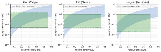
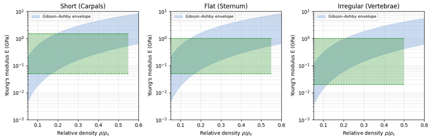
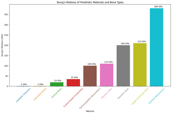
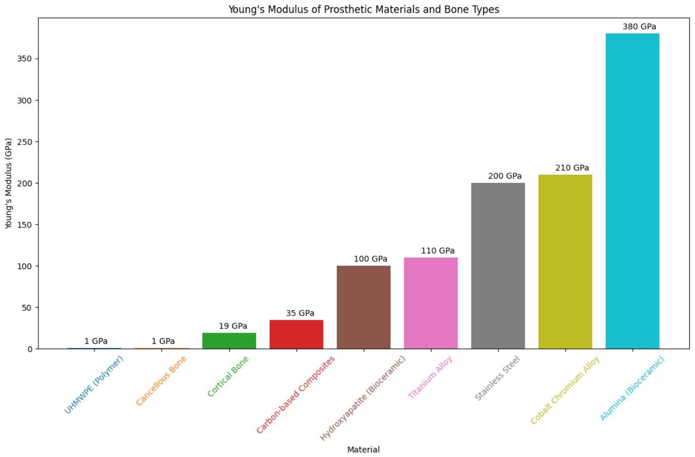
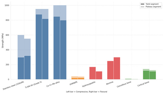
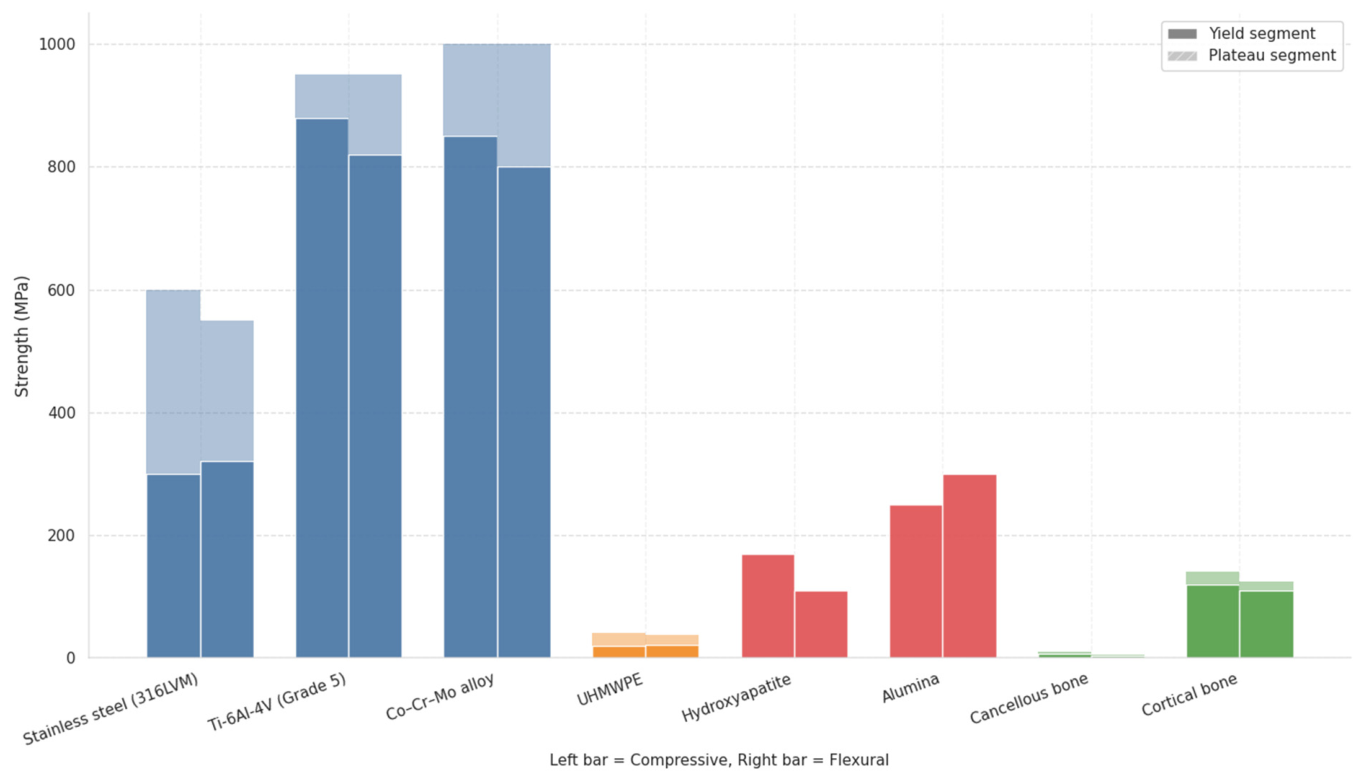
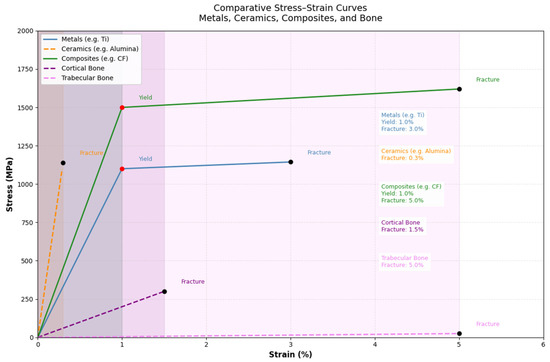
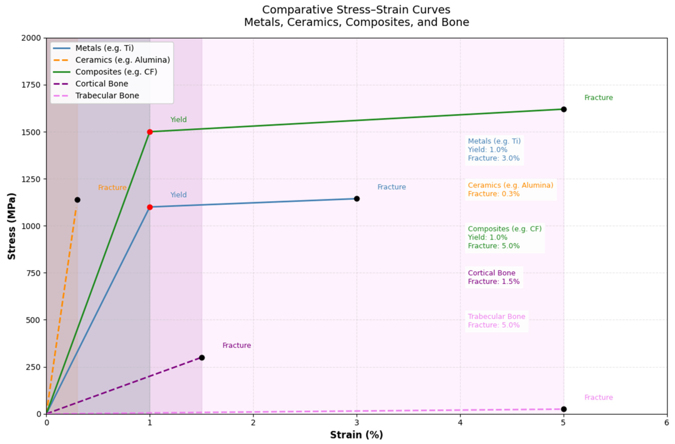
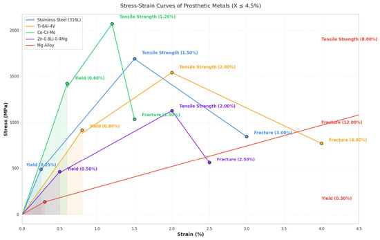
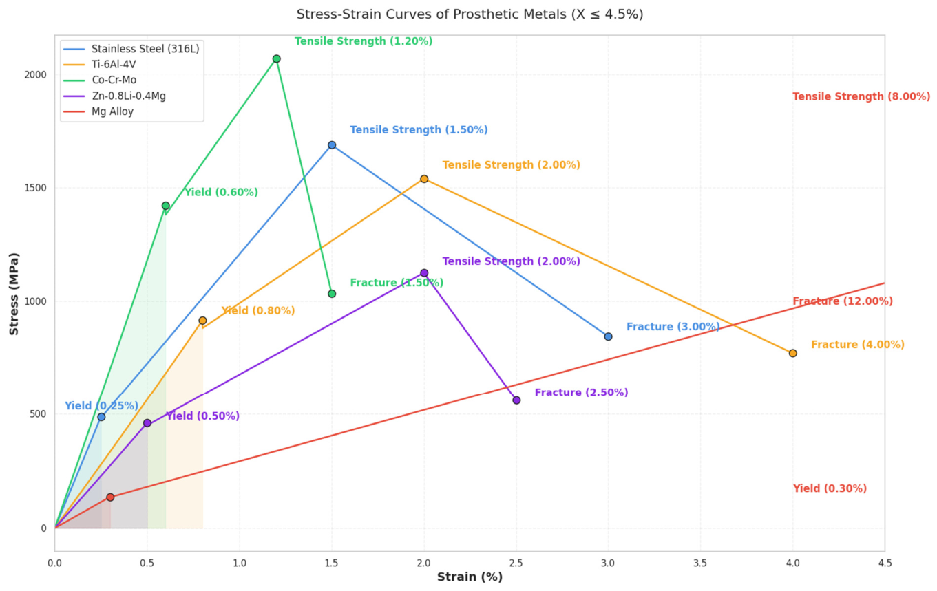
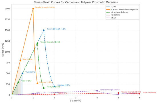
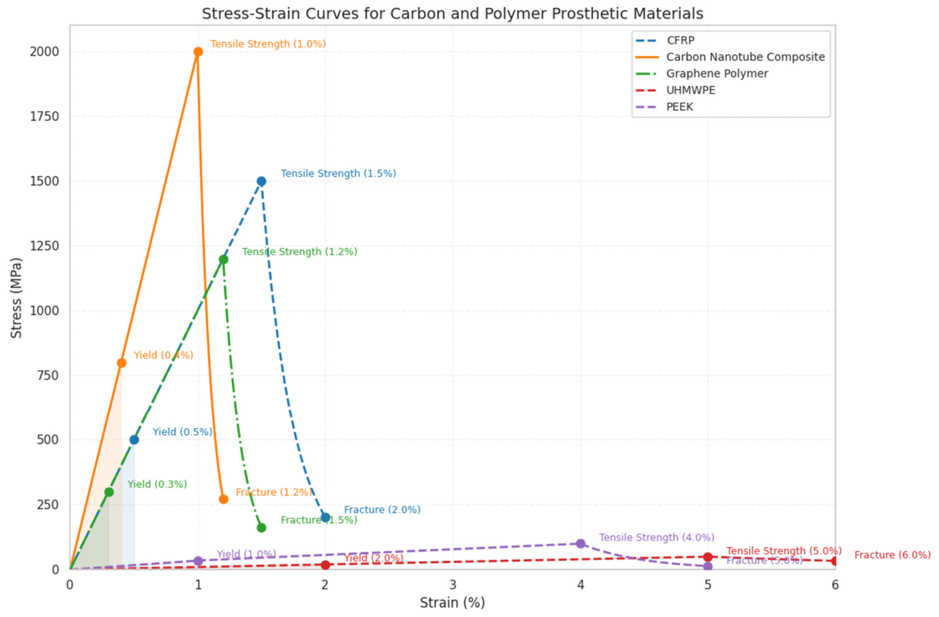
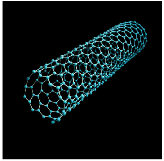
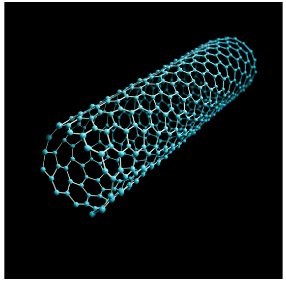
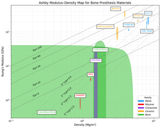
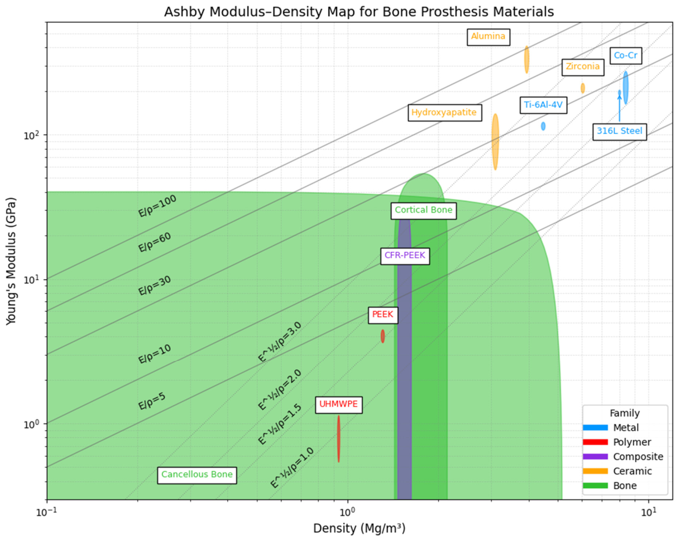
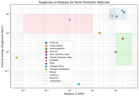
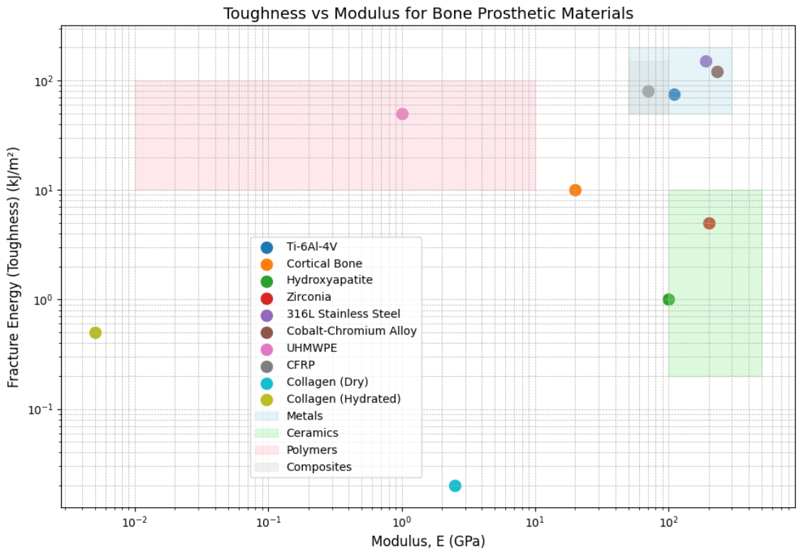
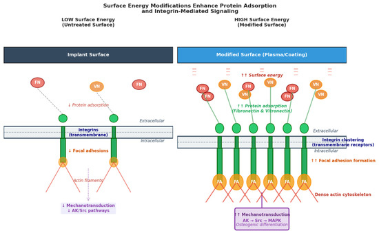
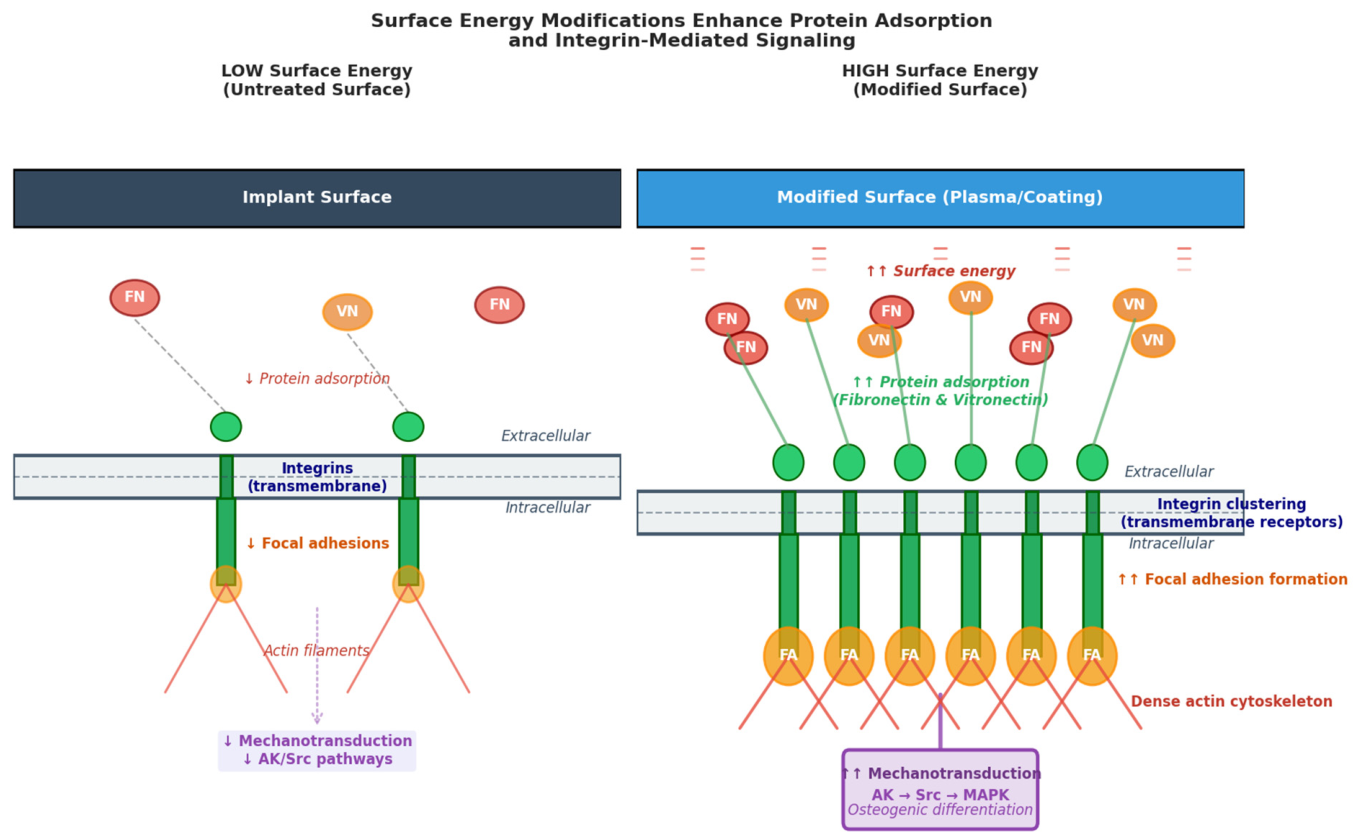
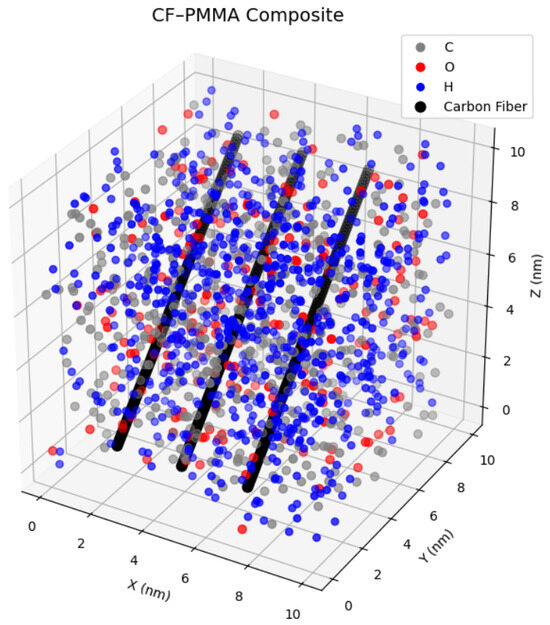
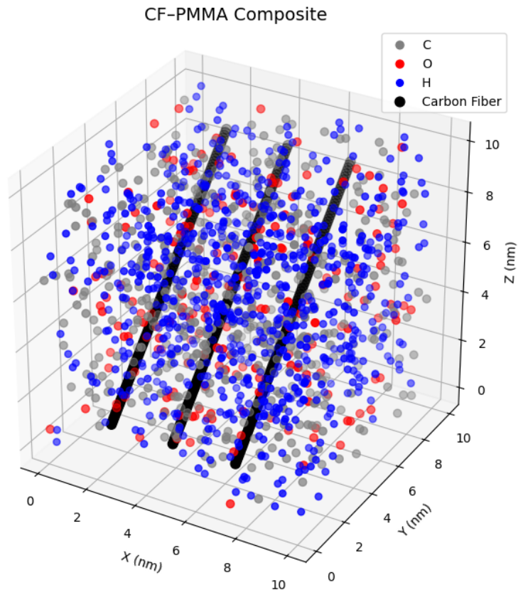
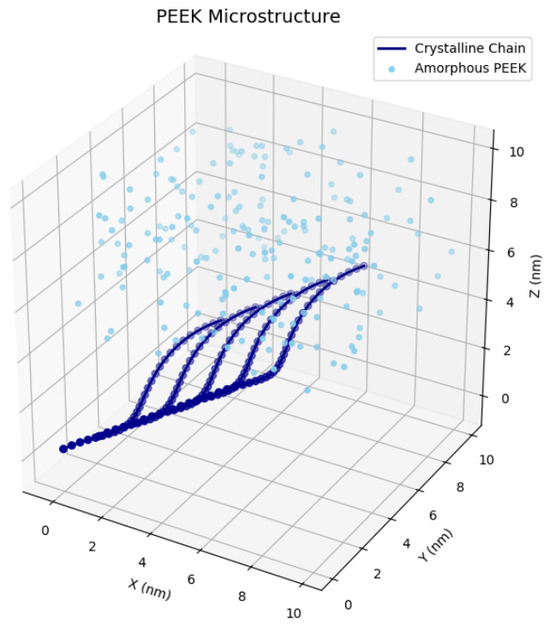
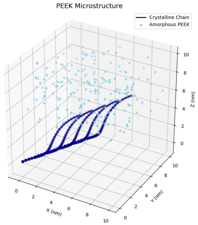

Disclaimer/Publisher’s Note: The statements, opinions and data contained in all publications are solely those of the individual author(s) and contributor(s) and not of MDPI and/or the editor(s). MDPI and/or the editor(s) disclaim responsibility for any injury to people or property resulting from any ideas, methods, instructions or products referred to in the content. |
© 2026 by the authors. Licensee MDPI, Basel, Switzerland. This article is an open access article distributed under the terms and conditions of the Creative Commons Attribution (CC BY) license.