


Abstract
Background: Combined bone and soft-tissue loss of the lower extremity requires complex reconstructive pathways. While acute post-traumatic infected defects and chronic osteomyelitis differ clinically, both present reconstructive challenges that demand radical debridement, infection-directed therapy, and radiology-guided soft-tissue coverage. Objective: To evaluate treatment options and functional recovery using a standardized orthoplastic workflow supported by radiological planning in patients with lower-extremity combined bone and soft-tissue loss of acute traumatic infectious etiology and chronic osteomyelitis. Methods: In this retrospective case series, 27 patients treated between May 2021 and December 2023 were included and divided into two groups: (A) acute post-traumatic bone and soft-tissue loss (n = 17) and (B) chronic osteomyelitis with fistula and soft-tissue loss (n = 10). Patients received multidisciplinary orthoplastic management with local and/or free flaps selected according to defect characteristics and radiology-guided vascular assessment (CT/MRI and angiographic CT; Doppler ultrasound for regional flap planning). Empirical broad-spectrum antibiotics were started after intraoperative biopsy sampling and later tailored to microbiological results. Healing was defined by bone union, infection/inflammation resolution, and flap healing. Functional outcomes were assessed using the Lower Limb Functional Index (LLFI). Due to baseline differences and a small sample size, within-group pre- vs. post-treatment changes in LLFI were analyzed separately. Results: Both groups demonstrated clinically relevant improvements in LLFI after treatment when assessed within each group. Flap selection and antibiotic strategy were implemented according to predefined multidisciplinary endpoints, achieving the study healing criteria in the included patients. Conclusions: A standardized orthoplastic reconstruction strategy supported by radiology-guided planning appears feasible in distinct clinical contexts of acute infected traumatic defects and chronic osteomyelitis, with improvement in lower-limb function assessed within groups.
1. Introduction
Lower extremity injuries in the form of open fractures are a very complicated and difficult therapeutic issue in orthopedics and traumatology. Simultaneous bone and soft tissue injuries are problems quite common in daily practice [1,2]. The difficulties that come to be confronted are mainly the primary injury extent with open entry points for infection of the traumatic site. The main causes of extremity defects are trauma, tumors, and chronic osteomyelitis [2].
Orthoplastic treatment is a multidisciplinary form of treatment that combines orthopedic and plastic surgery, and with this kind of treatment, it is possible to use microsurgical techniques and local flaps. Combined treatment with specialists, such as radiologists, internal medicine specialists, diabetologists, microbiologists, and psychologists, is necessary for therapeutic success. The goal of orthoplasty and reconstructive surgery is to choose the optimal form of treatment that provides the best functional and aesthetic results, provides a quick recovery, and limits disability.
Options for lower extremity reconstruction must provide adequate coverage for exposed bone, muscle, joints, and tendons while maximizing function and providing acceptable aesthetics [3]. As shown in previous publications, the debridement of infected bones and soft tissues must be as radical as necessary. This procedure seems to be crucial for good clinical outcomes [4], and many different forms of therapy can be used to achieve this goal. The latest research indicates the factors that should be taken into account when choosing the form and method of treatment. The most important prognostic factors are the patient’s condition and the characteristics of the injury [5].
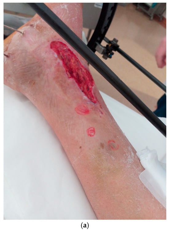
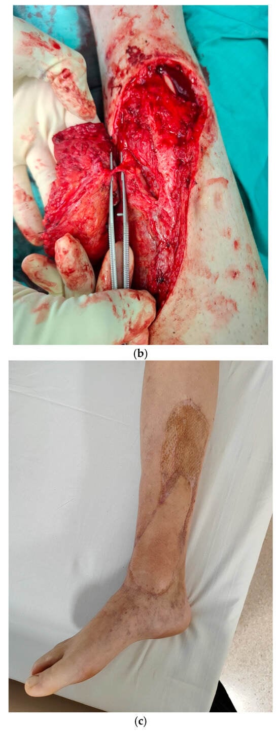
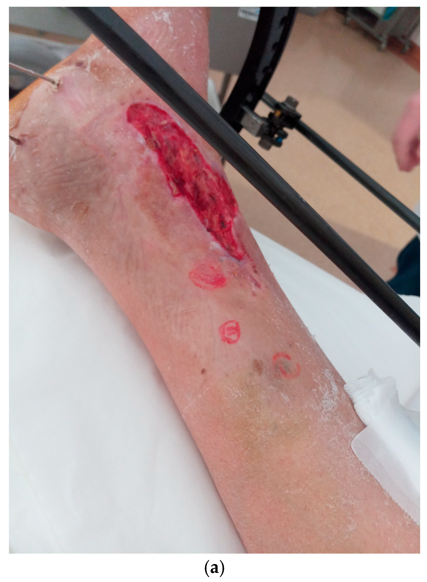
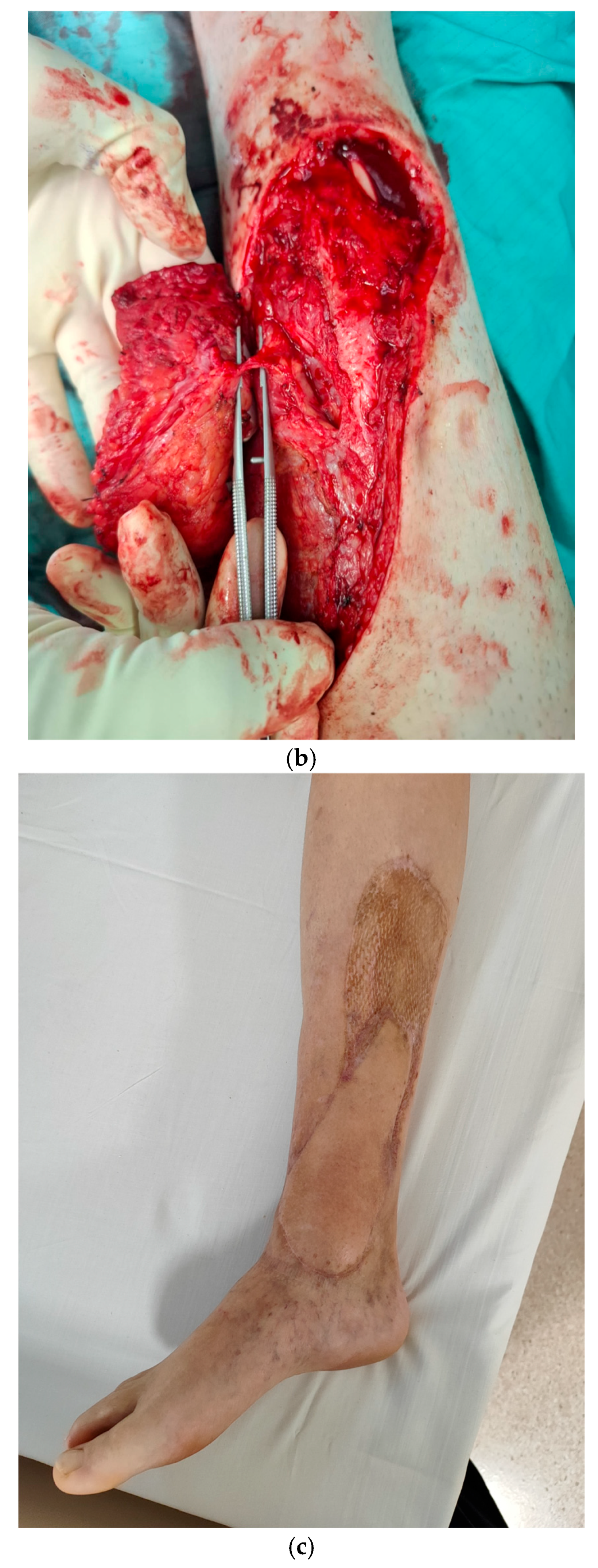
In our clinical practice, we usually use regional flaps, such as propeller flaps (Figure 1) and free flaps such as antero-lateral thigh flaps, as well as muscle flaps depending on the etiology and the location and size of the defect [2,4,5].
Figure 1.
A 41-year-old man suffered a factory accident, resulting in an open fracture Gustilo–Anderson IIIb of the right distal tibia segment, treated with ex-fix: (a) preoperative condition of the skin and surrounding tissues, clinical soft-tissue defect with an open, infected tibia bone; (b) intraoperative view of the propellar flap with prepared perforator; (c) healed propellar flap with good coverage. The time to follow-up was 16 months.
The aim of the study was to evaluate the treatment options for patients with severe bone and soft-tissue defects of a traumatic and infectious etiology of the lower extremities and that allow for improvements to the functionality of the limb allowing improvements in limb function and successful healing based on predefined endpoints.
2. Materials and Methods
This study was approved by the institutional review board (number: 41/2021), and informed consent was obtained from patients for their participation. Written informed consent for the publication of information and photos was obtained from all study participants presented.
All patients presenting to our department with combined bone and soft-tissue defects of the lower extremity between May 2021 and December 2023 were consecutively screened for eligibility. A total of 27 patients met the inclusion criteria and were enrolled in the study. No patients were excluded after screening, and no patients were lost to follow-up during the study period.
A retrospective case series of 27 patients was divided into two groups: (A) acute post-traumatic bone and soft-tissue loss (17 patients) and (B) chronic osteomyelitis with fistula and soft-tissue loss (10 patients). Because of the retrospective nature of this case series, the exact duration from injury to definitive orthoplastic reconstruction (i.e., in hours) was not consistently documented in the available records for all patients. Therefore, we could not reliably calculate the time to reconstruction on an individual basis, nor report the proportion of patients reconstructed within 72 h or within the first 7 days. Nevertheless, patients were managed according to standard clinical procedures, considering tissue condition and infection status. In the acute post-traumatic group, surgical supply/orthoplastic reconstruction was performed within the time frame of up to 3 weeks after injury. Patients in the chronic group had defects related to chronic disease lasting longer than 6 months. We provided multidisciplinary treatment for bone and soft-tissue healing using various local and free flaps from May 2021 to December 2023. Our standard approach to preoperative planning is to use bone segment X-rays and extremity X-rays. For detailed imaging, we used computed tomography (CT) and magnetic resonance imaging (MRI); for vascular imaging, Angiography-Computed Tomography (angio-CT); for regional flap planning, we used Doppler ultrasound [6]. We based the description of radiological procedures on articles available in the PubMed database between 2003–2024. All published studies that analyzed the applicability of Doppler ultrasound or angio-CT, in the evaluation of arterial perforators and used the terms ‘arterial perforators’ OR ‘ultrasound arterial perforators’ OR ‘Doppler ultrasound arterial perforators’ OR ‘angio-CT arterial perforators’ AND ‘lower limb flap’ OR ‘propeller flap’ OR ‘ALT flap’ OR ‘distal crus’ were identified and reviewed. The search limits were defined as ‘English’ (language) and ‘the beginning of a given database through to 31 December 2024. All 14 patients who underwent propeller or anterolateral thigh flap reconstruction underwent both color Doppler ultrasound and CT angiography. A total of 18 potentially suitable perforators were identified because more than one perforator was detected in some patients. One perforator was selected as the flap pedicle in each patient. The selected perforator was subsequently verified intraoperatively. Therefore, 14 selected perforators were available for imaging–operative concordance assessment, whereas the remaining four non-selected perforators were not systematically verified during surgery. In Group A, nine patients did not have perforator identification in the radiological examination, and they underwent a total of nine flaps–muscle (n = 6), reverse sural (n = 1), flag-ship (n = 1), and cross-leg (n = 1). In Group B, four patients also had no perforator identification in the radiological examination, and these patients underwent a total of six flaps–muscle (n = 4), reverse sural (n = 1), and flag-ship (n = 1). Clinical assessments were based on the Lower Limb Functional Index [7]. All patients received empirical intravenous broad-spectrum antibiotic therapy immediately after the intraoperative collection of at least five tissue biopsies for microbiological examination (standard culture and sonication). All patients initially received empirical intravenous broad-spectrum antibiotic therapy tailored to cover the most common pathogens associated with bone and soft-tissue infections. This regimen was standardized across cases based on current clinical guidelines. However, once microbiological results were available from intraoperative tissue biopsies, antibiotic treatments were customized to target specific pathogens identified in each patient. Targeted oral antibiotic therapy was continued for 6 weeks, with CRP levels monitored every 2 weeks to assess treatment efficacy and adjust as necessary. This approach ensured both broad initial coverage and a precise, pathogen-specific treatment strategy. PMMA with antibiotics was used both as a filler for bone infection cavities and to cover the intramedullary nails used to stabilize open fractures and intramedullary infections [8,9]. Cases of acute tibial segment injury were evaluated based on the Gustilo–Anderson classification [10], whereas patients with chronic defects and inflammation were evaluated based on the Cierny–Mader classification [11].
The inclusion criteria were as follows: patients with combined bone loss and/or osteomyelitis with concomitant soft-tissue defects of the lower extremity requiring orthoplastic reconstruction. Etiologies were categorized as acute post-traumatic infected defects or chronic osteomyelitis with fistula, reflecting clinically distinct cohorts that were managed using a unified reconstructive planning framework. The end of the treatment process, as well as the end of the study, was healing in patients who met all three of the following criteria (i.e., healing time): (1) bone union, (2) healing of the inflammatory process, and (3) healing of the flap.
2.1. Statistical Analysis
The data comprised 27 patients divided into two groups: (A) acute post-traumatic bone and soft-tissue loss (17 patients) and (B) chronic osteomyelitis with fistula and soft-tissue loss (10 patients). Due to the small sample size and non-normal distribution of the data, confirmed by Shapiro–Wilk tests, we employed non-parametric statistical methods. Specifically, the Wilcoxon signed-rank test was used to evaluate changes in the Lower Limb Functional Index (LLFI) pre- and post-treatment.
The initial between-group comparisons were found to be inappropriate due to significant differences in age, baseline LLFI scores, and follow-up duration between the acute and chronic groups. These differences violate the assumption of homogeneity necessary for valid comparative analysis. The Minimal Clinically Important Difference (MCID) for LLFI scores was not defined in this study, which limits the interpretation of the practical significance of the improvements observed. Accordingly, to avoid inappropriate etiologic comparisons, we performed within-group analyses of LLFI pre- vs. post-treatment for each cohort separately.
On the basis of the Shapiro–Wilk tests, we did not achieve a normal distribution for all variables; therefore, we used the Wilcoxon signed-rank test. Perforator diameter and flow-velocity measurements were summarized descriptively at the vessel level. Because more than one perforator could be identified in the same patient, individual perforators were not treated as independent patient-level observations. Therefore, no inferential statistical comparisons were performed for vessel-level imaging measurements. The analysis of the data included descriptive statistics and was performed with Stata v. 11.0 (StataCorp, Collage Station, TX, USA) and Excel 2024 (Microsoft, Redmond, WA, USA). Statistical significance was set at p < 0.05. These statistical methods were selected to align with the data characteristics, providing a reliable analysis of treatment outcomes.
2.2. Radiological Evaluation of Perforators
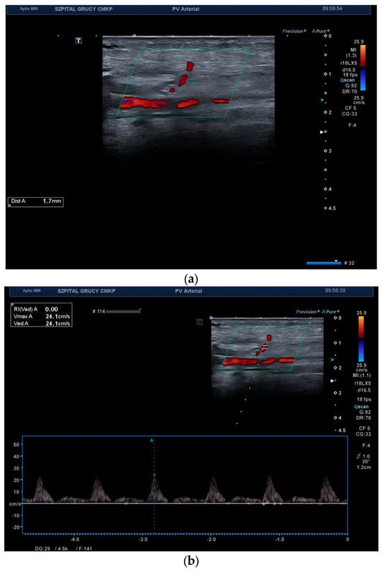
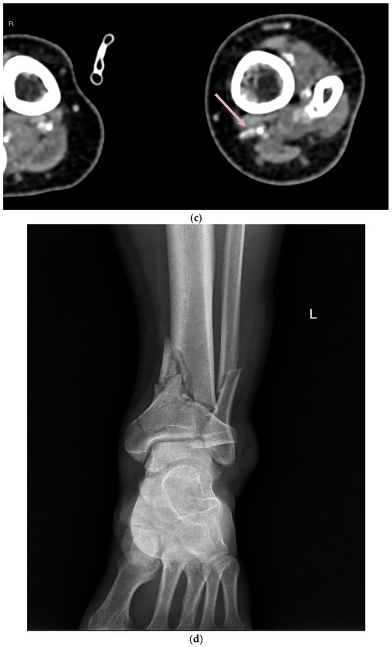
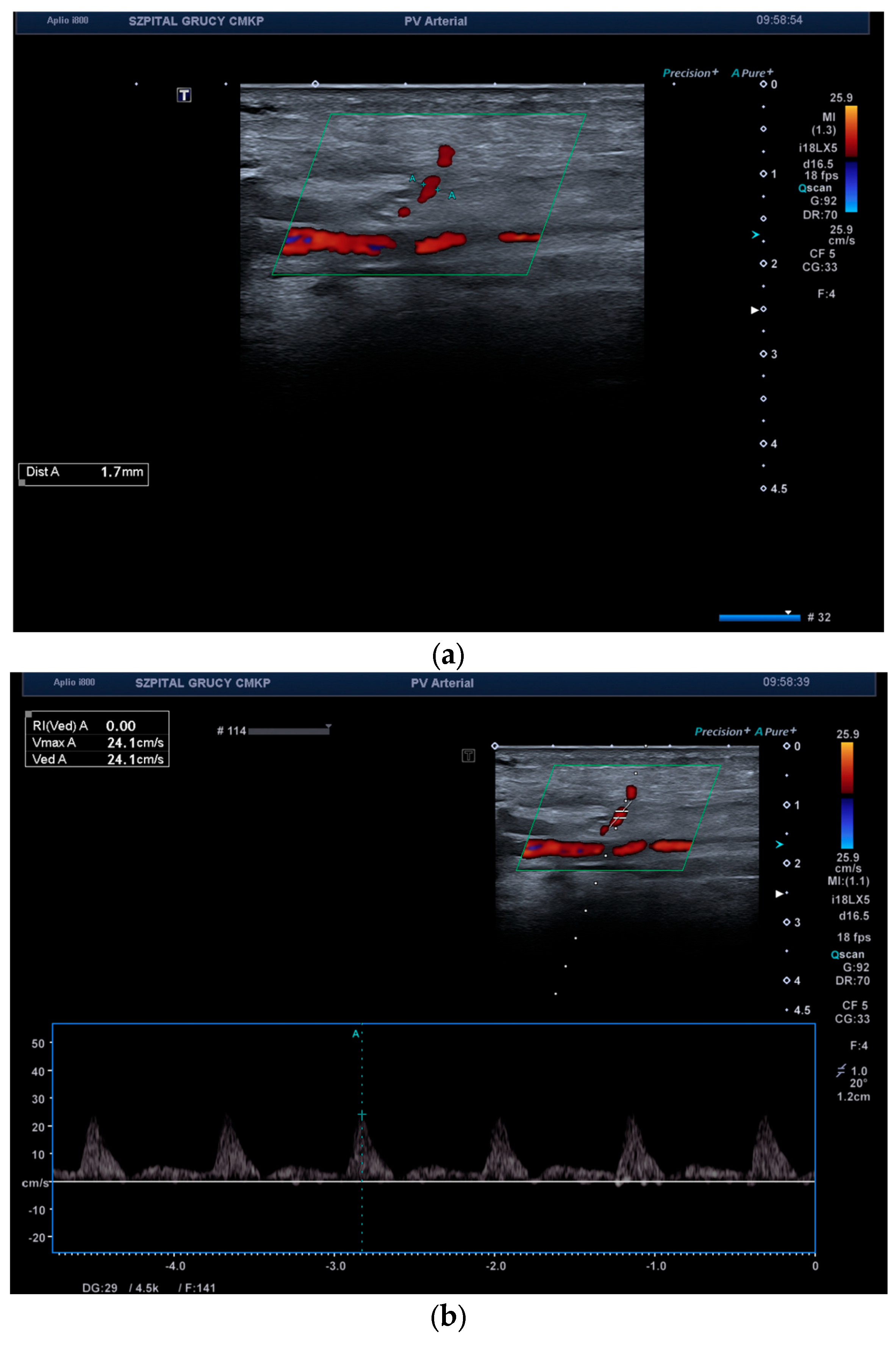
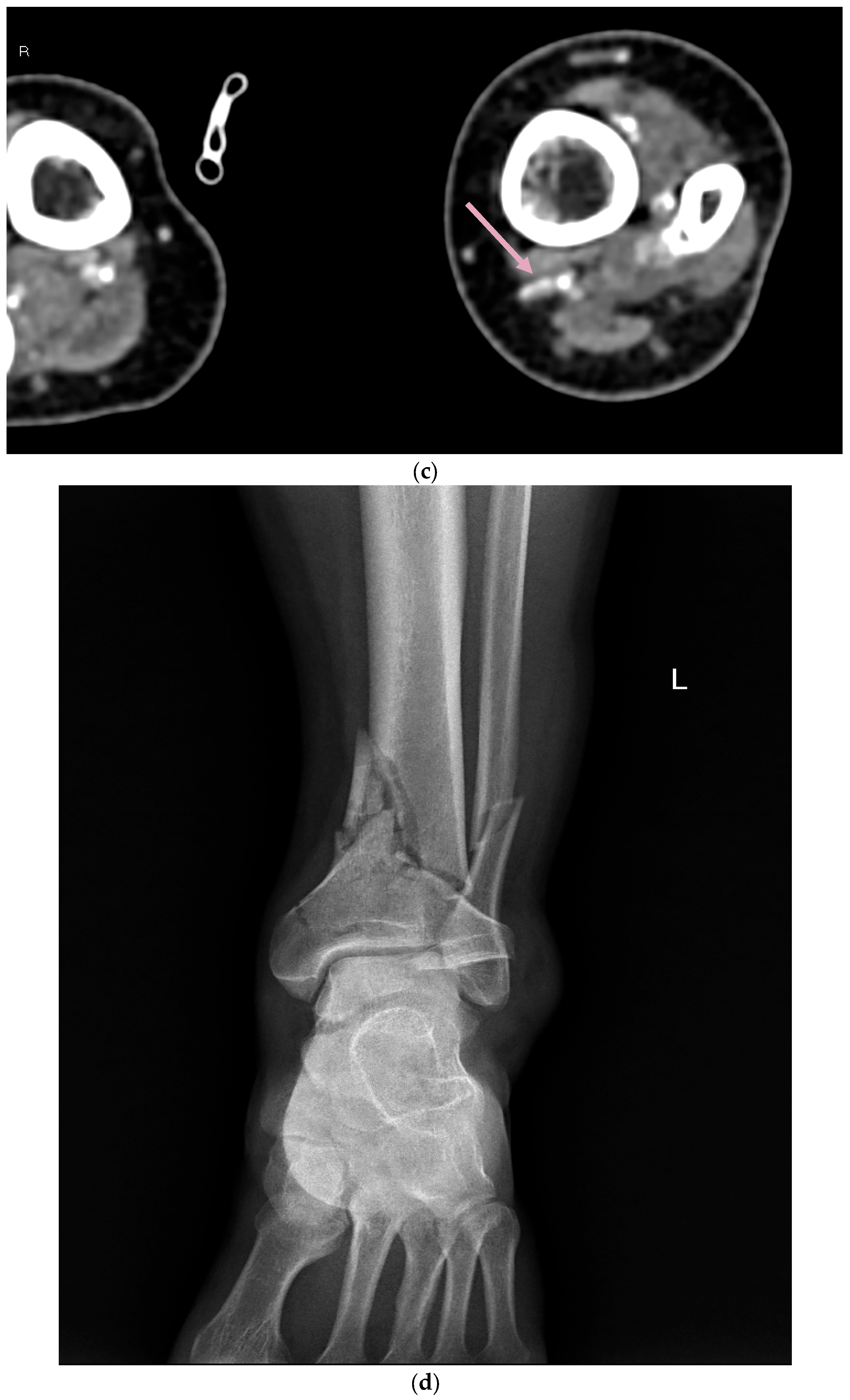
In routine clinical practice, Doppler ultrasound was our first-line imaging method, with CT angiography or, less frequently, MR angiography performed when additional vascular assessment was required [12]. However, for the purpose of this study, all 14 patients undergoing propeller or ALT flap reconstruction underwent both color Doppler ultrasound and CT angiography. Representative findings are shown in Figure 2. For the purpose of this research, patients who qualified for propeller and ALT flaps underwent Doppler USG and angio-CT examination. An example of vascular imaging studies is shown in Figure 2.
Figure 2.
A 49-year-old male with acute osteomyelitis of the distal tibia: (a) Doppler ultrasound examination of the distal crus perforator branching from the posterior tibial artery. Evaluation of perforator diameter at the level of fascia penetration; (b) evaluation of velocity and flow spectrum at the level of fascia penetration; (c) same perforator seen on angio-CT with accompanying vein (arrow); (d) preoperative A-P X-ray of the ankle.
We used an 11–18 MHz linear transducer, with the majority of examinations on the peripheral artery preset. Based on the location of the skin defect in the distal calf, we focused on perforators originating either from a peroneal or tibial artery. It is important to adjust the flow speed (PRF in cm/s) and gain scales accordingly in the color Doppler or power Doppler option. Gain and scale should be set so that the color fills the entire lumen of the arterial vessel, “touching” but not exceeding the inner boundary of the intima-media complex (IMT). A symptom of excessive gain is the phenomenon of “blooming” of color outside the vessel wall, into the adjacent tissues. In that case, we should either lower the gain or increase the scale (PRF). From our experience, the only exception is when we want to find a perforator with a relatively low velocity and small diameter; then, sensitivity is a greater priority. Increasing gain and decreasing scale will indeed cause the appearance of many blooming artifacts, but will potentially allow the identification of a small arterial perforator. Sensitivity also depends on the color box size. The smaller the color box size, the greater the sensitivity.
After determining the site of search for perforators with the orthopedists and after appropriate adjustment of the device, based on experience and data from scientific publications, one should carefully search for arteries branching off the peroneal or posterior tibial artery and visualize in B-mode the pulsating perforator that passes through the deep fascia to the subcutaneous tissue [13]. At the level of the deep fascia puncture, the vessel diameter and flow velocity should be measured. The color Doppler gate should be tilted at an angle of <60° relative to the long axis of the vessel; it is not recommended to examine the flow spectrum in B-mode. The recommended width of the spectral gate should correspond to 30–70% of the diameter of the analyzed vessel [14]. According to Song et al., the mean arterial flow velocity of perforators at the level of fascia penetration was 28.06 ± 7.94 cm/s, which matches other authors with PSV of 21.9 cm/s [15,16].
It is important to document adequate flow from the perforator pedicle to the propeller flap due to the adequate blood supply and consequently nourishment of the flap in order to avoid necrosis, which is the primary reason for flap failure [15,17].
The place where the perforators pass through the deep fascia should be carefully analyzed in at least two planes to be sure that this is the real place of puncture of the fascia. Quite often, the perforating vessel in the proximal section reaches the deep fascia and then continues subfascial, adhering to the fascia, and the real puncture of the fascia occurs even a few centimeters further. It is crucial information because it is a pivot point around which the propeller flap is rotated on its own axis. The closer the pivot point is to the skin defect, the greater the chance of covering the entire lesion, as the larger portion of healthy proximal skin can be mobilized and positioned over the skin defect; also, the shortest possible arc of rotation can prevent postoperative complications [6,18].
Another important piece of information is the number and direction of the perforator branches in the subcutaneous section [19].
In the distal third of the calf, we were usually able to identify one to three perforators for both the medial and lateral sides. The fascia puncture locations were marked with a marker on the skin.
Preoperative evaluation of potential perforators using Doppler ultrasound allows for the assessment of key parameters such as their number, location, size, origin, course, and branching patterns. This detailed assessment enables the surgeon/orthopedist to plan the extent of the operation and, in cases where suitable perforators are absent, to consider alternative treatments or extended diagnostic imaging [20,21]. Additionally, the measurement of vessel diameter and flow velocity at the point where the perforator pierces the deep fascia is critical in reducing the risk of postoperative complications, such as flap necrosis, and in selecting the optimal vessel for the procedure [6,13,15,22]. Evaluating the main arterial trunks of the lower limb is also important; it identifies any vascular anomalies as well as peripheral artery disease and potential arterial occlusion [12]. However, assessing the distal third of the lower limb can be challenging due to the progressively smaller diameter of the perforators in this region, which increases the procedure’s user dependency [16,21]. According to Jakubietz et al., in color Doppler ultrasound, false–positive results were present in 21% of cases; on the other hand, Gunnarsson et al. showed 0% false-positive cases [13,23].
To minimize intra and postoperative complications, assessing perforators through ultrasound or CT angiography is critical [12,24,25]. Despite preoperative mapping of perforators using Doppler ultrasound, their exact location where they penetrate the fascia may differ in practice. This sometimes necessitates intraoperative adjustments to the flap design to match the actual location of the perforator [21].
All in all, the ultrasound method is sufficient for preoperative scanning [18,26,27,28].
2.3. Angio-CT
We used the angio-CT (CTA) method when Doppler ultrasound provided insufficient data, i.e., comprehensive assessment of the limb vascularization and simultaneous assessment of the condition of bone structures (extent of defects, presence of sequestra), vascular anomalies were visible, obstruction of the main arterial trunks, no perforators were detected, or they were not suitable for propeller flap.
Performing angio-CT is associated with obvious limitations such as allergy to contrast agents or renal failure. An additional potential problem is insufficient filling of the vascular bed with a contrast agent, which makes it difficult and sometimes impossible to detect perforators in a particular area of interest. Another limitation is the inability to determine the flow rate in the perforator and its precise course in the subcutaneous tissue. However, it should be remembered that after performing CTA and locating the appropriate perforator, the Doppler USG examination can be repeated to assess the quality of the vessel.
Feng et al. conducted a direct comparative analysis between CTA and Doppler ultrasound in terms of the usefulness and sensitivity of both examinations in the context of the evaluation of lower limb perforators. He concluded that Doppler ultrasound shows a mean error of 1.11 ± 1.29 mm and CTA shows a mean error of 2.55 ± 2.63 mm in the context of the demonstrated site of deep fascia penetration and its actual location during the surgery. The success of color Doppler ultrasound has been directly related to the experience of radiologists; however, the same has not been true for CT angiography [29].
What is more, Color Doppler ultrasound is capable of detecting small perforators with a caliber up to 0.5 mm [30].
3. Results
This retrospective study group consisted of 27 patients. The mean age in Group A was 46.9 years, and that in Group B was 66.7 years. The mean time to healing was 8.2 in Group A and 5.9 in Group B. The mean follow-up time was 17.87 months in Group A and 24.4 months in Group B. The most common location of defects and soft-tissue- and bone-healing problems was in the lower extremity of our material, which is the distal third of the tibia segment, as recorded for 17 patients (62%) (Table 1). All patients with defects in the lower extremities qualified for surgery. During surgery, six types of flaps were used. Overall, we used a propeller flap in eleven patients (39%), a medial gastrocnemius muscle flap in six patients (21%), a soleus muscle flap in four patients (14%), ALT flap in three (10%) a retrograde sural three times (10%), flagship flap in two patients (7%) and one cross-leg flap (3%). All patients in Group A required bone stabilization. In Group B, eight patients required bone stabilization, and one patient required a knee endoprosthesis. We mainly used LCPs for bone stabilization, that is, seven in Group A and four in Group B, and intramedullary nails, that is, ten in Group A and four in Group B. Plain radiographs formed part of the standard preoperative assessment and were used to evaluate the osseous injury, bone loss, alignment, and fixation material. The distal third of the tibia was the most frequently affected region, occurring in 17 of 27 patients (62%).
Table 1.
Demographic data of patients who underwent lower extremity reconstruction.
All 14 patients included in the imaging subgroup underwent both color Doppler ultrasound and CT angiography. A total of 18 potentially suitable perforators were identified, including 11 perforators in Group A and seven in Group B. The Doppler ultrasound and CT angiography findings were concordant. One perforator was selected as the flap pedicle in each patient, and all 14 selected perforators were confirmed intraoperatively. The remaining four imaging-positive perforators were not selected and were not systematically verified during surgery. Therefore, they could not be classified as true-positive or false-positive findings. No false-positive findings were observed among the 14 surgically verified selected perforators. Perforator diameter and flow velocity were assessed descriptively for all 18 imaging-identified perforators. The mean flow velocity at the fascial penetration point was 27.89 cm/s, and the mean vessel diameter was 1.55 mm. Group-specific results are presented in Table 2 and Table 3. A high standard deviation (SD) indicates substantial variability within the groups.
Table 2.
Imaging characteristics of patients undergoing propeller or ALT flap reconstruction.
Table 3.
Concordance of preoperative perforator imaging with intraoperative findings.
Twelve patients were treated with PMMA-supported antibiotics for osteomyelitis. We used gentamicin–clindamycin–vancomycin for all of these patients. During surgery, material was collected for microbiological examinations (sonication and standard culture) [31]. In most patients, more than one type of bacteria was found in the collected material. The most common pathogens were Enterococcus faecalis and methicillin-resistant coagulase-negative staphylococci (MRCNS). Mixed bacterial flora was more common in Group A.
We observed no difference in the length of surgery, with the mean time in Group A being 324.3 min and in Group B being 362.2 min. The length of stay in hospital averaged 4.12 weeks in Group A and 3.44 weeks in Group B. In four Group A and three Group B patients, we were forced to perform a flap revision, mainly because of venous congestion. For the first few cases, we used hirudin therapy, but in time, we abandoned this treatment due to Pseudomonas infection (two cases in Group A). We treated subsequent cases of venous congestion with punctures and injections of heparin and intravenous dextran and dexaven. If the venous congestion persisted despite interim treatment, we performed revision surgery (Table 4).
Table 4.
Analysis of the operation and hospitalization data of patients who underwent lower extremity reconstruction.
No major complications such as flap failure, wound dehiscence, or infection recurrence were observed during the follow-up period. Patient satisfaction was assessed using structured interviews and visual analog scales (VAS). All patients reported high satisfaction regarding both functional and aesthetic aspects. No standardized, validated aesthetic scale was used to quantify aesthetic outcome. These data underscore the effectiveness of combining radiologically guided flap planning and multidisciplinary care in achieving favorable clinical outcomes.
The LLFI was used to assess patients’ quality of life and satisfaction before surgery and at the end of treatment (healing time) during follow-up in the ambulatory clinic. The LLFI improved significantly after surgery in both groups (p < 0.05 for each within-group comparison). In Group A, the mean LLFI increased from 9.53 (±6.09) preoperatively to 66.26 (±8.09) postoperatively. In Group B, the mean LLFI increased from 27.0 (±12.08) to 69.6 (±9.02). Before reconstructive surgery, the LLFI was 9.53 (±6.09) in Group A and 27.0 (±12.08) in Group B. This result is, one might say, obvious because of the comparison of an acute and sudden traumatic loss of limb function and experiencing a state of chronic limited functioning in which certain compensatory systems (“habituation”, bracing, and the acceptance of the disease) have already taken effect. After orthoplastic treatment, the postoperative LLFI scores were 66.26 (±8.09) in Group A and 69.6 (±9.02) in Group B (Table 5).
Table 5.
Functional and clinical outcomes: LLFI pre- and post-operation for lower extremity reconstruction.
4. Discussion
As highlighted, the treatment of patients with bone and soft-tissue defects is very difficult. Good qualifications and properly performed operations are crucial. Many flap-based surgical techniques are described in the literature, and each has its advantages and disadvantages. There are three permanent perforators on the lower extremity that allow for the proper realization of the propeller flap. The perforating vessel should be as close as possible to the beginning of the skin defect. The flap must be at least 1 cm longer and at least 0.5 cm wider than the defect. It is very important to cut the perforator to a sufficient length past the fascia to avoid twist points [6]. Song et al. showed how significant the direction of rotation of a flap is. The correct direction of rotation can reduce the risk of flap loss, especially with large flaps [15]. The other options are free flaps, for example, an ALT. Wong et al. evaluated an anterolateral thigh flap. In their opinion, an ALT can reliably and safely be harvested in most patients [32]. The purported difficulty with the use of this flap lies in the anatomical variations that may render this flap unreliable. Despite anatomical inconstancy, an anterolateral thigh free flap is relatively easy to harvest and utilize. Its pedicle can be used to bridge a vascular defect in the extremity [33]. Other free flaps are latissimus dorsi muscle flaps and dorsali pedis artery-based flaps. These are very good flaps, but our experience with cases of infectious and post-traumatic tissue changes speaks for the use of local flaps in the first place due to their easy availability, low cost, and shorter operating time and, perhaps, a lack of experience in making free microsurgical flaps. Innocenti et al. compared the costs and efficiency of free and propeller flaps. Complications arose for 6% of the free flaps and 3.7% of the propeller flaps. The average cost of a free flap is EUR 5077.5 per patient, and the cost of a propeller flap is EUR 1595.6 per patient. The choice of surgical option should only be made after a careful evaluation of the soft tissue surrounding the defect. For small and medium defects in healthy extremities, a propeller flap should be used. Free flaps should be chosen for large defects caused by trauma or vascular disease [34]. Propeller flaps have one more limitation. They should be avoided if underlying metalwork is required for bony fixation [35]. Furthermore, as our results show, local flaps are a very good solution for the complex treatment of soft-tissue damage and defects of various etiologies. A distal-based sural flap is a useful alternative to free tissue transfer for simple and complex defects in the lower third tibia segment, foot, and ankle. This procedure includes the lesser saphenous vein, used to augment venous drainage. The flap should be used with caution in elderly patients and those with venous insufficiency. Patients with venous insufficiency have been found to have a nine times greater risk of complications [36]. A gastrocnemius flap is widely used to reconstruct the knee and upper tibia segment, whereas a soleus flap is used to cover middle and lower tibia segment defects. The soleus muscle can be divided longitudinally and used as a hemisoleus flap. Muscle flaps fill defects with significant soft-tissue thickness; however, they show scarring, contractures, and complications at the donor site [37]. Al-Mugaren et al. published an algorithm for their approach to lower extremity defects and the selection of appropriate flaps for reconstruction [38]. They consider the propeller blade and keystone as “new trends” in reconstructive surgery. Nevertheless, classic flaps, such as gastrocnemius flaps, soleus flaps, and inverted calf flaps, as well as free microsurgical flaps, also have their advantages and are used in severe defects of the lower extremity. In the end, flap choice should be made depending on the defect’s etiology, resources available, surgeon’s experience, and the patient’s comorbidities and expectations [39]. There are also treatments that do not require the use of microsurgery. In the absence of appropriate equipment, experienced surgeons use external fixators, and bone transport and soft-tissue distraction can also be used, although they have no advantages over microvascular techniques [40].
Orthoplasty combines orthopedic treatment (bone loss and osteomyelitis) and plastic treatment (soft-tissue loss). In cases of patients with osteomyelitis, antibiotic cement-coated interlocking nails seem to be the best option for treatment. They are advantageous over external fixators, as they eliminate the complications of external fixators and have good patient compliance [41]. The antibiotics that are used should be heat-stable, exhibit broad-spectrum activity, have good elution properties from the cement, and have low allergenicity [42]. In our study, we used gentamicin–clindamycin–vancomycin and a PMMA as a donor. The use of antibiotic-impregnated cement-coated intramedullary nails for infected non-unions of tibia and femur fractures has been well documented in the literature. It may also reduce the morbidity of the two surgical procedures and inpatient admissions required for the two-stage procedures or for the application and removal of external fixators to that of a single procedure and inpatient admission [43]. Lan et al. reported that patients with a Gustilo–Anderson III open tibia fracture treated with the Masquelet technique have a shorter average union time and a lower risk of infection than patients treated with a vascularized bone graft [44]. Sometimes, because of the removal of a large amount of tissue, it is necessary to perform massive reconstructive surgery [4]. In difficult cases that do not respond to treatment or in departments without an experienced flap surgeon, other methods of therapy should be considered, one of which is bone transport with the Ilizarov technique. Bone transport treatment with a soft-tissue transport technique can provide satisfactory clinical results in most cases [42]. Novel treatment methods, such as an osteoinductive autologous bone graft substitute, are described. The aim of this method is to accelerate bone regeneration [45]. The clinical evidence regarding the use of bone morphogenetic proteins in the treatment of fractures, non-unions, and osteonecrosis is still controversial, and there are not enough clinical reports [46]. In our material, there were no complications of non-union, so we did not have to use another form of treatment.
Various methods of delivering antibiotics have been investigated. Patel et al. showed that PMMA beads with antibiotics are very effective in the treatment of chronic osteomyelitis and in preventing infection recurrence [47]. The sonication of material collected during an operation is a well-known technique used for implant-associated infections. This method, which is based on the application of long-wave ultrasound radiating in a liquid medium, has recently been validated for the diagnosis of prosthetic joint infections [32]. The treatment of patients with reconstructions because of extremity fractures is a long-term process. After their first operation, patients often require revision. Aydogan et al. showed that after revision, patients had poor outcomes, had a low quality of life, and needed a long time to go back to work. Patients suffering from flap ischemia have worse outcomes than those without flap ischemia [48]. In our study, the complications that occurred after primary surgeries were due to venous congestion, which remains a major postoperative complication [21]. Venous congestion can cause necrosis of the distal flap. In the first few cases, we used hirudin therapy according to a study conducted by de Chalain [49]. Later, we switched from the use of hirudinotherapy to injections with heparin and an intravenous supply of dextran and dexaven.
Radiology plays a crucial role in lower leg microsurgery by providing essential imaging support that enhances surgical planning, execution, and postoperative evaluation. Radiological imaging assists in mapping the vascular supply to the lower leg. Techniques like Doppler ultrasound and angiography allow surgeons to visualize blood vessels, ensuring that the surgical site will receive adequate blood supply post-surgery. The anatomical study of lateral lower leg perforators provides significant insights into their distribution, characteristics, and clinical applications. This study revealed that 73.1% of the perforators had a septocutaneous course, while 26.9% were musculocutaneous. This suggests that septocutaneous perforators may be more prevalent in the lateral lower leg, which could influence surgical approaches for flap harvesting. The anterior tibial artery was identified as the primary source of perforators (53.0%), followed by the peroneal artery (41.6%) and the popliteal artery (5.1%). Notably, popliteal artery perforators were significantly larger than those from the other arteries, indicating their potential utility in reconstructive procedures. Perforators in the proximal third were significantly longer and larger compared to those in the distal third. Musculocutaneous perforators were found to be longer and wider than septocutaneous ones, which may influence the choice of perforator for different types of tissue transfer [49].
The article by Lichte et al. discusses the use of routine preoperative color Doppler duplex ultrasound scanning to localize perforators in ALT flaps. This study found a strong correlation between the accuracy of identification and the patients’ body mass index (BMI), but not with age or sex. The authors conclude that color Doppler ultrasound is a reliable and effective method for identifying perforators in ALT flaps, suggesting it could become a standard procedure in preoperative planning. The method is non-invasive, cost-effective, and avoids radiation exposure, making it favorable compared to other imaging techniques [50]. The use of Color Duplex Sonography (CDS) for preoperative localization of perforator arteries improved surgical accuracy and facilitated the design and execution of the flap procedures. Liu et al. investigated the clinical efficacy of using bilateral anterolateral thigh perforator (ALTP) flaps for repairing large skin and soft tissue defects in the extremities. Twelve patients with defects ranging from 18.0 × 11.0 cm2 to 38.0 × 15.0 cm2, caused by various injuries, underwent this surgical procedure. The study employed CDS to accurately locate perforator arteries preoperatively, which improved surgical outcomes [51]. The study by Tang et al. focuses on improving the localization of perforators in the ALT for soft tissue reconstructive surgery using augmented reality (AR) combined with CTA. The research involved 14 patients who underwent CTA mapping of their lateral circumflex femoral artery (LCFA) and its perforators, which were then projected onto the skin surface during surgery for accurate flap harvesting. The combined CTA and AR technique achieved a 90.2% accuracy rate and a 97.4% sensitivity in identifying perforators, significantly outperforming handheld Doppler ultrasound, which had an accuracy of 82.4% [52]. The study conducted by Ahmad Alwi et al. provides valuable insights into the feasibility of using the medial plantar artery flap for diabetic patients with suspected vascular insufficiency. The study found that more than 50% of the lower limbs in diabetic patients with vascular compromise still exhibited a patent medial plantar artery. This suggests that the medial plantar artery flap may be a viable option for reconstructive procedures in this patient population. The findings highlight the necessity of conducting a CTA prior to surgical intervention to accurately assess the patency of the medial plantar artery. The study demonstrated that bedside clinical assessments, such as palpation and hand-held Doppler evaluations of the posterior tibial artery, were inadequate predictors of medial plantar artery patency [53].
Our findings support the role of Doppler ultrasound and CT angiography in preoperative perforator mapping. Doppler ultrasound and CT angiography showed concordant findings in the imaging subgroup. One perforator was selected as the flap pedicle in each of the 14 patients, and all 14 selected perforators were confirmed intraoperatively. No false-positive findings were observed among the surgically verified selected perforators. However, four additional imaging-positive perforators were not selected and were not systematically explored intraoperatively. These vessels could therefore not be classified as true-positive or false-positive findings. Imaging-negative anatomical sites were also not systematically verified, precluding calculation of sensitivity, specificity, and negative predictive value. The results should therefore be interpreted as evidence of positive imaging–operative concordance rather than complete diagnostic accuracy. Jakubietz et al. reported a 21% false- positive rate with Doppler ultrasound [24], whereas Gunnarsson et al. reported no false-positive findings [13]. Furthermore, the consistent application of these assessments facilitated ongoing outcome monitoring and improvements in surgical planning, aligning with the continuous quality improvement framework. This approach underscores the importance of multidisciplinary collaboration and data-driven decision-making in advancing the quality of complex orthoplastic reconstructions.
It is also worth mentioning new methods of preventing musculoskeletal infections after surgical treatment. Ren et al. presented a Janus-like adhesive hydrogel PSAP/DXP@AgNPs with a built-in “attack-and-defense” mechanism, designed to simultaneously kill existing bacteria and reduce secondary contamination from the environment. The inner DXP@AgNPs layer contains in situ reduced silver nanoparticles (AgNPs), which exhibit antibacterial activity against E. coli and S. aureus (~99% and ~88% efficacy). The outer PSAP layer with PSBMA provides a hydration-based antifouling surface (zwitterionic effect), which is expected to reduce bacterial adhesion and the risk of secondary infection. Additionally, the material demonstrates tissue adhesion and biocompatibility in cell-based studies [54]. Biofilm infections are difficult to treat, among other reasons, due to the ability of biofilms to neutralize reactive oxygen species. In this context, Zhou et al. proposed a strategy to increase hydrogen peroxide (H2O2) retention in biofilms by using nitric oxide (NO) released from biofilm-anchored carriers under the influence of ultrasound. NO is thought to limit H2O2 degradation by biofilm scavengers (e.g., catalase and glutathione), while the presence of glucose oxidase supports H2O2 generation and its conversion to highly reactive •OH radicals. The authors report effective biofilm sterilization in various infection models, demonstrating the potential of the NO–ROS synergy approach and providing new insights into the design of NO-enhanced biomaterials [55].
Each department should develop a scheme for the prevention and treatment of venous congestion after reconstructive surgery. This study has some limitations, such as a retrospective design and a small and heterogeneous group of patients.
5. Conclusions
A multidisciplinary orthoplastic strategy combining radical debridement, targeted antimicrobial therapy, and appropriate reconstruction with local and/or free flaps is feasible and clinically effective for complex acute and chronic lower-extremity injuries characterized by bone/soft-tissue loss and infection. In both acute and chronic patient groups, treatment was associated with an improvement in the LLFI from preoperative to postoperative status (assessed using within-group non-parametric analysis), supporting functional recovery after staged reconstruction. Preoperative perforator mapping using Doppler ultrasound, with selective extension to CT angiography when indicated, supports evidence-based planning of flap type and improves the reliability of vascular selection in patients with distal lower-leg defects. The selection of reconstructive technique should be individualized according to the defect etiology (acute traumatic vs. chronic osteomyelitis), the underlying classification of bone pathology, and the radiological vascular anatomy, to optimize both functional outcomes and acceptable aesthetic reconstruction.
This study has several limitations. First, its retrospective, single-center design and relatively small sample size (n = 27) limit the generalizability of the findings. Second, all patients in this cohort achieved successful healing, with no treatment failures or losses to follow-up recorded. While this reflects favorable outcomes in our series, it may introduce an inherent bias and should not be interpreted as a 100% success rate of the method. The absence of failures may be attributable to the small sample size, the relatively short follow-up period, and the specialized nature of our referral center. Third, no patients in the subacute phase (3 weeks–6 months) were included, as our institution receives patients either acutely after trauma or in the chronic phase following referral from other centers; this limits the applicability of our findings to subacute presentations. Fourth, the MCID for the LLFI has not been established in this population, limiting the clinical interpretability of functional improvements.
To enhance the evaluation of treatment effectiveness, future studies should integrate a broader range of Patient Reported Outcomes (PROs). These could include measures such as pain levels, mobility, daily activity limitations, and psychological well-being.
The heterogeneity of the patient cohort, varying in injury severity, defect size, and comorbidities, may affect the generalizability of our findings. Our cohort consisted exclusively of adults; these findings should be interpreted as applicable primarily to adult patients. Additionally, in some cases, the radiological identification of perforators was unsuccessful, necessitating alternative intraoperative strategies. Furthermore, a complete diagnostic accuracy analysis of Doppler ultrasound and CT angiography was not possible because non-selected imaging-positive perforators and imaging-negative anatomical sites were not systematically verified intraoperatively. Therefore, sensitivity, specificity, and negative predictive value could not be calculated.
These limitations highlight the need for prospective, controlled studies to validate the efficacy of standardized radiological assessment protocols. Future research should aim to develop and implement standardized criteria for perforator identification and flap planning, which could improve preoperative accuracy and operative outcomes. Incorporating continuous quality monitoring tools will also be essential to track process efficiencies and patient outcomes over time, improving quality in orthoplastic surgical practice.
Author Contributions
Conceptualization, K.K. and M.S.; data curation, K.K., M.S., and R.G.; formal analysis, K.K., M.S., R.G., K.C., Ż.N., and M.Z.; investigation, K.K., M.S., and R.G.; methodology, K.K. and M.S.; project administration, K.K., M.S., R.G., M.Z., and J.C.; resources, K.K., M.S., and R.G.; software, K.K., K.C., Ż.N., and M.S.; supervision, R.G., M.Z., and J.C.; validation, M.Z. and J.C.; visualization, K.K. and M.S.; writing—original draft, K.K., K.C., Ż.N., and M.S.; writing—review and editing, K.K., M.S., R.G., M.Z., K.C., Ż.N., and J.C. All authors have read and agreed to the published version of the manuscript.
Funding
The open access fee for this study was sourced from statutory funds for research number 50110182322/2026 at the Department of Orthopaedics, Children’s Orthopaedics and Traumatology, Centre of Postgraduate Medical Education, A. Gruca Teaching Hospital, Konarskiego 13, 05-400 Otwock, Poland.
Institutional Review Board Statement
This study was conducted according to the guidelines of the Declaration of Helsinki and approved by the Institutional Review Board of the Medical Centre of Postgraduate Education (41/2021, 21 May 2021, Warsaw).
Informed Consent Statement
Informed consent was obtained from all subjects involved in the study.
Data Availability Statement
The underlying data from the electronic database is available on demand.
Conflicts of Interest
The authors declare no conflicts of interest.
References
- Nelson, J.A.; Fischer, J.P.; Brazio, P.S.; Kovach, S.J.; Rosson, G.D.; Rad, A.N. A review of propeller flaps for distal lower extremity soft tissue reconstruction: Is flap loss too high? Microsurgery 2013, 33, 578–586. [Google Scholar] [CrossRef] [Scilit] [PubMed]
- Soltanian, H.; Garcia, R.M.; Hollenbeck, S.T. Current Concepts in Lower Extremity Reconstruction. Plast. Reconstr. Surg. 2015, 136, 815e–829e. [Google Scholar] [CrossRef] [Scilit] [PubMed]
- Mastroianni, M.; Barone, A.A.L.; Shanmugarajah, K.; Leonard, D.A.; Di Rosa, L.; Feingold, R.S.; Israeli, R.; Cetrulo, C.L. Lower extremity soft tissue defect reconstruction with the serratus anterior flap. Microsurgery 2014, 34, 183–187. [Google Scholar] [CrossRef] [Scilit] [PubMed]
- Sambri, A.; Pignatti, M.; Tedeschi, S.; Lozano Miralles, M.E.; Giannini, C.; Fiore, M.; Filippini, M.; Cipriani, R.; Viale, P.; De Paolis, M. Combined Orthoplastic Approach in Fracture-Related Infections of the Distal Tibia. Microorganisms 2022, 10, 1640. [Google Scholar] [CrossRef] [Scilit] [PubMed]
- Battiston, B.; Castrogiovanni, M.C.; Beltramo, C. The orthoplastic approach in the mangled lower limb. Scalpello J. 2023, 37, 4–8. [Google Scholar] [CrossRef] [Scilit]
- Ibrahim, R.M.; Gunnarsson, G.L.; Akram, J.; Sørensen, J.A.; Thomsen, J.B. Color Doppler ultrasonography targeted reconstruction using pedicled perforator flaps—A systematic review and meta-analysis. Eur. J. Plast. Surg. 2018, 41, 495–504. [Google Scholar] [CrossRef] [Scilit] [PubMed]
- Bejer, A.; Bieś, A.; Kyc, S.; Lorenc, M.; Mataczyński, P.; Domka-Jopek, E.; Melloh, M.; Gabel, C.P. Polish Cross-Cultural Adaptation of the Lower Limb Functional Index (LLFI) Demonstrates a Valid Outcome Measure for the Lower Limb Region and Joints. Int. J. Environ. Res. Public Health 2021, 18, 9894. [Google Scholar] [CrossRef] [Scilit] [PubMed]
- Kalbas, Y.; Klingebiel, F.; Pape, H.-C. Antibiotic coated nails: Rationale, development, indications and outcomes. J. Orthop. Surg. 2022, 30, 10225536221118521. [Google Scholar] [CrossRef] [Scilit] [PubMed]
- Walter, N.; Rupp, M.; Krückel, J.; Alt, V. Individual and commercially available antimicrobial coatings for intramedullary nails for the treatment of infected long bone non-unions: A systematic review. Injury 2022, 53, S74–S80. [Google Scholar] [CrossRef] [Scilit] [PubMed]
- Gustilo, R.B.; Anderson, J.T. Prevention of infection in the treatment of one thousand and twenty-five open fractures of long bones: Retrospective and prospective analyses. J. Bone Jt. Surg. Am. 1976, 58, 453–458. [Google Scholar]
- Cierny, G.; Mader, J.T.; Penninck, J.J. The Classic: A clinical staging system for adult osteomyelitis. Clin. Orthop. Relat. Res. 2003, 414, 7–24. [Google Scholar] [CrossRef] [Scilit] [PubMed]
- Gravvanis, A.; Petrocheilou, G.; Tsoutsos, D.; Delikonstantinou, I.; Karakitsos, D. Integrating imaging techniques in lower limb microsurgical reconstruction: Focusing on ultrasonography versus computed tomography angiography. In Vivo 2013, 27, 371–375. [Google Scholar] [PubMed]
- Gunnarsson, G.L.; Tei, T.; Thomsen, J.B. Color Doppler ultrasonography–targeted perforator mapping and angiosome-based flap reconstruction. Ann. Plast. Surg. 2016, 77, 464–468. [Google Scholar] [CrossRef] [Scilit] [PubMed]
- Zubair, A.; Lotfollahzadeh, S. Peripheral Arterial Duplex Assessment, Protocols, and Interpretation. In Peripheral Arterial Duplex Ultrasonography; StatPearls Publishing: Treasure Island, FL, USA, 2026. [Google Scholar] [PubMed]
- Song, S.; Jeong, H.H.; Lee, Y.; Powers, H.; Suh, Y.C.; Christoffi, P.; Suh, H.P.; Hong, J.P.J. Direction of Flap Rotation in Propeller Flaps: Does It Really Matter? J. Reconstr. Microsurg. 2019, 35, 549–556. [Google Scholar] [CrossRef] [Scilit] [PubMed]
- Lovětínská, V.; Sukop, A.; Sulženko, J.; Hora, A.; Patzelt, M.; Kožnar, B.; Kovačič, K.; Kamenistý, M.; Kučerák, J. Stable Arterial Perforators Mapping in Lower Leg Using Color-Coded Doppler Sonography, Acoustic Doppler, and Thermal Imaging Camera in Patients Undergoing Digital Subtraction Angiography. J. Reconstr. Microsurg. Open 2024, 9, e52–e63. [Google Scholar] [CrossRef] [Scilit]
- Ozdemir, R.; Kocer, U.; Sahin, B.; Oruc, M.; Kilinc, H.; Tekdemir, I. Examination of the skin perforators of the posterior tibial artery on the leg and the ankle region and their clinical use. Plast. Reconstr. Surg. 2006, 117, 1619–1630. [Google Scholar] [CrossRef] [Scilit] [PubMed]
- Teo, T.C. The propeller flap concept. Clin. Plast. Surg. 2010, 37, 615–626. [Google Scholar] [CrossRef] [Scilit] [PubMed]
- Yu, D.; Hou, Q.; Liu, A.; Tang, H.; Fang, G.; Zhai, X.; Jiang, H.; Cao, X. Delineation the anatomy of posterior tibial artery perforator flaps using human cadavers with a modified technique. Surg. Radiol. Anat. 2016, 38, 1075–1081. [Google Scholar] [CrossRef] [Scilit] [PubMed]
- Shen, L.; Liu, Y.; Zhang, C.; Guo, Q.; Huang, W.; Wong, K.K.L.; Chang, S. Peroneal perforator pedicle propeller flap for lower leg soft tissue defect reconstruction: Clinical applications and treatment of venous congestion. J. Int. Med. Res. 2017, 45, 1074–1089. [Google Scholar] [CrossRef] [Scilit] [PubMed] [PubMed Central]
- Bhattacharya, V.; Deshpande, S.B.; Watts, R.K.; Reddy, G.R.; Singh, S.K.; Goyal, S. Measurement of perfusion pressure of perforators and its correlation with their internal diameter. Br. J. Plast. Surg. 2005, 58, 759–764. [Google Scholar] [CrossRef] [Scilit] [PubMed]
- Gravvanis, A.; Karakitsos, D.; Dimitriou, V.; Zogogiannis, I.; Katsikeris, N.; Karabinis, A.; Tsoutsos, D. Portable duplex ultrasonography: A diagnostic and decision-making tool in reconstructive microsurgery. Microsurgery 2010, 30, 348–353. [Google Scholar] [CrossRef] [Scilit] [PubMed]
- Jakubietz, R.G.; Jakubietz, M.G.; Grünert, J.G.; Zahn, R.K.; Meffert, R.H.; Schmidt, K. Propeller-Lappenplastiken: Die Reliabilität der präoperativen, unidirektionalen Dopplersonografie [Propeller flaps: The reliability of preoperative, unidirectional Doppler sonography]. Handchir. Mikrochir. Plast. Chir. 2011, 43, 76–80. (In German) [Google Scholar] [CrossRef] [Scilit] [PubMed]
- Hölzle, F.; Ristow, O.; Rau, A.; Mücke, T.; Loeffelbein, D.J.; Mitchell, D.A.; Wolff, K.-D.; Kesting, M.R. Evaluation of the vessels of the lower leg before microsurgical fibular transfer. Part I: Anatomical variations in the arteries of the lower leg. Br. J. Oral Maxillofac. Surg. 2011, 49, 270–274. [Google Scholar] [CrossRef] [Scilit] [PubMed]
- Tsukino, A.; Kurachi, K.; Inamiya, T.; Tanigaki, T. Preoperative color Doppler assessment in planning of anterolateral thigh flaps. Plast. Reconstr. Surg. 2004, 113, 241–246. [Google Scholar] [CrossRef] [Scilit] [PubMed]
- Higueras Suñé, M.C.; López Ojeda, A.; Narváez García, J.A.; De Albert De Las Vigo, M.; Roca Mas, O.; Perez Sidelnikova, D.; Carrasco López, C.; Palacin Porte, J.A.; Serra Payró, J.M.; Viñals, J.M. Use of angioscanning in the surgical planning of perforator flaps in the lower extremities. J. Plast. Reconstr. Aesthet. Surg. 2011, 64, 1207–1213. [Google Scholar] [CrossRef] [Scilit] [PubMed]
- Ribuffo, D.; Atzeni, M.; Saba, L.; Guerra, M.; Mallarini, G.; Proto, E.B.; Grinsell, D.; Ashton, M.W.; Rozen, W.M. Clinical study of peroneal artery perforators with computed tomographic angiography: Implications for fibular flap harvest. Surg. Radiol. Anat. 2010, 32, 329–334. [Google Scholar] [CrossRef] [Scilit] [PubMed]
- Fukaya, E.; Saloner, D.; Leon, P.; Wintermark, M.; Grossman, R.F.; Nozaki, M. Magnetic resonance angiography to evaluate septocutaneous perforators in free fibula flap transfer. J. Plast. Reconstr. Aesthet. Surg. 2010, 63, 1099–1104. [Google Scholar] [CrossRef] [Scilit] [PubMed]
- Tenekeci, G. Perforator Flaps: Principles and Techniques. In IIssues in Flap SurgerynTech; InTech: London, UK, 2018. [Google Scholar] [CrossRef] [Scilit]
- Feng, S.; Min, P.; Grassetti, L.; Lazzeri, D.; Sadigh, P.; Nicoli, F.; Torresetti, M.; Gao, W.; Di Benedetto, G.; Zhang, W.; et al. A Prospective Head-to-Head Comparison of Color Doppler Ultrasound and Computed Tomographic Angiography in the Preoperative Planning of Lower Extremity Perforator Flaps. Plast. Reconstr. Surg. 2016, 137, 335–347. [Google Scholar] [CrossRef] [Scilit] [PubMed]
- Oliva, A.; Pavone, P.; D’Abramo, A.; Iannetta, M.; Mastroianni, C.M.; Vullo, V. Role of Sonication in the Microbiological Diagnosis of Implant-Associated Infections: Beyond the Orthopedic Prosthesis. In Advances in Microbiology, Infectious Diseases and Public Health; Springer: Cham, Switzerland, 2015; Volume 897, pp. 85–102. [Google Scholar]
- Wong, C.; Wei, F. Anterolateral Tigh Flap. Head Neck 2010, 32, 529–540. [Google Scholar] [CrossRef] [Scilit] [PubMed]
- Lim, J.; Kwon, H.; Lee, K.M.; Pak, C. Immediate Lower Extremity Reconstruction Using an Anterolateral Thigh Free Flap With Simultaneous Interposition Graft of Descending Branches of Lateral Circumflex Femoral Vessels. Int. J. Low. Extrem. Wounds 2019, 18, 89–93. [Google Scholar] [CrossRef] [Scilit] [PubMed]
- Innocenti, M.; Dell’Acqua, I.; Famiglietti, M.; Vignini, L.; Menichini, G. Free perforator flaps vs. propeller flaps in lower extremity reconstruction: A cost/effectiveness analysis on a series of 179 cases. Injury 2019, 50, S11–S16. [Google Scholar] [CrossRef] [Scilit] [PubMed]
- Guillier, D.; Sapino, G.; Schaffer, C.; Borens, O.; Thein, E.; Bramhall, R.J.; di Summa, P. Lower extremity reconstruction involving osteosynthesis material: A retrospective study on propeller flaps outcomes. Injury 2021, 52, 3117–3123. [Google Scholar] [CrossRef] [Scilit] [PubMed]
- de Blacam, C.; Colakoglu, S.; Ogunleye, A.A.; Nguyen, J.T.; Ibrahim, A.M.; Lin, S.J.; Kim, P.S.; Lee, B.T. Risk factors associated with complications in lower-extremity reconstruction with the distally based sural flap: A systematic review and pooled analysis. J. Plast. Reconstr. Aesthetic Surg. 2014, 67, 607–616. [Google Scholar] [CrossRef] [Scilit] [PubMed]
- Pu, L.L.Q. Further experience with the medial hemisoleus muscle flap for soft-tissue coverage of a tibial wound in the distal third of the leg. Plast. Reconstr. Surg. 2008, 121, 2024–2028. [Google Scholar] [CrossRef] [Scilit] [PubMed]
- AlMugaren, F.M.; Pak, C.J.; Suh, H.P.; Hong, J.P. Best Local Flaps for Lower Extremity Reconstruction. Plast. Reconstr. Surg. Glob. Open 2020, 8, e2774. [Google Scholar] [CrossRef] [Scilit] [PubMed]
- Scampa, M.; Mégevand, V.; Suva, D.; Kalbermatten, D.F.; Oranges, C.M. Free Versus Pedicled Flaps for Lower Extremity Reconstruction: A Meta-Analysis of Comparative Studies. J. Clin. Med. 2022, 11, 3672. [Google Scholar] [CrossRef] [Scilit] [PubMed]
- Xu, Y.-Q.; Fan, X.-Y.; He, X.-Q.; Wen, H.-J. Reconstruction of massive tibial bone and soft tissue defects by trifocal bone transport combined with soft tissue distraction: Experience from 31 cases. BMC Musculoskelet. Disord. 2021, 22, 34, Correction in BMC Musculoskelet. Disord. 2021, 22, 130. https://doi.org/10.1186/s12891-020-03894-y. [Google Scholar] [CrossRef] [Scilit] [PubMed]
- Bhatia, C.; Tiwari, A.K.; Sharma, S.B.; Thalanki, S.; Rai, A. Role of Antibiotic Cement Coated Nailing in Infected Nonunion of Tibia. Malays. Orthop. J. 2017, 11, 6–11. [Google Scholar] [CrossRef] [Scilit] [PubMed]
- van Vugt, T.A.G.; Arts, J.J.; Geurts, J.A.P. Antibiotic-Loaded Polymethylmethacrylate Beads and Spacers in Treatment of Orthopedic Infections and the Role of Biofilm Formation. Front. Microbiol. 2019, 10, 1626. [Google Scholar] [CrossRef] [Scilit] [PubMed]
- Thonse, R.; Conway, J. Antibiotic cement-coated interlocking nail for the treatment of infected nonunions and segmental bone defects. J. Orthop. Trauma 2007, 21, 258–268. [Google Scholar] [CrossRef] [Scilit] [PubMed]
- Lan, C.-Y.; Lien, P.-H.; Lien, Y.-T.; Lin, C.-H.; Hsu, C.-C.; Lin, C.-H.; Chen, S.-H.; Yu, Y.-H. Comparison of the clinical outcomes between vascularized bone graft and the Masquelet technique for the reconstruction of Gustilo type III openvtibial fractures. BMC Musculoskelet. Disord. 2022, 23, 1036. [Google Scholar] [CrossRef] [Scilit] [PubMed]
- Dumic-Cule, I.; Peric, M.; Kucko, L.; Grgurevic, L.; Pecina, M.; Vukicevic, S. Bone morphogenetic proteins in fracture repair. Int. Orthop. 2018, 42, 2619–2626. [Google Scholar] [CrossRef] [Scilit] [PubMed]
- Krishnakumar, G.S.; Roffi, A.; Reale, D.; Kon, E.; Filardo, G. Clinical application of bone morphogenetic proteins for bone healing: A systematic review. Int. Orthop. 2017, 41, 1073–1083. [Google Scholar] [CrossRef] [Scilit] [PubMed]
- Patel, K.H.; Bhat, S.N.; H, M. Outcome analysis of antibiotic-loaded poly methyl methacrylate (PMMA) beads in musculoskeletal infections. J. Taibah Univ. Med. Sci. 2021, 16, 177–183. [Google Scholar] [CrossRef] [Scilit] [PubMed]
- Aydogan, E.; Langer, S.; Josten, C.; Fakler, J.K.M.; Henkalmann, R. Outcomes of tissue reconstruction in distal lower leg fractures: A retrospective cohort study. BMC Musculoskelet. Disord. 2020, 21, 799. [Google Scholar] [CrossRef] [Scilit] [PubMed]
- Hupkens, P.; Schijns, W.; Van Abeelen, M.; Kooloos, J.G.; Slater, N.J.; Ulrich, D.J. Lateral lower leg perforator flaps: An anatomical study to localize and classify lateral lower leg perforators. Microsurgery 2015, 35, 140–147. [Google Scholar] [CrossRef] [Scilit] [PubMed]
- Lichte, J.; Teichmann, J.; Loberg, C.; Kloss-Brandstätter, A.; Bartella, A.; Steiner, T.; Modabber, A.; Hölzle, F.; Lethaus, B. Routine pre-operative colour Doppler duplex ultrasound scanning in anterolateral thigh flaps. Br. J. Oral Maxillofac. Surg. 2016, 54, 909–913. [Google Scholar] [CrossRef] [Scilit] [PubMed]
- Liu, C.; Gao, F.; Liu, X.-L.; Wu, G.-Z. A clinical study of ultrasonic localization-assisted combined transplantation of a bilateral anterolateral thigh perforator flap for the repair of large-area skin and soft tissue defects of the extremities. Eur. Rev. Med. Pharmacol. Sci. 2023, 27, 2871–2880. [Google Scholar] [CrossRef] [Scilit] [PubMed]
- Tang, Y.; Guo, Q.; Li, X.; Huang, Y.; Kuang, W.; Luo, L. Augmented reality-assisted systematic mapping of anterolateral thigh per-forators. BMC Musculoskelet. Disord. 2022, 23, 1047. [Google Scholar] [CrossRef] [Scilit] [PubMed] [PubMed Central]
- Alwi, A.A.A.; Low, C.L.; Kow, R.Y.; Tajudin, F.A.; Lim, B.C.; Lim, A.A.H.; Raghib, A.R.M.R.M. Evaluation of Feasibility of Medial Plantar Artery Flap with Computed Tomography Angiography in Diabetic Patients: A Pilot Radioanatomic Study. Cureus 2023, 15, e48067. [Google Scholar] [CrossRef] [Scilit] [PubMed] [PubMed Central]
- Ren, K.; Ke, X.; Zhang, M.; Ding, Y.; Wang, H.; Chen, H.; Xie, J.; Li, J. A janus adhesive hydrogel with integrated attack and defense for bacteria killing and antifouling. BME Front. 2024, 5, 0059. [Google Scholar] [CrossRef] [Scilit] [PubMed]
- Wu, Y.; Ren, X.; Xiao, D.; Liu, P.; Ke, Y.; Cui, W.; Chu, P.K.; Wang, H. Sacrificing Knight for Pawn-Queen Promotion: Sonosensitive Nano-Xanthiums Orchestrate NO to Retain H2O2 for Antibiofilm Therapy. Adv. Mater. 2026, 38, e21809. [Google Scholar] [CrossRef] [Scilit] [PubMed]

Disclaimer/Publisher’s Note: The statements, opinions and data contained in all publications are solely those of the individual author(s) and contributor(s) and not of MDPI and/or the editor(s). MDPI and/or the editor(s) disclaim responsibility for any injury to people or property resulting from any ideas, methods, instructions or products referred to in the content. |
© 2026 by the authors. Licensee MDPI, Basel, Switzerland. This article is an open access article distributed under the terms and conditions of the Creative Commons Attribution (CC BY) license.