


Abstract
Background: This study aims to evaluate the effect of postoperative posturing on graft detachment and rebubbling rates following Descemet Membrane Endothelial Keratoplasty (DMEK). Methods: This was an interventional comparative cohort study, assessing patients with and without posturing. All consecutive eyes requiring DMEK or phaco-DMEK were included in two chronological cohorts. The first cohort received strict 48 h postoperative posturing instructions, while the subsequent cohort received no posturing advice. Primary outcomes were graft detachment and rebubbling rates. Secondary outcomes were other adverse events and 6 months functionality. Results: A total of 119 eyes were included: 29 eyes in the posturing cohort (PC) and 90 eyes in the no-posturing cohort (no-PC). Forty-five eyes developed a graft detachment and rebubbling was performed in 38 eyes (37.8% resp. 31.9%). There was no significant difference in detachments (37.9% PC vs. 37.8% no-PC, p = 0.99) or rebubbling (31.0% PC vs. 32.2% no-PC, p = 0.91) between cohorts. Graft failure occurred twice in both cohorts (6.9% PC vs. 2.2% no-PC, p = 0.23). Conclusions: Abstaining from postoperative posturing did not impact the clinical outcomes of DMEK surgery. Eliminating posturing could reduce patient discomfort, health risks, and broaden eligibility for endothelial keratoplasty.
1. Introduction
Descemet Membrane Endothelial Keratoplasty (DMEK) has become an established standard in corneal transplantation, with 17.116 procedures performed in the United States in 2023 [1]. The literature endorses DMEK as an effective treatment for corneal visual impairment, such as Fuchs endothelial dystrophy [2,3]. DMEK involves replacing the innermost two layers of the cornea—the endothelium and Descemet membrane—with donor tissue. This donor graft is a thin layer (10–15 µm) that has a tendency to roll up; when free floating in fluid grafts it can form a scroll of variable tightness. To promote proper adhesion of the delicately unfolded graft, an air or gas bubble is introduced into the anterior chamber to prevent the graft from rolling up again [4,5]. A notable complication of this procedure is graft detachment, which may require reinjection of a gas bubble; so-called rebubbling. Established rebubbling rates vary between 0.2% and 76% (mean 28.8%), and are significantly associated with surgeon experience [3,6,7,8,9,10].
Nowadays, standard practice patterns request patients to lie flat (posturing) after DMEK surgery. Although posturing is widely recommended after DMEK, its duration varies considerably in clinical practice. Some surgeons limit posturing to the immediate postoperative period (e.g., 1–2 h), while others advise prolonged posturing up to several days. This variability reflects the lack of consensus and standardized guidelines [3,11,12]. Posturing instructions are intended to facilitate proper graft attachment by supporting the air or gas bubble, potentially reducing the incidence of graft detachment and the need for invasive rebubbling procedures [2,11].
Interestingly, there are no validated guidelines concerning postoperative posturing in DMEK and the actual benefit on graft adhesion remains uncertain [11,13,14,15]. A recent international Delphi consensus paper highlighted the inability of endothelial keratoplasty experts to agree on the implementation and duration of posturing instructions, reflecting significant variability in clinical practice [12]. Additionally, previous research showed no difference in the incidence of graft detachments between different intraoperative tamponade times [16]. Moreover, Kladny et al. (2024) conducted a randomized controlled trial comparing 5 days versus 1 day of postoperative posturing and found no clinically relevant difference in graft attachment outcomes [15]. However, a true no-posturing cohort was not included, leaving uncertainty regarding the necessity of posturing itself. Evidently, posturing is experienced as burdensome by many patients. Back and neck pain are frequently reported and progressively worsen with prolonged posturing duration [15]. The immobilization required to adhere to posturing can even introduce health risks, including muscle loss, thromboembolism, pulmonary dysfunction, and loss of overall fitness [17,18]. These risks are particularly concerning and more prevalent in older patient populations, such as the population undergoing DMEK surgery [19,20].
Recent studies suggest that DMEK without any postoperative posturing did not suffer significantly increased rates of graft detachment or rebubbling [13,14]. However, these studies were conducted without an initial comparison group and clinical practice protocols for postoperative posturing still vary greatly from minutes up to several days. Though posturing is considered a standard practice pattern, the benefits are not researched, there are no validated guidelines, and posturing can cause significant discomfort and health risks, particularly among elderly patients. This reflects the need for an evidence-based recommendation regarding a potential benefit of posturing after DMEK with a comparative study design to improve patient comfort and safety [11,15]. Our hypothesis states that postoperative posturing will not significantly affect the risk of graft detachment and rebubbling procedures after DMEK. The aim of this study is to compare the incidence of graft detachment and rate of rebubbling with and without postoperative posturing after DMEK surgery.
2. Materials and Methods
2.1. Study Design
This study represents a comparative interventional cohort study with an equivalence design, issued by the Medical research Ethics Committee United under registration number W25.045. All consecutive DMEK procedures performed by surgeon R.W. at Xpert Clinics Eyecare (Zeist, The Netherlands) between 24 May 2023 and 12 June 2024 were reviewed. Data were compiled between 4 November 2024 and 23 December 2024 by reviewing medical records of all DMEK patients. All data were stored securely on the local hard drive system and were analysed on-site. No data were transferred to or stored on any external server. Access to the data was restricted to authorized study personnel only.
Inclusion criteria were eyes of adult patients with any cause of corneal endothelial failure, including graft decompensation, eligible for DMEK surgery. The minimum follow-up duration for inclusion was six months. Combined cataract surgery was not considered an exclusion criterion. When patients underwent a second DMEK procedure for the fellow eye, both surgeries were included for analysis. When a patient underwent a second DMEK procedure on the same eye (in case of primary rejection), only the first eye was included for analysis. Eyes were divided into two cohorts based on a protocol change regarding postoperative posturing instructions implemented on the 24 October 2023. The eyes treated prior to the protocol change constituted the posturing cohort (PC), while the eyes treated after the change constituted the no-posturing cohort (no-PC) in a 1:3 ratio.
2.2. Surgical Procedure and Postoperative Management
All surgical procedures were performed following a standardized procedure by one surgeon (R.W.). For all surgeries, a 2.2 mm corneo-scleral incision was made, followed by a 9 mm descemetorhexis performed under air. The donor graft was stained with tryptan-blue 0.05% (RS Blue, Alchimea SRL, Ponte San Nicolò, Italy) and inserted into the anterior chamber using a single use 2.4 mm glass injector (DMEK inserter, Geuder AG, Heidelberg, Germany). After unscrolling and centring the graft, the anterior chamber was maximally filled with air at overpressure and cefuroxime 1 mg was deposited in the anterior chamber. Donor grafts from organ culture were supplied pre-stripped by Amnitrans Eyebank (Rotterdam, The Netherlands) or ETB-Bislife (Haarlem, The Netherlands) in an equal ratio. Donor tissue was processed according to Standardized European Eye Bank protocols, ensuring preservation shortly after donor death. The grafts required a minimum endothelial cell density (ECD) of 2300 cells/mm2 and had a standard diameter of 8.5 mm. All grafts were marked with two peripheral punches to ensure correct intraoperative orientation.
Preoperatively, 11 eyes had pre-existing peripheral iridotomies (PI; re-grafting procedures) or had preoperatively undergone a YAG laser PI at the 6 o’clock position (in phakic DMEK procedures). In the remaining 108 eyes, an intraoperative surgical PI was performed at the 6 o’clock position using a 30-gauge needle and Price hook. In combined procedures, the DMEK was preceded by a routine phacoemulsification and intraocular lens (IOL) implantation. After implantation of the IOL, intracameral 3:1 dilute pilocarpine 20 mg/mL was used to induce miosis.
Patients were re-examined at the slit lamp one hour after DMEK completion, ensuring an 80–90% air fill ensuring the air bubble exceeded graft diameter, without blocking the iridectomy. Any irregularities were managed at the surgeon’s discretion by ad hoc removing or instilling of air. In the PC, patients remained strictly supine in the clinic and were instructed to maximally maintain this position at home for the subsequent 48 h. In contrast, patients in the no-PC were permitted to sit upright in a chair immediately following surgery and received no further posturing instructions.
The follow-up period following DMEK was six months, with visits scheduled for each patient at one day, one week, one month, and six months postoperatively. Additional visits were performed when indicated. Ophthalmic examinations consisted of slit lamp and fundus examination, intraocular pressure (IOP) measurement, Scheimpflug tomography (baseline), anterior segment OCT (one day, one week), and ECD assessed by light microscopy (six months). Best-corrected visual acuity (BCVA) was measured using a Snellen chart at six meters.
2.3. Outcomes
Primary outcomes were the incidence of adverse events (AE), specifically the rate of graft detachment and rebubbling. Graft detachments were defined as any form of graft non-adherence identified by corneal OCT imaging or slit lamp microscopy, within one month post-DMEK. The indication for rebubbling was based on predefined clinical criteria. Rebubbling was performed primarily when the graft detachment exceeded 25% of the graft area or when the visual axis was affected. Typically, the rebubbling procedure was performed at the slit lamp with a 30-gauge needle, under topical anaesthesia, and after one drop of povidone iodine 50 mg/mL. The rebubbling procedure was repeated when proper adherence was not achieved. If follow-up indicated that the graft was not positioned correctly, the surgeon could opt to surgically reposition the graft (n = 4).
Secondary outcomes included other AEs, such as acute glaucoma and primary graft failure, the interval between DMEK and the occurrence of detachments, maximum BCVA at follow-up, and postoperative ECD loss.
2.4. Sample Size and Statistical Analysis
A post-hoc power calculation was executed based on the incidence of rebubbling, assuming a significance level (α) of 0.05, a power of 80%, an equivalence margin on risk difference of 25%, an allocation ratio of 3:1, and an expected proportion of rebubbling incidence of 28.8% in both cohorts. A sample size of 28 eyes in the PC and 84 eyes in the no-PC was considered necessary to investigate equivalence.
An intention-to-treat analysis was conducted for all primary outcomes. When graft failure occurred, all results prior to the event were included in the analysis, whereas secondary outcome data collected after graft failure were excluded. Descriptive statistics were utilised to summarize surgical, patient, and graft donor characteristics, which were presented in the baseline table. Missing secondary outcome data were treated as missing, and analyses were performed using the available observations. Measurements unavailable following graft failure were excluded from subsequent secondary outcome analyses. No imputation was applied. Baseline variables were visualized and assessed for normality using the Shapiro–Wilk test. A normal distribution justified the use of parametric tests. Frequencies, means or medians of the outcome measurements were calculated and converted into differences. For categorical outcome analysis, chi-square tests were applied to determine differences between both cohorts. Associations between posturing and graft detachment or rebubbling were additionally assessed using generalized estimating equations (GEE), with patient as the clustering variable, to account for the inclusion of both eyes in a subset of patients. Continuous outcomes were analysed for differences using independent-samples T-tests. To provide additional context for certain difference values, 95% confidence intervals (CI) were calculated. All reported p-values are two-tailed and were considered statistically significant at <0.05. Results were compiled and presented in the outcome tables. IBM SPSS statistics version 29.0.1 was used.
3. Results
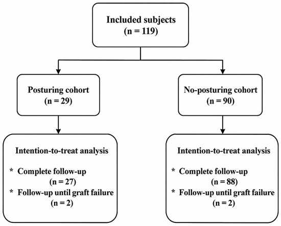
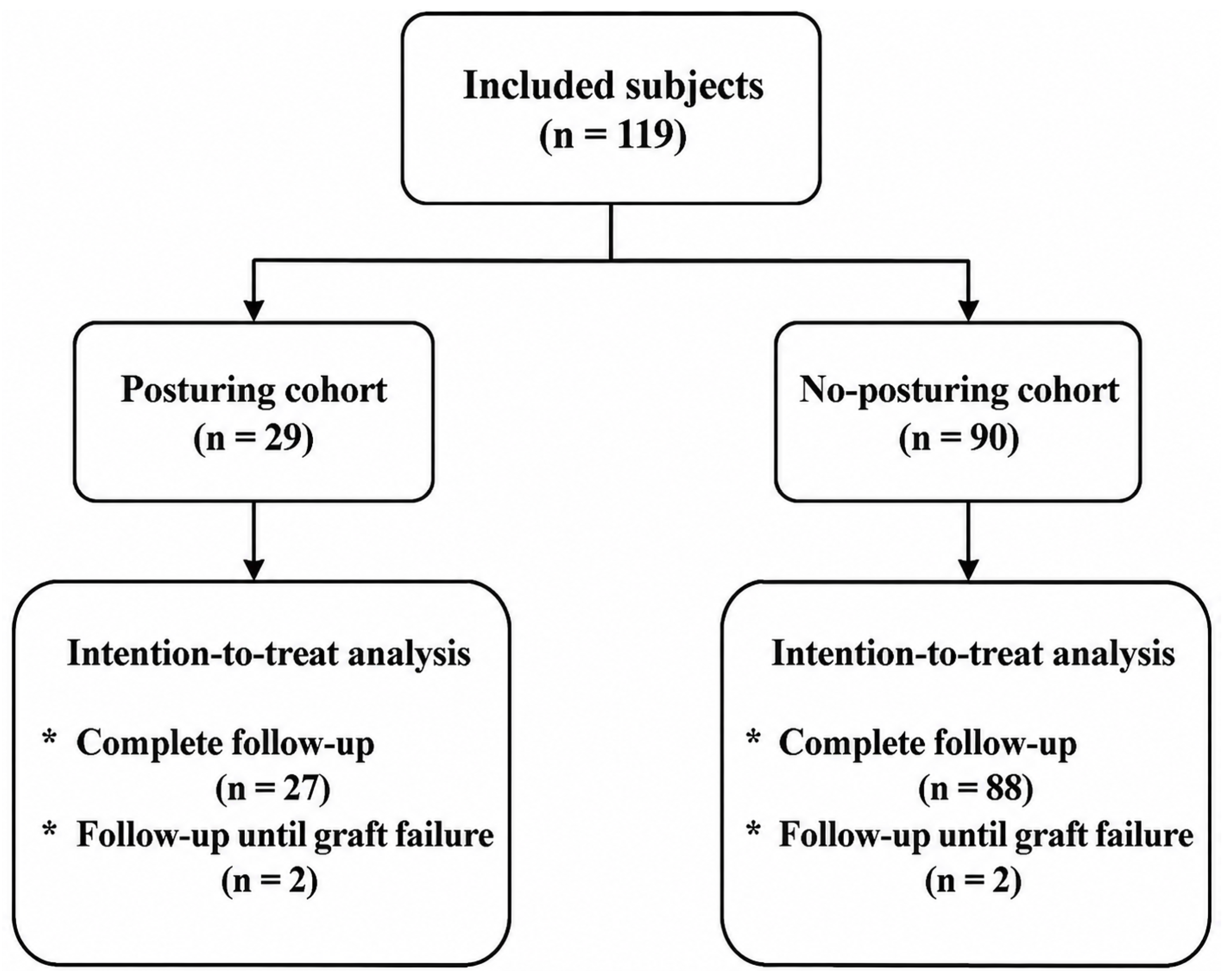
A total of 119 eyes of 97 patients received either the PC (n = 29) or the no-PC (n = 90) following DMEK and were included in the analysis (Figure 1). Mean age at the time of surgery was 70.8 years (SD ± 9.1). Fifty-four (45.4%) were female eyes. The primary indication for DMEK surgery was Fuchs endothelial dystrophy (94.1%). Additional indications were bullous keratopathy (3.4%), graft decompensation (1.7%) and herpetic endothelial failure (0.9%). DMEK was combined with phacoemulsification and IOL implantation (Triple procedure) in 54 eyes (45.4%). Baseline characteristics of patients and donors are reported in Table 1 and were normally distributed according to the Shapiro–Wilk test (all p > 0.05). Four eyes were lost to follow-up, all of whom underwent re-transplantation due to primary graft failure and were subsequently excluded from that point forward.
Figure 1.
Clinical flowchart of patient stratification and analysis. Patients underwent DMEK between May 2023 and June 2024 at Xpert Clinics Eyecare. Patients received the posturing protocol before protocol change in October 2023 and afterwards patients received the no-posturing protocol. No selection was made. An intention-to-treat analysis was conducted for all primary outcomes. Graft failure occurred in four eyes; all results prior to graft failure were included in the analysis, while secondary outcome data following graft failure were excluded.
Table 1.
Baseline characteristics of patient and donor.
Clinical Outcomes
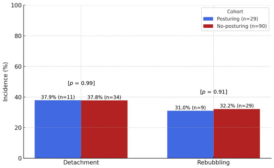
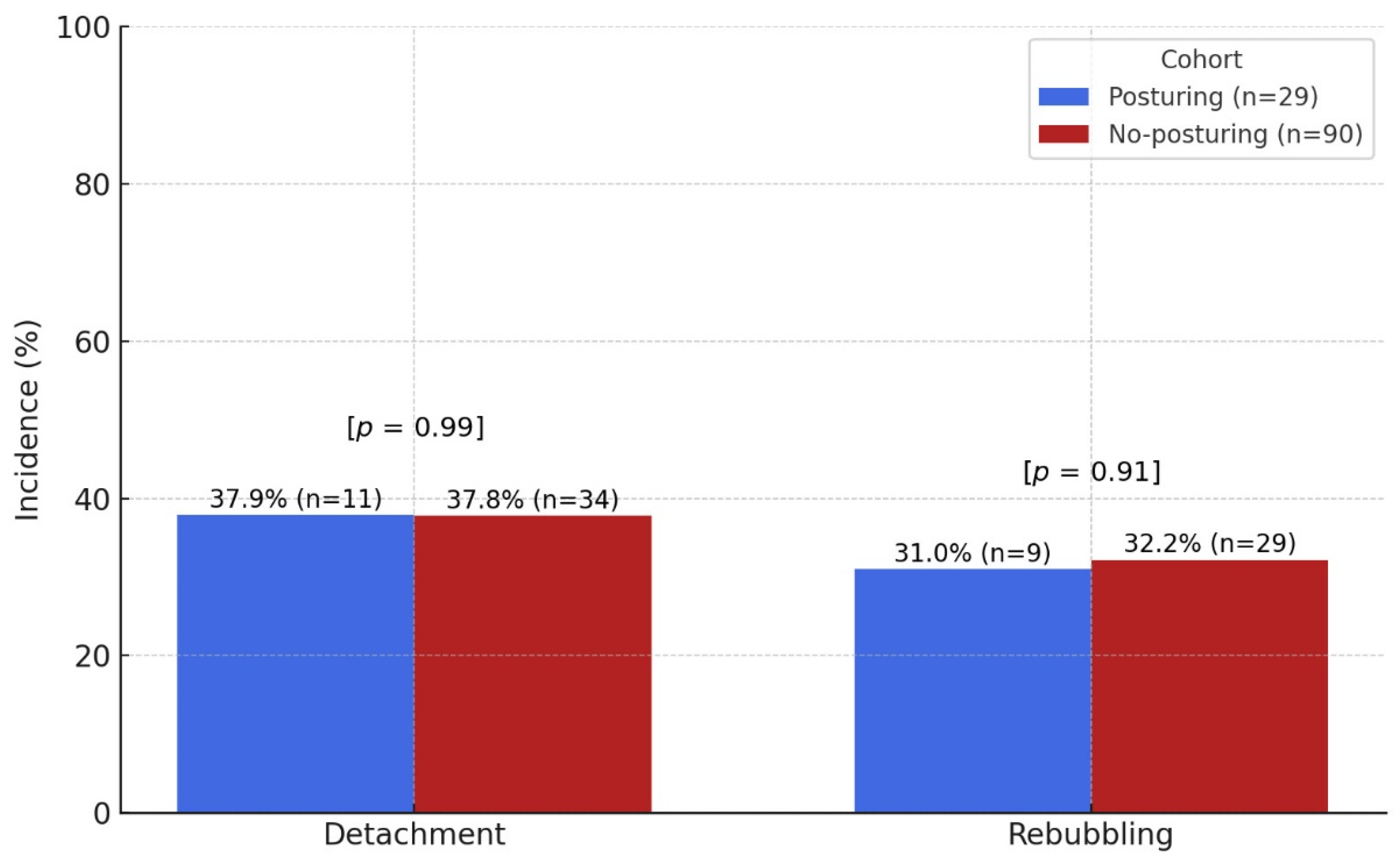
Overall, 45 of 119 eyes (37.8%) developed a graft detachment and rebubbling was performed in 38 of 119 eyes (31.9%). A detailed overview of the primary outcomes and its determinants is shown in Table 2 with visualization of these outcomes in Figure 2. Graft detachments were observed in 11 of 29 eyes (37.9%) in the PC after DMEK surgery, versus 34 of 90 eyes (37.8%; p = 0.99) in the no-PC. The mean duration between DMEK and detachment was 7.2 days (SD: 4.0), and after splitting into both cohorts 8.6 days in the PC and 6.8 days in the no-PC. Considering surgical interventions, 9 of 29 eyes (31.0%) in the PC underwent a rebubbling procedure versus 29 of 90 eyes (32.2%; p = 0.91) in the no-PC. To account for the correlation between eyes of the same patient (22 patients with bilateral inclusion), the association between posturing and the primary outcomes was assessed using generalized estimating equations (GEE). After accounting for clustering, there remained no significant association between posturing and graft detachment (OR 1.00, p = 1.00) or rebubbling (OR 1.00, p = 0.99). A rebubbling procedure was performed twice in 9 eyes and thrice in 2 eyes.
Table 2.
Detailed primary outcomes.
Figure 2.
Incidence of primary outcomes. Bar chart visualization of the primary outcome incidence: graft detachments and rebubbling procedure, stratified by the posturing (blue) and the no-posturing (red) cohort.
Considering secondary outcomes; grafts survived in 115 of 119 eyes (96.6%) at six months follow-up. Graft failure occurred twice in both cohorts (6.9% PC versus 2.2% no-PC, p = 0.23). A diagnosis of graft failure was established after two rebubbling procedures in two cases, or after three rebubbling procedures in one case. The other case of graft failure had a particularly complicated surgical course. The other adverse events of iatrogenic glaucoma (6.9% PC versus 4.4% no-PC, p = 0.60), IOP steroid responses (10.3% PC versus 11.1% no-PC, p = 0.91), and Cystoid Macular Edema (6.9% PC versus 6.7% no-PC, p = 0.97) occurred in equal proportions between both cohorts. The only outcome measure that showed a significant difference between the two cohorts was BCVA at six months follow-up, in favour of the no-PC (mean Snellen = 0.68 PC versus 0.78 no-PC, p = 0.03). An overview of all important clinical outcomes is shown in Table 3.
Table 3.
Clinical outcomes overview.
4. Discussion
The aim of this study was to compare the incidence of graft detachment and rebubbling with and without postoperative posturing after DMEK surgery. Though posturing is considered a standard practice pattern, the benefits are never properly researched and there are no validated guidelines [11,13,14,15]. Moreover, posturing can cause significant discomfort and health risks, particularly among elderly patients [15,17,18,19,20].
In this study, we found that the incidence of postoperative graft detachment and rebubbling following DMEK in the PC is comparable to that observed in the no-PC (risk differences for graft detachment and rebubbling 0.1% and −1.2%, respectively). The risk difference in rebubbling was −1.2 percentage points (95% CI, −0.21; 0.18). As the entire confidence interval fell within the stated equivalence margin of ±25 percentage points, these findings support statistical equivalence for rebubbling.
Our findings are consistent with those reported by Kladny et al. (2024), who demonstrated in a randomized controlled trial that prolonged posturing for five days did not improve graft attachment compared with one day of posturing [15]. While that study compared different durations of postoperative posturing, the present study expands on these findings by evaluating a strategy in which postoperative posturing is omitted altogether. The comparable rates of graft detachment and rebubbling observed in our study further support the notion that prolonged postoperative posturing may not provide additional clinical benefit.
The mean reported rate of rebubbling in the literature is 28.8% [3]. Our observed mean rebubbling rate shows slight deviation (31.9%) and may reflect a relatively low threshold for performing rebubbling where other surgeons might not have intervened. Notwithstanding, our findings support our assumption that postoperative posturing does not have a positive effect on detachments and rebubbling rates. On the contrary, it exposes patients to unnecessary burdens. The physical burden of posturing may even discourage patients from undergoing DMEK, thereby preventing them from benefiting from potential improvements in vision and quality of daily life [3].
Considering the secondary outcomes, the incidence rates of other common adverse events such as graft failure and Cystoid Macular Edema also showed no significant difference between the PC and no-PC. Furthermore, the graft failure rate of 2.2% in this study is notably lower than the reported graft failure incidence of 7.0% documented in a large multicentre trial [21]. The only outcome that showed a significant difference between the two cohorts was the BCVA at six months follow-up. Remarkably, this difference is not supported by prior literature, and its cause remains unclear, possibly reflecting random variability. The study was not powered to investigate differences in postoperative BCVA.
The equivalence margin was based on rebubbling incidence rates by literature review in consultation with the corneal surgeon [3]. Despite this being a retrospective study, we decided to perform a power calculation to provide additional strength to the validity of the primary outcome measures. The power calculation determined that the sample size for this study is sufficient to meet the required statistical power.
It is worth mentioning that the percentage of perioperative manual PI differed by 7.7% between the two cohorts. A previous study investigating risk factors for graft detachment following endothelial keratoplasty demonstrated that preoperative laser PI had a slight protective effect against graft detachment compared to surgical PI [22]. This protective effect of preoperative laser PI may be attributed to the increased intraoperative fibrin formation associated with surgical PI [23]. Yet, our study did not identify any protective effect of pre-existing PI’s. The major indication for a laser PI was a phakic DMEK procedure (n = 6), where an intraoperative surgical PI could not be performed due to risk of cataract formation. This direct relationship impedes causation.
Furthermore, it should be acknowledged that a full air or gas fill at the conclusion of DMEK surgery is an essential component in promoting graft adherence [24]. Although our study focused on postoperative posturing, the impact of intraoperative tamponade techniques deserves consideration in future research.
Additionally, frequent postoperative follow-up with adequate corneal imaging is crucial to ensure optimal outcomes after DMEK. Timely intervention with rebubbling effectively resolves most detachments, often leading to favourable visual results. Small peripheral detachments may resolve spontaneously, reducing the need for immediate intervention. These findings emphasize that careful monitoring may be more critical than strict posturing protocols.
Several considerations deserve mentioning. This was a retrospective study, which is inherently subject to potential treatment bias. Because cohort allocation was determined chronologically, secular changes during the study period, including subtle changes in surgeon performance, cannot be fully excluded. Although all procedures were performed by the same surgeon using a standardized technique, the non-concurrent cohort design remains susceptible to temporal confounding. Yet, all consecutive DMEK procedures were included in the study, and the protocol change was complete, without exceptions. Additionally, the exact extent of graft detachment was not consistently documented in our medical records, which precluded a detailed stratification of detachments according to size. In clinical practice, however, the decision to perform rebubbling is typically based on pragmatic clinical thresholds such as involvement of the visual axis or detachment exceeding 25% of the graft, rather than precise quantitative measurements. Therefore, this is unlikely to have substantially influenced the outcomes of this study. Furthermore, a limitation is that postoperative posturing was not objectively monitored. Consequently, cohort allocation reflects the postoperative instructions provided rather than verified posturing behaviour. This potential exposure misclassification reduces the contrast between the two strategies and could bias the estimated effect. Therefore, our findings should be interpreted as a comparison of posturing instructions rather than precisely measured postoperative posturing behaviour.
A randomised prospective trial assessing actual posturing time may be necessary to confirm these findings. Another option could be a fellow eye analysis, where one eye undergoes DMEK with posturing and the other without posturing. This could provide further insight by minimizing variability and directly comparing outcomes within the same patient. Future prospective studies should also focus on exploring specific subsets of patients who might still benefit from posturing, such as those with limited access to follow-up care or with high-risk factors for graft detachment [22]. Studies better predicting the incidence of a graft detachment might better tailor postoperative check-ups to patients at risk [25,26]. Identifying these groups could help refine postoperative protocols, ensuring tailored care while minimizing unnecessary burdens. Such efforts would contribute to a more patient-centred approach and broaden the applicability of DMEK to diverse patient populations.
5. Conclusions
In conclusion, this equivalence study on postoperative posturing after DMEK identified no benefit of postoperative posturing considering the risk of graft detachment, rebubbling, or any other postoperative outcome. These findings suggest that eliminating posturing instructions enhances patient comfort and broadens the eligibility for DMEK without compromising clinical outcomes.
Author Contributions
R.P.L.W. and C.B.J.T. contributed equally as joint first authors. Conceptualization, R.P.L.W., C.B.J.T. and C.v.d.Z.; Methodology, R.P.L.W., C.B.J.T. and C.v.d.Z.; Software, C.B.J.T. and C.v.d.Z.; Validation, R.P.L.W., C.B.J.T., N.G.d.K.-T. and C.v.d.Z.; Formal Analysis, R.P.L.W., C.B.J.T. and C.v.d.Z.; Investigation, R.P.L.W., C.B.J.T. and C.v.d.Z.; Resources, R.P.L.W., C.B.J.T. and N.G.d.K.-T.; Data Curation, R.P.L.W., C.B.J.T. and C.v.d.Z.; Writing—Original Draft Preparation, R.P.L.W., C.B.J.T., N.G.d.K.-T. and C.v.d.Z.; Writing—Review & Editing, R.P.L.W., C.B.J.T., N.G.d.K.-T. and C.v.d.Z.; Visualization, C.B.J.T. and C.v.d.Z.; Supervision, R.P.L.W. and C.v.d.Z.; Project Administration, R.P.L.W., C.B.J.T. and C.v.d.Z. All authors have read and agreed to the published version of the manuscript.
Funding
This research received no external funding.
Institutional Review Board Statement
The study was conducted in accordance with the Declaration of Helsinki, and approved by the Medical Research Ethics Committees United (MEC-U; registration number W25.045) on 19 March 2025. The Ethics Committee reviewed the study and issued a non-WMO declaration, indicating that the study does not fall under the scope of the Dutch Medical Research Involving Human Subjects Act (WMO).
Informed Consent Statement
Written informed consent for participation in the study and use of clinical data was obtained from all subjects involved in the study.
Data Availability Statement
Data are available from the corresponding author, C.B.J. Thijssen, upon reasonable request.
Acknowledgments
We express our gratitude to the ophthalmological assistants, nurses, and management staff at Xpert Clinics Eyecare for providing the facilities and support necessary to conduct this study. Their contribution to the data collection and clinical infrastructure was invaluable to the successful completion of this research.
Conflicts of Interest
Authors Robert P. L. Wisse and Nayyirih G. de Koning-Tahzib were employed by the company Xpert Clinics Eyecare. Author R.P.L.W. is shareholder and consultant for Easee BV, paid speaker for Alcon Benelux BV, paid speaker for Carl Zeiss GmbH and paid speaker for Thea Pharma BV. The remaining authors declare that the research was conducted in the absence of any commercial or financial relationships that could be construed as a potential conflict of interest.
References
- Eye Bank Association of America. The 2023 Eye Banking Statistical Report 2023. Available online: https://restoresight.org/members/publications/statistical-report/#:~:text=U.S.%20eye%20banks%20reported%20137%2C697,which%20approximates%20pre%2DCOVID%20levels (accessed on 18 October 2024).
- Bunya, V.Y.; Woodward, M.A.; DeMill, D.L.; Halfpenny, C.; Syed, Z.A.; Bert, B.B. Descemet Membrane Endothelial Keratoplasty. EyeWiki 2024. Available online: https://eyewiki.org/Descemet_Membrane_Endothelial_Keratoplasty#cite_note-3 (accessed on 15 October 2024).
- Deng, S.X.; Lee, W.B.; Hammersmith, K.M.; Kuo, A.N.; Li, J.Y.; Shen, J.F.; Weikert, M.P.; Shtein, R.M. Descemet Membrane Endothelial Keratoplasty: Safety and Outcomes: A Report by the American Academy of Ophthalmology. Ophthalmology 2018, 125, 295–310. [Google Scholar] [CrossRef] [Scilit] [PubMed]
- Melles, G.R.J.; Ong, T.S.; Ververs, B.; van der Wees, J. Descemet membrane endothelial keratoplasty (DMEK). Cornea 2006, 25, 987–990. [Google Scholar] [CrossRef] [Scilit]
- Ang, M.; Wilkins, M.R.; Mehta, J.S.; Tan, D. Descemet membrane endothelial keratoplasty. Br. J. Ophthalmol. 2016, 100, 15–21. [Google Scholar] [CrossRef] [Scilit] [PubMed]
- Feng, M.T.; Burkhart, Z.N.; Price, F.W.; Price, M.O. Effect of donor preparation-to-use times on Descemet membrane endothelial keratoplasty outcomes. Cornea 2013, 32, 1080–1082. [Google Scholar] [CrossRef] [Scilit] [PubMed]
- Dapena, I.; Ham, L.; Droutsas, K.; van Dijk, K.; Moutsouris, K.; Melles, G.R.J. Learning Curve in Descemet’s Membrane Endothelial Keratoplasty: First Series of 135 Consecutive Cases. Ophthalmology 2011, 118, 2147–2154. [Google Scholar] [CrossRef] [Scilit] [PubMed]
- Dirisamer, M.; Ham, L.; Dapena, I.; Moutsouris, K.; Droutsas, K.; van Dijk, K.; Frank, L.E.; Oellerich, S.; Melles, G.R. Efficacy of descemet membrane endothelial keratoplasty: Clinical outcome of 200 consecutive cases after a learning curve of 25 cases. Arch. Ophthalmol. 2011, 129, 1435–1443. [Google Scholar] [CrossRef] [Scilit] [PubMed]
- Dunker, S.; Winkens, B.; van den Biggelaar, F.; Nuijts, R.; Kruit, P.J.; Dickman, M.; Dutch Cornea Consortium. Rebubbling and graft failure in Descemet membrane endothelial keratoplasty: A prospective Dutch registry study. Br. J. Ophthalmol. 2023, 107, 17–23. [Google Scholar] [CrossRef] [Scilit] [PubMed]
- Muijzer, M.B.; Delbeke, H.; Dickman, M.M.; Nuijts, R.M.; Noordmans, H.J.; Imhof, S.M.; Wisse, R.P. Outcomes of the advanced visualization in corneal surgery evaluation trial; a non-inferiority randomized control trial to evaluate the use of intraoperative OCT during Descemet membrane endothelial keratoplasty. Front. Ophthalmol. 2022, 2, 1041778. [Google Scholar] [CrossRef] [Scilit] [PubMed]
- Rapoport, Y.; Veldman, P. A Comprehensive Review of Postoperative Management of Descemet’s Membrane Endothelial Keratoplasty. Semin. Ophthalmol. 2017, 32, 104–110. [Google Scholar] [CrossRef] [Scilit] [PubMed]
- Bhogal, M.F.; Gupta, N.; Giobellina, T.; Kobayashi, A.; Males, J.M.; Mehta, J.S.F.; Romano, V.; Allan, B.F.; Busin, M.; Cursiefen, C.; et al. Delphi-Based Global Consensus on Adopting Endothelial Keratoplasty: An Endothelial Keratoplasty Learners Group Initiative. Cornea 2025, 44, 1010–1021. [Google Scholar] [CrossRef] [Scilit] [PubMed]
- Roberts, H.W.; Kit, V.; Phylactou, M.; Din, N.; Wilkins, M.R. “Posture-Less” DMEK: Is Posturing After Descemet Membrane Endothelial Keratoplasty Actually Necessary? Am. J. Ophthalmol. 2022, 240, 23–29. [Google Scholar] [CrossRef] [Scilit] [PubMed]
- Parker, J.S.; Parker, J.S.; Tate, H.; Melles, G.R.J. DMEK Without Postoperative Supine Posturing. Cornea 2023, 42, 32–35. [Google Scholar] [CrossRef] [Scilit] [PubMed]
- Kladny, A.-M.S.; Glatz, A.; Lieberum, J.-L.; Zander, D.B.; Siegel, H.; Jiang, J.; Brandi-Dohrn, F.; Maier, P.C.; Lapp, T.; Lang, S.J.; et al. Supine Positioning for Graft Attachment After Descemet Membrane Endothelial Keratoplasty: A Randomized Controlled Trial. Am. J. Ophthalmol. 2024, 263, 117–125. [Google Scholar] [CrossRef] [Scilit] [PubMed]
- Santander-García, D.; Peraza-Nieves, J.; Müller, T.M.; Gerber-Hollbach, N.; Baydoun, L.; Liarakos, V.S.; Dapena, I.; Oellerich, S.; Van Dijk, K.; Melles, G.R.J. Influence of Intraoperative Air Tamponade Time on Graft Adherence in Descemet Membrane Endothelial Keratoplasty. Cornea 2019, 38, 166–172. [Google Scholar] [CrossRef] [Scilit] [PubMed]
- Truong, A.D.; Fan, E.; Brower, R.G.; Needham, D.M. Bench-to-bedside review: Mobilizing patients in the intensive care unit--from pathophysiology to clinical trials. Crit. Care 2009, 13, 216. [Google Scholar] [CrossRef] [Scilit] [PubMed]
- Pottier, P.; Hardouin, J.B.; Lejeune, S.; Jolliet, P.; Gillet, B.; Planchon, B. Immobilization and the risk of venous thromboembolism. A meta-analysis on epidemiological studies. Thromb. Res. 2009, 124, 468–476. [Google Scholar] [CrossRef] [Scilit] [PubMed]
- Rommersbach, N.; Wirth, R.; Lueg, G.; Klimek, C.; Schnatmann, M.; Liermann, D.; Janssen, G.; Müller, M.J.; Pourhassan, M. The impact of disease-related immobilization on thigh muscle mass and strength in older hospitalized patients. BMC Geriatr. 2020, 20, 500. [Google Scholar] [CrossRef] [Scilit] [PubMed]
- Tana, C.; Lauretani, F.; Ticinesi, A.; Prati, B.; Nouvenne, A.; Meschi, T. Molecular and Clinical Issues about the Risk of Venous Thromboembolism in Older Patients: A Focus on Parkinson’s Disease and Parkinsonism. Int. J. Mol. Sci. 2018, 19, 1299. [Google Scholar] [CrossRef] [Scilit] [PubMed]
- Oellerich, S.; Baydoun, L.; Peraza-Nieves, J.; Ilyas, A.; Frank, L.; Binder, P.S.; Melles, G.R.J. Multicenter Study of 6-Month Clinical Outcomes After Descemet Membrane Endothelial Keratoplasty. Cornea 2017, 36, 1467–1476. [Google Scholar] [CrossRef] [Scilit] [PubMed]
- Muijzer, M.B.; Hoven, C.M.W.; Frank, L.E.; Vink, G.; Wisse, R.P.L.; Netherlands Corneal Transplant Network (NCTN). A machine learning approach to explore predictors of graft detachment following posterior lamellar keratoplasty: A nationwide registry study. Sci. Rep. 2022, 12, 17705. [Google Scholar] [CrossRef] [Scilit] [PubMed]
- Benage, M.; Korchak, M.; Boyce, M.; Mayko, Z.M.; Bauer, A.; Straiko, M.D.; Terry, M.A.; Sáles, C.S.; Greiner, M.A. Intraoperative fibrin formation during Descemet membrane endothelial keratoplasty. Am. J. Ophthalmol. Case Rep. 2020, 18, 100686. [Google Scholar] [CrossRef] [Scilit] [PubMed]
- Pilger, D.; Wilkemeyer, I.; Schroeter, J.; Maier, A.-K.B.; Torun, N. Rebubbling in Descemet Membrane Endothelial Keratoplasty: Influence of Pressure and Duration of the Intracameral Air Tamponade. Am. J. Ophthalmol. 2017, 178, 122–128. [Google Scholar] [CrossRef] [Scilit] [PubMed]
- Muijzer, M.B.; Heslinga, F.G.; Couwenberg, F.; Noordmans, H.-J.; Oahalou, A.; Pluim, J.P.W.; Veta, M.; Wisse, R.P.L. Automatic evaluation of graft orientation during Descemet membrane endothelial keratoplasty using intraoperative OCT. Biomed. Opt. Express 2022, 13, 2683–2694. [Google Scholar] [CrossRef] [Scilit] [PubMed]
- Muijzer, M.B.M.; Noordmans, H.-J.; Delbeke, H.; Dickman, M.M.; Nuijts, R.M.M.A.; Dunker, S.; Imhof, S.M.; Wisse, R.P.L. Establishing a Biomarker for the Prediction of Short-Term Graft Detachment After Descemet Membrane Endothelial Keratoplasty. Cornea 2023, 42, 204–210. [Google Scholar] [CrossRef] [Scilit] [PubMed]

Disclaimer/Publisher’s Note: The statements, opinions and data contained in all publications are solely those of the individual author(s) and contributor(s) and not of MDPI and/or the editor(s). MDPI and/or the editor(s) disclaim responsibility for any injury to people or property resulting from any ideas, methods, instructions or products referred to in the content. |
© 2026 by the authors. Licensee MDPI, Basel, Switzerland. This article is an open access article distributed under the terms and conditions of the Creative Commons Attribution (CC BY) license.