


Abstract
Libidibia coriaria (Cascalote), an underutilized leguminous species native to the tropical Americas, is a promising source of bioactive phenolic compounds with pharmaceutical and nutraceutical potential. This study compared microwave-assisted extraction (MAE), ultrasound-assisted extraction (UAE), and their hybrid combination (UMAE) for the recovery of hydrolyzable and condensed phenolics from cascalote pods and evaluated the antioxidant capacity of the resulting extracts. UMAE achieved the highest total phenolic content (223.49 mg GAE/g dry matter), while antioxidant capacity varied by assay: MAE showed the highest DPPH radical scavenging activity and FRAP reducing power, whereas UAE exhibited the highest ABTS radical-scavenging activity. Nevertheless, UMAE showed the lowest IC50 values for DPPH and ABTS (228.91 and 155.65 mg/L, respectively), indicating greater concentration-dependent radical-scavenging efficiency. Qualitative HPLC–MS tentatively identified gallotannins, ellagitannins, and hydroxybenzoic acid derivatives. UAE yielded the broadest phenolic profile, whereas MAE and UMAE shared several major tannin-related compounds. FTIR analysis supported the presence of characteristic functional groups associated with gallotannins and ellagitannins. Overall, under the evaluated conditions, UMAE was the most effective extraction method for improving phenolic recovery. These findings highlight the potential of this hybrid extraction approach for the valorization of L. coriaria pods as a source of high-value bioactive phenolic compounds.
1. Introduction
Global food security remains a major challenge due to population growth, declining crop productivity, and climate change. Millions of people still face hunger, hindering progress toward sustainable development. One contributing factor is the limited use of nutrient-rich, yet neglected, crops [1]. Promoting the cultivation and utilization of underexploited legumes offers a sustainable strategy to improve food availability, as these species are nutrient-dense and provide functional properties that could play a critical role in maintaining consumer well-being [2]. In this regard, Libidibia coriaria (Jacq.) Schltdl. represents a promising yet underutilized resource for strengthening food system resilience.
L. coriaria (Jacq.) Schltdl. (Syn. Caesalpinia coriaria (Jacq.) Willd.), commonly known as “cascalote” or “divi-divi,” is a leguminous tree native to the tropical regions of the Americas and the Antilles. In México, it is widely distributed and recognized for its distinctive curved pods, which are 3 to 7 cm long and change from green to dark brown during maturation [3,4]. Cascalote pods are rich in phenolic compounds, containing approximately 40–45% tannins [5]. The major bioactive constituents include corilagin, lagerstannin, geraniin, and ellagic acid [3], which have demonstrated anticancer [6], antibacterial [7], and antiparasitic activities [8]. Despite this potential, studies evaluating the efficacy and formulation performance of cascalote pod extract in food systems remain limited. Consequently, their application as functional food ingredients or natural preservatives is still incipient, and the species remains underutilized because of the lack of efficient and sustainable extraction methods.
Conventional extraction of phenolic compounds relies largely on organic solvent-intensive processes that require long extraction times and generate environmental concerns [9]. Green extraction technologies have emerged as sustainable alternatives that reduce processing time while enabling the use of safer solvents, such as water or ethanol–water mixtures [10]. Among these technologies, ultrasound-assisted extraction (UAE) and microwave-assisted extraction (MAE) have attracted considerable interest because they improve extraction efficiency, shorten processing times, and reduce energy consumption compared with conventional extraction methods [11].
UAE employs high-frequency sound waves to disrupt plant cell walls, facilitating the release of intracellular compounds into the extraction solvent [12]. This process is driven by acoustic cavitation, in which the formation and collapse of microbubbles generate localized energy that disrupts cell structures and enhances mass transfer [13,14]. In contrast, MAE uses electromagnetic radiation to rapidly and uniformly heat the solvent–sample matrix, thereby increasing mass transfer, accelerating solute diffusion, and improving extraction efficiency. MAE is recognized for its short processing time, reduced solvent consumption, and high efficiency in recovering thermolabile bioactive compounds [3,15].
More recently, ultrasound/microwave-assisted extraction (UMAE) has emerged as an innovative hybrid approach that combines the mechanical effects of ultrasound with the rapid heating capability of microwaves. This approach enables fast, uniform, and relatively low-temperature extraction, thereby reducing compound degradation, enhancing extraction yields, and minimizing solvent consumption [16,17]. UMAE has been successfully applied to recover phenolic compounds from various plant matrices, including xoconostle fruit [18], eucalyptus leaves [19], moringa leaves [20], creosote bush, tarbush and soursoup [21], as well as agroindustrial by-products such as grape pomace [22], sorghum husk [23], pistachio hulls [24], mango peels [25], pomelo peels [26], and rambutan peels [27]. Collectively, these studies demonstrate the applicability of UMAE to chemically and structurally diverse phenolic-rich plant matrices. Several of these materials are relevant sources of hydrolyzable and condensed tannins, as are L. coriaria pods. Furthermore, the use of water as the extraction medium provides a solvent system consistent with green-extraction principles and suitable for recovering polar phenolic constituents. However, to the best of our knowledge, a comparative evaluation of UAE, MAE, and UMAE for phenolic recovery from L. coriaria pods has not yet been reported. Given the distinctive phenolic composition of cascalote pods, particularly their abundance of hydrolyzable and condensed tannins [3,28,29], evaluating the behavior of these extraction technologies in this way provides insights that cannot be directly extrapolated from previously studied plant materials. Therefore, this study aimed to compare the performance of MAE, UAE, and UMAE under predefined operating conditions for the recovery of phenolic compounds from cascalote pods and to evaluate the antioxidant capacity and phenolic profiles of the resulting extracts.
2. Materials and Methods
2.1. Plant Material
The cascalote pods used in the current study were obtained from the same sampling location previously reported by López-Cárdenas et al. [3], located in the tropical region of southern Mexico. After collection, the pods were manually cleaned to remove impurities and debris. They were then dried in a convection oven at 50 °C for 24 h prior to storage and further analysis.
2.2. Chemical Reagents
Ethanol, methanol, and 1-butanol were acquired from JT BakerTM (Phillipsburg, NJ, USA). DPPH (2,2-diphenyl-1-picrylhydrazyl), ABTS (2,2′-azino-bis(3-ethylbenzothiazoline-6-sulfonic acid) diammonium salt), Trolox (6-hydroxy-2,5,7,8-tetramethylchroman-2-carboxylic acid) standard, potassium persulfate, hydrochloric acid, gallic acid standard, Folin–Ciocalteu reagent, sodium carbonate, ferric chloride (FeCl3·6H2O), TPTZ (2,4,6-tri(2-pyridyl)-s-triazine), sodium acetate, ferrous sulfate (FeSO4·7H2O) standard, and formic acid were obtained from Sigma-Aldrich® (Sigma-Aldrich, St. Louis, MO, USA). Acetonitrile and methanol (HPLC grade) were used for chromatographic analyses. Amberlite XAD-16 resin (Sigma-Aldrich, St. Louis, MO, USA) and distilled water were employed to prepare all aqueous solutions. Unless otherwise specified, all reagents were of analytical grade.
2.3. Extraction of Samples
Phenolic compounds were extracted from cascalote pods using MAE, UAE and UMAE. The extraction conditions were adapted from the protocol reported by Estrada-Gil et al. [27] and used as predefined reference conditions for the comparative evaluation of the three extraction methods. These conditions have been successfully applied to different phenolic-rich plant matrices [18,19,20,21,22,24,25], supporting their use as a reference framework for evaluating the extraction behavior of cascalote pods. Nevertheless, these conditions were not specifically optimized for L. coriaria pods. UAE was performed at 25 kHz and 40 W in pulse mode (ton/toff = 10 s/3 s) for 20 min at room temperature (28 ± 2 °C). MAE was carried out at 2450 MHz and 400 W for 5 min, reaching a target temperature of 70 °C. For UMAE, extraction was performed using a programmed three-stage protocol. Initially, ultrasound was applied at 25 kHz and 40 W in pulsed mode (10 s on/3 s off) for 5 min. Subsequently, microwave irradiation (2450 MHz, 400 W) was applied for 5 min while ultrasound treatment continued simultaneously under the same operating conditions. During this simultaneous ultrasound–microwave stage, the extraction temperature was controlled at 70 °C using the temperature-control system integrated into the ATPIO-SM300 equipment (Nanjing ATPIO Instruments Manufacture Co., Ltd., Nanjing, China). After microwave irradiation was discontinued, ultrasound treatment alone was continued for an additional 10 min. Thus, the total ultrasound treatment time was 20 min, whereas microwave irradiation was applied for 5 min. The pulsed ultrasound regime provided intermittent acoustic cavitation while allowing heat dissipation during the off-periods. All extractions were performed using an ATPIO-SM300 system (Nanjing ATPIO Instruments Manufacture Co., Ltd., Nanjing, China). For each extraction, 62.5 g of ground cascalote pods (particle size: 0.84 mm, moisture content: 7%) was mixed with 1000 mL of distilled water in a glass reactor at a sample-to-solvent ratio of 1:16 (w/v). The resulting extracts were filtered through cotton followed by Whatman No. 1 filter paper under low-light conditions to remove solid residues. The filtrates were collected in amber glass bottles and stored at −18 °C until analysis.
2.4. Chromatography Fractionation
The aqueous extracts obtained from cascalote pods were fractionated using Amberlite XAD-16 resin according to the method described by Estrada-Gil et al. [27]. A glass column (60 cm height × 2.5 cm diameter) was packed with the resin and equilibrated with distilled water before loading 200 mL of each aqueous extract. The column was then washed with 2 L of distilled water under gravity flow to remove low-molecular-weight and non-phenolic compounds. Retained phenolic compounds were subsequently eluted with 96% (v/v) ethanol. The ethanolic eluate was concentrated under reduced pressure at 40 °C using a rotary evaporator (BUCHI® R-100 HB, Flawil, Switzerland) until complete solvent removal. The resulting residue was reconstituted in a minimal volume of ethanol and dried in a convection oven at 40 °C for 24 h to obtain a powdered semi-purified phenolic fraction. For clarity, this semi-purified phenolic fraction is hereafter referred to as the “extract” throughout the analytical procedures.
2.5. Analytical Methods
2.5.1. Hydrolyzable Phenolics (HP)
The quantification of hydrolyzable phenolics was determined according to the method of Ordoñez-Cano et al. [30], with minor modifications. A known amount of the extract was dissolved in absolute ethanol, and a 20 µL aliquot was mixed with 20 µL of Folin–Ciocalteu reagent in a 96-well microplate. After incubation for 5 min at room temperature, 20 µL of 0.01 M sodium carbonate solution was added, followed by a second 5 min of incubation. Subsequently, 125 µL of distilled water was added to each well, and the absorbance was measured at 790 nm using a MULTISKAN GO microplate reader (Thermo Scientific™, Vantaa, Finland). Hydrolyzable phenolics content was quantified using a gallic acid calibration curve (0–200 mg/L) and expressed as milligrams of gallic acid equivalents per gram of dry matter (mg GAE/g dm). All measurements were performed in quadruplicate.
2.5.2. Condensed Phenolics (CP)
Condensed phenolics content was determined according to the method of Ordoñez-Cano et al. [30], with minor modifications. A 250 µL aliquot of the extract was transferred to screw-cap tubes, followed by the addition of 1500 µL of 10% HCl–n-butanol reagent and 50 µL of ferric chloride (FeCl3) solution. The tubes were tightly sealed and incubated in a boiling water bath for 40 min. After cooling to room temperature, 200 µL of each reaction mixture was transferred to a 96-well microplate, and the absorbance was measured at 460 nm using a microplate reader. CP content was quantified using a catechin calibration curve (0–200 mg/L) and expressed as milligrams of catechin equivalents per gram of dry matter (mg CE/g dm). All analyses were performed in quadruplicate.
The total phenolic content (TPC) was calculated as the sum of HP and CP values and expressed as milligrams per gram of dry matter (mg/g dm).
2.5.3. Antioxidant Capacity
DPPH radical scavenging assay: The assay was conducted according to Meléndez et al. [31]. Briefly, 7 μL of the extract (previously diluted in absolute ethanol) was mixed with 193 μL of a 60 μM DPPH● (2,2-diphenyl-1-picrylhydrazyl) solution prepared in methanol in a 96-well microplate. The reaction mixture was incubated in the dark at room temperature for 30 min, and the absorbance was measured at 517 nm using a microplate reader. The control consisted of 7 μL of absolute ethanol and 193 μL of the DPPH● solution. Antioxidant capacity was quantified using a Trolox calibration curve (0–800 μmol/L) and expressed as μmol Trolox equivalents per g of dry matter (μmol TE/g dm). All measurements were performed in quadruplicate.
ABTS radical scavenging assay: The ABTS●+ radical scavenging activity was determined according to Rojo-Gutiérrez et al. [32]. The ABTS●+ radical cation was generated by reacting 7 mM ABTS [2,2-azino-bis(3-ethylbenzothiazoline-6-sulfonic acid)] with 2.45 mM potassium persulfate in a 2:1 (v/v) ratio. The mixture was incubated in the dark at room temperature for 16 h, and the absorbance was adjusted to 0.70 ± 0.02 at 734 nm with absolute ethanol. For the assay, 10 μL of the diluted extract was mixed with 190 μL of the ABTS●+ solution in a 96-well microplate and incubated in the dark at room temperature for 1 min. The absorbance was measured at 734 nm using a microplate reader. Antioxidant capacity was calculated using a Trolox calibration curve (0–800 μmol/L) and expressed as μmol Trolox equivalents per g of dry matter (μmol TE/g dm). All measurements were performed in quadruplicate.
FRAP reducing power assay: The ferric-reducing antioxidant power (FRAP) of the extract was determined according to Ordoñez-Cano et al. [30]. The FRAP reagent was prepared by mixing 0.3 M acetate buffer at pH 3.6, 10 mM TPTZ in 40 mM HCl, and 20 mM FeCl3·6H2O at a 10:1:1 (v/v/v) ratio, followed by incubation at 37 °C for 30 min. For the assay, 6 µL of the extract, 18 µL of distilled water, and 180 µL of FRAP reagent were added to a 96-well microplate and incubated for 1 h. The absorbance was measured at 593 nm using a microplate reader. Antioxidant capacity was quantified using a FeSO4·7H2O calibration curve (0–4000 μmol/L) and expressed as μmol Fe2+ equivalents per g of dry matter (μmol Fe2+/g dm). All measurements were performed in quadruplicate.
IC50 Determinations: IC50 values were determined using extract concentrations ranging from 100 to 400 mg/L, prepared from a 1000 mg/L stock solution. Radical scavenging activity was calculated as percentage of inhibition according to Equation (1):
where Acontrol represents the absorbance of the control, and Asample corresponds to the absorbance of each sample. The IC50 value, defined as the extract concentration required to inhibit 50% of the DPPH• or ABTS•+ radicals, was determined by linear regression of extract concentration against percentage inhibition [33].
2.5.4. HPLC-MS Identification
Phenolic compounds in the extract were identified by high-performance liquid chromatography coupled with mass spectrometry (HPLC–MS), according to Cerda-Cejudo et al. [34]. Analyses were performed using a ProStar 410 HPLC system (Varian, Palo Alto, CA, USA) equipped with a photodiode array detector set at 253 nm. Before analysis, samples were filtered through 0.45 µm nylon membranes. Chromatographic separation was carried out on a Denali™ C18 reversed-phase column (150 × 2.1 mm, 3 μm; Grace™, Deerfield, IL, USA) maintained at 30 °C, with an injection volume of 10 µL. The mobile phase consisted of acetonitrile (solvent A) and 3% (v/v) aqueous acetic acid (solvent B). The elution gradient was as follows: 3% A (0–5 min), 9% A (5–15 min), 16% A (15–30 min), 33% A (30–33 min), 90% A (33–35 min), followed by re-equilibration at 3% A (35–42 min). The flow rate was 1.2 mL/min. Mass spectrometric detection was performed using a Varian 500-MS ion trap mass spectrometer equipped with an electrospray ionization (ESI) source operating in negative-ion mode. The capillary voltage was set to 90 V, and spectra were acquired over an m/z range of 100–2000. Compounds were tentatively identified by comparing the obtained mass spectra with a reference database of bioactive compounds using MS WorkStation version 6.9 software (Varian).
2.5.5. Fourier Transform Infrared (FTIR) Spectroscopy Analysis
The functional groups of the semi-purified phenolic extract were characterized by Fourier transform infrared (FTIR) spectroscopy using an Agilent Cary 630 spectrometer (Agilent Technologies, Santa Clara, CA, USA) equipped with an attenuated total reflectance (ATR) accessory fitted with a zinc selenide (ZnSe) crystal, according to Bautista-Hernández et al. [35]. Spectra were acquired directly from the samples without pretreatment over the range of 4000–650 cm−1 at a spectral resolution of 2 cm−1 using 32 scans per sample. Data acquisition and processing were performed with MicroLab Expert version 1.2 software (Agilent Technologies), and spectra were visualized using OriginPro 8 (OriginLab Corporation, Northampton, MA, USA).
2.6. Statistical Analysis
A completely randomized design (CRD) was used to evaluate the effect of the extraction method (MAE, UAE, and UMAE) on hydrolysable, condensed, and total phenolic contents, as well as antioxidant capacity determined by DPPH●, ABTS•+ and FRAP assays. Each extraction method was performed in quadruplicate, and all analytical determinations were conducted for each independent extract. Data were analyzed by one-way analysis of variance (ANOVA), and treatments means were compared using Tukey’s honestly significant difference (HSD) test at a significance level of p ≤ 0.05. Statistical analyses were performed using SAS software (version 9.0; SAS Institute Inc., Cary, NC, USA).
3. Results and Discussion
3.1. Phenolic Content Assays
3.1.1. Hydrolyzable Phenolics (HP)
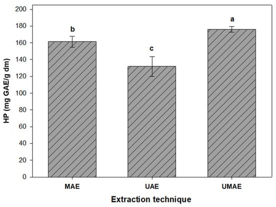
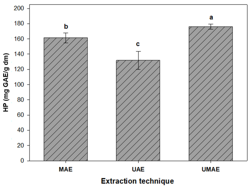
The HP content of the semi-purified extracts obtained from cascalote pods was significantly affected by the extraction method (MAE, UAE and UMAE) (p ≤ 0.05) (Figure 1). UMAE produced the highest HP content (177.49 mg GAE/g dm), followed by MAE (164.54 mg GAE/g dm) and UAE (131.87 mg GAE/g dm).
Figure 1.
Effect of microwave-assisted extraction (MAE), ultrasound-assisted extraction (UAE), and hybrid ultrasound–microwave-assisted extraction (UMAE) on the hydrolyzable phenolic (HP) content of semi-purified cascalote pod extracts. Values are presented as the mean ± standard error (n = 4). Different letters indicate significant differences (Tukey, p < 0.05).
The greater HP recovery obtained with UMAE can be attributed to the complementary effects of acoustic cavitation and microwave dielectric heating. Ultrasound generates cavitation bubbles that collapse violently, producing localized mechanical forces that disrupt plant cell structures and enhance solvent penetration [36,37]. Simultaneously, microwave irradiation provides rapid and uniform heating, accelerating solvent diffusion and the release of phenolic compounds into the extraction medium [38,39]. The combined action of these mechanisms enhances mass transfer and promotes the extraction of bound phenolic compounds. Similar improvements in phenolic recovery using UMAE have been reported for rambutan peel and other plant materials [16,27].
Although MAE yielded a higher HP content than UAE, its extraction efficiency remained lower than that of UMAE. This result suggests that microwave heating alone enhances cell disruption and phenolic solubilization but lacks the mechanical effects generated by acoustic cavitation, which may be particularly important for lignified, tannin-rich matrices such as cascalote pods [3]. In contrast, UAE showed the lowest HP recovery among the assisted extraction methods. This finding agrees with Nipornram et al. [40], who reported that ultrasound alone is less effective at releasing hydrolyzable phenolics tightly bound to polymeric matrices unless combined with additional thermal or chemical energy. The higher HP recovery obtained with UMAE may also be explained by the complementary effects of microwave heating and ultrasonic cavitation. As described by Chemat et al. [41,42], microwave irradiation promotes rapid heating of polar molecules and enhances solvent penetration, while ultrasound improves mass transfer through cavitation-induced disruption of plant tissues. The combined action of these mechanisms facilitates the extraction of hydrolyzable tannins from complex plant matrices, resulting in greater phenolic recovery than either technique applied individually.
3.1.2. Condensed Phenolics (CP)
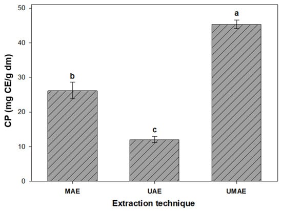
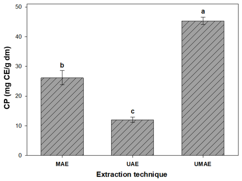
The CP content was significantly affected by the extraction method (p ≤ 0.05) (Figure 2). UMAE produced the highest CP content (45.99 mg CE/g dm), followed by MAE (26.21 mg CE/g dm) and UAE (12.05 mg CE/g dm), demonstrating that extraction efficiency depended on the extraction technique.
Figure 2.
Effect of microwave-assisted extraction (MAE), ultrasound-assisted extraction (UAE), and hybrid ultrasound–microwave-assisted extraction (UMAE) on the condensed phenolics (CP) content of semi-purified cascalote pod extracts. Values represent mean ± standard error (n = 4). Different letters indicate significant differences (Tukey, p < 0.05).
The higher CP recovery obtained with UMAE can be attributed to the complementary effects of ultrasonic cavitation and microwave heating. Acoustic cavitation disrupts plant cell structures and enhances mass transfer, whereas microwave irradiation provides rapid volumetric heating that increases solvent penetration and facilitates the extraction of high-molecular-weight phenolic compounds, including condensed tannins [11,43]. Similar improvements in condensed phenolic recovery have been reported for proanthocyanidin-rich matrices such as rambutan peel, mango peel, and grape pomace [27,34,44]. MAE also improved CP extraction compared with UAE, likely because dielectric heating enhances solvent diffusion and promotes the release of bound phenolic compounds [38,44]. However, in the absence of cavitation, cell disruption may be less effective than under hybrid extraction conditions [45]. These results indicate that combining thermal and mechanical energy enhances the extraction of condensed phenolics from the lignified, tannin-rich matrix of cascalote pods.
3.1.3. Total Phenolic Content (TPC)
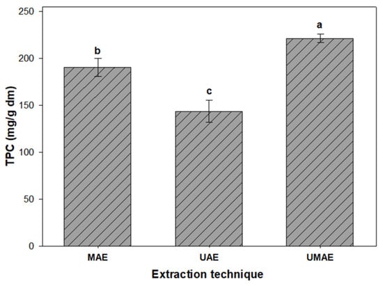
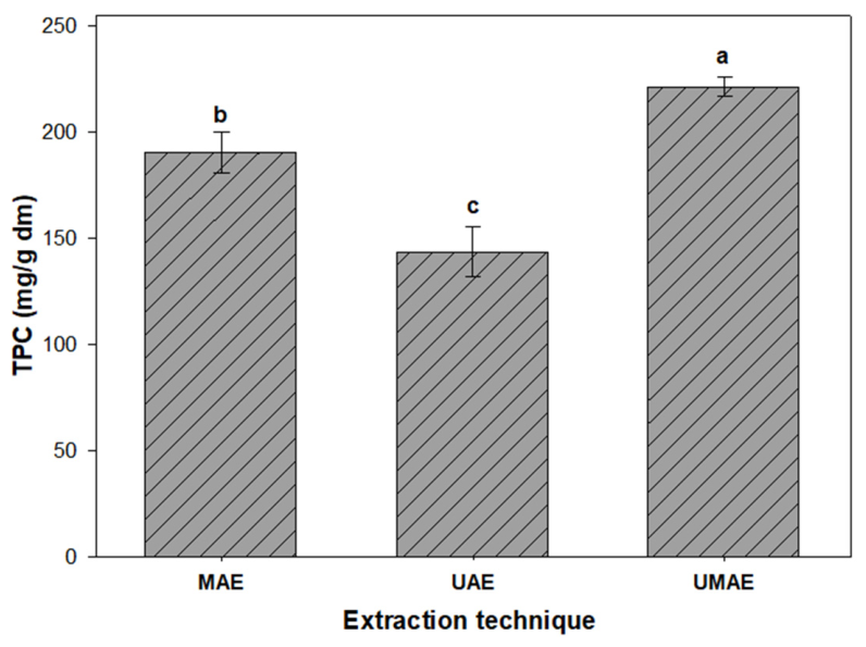
A similar trend was also observed for TPC, which combined the contribution of both hydrolyzable and condensed fractions (Figure 3). Significant differences were observed among methods (p ≤ 0.05), with the highest TPC for UMAE (223.49 mg GAE/g dm), followed by MAE (190.75 mg GAE/g dm) and UAE (143.93 mg GAE/g dm).
Figure 3.
Effect of microwave-assisted extraction (MAE), ultrasound-assisted extraction (UAE), and hybrid ultrasound–microwave-assisted extraction (UMAE) on the total phenolic content (TPC) of cascalote pod extracts after XAD-16 semi-purification. Values represent mean ± standard error (n = 4). Different letters indicate statistically significant differences (Tukey, α < 0.05).
The higher extraction yield of UMAE further supports the benefit of combining ultrasonic cavitation with microwave-induced volumetric heating. This combined effect can better facilitate the efficient extraction of low-molecular-weight phenolics (hydrolyzable compounds) and complex phenolic polymers (condensed tannins) [27,41]. Earlier studies have also reported similar trends in different plant materials, where UMAE can cause extensive cell disruption and higher release of intracellular phenolic metabolites [11,37,43].
MAE also showed higher extraction yields than UAE, which could be attributed to the rapid, uniform heating from microwaves, resulting in enhanced dipolar rotation and ionic conduction within the cellular matrix and further improving solvent–solute interactions and polyphenol solubilization [38,44,45]. However, its thermal mechanism may be less effective in disrupting macromolecular complexes, especially those involving lignin, tannin, and protein interactions, thereby restricting complete phenolic recovery [36].
UAE, on the other hand, showed the lowest TPC among the different methods. Although acoustic cavitation can partially disrupt cell walls and release free phenolics [40,46], its limited penetration into polymeric or lignified structures can limit its efficiency in complex plant materials such as cascalote pods [3,6]. The improved extraction efficiency of UMAE further supports its suitability as an energy-assisted, high-performance technique for maximizing total phenolic recovery from cascalote pods.
The apparent phenolic recovery yields, estimated by comparing the total phenolic content of semi-purified extracts with the total phenolic content previously reported for raw cascalote pods by López-Cárdenas et al. [3] (56.73 mg/g dm), further illustrate the differences among the extraction methods. UMAE showed the highest apparent recovery (394%), followed by MAE (336%) and UAE (254%), consistent with the trend observed for hydrolyzable, condensed, and total phenolic contents. Because these values were calculated from semi-purified fractions obtained after Amberlite XAD-16 chromatographic purification, they represent enrichment relative to the phenolic content of the raw material rather than absolute extraction yields.
3.2. Antioxidant Capacity
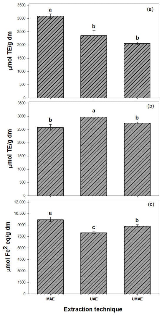
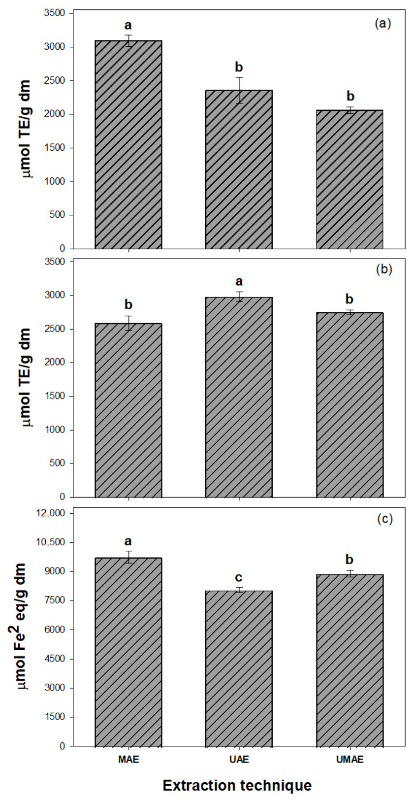
The antioxidant capacity of the semi-purified phenolic extracts obtained from cascalote pods was significantly affected by the extraction method (p ≤ 0.05) (Figure 4). In the DPPH assay, MAE extracts exhibited the highest radical-scavenging activity (3090.83 µmol TE/g dm), followed by UAE (2352.29 µmol TE/g dm) and UMAE (2057.61 µmol TE/g dm).
Figure 4.
Antioxidant activity of semi-purified cascalote pod extracts obtained by microwave-assisted extraction (MAE), ultrasound-assisted extraction (UAE), and hybrid ultrasound–microwave-assisted extraction (UMAE), as determined by the DPPH (a), ABTS (b), and FRAP (c) assays. Values are presented as the mean ± standard error (n = 4). Different letters indicate significant differences according to Tukey’s test (p < 0.05).
Similarly, MAE exhibited the highest ferric-reducing antioxidant power (FRAP) (9738.09 µmol Fe2+/g dm), indicating the greatest reducing capacity among the extraction methods. In contrast, the highest ABTS radical scavenging activity was observed in UAE extracts (2978.17 µmol TE/g dm), followed by UMAE (2747.57 µmol TE/g dm) and MAE (2586.30 µmol TE/g dm).
The higher FRAP values observed for MAE extracts indicate a greater ferric-reducing capacity, reflecting an enhanced ability to donate electrons and reduce ferric (Fe3+) to ferrous (Fe2+) ions [47]. This activity is commonly associated with phenolic compounds containing multiple hydroxyl groups and conjugated aromatic rings, particularly ellagitannins and galloylated derivatives [48]. The predominance of these high-molecular-weight hydrolyzable tannins may therefore explain the greater reducing capacity of MAE extracts observed in the FRAP assay.
These findings demonstrate that the extraction method differentially influences phenolic recovery and antioxidant performance. Although UMAE achieved the highest total phenolic content, it did not exhibit the highest antioxidant capacity in the DPPH, ABTS, or FRAP assays. This finding indicates that total phenolic content alone does not determine the antioxidant response of the extracts. Instead, differences in phenolic composition and molecular structure may influence the predominant antioxidant mechanisms, including radical scavenging and ferric-reducing capacity [28]. This behavior may be associated with differences in the relative abundance and structural characteristics of hydrolyzable tannins, including ellagitannins such as geraniin and related galloyl- and HHDP-containing derivatives previously reported in Libidibia coriaria [3,49]. These compounds can participate in multiple antioxidant mechanisms, including hydrogen atom transfer, single-electron transfer, and metal chelation [50].
Similar trends have been reported for other agri-food by-products, where MAE frequently enhances reducing capacity, whereas UAE or hybrid extraction systems promote the recovery of phenolic compounds with different antioxidant properties [51,52]. For example, Rojo-Gutiérrez et al. [24] reported that MAE improved DPPH radical-scavenging activity in pistachio hulls, whereas UAE favored the extraction of a broader range of phenolic compounds, including thermolabile anthocyanins. These findings highlight that extraction technology can influence phenolic composition and, consequently, antioxidant behavior without necessarily resulting in a direct relationship between total phenolic content and antioxidant capacity.
Purification with Amberlite XAD-16 selectively enriches phenolic compounds through hydrophobic adsorption while removing low-molecular-weight polar constituents, such as sugars and organic acids, resulting in phenolic-enriched fractions with enhanced antioxidant capacity [53]. This enrichment was reflected in the IC50 values of the semi-purified extracts (Table 1). UMAE exhibited the lowest IC50 values in both the DPPH (228.91 mg/L) and ABTS (155.65 mg/L) assays. Overall, IC50 values ranged from 228.91 to 284.93 mg/L for DPPH and from 155.65 to 194.43 mg/L for ABTS. Because lower IC50 values indicate greater radical-scavenging efficiency [46], these results indicate that UMAE extracts exhibited the greatest concentration-dependent radical-scavenging efficiency in both assays.
Table 1.
IC50 values of DPPH and ABTS radical scavenging activities of semi-purified cascalote pod extracts obtained by UAE, MAE, and UMAE.
It should be noted that IC50 values represent concentration-dependent radical-scavenging efficiency and should therefore be interpreted separately from antioxidant capacity values expressed per gram of dry matter. Thus, the lower IC50 values observed for UMAE do not contradict the higher DPPH and ABTS capacity values observed for MAE and UAE, respectively.
Estrada-Gil et al. [27] reported IC50 values of 98 and 335 mg/L for the DPPH and ABTS assays, respectively, in an amberlite XAD-16-purified UMAE extract from rambutan peel. Similarly, Shukla and Mehta [54] reported IC50 values of 74.73 and 86.31 mg/L for crude aqueous and ethanolic extracts of Caesalpinia bonducella, respectively. The comparatively higher IC50 values observed in the present study may reflect differences in phenolic composition, particularly in the relative abundance of hydrolyzable and condensed tannins. The antioxidant capacity of tannin-rich extracts is strongly influenced by factors such as molecular size, hydroxyl-group density, and degree of polymerization, which affect the accessibility and reactivity of phenolic hydroxyl groups toward free radicals and other reactive species [55,56]. Because these studies involved different plant species, extraction methods, and purification procedures, direct comparisons should be interpreted with caution.
3.3. Profiling of Phenolic Compounds
The putative phenolic compounds detected in cascalote pod extracts obtained by UAE, MAE, and UMAE are presented in Table 2. Overall, UAE yielded the greatest diversity of phenolic compounds, with six chromatographic peaks tentatively assigned to five compounds: di-galloyl glucose (m/z 482.8), 3,3′-di-O-methylellagic acid 4′-glucoside (m/z 494.9), geraniin (m/z 950.5), ellagic acid (m/z 300.8) detected as two different chromatographic peaks, and 5-5′-dehydrodiferulic acid (m/z 385). In contrast, MAE and UMAE exhibited less diverse phenolic profiles, predominantly composed of ellagitannin-related compounds and hydroxybenzoic acid derivatives.
Table 2.
Putative phenolic compounds identified in semi-purified cascalote pod extracts obtained by UAE, MAE, and UMAE using HPLC-MS analysis.
Geraniin (m/z 950.5) and di-galloyl glucose (m/z 482.8) were consistently detected in UAE extracts and are representative ellagitannin-related compounds previously associated with antioxidant, cytoprotective, and antimicrobial activities [30,57]. The simultaneous detection of ellagic acid (m/z 300.8) and its glycosylated derivative suggests the presence of structurally related ellagitannins and their transformation products. Acoustic cavitation generated during UAE enhances cell disruption and mass transfer through microstreaming and shear forces while minimizing thermal degradation [58]. These extraction conditions may therefore favor the recovery of structurally diverse phenolic compounds, including both high-molecular-weight ellagitannins and lower-molecular-weight derivatives. Accordingly, UAE produced the greatest qualitative diversity of phenolic compounds among the extraction methods evaluated. Although geraniin and di-galloyl glucose were also detected in MAE and UMAE extracts, their overall phenolic profiles were less diverse than those obtained by UAE.
These findings are in agreement with previous studies reporting the recovery of complex ellagitannins by UAE. Hernández-Hernández et al. [16] identified geraniin and ellagic acid pentoside as the major phenolic compounds in UAE extracts, whereas Insuan et al. [59] reported that UAE effectively recovered thermolabile compounds such as brazilin from Caesalpinia sappan heartwood. Collectively, these studies indicate that ultrasound-assisted extraction favors the recovery of structurally diverse phenolic compounds while minimizing thermal degradation in tannin-rich plant matrices.
In contrast, MAE and UMAE produced less diverse phenolic profiles, predominantly composed of ellagitannin-related compounds and hydroxybenzoic acid derivatives. Rapid dielectric heating during MAE enhances solvent penetration and accelerates the diffusion of phenolic compounds, improving extraction efficiency [3,27,57]. Under certain extraction conditions, microwave heating may also promote the hydrolysis or transformation of larger phenolic compounds, contributing to differences in the resulting phenolic profile. A similar trend was reported by Estrada-Gil et al. [27], who identified geraniin, corilagin, and ellagic acid as the major phenolic compounds extracted from Mexican rambutan peel using assisted extraction techniques. Despite the combined extraction mechanisms involved in UMAE, its phenolic profile was comparable to that of MAE, suggesting that microwave heating may have a greater influence on extraction selectivity under the conditions evaluated.
These findings are consistent with previous studies that have documented that extraction technology influences phenolic composition. Rojo-Gutiérrez et al. [24] found that microwave-assisted extraction produced a more selective phenolic profile, whereas ultrasound-assisted extraction recovered a broader range of phenolic compounds. These differences have been attributed to the distinct extraction mechanisms of each technique: microwave-assisted extraction relies on rapid volumetric heating to enhance solvent diffusion, whereas ultrasound-assisted extraction promotes cell disruption and mass transfer through acoustic cavitation [12,60]. Under the conditions evaluated in this study, UAE yielded the greatest qualitative diversity of phenolic compounds, whereas MAE and UMAE produced comparatively less diverse phenolic profiles.
3.4. FTIR Spectral Analysis
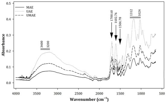
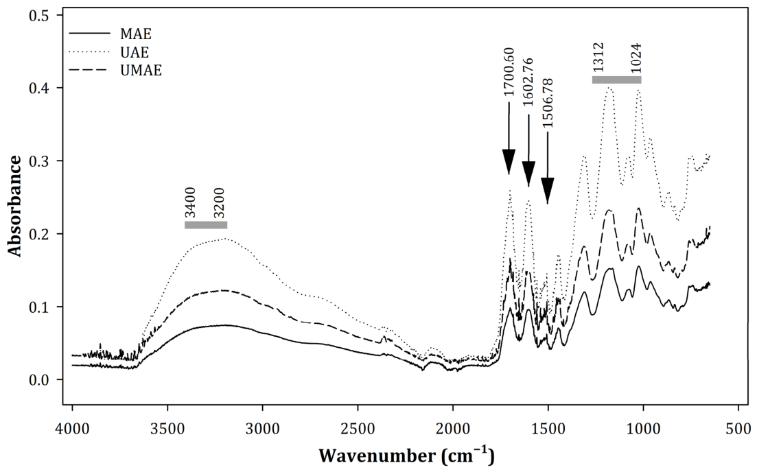
The FTIR spectra of the semi-purified cascalote pod extracts differed according to the extraction method (UAE, MAE, and UMAE) (Figure 5). UAE exhibited more intense absorption bands in regions associated with phenolic compounds, particularly the broad band at 3300–3400 cm−1, assigned to O–H stretching vibrations, and the band at 1733 cm−1, attributed to C=O stretching vibrations of ester and carboxyl groups [61]. These absorption bands are consistent with the presence of oxygen-containing functional groups commonly associated with hydrolyzable tannins and phenolic acids [62].
Figure 5.
FTIR spectra of cascalote pod extracts obtained by UAE, MAE, and UMAE.
The FTIR absorption bands were consistent with the phenolic compounds tentatively identified by HPLC–MS, including ellagitannins such as geraniin and galloylated derivatives. The agreement between the FTIR and HPLC–MS results supports the presence of phenolic compounds containing hydroxyl and carbonyl functional groups in the semi-purified extracts. Similar relationships between FTIR spectral features and phenolic composition have been reported by Nogales-Bueno et al. [63] and Vendramini et al. [64].
Conversely, MAE extracts exhibited lower band intensities than UAE extracts, suggesting differences in the abundance of phenolic-associated functional groups. This observation is consistent with the lower qualitative diversity of phenolic compounds detected by HPLC–MS. The differences may be related to the extraction mechanism associated with microwave-assisted extraction, which promotes rapid dielectric heating and can influence the recovery of phenolic compounds depending on their structural characteristics and thermal stability [65].
The FTIR spectrum of the UMAE extract exhibited an intermediate profile, with a pronounced absorption band at 1499 cm−1 assigned to aromatic C=C stretching vibrations [54]. This band is characteristic of aromatic phenolic structures and is consistent with the presence of tannin-derived compounds identified by HPLC–MS.
Overall, the absorption bands observed at 1733 cm−1 and within the 1312–1024 cm−1 region provide complementary evidence for functional groups commonly associated with ellagitannins and gallotannins [66]. The combined FTIR and HPLC–MS analyses therefore support the presence of these phenolic classes in the semi-purified extracts and demonstrate that the extraction method influences the resulting phenolic profile.
This study has several limitations that should be considered when interpreting the results. First, conventional extraction methods, such as hot-water and organic-solvent extraction, were not included as reference treatments. Therefore, the performance of the evaluated green extraction technologies could not be directly compared with conventional approaches in terms of extraction efficiency, phenolic selectivity, and process sustainability. Second, although the extraction conditions used in this study were based on a previously established protocol applied to different phenolic-rich plant matrices, they were not specifically optimized for L. coriaria pods. Matrix-specific characteristics, including cell-wall architecture, chemical composition, and the distribution and accessibility of phenolic compounds, can substantially influence extraction performance. Therefore, the conditions evaluated herein should not be regarded as optimal for cascalote pods. Independent optimization of MAE, UAE, and UMAE could result in different extraction efficiencies and potentially alter the relative performance of the three technologies. Third, because ultrasound and microwave treatments were not evaluated within a factorial design incorporating absent/present levels and a non-assisted control, the experimental design does not allow statistical estimation of an ultrasound × microwave interaction. Therefore, the greater phenolic recovery observed for UMAE under the evaluated conditions should not be interpreted as evidence of a synergistic interaction between ultrasound and microwave energy. Rather, the results demonstrate the comparative performance of the three extraction configurations under the selected operating conditions. Future studies employing independently optimized conditions and factorial experimental designs are needed to determine whether the simultaneous application of ultrasound and microwave energy produces synergistic, additive, or antagonistic effects. Finally, quantitative sustainability indicators, including energy consumption, solvent-use efficiency, E-factor, operating costs, equipment requirements, and techno-economic aspects, were not evaluated. As a result, the practical applicability, scalability, and overall sustainability of the evaluated extraction technologies could not be comprehensively assessed. Addressing these aspects in future studies would enable a more rigorous evaluation of the practical feasibility and sustainability of MAE, UAE, and UMAE.
4. Conclusions
Among the extraction methods evaluated, UMAE was the most effective for recovering phenolic compounds from cascalote pods under the selected operating conditions, yielding the highest hydrolyzable, condensed, and total phenolic contents. However, greater phenolic recovery did not directly correspond to higher antioxidant capacity across all assays. Antioxidant performance was assay-dependent: MAE exhibited the highest DPPH radical-scavenging activity and ferric-reducing power (FRAP), whereas UAE showed the highest ABTS radical-scavenging activity. Conversely, UMAE exhibited the lowest IC50 values in both radical-scavenging assays, indicating greater concentration-dependent radical-scavenging efficiency.
In contrast, UAE yielded the greatest qualitative diversity of phenolic compounds, whereas MAE and UMAE produced less diverse phenolic profiles dominated by ellagitannin-related compounds and hydroxybenzoic acid derivatives. The complementary HPLC–MS and FTIR analyses provided consistent evidence for the presence of gallotannins, ellagitannins, and related phenolic compounds in the semi-purified extracts. Overall, the results indicate that the selection of an extraction method should be guided by the targeted phenolic recovery, phenolic profile, antioxidant response, and intended application. These findings highlight the potential of UMAE as an efficient extraction approach consistent with green-extraction principles for producing phenolic-rich extracts from L. coriaria pods with prospective applications in food, nutraceutical, and pharmaceutical products.
Author Contributions
E.O.-R.: Writing—original draft preparation, Methodology, Investigation, Formal analysis, Software, Data curation. M.C.-C.: Methodology, Formal analysis, Investigation. E.R.-G.: Writing—original draft preparation, Validation, Writing—review and editing. J.C.T.-A.: Conceptualization, Validation, Resources, Writing—review and editing, Supervision. R.R.-H.: Resources, Writing—review and editing. J.A.A.-V.: Resources, Data curation, formal analysis, Visualization, Writing—review and editing. J.J.B.-F.: Conceptualization, Software, Resources, Data curation, writing—review and editing, Supervision, Project administration, funding acquisition. All authors have read and agreed to the published version of the manuscript.
Funding
This research received no external funding.
Institutional Review Board Statement
Not applicable.
Informed Consent Statement
Not applicable.
Data Availability Statement
The original contributions presented in this study are included in the article. Further inquiries can be directed to the corresponding authors.
Acknowledgments
The authors thank Biologist Raciel Vergara Almeda for his support in the sampling of cascalote pods. The authors gratefully acknowledge the use of ChatGPT (OpenAI, GPT-5.50) for assistance with language editing and improving the clarity, organization and readability of the manuscript. The AI tool was used solely as a writing support tool and did not contribute to the study design, experimental procedures, data analysis, interpretation of the results, or scientific conclusions. The authors reviewed and verified all AI-assisted text and take full responsibility for the accuracy and integrity of the manuscript.
Conflicts of Interest
The authors declare no conflicts of interest.
References
- Popoola, J.O.; Ojuederie, O.B.; Aworunse, O.S.; Adelekan, A.; Oyelakin, A.S.; Oyesola, O.L.; Akinduti, P.A.; Dahunsi, S.O.; Adegboyega, T.T.; Oranusi, S.U.; et al. Nutritional, functional, and bioactive properties of african underutilized legumes. Front. Plant Sci. 2023, 14, 1105364. [Google Scholar] [CrossRef] [Scilit]
- Samal, I.; Bhoi, T.K.; Raj, M.N.; Majhi, P.K.; Murmu, S.; Pradhan, A.K.; Kumar, D.; Paschapur, A.U.; Joshi, D.C.; Guru, P.N. Underutilized legumes: Nutrient status and advanced breeding approaches for qualitative and quantitative enhancement. Front. Nutr. 2023, 10, 1110750. [Google Scholar] [CrossRef] [Scilit] [PubMed]
- López-Cárdenas, F.; Ochoa-Reyes, E.; Baeza-Jiménez, R.; Tafolla-Arellano, J.C.; Ascacio-Valdés, J.A.; Buenrostro-Figueroa, J.J. Solid-State Fermentation as a Sustainable Tool for Extracting Phenolic Compounds from Cascalote Pods. Fermentation 2023, 9, 823. [Google Scholar] [CrossRef] [Scilit]
- Pineda-Peña, E.A.; Capistran-Amezcua, D.; Reyes-Ramírez, A.; Xolalpa-Molina, S.; Chávez-Piña, A.E.; Figueroa, M.; Navarrete, A. Gastroprotective effect methanol extract of Caesalpinia coriaria pods against indomethacin- and ethanol-induced gastric lesions in Wistar rats. J. Ethnopharmacol. 2023, 305, 116057. [Google Scholar] [CrossRef] [Scilit] [PubMed]
- Palma García, J.M. Caesalpinia coriaria (Jacq.) Wild. In Recursos Arboreos y Arbustivos Tropicales; Palma García, J.M., González-Reveles, C., Eds.; Universidad de Colima: México, Mexico, 2018; pp. 30–34. [Google Scholar]
- Sánchez-Carranza, J.N.; Alvarez, L.; Marquina-Bahena, S.; Salas-Vidal, E.; Cuevas, V.; Jiménez, E.W.; Veloz G., R.A.; Carraz, M.; González-Maya, L. Phenolic Compounds Isolated from Caesalpinia coriaria Induce S and G2/M Phase Cell Cycle Arrest Differentially and Trigger Cell Death by Interfering with Microtubule Dynamics in Cancer Cell Lines. Molecules 2017, 22, 666. [Google Scholar] [CrossRef] [Scilit] [PubMed]
- Olmedo-Juárez, A.; Briones-Robles, T.I.; Zaragoza-Bastida, A.; Zamilpa, A.; Ojeda-Ramírez, D.; Mendoza de Gives, P.; Olivares-Pérez, J.; Rivero-Perez, N. Antibacterial activity of compounds isolated from Caesalpinia coriaria (Jacq) Willd against important bacteria in public health. Microb. Pathog. 2019, 136, 103660. [Google Scholar] [CrossRef] [Scilit] [PubMed]
- García-Hernández, C.; Rojo-Rubio, R.; Olmedo-Juárez, A.; Zamilpa, A.; Mendoza de Gives, P.; Antonio-Romo, I.A.; Aguilar-Marcelino, L.; Arece-García, J.; Tapia-Maruri, D.; González-Cortazar, M. Galloyl derivatives from Caesalpinia coriaria exhibit in vitro ovicidal activity against cattle gastrointestinal parasitic nematodes. Exp. Parasitol. 2019, 200, 16–23. [Google Scholar] [CrossRef] [Scilit] [PubMed]
- Mungwari, C.P.; King’ondu, C.K.; Sigauke, P.; Obadele, B.A. Conventional and modern techniques for bioactive compounds recovery from plants: Review. Sci. Afr. 2025, 27, e02509. [Google Scholar] [CrossRef] [Scilit]
- Osorio-Tobón, J.F. Recent advances and comparisons of conventional and alternative extraction techniques of phenolic compounds. J. Food Sci. Technol. 2020, 57, 4299–4315. [Google Scholar] [CrossRef] [Scilit] [PubMed]
- Biswas, R.; Sarkar, A.; Alam, M.; Roy, M.; Mahdi Hasan, M.M. Microwave and ultrasound-assisted extraction of bioactive compounds from Papaya: A sustainable green process. Ultrason. Sonochem. 2023, 101, 106677. [Google Scholar] [CrossRef] [Scilit] [PubMed]
- Shen, L.; Pang, S.; Zhong, M.; Sun, Y.; Qayum, A.; Liu, Y.; Rashid, A.; Xu, B.; Liang, Q.; Ma, H.; et al. A comprehensive review of ultrasonic assisted extraction (UAE) for bioactive components: Principles, advantages, equipment, and combined technologies. Ultrason. Sonochem. 2023, 101, 106646. [Google Scholar] [CrossRef] [Scilit] [PubMed]
- Chaji, S.; Capaldi, G.; Gallina, L.; Grillo, G.; Boffa, L.; Cravotto, G. Semi-industrial ultrasound-assisted extraction of grape-seed proteins. J. Sci. Food Agric. 2024, 104, 5689–5697. [Google Scholar] [CrossRef] [Scilit] [PubMed]
- Singh, S.; Shahi, N.C.; Lohani, U.C.; Bhat, M.I.; Sirohi, R.; Singh, S. Process optimization for the extraction of bioactive compounds from defatted flaxseed cake (Linum usitatissimu) using ultrasound-assisted extraction method and its characterization. J. Food Process Eng. 2023, 46, e14217. [Google Scholar] [CrossRef] [Scilit]
- Hijazi, A.; Bandar, H.; Rammal, H.; Hachem, A.; Saad, Z.; Badran, B. Techniques for the extraction of bioactive compounds from Lebanese Urtica dioica. Am. J. Phytomed. Clin. Ther. 2013, 1, 507–513. [Google Scholar]
- Hernández-Hernández, C.; Aguilar, C.N.; Flores-Gallegos, A.C.; Sepúlveda, L.; Rodríguez-Herrera, R.; Morlett-Chávez, J.; Govea-Salas, M.; Ascacio-Valdés, J. Preliminary testing of ultrasound/microwave-assisted extraction (U/M-AE) for the isolation of geraniin from Nephelium lappaceum L. (Mexican variety) peel. Processes 2020, 8, 572. [Google Scholar] [CrossRef] [Scilit]
- Zhang, S.; Xie, H.; Huang, J.; Chen, Q.; Li, X.; Chen, X.; Liang, J.; Wang, L. Ultrasound-assisted extraction of polyphenols from pine needles (Pinus elliottii): Comprehensive insights from RSM optimization, antioxidant activity, UHPLC-Q-Exactive Orbitrap MS/MS analysis and kinetic model. Ultrason. Sonochem. 2024, 102, 106742. [Google Scholar] [CrossRef] [Scilit] [PubMed]
- Coutiño-Laguna, B.d.C.; Flores-Gallegos, A.C.; Ascacio-Valdés, J.A.; Iliná, A.; Sáenz-Galindo, A.; Castañeda-Facio, A.O.; Esparza-González, S.C.; Rodríguez-Herrera, R. Physicochemical and functional properties of the undervalued fruits of cactus Cylindropuntia imbricate (“xoconostle”) and antioxidant potential. Biocatal. Agric. Biotechnol. 2022, 39, 102245. [Google Scholar] [CrossRef] [Scilit]
- Sánchez-Loredo, E.; Estrada-Gil, L.E.; Sepúlveda, L.; Jorge, E.; Palomo-Ligas, L.; Rodríguez-Herrera, R.; Aguilar, C.N.; Ascacio-Valdés, J.A. Recuperación de elagitaninos de Eucalyptus camaldulensis: Comparación de tecnologías híbridas convencionales y emergentes, su efecto antioxidante y antigiardia. Biotecnia 2025, 27, e2503. [Google Scholar] [CrossRef] [Scilit]
- Esparza, C.; Estrada, R.; Salazar Sanchez, D.; Saenz-Galindo, A.; Ascacio Valdes, J.A.; Flores-Gallegos, A.C.; Rodríguez-Herrera, R. Ultrasound and microwave extraction from Moringa oleifera Lam.: Characterization and antiproliferative effect. Int. J. Second. Metab. 2024, 11, 292–304. [Google Scholar] [CrossRef] [Scilit]
- Castillo-Reyes, F.; De León-Juárez, E.; Nery-Flores, S.D.; Flores-Gallegos, A.C.; Campos-Muzquiz, L.G.; Ascacio-Valdés, J.A.; Rodríguez-Herrera, R. Polyphenols extraction from creosote bush, tarbush, and soursop using ultrasound-microwave and their effect against Alternaria alternata and Fusarium solani. Rev. Mex. Fitopatol. 2022, 40, 349–376. [Google Scholar] [CrossRef] [Scilit]
- Cano-Gonzalez, C.N.; Contreras-Esquivel, J.C.; Rodriguez-Herrera, R.; Ascacio Valdes, J.A.; Aguilar González, C.N.; Aguirre-Loredo, R.Y.; Soriano-Melgar, L.d.A.A. Eco-Friendly and Efficient Extraction of Flavonoid Glucosides From Grape Pomace With Antioxidant and Antihyperglycemic Activities: In Vitro and In Silico Approach. Environ. Qual. Manag. 2025, 34, e70076. [Google Scholar] [CrossRef] [Scilit]
- Wizi, J.; Wang, L.; Hou, X.; Tao, Y.; Ma, B.; Yang, Y. Ultrasound-microwave assisted extraction of natural colorants from sorghum husk with different solvents. Ind. Crops Prod. 2018, 120, 203–213. [Google Scholar] [CrossRef] [Scilit]
- Rojo-Gutiérrez, E.; Cordero-Nieto, X.A.; Ochoa-Reyes, E.; Ordoñez-Cano, A.J.; Tafolla-Arellano, J.C.; Rodríguez-Herrera, R.; Ascacio-Valdés, J.A.; Buenrostro Figueroa, J.J. Extracción verde de compuestos polifenólicos de ruezno de pistache: Potencial antioxidante e identificación mediante HPLC/ESI/MS. Biotecnia 2026, 28, e2714. [Google Scholar] [CrossRef] [Scilit]
- Ordoñez-Torres, A.; Torres-León, C.; Hernández-Almanza, A.; Flores-Guía, T.; Luque-Contreras, D.; Aguilar, C.N.; Ascacio-Valdés, J. Ultrasound-microwave-assisted extraction of polyphenolic compounds from Mexican “Ataulfo” mango peels: Antioxidant potential and identification by HPLC/ESI/MS. Phytochem. Anal. 2021, 32, 495–502. [Google Scholar] [CrossRef] [Scilit] [PubMed]
- Liew, S.Q.; Ngoh, G.C.; Yusoff, R.; Teoh, W.H. Sequential ultrasound-microwave assisted acid extraction (UMAE) of pectin from pomelo peels. Int. J. Biol. Macromol. 2016, 93, 426–435. [Google Scholar] [CrossRef] [Scilit] [PubMed]
- Estrada-Gil, L.; Contreras-Esquivel, J.C.; Flores-Gallegos, C.; Zugasti-Cruz, A.; Govea-Salas, M.; Mata-Gómez, M.A.; Rodríguez-Herrera, R.; Ascacio-Valdés, J.A. Recovery of bioactive ellagitannins by Ultrasound/Microwave-Assisted Extraction from Mexican Rambutan Peel (Nephelium lappaceum L.). Molecules 2022, 27, 1592. [Google Scholar] [CrossRef] [Scilit] [PubMed]
- Cipriano-Salazar, M.; Salem, M.Z.M.; Elghandour, M.M.M.Y.; Selim, S.; Lackner, M.; Salem, A.Z.M. Phytochemical profile and biological activities of Caesalpinia coriaria extract: A review. Front. Vet. Sci. 2025, 12, 1629447. [Google Scholar] [CrossRef] [Scilit] [PubMed]
- Mora-Santacruz, A.; Roman-Miranda, M.L.; González-Cueva, G.; Barrientos-Ramírez, L. Chemical composition of cascalote Caesalpinia coriaria (Jacq.) Willd. and diversity of uses in the rural areas of dry tropics. Rev. Investig. Desarro. 2018, 4, 24–28. Available online: https://www.ecorfan.org/spain/researchjournals/Investigacion_y_Desarrollo/vol4num12/Revista_de_Investigacion_y_Desarrollo_V4_N12.pdf (accessed on 2 July 2026). [CrossRef] [Scilit]
- Ordoñez-Cano, A.J.; Ramírez-Esparza, U.; Méndez-González, F.; Alvarado-González, M.; Baeza-Jiménez, R.; Sepúlveda-Torre, L.; Prado-Barragán, L.A.; Buenrostro-Figueroa, J.J. Recovery of phenolic compounds with antioxidant capacity through solid-state fermentation of pistachio green hull. Microorganisms 2025, 13, 35. [Google Scholar] [CrossRef] [Scilit] [PubMed]
- Meléndez, N.P.; Nevárez-Moorillón, V.; Rodriguez-Herrera, R.; Espinoza, J.C.; Aguilar, C.N. A microassay for quantification of 2,2-diphenyl-1-picrylhydracyl (DPPH) free radical scavening. Afr. J. Biochem. Res. 2014, 8, 14–18. [Google Scholar] [CrossRef] [Scilit]
- Rojo-Gutiérrez, E.; Carrasco-Molinar, O.; Tirado-Gallegos, J.M.; Levario-Gómez, A.; Chávez-González, M.L.; Baeza-Jiménez, R.; Buenrostro-Figueroa, J.J. Evaluation of green extraction processes, lipid composition and antioxidant activity of pomegranate seed oil. J. Food Meas. Charact. 2021, 15, 2098–2107. [Google Scholar] [CrossRef] [Scilit]
- Scherer, R.; Godoy, H.T. Antioxidant activity index (AAI) by the 2,2-diphenyl-1-picrylhydrazyl method. Food Chem. 2009, 112, 654–658. [Google Scholar] [CrossRef] [Scilit]
- Cerda-Cejudo, N.D.; Buenrostro-Figueroa, J.J.; Sepúlveda, L.; Torres-Leon, C.; Chávez-González, M.L.; Ascacio-Valdés, J.A.; Aguilar, C.N. Recovery of ellagic acid from mexican rambutan peel by solid-state fermentation-assisted extraction. Food Bioprod. Process. 2022, 134, 86–94. [Google Scholar] [CrossRef] [Scilit]
- Bautista-Hernández, I.; Aranda-Ledesma, N.E.; Rojas, R.; Tafolla-Arellano, J.C.; Martínez-Ávila, G.C.G. Antioxidant activity of polyphenolic compounds obtained from Euphorbia antisyphilitica by-products. Heliyon 2021, 7, e06734. [Google Scholar] [CrossRef] [Scilit] [PubMed]
- Medina-Torres, N.; Ayora-Talavera, T.; Espinosa-Andrews, H.; Sánchez-Contreras, A.; Pacheco, N. Ultrasound assisted extraction for the recovery of phenolic compounds from vegetable sources. Agronomy 2017, 7, 47. [Google Scholar] [CrossRef] [Scilit]
- Usman, I.; Hussain, M.; Imran, A.; Afzaal, M.; Saeed, F.; Javed, M.; Afzal, A.; Ashfaq, I.; Al Jbawi, E.; Saewan, S. Traditional and innovative approaches for the extraction of bioactive compounds. Int. J. Food Prop. 2022, 25, 1215–1233. [Google Scholar] [CrossRef] [Scilit]
- Azmir, J.; Zaidul, I.S.M.; Rahman, M.M.; Sharif, K.M.; Mohamed, A.; Sahena, F.; Jahurul, M.H.A.; Ghafoor, K.; Norulaini, N.A.N.; Omar, A.K.M. Techniques for extraction of bioactive compounds from plant materials: A review. J. Food Eng. 2013, 117, 426–436. [Google Scholar] [CrossRef] [Scilit]
- Chuyen, H.V.; Nguyen, M.H.; Roach, P.D.; Golding, J.B.; Parks, S.E. Microwave-assisted extraction and ultrasound-assisted extraction for recovering carotenoids from Gac peel and their effects on antioxidant capacity of the extracts. Food Sci. Nutr. 2018, 6, 189–196. [Google Scholar] [CrossRef] [Scilit] [PubMed]
- Nipornram, S.; Tochampa, W.; Rattanatraiwong, P.; Singanusong, R. Optimization of low power ultrasound-assisted extraction of phenolic compounds from mandarin (Citrus reticulata Blanco cv. Sainampueng) peel. Food Chem. 2018, 241, 338–345. [Google Scholar] [CrossRef] [Scilit] [PubMed]
- Chemat, F.; Abert Vian, M.; Fabiano-Tixier, A.-S.; Nutrizio, M.; Režek Jambrak, A.; Munekata, P.E.S.; Lorenzo, J.M.; Barba, F.J.; Binello, A.; Cravotto, G. A review of sustainable and intensified techniques for extraction of food and natural products. Green Chem. 2020, 22, 2325–2353. [Google Scholar] [CrossRef] [Scilit]
- Chemat, S.; Aissa, A.; Boumechhour, A.; Arous, O.; Ait-Amar, H. Extraction mechanism of ultrasound assisted extraction and its effect on higher yielding and purity of artemisinin crystals from Artemisia annua L. leaves. Ultrason. Sonochem. 2017, 34, 310–316. [Google Scholar] [CrossRef] [Scilit] [PubMed]
- Garcia-Vaquero, M.; Ummat, V.; Tiwari, B.; Rajauria, G. Exploring ultrasound, microwave and ultrasound–microwave assisted extraction technologies to increase the extraction of bioactive compounds and antioxidants from brown macroalgae. Mar. Drugs 2020, 18, 172. [Google Scholar] [CrossRef] [Scilit] [PubMed]
- da Rocha, C.B.; Noreña, C.P.Z. Microwave-Assisted Extraction and Ultrasound-Assisted Extraction of Bioactive Compounds from Grape Pomace. Int. J. Food Eng. 2020, 16, 20190191. [Google Scholar] [CrossRef] [Scilit]
- Ngamkhae, N.; Monthakantirat, O.; Chulikhit, Y.; Boonyarat, C.; Maneenet, J.; Khamphukdee, C.; Kwankhao, P.; Pitiporn, S.; Daodee, S. Optimization of extraction method for Kleeb Bua Daeng formula and comparison between ultrasound-assisted and microwave-assisted extraction. J. Appl. Res. Med. Aromat. Plants 2022, 28, 100369. [Google Scholar] [CrossRef] [Scilit]
- Carrera, C.; Ruiz-Rodríguez, A.; Palma, M.; Barroso, C.G. Ultrasound assisted extraction of phenolic compounds from grapes. Anal. Chim. Acta 2012, 732, 100–104. [Google Scholar] [CrossRef] [Scilit] [PubMed]
- Thanarukwuttikorn, K.; Prabsangob, N.; Maisuthisakul, P.; Harnsilawat, T. Optimization of microwave-assisted extraction conditions for bioactive compounds from Cannabis sativa L. leaves using response surface methodology. Meas. Food 2025, 19, 100237. [Google Scholar] [CrossRef] [Scilit]
- Ahmad, Z.; Rauf, A.; Orhan, I.E.; Mubarak, M.S.; Akram, Z.; Islam, M.R.; Imran, M.; Edis, Z.; Kondapavuluri, B.K.; Thangavelu, L.; et al. Antioxidant potential of polyphenolic compounds, sources, extraction, purification and characterization techniques: A focused review. Food Sci. Nutr. 2025, 13, e71259. [Google Scholar] [CrossRef] [Scilit] [PubMed]
- do Nascimento, B.O.; David, J.M. Chemical composition, biological activities and traditional uses of plants from the segregated genus Caesalpinia sensu lato. Phytochem. Rev. 2024, 23, 1–93. [Google Scholar] [CrossRef] [Scilit]
- Chen, B.; Li, X.; Liu, J.; Li, Y.; Dai, W.; Chen, Y.; Chen, D. Ferroptosis-Inhibitory Effect and Possible Mechanisms of Ellagitannin Geraniin. ChemistryOpen 2021, 10, 737–739. [Google Scholar] [CrossRef] [Scilit] [PubMed]
- Alaydi, H.; Zhu, X.; Mondala, J.R.M.; Tiwari, B.K.; Kumari, B.; Curtin, J.F.; Downey, P.; McKeon-Bennett, M.; Beletskaya, T. Valorisation of phytochemical from Sitka spruce (Picea sitchensis) needles: Impact of ultrasound/microwave-assisted extraction. Food Bioprod. Process. 2023, 139, 98–108. [Google Scholar] [CrossRef] [Scilit]
- Palos-Hernández, A.; González-Paramás, A.M.; Santos-Buelga, C. Latest advances in green extraction of polyphenols from plants, foods and food by-products. Molecules 2025, 30, 55. [Google Scholar] [CrossRef] [Scilit] [PubMed]
- Seif Zadeh, N.; Zeppa, G. Recovery and concentration of polyphenols from roasted hazelnut skin extract using macroporous resins. Foods 2022, 11, 1969. [Google Scholar] [CrossRef] [Scilit] [PubMed]
- Shukla, S.; Mehta, A. Antioxidant, total phenolics and total flavonoid content of the aqueous extract of Caesalpinia bonducella seeds. Chiang Mai J. Sci. 2017, 44, 929–938. [Google Scholar]
- Xia, S.; Wei, Z.; Kong, X.; Jia, B.; Han, S. Antioxidative properties of bayberry tannins with different mean degrees of polymerization: Controlled degradation based on hydroxyl radicals. Food Res. Int. 2022, 162, 112078. [Google Scholar] [CrossRef] [Scilit] [PubMed]
- Cosme, F.; Aires, A.; Pinto, T.; Oliveira, I.; Vilela, A.; Gonçalves, B. A Comprehensive Review of Bioactive Tannins in Foods and Beverages: Functional Properties, Health Benefits, and Sensory Qualities. Molecules 2025, 30, 800. [Google Scholar] [CrossRef] [Scilit] [PubMed]
- Souadia, A.; Djemoui, A.; Souli, L.; Haiouani, K.; Atoki, A.V.; Djemoui, D.; Messaoudi, M.; Hegazy, S.; Alsaeedi, H.; Barhoum, A. Ultrasound-assisted extraction for maximizing total phenolics, flavonoid content, and antioxidant activity in Thymus algeriensis: Box-Behnken experimental design. Biomass Convers. Biorefinery 2025, 15, 12979–12994. [Google Scholar] [CrossRef] [Scilit]
- Mehta, N.; S., J.; Kumar, P.; Verma, A.K.; Umaraw, P.; Khatkar, S.K.; Khatkar, A.B.; Pathak, D.; Kaka, U.; Sazili, A.Q. Ultrasound-assisted extraction and the encapsulation of bioactive components for food applications. Foods 2022, 11, 2973. [Google Scholar] [CrossRef] [Scilit] [PubMed]
- Insuan, W.; Sillawatthumrong, N.; Chahomchuen, T.; Khamchun, S.; Chueahongthong, F.; Insuan, O. Brazilin content and potential biological properties of Caesalpinia sappan L. heartwood extracts from different extraction methods. Discov. Appl. Sci. 2024, 6, 509. [Google Scholar] [CrossRef] [Scilit]
- Sparr Eskilsson, C.; Björklund, E. Analytical-scale microwave-assisted extraction. J. Chromatogr. A 2000, 902, 227–250. [Google Scholar] [CrossRef] [Scilit] [PubMed]
- Fatollahzadeh Dizaji, M.; Ghasemishayan, R.; Edjlali, L. Design, synthesis, and reactivity study of (3-(4-bromophenyl)isoxazole-5-yl)methanol with isobutyric acid. Discov. Chem. 2025, 2, 332. [Google Scholar] [CrossRef] [Scilit]
- Wongsa, P.; Phatikulrungsun, P.; Prathumthong, S. FT-IR characteristics, phenolic profiles and inhibitory potential against digestive enzymes of 25 herbal infusions. Sci. Rep. 2022, 12, 6631. [Google Scholar] [CrossRef] [Scilit] [PubMed]
- Nogales-Bueno, J.; Baca-Bocanegra, B.; Rooney, A.; Hernández-Hierro, J.M.; Byrne, H.J.; Heredia, F.J. Study of phenolic extractability in grape seeds by means of ATR-FTIR and Raman spectroscopy. Food Chem. 2017, 232, 602–609. [Google Scholar] [CrossRef] [Scilit] [PubMed]
- Vendramini, C.F.; de Campos, T.A.F.; da Silva, N.M.; Matiucci, M.A.; Alves, E.S.; dos Santos, P.D.S.; Barão, C.E.; de Oliveira, O.; Cardozo-Filho, L.; Feihrmann, A.C. Ultrasound-assisted extraction of phenolic compounds from Tricosanthes cucumerina leaves: Microencapsulation and characterization. Processes 2025, 13, 397. [Google Scholar] [CrossRef] [Scilit]
- Bin Mokaizh, A.A.; Hamid Nour, A.; Ali, G.A.M.; Ishmael Ukaegbu, C.; Faraj Hawege, E. Eco-friendly and efficient extraction of phenolic compounds from Commiphora gileadensis bark using microwave-assisted extraction. J. Ind. Eng. Chem. 2025, 142, 321–328. [Google Scholar] [CrossRef] [Scilit]
- Quaratesi, I.; Ferrara, V.; Călinescu, I.; Chipurici, P.; Popa, I.; Carşote, C.; La Manna, P.; Badea, E. Sustainable vegetable tanning: Ultrasound-assisted tannin extraction of oak bark and analytical approaches to collagen–tannin interactions. Int. J. Biol. Macromol. 2025, 330, 148194. [Google Scholar] [CrossRef] [Scilit] [PubMed]

Disclaimer/Publisher’s Note: The statements, opinions and data contained in all publications are solely those of the individual author(s) and contributor(s) and not of MDPI and/or the editor(s). MDPI and/or the editor(s) disclaim responsibility for any injury to people or property resulting from any ideas, methods, instructions or products referred to in the content. |
© 2026 by the authors. Licensee MDPI, Basel, Switzerland. This article is an open access article distributed under the terms and conditions of the Creative Commons Attribution (CC BY) license.