


Abstract
Background/Objectives: Vector-borne diseases (VBDs) remain a major global public health challenge, particularly in tropical and subtropical regions. In eastern Colombia, the department of Meta reports a high incidence of arboviral infections such as dengue, as well as parasitic diseases including malaria and leishmaniasis. This study aimed to conduct baseline entomological surveillance and molecular screening of Diptera vectors to detect the circulation of arboviruses and parasitic pathogens in two municipalities of Meta, Fuente de Oro and Vista Hermosa. Methods: Adult mosquitoes and sand flies were collected in both municipalities and identified primarily at the genus level, with Anopheles specimens identified to species level. A total of 790 insects were collected, of which 780 were processed in 148 pools and 10 were analyzed individually. Molecular detection of pathogens was performed using PCR and RT-PCR to screen for dengue virus (DENV) serotypes, Zika virus (ZIKV), Chikungunya virus (CHIKV), Oropouche virus (OROV), Plasmodium spp., and Leishmania spp. Results: DENV was detected in 34.8% (55/158) of the processed pools, with DENV-1 identified as the most prevalent serotype. Culex was the most abundant genus overall, particularly in Fuente de Oro, while Aedes predominated in Vista Hermosa. MIR estimates indicated higher molecular detection likelihood in Aedes compared with Culex. Plasmodium vivax and P. falciparum were detected in pools of Anopheles darlingi and Anopheles rangeli, respectively. No molecular evidence of Leishmania DNA was detected in Lutzomyia specimens, and no positive detections were observed for ZIKV, CHIKV, or OROV. Conclusions: The molecular detection of DENV and Plasmodium spp. in field-collected vectors provides valuable baseline evidence of pathogen circulation in Meta, Colombia. While the findings do not imply vector competence, they highlight the importance of sustained entomological surveillance to inform integrated vector control strategies and guide future studies incorporating species-level identification and longitudinal sampling in endemic regions.
1. Introduction
Vector-borne diseases (VBDs), caused by bacteria, parasites, and viruses, represent a major global public health challenge, accounting for approximately 17% of all infectious diseases and an estimated 700,000 deaths annually. The burden of VBDs is highest in tropical and subtropical regions, particularly in sub-Saharan Africa and Southeast Asia, although the Americas have experienced a substantial increase in arboviral outbreaks in recent decades, including dengue, chikungunya, and Zika since 2014 [1]. These arboviral diseases are primarily transmitted by mosquitoes of the family Culicidae, notably Aedes aegypti and Aedes albopictus [2,3,4]. Beyond arboviruses, parasitic VBDs also contribute significantly to global morbidity and mortality. Malaria, transmitted by Anopheles mosquitoes, was responsible for approximately 597,000 deaths in 2023 according to the World Malaria Report 2024 [5], while leishmaniasis, transmitted by phlebotomine sand flies, remains a major public health concern in many endemic regions [6]. The broad geographic distribution and hematophagous behavior of these vectors facilitate pathogen transmission across both rural and urban environments [7].
Molecular biology techniques, including PCR and RT-PCR, have long been integral to entomological surveillance in endemic settings, enabling sensitive and specific detection of pathogens circulating in vector populations [8]. These approaches have identified arboviruses in mosquito species that are not considered primary vectors, underscoring the complexity of arbovirus ecology and the potential involvement of multiple vector species in maintaining transmission cycles [9]. While surveillance efforts have historically focused on Aedes aegypti and Aedes albopictus as the principal vectors of dengue virus (DENV), other mosquito genera—such as Culex (subgenera Culex and Melanoconion), Haemagogus, Sabethes, Coquillettidia, Mansonia, and Psorophora—have been implicated as potential vectors, although their vectorial capacity remains incompletely understood [10,11,12,13]. In parallel, sustained entomological surveillance is also critical for parasitic VBDs, particularly malaria, where elimination and control programs have been implemented in several countries. In this context, entomological monitoring plays a fundamental role in identifying vector species and the Plasmodium parasites they harbor [14].
In Colombia, entomological studies conducted in coastal ecosystems of the Caribbean region have detected several arboviruses in Culex mosquito pools, including St. Louis encephalitis virus (SLEV), West Nile virus (WNV), Venezuelan equine encephalitis virus (VEEV), and Culex flavivirus (CxFV), as well as VEEV in pools of Psorophora mosquitoes [2]. Additionally, multiple studies have documented the molecular detection of Plasmodium spp. and Leishmania spp. in insect vectors collected in endemic regions of the Atlantic, Pacific, and Amazon areas of Colombia [15,16,17,18,19,20]. These investigations have largely relied on molecular methods, which offer improved sensitivity and specificity over traditional techniques and enable the molecular detection in Anopheles mosquitoes and phlebotomine sand flies [15,16,17,18,19,20].
Despite this progress, other endemic regions of Colombia, such as the Orinoquia, remain understudied with respect to entomological surveillance [21,22]. This region reports a high incidence of medically important arboviruses, including DENV, as well as parasitic infections such as malaria and leishmaniasis [21,22]. However, studies aimed at detecting the pathogens responsible for these diseases in insect vectors from the Orinoquia are scarce.
Given the field-based nature of this study, mosquitoes were identified primarily at the genus level and analyzed in whole-body pools. Accordingly, the objective of this work was to provide baseline molecular evidence of pathogen circulation rather than species-specific assessments of vector competence or transmission risk. Specifically, we screened insect vectors from the department of Meta, one of Colombia’s main endemic foci, to detect arboviruses and the pathogens that cause malaria (Plasmodium spp.) and leishmaniasis (Leishmania spp.). Within Meta, the municipalities of Fuente de Oro and Vista Hermosa were selected due to their high reported incidence of dengue and malaria cases [23].
2. Materials and Methods
2.1. Dipteran Sampling and Collection Area
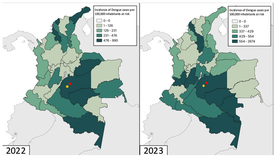
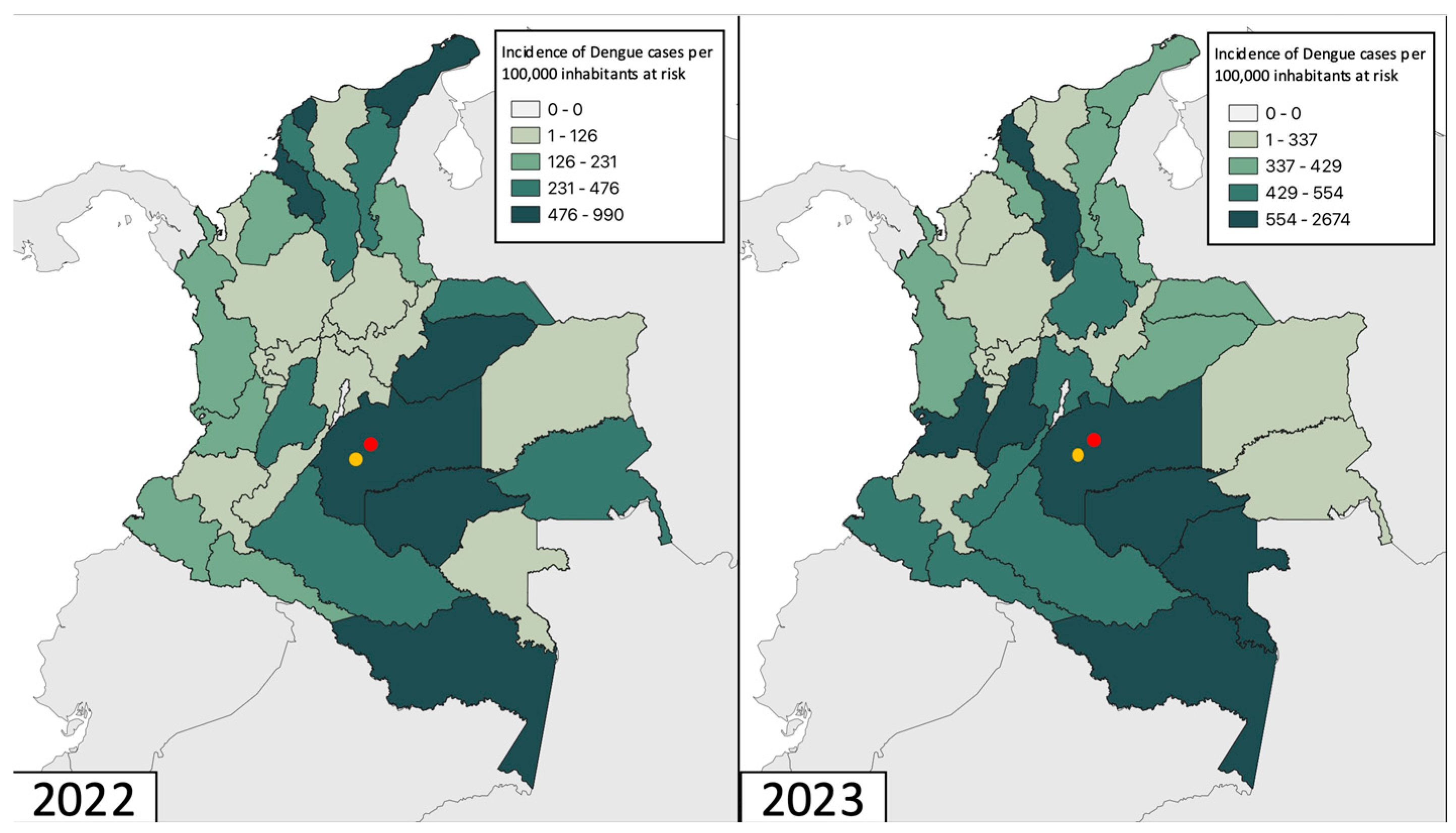
Diptera collection was carried out in two different municipalities (Fuente de Oro and Vista Hermosa) in the department of Meta, Colombia, one of the departments most affected by dengue during the last epidemic cycle in 2023, with an incidence of 1560 cases per 100,000 population at risk for Dengue (Figure 1). Sampling was conducted at selected sites and time points, providing a snapshot of pathogen circulation rather than continuous or longitudinal coverage. The municipality of Fuente de Oro has a total area of 576 km2, with 44.4% of the population living in rural areas and an incidence of 5152 [23]. On the other hand, the municipality of Vista Hermosa has a total area of 4749 km2, with 56.1% of the population living in rural areas and an incidence of dengue fever of 6207 [23]. Sampling sites were selected based on incidence values and strategic points of mosquito breeding. Non-probabilistic convenience sampling was conducted with the help of the Departmental Secretary of Meta on 31 October, 1 November 2022, and 17 and 18 May 2023 (rainy season). Black light traps were set for 24 h in the selected areas, and/or insects were caught manually during the day with mechanical vacuum cleaners. The most entomologically important insects from the family Culicidae and Psychodidae were selected at each sampling point and grouped into pools of 1 to 10 individuals, based on genus identification through morphological evaluation (Table S1). Insects were identified to the genus level due to logistical constraints associated with field collection, specimen preservation, and processing throughput. While species-level identification provides higher ecological resolution, genus-level identification is commonly used in surveillance studies to detect circulating pathogens across vector groups. Whole-body pools were analyzed to maximize detection sensitivity under field surveillance conditions. As a result, positive molecular detections may reflect the presence of viral RNA from recent blood meals rather than active infection or transmission competence. The Technical Research Committee at the University of Rosario in Bogotá, Colombia, with approval code DVO005 1585-CV1427 on 8 June 2021: “Molecular Surveillance of Vector Borne Diseases (VBDs) and Emerging Diseases in the Orinoco region”.
Figure 1.
Dengue incidence in Colombia during 2022–2023 and study sites. Map of Colombia, showing Dengue incidence data (reported cases per 100,000 population at risk). The sampled municipalities are highlighted with a red dot Fuente de Oro and the yellow dot indicates Vista Hermosa. The data used for these maps were obtained from Colombia’s Public Health Surveillance System (Sivigila) (Instituto Nacional de Salud, 2022–2023).
The samples were preserved in DNA/RNA shield (Zymo. R1100-50) at −4 °C in vials labelled with the coordinates and sampling site. Finally, the samples were transported to the Microbiology Laboratory of the Universidad del Rosario in Bogotá, Colombia, for processing and molecular analysis.
2.2. Nucleic Acids Extraction and cDNA Synthesis
Pooled and individual entomological material (Table S1) was homogenized using ZR BashingBeadTM lysis tubes (Lysis Tubes-ref. S6003-50) (Zymo Research, Irvine, CA, USA) with 200 μL DNA/RNA shield buffer at 30 rpm for 20 min in the TissueLyser II® tissue homogeniser (Qiagen, Hilden, Germany), followed by centrifugation at 10,000 rpm for 2 min. Nucleic acid extraction was performed from the supernatant obtained in the previous step using the Hamilton Microlab Star automated system and the MagBead Quick-DNA/Viral RNA Kit (Zymo Research, Irvine, CA, USA) according to the manufacturer’s recommended instructions. Once the DNA and RNA was obtained, its concentration and quality were quantified using the Nanodrop-2000 spectrophotometer (Thermo Fisher Scientific, Waltham, MA, USA) and stored at −80 °C. RT-PCR was then performed on the RNA to generate cDNA (complementary DNA) using the LunaScript RT SuperMix Reverse Transcriptase Kit enzyme (NEB #E3010) (New England Biolabs, Ipswich, MA, USA). The cDNA was stored at −30 °C.
2.3. Conventional Polymerase Chain Reaction for the Identification of Dengue Virus Serotypes and Plasmodium Species
Detection of DENV serotypes (DENV 1-4) was performed only in the mosquitoes of the genera belonging to the subfamily Culicinae by multiplex PCR using previously reported primers for the region of the C-prM gene (Table 1). PCR conditions were performed according to the previously described protocol [24].
Table 1.
List of primers and Taqman probes used in PCR for the detection of DENV serotypes DENV1–4, CHIKV, ZIKV, OROV, Plasmodium y Leishmania.
For Plasmodium, amplification of the 18S ribosomal RNA (18S rRNA) gene was carried out. PCR only identified in mosquitoes of the genus Anopheles, reactions were performed in a final volume of 12.5 μL containing 2× GoTaq® Colorless Master Mix (Promega, Madison, WI, USA), 10 μM of each primer (rPLU5 and rPLU6; Table 1) and 0.8 μL of cDNA. The thermal profile consisted of an initial denaturation cycle of 95 °C for 5 min, 25 cycles of 58 °C for 2 min, 72 °C for 2 min, 94 °C for 1 min, and finally a final extension cycle of 72 °C for 5 min. Plasmodium positive pools were processed to identify the species, Plasmodium falciparum and Plasmodium vivax. PCR reactions were performed in a final volume of 12.5 μL containing 2× GoTaq® Colorless Master Mix (Promega, # M7133), 10 μM of each primer (rFAL1/rVIV1 and rFAL2/rVIV2; Table 1) and 0.8 μL of cDNA. The thermal profile consisted of an initial denaturation cycle of 94 °C for 4 min, 35 cycles of 94 °C for 30 seg, 58 °C for 1 min, 72 °C for 1 min, and finally a final extension cycle of 72 °C for 4 min. Visualization of the gene fragments was carried out using 2% agarose gel electrophoresis in 1X TBE buffer and 1 μL of SYBR® Safe (Invitrogen®, Carlsbad, CA, USA) as an intercalating agent, then the gel was exposed to UV light to observe the amplification band of each, a band of 208 base pairs (bp) was observed for DENV-1, a band of 119 bp for DENV-2, a band of 288 bp for DENV-3, a band of 260 bp for DENV-4, a band of 1200 pb for Plasmodium, a band of 205 pb for Plasmodium falciparum and a band of 117 pb for Plasmodium vivax.
2.4. Real-Time Polymerase Chain Reaction for the Detection of Chikungunya, Zika, Oropouche and Leishmania
Identification of CHIKV, ZIKV, OROV and Leishmania was performed by real-time reverse transcription–polymerase chain reaction (qRT-PCR) using the appropriate primers and Taqman probes proposed previously (Table 1). The qRT-PCR enzyme used was PerfeCTa® qPCR ToughMixTM, Low ROXTM (Quantabio, Carlsbad, CA, USA). CHIKV, ZIKV y OROV was performed only in the mosquitoes of the genera belonging to the subfamily Culicinae. For CHIKV, 10 μM of each primer, 5 μM of probe and 1.2 μL of cDNA were used. The thermal profile consisted of an initial denaturation at 95 °C for 2 min, 40 cycles of 95 °C for 15 s, 55 °C for 45 s. For ZIKV, 10 μM of each primer, 25 μM of the probe and 1.2 μL of cDNA were used. Amplification conditions were: one cycle at 95 °C for 1 min, 40 cycles of 95 °C for 15 s and 56 °C for 1 min. For OROV, 20 μM of each primer, 20 μM of probe and 1.2 μL of cDNA were used. The amplification conditions were: one cycle of 95 °C for 1 min, 40 cycles of 95 °C for 15 s and 59 °C for 1 min. Detection of Leishmania was performed only in the insects of the genus Lutzomyia, 10 μM of each primer, 5 μM of probe and 1.2 μL of cDNA were used. The amplification conditions were: one cycle of 95 °C for 5 min, 40 cycles of 94 °C for 15 s and 60 °C for 1 min. To perform the real-time PCR, the cut-off Ct value was ≤39 based on several criteria, including the Centers for Disease Control and Prevention (CDC) interpretation criteria, where a sample was considered positive if primer sets showed amplification with cycle threshold (Ct) values ≤ 38.5. Similarly, positive tests were confirmed in duplicate. We used as positive controls Synthethic DNA provided by TWIST and as negative controls, samples previously tested as negative for these microorganisms.
2.5. Statistical Analysis
First, the results of DENV molecular detection were analyzed in terms of absolute and relative frequencies for the variables considered in the study (municipalities and vector genus). The frequency of molecular detection was calculated as the minimum infection rate (MIR), the ratio of the number of positive pools to the total number of individuals. The MIR was calculated for each mosquito genus for each of the DENV serotypes. Plots were constructed using the ggplot2 package included in the R software (RStudio Team, Boston, MA, USA, v.5.0), and maps were created using the Orange V3.37 software.
3. Results
3.1. Insect Diversity and Geographic Distribution
A total of 790 insects were captured at the two municipalities, of which 780 were processed in pools (n = 148) and 10 individually (Table S1). The pools were constructed by genera to assess the circulation of arboviruses and parasites at a general level in the study municipalities, rather than to characterize insect diversity. Eight mosquito genera were identified, Culex was the genus found in the highest abundance (51.1% n = 404; Table 2) followed by Aedes (50 pools, 31.6%), Psorophora (12 pools, 7.6%), Lutzomyia (7 pools, 4.4%), Anopheles (3 pools, 1.9%), Coquilletidia (2 pools, 1.3%), Mansonia (1 pool, 0.6%), and Limatus (1 pool, 0.6%) (Figure 2A and Table S1). By municipality, Aedes is the genus found in greatest abundance in Vista Hermosa (96% n = 48; Table 2), while in Fuente de Oro, Culex is the genus found in greatest abundance (87.8% n = 72; Table 2). Vista Hermosa has the highest diversity of mosquito genera captured, with six genera identified and the genera Anopheles, Limatus and Lutzomyia were found only at this municipality (Table 2).
Table 2.
Frequency of pools collected by genus and by each municipality sampled (Fuente de Oro and Vista Hermosa). Calculated MIR value for each DENV serotype by mosquito genus. Numbers in parentheses refer to percentage per pool.
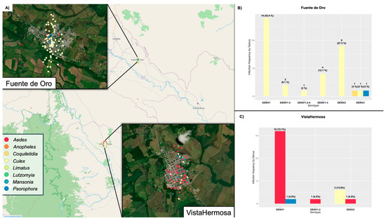
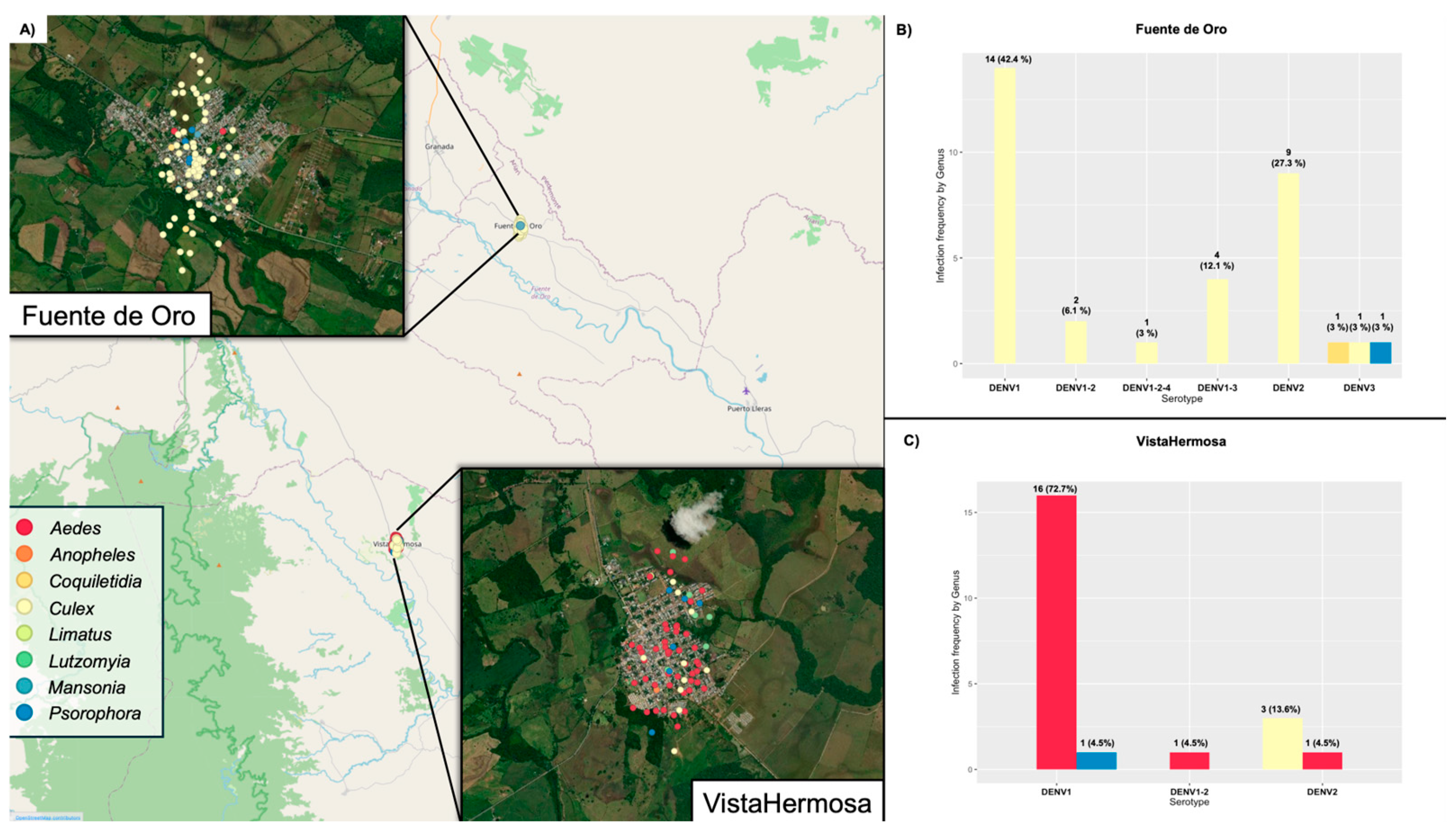
Figure 2.
Spatial distribution of insects and the frequency of molecular detection by DENV serotypes. (A) Map of the two sampling municipalities, Fuente de Oro and Vista Hermosa, showing the distribution of the captured insects and the frequency of molecular detection by DENV serotypes in (B) Fuente de Oro and (C) Vista Hermosa.
3.2. Arbovirus Detection and MIR of Dengue Viruses
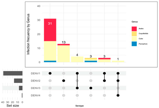
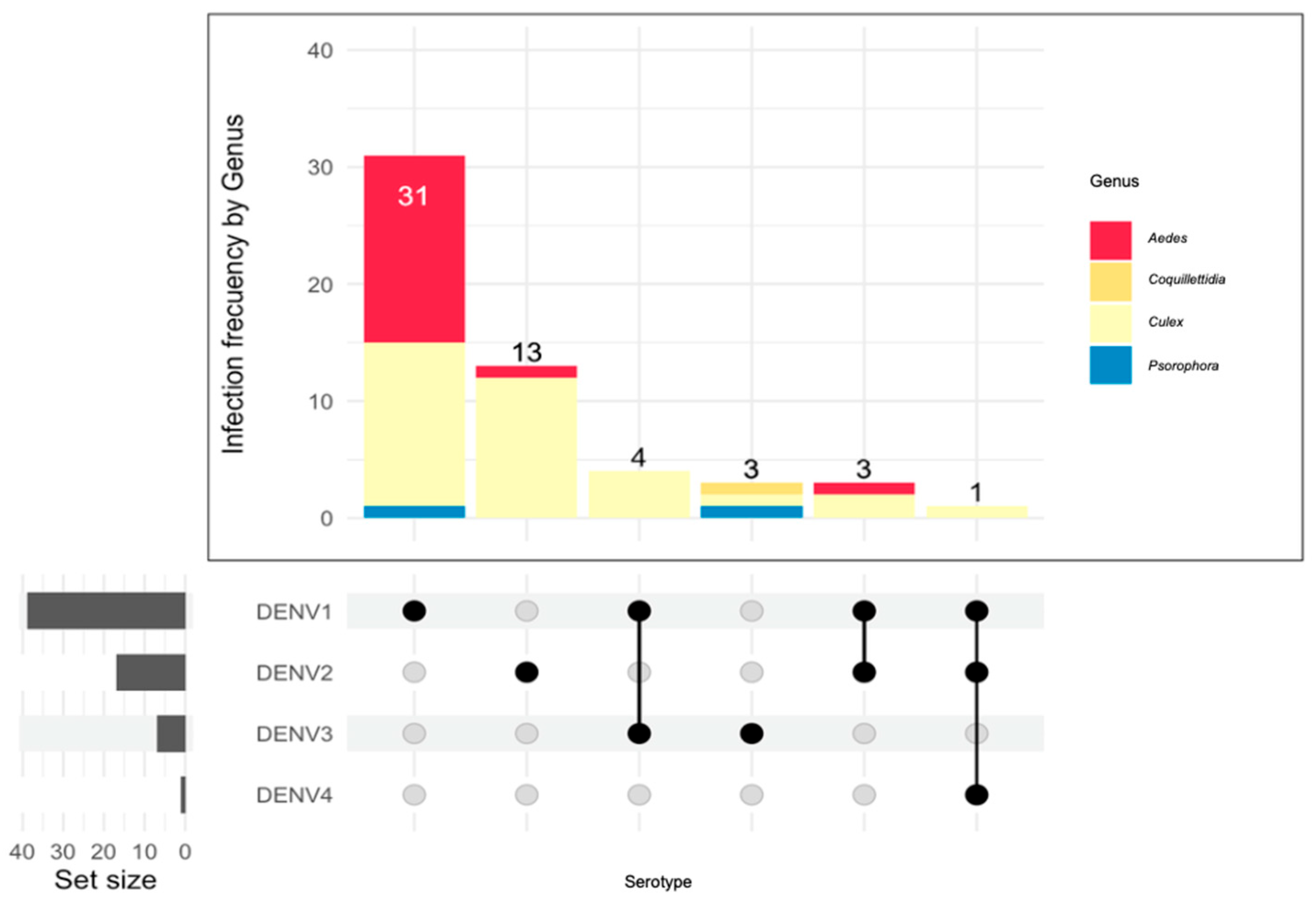
Of the 158 pools processed, the overall DENV molecular detection rate was 34.8% (55/158), while no infected pools were found for the other arboviruses (CHIKV, ZIKV, OROV). A higher rate of DENV molecular detection was observed in the genus Culex, most of which were found in Fuente de Oro. The most abundant DENV serotype was DENV1, present in 39 pools (24.68%;39/158) (Figure 3), mainly in pools of the genus Culex (53.8%;21/39), followed by the genus Aedes (43.6%;17/39) and one pool of the genus Psorophora (Figure 3). In the Culex genus pools all serotypes DENV1 to 4 were identified, whereas in the Aedes genus pools only DENV1 and 2 were identified. In the Psoropohora genus pools only DENV1 and 3 were identified and in the Coquilletidia genus only one pool infected with DENV3 was found (Figure 3). In the two municipalities of Fuente de Oro and Vista Hermosa, the serotype with the highest molecular detection rate was DENV1, but in Fuente de Oro it was found only in individuals of the genus Culex (21/33), whereas in Vista Hermosa it was found in the genus Aedes (17/22) and in the genus Psorophora (1/22) (Figure 2). The four serotypes were identified in Fuente de Oro and almost all co-infected pools with more than one serotype: DENV1-2 (1/2), DENV1-3 (4/4) and DENV1-2-4 (1). In Vista Hermosa, on the other hand, only serotypes DENV1 and 2 were identified, and only one pool of the Aedes genus was found to be co-infected with these two serotypes (Figure 2). In terms of MIR values, DENV1 has the highest values in the Culex and Aedes genera, the latter being the highest with a value of 62.96. DENV2 values were higher in the Culex genus (37.13) than in the Aedes genus. Finally, DENV4 was only identified in the genus Culex with a value of 2.48 (Table 2).
Figure 3.
Frequency of molecular detection and co-occurrence by Dengue serotypes in the mosquito genera collected.
3.3. Plasmodium and Leishmania Detection
A total of 43 individuals from the genus Lutzomyia were captured and processed in seven pools; however, none tested positive for Leishmania. On the other hand, 17 individuals of the genus Anopheles were captured and processed in 3 pools, two of which were positive for Plasmodium. The insects from these two pools were captured in the village of Campo Alegría, in the municipality of Vista Hermosa. One of the pools corresponds to Anopheles darlingi with 10 individuals positive for Plasmodium falciparum and the other to Anopheles rangeli with 4 individuals positive for Plasmodium vivax.
4. Discussion
The design of effective arbovirus prevention and vector control strategies relies on knowledge of the presence and baseline circulation of pathogens in their vectors and/or potential vectors. Entomovirological surveillance plays a central role in this process by providing essential information on the local transmission of arboviruses. In this study, we performed molecular detection of relevant arboviruses, Plasmodium, and Leishmania in insects collected from two municipalities, Fuente de Oro and Vista Hermosa, in the department of Meta, Colombia.
In this study, given the field-based nature of this study, we chose to identify insects at the genus level rather than species level due to morphological variability, field-based logistical constraints, and the high ecological redundancy observed among species within the same genus in tropical regions. This approach enables robust characterization of vector communities, particularly when the primary goal is to assess patterns of abundance, distribution, and relative dominance. The Culex genus, for instance, comprises numerous species that share key ecological traits such as generalist feeding behavior, tolerance to diverse habitats, and vector competence for pathogens like West Nile virus and Venezuelan equine encephalitis virus [31]. Moreover, previous studies have shown that genus-level trends often mirror epidemiological risk patterns in endemic regions and are commonly used in baseline or exploratory entomological surveillance [32]. Considering these factors—and given that in areas like Fuente de Oro, 44.4% of the population resides in rural settings with limited access to molecular identification tools or trained taxonomists—genus-level identification represents a practical and scientifically valid strategy for initial ecological assessments.
We first identified the genera of captured mosquitoes and found that Culex was the most abundant (51.1%; N = 82) (Table 2), representing more than half of all mosquitoes collected. This dominance was especially evident in Fuente de Oro, where 87.8% of mosquitoes belonged to the Culex genus. The high prevalence of Culex has also been documented in other studies, where species such as Cx. pipiens accounted for 75% of all mosquitoes captured [33]. Moreover, that study reported the presence of this species across three habitat types—urban, peri-urban, and rural—supporting the idea that most Culex species exhibit generalist behavior and a broad distribution range. This is consistent with our findings, as 44.4% of the population in Fuente de Oro resides in rural areas [34], which comprise diverse habitats (urban, peri-urban, and rural).
The same study also evaluated gene flow in Cx. pipiens across different habitats using ten microsatellite markers, revealing extensive gene flow both within and between ecological zones. Notably, urban environments neither promoted nor restricted gene flow among populations [33], suggesting continuous movement of Culex mosquitoes across habitats. This observation is relevant because certain Culex species have been implicated as vectors of arboviruses such as West Nile virus [3]. Given their high abundance and documented involvement in the transmission of some arboviruses, Culex mosquitoes may contribute to the maintenance of arbovirus circulation. However, this potential role remains speculative and requires confirmation through targeted entomovirological studies assessing vector competence and epidemiological relevance.
In our study, molecular detection revealed active circulation of DENV within the sampled communities, with an overall molecular detection rate of 34.8%. DENV1 was the most frequently detected serotype and was predominantly identified in Culex pools (53.8%). However, MIR estimates revealed a contrasting pattern: Aedes mosquitoes exhibited higher MIR values (MIR-DENV1: 62.96; Table 2) than Culex (MIR-DENV1: 51.98; Table 2). This suggests that Aedes individuals were more likely to be infected with DENV1 (6.3%), consistent with previous reports from Mexico where MIR-DENV values of up to 125 have been described for Aedes spp. [35]. These findings are in line with the well-established role of Aedes mosquitoes as the primary vectors of DENV, supported by their strong anthropophilic feeding behavior, in contrast to Culex species, which preferentially feed on non-human hosts such as domestic animals [36].
Interestingly, the MIR values observed for Culex in this study were higher than those reported in some previous surveillance efforts. For example, MIR values of 1.6 have been documented for Cx. quinquefasciatus in Mexico [19], and positive pools of Cx. (Melanoconion) vaxus (DENV2) and Cx. quinquefasciatus (DENV4) have been reported in Brazil [21,22]. These differences may partly reflect the inherent limitations of MIR calculations, which are influenced by pool size and composition and are not standardized across studies. As such, MIR values are more appropriately interpreted for within-study comparisons rather than direct cross-study evaluations, although they remain useful indicators of local arbovirus circulation [10,37]. Additional factors, such as differences in viral load or vector–virus interactions, may also influence detection. For instance, studies on Mayaro virus in Ae. aegypti have shown that low viral loads can limit molecular detection [38], underscoring the importance of pool composition and sample size.
Despite the lack of conclusive evidence supporting Culex mosquitoes as competent biological vectors of DENV, we observed relatively high molecular detection rates and MIR values in Culex pools (Table 2). Entomovirological surveillance has historically focused on Ae. aegypti and Ae. albopictus, while Culex species have received comparatively less attention. Experimental studies assessing the vector competence of Cx. quinquefasciatus for DENV have reported very low levels of viral replication, suggesting limited capacity for transmission [39]. Nevertheless, emerging evidence indicates that insect-specific viruses may modulate arbovirus molecular detection dynamics [40], and notable differences in microbiota—particularly the virome—have been observed between laboratory-reared and wild mosquito populations [41]. Moreover, field studies from northeastern Brazil have reported natural DENV2 and DENV3 molecular detections in Cx. quinquefasciatus pools during outbreaks [42]. While Culex species are not considered primary DENV vectors, these observations suggest they may act as incidental hosts or epidemiological indicators of viral circulation in settings where they coexist with established vectors. Collectively, our findings highlight the need for further integrated studies combining species-level identification, tissue-specific analyses, and experimental validation to clarify the role of Culex mosquitoes in arbovirus ecology.
With regard to Leishmania, no Lutzomyia specimens were found to be infected, which contrasts with the reported cutaneous leishmaniasis incidence in Meta (115.88 cases per 100,000 inhabitants at risk) [43]. However, disaggregated data show that the highest burden occurs in southern municipalities bordering Guaviare, the department with the highest incidence in Colombia in 2022 (961.02) [43]. By contrast, our collections were conducted in central Meta, where leishmaniasis prevalence is lower. Additionally, Lu. gomezi is the only Lutzomyia species reported in Meta, suggesting that either geographical or ecological barriers may limit its distribution into northern municipalities, or that no active transmission cycles were ongoing at the time of collection [43]. Broader and more systematic sampling will be necessary to test these hypotheses.
For malaria, the main Colombian vectors are An. darlingi, An. nuneztovari, and An. albimanus, in which natural detections with Plasmodium vivax and P. falciparum have been identified in departments such as Chocó, Valle, and Córdoba [44]. Our findings expand this knowledge by documenting 13 individuals (two pools) of An. darlingi circulating in Vista Hermosa, with one pool testing positive for P. falciparum. Although Meta reports a relatively low malaria incidence (2.7), the detection of this parasite in a primary vector poses a significant risk for local transmission. We also detected P. vivax in one pool of An. rangeli (four individuals). While considered a secondary vector in Colombia, An. rangeli has been reported as naturally infected with P. vivax in southern regions [45]. Together with our data, this suggests that non-primary Anopheles species should be further investigated to better understand their role in malaria circulation and transmission dynamics.
Our study has several limitations inherent to field-based entomological surveillance that should be considered when interpreting the results. First, the restricted spatial and temporal sampling effort, while valuable for baseline assessment, limits the generalizability of our findings beyond the specific study sites. Second, mosquitoes were identified at the genus level, which reduces ecological resolution and precludes species-specific inferences regarding the role of individual taxa in DENV transmission, particularly within the Culex complex.
In addition, molecular analyses were performed on whole-body mosquito pools rather than dissected tissues. Consequently, the detection of viral RNA may reflect recent blood meal ingestion rather than active infection, viral dissemination to salivary glands, or transmission competence. This limitation is further compounded by the absence of pre-processing classification by sex or gonotrophic status (e.g., blood-fed, gravid, unfed), which restricts our ability to distinguish between passive detection of pathogen nucleic acids and biologically meaningful infection. This consideration is especially relevant for Culex mosquitoes, which were highly abundant in the study area; therefore, positive detections within this genus should be interpreted cautiously, as they may primarily reflect numerical dominance rather than epidemiological significance.
Despite these constraints, the data provide original and informative baseline evidence of the local circulation of DENV, Plasmodium, and Leishmania in Fuente de Oro and Vista Hermosa. These findings highlight the utility of molecular surveillance for detecting pathogen circulation in endemic regions and underscore the need for future studies incorporating species-level identification, tissue-specific analyses, gonotrophic status assessment, and expanded longitudinal sampling to more accurately evaluate vectorial capacity and transmission dynamics. Such efforts will be essential for informing integrated surveillance frameworks and evidence-based vector control strategies in Colombia.
In conclusion, our study underscores the value of entomological surveillance and molecular tools in identifying pathogens circulating locally and driving vector-borne disease (VBD) transmission. Importantly, our results reveal the contribution of species not traditionally considered primary vectors to rural VBD transmission, potentially sustaining ongoing circulation. Integrative approaches that combine mosquito identification with the detection of natural arbovirus and parasite molecular detection may provide a powerful tool for characterizing local VBD transmission dynamics.
Supplementary Materials
The following supporting information can be downloaded at: https://www.mdpi.com/article/10.3390/epidemiologia7030076/s1, Table S1: Collection information and insect pools.
Author Contributions
P.U., D.M. (Davinzon Martínez) and C.A. designed the protocol and carried out the capture of the individuals. D.M. (David Martinez) performed the processing of the samples in the laboratory. D.M. (Davinzon Martínez) and J.D.R. drafted the manuscript. C.H., L.H.P., M.G.-R., M.M., N.P.-E., L.S.-L. and O.C.-B. contributed to interpretation and critical review. All authors have read and agreed to the published version of the manuscript.
Funding
This work was supported by the Ministry of Science, Technology, and Innovation of Colombia (MinCiencias) grant number 143889685192-2021.
Institutional Review Board Statement
The study was approved by the Technical Research Committee at the University of Rosario in Bogotá, Colombia, with approval code DVO005 1585-CV1427 on 8 June 2021.
Informed Consent Statement
Not applicable.
Data Availability Statement
Data is contained within the article or Supplementary Materials. The original contributions presented in this study are included in the article/Supplementary Materials. Further inquiries can be directed to the corresponding authors.
Acknowledgments
We thank the local health authorities from the Meta department for their support and assistance with the fieldwork.
Conflicts of Interest
The authors declare that the research was conducted in the absence of any commercial or financial relationships that could be construed as a potential conflict of interest.
References
- WHO. Vector Borne Diseases. Available online: https://www.who.int/news-room/fact-sheets/detail/vector-borne-diseases (accessed on 10 October 2024).
- Hoyos-López, R.; Suaza-Vasco, J.; Rúa-Uribe, G.; Uribe, S.; Gallego-Gómez, J.C. Molecular detection of flaviviruses and alphaviruses in mosquitoes (Diptera: Culicidae) from coastal ecosystems in the Colombian Caribbean. Mem. Inst. Oswaldo Cruz 2016, 111, 625–634. [Google Scholar] [CrossRef] [Scilit] [PubMed]
- Jiang, S.; Xing, D.; Li, C.; Dong, Y.; Zhao, T.; Guo, X. Replication and transmission of West Nile virus in simulated overwintering adults of Culex pipiens pallens (Diptera: Culicidae) in China. Acta Trop. 2023, 237, 106720. [Google Scholar] [CrossRef] [Scilit] [PubMed]
- Kampen, H.; Tews, B.A.; Werner, D. First Evidence of West Nile Virus Overwintering in Mosquitoes in Germany. Viruses 2021, 13, 2463. [Google Scholar] [CrossRef] [Scilit] [PubMed]
- Malaria. Available online: https://www.who.int/news-room/fact-sheets/detail/malaria (accessed on 10 October 2024).
- Mann, S.; Frasca, K.; Scherrer, S.; Henao-Martínez, A.F.; Newman, S.; Ramanan, P.; Suarez, J.A. A Review of Leishmaniasis: Current Knowledge and Future Directions. Curr. Trop. Med. Rep. 2021, 8, 121–132. [Google Scholar] [CrossRef] [Scilit]
- Ma, J.; Guo, Y.; Gao, J.; Tang, H.; Xu, K.; Liu, Q.; Xu, L. Climate Change Drives the Transmission and Spread of Vector-Borne Diseases: An Ecological Perspective. Biology 2022, 11, 1628. [Google Scholar] [CrossRef] [Scilit]
- Hoyos, J.; Carrasquilla, M.C.; León, C.; Montgomery, J.M.; Salyer, S.J.; Komar, N.; González, C. Host selection pattern and flavivirus screening of mosquitoes in a disturbed Colombian rainforest. Sci. Rep. 2021, 11, 18656. [Google Scholar] [CrossRef] [Scilit]
- de Abreu, F.V.S.; Ribeiro, I.P.; Ferreira-De-Brito, A.; dos Santos, A.A.C.; de Miranda, R.M.; Bonelly, I.d.S.; Neves, M.S.A.S.; Bersot, M.I.; dos Santos, T.P.; Gomes, M.Q.; et al. Haemagogus leucocelaenus and Haemagogus janthinomys are the primary vectors in the major yellow fever outbreak in Brazil, 2016–2018. Emerg. Microbes Infect. 2019, 8, 218–231. [Google Scholar] [CrossRef] [Scilit]
- Martínez, D.; Hernández, C.; Muñoz, M.; Armesto, Y.; Cuervo, A.; Ramírez, J.D. Identification of Aedes (Diptera: Culicidae) Species and Arboviruses Circulating in Arauca, Eastern Colombia. Front. Ecol. Evol. 2020, 8, 602190. [Google Scholar] [CrossRef] [Scilit]
- Gómez, M.; Martínez, D.; Hernández, C.; Luna, N.; Patiño, L.H.; Bohórquez Melo, R.; Suarez, L.A.; Palma-Cuero, M.; Murcia, L.M.; Páez, L.G.; et al. Arbovirus infection in Aedes aegypti from different departments of Colombia. Front. Ecol. Evol. 2022, 10, 999169. [Google Scholar] [CrossRef] [Scilit]
- Li, S.L.; Acosta, A.L.; Hill, S.C.; Brady, O.J.; de Almeida, M.A.B.; Cardoso, J.d.C.; Hamlet, A.; Mucci, L.F.; de Deus, J.T.; Iani, F.C.M.; et al. Mapping environmental suitability of Haemagogus and Sabethes spp. mosquitoes to understand sylvatic transmission risk of yellow fever virus in Brazil. PLoS Negl. Trop. Dis. 2022, 16, e0010019. [Google Scholar] [CrossRef] [Scilit]
- Mishra, A.K.; Bharti, P.K.; Vishwakarma, A.; Nisar, S.; Rajvanshi, H.; Sharma, R.K.; Saha, K.B.; Shukla, M.M.; Jayswar, H.; Das, A.; et al. A study of malaria vector surveillance as part of the Malaria Elimination Demonstration Project in Mandla, Madhya Pradesh. Malar. J. 2020, 19, 447. [Google Scholar] [CrossRef] [Scilit]
- Betson, M.; Jawara, M.; Awolola, T.S. Status of insecticide susceptibility in Anopheles gambiae s.l. from malaria surveillance sites in the Gambia. Malar. J. 2009, 8, 187. [Google Scholar] [CrossRef] [Scilit]
- Murillo, E.; Muskus, C.; Agudelo, L.A.; Vélez, I.D.; Ruiz-Lopez, F. A new high-resolution melting analysis for the detection and identification of Plasmodium in human and Anopheles vectors of malaria. Sci. Rep. 2019, 9, 1674. [Google Scholar] [CrossRef] [Scilit]
- Rojas, M.A.; Ahumada, M.L.; Velasco-Pareja, M.C.; Chaparro, P.; Yasnot-Acosta, M.F.; Orjuela, L.I. Anopheles species associated with malaria transmission in gold-mining areas of northwest Colombia. Malar. J. 2025, 24, 285. [Google Scholar] [CrossRef] [Scilit]
- Ahumada, M.L.; Orjuela, L.I.; Pareja, P.X.; Conde, M.; Cabarcas, D.M.; Cubillos, E.F.G.; Lopez, J.A.; Beier, J.C.; Herrera, S.; Quiñones, M.L. Spatial distributions of Anopheles species in relation to malaria incidence at 70 localities in the highly endemic Northwest and South Pacific coast regions of Colombia. Malar. J. 2016, 15, 407. [Google Scholar] [CrossRef] [Scilit]
- González, C.; León, C.; Paz, A.; López, M.; Molina, G.; Toro, D.; Ortiz, M.; Cordovez, J.M.; Atencia, M.C.; Aguilera, G.; et al. Diversity patterns, Leishmania DNA detection, and bloodmeal identification of Phlebotominae sand flies in villages in northern Colombia. PLoS ONE 2018, 13, e0190686. [Google Scholar] [CrossRef] [Scilit] [PubMed]
- Sandoval-Ramírez, C.M.; Hernández, C.; Teherán, A.A.; Gutierrez-Marin, R.; Martínez-Vega, R.A.; Morales, D.; Hoyos-Lopez, R.; Araque-Mogollón, A.; Ramírez, J.D. Complex ecological interactions across a focus of cutaneous leishmaniasis in Eastern Colombia: Novel description of Leishmania species, hosts and phlebotomine fauna. R. Soc. Open Sci. 2020, 7, 200266. [Google Scholar] [CrossRef] [Scilit] [PubMed]
- Caviedes-Triana, K.; Duque-Granda, D.; Cadavid-Restrepo, G.; Moreno-Herrera, C.X.; Vivero-Gomez, R. Taxonomic, molecular and ecological approach reveals high diversity of vector sand flies, varied blood source supply and a high detection rate of Leishmania DNA in Colombian Amazon region. PLoS Negl. Trop. Dis. 2025, 19, e0013445. [Google Scholar] [CrossRef] [Scilit]
- Padilla, J.C.; Lizarazo, F.E.; Murillo, O.L.; Mendigaña, F.A.; Pachón, E.; Vera, M.J. Epidemiología de las principales enfermedades transmitidas por vectores en Colombia, 1990–2016. Biomédica 2017, 37, 27. [Google Scholar] [CrossRef] [Scilit]
- Padilla-Rodríguez, J.C. Panorama epidemiológico de las enfermedades transmitidas por vectores: Lecciones aprendidas y retos para romper el círculo. Biomédica 2023, 43, 422–426. [Google Scholar] [CrossRef] [Scilit] [PubMed]
- Secretaria del Meta. Boletin Epidemiologico Departamento de Meta. 2024. Available online: https://devx.meta.gov.co/media/centrodocumentacion/2023/04/21/Boletin_Eventos_I_Trimestre_2023-SSM.pdf (accessed on 1 November 2025).
- Martínez, D.; Gómez, M.; Hernández, C.; Campo-Palacio, S.; González-Robayo, M.; Montilla, M.; Pavas-Escobar, N.; Tovar-Acero, C.; Geovo-Arias, L.; Valencia-Urrutia, E.; et al. Cryptic transmission and novel introduction of Dengue 1 and 2 genotypes in Colombia. Virus Evol. 2024, 10, veae068. [Google Scholar] [CrossRef] [Scilit] [PubMed]
- Chien, L.-J.; Liao, T.-L.; Shu, P.-Y.; Huang, J.-H.; Gubler, D.J.; Chang, G.-J.J. Development of real-time reverse transcriptase PCR assays to detect and serotype dengue viruses. J. Clin. Microbiol. 2006, 44, 1295–1304. [Google Scholar] [CrossRef] [Scilit]
- Singh, B.; Bobogare, A.; Cox-Singh, J.; Snounou, G.; Abdullah, M.S.; Rahman, H.A. A genus- and species-specific nested polymerase chain reaction malaria detection assay for epidemiologic studies. Am. J. Trop. Med. Hyg. 1999, 60, 687–692. [Google Scholar] [CrossRef] [Scilit] [PubMed]
- Filgueira, C.P.B.; Moreira, O.C.; Cantanhêde, L.M.; de Farias, H.M.T.; Porrozzi, R.; Britto, C.; Boité, M.C.; Cupolillo, E. Comparison and clinical validation of qPCR assays targeting Leishmania 18S rDNA and HSP70 genes in patients with American Tegumentary Leishmaniasis. PLoS Negl. Trop. Dis. 2020, 14, e0008750. [Google Scholar] [CrossRef] [Scilit]
- Álvarez-Díaz, D.A.; Valencia-Álvarez, E.; Rivera, J.A.; Rengifo, A.C.; Usme-Ciro, J.A.; Peláez-Carvajal, D.; Lozano-Jiménez, Y.Y.; Torres-Fernández, O. An updated RT-qPCR assay for the simultaneous detection and quantification of chikungunya, dengue and zika viruses. Infect. Genet. Evol. 2021, 93, 104967. [Google Scholar] [CrossRef] [Scilit]
- Faye, O.; Faye, O.; Diallo, D.; Diallo, M.; Weidmann, M.; Sall, A.A. Quantitative real-time PCR detection of Zika virus and evaluation with field-caught mosquitoes. Virol. J. 2013, 10, 311. [Google Scholar] [CrossRef] [Scilit]
- Rojas, A.; Stittleburg, V.; Cardozo, F.; Bopp, N.; Cantero, C.; López, S.; Bernal, C.; Mendoza, L.; Aguilar, P.; Pinsky, B.A.; et al. Real-time RT-PCR for the detection and quantitation of Oropouche virus. Diagn. Microbiol. Infect. Dis. 2020, 96, 114894. [Google Scholar] [CrossRef] [Scilit]
- Farajollahi, A.; Fonseca, D.M.; Kramer, L.D.; Kilpatrick, A.M. “Bird Biting” Mosquitoes and Human Disease: A Review of the Role of Culex Pipiens Complex Mosquitoes In Epidemiology. Infect. Genet. Evol. 2011, 11, 1577–1585. [Google Scholar] [CrossRef] [Scilit]
- Rochlin, I.; Harding, K.; Ginsberg, H.S.; Campbell, S.R. Comparative analysis of distribution and abundance of West Nile and eastern equine encephalomyelitis virus vectors in Suffolk County, New York, using human population density and land use/cover data. J. Med. Entomol. 2008, 45, 563–571. [Google Scholar] [CrossRef]
- Honnen, A.-C.; Monaghan, M.T. City-Dwellers and Country Folks: Lack of Population Differentiation Along an Urban–Rural Gradient in the Mosquito Culex pipiens (Diptera: Culicidae). J. Insect Sci. 2017, 17, 107. [Google Scholar] [CrossRef] [Scilit] [PubMed]
- DANE—Censo General 2005 [Internet]. Available online: https://www.dane.gov.co/index.php/estadisticas-por-tema/demografia-y-poblacion/censo-general-2005-1#estimaciones-demograficas-linea-base-2005 (accessed on 23 September 2024).
- Izquierdo-Suzán, M.; Zavala-Guerrero, P.B.; Mendoza, H.; Portela Salomão, R.; Vázquez-Pichardo, M.; Von Thaden, J.J.; Medellín, R.A. Mosquito (Diptera: Culicidae) diversity and arbovirus detection across an urban and agricultural landscape. Acta Trop. 2024, 257, 107321. [Google Scholar] [CrossRef] [Scilit] [PubMed]
- Olson, M.F.; Ndeffo-Mbah, M.L.; Juarez, J.G.; Garcia-Luna, S.; Martin, E.; Borucki, M.K.; Frank, M.; Estrada-Franco, J.G.; Rodríguez-Pérez, M.A.; Fernández-Santos, N.A.; et al. High Rate of Non-Human Feeding by Aedes aegypti Reduces Zika Virus Transmission in South Texas. Viruses 2020, 12, 453. [Google Scholar] [CrossRef] [Scilit]
- Gu, W.; Lampman, R.; Novak, R.J. Assessment of arbovirus vector infection rates using variable size pooling. Med. Vet. Entomol. 2004, 18, 200–204. [Google Scholar] [CrossRef] [Scilit]
- Maia, L.M.S.; Bezerra, M.C.F.; Costa, M.C.S.; Souza, E.M.; Oliveira, M.E.B.; Ribeiro, A.L.M.; Miyazaki, R.D.; Slhessarenko, R.D. Natural vertical infection by dengue virus serotype 4, Zika virus and Mayaro virus in Aedes (Stegomyia) aegypti and Aedes (Stegomyia) albopictus. Med. Vet. Entomol. 2019, 33, 437–442. [Google Scholar] [CrossRef] [Scilit]
- Vazeille-Falcoz, M.; Rosen, L.; Mousson, L.; Rodhain, F. Replication of dengue type 2 virus in Culex quinquefasciatus (Diptera: Culicidae). Am. J. Trop. Med. Hyg. 1999, 60, 319–321. [Google Scholar] [CrossRef] [Scilit] [PubMed]
- Gómez, M.; Martínez, D.; Páez-Triana, L.; Luna, N.; Ramírez, A.; Medina, J.; Cruz-Saavedra, L.; Hernández, C.; Castañeda, S.; Melo, R.B.; et al. Influence of dengue virus serotypes on the abundance of Aedes aegypti insect-specific viruses (ISVs). J. Virol. 2024, 98, e01507-23. [Google Scholar] [CrossRef] [Scilit] [PubMed]
- Liu, Q.; Cui, F.; Liu, X.; Fu, Y.; Fang, W.; Kang, X.; Lu, H.; Li, S.; Liu, B.; Guo, W.; et al. Association of virome dynamics with mosquito species and environmental factors. Microbiome 2023, 11, 101. [Google Scholar] [CrossRef] [Scilit] [PubMed]
- Krokovsky, L.; Paiva, M.H.S.; Guedes, D.R.D.; Barbosa, R.M.R.; De Oliveira, A.L.S.; Anastácio, D.B.; Pontes, C.R.; Ayres, C.F.J. Arbovirus Surveillance in Field-Collected Mosquitoes from Pernambuco-Brazil, During the Triple Dengue, Zika and Chikungunya Outbreak of 2015–2017. Front. Trop. Dis. 2022, 3, 875031. [Google Scholar] [CrossRef] [Scilit]
- Ferro, C.; López, M.; Fuya, P.; Lugo, L.; Cordovez, J.M.; González, C. Spatial Distribution of Sand Fly Vectors and Eco-Epidemiology of Cutaneous Leishmaniasis Transmission in Colombia. PLoS ONE 2015, 10, e0139391. [Google Scholar] [CrossRef] [Scilit]
- Montoya-Lerma, J.; Solarte, Y.A.; Giraldo-Calderón, G.I.; Quiñones, M.L.; Ruiz-López, F.; Wilkerson, R.C.; González, R. Malaria vector species in Colombia: A review. Mem. Inst. Oswaldo Cruz 2011, 106, 223–238. [Google Scholar] [CrossRef] [Scilit]
- Méndez, C.; Calderón, J. Malaria 2020. Informe de Evento. Available online: https://www.ins.gov.co/buscador-eventos/Informesdeevento/MALARIA_2020.pdf (accessed on 1 November 2025).

Disclaimer/Publisher’s Note: The statements, opinions and data contained in all publications are solely those of the individual author(s) and contributor(s) and not of MDPI and/or the editor(s). MDPI and/or the editor(s) disclaim responsibility for any injury to people or property resulting from any ideas, methods, instructions or products referred to in the content. |
© 2026 by the authors. Licensee MDPI, Basel, Switzerland. This article is an open access article distributed under the terms and conditions of the Creative Commons Attribution (CC BY) license.