


Abstract
Retinol-binding protein 4 (RBP4) is the primary carrier of retinol in the circulation and is produced mainly by the liver. Interest in RBP4 expanded after reports of elevated circulating levels in insulin-resistant mice and some human populations, leading to its proposal as an adipokine, an immunometabolic mediator, and a biomarker of obesity-related metabolic disease. However, subsequent research has revealed that its biology is considerably more complex. The metabolic effects of RBP4 are influenced by multiple factors, including its tissue of origin, retinol-binding status, association with transthyretin, receptor interactions, renal clearance, and the surrounding inflammatory environment. This review summarizes current evidence linking RBP4 to insulin resistance, adipose tissue dysfunction, and type 2 diabetes, while distinguishing classical retinol-bound holo-RBP4–dependent signaling through STRA6 (stimulated by retinoic acid 6) from retinol- and STRA6-independent mechanisms involved in innate and adaptive immune activation. We also discuss the transcriptional regulation of RBP4, focusing on PPARγ (peroxisome proliferator-activated receptor γ), HMGA1 (high mobility group A1 protein), and HIF-1α (hypoxia-inducible factor 1α)-related hypoxia-responsive pathways. Available evidence suggests that these factors do not operate within a simple HIF-1α-PPARγ-RBP4 signaling axis but rather participate in interconnected regulatory networks that shape adipocyte differentiation, secretory function, and metabolic adaptation. Although higher circulating RBP4 concentrations have been associated with adverse metabolic traits and an increased risk of type 2 diabetes, their clinical interpretation remains challenging because of the influence of renal function, vitamin A status, molecular heterogeneity, and methodological differences between assays. Pharmacological approaches to lower RBP4 are now feasible, but whether systemic reduction is beneficial remains uncertain. Targeting the specific pathogenic actions of RBP4, while preserving its essential role in retinol transport, may offer a more effective therapeutic strategy for metabolic disease.
1. Introduction
Obesity-associated insulin resistance is a complex disorder that cannot be explained by impaired glucose metabolism alone. Rather, it results from coordinated alterations across multiple organs, including adipose tissue, liver, skeletal muscle, pancreatic β-cells, and the immune system. Among these, adipose tissue plays a central role because its expansion is accompanied by profound changes in lipid handling, extracellular matrix remodeling, vascularization, immune-cell infiltration, and endocrine signaling. These alterations reshape systemic metabolic homeostasis and contribute to the development of chronic low-grade inflammation. Within this context, retinol-binding protein 4 (RBP4) has emerged as an important candidate linking vitamin A metabolism with metabolic and inflammatory regulation [1,2,3,4,5]. RBP4 was initially characterized as the major plasma carrier responsible for transporting retinol from hepatic stores to peripheral tissues [6,7]. Interest in its metabolic role increased after reports showing elevated circulating RBP4 levels in insulin-resistant mice and in several cohorts of individuals with obesity or type 2 diabetes, leading to the proposal that RBP4 functions as an adipokine that promotes systemic insulin resistance [1,2]. Subsequent studies, however, have revealed a more nuanced picture. Although adipocytes express RBP4, hepatocytes are now recognized as the principal source of circulating protein, as demonstrated by tissue-specific mouse models [8]. In addition, moderate liver-specific overexpression of RBP4 failed to impair glucose tolerance or insulin sensitivity in one experimental model [9], suggesting that circulating RBP4 concentration alone is insufficient to drive metabolic dysfunction. These findings indicate that the biological effects of RBP4 depend not only on its circulating levels but also on its tissue of origin, molecular form, and mechanisms of action [4,8,9]. Beyond its established role in retinol transport, RBP4 can activate receptor-dependent signaling pathways and modulate inflammatory responses through mechanisms that are either dependent on, or independent of, classical retinoid transport [10,11,12,13]. Interpretation of clinical studies is further complicated by factors such as renal function, vitamin A status, transthyretin binding, the relative proportions of apo- and holo-RBP4, and differences in analytical methods used to quantify the protein [4,14,15,16,17].
This review considers the complex role of RBP4 in metabolic disease. Although RBP4 is often described as a biomarker of metabolic risk or a direct contributor to insulin resistance and inflammation, its effects may depend on where it is produced, the molecular form in which it circulates, and the pathways through which it acts. Drawing on findings from experimental models, human genetic and clinical studies, and therapeutic research, we first describe the development of insulin resistance and adipose tissue dysfunction. We then examine the structure and distribution of RBP4, its molecular forms, and its roles in cell signaling, immune regulation, and gene expression. Particular attention is given to its relationship with peroxisome proliferator-activated receptor γ (PPARγ), high mobility group A1 protein (HMGA1), and hypoxia-inducible factor 1α (HIF-1α). We conclude by considering the clinical relevance of RBP4 and whether it represents a viable therapeutic target in obesity-related metabolic dysfunction.
Literature Search Strategy
A narrative literature search was manually performed using PubMed/MEDLINE, Scopus, Web of Science, and ClinicalTrials.gov to identify studies relevant to RBP4 biology and obesity-associated metabolic dysfunction. Searches included studies published from database inception to July 2026 and combined the terms “retinol-binding protein 4”, “RBP4”, “obesity”, “insulin resistance”, “type 2 diabetes”, “adipose tissue”, “retinol”, “transthyretin”, “STRA6”, “inflammation”, “TLR2”, “TLR4”, “NLRP3”, “PPARγ”, “HMGA1”, “hypoxia”, and “therapeutic targeting”.
English-language original studies, clinical trials, authoritative reviews, meta-analyses, and relevant ClinicalTrials.gov records were prioritized when they informed RBP4 structure and function, metabolic phenotypes, clinical measurement, or therapeutic targeting. Reference lists of key articles were also screened to identify additional relevant studies. Experimental studies were selected to illustrate mechanistic evidence, tissue-specific effects, and conflicting causal findings, whereas human studies were selected to represent different populations, study designs, analytical methods, and major confounders, when available. Relevant findings from the authors’ previous original research were also incorporated when they directly contributed to the mechanistic or interpretative framework of the review.
Before addressing RBP4 biology in detail, the following section briefly outlines the pathophysiological context of obesity-associated insulin resistance and adipose tissue dysfunction.
2. Insulin Resistance and Adipose Tissue Dysfunction
2.1. Molecular Basis and Progression of Insulin Resistance
Insulin resistance is defined as a reduced responsiveness of target tissues to the metabolic actions of insulin and typically develops years before overt type 2 diabetes. In the early stages, pancreatic β-cells compensate by increasing insulin secretion, allowing blood glucose concentrations to remain within the normal range. As insulin resistance worsens, this compensatory response gradually fails, resulting in persistent hyperglycemia and the onset of type 2 diabetes [18,19]. At the cellular level, insulin binding triggers autophosphorylation of the insulin receptor and recruitment of insulin receptor substrates (IRS), leading to activation of the phosphoinositide 3-kinase (PI3K)-Akt signaling pathway. Akt is a central regulator of insulin actions, promoting glucose transporter 4 (GLUT4) translocation and glucose uptake in skeletal muscle and adipose tissue while coordinating glycogen synthesis, lipid metabolism, protein synthesis, and suppression of hepatic glucose production [20,21].
Obesity disrupts this finely regulated signaling cascade through a combination of metabolic and inflammatory stress. Stress-responsive kinases, including c-Jun N-terminal kinase (JNK) and IκB kinase (IKK), phosphorylate IRS proteins at inhibitory sites, weakening PI3K-Akt signaling and reducing insulin-stimulated glucose uptake. As a consequence, insulin becomes less effective at suppressing lipolysis in adipose tissue and glucose production in the liver [22,23,24,25,26]. Additional factors, including lipotoxicity, oxidative stress, mitochondrial dysfunction, and endoplasmic reticulum stress, further impair insulin action and contribute to the development of dyslipidemia, metabolic dysfunction-associated steatotic liver disease, and cardiovascular complications [19,27,28]. Insulin sensitivity is also influenced by transcriptional control of the insulin receptor. HMGA1 promotes assembly of the transcriptional machinery at the insulin receptor (INSR) promoter [29,30]. More specifically, HMGA1 is part of a transcriptionally active multiprotein complex at the INSR promoter, where it cooperates with Sp1 and C/EBPβ. Through direct interactions with these transcription factors, HMGA1 facilitates their binding to the promoter and enhances their transcriptional synergy, thereby supporting the full activation of the INSR and other genes involved in glucose metabolism [31]. Reduced HMGA1 expression or activity decreases INSR abundance, thereby limiting cellular responsiveness to insulin [30,32]. This mechanism is particularly relevant in the context of RBP4, as HMGA1 also directly regulates transcription of the RBP4 gene [33,34].
2.2. Adipose Tissue Expansion and Metabolic Dysfunction
Adipose tissue is now recognized as far more than a passive reservoir for energy storage. It is a highly active metabolic and endocrine organ that regulates lipid handling, insulin sensitivity, and communication with other tissues through a complex secretome composed of proteins, lipids, metabolites, cytokines, and extracellular vesicles [35,36]. Healthy expansion of adipose tissue depends on coordinated adipogenesis, angiogenesis, extracellular matrix remodeling, and sufficient capacity to store triglycerides. During obesity, especially within visceral fat depots, these adaptive processes become progressively impaired. As a result, tissue growth occurs mainly through hypertrophy of existing adipocytes rather than the formation of new ones. Enlarged adipocytes respond poorly to insulin and fail to adequately suppress lipolysis, leading to increased release of non-esterified fatty acids and glycerol. The excess lipid flux promotes ectopic fat deposition in the liver and skeletal muscle, contributing to systemic insulin resistance [35,36,37,38].
As adipocytes become stressed, the surrounding tissue also undergoes marked remodeling. Extracellular matrix deposition increases, damaged adipocytes accumulate, and immune cells are progressively recruited [39]. Macrophages surround stressed or dying adipocytes and engage in continuous crosstalk with them through cytokines, chemokines, fatty acids, and reactive oxygen species [35]. Over time, these interactions shift adipose tissue from an efficient energy-buffering organ to a chronic source of inflammatory and metabolic signals that promote whole-body insulin resistance [26].
2.3. Adipose Tissue Hypoxia and HIF-1-Dependent Remodeling
Among the mechanisms contributing to adipose tissue dysfunction, hypoxia has emerged as an important driver of maladaptive remodeling in obesity. As adipocytes enlarge, oxygen diffusion becomes less efficient, while angiogenesis and tissue perfusion may fail to keep pace with tissue expansion. Local oxygen availability may be further affected by changes in mitochondrial activity and oxygen consumption. Adipose tissue hypoxia should therefore be understood as a dynamic imbalance between oxygen supply and metabolic demand, rather than solely as a consequence of reduced capillary density [39,40,41,42,43]. Reduced oxygen availability stabilizes hypoxia-inducible factor 1-alpha (HIF-1α) by preventing its hydroxylation-dependent degradation, thereby enabling its nuclear accumulation and the activation of hypoxia-responsive genes. Although transient HIF-1 activation supports adaptive responses to reduced oxygen tension, persistent activation promotes extracellular matrix deposition, fibrosis, inflammatory signaling, and impaired adipocyte differentiation, ultimately contributing to adipose tissue dysfunction and insulin resistance [44,45,46,47]. In line with these effects, proteomic studies have shown that hypoxia extensively remodels the adipocyte secretome, altering proteins involved in extracellular matrix organization, lipid metabolism, and insulin sensitivity [48]. Importantly, the metabolic consequences of adipose tissue hypoxia extend beyond canonical HIF-1 signaling. Hypoxia can also reshape adipocyte transcriptional and post-transcriptional programs through HIF-1α-associated nuclear regulators, including HMGA1, as well as through mechanisms such as miR-128-mediated regulation of insulin receptor expression [48,49]. Together, these findings indicate that adipose tissue hypoxia acts through multiple interconnected pathways rather than through a single transcriptional program.
2.4. Potential Links Between Hypoxia and RBP4
The broad effects of hypoxia on adipocyte differentiation, inflammation, and secretory function suggest that it may also influence RBP4 biology. However, direct transcriptional regulation of RBP4 by HIF-1α has not been demonstrated. To date, neither HIF-1α binding to the RBP4 promoter nor a functional hypoxia-response element controlling RBP4 transcription has been conclusively identified. Instead, hypoxia is more likely to influence RBP4 indirectly by modifying adipocyte differentiation, PPARγ-dependent transcriptional programs, inflammatory pathways, or the overall composition of the adipose secretome. Importantly, inflammatory signaling does not necessarily increase RBP4 expression. In human adipocytes, both tumor necrosis factor alpha (TNF-α) and interleukin-1 beta (IL-1β) have been reported to suppress RBP4 production, underscoring the complexity of its regulation [50,51].
HMGA1 represents a possible point of convergence between these pathways. Previous work from our group demonstrated that HMGA1 binds regulatory regions of the RBP4 gene and is required for both basal and cAMP-induced transcription [33,34]. Given that HMGA1 also cooperates with HIF-1 in regulating selected hypoxia-responsive genes, it is plausible that hypoxia alters the transcriptional environment in which RBP4 is expressed. Nevertheless, direct evidence for an HIF-1α-HMGA1 complex controlling RBP4 transcription is currently lacking. Taken together, the available data support a model in which hypoxia shapes a metabolic, inflammatory, and transcriptional milieu capable of influencing RBP4 expression or function through indirect, context-dependent mechanisms, rather than through a direct linear HIF-1α-RBP4 signaling pathway.
3. RBP4: From Retinol Carrier to Metabolic and Immune Signal
The preceding sections highlight how adipose tissue dysfunction is characterized by altered lipid metabolism, chronic inflammation, and remodeling of the adipose secretome. Within this context, RBP4 has attracted considerable attention because it serves not only as the principal carrier of retinol but also as an active regulator of metabolic and immune signaling. These functions are closely interconnected, as the biological activity of RBP4 depends on both its molecular form and its tissue of origin.
3.1. Genetics, Structure, Molecular Forms and Physiological Function
The human RBP4 gene is located on chromosome 10q23.33 [52]. Its transcription is regulated by a complex 5′-flanking promoter region containing multiple cis-regulatory elements that support basal, cell-specific, and signal-responsive expression [53,54]. Functional studies have identified enhancer, negative regulatory, and promoter elements within this region [53,54]. In addition, the RBP4 promoter contains retinoic acid-responsive elements (RAREs) separated by an intervening GC-rich regulatory sequence, which may contribute to the cooperative assembly or stabilization of transcriptional complexes required for retinoid sensitivity [55]. Furthermore, its basal and inducible activity can be modulated by metabolic and hormonal signals, including cAMP-HMGA1-dependent mechanisms [33,34]. Regulatory genetic variation and additional cis-regulatory regions may further influence RBP4 transcriptional activity [56,57]. The encoded protein, RBP4, is synthesized as a 201-amino-acid precursor containing an N-terminal signal peptide. After cleavage of this signal peptide, the mature secreted protein consists of 183 amino acids and has a molecular mass of approximately 21 kDa [4,6]. As a member of the lipocalin family, RBP4 adopts the characteristic eight-stranded antiparallel β-barrel that forms a hydrophobic binding pocket for retinol, allowing efficient transport of this poorly water-soluble vitamin through the circulation [58,59]. In hepatocytes, retinol mobilized from hepatic stores binds RBP4 before being released into the bloodstream [7].
Once in circulation, retinol-bound holo-RBP4 forms a complex with transthyretin [3,14], increasing its effective molecular size and reducing renal filtration [14,58]. Retinol-free apo-RBP4 also circulates but differs from holo-RBP4 in stability, clearance, receptor interactions, and signaling properties [4]. In plasma, retinol-bound holo-RBP4 associates with transthyretin (TTR), increasing the effective molecular size of the complex and limiting renal filtration [3,4,14]. Retinol-free apo-RBP4 also circulates, but it binds TTR less efficiently and is cleared more readily than holo-RBP4 [14]. These molecular differences are biologically relevant because holo-RBP4 is the principal form involved in retinol transport and the best-characterized ligand for STRA6-mediated retinol uptake and JAK2/STAT signaling [10,60]. By contrast, retinol- and STRA6-independent inflammatory effects of RBP4 have been described in macrophage–adipocyte systems [11,13]. Therefore, the biological effects attributed to RBP4 should be interpreted in relation to its molecular form and the experimental system used. Total circulating RBP4 is therefore an incomplete descriptor of RBP4 biology. Renal dysfunction can increase apo-RBP4, holo-RBP4, and C-terminally truncated RBP4 species [61], while circulating RBP4 and TTR concentrations may vary with age and sex even in metabolically healthy individuals [62]. Accordingly, measurement of total RBP4 may not accurately reflect the abundance of the molecular forms most relevant to metabolic signaling. Because molecular-form information is rarely available in metabolic cohorts, this limitation complicates causal interpretation and the development of form-selective therapeutic strategies, as discussed in the following sections.
3.2. Tissue Sources and Regulation of RBP4
The liver is the principal source of circulating RBP4, with hepatocytes coordinating its synthesis and secretion according to retinol availability [7,8]. Tissue-specific knockout studies in mice indicate that hepatocytes account for the majority of circulating RBP4 under physiological conditions [8]. Adipose tissue also expresses and secretes RBP4, although its contribution to plasma concentrations appears relatively small and may become more relevant under conditions of metabolic stress [8,63,64]. Interest in adipose-derived RBP4 began with the landmark study by Yang et al., which identified RBP4 as an adipokine whose expression increased in insulin-resistant mice and whose manipulation altered systemic insulin sensitivity [1]. Subsequent studies confirmed that adipocytes produce RBP4 [65,66] but also revealed marked variability depending on species, adipose depot, differentiation state, obesity, and overall metabolic health [63,65,67].
Compartmentalization is central to the interpretation of RBP4 biology. Hepatic RBP4 primarily sustains systemic retinol transport, whereas adipocyte-derived RBP4 may act mainly through local autocrine or paracrine mechanisms that influence adipose metabolism and inflammation [63,64]. This tissue specificity is further supported by evidence from brown adipose tissue (BAT), where RBP4 overexpression enhanced adrenergic signaling, thermogenesis, energy expenditure, and glucose homeostasis in mice [68]. The molecular mechanisms regulating RBP4 expression also differ between the liver and adipose tissue (Figure 1). In hepatocytes, retinol availability and fasting-related pathways involving cAMP, glucagon, and forkhead box O1 (FOXO1) play major regulatory roles [33,34,69]. HMGA1 further enhances both basal and cAMP-induced transcription by promoting assembly of a transcriptional complex at the RBP4 promoter [33,34]. In adipocytes, RBP4 expression is influenced by differentiation status, nuclear receptor signaling, insulin sensitivity, and inflammatory stimuli. Importantly, these regulatory relationships are context-dependent. Although circulating RBP4 decreases following PPARγ agonist treatment in some studies [1,5,66], troglitazone increased RBP4 production in primary human adipocytes, whereas TNF-α reduced it [50]. These observations suggest that neither PPARγ activation nor inflammatory signaling exerts a uniform effect on RBP4 expression across different biological settings.
Figure 1.
Tissue-specific regulation and actions of RBP4 in hepatocytes and adipocytes. Schematic overview of the tissue-specific biology of retinol-binding protein 4 (RBP4) in hepatocytes and adipocytes. In the liver, retinol released from hepatic retinyl ester stores is loaded onto RBP4, allowing secretion of holo-RBP4, which predominantly circulates in complex with transthyretin (TTR). Under conditions of limited retinol availability, apo-RBP4 is produced, displays reduced TTR binding, and is more readily cleared by the kidney. Hepatic RBP4 expression is stimulated during fasting through glucagon/cAMP signaling and the transcription factors FOXO1 and HMGA1. In adipose tissue, nutrient excess promotes adipocyte hypertrophy, inflammation, hypoxia, cellular stress, impaired differentiation, and altered secretory activity. In this setting, RBP4 expression is regulated by multiple pathways, including HMGA1, PPARγ/RXR-dependent adipogenic programs, hypoxia-responsive signaling, and inflammatory mediators such as TNF-α and IL-1β. Adipocyte-derived RBP4 may exert local autocrine and paracrine effects that contribute to impaired insulin signaling. Solid arrows indicate direct or experimentally supported biological processes, including transcriptional activation, protein synthesis, retinol loading, complex formation, secretion, and cellular responses. Blunt-ended lines indicate inhibitory effects. Dashed arrows indicate indirect, proposed, or context-dependent relationships that may require intermediate molecular pathways or changes in cellular state/tissue microenvironment. Dashed lines in the apo-RBP4 route indicate an alternative pathway favored by limited retinol availability. Dotted arrows indicate downstream functional consequences rather than direct molecular interactions. Overall, the figure emphasizes that RBP4 biology depends on tissue origin, retinol-loading status, TTR association, and the metabolic and inflammatory state of adipose tissue.
3.3. Human Genetics of RBP4 and STRA6
Candidate-gene studies suggest that genetic variation at the RBP4 locus may influence RBP4 expression and metabolic traits, although the evidence remains heterogeneous. Promoter and regulatory variants, including rs3758539, have been associated with adipose RBP4 expression, circulating RBP4 concentrations, insulin resistance, body-mass index, type 2 diabetes, or hypertriglyceridemia in Mongolian, European, and Chinese cohorts [70,71,72,73,74]. These associations are biologically plausible because promoter variation may affect tissue-specific gene expression; however, effect sizes, associated alleles, and metabolic phenotypes have not been consistent across populations.
Replication studies have provided mixed results. In a South Indian case–control study including 2002 participants, individual RBP4 single-nucleotide polymorphisms were not independently associated with type 2 diabetes, although an RBP4 haplotype and selected STRA6 variants showed significant associations [75]. Differences in ancestry, linkage disequilibrium, adipose distribution, renal function, vitamin A status, and sample size may contribute to these discrepancies. Therefore, candidate-gene associations should not be interpreted as genetic proof that increased circulating RBP4 directly causes insulin resistance. Genome-scale analyses further support the genetic regulation of the retinol-transport system. A recent genome-wide study identified loci influencing circulating retinol and highlighted the relevance of the RBP4-TTR complex in human retinoid biology [76]. However, these data do not establish a direct causal pathway from genetically regulated RBP4 or retinol transport to obesity-associated insulin resistance or type 2 diabetes. Overall, human genetics supports population-dependent regulation of RBP4-related pathways, while leaving the metabolic causal role of RBP4 unresolved.
4. Transcriptional Regulation of RBP4
The tissue-specific functions of RBP4 emerge from complex transcriptional networks that govern adipocyte identity, lipid storage, insulin responsiveness, and adaptation to metabolic stress. Among these regulators, PPARγ is the master determinant of the differentiated adipocyte phenotype, while HMGA1 and hypoxia-responsive pathways provide additional layers of regulation that intersect with, but are not fully dependent on, PPARγ signaling.
4.1. PPARγ and Adipocyte Metabolic Competence
PPARγ is a ligand-activated nuclear receptor that plays a central role in adipocyte differentiation and metabolic function. Two major isoforms have been identified: PPARγ1, which is expressed in several tissues, and PPARγ2, which is largely restricted to adipose tissue. Structurally, the receptor contains an N-terminal transactivation domain, a conserved DNA-binding domain, and a C-terminal ligand-binding domain. Upon ligand binding, PPARγ heterodimerizes with retinoid X receptor (RXR) and binds peroxisome proliferator response elements (PPREs) within regulatory regions of target genes [77,78,79].
Rather than controlling a single metabolic pathway, PPARγ orchestrates a broad transcriptional program that supports fatty-acid uptake, triglyceride synthesis, lipid-droplet formation, insulin-stimulated glucose utilization, adipokine secretion, and the differentiation of metabolically competent adipocytes [77,78,80,81,82]. By expanding the lipid-storage capacity of adipose tissue, PPARγ also limits ectopic fatty acid deposition in the liver and skeletal muscle, thereby contributing to whole-body insulin sensitivity.
In obesity, impaired PPARγ function cannot simply be attributed to reduced receptor expression. Its transcriptional activity is influenced by ligand availability, cofactor recruitment, inflammatory signaling, and post-translational modifications. For example, obesity-associated phosphorylation selectively alters the expression of metabolically relevant target genes while preserving much of the receptor’s adipogenic activity [83].
4.2. Hypoxia and Inflammation as Modifiers of PPARγ Signaling
Chronic hypoxia and inflammation can disrupt PPARγ-dependent transcriptional programs within adipose tissue. HIF-1α induces the transcriptional repressor DEC1/Stra13, which suppresses PPARγ2 expression and inhibits adipocyte differentiation [84]. Reduced recruitment of new insulin-sensitive adipocytes may consequently favor hypertrophy of existing cells. In parallel, inflammatory mediators including TNF-α, NF-κB, and JNK antagonize PPARγ activity and further impair adipocyte function [24,85,86]. Together, these processes diminish adipose tissue plasticity, promote lipolysis and fibrosis, and sustain immune-cell infiltration.
However, the interaction between hypoxia and PPARγ is not uniform. The effects of HIF-1 signaling depend on the duration and severity of hypoxia, the stage of adipocyte differentiation, and the adipose depot involved [41,46]. Rather than acting as a universal inhibitor, hypoxia is better viewed as a context-dependent modulator of PPARγ-driven transcription.
4.3. The Relationship Between PPARγ and RBP4 Is Not Uniformly Inverse
Because PPARγ promotes adipocyte insulin sensitivity, reduced PPARγ activity has often been assumed to increase RBP4 expression. Experimental findings, however, indicate that this relationship is considerably more complex.
In brown adipocytes, activation of PPARγ and PPARα stimulates both RBP4 transcription and protein secretion. A functional PPRE has been identified within the RBP4 promoter, and PGC-1α further enhances RBP4 expression during thermogenic activation [87]. Similarly, PPARγ agonists increase RBP4 expression in differentiated white adipocytes, and troglitazone has been shown to stimulate RBP4 production in primary human adipocytes [50]. In contrast, several studies have reported lower circulating RBP4 concentrations following treatment with thiazolidinediones [1,5,66]. These systemic changes are more likely to reflect improvements in insulin sensitivity, altered lipid partitioning, changes in hepatic RBP4 production, renal clearance, retinol availability, or shifts in the relative contribution of individual adipose depots than direct transcriptional repression by PPARγ.
Accordingly, reduced PPARγ activity during adipose hypoxia or inflammation should not automatically be interpreted as a stimulus for RBP4 expression. The direction and magnitude of this relationship vary according to species, adipose depot, differentiation state, pharmacological intervention, and whether RBP4 is measured locally or in the circulation. Moreover, although RBP4 is the principal transporter of retinol, it is not itself a PPARγ ligand. Any influence of RBP4 on PPARγ-dependent transcription would therefore be expected to occur indirectly through changes in retinoid delivery, intracellular retinoid metabolism, or downstream receptor signaling.
4.4. HMGA1 as a Regulator of Insulin Signaling and RBP4 Transcription
Compared with PPARγ, HMGA1 provides a more direct mechanistic link between RBP4 expression and insulin sensitivity. HMGA1 binds regulatory regions of the RBP4 gene and promotes assembly of the transcriptional complexes required for both basal and cAMP-induced gene expression [33,34]. The cAMP-HMGA1-RBP4 axis therefore represents one of the best-characterized pathways controlling RBP4 transcription. HMGA1 also regulates expression of the insulin receptor. Reduced HMGA1 expression or activity lowers insulin receptor abundance and contributes to impaired insulin signaling in both experimental models and humans [29,30,32]. Through these complementary actions, HMGA1 occupies a strategic position at the interface between insulin responsiveness and RBP4 regulation.
Its metabolic effects, however, extend beyond these individual targets. Adipose-specific overexpression of HMGA1 impairs adipocyte differentiation while simultaneously protecting mice from diet-induced obesity and insulin resistance, highlighting its broader influence on adipose tissue remodeling and systemic metabolism [88]. HMGA1 also cooperates with HIF-1 during the hypoxic transcriptional response, enhancing the expression of VEGF and visfatin in hypoxic adipocytes [48]. At present, however, there is no evidence that HMGA1 and HIF-1 cooperate directly at the RBP4 promoter, nor has a direct physical interaction between HMGA1 and PPARγ been demonstrated.
4.5. An Integrated Model
Current evidence does not support a single, direct HIF-1α-PPARγ-RBP4 signaling pathway. Instead, RBP4 expression appears to be regulated within a broader transcriptional network in which chronic hypoxia and inflammation reshape adipose tissue through coordinated changes in HIF-1-dependent responses, PPARγ-controlled adipocyte programs, HMGA1-mediated transcription, and the overall secretory phenotype. Within this framework, HMGA1 directly regulates both basal and cAMP-induced RBP4 transcription [33,34], whereas PPARγ can promote RBP4 expression in selected adipocyte contexts, particularly during differentiation and thermogenic activation [50,87]. HIF-1α suppresses adipocyte differentiation [84] and cooperates with HMGA1 on a subset of hypoxia-responsive genes [47], but direct HIF-1α-mediated transcriptional regulation of RBP4 has not been demonstrated.
RBP4 should therefore be regarded as one component of an integrated metabolic and transcriptional network rather than the endpoint of a single signaling cascade. Its expression and biological effects are shaped by the interplay among tissue origin, adipocyte differentiation state, retinoid availability, inflammation, hypoxia, and systemic metabolic status. An overview of the tissue-specific regulation and function of RBP4 in hepatocytes and adipocytes is presented in Figure 1.
5. Biological Actions of RBP4
5.1. STRA6-Dependent Signaling and Insulin Action
Beyond its role in retinol transport, RBP4 can directly influence insulin signaling through several complementary mechanisms. In adipocytes, RBP4 exposure has been associated with reduced insulin-stimulated phosphorylation of IRS and Akt, impaired GLUT4 translocation, and diminished glucose uptake [1,5,10]. Many of these effects involve signaling through STRA6, a membrane protein that functions both as a retinol transporter and as a signaling receptor.
Binding of holo-RBP4 to STRA6 activates JAK2 and downstream STAT signaling, leading to increased expression of suppressor of cytokine signaling 3 (SOCS3). SOCS3 subsequently inhibits insulin receptor and IRS signaling, thereby attenuating activation of the PI3K-Akt pathway [10,60,89]. STRA6-mediated signaling has also been implicated in pancreatic β-cell dysfunction associated with RBP4 [90]. However, STRA6 does not account for all biological effects of RBP4, and its expression varies considerably among tissues and metabolic states. In preclinical in vitro and in vivo obesity models, Zemany et al. reported marked downregulation of STRA6 in adipocytes and adipose stromovascular cells, while adipocyte-specific Stra6 knockdown produced only modest effects on insulin sensitivity under high-fat-diet conditions [91]. Moreover, formation of the RBP4-TTR complex may limit receptor accessibility [4], and several pro-inflammatory effects of RBP4 occur independently of both retinol and STRA6 [11], indicating that alternative signaling mechanisms also contribute.
RBP4 has likewise been implicated in hepatic glucose metabolism. Increased systemic RBP4 enhanced phosphoenolpyruvate carboxykinase expression and hepatic glucose production in several experimental models [1]. By contrast, moderate hepatocyte-specific overexpression failed to impair glucose tolerance or insulin sensitivity [9]. Together, these findings suggest that the metabolic consequences of RBP4 depend not simply on circulating concentrations but also on the duration of exposure, tissue source, retinol-loading status, and accompanying inflammatory environment.
5.2. Local Adipose Actions and Inter-Organ Communication
Although adipose tissue contributes relatively little to circulating RBP4, locally produced RBP4 may exert important effects within the adipose microenvironment. Adipocyte-specific overexpression has been linked to increased inflammation, enhanced lipolysis, and greater release of fatty acids from adipose tissue, changes that are followed by ectopic lipid accumulation and hepatic steatosis [64]. In cultured human adipocytes, RBP4 also stimulates lipolysis and promotes communication between adipocytes and macrophages [63]. These observations support the concept that adipose-derived RBP4 acts primarily through autocrine and paracrine mechanisms, amplifying local inflammation and lipid mobilization before indirectly affecting distant organs through altered lipid flux and inflammatory mediators [64]. This model helps explain why adipocyte-derived RBP4 can exert substantial metabolic effects despite contributing only modestly to circulating RBP4 concentrations [8,64]. Whether this mechanism plays an equivalent role in humans remains less clear [63]. Adipose RBP4 expression is not consistently elevated in obesity or insulin resistance and likely varies according to adipose depot, disease stage, retinoid availability, and cellular composition of the tissue.
5.3. Innate and Adaptive Immune Activation
Accumulating evidence indicates that RBP4 also contributes to metabolic dysfunction by modulating immune responses. In macrophages, RBP4 activates JNK and Toll-like receptor-associated signaling pathways, promoting production of inflammatory mediators that impair insulin signaling in neighboring adipocytes. Notably, these effects can occur independently of retinol binding and STRA6 [11]. RBP4 also influences immunity by enhancing antigen presentation and promoting CD4+ T-cell activation. In mouse models with elevated RBP4 expression, blockade of T-cell co-stimulation using CTLA4-Ig reduced adipose inflammation and improved insulin sensitivity, supporting a direct contribution of adaptive immune activation of RBP4-mediated metabolic dysfunction [12]. More recently, RBP4 has been shown to prime the NLR family pyrin domain containing 3 (NLRP3) inflammasome through toll-like receptor 2 and 4 (TLR2/TLR4)-dependent pathways, increasing macrophages’ production of interleukin-1β following an appropriate secondary stimulus [13]. These findings place RBP4 within the broader immunometabolic network linking nutrient excess, innate immune activation, and insulin resistance.
5.4. Tissue Specificity and Unresolved Causality
Collectively, experimental studies support an important role for RBP4 in metabolic regulation and inflammation [11,13], but they do not demonstrate that every increase in circulating RBP4 is inherently pathogenic [9]. Much of the apparent inconsistency across experimental models likely reflects tissue-specific biology. Hepatocyte-derived RBP4 dominates the circulating pool [8], whereas adipocyte-derived RBP4 may act predominantly within the local tissue environment [63,64]. Likewise, apo- and holo-RBP4 differ in receptor interactions, while association with transthyretin influences both protein stability and receptor accessibility [4,14]. The biological effects of RBP4 also vary across adipose depots [64,68]. Recent work has shown that brown adipocyte-specific overexpression enhances adrenergic signaling, fatty-acid oxidation, thermogenesis, energy expenditure, and glucose clearance [68]. Although these findings do not exclude pathogenic actions in white adipose tissue or immune cells, they clearly demonstrate that the effects of RBP4 are highly context-dependent. Taken together, current evidence supports viewing RBP4 not as a uniformly pathogenic adipokine but as a retinoid carrier whose signaling functions are shaped by tissue type, metabolic state, and molecular context. This perspective is particularly important when interpreting circulating RBP4 measurements, investigating its transcriptional regulation, or considering therapeutic strategies aimed at modulating RBP4 activity. Table 1 summarizes the key preclinical studies supporting the tissue- and context-dependent actions of RBP4.
Table 1.
Key preclinical evidence on RBP4 in obesity-associated metabolic dysfunction.
6. Human Clinical Evidence on RBP4
6.1. Evidence from Human Metabolic Studies
The discovery of a link between RBP4 and insulin resistance in experimental models prompted considerable interest in its clinical relevance. Early cross-sectional studies consistently reported higher circulating RBP4 concentrations in individuals with obesity, impaired glucose tolerance, and type 2 diabetes, with several showing an inverse relationship between RBP4 levels and insulin-stimulated glucose disposal [1,2]. Elevated RBP4 has also been associated with visceral adiposity, dyslipidemia, hypertension, MASLD, and other features of the metabolic syndrome [63,95,96].
Longitudinal studies have provided further support for a role of RBP4 as a marker of metabolic risk, although the findings have not always been consistent. Higher baseline RBP4 concentrations have been linked to an increased risk of developing type 2 diabetes in several adult cohorts [97,98]. Similar associations have been reported in individuals with prediabetes, although some studies suggest that the relationship may be non-linear rather than simply proportional to circulating RBP4 levels [99]. In younger populations, elevated RBP4 has been associated with the later development of insulin resistance or metabolic syndrome [100]. Nevertheless, these observations require confirmation across larger and more diverse populations.
Taken together, these studies suggest that elevated RBP4 may identify individuals with an unfavorable metabolic profile before overt diabetes becomes clinically apparent. However, they do not establish RBP4 as a causal driver of disease progression. Indeed, several longitudinal studies have reported that changes in circulating RBP4 do not consistently mirror improvements in insulin sensitivity, while others found stronger associations with adiposity, lipid metabolism, or systemic inflammation than with direct measures of insulin action [101,102]. The principal human studies examining circulating or adipose RBP4 in relation to insulin resistance, adiposity, renal function, and diabetes risk are summarized in Table 2.
Table 2.
Human studies of RBP4 in obesity-associated metabolic dysfunction. Findings are observational unless explicitly stated.
6.2. Assay Limitations and Clinical Confounders
Interpreting clinical findings and associations is further complicated by the biology of circulating RBP4 itself. Most clinical assays quantify total RBP4 and do not distinguish between apo-RBP4 and holo-RBP4, despite evidence that RBP4-to-retinol ratio may correlate more closely with metabolic dysfunction than total RBP4 concentrations alone [4,15]. In addition, circulating levels are influenced by several physiological and methodological factors, including transthyretin binding, vitamin A status, age, sex, liver function, nutritional status, and assay variability. Renal function represents another major source of variability. Because the kidney plays a central role in RBP4 filtration and catabolism, impaired renal function, including reduced glomerular filtration, microalbuminuria, and diabetic kidney disease, can substantially increase circulating RBP4 independently of insulin resistance [16,17,103]. These factors have not been systematically addressed in most clinical studies. Frey et al. showed that chronic kidney disease is associated with increased apo-RBP4, holo-RBP4, and C-terminally truncated RBP4 isoforms, indicating that impaired renal handling affects not only total RBP4 concentrations but also the distribution of circulating molecular forms [61]. More recently, quantitative LC-MS/MS analyses in metabolically healthy individuals showed that serum RBP4 and TTR concentrations vary according to age and sex, while kinetic modeling further emphasized the role of RBP4-TTR-retinol interactions in determining circulating RBP4 levels [62].
Future clinical studies should therefore adjust for estimated glomerular filtration rate and albuminuria and, whenever possible, include measurements of retinol, TTR, and the RBP4-to-retinol ratio. Assessment of the molecular and binding state of RBP4 would also improve interpretation of circulating RBP4 levels and their relationship with metabolic outcomes.
Overall, the available evidence supports RBP4 as a promising candidate biomarker of metabolic risk rather than an established clinical marker. At present, standardized diagnostic thresholds are lacking, and it remains uncertain whether measuring RBP4 provides clinically meaningful information beyond established clinical and laboratory indicators such as adiposity, fasting glucose, HbA1c, triglycerides, and renal function.
7. Therapeutic Targeting of RBP4
7.1. Indirect Modulation of RBP4: Thiazolidinediones
Thiazolidinediones, including pioglitazone and rosiglitazone, improve insulin sensitivity primarily through activation of PPARγ, leading to profound remodeling of adipose tissue. Their metabolic effects include enhanced adipocyte differentiation, greater subcutaneous lipid-storage capacity, suppression of excessive lipolysis, increased adiponectin secretion, and reduced ectopic lipid accumulation [82,83,104].
Several experimental and clinical studies have shown that improvements in insulin sensitivity during thiazolidinedione therapy are accompanied by reductions in circulating or adipose RBP4 [1,5,104]. These observations initially raised the possibility that RBP4 suppression might contribute to their beneficial metabolic effects. Subsequent work, however, has revealed a more complex picture. PPARγ activation can directly stimulate RBP4 transcription in differentiated adipocytes [50,87], indicating that changes in RBP4 are unlikely to represent a simple pharmacological response.
Instead, alterations in circulating RBP4 during treatment probably reflect broader improvements in metabolic homeostasis, including changes in adipose tissue composition, hepatic secretion, renal handling, and retinoid metabolism. Given the broad range of pathways regulated by PPARγ, it is not possible to attribute the insulin-sensitizing effects of thiazolidinediones specifically to modulation of RBP4.
Their clinical use is further limited by well-recognized adverse effects, including weight gain, fluid retention, edema, worsening or new-onset heart failure, and an increased risk of fractures. Pioglitazone should also be used cautiously in patients with active bladder cancer or a relevant clinical history. Consequently, although thiazolidinediones remain valuable for understanding adipose tissue biology, they cannot be regarded as pharmacological validation of RBP4 as a therapeutic target.
7.2. Strategies Aimed at Reducing Circulating RBP4
7.2.1. Fenretinide and Early Attempts to Lower Circulating RBP4
Fenretinide, a synthetic retinoid, was one of the first compounds used to investigate whether lowering circulating RBP4 could improve metabolic dysfunction. By binding to RBP4 and disrupting its interaction with transthyretin, fenretinide enhances renal clearance of the protein, resulting in reduced circulating concentrations of both RBP4 and retinol [1]. In both diet-induced and genetic mouse models of obesity, fenretinide improved glucose tolerance, enhanced insulin sensitivity, reduced adiposity, and attenuated hepatic steatosis [1,93,94]. Clinical evidence is more limited but encouraging. In overweight women enrolled in a breast cancer prevention trial, fenretinide treatment was associated with improvement in a surrogate measure of insulin sensitivity [105].
Despite these findings, the interpretation of fenretinide studies requires caution. The compound is not a selective inhibitor of RBP4 but exerts multiple biological effects, including modulation of retinoid signaling, ceramide metabolism, oxidative stress, and other cellular pathways. Consistent with these broader mechanisms of action, several of its metabolic benefits are preserved in RBP4-deficient mice, indicating that they cannot be explained solely by reductions in circulating RBP4 [93]. Moreover, as a retinoid derivative, fenretinide has the potential to interfere with vitamin A homeostasis during long-term treatment, raising concerns about visual adaptation and other retinoid-dependent physiological processes. For these reasons, the available evidence does not demonstrate that selective inhibition of RBP4 alone is sufficient to improve insulin resistance.
7.2.2. Non-Retinoid RBP4 Antagonists and Evidence of Target Engagement
Non-retinoid RBP4 antagonists were developed to reduce circulating RBP4 without activating classical retinoid receptors. Among the earliest compounds, A1120 binds RBP4 and disrupts its interaction with transthyretin, leading to marked reductions in circulating RBP4 and retinol in mice. Although this clearly demonstrated effective target engagement, treatment with A1120 failed to improve glucose tolerance or insulin sensitivity in high-fat-diet-fed animals [92]. These findings suggest that simply lowering circulating RBP4 is unlikely to be sufficient to reverse insulin resistance. They also raise the possibility that the metabolic actions of fenretinide are mediated, at least in part, through mechanisms independent of RBP4. Another explanation is that the pathogenic effects of RBP4 depend less on its overall circulating concentration than on its tissue-specific production, particular molecular forms, or activation of inflammatory pathways that are not adequately suppressed by reducing the systemic pool.
More recently, non-retinoid RBP4 antagonists, including BPN-14136, STG-001, and tinlarebant, have further demonstrated the feasibility of pharmacological RBP4 modulation. These agents have been developed primarily for inherited retinal diseases and geographic atrophy, where reduction of circulating RBP4 is intended to limit retinol delivery to the retina. BPN-14136 reduced circulating RBP4 and retinal bisretinoid formation in preclinical models [106]. STG-001 has been evaluated in a completed Phase 2a study in Stargardt disease [107]. Tinlarebant has progressed further, with completion of the Phase 3 DRAGON study in adolescent patients with Stargardt disease and an active, not recruiting Phase 3 PHOENIX study in geographic atrophy [108,109]. Together, these clinical trial programs provide human evidence of target engagement and pharmacological control of systemic retinol transport, but they do not establish that RBP4 inhibition improves insulin sensitivity, glycemic control, adipose inflammation, or other metabolic outcomes.
7.3. Limitations of Systemic RBP4 Lowering and Rationale for Selective Targeting
Whether systemic RBP4 inhibition represents an effective treatment for metabolic disease remains uncertain. Several biological considerations suggest that indiscriminate suppression may not be the optimal approach. RBP4 plays a fundamental role in mobilizing hepatic vitamin A stores and delivering retinol to peripheral tissues [3,7]. Although alternative transport pathways can partially compensate under conditions of adequate vitamin A availability, sustained reductions in circulating RBP4 inevitably lower plasma retinol and could compromise physiological processes such as dark adaptation, night vision, and other retinoid-dependent functions [3,4,110]. These considerations underscore a key limitation of broadly lowering circulating RBP4 and support the development of more selective strategies that preserve physiological retinol transport while targeting pathological RBP4 signaling.
7.3.1. Inhibition of RBP4-STRA6 Signaling
Selective inhibition of RBP4-STRA6 signaling may represent one approach to limit pathogenic RBP4 activity without broadly reducing circulating RBP4 levels. RBP4-STRA6 signaling has been linked to insulin resistance through JAK2/STAT activation and SOCS3 induction, which impair INSR and IRS activities [10,60]. In vivo, Stra6 ablation protected mice from RBP4-induced suppression of insulin signaling while producing only modest effects on retinoid homeostasis outside the eye [111]. However, the relevance of this pathway appears context-dependent. STRA6 expression varies across tissues and metabolic states, and adipocyte-specific Stra6 knockdown produced only modest metabolic effects under high-fat-diet conditions [91]. Thus, inhibition of RBP4-STRA6 signaling may offer a more selective alternative to systemic RBP4 lowering, but its therapeutic value in metabolic disease remains to be established.
7.3.2. Inhibition of RBP4-Mediated TLR2/TLR4 Signaling
RBP4-mediated Toll-like receptor signaling represents another potential strategy to limit the pro-inflammatory actions of RBP4. RBP4 can activate macrophages through a retinol-independent TLR4/JNK pathway, increasing pro-inflammatory cytokine production and impairing insulin signaling in neighboring adipocytes [11]. More recent evidence further implicates TLR2 and TLR4 in RBP4-induced inflammatory activation, including upstream priming of the NLRP3 inflammasome [13].
Selective inhibition of RBP4-mediated TLR2/TLR4 signaling could therefore attenuate adipose inflammation and downstream metabolic dysfunction without broadly lowering circulating RBP4. However, whether this approach can improve metabolic outcomes while preserving physiological innate immune responses remains unknown.
7.3.3. Inhibition of RBP4-Associated NLRP3 Inflammasome Activation
As noted above, NLRP3 inflammasome activation appears to be a downstream component of RBP4-associated inflammatory signaling. RBP4 can prime NLRP3 through TLR2- and TLR4-dependent mechanisms, thereby increasing IL-1β production after a secondary inflammatory stimulus. Genetic disruption of TLR2, TLR4, NLRP3, or caspase-1 attenuated RBP4-induced inflammatory responses, whereas pharmacological TLR4 inhibition reduced adipose tissue inflammation and improved insulin sensitivity in RBP4-overexpressing mice [13].
These findings support the potential relevance of the RBP4-TLR2/TLR4-NLRP3 pathway in metabolic inflammation. Consistent with the broader role of NLRP3 in metabolic disease, the selective NLRP3 inhibitor VTX3232 reduced metabolic inflammation, hyperglycemia, and hepatic steatosis in obese mice and improved insulin resistance in primary human hepatocytes and adipocytes [112]. However, these data do not establish that NLRP3 inhibition specifically reverses RBP4-driven metabolic dysfunction. The therapeutic value of targeting the RBP4-associated NLRP3 pathway therefore remains to be determined.
7.3.4. Selective Targeting of Adipose-Derived RBP4 and Future Therapeutic Directions
As discussed in Section 3.2, hepatocytes account for most circulating RBP4 under physiological conditions [8], whereas adipocyte-derived RBP4 appears to exert predominantly local inflammatory and lipolytic effects [63,64]. Systemic RBP4 depletion may disproportionately affect physiological retinol transport while leaving pathogenic signaling within adipose tissue or immune cells only partially inhibited. These observations provide a rationale for exploring strategies that selectively limit pathogenic RBP4 actions within white adipose tissue or adipose-resident immune cells. However, direct evidence that RBP4 can be selectively modulated in white adipose tissue while preserving its functions in other compartments remains limited. This limitation is particularly important because RBP4 activity appears to differ across adipose depots. In brown adipose tissue, RBP4 has been reported to support adrenergic signaling, fatty-acid oxidation, energy expenditure, glucose homeostasis, and adaptive thermogenesis [68]. Therefore, non-selective targeting of adipose-derived RBP4 could theoretically interfere with beneficial BAT-dependent metabolic adaptation. Future therapeutic strategies should therefore distinguish pathogenic RBP4 signaling in dysfunctional white adipose tissue from potentially beneficial RBP4 actions in brown adipose tissue. Rather than complete systemic depletion, partial or pathway-selective inhibition may offer a more favorable therapeutic window by reducing inflammatory or lipolytic RBP4 actions while preserving retinol transport and BAT thermogenic function. Careful patient selection is also likely to influence therapeutic success. Factors such as renal function, vitamin A status, transthyretin concentration, adipose distribution, and the predominant molecular form of circulating RBP4 may help identify individuals most likely to benefit. Patients with disproportionately elevated RBP4 relative to retinol, preserved renal function, and evidence of RBP4-associated inflammatory activation may represent a more biologically homogeneous population than unselected individuals with obesity or type 2 diabetes. Future clinical trials should also move beyond plasma RBP4 reduction as the primary pharmacodynamic endpoint. Demonstrating clinical benefit will require comprehensive assessment of insulin sensitivity, hepatic glucose production, adipose tissue inflammation, lipid metabolism, glycemic control, and hepatic steatosis. Equally important will be careful monitoring of retinol status, retinal function, dark adaptation, and other consequences of impaired vitamin A transport to establish the long-term safety of this therapeutic approach.
8. Conclusions
RBP4 lies at the crossroads of retinoid transport, adipose tissue biology, immune regulation, and glucose metabolism. Experimental evidence indicates that RBP4 can impair insulin signaling, promote lipolysis, activate macrophages and T cells, and enhance NLRP3 inflammasome activity. These findings provide biologically plausible mechanisms linking RBP4 to insulin resistance and adverse metabolic phenotypes.
At the same time, current evidence argues against viewing RBP4 as uniformly harmful. Its biological effects depend on tissue origin, molecular form, receptor interactions, and the surrounding inflammatory milieu. Hepatic and adipose-derived RBP4 appear to have distinct functions, while evidence from brown adipose tissue suggests that RBP4 may support thermogenesis and glucose homeostasis in specific contexts. This context dependence cautions against indiscriminate systemic depletion and favors therapeutic strategies that selectively target pathogenic RBP4 signaling while preserving physiological retinol transport.
The regulatory network involving adipose hypoxia, PPARγ, and HMGA1 is similarly more nuanced than a simple linear pathway. HMGA1 directly controls the transcription of both RBP4 and the insulin receptor, whereas PPARγ can stimulate RBP4 expression under specific adipocyte conditions. Although HIF-1 influences adipocyte differentiation and cooperates with HMGA1 in regulating selected hypoxia-responsive genes, direct transcriptional control of RBP4 by HIF-1 has yet to be convincingly demonstrated. Clinical studies consistently associate circulating RBP4 with markers of metabolic dysfunction, supporting its potential as a biomarker of metabolic risk. However, several factors, including renal function, vitamin A status, molecular heterogeneity, and differences between analytical assays, continue to limit its clinical implementation. Likewise, while pharmacological strategies can successfully reduce circulating RBP4 concentrations, current evidence has not shown that lowering total RBP4 alone translates into meaningful improvements in metabolic disease.
Taken together, the current literature identifies RBP4 as a compelling biological mediator and a promising therapeutic target, though not yet as a clinically validated metabolic target. Future research should distinguish its physiological role in retinol transport from its pathological signaling functions and prioritize interventions that selectively disrupt the tissue-specific or receptor-dependent mechanisms underlying its adverse metabolic effects.
Author Contributions
Conceptualization, M.M. and A.B.; writing—original draft preparation, R.S. (Rosa Scalise) and B.Z.; writing—review and editing, M.M., D.P.F. and A.B.; visualization, R.S. (Riyaz Syed) and P.R.Y.; project administration, A.B. All authors have read and agreed to the published version of the manuscript.
Funding
This research was, in part, funded by the Italian Ministry of Health, grant number PNRR-MCNT2-2023-12377884 (to A.B.).
Institutional Review Board Statement
Not applicable.
Informed Consent Statement
Not applicable.
Data Availability Statement
No new data were created or analyzed in this study. Data sharing is not applicable to this article.
Conflicts of Interest
The authors declare no conflicts of interest.
References
- Yang, Q.; Graham, T.E.; Mody, N.; Preitner, F.; Peroni, O.D.; Zabolotny, J.M.; Kotani, K.; Quadro, L.; Kahn, B.B. Serum retinol binding protein 4 contributes to insulin resistance in obesity and type 2 diabetes. Nature 2005, 436, 356–362. [Google Scholar] [CrossRef] [Scilit] [PubMed]
- Graham, T.E.; Yang, Q.; Blüher, M.; Hammarstedt, A.; Ciaraldi, T.P.; Henry, R.R.; Wason, C.J.; Oberbach, A.; Jansson, P.-A.; Smith, U.; et al. Retinol-binding protein 4 and insulin resistance in lean, obese, and diabetic subjects. N. Engl. J. Med. 2006, 354, 2552–2563. [Google Scholar] [CrossRef] [Scilit] [PubMed]
- Blaner, W.S. Vitamin A signaling and homeostasis in obesity, diabetes, and metabolic disorders. Pharmacol. Ther. 2019, 197, 153–178. [Google Scholar] [CrossRef] [Scilit] [PubMed]
- Steinhoff, J.S.; Lass, A.; Schupp, M. Biological functions of RBP4 and its relevance for human diseases. Front. Physiol. 2021, 12, 659977. [Google Scholar] [CrossRef] [Scilit] [PubMed]
- Nono Nankam, P.A.; Blüher, M. Retinol-binding protein 4 in obesity and metabolic dysfunctions. Mol. Cell. Endocrinol. 2021, 531, 111312. [Google Scholar] [CrossRef] [Scilit] [PubMed]
- Rask, L.; Anundi, H.; Fohlman, J.; Peterson, P.A. The complete amino acid sequence of human serum retinol-binding protein. Ups. J. Med. Sci. 1987, 92, 115–146. [Google Scholar] [CrossRef] [Scilit] [PubMed]
- Kanai, M.; Raz, A.; Goodman, D.S. Retinol-binding protein: The transport protein for vitamin A in human plasma. J. Clin. Investig. 1968, 47, 2025–2044. [Google Scholar] [CrossRef] [Scilit] [PubMed]
- Thompson, S.J.; Sargsyan, A.; Lee, S.-A.; Yuen, J.J.; Cai, J.; Smalling, R.; Ghyselinck, N.; Mark, M.; Blaner, W.S.; Graham, T.E. Hepatocytes are the principal source of circulating RBP4 in mice. Diabetes 2017, 66, 58–63. [Google Scholar] [CrossRef] [Scilit] [PubMed]
- Fedders, R.; Muenzner, M.; Weber, P.; Sommerfeld, M.; Knauer, M.; Kedziora, S.; Kast, N.; Heidenreich, S.; Raila, J.; Weger, S.; et al. Liver-secreted RBP4 does not impair glucose homeostasis in mice. J. Biol. Chem. 2018, 293, 15269–15276. [Google Scholar] [CrossRef] [Scilit] [PubMed]
- Berry, D.C.; Jin, H.; Majumdar, A.; Noy, N. Signaling by vitamin A and retinol-binding protein regulates gene expression to inhibit insulin responses. Proc. Natl. Acad. Sci. USA 2011, 108, 4340–4345. [Google Scholar] [CrossRef] [Scilit] [PubMed]
- Norseen, J.; Hosooka, T.; Hammarstedt, A.; Yore, M.M.; Kant, S.; Aryal, P.; Kiernan, U.A.; Phillips, D.A.; Maruyama, H.; Kraus, B.J.; et al. Retinol-binding protein 4 inhibits insulin signaling in adipocytes by inducing proinflammatory cytokines in macrophages through a c-Jun N-terminal kinase- and toll-like receptor 4-dependent and retinol-independent mechanism. Mol. Cell. Biol. 2012, 32, 2010–2019. [Google Scholar] [CrossRef] [Scilit] [PubMed]
- Moraes-Vieira, P.M.; Castoldi, A.; Aryal, P.; Wellenstein, K.; Peroni, O.D.; Kahn, B.B. Antigen presentation and T-cell activation are critical for RBP4-induced insulin resistance. Diabetes 2016, 65, 1317–1327. [Google Scholar] [CrossRef] [Scilit] [PubMed]
- Moraes-Vieira, P.M.; Yore, M.M.; Sontheimer-Phelps, A.; Castoldi, A.; Norseen, J.; Aryal, P.; Sjödin, K.S.; Kahn, B.B. Retinol binding protein 4 primes the NLRP3 inflammasome by signaling through Toll-like receptors 2 and 4. Proc. Natl. Acad. Sci. USA 2020, 117, 31309–31318. [Google Scholar] [CrossRef] [Scilit] [PubMed]
- Mody, N.; Graham, T.E.; Tsuji, Y.; Yang, Q.; Kahn, B.B. Decreased clearance of serum retinol-binding protein and elevated levels of transthyretin in insulin-resistant ob/ob mice. Am. J. Physiol. Endocrinol. Metab. 2008, 294, E785–E793. [Google Scholar] [CrossRef] [Scilit] [PubMed][Green Version]
- Erikstrup, C.; Mortensen, O.H.; Nielsen, A.R.; Fischer, C.P.; Plomgaard, P.; Petersen, A.M.; Krogh-Madsen, R.; Lindegaard, B.; Erhardt, J.G.; Ullum, H.; et al. RBP-to-retinol ratio, but not total RBP, is elevated in patients with type 2 diabetes. Diabetes Obes. Metab. 2009, 11, 204–212. [Google Scholar] [CrossRef] [Scilit] [PubMed]
- Raila, J.; Henze, A.; Spranger, J.; Möhlig, M.; Pfeiffer, A.; Schweigert, F. Microalbuminuria is a major determinant of elevated plasma retinol-binding protein 4 in type 2 diabetic patients. Kidney Int. 2007, 72, 505–511. [Google Scholar] [CrossRef] [Scilit] [PubMed][Green Version]
- Henze, A.; Frey, S.K.; Raila, J.; Tepel, M.; Scholze, A.; Pfeiffer, A.F.H.; Weickert, M.O.; Spranger, J.; Schweigert, F.J. Evidence that kidney function but not type 2 diabetes determines retinol-binding protein 4 serum levels. Diabetes 2008, 57, 3323–3326. [Google Scholar] [CrossRef] [Scilit] [PubMed]
- DeFronzo, R.A.; Ferrannini, E.; Groop, L.; Henry, R.R.; Herman, W.H.; Holst, J.J.; Hu, F.B.; Kahn, C.R.; Raz, I.; Shulman, G.I.; et al. Type 2 diabetes mellitus. Nat. Rev. Dis. Primers 2015, 1, 15019. [Google Scholar] [CrossRef] [Scilit] [PubMed]
- Petersen, M.C.; Shulman, G.I. Mechanisms of insulin action and insulin resistance. Physiol. Rev. 2018, 98, 2133–2223. [Google Scholar] [CrossRef] [Scilit] [PubMed]
- Saltiel, A.R. Insulin signaling in health and disease. J. Clin. Investig. 2021, 131, e142241. [Google Scholar] [CrossRef] [Scilit] [PubMed]
- Yunn, N.O.; Kim, J.; Ryu, S.H.; Cho, Y. A stepwise activation model for the insulin receptor. Exp. Mol. Med. 2023, 55, 2147–2161. [Google Scholar] [CrossRef] [Scilit] [PubMed]
- Martínez Báez, A.; Ayala, G.; Pedroza-Saavedra, A.; González-Sánchez, H.M.; Chihu Amparan, L. Phosphorylation codes in IRS-1 and IRS-2 are associated with the activation/inhibition of insulin canonical signaling pathways. Curr. Issues Mol. Biol. 2024, 46, 634–649. [Google Scholar] [CrossRef] [Scilit] [PubMed]
- Copps, K.D.; White, M.F. Regulation of insulin sensitivity by serine/threonine phosphorylation of insulin receptor substrate proteins IRS1 and IRS2. Diabetologia 2012, 55, 2565–2582. [Google Scholar] [CrossRef] [Scilit] [PubMed]
- Feng, J.; Lu, S.; Ou, B.; Liu, Q.; Dai, J.; Ji, C.; Zhou, H.; Huang, H.; Ma, Y. The role of JNK signaling pathway in obesity-driven insulin resistance. Diabetes Metab. Syndr. Obes. 2020, 13, 1399–1406. [Google Scholar] [CrossRef] [Scilit] [PubMed]
- Arkan, M.C.; Hevener, A.L.; Greten, F.R.; Maeda, S.; Li, Z.-W.; Long, J.M.; Wynshaw-Boris, A.; Poli, G.; Olefsky, J.; Karin, M. IKK-beta links inflammation to obesity-induced insulin resistance. Nat. Med. 2005, 11, 191–198. [Google Scholar] [CrossRef] [Scilit] [PubMed]
- Hotamisligil, G.S. Inflammation, metaflammation and immunometabolic disorders. Nature 2017, 542, 177–185. [Google Scholar] [CrossRef] [Scilit] [PubMed]
- Li, Y.; Yang, P.; Ye, J.; Xu, Q.; Wu, J.; Wang, Y. Updated mechanisms of MASLD pathogenesis. Lipids Health Dis. 2024, 23, 117. [Google Scholar] [CrossRef] [Scilit] [PubMed]
- Zheng, H.; Sechi, L.A.; Navarese, E.P.; Casu, G.; Vidili, G. Metabolic dysfunction-associated steatotic liver disease and cardiovascular risk: A comprehensive review. Cardiovasc. Diabetol. 2024, 23, 346. [Google Scholar] [CrossRef] [Scilit] [PubMed]
- Brunetti, A.; Manfioletti, G.; Chiefari, E.; Goldfine, I.D.; Foti, D. Transcriptional regulation of human insulin receptor gene by the high-mobility group protein HMGI(Y). FASEB J. 2001, 15, 492–500. [Google Scholar] [CrossRef] [Scilit] [PubMed]
- Chiefari, E.; Foti, D.P.; Sgarra, R.; Pegoraro, S.; Arcidiacono, B.; Brunetti, F.S.; Greco, M.; Manfioletti, G.; Brunetti, A. Transcriptional regulation of glucose metabolism: The emerging role of the HMGA1 chromatin factor. Front. Endocrinol. 2018, 9, 357. [Google Scholar] [CrossRef] [Scilit] [PubMed]
- Foti, D.; Iuliano, R.; Chiefari, E.; Brunetti, A. A nucleoprotein complex containing Sp1, C/EBP beta, and HMGI-Y controls human insulin receptor gene transcription. Mol. Cell. Biol. 2003, 23, 2720–2732. [Google Scholar] [CrossRef] [Scilit] [PubMed]
- Foti, D.; Chiefari, E.; Fedele, M.; Iuliano, R.; Brunetti, L.; Paonessa, F.; Manfioletti, G.; Barbetti, F.; Brunetti, A.; Croce, C.M.; et al. Lack of the architectural factor HMGA1 causes insulin resistance and diabetes in humans and mice. Nat. Med. 2005, 11, 765–773. [Google Scholar] [CrossRef] [Scilit] [PubMed]
- Chiefari, E.; Paonessa, F.; Iiritano, S.; Le Pera, I.; Palmieri, D.; Brunetti, G.; Lupo, A.; Colantuoni, V.; Foti, D.; Gulletta, E.; et al. The cAMP-HMGA1-RBP4 system: A novel biochemical pathway for modulating glucose homeostasis. BMC Biol. 2009, 7, 24. [Google Scholar] [CrossRef] [Scilit] [PubMed]
- Bianconcini, A.; Lupo, A.; Capone, S.; Quadro, L.; Monti, M.; Zurlo, D.; Fucci, A.; Sabatino, L.; Brunetti, A.; Chiefari, E.; et al. Transcriptional activity of the murine retinol-binding protein gene is regulated by a multiprotein complex containing HMGA1, p54 nrb/NonO, protein-associated splicing factor (PSF) and steroidogenic factor 1 (SF1)/liver receptor homologue 1 (LRH-1). Int. J. Biochem. Cell Biol. 2009, 41, 2189–2203. [Google Scholar] [CrossRef] [Scilit] [PubMed][Green Version]
- Guilherme, A.; Virbasius, J.V.; Puri, V.; Czech, M.P. Adipocyte dysfunctions linking obesity to insulin resistance and type 2 diabetes. Nat. Rev. Mol. Cell Biol. 2008, 9, 367–377. [Google Scholar] [CrossRef] [Scilit] [PubMed]
- Pogodziński, D.; Ostrowska, L.; Smarkusz-Zarzecka, J.; Zyśk, B. Secretome of adipose tissue as the key to understanding the endocrine function of adipose tissue. Int. J. Mol. Sci. 2022, 23, 2309. [Google Scholar] [CrossRef] [Scilit] [PubMed]
- Kahn, S.E.; Hull, R.L.; Utzschneider, K.M. Mechanisms linking obesity to insulin resistance and type 2 diabetes. Nature 2006, 444, 840–846. [Google Scholar] [CrossRef] [Scilit] [PubMed]
- Czech, M.P. Insulin action and resistance in obesity and type 2 diabetes. Nat. Med. 2017, 23, 804–814. [Google Scholar] [CrossRef] [Scilit] [PubMed]
- Crewe, C.; An, Y.A.; Scherer, P.E. The ominous triad of adipose tissue dysfunction: Inflammation, fibrosis, and impaired angiogenesis. J. Clin. Investig. 2017, 127, 74–82. [Google Scholar] [CrossRef] [Scilit] [PubMed]
- Herold, J.; Kalucka, J. Angiogenesis in adipose tissue: The interplay between adipose and endothelial cells. Front. Physiol. 2021, 11, 624903. [Google Scholar] [CrossRef] [Scilit] [PubMed]
- Engin, A. Adipose tissue hypoxia in obesity: Clinical reappraisal of hypoxia hypothesis. Adv. Exp. Med. Biol. 2024, 1460, 329–356. [Google Scholar] [CrossRef] [Scilit] [PubMed]
- Lee, Y.S.; Kim, J.-W.; Osborne, O.; Oh, D.Y.; Sasik, R.; Schenk, S.; Chen, A.; Chung, H.; Murphy, A.; Watkins, S.M.; et al. Increased adipocyte O2 consumption triggers HIF-1alpha, causing inflammation and insulin resistance in obesity. Cell 2014, 157, 1339–1352. [Google Scholar] [CrossRef] [Scilit] [PubMed]
- Pasarica, M.; Sereda, O.R.; Redman, L.M.; Albarado, D.C.; Hymel, D.T.; Roan, L.E.; Rood, J.C.; Burk, D.H.; Smith, S.R. Reduced adipose tissue oxygenation in human obesity: Evidence for rarefaction, macrophage chemotaxis, and inflammation without an angiogenic response. Diabetes 2009, 58, 718–725. [Google Scholar] [CrossRef] [Scilit] [PubMed]
- Kaelin, W.G., Jr.; Ratcliffe, P.J. Oxygen sensing by metazoans: The central role of the HIF hydroxylase pathway. Mol. Cell 2008, 30, 393–402. [Google Scholar] [CrossRef] [Scilit] [PubMed]
- Semenza, G.L. Hypoxia-inducible factors in physiology and medicine. Cell 2012, 148, 399–408. [Google Scholar] [CrossRef] [Scilit] [PubMed]
- He, Q.; Gao, Z.; Yin, J.; Zhang, J.; Yun, Z.; Ye, J. Regulation of HIF-1alpha activity in adipose tissue by obesity-associated factors: Adipogenesis, insulin, and hypoxia. Am. J. Physiol. Endocrinol. Metab. 2011, 300, E877–E885. [Google Scholar] [CrossRef] [Scilit] [PubMed]
- Halberg, N.; Khan, T.; Trujillo, M.E.; Wernstedt-Asterholm, I.; Attie, A.D.; Sherwani, S.; Wang, Z.V.; Landskroner-Eiger, S.; Dineen, S.; Magalang, U.J.; et al. Hypoxia-inducible factor 1alpha induces fibrosis and insulin resistance in white adipose tissue. Mol. Cell. Biol. 2009, 29, 4467–4483. [Google Scholar] [CrossRef] [Scilit] [PubMed]
- Messineo, S.; Laria, A.E.; Arcidiacono, B.; Chiefari, E.; Luque Huertas, R.M.; Foti, D.P.; Brunetti, A. Cooperation between HMGA1 and HIF-1 contributes to hypoxia-induced VEGF and visfatin gene expression in 3T3-L1 adipocytes. Front. Endocrinol. 2016, 7, 73. [Google Scholar] [CrossRef] [Scilit] [PubMed]
- Arcidiacono, B.; Chiefari, E.; Foryst-Ludwig, A.; Currò, G.; Navarra, G.; Brunetti, F.S.; Mirabelli, M.; Corigliano, D.M.; Kintscher, U.; Britti, D.; et al. Obesity-related hypoxia via miR-128 decreases insulin-receptor expression in human and mouse adipose tissue promoting systemic insulin resistance. EBioMedicine 2020, 59, 102912. [Google Scholar] [CrossRef] [Scilit] [PubMed]
- Sell, H.; Eckel, J. Regulation of retinol binding protein 4 production in primary human adipocytes by adiponectin, troglitazone and TNF-alpha. Diabetologia 2007, 50, 2221–2223. [Google Scholar] [CrossRef] [Scilit] [PubMed][Green Version]
- Kotnik, P.; Keuper, M.; Wabitsch, M.; Fischer-Posovszky, P. Interleukin-1beta downregulates RBP4 secretion in human adipocytes. PLoS ONE 2013, 8, e57796. [Google Scholar] [CrossRef] [Scilit] [PubMed][Green Version]
- Rocchi, M.; Covone, A.; Romeo, G.; Faraonio, R.; Colantuoni, V. Regional mapping of RBP4 to 10q23-q24 and RBP1 to 3q21-q22 in man. Somat. Cell Mol. Genet. 1989, 15, 185–190. [Google Scholar] [CrossRef] [Scilit] [PubMed]
- D’Onofrio, C.; Colantuoni, V.; Cortese, R. Structure and cell-specific expression of a cloned human retinol binding protein gene: The 5′-flanking region contains hepatoma specific transcriptional signals. EMBO J. 1985, 4, 1981–1989. [Google Scholar] [CrossRef] [Scilit] [PubMed]
- Colantuoni, V.; Pirozzi, A.; Blance, C.; Cortese, R. Negative control of liver-specific gene expression: Cloned human retinol-binding protein gene is repressed in HeLa cells. EMBO J. 1987, 6, 631–636. [Google Scholar] [CrossRef] [Scilit] [PubMed]
- Panariello, L.; Quadro, L.; Trematerra, S.; Colantuoni, V. Identification of a novel retinoic acid response element in the promoter region of the retinol-binding protein gene. J. Biol. Chem. 1996, 271, 25524–25532. [Google Scholar] [CrossRef] [Scilit] [PubMed]
- Munkhtulga, L.; Nagashima, S.; Nakayama, K.; Utsumi, N.; Yanagisawa, Y.; Gotoh, T.; Omi, T.; Kumada, M.; Zolzaya, K.; Lkhagvasuren, T.; et al. Regulatory SNP in the RBP4 gene modified the expression in adipocytes and associated with BMI. Obesity 2010, 18, 1006–1014. [Google Scholar] [CrossRef] [Scilit] [PubMed]
- Mohd, M.A.; Ahmad Norudin, N.A.; Muhammad, T.S.T. Transcriptional regulation of retinol binding protein 4 by Interleukin-6 via peroxisome proliferator-activated receptor α and CCAAT/enhancer binding proteins. Mol. Cell. Endocrinol. 2020, 505, 110702. [Google Scholar] [CrossRef] [Scilit] [PubMed]
- Newcomer, M.E.; Ong, D.E. Plasma retinol binding protein: Structure and function of the prototypic lipocalin. Biochim. Biophys. Acta 2000, 1482, 57–64. [Google Scholar] [CrossRef] [Scilit] [PubMed]
- Cowan, S.W.; Newcomer, M.E.; Jones, T.A. Crystallographic refinement of human serum retinol binding protein at 2A resolution. Proteins 1990, 8, 44–61. [Google Scholar] [CrossRef] [Scilit] [PubMed]
- Gliniak, C.M.; Brown, J.M.; Noy, N. The retinol-binding protein receptor STRA6 regulates diurnal insulin responses. J. Biol. Chem. 2017, 292, 15080–15093. [Google Scholar] [CrossRef] [Scilit] [PubMed]
- Frey, S.K.; Nagl, B.; Henze, A.; Raila, J.; Schlosser, B.; Berg, T.; Tepel, M.; Zidek, W.; Weickert, M.O.; Pfeiffer, A.F.H.; et al. Isoforms of retinol binding protein 4 (RBP4) are increased in chronic diseases of the kidney but not of the liver. Lipids Health Dis. 2008, 7, 29. [Google Scholar] [CrossRef] [Scilit] [PubMed]
- Yadav, A.S.; Czuba, L.C.; Zhu, J.; LaFrance, J.; Authement, A.K.; Williams, L.; Chen, J.Y.; Williams, E.; Parr, Z.; Khandelwal, S.; et al. Sex and Age but Not Body Mass Index (BMI) Predict Serum Retinol Binding Protein 4 (RBP4) and Transthyretin (TTR) Concentrations. FASEB J. 2025, 39, e70842. [Google Scholar] [CrossRef] [Scilit] [PubMed]
- Kilicarslan, M.; de Weijer, B.A.; Sjödin, K.S.; Aryal, P.; ter Horst, K.W.; Cakir, H.; Romijn, J.A.; Ackermans, M.T.; Janssen, I.M.; Berends, F.J.; et al. RBP4 increases lipolysis in human adipocytes and is associated with increased lipolysis and hepatic insulin resistance in obese women. FASEB J. 2020, 34, 6099–6110. [Google Scholar] [CrossRef] [Scilit] [PubMed]
- Lee, S.A.; Yuen, J.J.; Jiang, H.; Kahn, B.B.; Blaner, W.S. Adipocyte-specific overexpression of retinol-binding protein 4 causes hepatic steatosis in mice. Hepatology 2016, 64, 1534–1546. [Google Scholar] [CrossRef] [Scilit] [PubMed]
- Janke, J.; Engeli, S.; Boschmann, M.; Adams, F.; Böhnke, J.; Luft, F.C.; Sharma, A.M.; Jordan, J. Retinol-binding protein 4 in human obesity. Diabetes 2006, 55, 2805–2810. [Google Scholar] [CrossRef] [Scilit] [PubMed]
- Yao-Borengasser, A.; Varma, V.; Bodles, A.M.; Rasouli, N.; Phanavanh, B.; Lee, M.-J.; Starks, T.; Kern, L.M.; Spencer, H.J.; Rashidi, A.A.; et al. Retinol binding protein 4 expression in humans: Relationship to insulin resistance, inflammation, and response to pioglitazone. J. Clin. Endocrinol. Metab. 2007, 92, 2590–2597. [Google Scholar] [CrossRef] [Scilit] [PubMed]
- Klöting, N.; Graham, T.E.; Berndt, J.; Kralisch, S.; Kovacs, P.; Wason, C.J.; Fasshauer, M.; Schön, M.R.; Stumvoll, M.; Blüher, M.; et al. Serum retinol-binding protein is more highly expressed in visceral than in subcutaneous adipose tissue and is a marker of intra-abdominal fat mass. Cell Metab. 2007, 6, 79–87. [Google Scholar] [CrossRef] [Scilit] [PubMed]
- Park, J.Y.; Ha, E.S.; Lee, J.; Brun, P.-J.; Kim, Y.; Chung, S.S.; Hwang, D.; Lee, S.-A.; Park, K.S. The brown fat-specific overexpression of RBP4 improves thermoregulation and systemic metabolism by activating the canonical adrenergic signaling pathway. Exp. Mol. Med. 2025, 57, 554–566. [Google Scholar] [CrossRef] [Scilit] [PubMed]
- Steinhoff, J.S.; Wagner, C.; E Dähnhardt, H.; Košić, K.; Meng, Y.; Taschler, U.; Pajed, L.; Yang, N.; Wulff, S.; Kiefer, M.F.; et al. Adipocyte HSL is required for maintaining circulating vitamin A and RBP4 levels during fasting. EMBO Rep. 2024, 25, 2878–2895. [Google Scholar] [CrossRef] [Scilit] [PubMed]
- Kovacs, P.; Geyer, M.; Berndt, J.; Klöting, N.; Graham, T.E.; Böttcher, Y.; Enigk, B.; Tönjes, A.; Schleinitz, D.; Schön, M.R.; et al. Effects of genetic variation in the human retinol binding protein-4 gene (RBP4) on insulin resistance and fat depot-specific mRNA expression. Diabetes 2007, 56, 3095–3100. [Google Scholar] [CrossRef] [Scilit] [PubMed]
- Munkhtulga, L.; Nakayama, K.; Utsumi, N.; Yanagisawa, Y.; Gotoh, T.; Omi, T.; Kumada, M.; Erdenebulgan, B.; Zolzaya, K.; Lkhagvasuren, T.; et al. Identification of a regulatory SNP in the retinol binding protein 4 gene associated with type 2 diabetes in Mongolia. Hum. Genet. 2007, 120, 879–888. [Google Scholar] [CrossRef] [Scilit] [PubMed]
- van Hoek, M.; Dehghan, A.; Zillikens, M.C.; Hofman, A.; Witteman, J.C.M.; Sijbrands, E.J.G. An RBP4 promoter polymorphism increases risk of type 2 diabetes. Diabetologia 2008, 51, 1423–1428. [Google Scholar] [CrossRef] [Scilit] [PubMed]
- Hu, C.; Jia, W.; Zhang, R.; Wang, C.; Lu, J.; Wu, H.; Fang, Q.; Ma, X.; Xiang, K. Effect of RBP4 gene variants on circulating RBP4 concentration and type 2 diabetes in a Chinese population. Diabet. Med. 2008, 25, 11–18. [Google Scholar] [CrossRef] [Scilit] [PubMed]
- Wu, Y.; Li, H.; Loos, R.J.F.; Qi, Q.; Hu, F.B.; Liu, Y.; Lin, X. RBP4 variants are significantly associated with plasma RBP4 levels and hypertriglyceridemia risk in Chinese Hans. J. Lipid Res. 2009, 50, 1479–1486. [Google Scholar] [CrossRef] [Scilit] [PubMed]
- Nair, A.K.; Sugunan, D.; Kumar, H.; Anilkumar, G. Case-control analysis of SNPs in GLUT4, RBP4, and STRA6: Association of SNPs in STRA6 with type 2 diabetes in a South Indian population. PLoS ONE 2010, 5, e11444. [Google Scholar] [CrossRef] [Scilit] [PubMed]
- Reay, W.R.; Atkins, J.R.; Carr, V.J.; Green, M.J.; Cairns, M.J. Genetic influences on circulating retinol and its relationship to human health. Nat. Commun. 2024, 15, 1490. [Google Scholar] [CrossRef] [Scilit] [PubMed]
- Lehrke, M.; Lazar, M.A. The many faces of PPARgamma. Cell 2005, 123, 993–999. [Google Scholar] [CrossRef] [Scilit] [PubMed]
- Hu, W.; Jiang, C.; Kim, M.; Xiao, Y.; Richter, H.J.; Guan, D.; Zhu, K.; Krusen, B.M.; Roberts, A.N.; Miller, J.; et al. Isoform-specific functions of PPARγ in gene regulation and metabolism. Genes Dev. 2022, 36, 300–312. [Google Scholar] [CrossRef] [Scilit] [PubMed]
- Yang, W.; Rachez, C.; Freedman, L.P. Discrete roles for peroxisome proliferator-activated receptor gamma and retinoid X receptor in recruiting nuclear receptor coactivators. Mol. Cell. Biol. 2000, 20, 8008–8017. [Google Scholar] [CrossRef] [PubMed]
- Rosen, E.D.; Spiegelman, B.M. Adipocytes as regulators of energy balance and glucose homeostasis. Nature 2006, 444, 847–853. [Google Scholar] [CrossRef] [Scilit] [PubMed]
- Siersbaek, R.; Nielsen, R.; Mandrup, S. PPARgamma in adipocyte differentiation and metabolism: Novel insights from genome-wide studies. FEBS Lett. 2010, 584, 3242–3249. [Google Scholar] [CrossRef] [Scilit] [PubMed]
- Hernandez-Quiles, M.; Broekema, M.F.; Kalkhoven, E. PPARgamma in metabolism, immunity, and cancer: Unified and diverse mechanisms of action. Front. Endocrinol. 2021, 12, 624112. [Google Scholar] [CrossRef] [Scilit] [PubMed]
- Choi, J.H.; Banks, A.S.; Estall, J.L.; Kajimura, S.; Boström, P.; Laznik, D.; Ruas, J.L.; Chalmers, M.J.; Kamenecka, T.M.; Blüher, M.; et al. Anti-diabetic drugs inhibit obesity-linked phosphorylation of PPARgamma by Cdk5. Nature 2010, 466, 451–456. [Google Scholar] [CrossRef] [Scilit] [PubMed]
- Yun, Z.; Maecker, H.L.; Johnson, R.S.; Giaccia, A.J. Inhibition of PPAR gamma 2 gene expression by the HIF-1-regulated gene DEC1/Stra13: A mechanism for regulation of adipogenesis by hypoxia. Dev. Cell 2002, 2, 331–341. [Google Scholar] [CrossRef] [Scilit] [PubMed]
- Sancar, G.; Birkenfeld, A.L. The role of adipose tissue dysfunction in hepatic insulin resistance and T2D. J. Endocrinol. 2024, 262, e240115. [Google Scholar] [CrossRef] [Scilit] [PubMed]
- Ruan, H.; Pownall, H.J.; Lodish, H.F. Troglitazone antagonizes tumor necrosis factor-alpha-induced reprogramming of adipocyte gene expression by inhibiting the transcriptional regulatory functions of NF-kappaB. J. Biol. Chem. 2003, 278, 28181–28192. [Google Scholar] [CrossRef] [Scilit] [PubMed]
- Rosell, M.; Hondares, E.; Iwamoto, S.; Gonzalez, F.J.; Wabitsch, M.; Staels, B.; Olmos, Y.; Monsalve, M.; Giralt, M.; Iglesias, R.; et al. Peroxisome proliferator-activated receptors-alpha and -gamma, and cAMP-mediated pathways, control retinol-binding protein-4 gene expression in brown adipose tissue. Endocrinology 2012, 153, 1162–1173. [Google Scholar] [CrossRef] [Scilit] [PubMed]
- Arce-Cerezo, A.; García, M.; Rodríguez-Nuevo, A.; Crosa-Bonell, M.; Enguix, N.; Peró, A.; Muñoz, S.; Roca, C.; Ramos, D.; Franckhauser, S.; et al. HMGA1 overexpression in adipose tissue impairs adipogenesis and prevents diet-induced obesity and insulin resistance. Sci. Rep. 2015, 5, 14487. [Google Scholar] [CrossRef] [Scilit] [PubMed]
- Fan, J.; Hu, J. Retinol binding protein 4 and type 2 diabetes: From insulin resistance to pancreatic β-cell function. Endocrine 2024, 85, 1020–1034. [Google Scholar] [CrossRef] [Scilit] [PubMed]
- Huang, R.; Bai, X.; Li, X.; Wang, X.; Zhao, L. Retinol-binding protein 4 activates STRA6, provoking pancreatic β-cell dysfunction in type 2 diabetes. Diabetes 2021, 70, 449–463. [Google Scholar] [CrossRef] [Scilit] [PubMed]
- Zemany, L.; Kraus, B.J.; Norseen, J.; Saito, T.; Peroni, O.D.; Johnson, R.L.; Kahn, B.B. Downregulation of STRA6 in adipocytes and adipose stromovascular fraction in obesity and effects of adipocyte-specific STRA6 knockdown in vivo. Mol. Cell. Biol. 2014, 34, 1170–1186. [Google Scholar] [CrossRef] [Scilit] [PubMed]
- Motani, A.; Wang, Z.; Conn, M.; Siegler, K.; Zhang, Y.; Liu, Q.; Johnstone, S.; Xu, H.; Thibault, S.; Wang, Y.; et al. Identification and characterization of a non-retinoid ligand for retinol-binding protein 4 which lowers serum retinol-binding protein 4 levels in vivo. J. Biol. Chem. 2009, 284, 7673–7680. [Google Scholar] [CrossRef] [Scilit] [PubMed]
- Preitner, F.; Mody, N.; Graham, T.E.; Peroni, O.D.; Kahn, B.B. Long-term fenretinide treatment prevents high-fat diet-induced obesity, insulin resistance, and hepatic steatosis. Am. J. Physiol. Endocrinol. Metab. 2009, 297, E1420–E1429. [Google Scholar] [CrossRef] [Scilit] [PubMed]
- Koh, I.-U.; Jun, H.-S.; Choi, J.S.; Lim, J.H.; Kim, W.H.; Yoon, J.B.; Song, J. Fenretinide ameliorates insulin resistance and fatty liver in obese mice. Biol. Pharm. Bull. 2012, 35, 369–375. [Google Scholar] [CrossRef] [Scilit] [PubMed]
- Ji, Y.; Song, J.; Wang, X.; Yang, J. Adipokine retinol binding protein 4 and cardiovascular diseases. Front. Physiol. 2022, 13, 856298. [Google Scholar] [CrossRef] [Scilit] [PubMed]
- Hu, R.; Yang, X.; He, X.; Song, G. The relationship between NAFLD and retinol-binding protein 4: A meta-analysis. Lipids Health Dis. 2023, 22, 7. [Google Scholar] [CrossRef] [Scilit] [PubMed]
- Sun, L.; Qi, Q.; Zong, G.; Ye, X.; Li, H.; Liu, X.; Zheng, H.; Hu, F.B.; Liu, Y.; Lin, X. Elevated plasma retinol-binding protein 4 is associated with increased risk of type 2 diabetes in middle-aged and elderly Chinese adults. J. Nutr. 2014, 144, 722–728. [Google Scholar] [CrossRef] [Scilit] [PubMed]
- Wang, Y.; Sun, L.; Lin, X.; Yuan, J.M.; Koh, W.P.; Pan, A. Retinol binding protein 4 and risk of type 2 diabetes in Singapore Chinese men and women: A nested case-control study. Nutr. Metab. 2019, 16, 3. [Google Scholar] [CrossRef] [Scilit] [PubMed]
- Fan, J.; Yin, S.; Lin, D.; Liu, Y.; Chen, N.; Bai, X.; Ke, Q.; Shen, J.; You, L.; Lin, X.; et al. Association of serum retinol-binding protein 4 levels and the risk of incident type 2 diabetes in subjects with prediabetes. Diabetes Care 2019, 42, 1574–1581. [Google Scholar] [CrossRef] [Scilit] [PubMed]
- Li, G.; Esangbedo, I.C.; Xu, L.; Fu, J.; Li, L.; Feng, D.; Han, L.; Xiao, X.; Li, M.; Mi, J.; et al. Childhood retinol-binding protein 4 levels predicting the 10-year risk of insulin resistance and metabolic syndrome: The BCAMS study. Cardiovasc. Diabetol. 2018, 17, 69. [Google Scholar] [CrossRef] [Scilit] [PubMed]
- Vitkova, M.; Klimcakova, E.; Kovacikova, M.; Valle, C.; Moro, C.; Polak, J.; Hanacek, J.; Capel, F.; Viguerie, N.; Richterova, B.; et al. Plasma levels and adipose tissue messenger ribonucleic acid expression of retinol-binding protein 4 are reduced during calorie restriction in obese subjects but are not related to diet-induced changes in insulin sensitivity. J. Clin. Endocrinol. Metab. 2007, 92, 2330–2335. [Google Scholar] [CrossRef] [Scilit] [PubMed]
- von Eynatten, M.; Lepper, P.M.; Liu, D.; Lang, K.; Baumann, M.; Nawroth, P.P.; Bierhaus, A.; Dugi, K.A.; Heemann, U.; Allolio, B.; et al. Retinol-binding protein 4 is associated with components of the metabolic syndrome, but not with insulin resistance, in men with type 2 diabetes or coronary artery disease. Diabetologia 2007, 50, 1930–1937. [Google Scholar] [CrossRef] [Scilit] [PubMed]
- Cao, X.; Zhong, G.; Jin, T.; Hu, W.; Wang, J.; Shi, B.; Wei, R. Diagnostic value of retinol-binding protein 4 in diabetic nephropathy: A systematic review and meta-analysis. Front. Endocrinol. 2024, 15, 1356131. [Google Scholar] [CrossRef] [Scilit] [PubMed]
- Soccio, R.E.; Chen, E.R.; Lazar, M.A. Thiazolidinediones and the promise of insulin sensitization in type 2 diabetes. Cell Metab. 2014, 20, 573–591. [Google Scholar] [CrossRef] [Scilit] [PubMed]
- Johansson, H.; Gandini, S.; Guerrieri-Gonzaga, A.; Iodice, S.; Ruscica, M.; Bonanni, B.; Gulisano, M.; Magni, P.; Formelli, F.; Decensi, A. Effect of fenretinide and low-dose tamoxifen on insulin sensitivity in premenopausal women at high risk for breast cancer. Cancer Res. 2008, 68, 9512–9518. [Google Scholar] [CrossRef] [Scilit] [PubMed]
- Racz, B.; Varadi, A.; Kong, J.; Allikmets, R.; Pearson, P.G.; Johnson, G.; Cioffi, C.L.; Petrukhin, K. A non-retinoid antagonist of retinol-binding protein 4 rescues phenotype in a model of Stargardt disease without inhibiting the visual cycle. J. Biol. Chem. 2018, 293, 11574–11588. [Google Scholar] [CrossRef] [Scilit] [PubMed]
- NCT04489511; A Phase 2a Study of the Safety, Pharmacokinetics and Pharmacodynamics of STG-001 in Subjects with Stargardt Disease (STGD1) Caused by Autosomal Recessive Mutation in ABCA4 Gene. National Library of Medicine: Bethesda, MD, USA, 2021.
- NCT05244304; Phase 3, Multicenter, Randomized, Double-Masked, Placebo-Controlled Study to Evaluate the Safety and Efficacy of Tinlarebant in the Treatment of Stargardt Disease in Adolescent Subjects (DRAGON). National Library of Medicine: Bethesda, MD, USA, 2022.
- NCT05949593; Phase 3, Randomized, Placebo-Controlled Study of Tinlarebant to Explore Safety and Efficacy in Geographic Atrophy. National Library of Medicine: Bethesda, MD, USA, 2023. Available online: https://clinicaltrials.gov/study/NCT05949593 (accessed on 20 August 2026).
- Decensi, A.; Torrisi, R.; Polizzi, A.; Gesi, R.; Brezzo, V.; Rolando, M.; Rondanina, G.; AntoniettaOrengo, M.; Formelli, F.; Costa, A. Effect of the synthetic retinoid fenretinide on dark adaptation and the ocular surface. J. Natl. Cancer Inst. 1994, 86, 105–110. [Google Scholar] [CrossRef] [Scilit] [PubMed]
- Berry, D.C.; Jacobs, H.; Marwarha, G.; Gely-Pernot, A.; O’Byrne, S.M.; DeSantis, D.; Klopfenstein, M.; Feret, B.; Dennefeld, C.; Blaner, W.S.; et al. The STRA6 receptor is essential for retinol-binding protein-induced insulin resistance but not for maintaining vitamin A homeostasis in tissues other than the eye. J. Biol. Chem. 2013, 288, 24528–24539. [Google Scholar] [CrossRef] [Scilit] [PubMed]
- Bultinck, J.; Yuan, S.; Cantuti-Castelvetri, L.; Brosens, L.; Bracke, D.; Collins, J.; Goethals, J.; Christianson, C.; Nuss, J.; Ogilvie, K. NLRP3 inhibition by VTX3232 tempers inflammation resulting in reduced body weight, hyperglycemia, and hepatic steatosis in obese male mice. Mol. Metab. 2026, 103, 102282. [Google Scholar] [CrossRef] [Scilit] [PubMed]
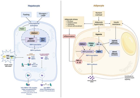
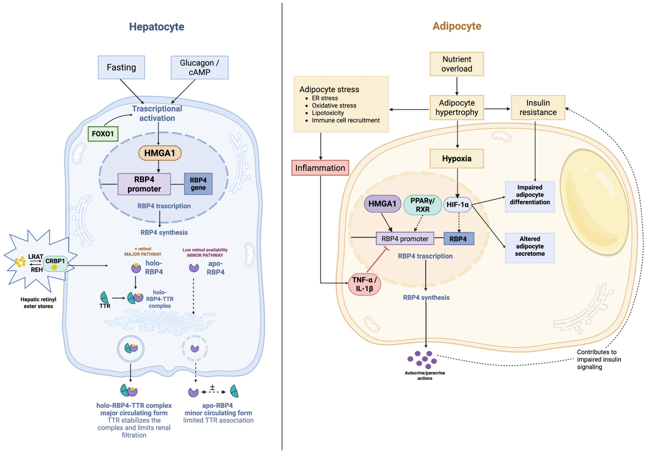

Disclaimer/Publisher’s Note: The statements, opinions and data contained in all publications are solely those of the individual author(s) and contributor(s) and not of MDPI and/or the editor(s). MDPI and/or the editor(s) disclaim responsibility for any injury to people or property resulting from any ideas, methods, instructions or products referred to in the content. |
© 2026 by the authors. Licensee MDPI, Basel, Switzerland. This article is an open access article distributed under the terms and conditions of the Creative Commons Attribution (CC BY) license.