


Abstract
Background: Diabetes in males leads to hyperglycemia-induced oxidative stress and inflammation in the testis, resulting in apoptosis. Diabetes-induced testicular impairment has been linked to the implication with the gut–brain axis. Sodium butyrate, a product of the gut microbiota fermentation process, has been targeted for its anti-diabetic, anti-inflammatory, and antioxidative functions. This research, therefore, explored sodium butyrate as a therapeutic potential intervention in mitigating diabetes-induced testicular dysfunction. Methods: Thirty-five male rats were randomly assigned to five groups after diabetes was confirmed: healthy control (CON), sodium butyrate-treated (SBUT; 200 mg/kg orally), diabetic control (T1DM; 50 mg/kg STZ intraperitoneally), diabetes + sodium butyrate (T1DM + SBUT), and diabetes + insulin (T1DM + INSL, 0.9 I.U/100 g/day). Body weight, testicular weight, glucose homeostasis (fasting blood sugar, oral glucose tolerance, and glycated hemoglobin), lipid profile, testosterone level, sperm analysis, histopathology of the testis, and anti-apoptotic markers (B-cell lymphoma-2 (Bcl-2), Bcl-associated protein X (Bax), and Caspase-3) were all assessed. Results: Along with elevated blood glucose and glycated hemoglobin levels, diabetic rats showed a notable decrease in body weight, testicular mass, gonadosomatic index, sperm count, motility, viability, and normal morphology. Elevated Bax and Caspase-3 expression with reduced Bcl-2 were observed alongside testicular fibrosis, accumulation of collagen, and carbohydrate-rich substances. Sodium butyrate treatment, by downregulating Bax and Caspase-3 and upregulating Bcl-2, conserved testicular histoarchitecture, restored sperm quality, controlled apoptotic signaling, and markedly improved metabolic indices. Conclusions: Sodium butyrate displayed a protective role in the testis in diabetic conditions via the modulation of hyperglycemia-induced apoptosis, identifying this as a potential therapeutic intervention in diabetes-induced testicular dysfunction.
1. Introduction
A major global health issue, infertility, affects approximately one in six adults of the total population [1]. Infertility affects over 55 million men, or 1.8% of all men worldwide, with an age-standardized prevalence of 1355 per 100,000 men (about 1.36%) [2,3]. This is expected to keep growing, especially in low- and middle-income areas. According to estimates, between 30% and 50% of men in sub-Saharan Africa are infertile [4]. Notably, male infertility has increased due to the rising incidence of diabetes—up to 90% of males with diabetes have been found to have varied degrees of testicular dysfunction [5,6,7]. In Nigeria, 35–50% of occurrences of infertility are caused by male factors [8]. Significant drops in semen characteristics were found in Nigerian men between 2015 and 2019, including decreases in progressive motility (87%), total progressively motile sperm count (78%), and normal morphology (50%) [9]. Male infertility cases rose by almost 75% between 1990 and 2021, indicating a worrying global deterioration in male reproductive health [10].
The risk of infertility is increased in both diabetic types, with oxidative stress, inflammation, and apoptosis triggered by hyperglycemia being pivotal factors in both comorbidities [11,12]. In diabetic models, there has been a display of elevated ROS levels, a decrease in antioxidant enzyme activity, an overexpression of Bax and Caspase-3, and mitochondrial dysfunction [13,14]. Testicular structure and reproductive functions are eventually compromised by these alterations, which start intrinsic apoptotic signaling pathways [15]. Overall, one significant biological link between diabetes and male reproductive failure is oxidative-stress-mediated apoptosis. These mechanisms show up in the testicular milieu as a compromised blood–testis barrier, morphological alterations, reduced sperm quality, and germ cell death [16,17,18]. These reproductive dysfunctions are made worse by diabetic dyslipidemia and dysregulated cholesterol metabolism [19,20]. These disruptions highlight the mechanism by which diabetes affects male reproductive health.
Despite numerous studies demonstrating the connection between diabetes and testicular dysfunction [21,22,23,24], therapeutic choices are still mostly conventional. The systemic metabolic consequences of gut-microbiota-targeted techniques, prebiotic/probiotic supplementation, and dietary change are attracting interest [25,26,27]. Environmental and dietary factors impact microbial homeostasis and reproductive health, and the gut microbiota has been connected to metabolic disorders and male fertility outcomes [28,29,30]. One important process that links gut health to systemic regulation is the metabolites, short-chain fatty acids (SCFAs), with the example, butyrate, generated from probiotics’ fermentation of prebiotics [31,32].
Butyrate has garnered substantial scientific attention due to its several biological advantages, such as insulin sensitivity, lipid metabolism, anti-inflammatory, antioxidant, and immunomodulatory qualities [33,34]. Butyrate (BUT), a salt derivative of butyric acid, affects gene expression, enhances insulin sensitivity, regulates glucose metabolism, and lowers systemic inflammation as a strong histone deacetylase (HDAC) inhibitor [35,36,37]. Despite growing evidence of its metabolic benefits, the effect of sodium butyrate in diabetes-induced testicular dysfunction remains poorly understood in vivo, particularly with respect to its control of oxidative stress and apoptotic signaling pathways. Furthermore, very few studies have systematically evaluated its effects on sperm parameters and apoptosis-related molecular markers in diabetic testicular tissue. Therefore, this research evaluates the role of sodium butyrate on sperm parameters, lipid profile indices, and apoptosis-related proteins in streptozotocin-induced diabetic rats.
2. Materials and Methods
2.1. Preparation of Sodium Butyrate (SBUT)
A stock solution of sodium butyrate (ChemCruz Biomedicals from Santa Cruz Biotechnology; sc-202341C, Dallas, TX, USA) was prepared by dissolving 250 mg of SBUT in 40 mL of distilled water.
2.2. Animal
Thirty-five (35) male Wistar rats with an average weight of 165 g were obtained from the University of Lagos’ animal care department. The animals were housed in a plastic cage with free access to drinking water, a pelletised feed, and ideal laboratory settings (excellent ventilation, temperature, and a 12-h light/dark cycle). The rats were acclimated to their surroundings for two weeks prior to the induction of experimental diabetic mellitus. The institutional Animal Care and Use Committee's (IACUC) and the National Research Council’s (1996) rules on animal handling are rigorously adhered to in the methods used in this work.
The Research Ethics Committee from the University of Lagos (University of Lagos Animal Care and Use Research Ethics Committee (UNILAGACUREC)) granted ethical permission for this study under reference number UNILAGACUREC/2024/05/AP02.
2.3. Induction of Type I Diabetes Mellitus
According to Aladaileh et al., diabetes was induced using 50 mg/kg streptozotocin (Sigma-Aldrich, St. Louis, MO, USA), administered intraperitoneally [14]. This was dissolved in a 0.1 M citrate buffer with a pH of 4.5, following an overnight fast. The rats’ blood glucose levels were assessed after 72 h. Rats having fasting blood sugar levels of at least 200 mg/dL were selected for this experiment.
2.4. Experimental Design
The animals were grouped into five groups (n = 7 in each group) at random when diabetes was confirmed based on the study design and institutional recommendations for the use of laboratory animals, as follows:
- CON: Healthy control.
- SBUT: Non-diabetic rats administered sodium butyrate (200 mg/kg body weight).
- T1DM: Animals induced with streptozotocin (50 mg/kg body weight).
- T1DM + SBUT: Animals induced with STZ (50 mg/kg body weight) and then treated with sodium butyrate (200 mg/kg body weight).
- T1DM + INSL: Animals induced with STZ (50 mg/kg body weight) and then treated with insulin (0.9 IU/100 g body weight).
Treatment commenced immediately after confirmation of diabetes. For six weeks, sodium butyrate was given orally once a day by gavage, while insulin (Mixtard Flexpen, Novo Nordisk Pharma Ltd., Bagsværd, Denmark) was administered subcutaneously once daily at the specified dosage regimen for six weeks. The total duration of the study was eight weeks, comprising two weeks of acclimatization followed by six weeks of treatment.
2.5. Oral Glucose Tolerance Test
At the start and end of the treatment session, body weights were noted. Following an overnight fast, all groups had an oral glucose tolerance test (OGTT) 48 h before the experiment’s conclusion (day 54). Prior to the delivery of glucose, baseline fasting blood glucose levels were assessed at 0 min. Oral glucose (2 g/kg body weight) was then given. A hand-held glucometer (Accu-Chek®, Mannheim, Germany) was used to monitor blood glucose levels at 30 min intervals for over two hours after glucose loading using blood drawn from the tail vein.
2.6. Sample Collection and Preparation
Immediately after the experiment was concluded, the animals were anesthetized using diethyl ether until an adequate surgical plane of anesthesia was achieved, as evidenced by loss of pedal withdrawal reflex. The blood was drawn via cardiac puncture, after which the epididymides and testicular tissue samples were collected. Blood samples were centrifuged for 15 min at room temperature at 3000 rpm. Before biochemical analysis, serum was isolated and kept at −20 °C. After being excised, the caudal epididymides were immediately collected for sperm analysis, and the testes were cleaned with cold saline and prepared for histochemical and immunohistochemical analyses.
2.7. Biochemical Analysis
Glycated hemoglobin (HbA1c) levels were determined from whole blood using commercially available assay kits (Fortress Diagnostics Limited, Antrim, UK; Product Code: BXC0670) per the guidelines provided by the manufacturer.
Serum lipid profile: triglycerides (Product Code: BXC0271), total cholesterol (Product Code: BXC0261), and high-density lipoprotein cholesterol (Product Code: BXC0421) were examined using Fortress Diagnostics Limited assay kits (Antrim, UK), per the guidelines provided by the manufacturer, as previously reported by Olaniyi et al. [38].
The serum concentration of testosterone (CAT: ELK8314) was determined using Rat Enzyme-Linked immunosorbent assay kits obtained from ELK Biotechnology Houston, TX, USA, following the manufacturer’s guidelines, as previously reported by Olaniyi et al. [38].
2.8. Sperm Analysis
The epididymis was carefully excised and processed for evaluation of four sperm parameters under a light microscope, including progressivity and viability, as previously described [13,23]. Sperm count was determined and expressed as the number of spermatozoa per milliliter of semen. The fraction of actively migrating sperm and their patterns of advancement were used to measure sperm motility. At least 200 spermatozoa were examined at ×400 magnification for sperm morphology, and percentages of head and tail abnormalities were noted. Eosin–nigrosin staining was used to evaluate sperm viability, and the percentage of viable (unstained) sperm cells was computed.
2.9. Histopathological and Immunohistochemical Analysis
2.9.1. Histochemical Staining (PAS and Masson Trichrome)
The testicular tissues that were used for histochemical techniques were fixed in 10% formol-saline immediately after extraction. After fixation, tissues were dehydrated in increasing ethanol grades, followed by clearing in xylene; afterward, the tissues were infiltrated and embedded in paraffin. As previously reported by Tahoon et al. [39], sections of 5 µm thickness were cut and stained with Masson’s trichrome stain to visualize collagen fibers and Periodic Acid-Schiff (PAS) to detect neutral mucopolysaccharides.
2.9.2. Immunohistochemical Analysis
The paraffin-embedded immunohistochemical technique was carried out using anti-PCNA, anti-Bax, anti-Bcl-2, and anti-Caspase-3 to assess apoptotic processes within testicular tissue across experimental groups. Thin paraffin tissue sections (5 µm) were placed on charged slides, deparaffinized in xylene, and then rehydrated using a decreasing graded ethanol medium and distilled water. Citrate buffer (pH 6.0) was used for antigen retrieval in a heat retrieval system. Then, 0.03% hydrogen peroxide in PBS was used to block endogenous peroxidase activity, and then the corresponding primary antibodies were incubated. Sections were then treated with a biotinylated secondary antibody conjugated with horseradish peroxidase. Immunoreactivity was visualized using 3,3′-diaminobenzidine (DAB), resulting in a brown precipitate. Sections were counterstained with hematoxylin, dehydrated, cleared, mounted, and examined under a light microscope, as previously described by Olajide et al. [40].
2.10. Statistical Analysis
The statistical analysis was conducted using the analytical software, GraphPad Prism version 9.5.1 (GraphPad Software, San Diego, CA, USA). The data are shown as mean ± SD. Group differences were investigated using Tukey’s multiple comparison post hoc test and one-way analysis of variance (ANOVA). Statistical significance was defined as a p-value of less than 0.05.
3. Results
3.1. Effect of Sodium Butyrate on Body Weight in T1DM Male Wistar Rats
As shown in Table 1, analysis of covariance (ANCOVA) was performed to determine whether final body weight differed among groups after adjustment for initial body weight. Initial body weight was a significant covariate, F(1,28) = 18.68, p = 0.0002, while group had a significant effect on final body weight after adjustment for initial body weight, F(4,28) = 66.54, p ˂ 0.0001. The assumptions of ANCOVA were satisfied, as the residuals were normally distributed (Shapiro–Wilk, p = 0.5057) and the Group*Initial body weight interaction was not significant, F(4,24) = 0.77, p = 0.5534, indicating homogeneity of regression slopes. The adjusted final body weight was highest in the SBUT group (271.83 g), followed by the control group (258.58 g), T1DM + INSL group (222.19 g), T1DM + SBUT group (178.83 g), and T1DM group (140.33 g). Compared with the T1DM group, adjusted final body weight was significantly higher in the T1DM + SBUT group (difference = 38.49 g; unadjusted p = 0.0031) and T1DM + INSL group (difference = 81.86 g; unadjusted p ˂ 0001). After Holm adjustment for multiple pairwise comparisons, these differences remained significant (p = 0.0125 and p ˂ 0.0001, respectively). The SBUT group did not differ significantly from the control group (p = 0.216).
Table 1.
Effect of sodium butyrate on final body weight: (a) p-value, (b) after adjustment for initial body weight in T1DM male Wistar rats.
3.2. Effect of Sodium Butyrate on Testicular Weight and Gonadosomatic Index in T1DM Male Wistar Rats
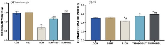
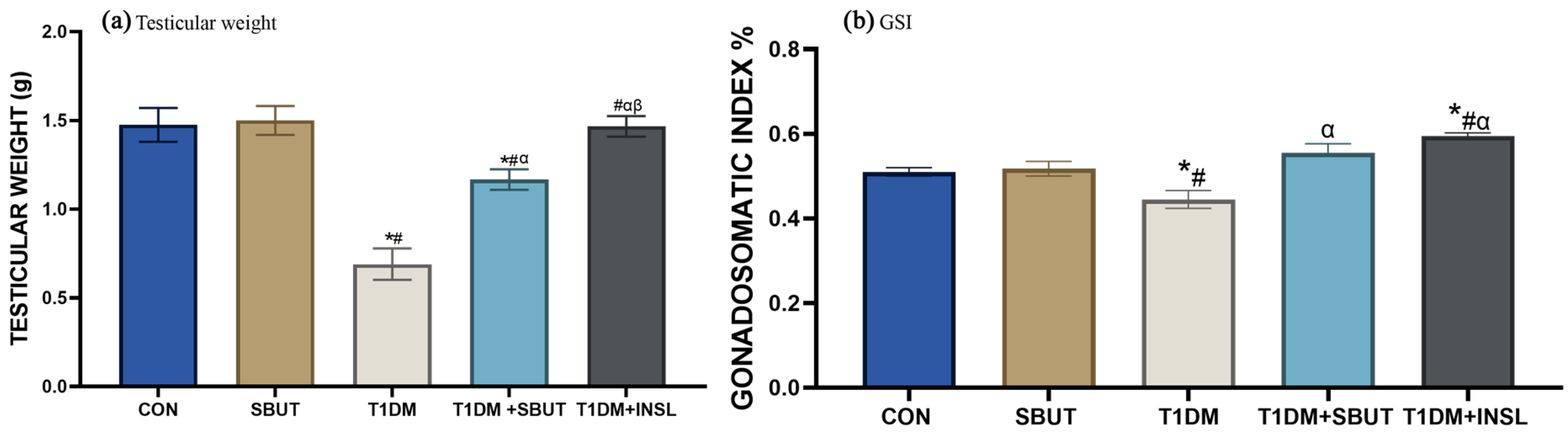
Testicular weight and gonadosomatic index (GSI) were significantly lower in T1DM rats than in controls (p < 0.05), as shown in Figure 1a,b. Compared with diabetic controls, administration of sodium butyrate significantly increased both parameters (p < 0.05). Similarly, insulin treatment significantly improved testicular weight and GSI compared with untreated diabetic rats (p < 0.05). Additionally, insulin treatment resulted in significantly higher improvements in testicular weight compared with sodium butyrate (p < 0.05). It is noteworthy that the GSI in the T1DM + INSL group is significantly higher than that of the control group; however, a higher GSI does not necessarily indicate a beneficial reproductive outcome.
Figure 1.
Bars showing the testicular weight (a) and GSI (b) levels in T1DM male Wistar rats. Bars were plotted with mean ± SD; n = 7. One-way ANOVA and Tukey’s test were used to analyze the data (*, p < 0.05 vs. CON; #, p < 0.05 vs. SBUT; α, p < 0.05 vs. T1DM; β, p < 0.05 vs. T1DM + SBUT). Control (CON); sodium butyrate (SBUT); type 1 diabetes mellitus (T1DM); insulin (INSL).
3.3. Effect of Sodium Butyrate on Glucose Homeostasis in T1DM Male Wistar Rats
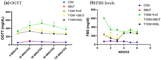
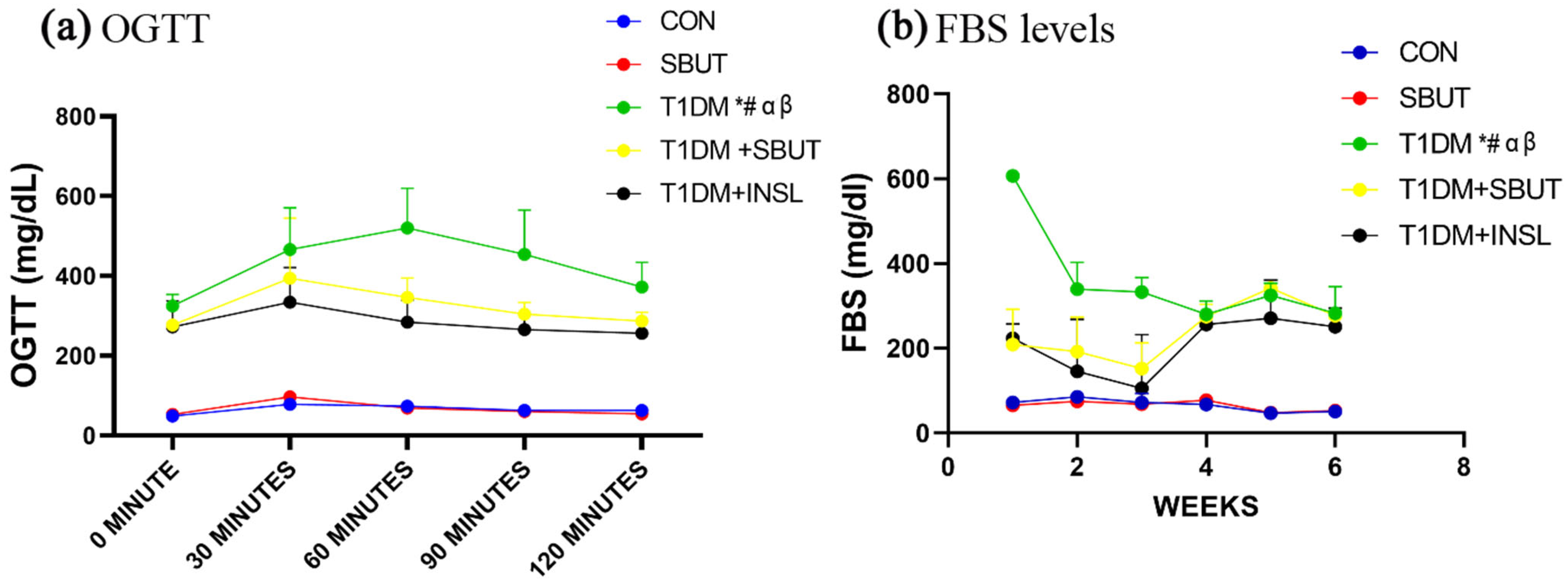
Figure 2a,b show that the T1DM group displayed significantly higher blood sugar levels compared with the control at all assessed time points during the oral glucose tolerance test as well as in fasting blood glucose (FBS) measurements (p < 0.05). However, T1DM + SBUT and T1DM + INSL showed a significant decrease in both the OGTT and FBS test compared to the T1DM group (p < 0.05). Notably, insulin treatment resulted in significantly lower OGTT and FBS values compared with sodium butyrate intervention (Figure 2a,b).
Figure 2.
Bars showing the OGTT (a) and FBS levels (b) throughout the experimental duration. Graphs were plotted with mean ± SD; n = 7. Two-way ANOVA and Tukey’s test were used to analyze the data (*, p < 0.05 vs. CON; #, p < 0.05 vs. SBUT; α, p < 0.05 vs. T1DM; β, p < 0.05 vs. T1DM + SBUT). Control (CON); fasting blood sugar (FBS); sodium butyrate (SBUT); type 1 diabetes mellitus (T1DM); insulin (INSL); oral glucose tolerance test (OGTT).
3.4. Effect of Sodium Butyrate on Glycated Hemoglobin in T1DM Male Wistar Rats
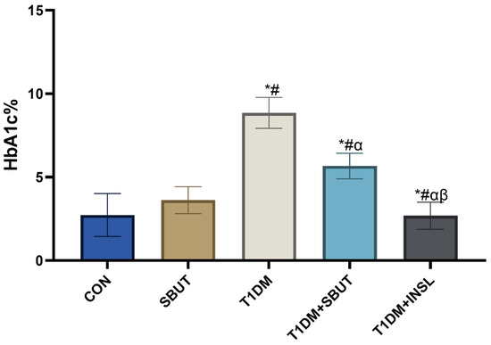
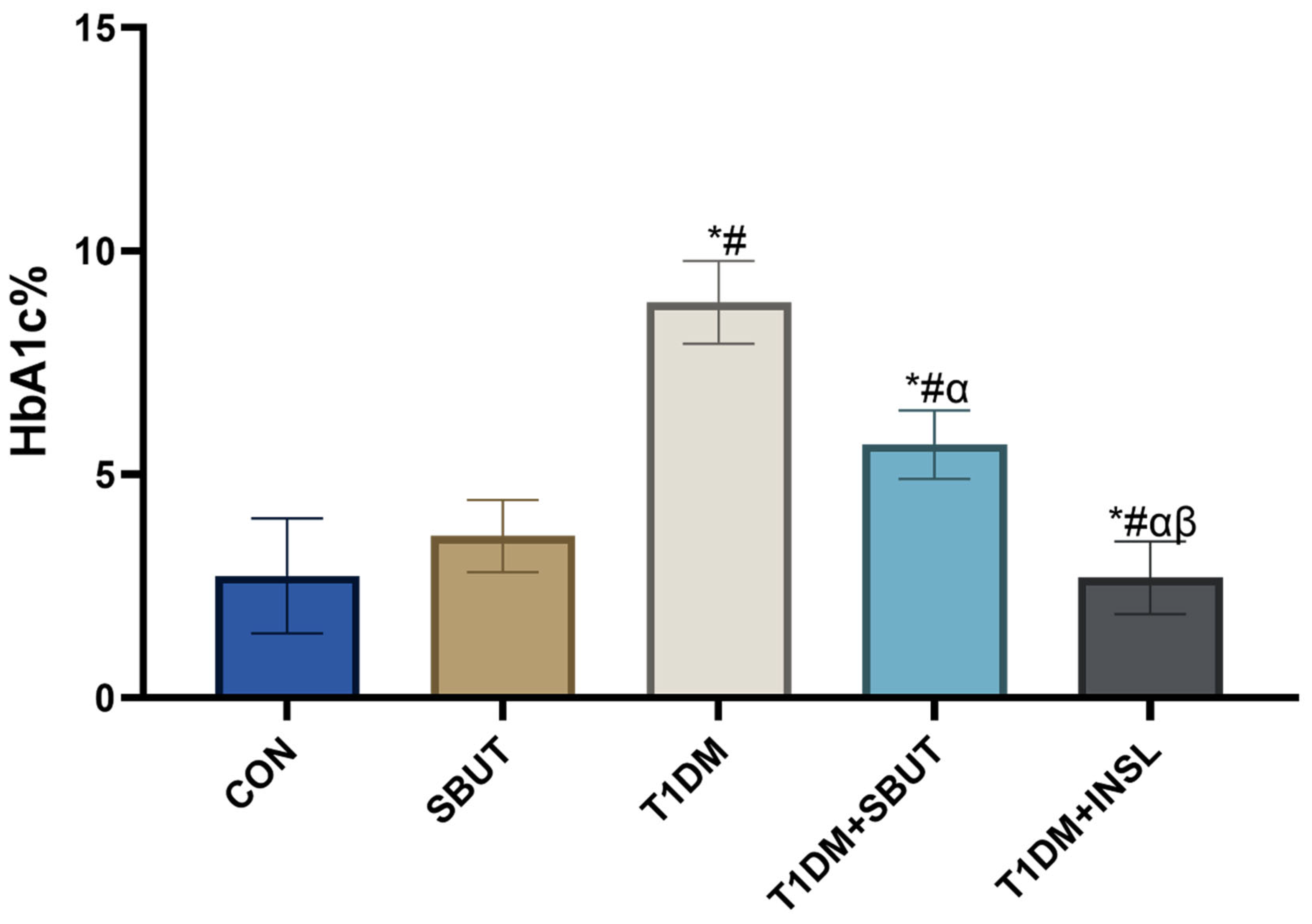
Figure 3 shows that glycated hemoglobin (HbA1c) was significantly elevated in T1DM animals compared to CON (p < 0.05). However, HbA1c levels significantly reduced in the T1DM + SBUT animals relative to T1DM animals (p < 0.05). Similarly, insulin administration resulted in a significant reduction in the T1DM + INSL group (p < 0.05). Notably, insulin treatment produced a significantly lower level compared to sodium butyrate treatment (p < 0.05).
Figure 3.
Effect of sodium butyrate on serum HbA1c levels in T1DM male Wistar rats. Bars were plotted with mean ± SD; n = 7. One-way ANOVA and Tukey’s test were used to analyze the data (*, p < 0.05 vs. CON; #, p < 0.05 vs. SBUT; α, p < 0.05 vs. T1DM; β, p < 0.05 vs. T1DM + SBUT). Control (CON); sodium butyrate (SBUT); type 1 diabetes mellitus (T1DM); insulin (INSL).
3.5. Effect of Sodium Butyrate on Lipid Profile in T1DM Male Wistar Rats
Table 2 reveals the role of sodium butyrate on serum lipid profile levels in T1DM male rats. T1DM rats exhibited significantly elevated total cholesterol (TC) and triglyceride (TG) levels, alongside a significant reduction in high-density lipoprotein (HDL) levels compared with controls (p < 0.05). In T1DM + SBUT, there was a significant reduction in TC and TG levels, while HDL increased relative to T1DM (p < 0.05). Insulin treatment significantly reduced TG levels and increased HDL levels (p < 0.05) but did not significantly alter TC levels compared with the T1DM group. Notably, TC levels were significantly lower in the T1DM + SBUT group compared with the T1DM + INSL group (p < 0.05). However, there was no disparity between the two treatment groups in TG and HDL levels.
Table 2.
Effects of sodium butyrate on TG, TC, and HDL levels in T1DM male Wistar rats.
3.6. Effect of Sodium Butyrate on Testosterone Level in T1DM Male Wistar Rats
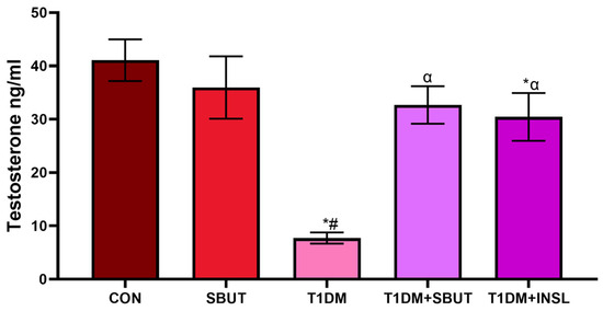
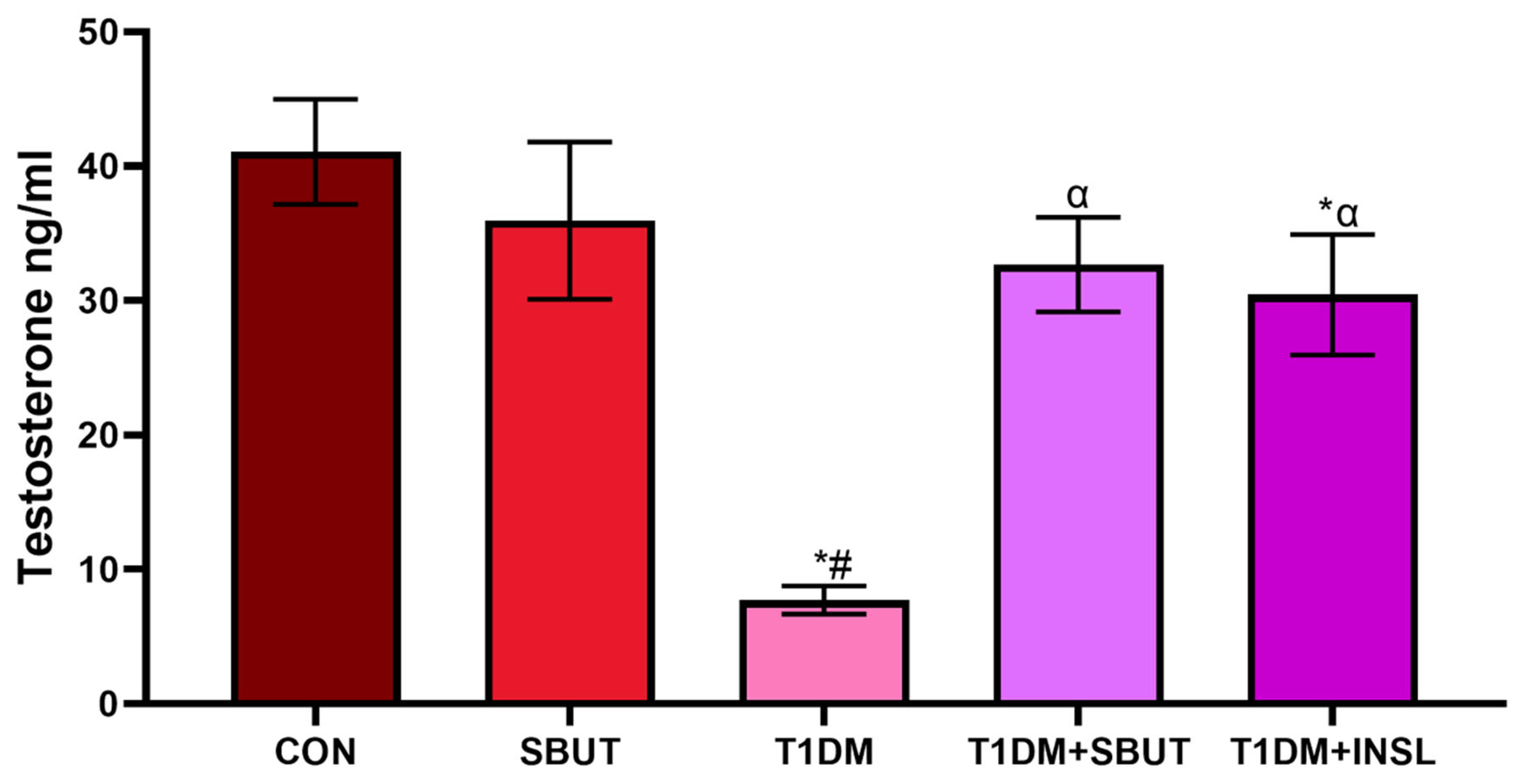
Figure 4 shows that serum testosterone was significantly reduced in T1DM animals compared with CON (p < 0.05). Sodium butyrate led to a significant increase in testosterone in the T1DM + SBUT relative to diabetic controls (p < 0.05). Similarly, treatment with insulin resulted in a significant increase in testosterone levels (p < 0.05) in the T1DM + INSL group. However, there was no disparity between the two treatment groups.
Figure 4.
Effect of sodium butyrate on serum testosterone levels in T1DM male Wistar rats. Bars were plotted with mean ± SD; n = 7. One-way ANOVA and Tukey’s test were used to analyze the data (*, p < 0.05 vs. CON; #, p < 0.05 vs. SBUT; α, p < 0.05 vs. T1DM). Control (CON); sodium butyrate (SBUT); type 1 diabetes mellitus (T1DM); insulin (INSL).
3.7. Effect of Sodium Butyrate on Sperm Parameters in T1DM Male Wistar Rats
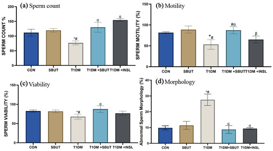
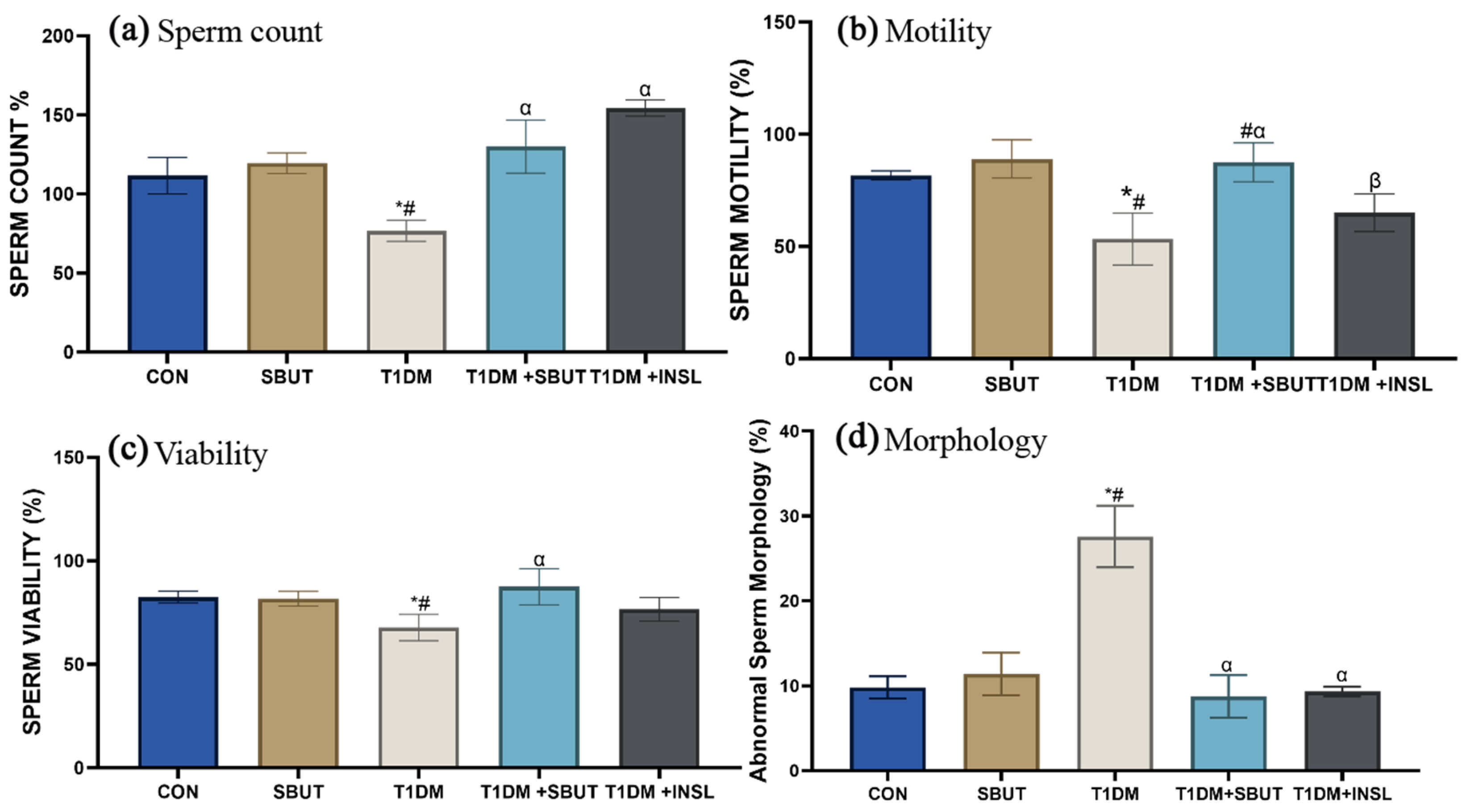
Figure 5 illustrates that diabetic animals exhibited a significant decrease in sperm count, motility, and viability compared to control groups (p < 0.05). Treatment with sodium butyrate (SBUT) significantly increased sperm count relative to the diabetic group, while insulin (INSL) significantly improved sperm count, motility, and viability in T1DM + INSL animals (p < 0.05). No significant differences were observed between the two treatment groups for sperm count and viability; however, sperm motility was significantly higher in the T1DM + SBUT group compared to the T1DM + INSL group. Diabetic animals also showed a significant increase in abnormal sperm morphology compared to controls. Both SBUT and INSL treatments significantly reduced abnormal morphology (p < 0.05), with no significant difference between the two treatments (Figure 5a–d).
Figure 5.
Effect of sodium butyrate on sperm count (a), motility (b), viability (c), and morphology (d) in T1DM male Wistar rats. Bars were plotted with mean ± SD; n = 7. One-way ANOVA and Tukey’s test were used to analyze the data (*, p < 0.05 vs. CON; #, p < 0.05 vs. SBUT; α, p < 0.05 vs. T1DM; β, p < 0.05 vs. T1DM + SBUT). Control (CON); sodium butyrate (SBUT); type 1 diabetes mellitus (T1DM) insulin (INSL).
3.8. Effect of Sodium Butyrate on Testicular Cellular Proliferation in T1DM Male Wistar Rats
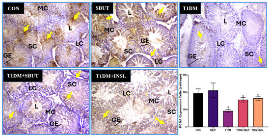
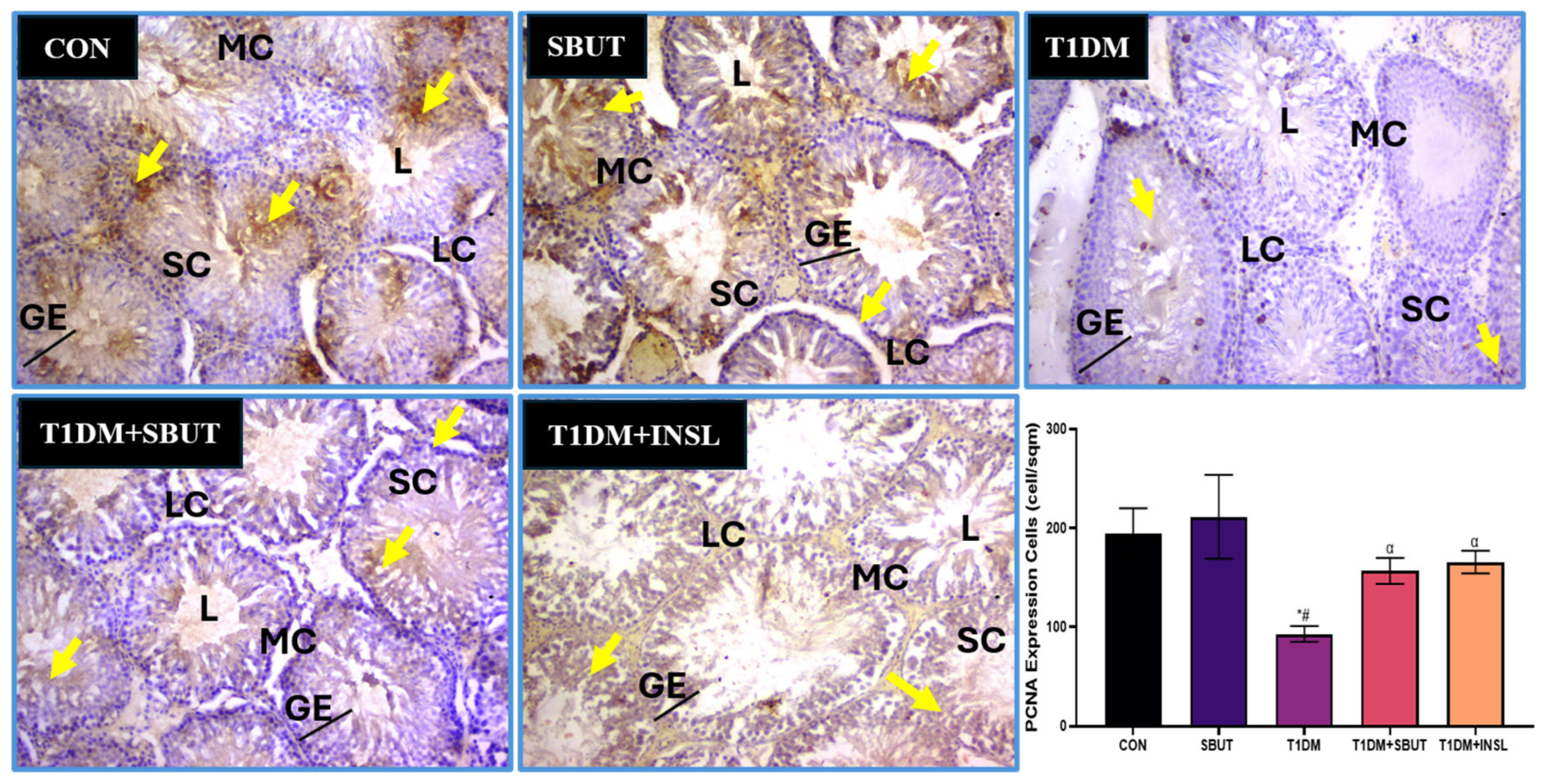
Figure 6 shows the effects of sodium butyrate on testicular cell proliferation in T1DM. The expression of proliferating cells was investigated with the PCNA immunohistochemical technique. The CON and SBUT groups have high PCNA expressions revealed through numerous positively stained nuclei reflecting normal proliferation. The T1DM group shows a significant decrease in PCNA expression via fewer positively stained nuclei when compared with the CON group, suggesting reduced germ cell proliferation and impaired spermatogenesis. Treatment with SBUT and insulin after T1DM induction significantly improved testicular expression of PCNA expression compared to the T1DM group.
Figure 6.
Photomicrograph and bar graph showing the expression of PCNA at ×100M in the testis across experimental groups investigated with the anti-PCNA immunohistochemistry technique. The bar graph shows the morphometric analysis and quantification of PCNA expression in the testis across experimental groups. Yellow arrows indicate immunoreactivity. (*, p < 0.05 vs. CON; #, p < 0.05 vs. SBUT; α, p < 0.05 vs. T1DM). Control (CON); sodium butyrate (SBUT); type 1 diabetes mellitus (T1DM); insulin (INSL).
3.9. Effect of Sodium Butyrate on Testicular Glycogen/Carbohydrate Deposition in T1DM Male Wistar Rats
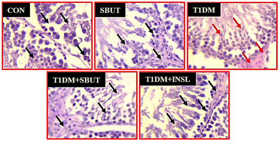
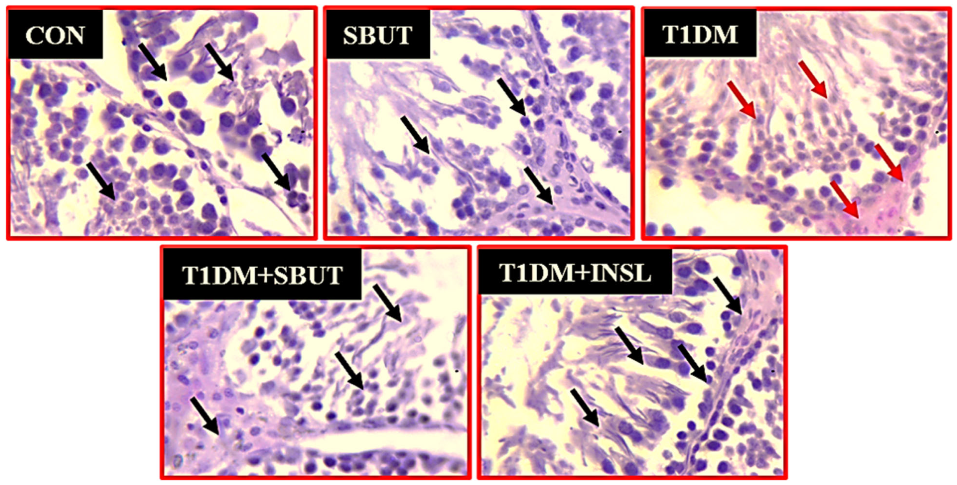
The photomicrograph in Figure 7 reveals the SBUT treatment on deposition and distribution in the testes following type 1 diabetic induction using the PAS staining technique. Normal testicular PAS-positive substances were seen in the control groups (CON and SBUT; black arrows). Increased PAS-positive substances were observed within the basal membrane, Sertoli, and epidermal cell layers (red arrows) following type 1 diabetic induction, indicating increased testicular accumulation and deposition of carbohydrates (glycogen, glycoproteins, glycolipids, and mucin). Treatment with sodium butyrate and insulin in T1DM shows normal testicular PAS-positive substances similar to those of CON.
Figure 7.
Periodic Acid-Schiff (PAS) staining of the testis across experimental groups at ×400M in the type 1 diabetic study. Black arrows indicate normal PAS intensity, Red arrows indicate increased PAS intensity. Control (CON); sodium butyrate (SBUT); type 1 diabetes mellitus (T1DM); insulin (INSL).
3.10. Effect of Sodium Butyrate on Testicular Collagen Distribution in T1DM Male Wistar Rats
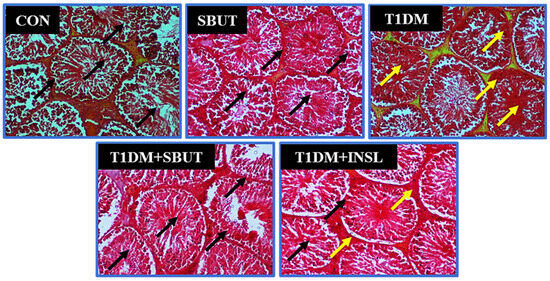
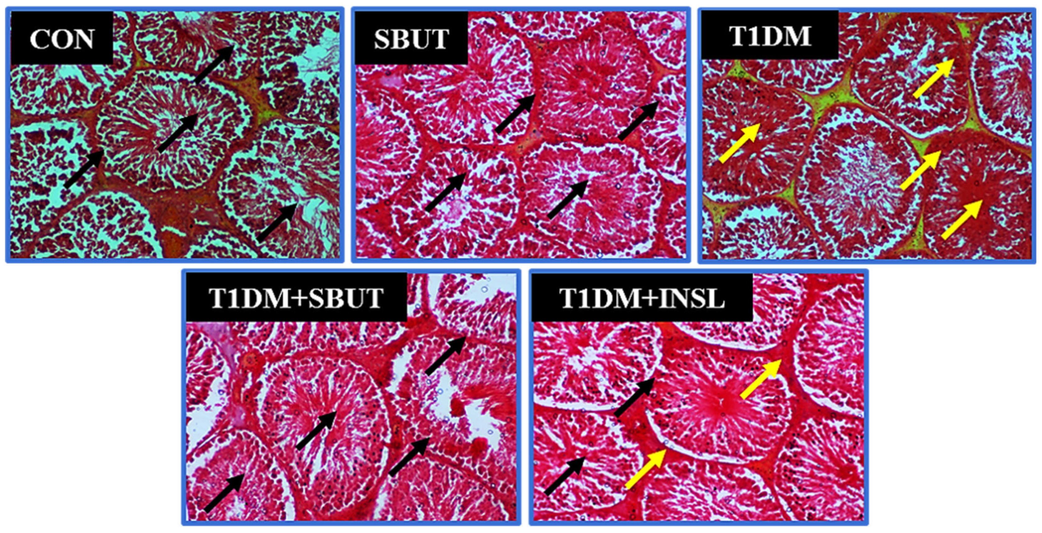
Figure 8 shows the SBUT treatment on collagen distribution in the testes following type 1 diabetes, using the Masson trichrome staining technique. Normal testicular collagen distribution was seen in the control groups (CON and SBUT; black arrows). Increased collagen distribution, which appears light green, was observed within the lumen, Sertoli, and epidermal cell layers (yellow arrows) following type 1 diabetic induction, indicating increased collagen accumulation and deposition. Treatment with sodium butyrate in type 1 diabetic rats showed normal testicular collagen distribution like that of the control groups.
Figure 8.
Masson trichrome staining of the testis across experimental groups at ×100M in the type 1 diabetic study. Black arrows indicate normal collagen; yellow arrows indicate increased collagen. Control (CON); sodium butyrate (SBUT); type 1 diabetes mellitus (T1DM); insulin (INSL).
3.11. Effect of Sodium Butyrate on Apoptosis in Testicular Tissues in T1DM Male Wistar Rats
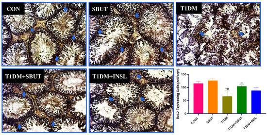
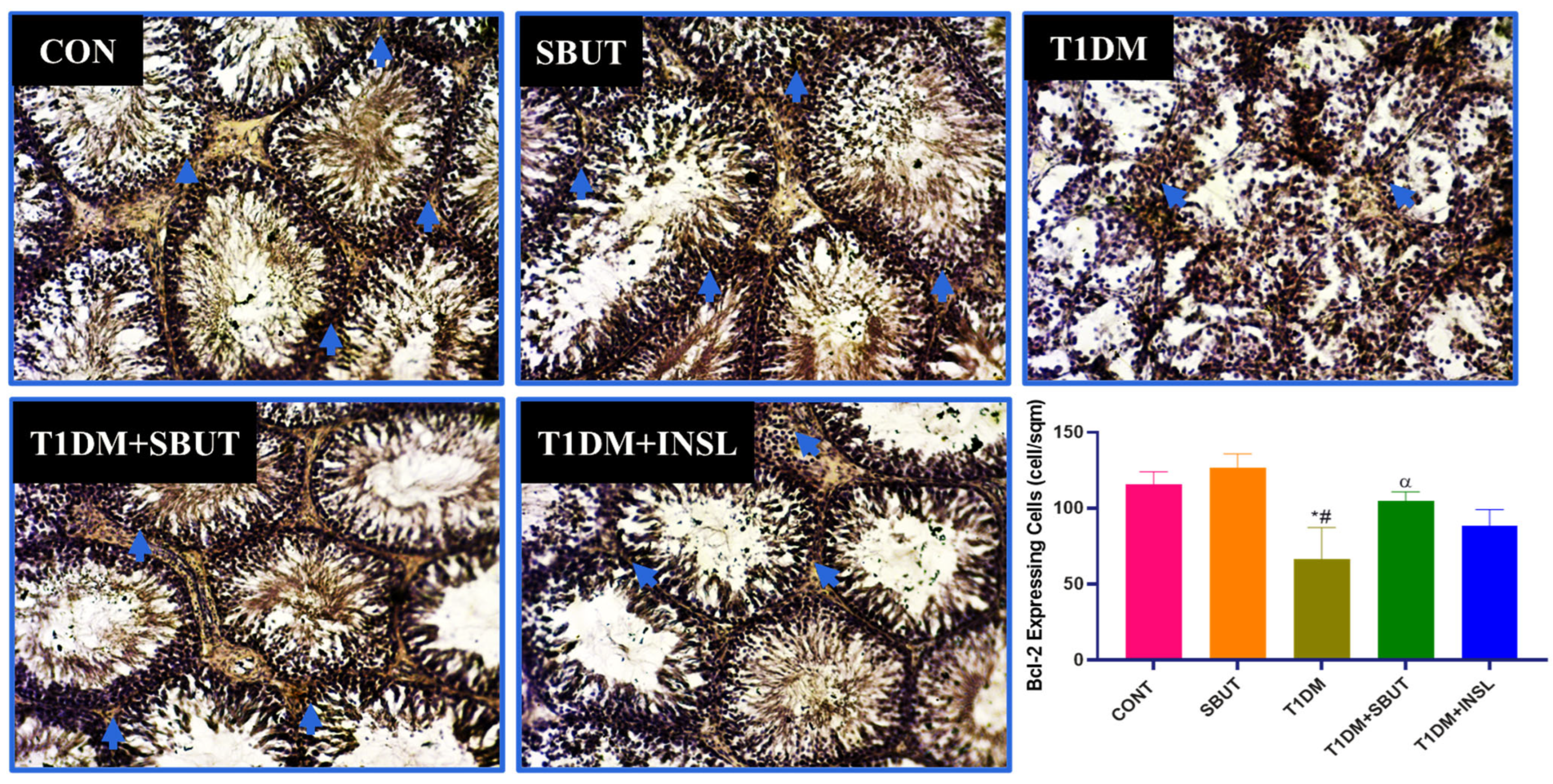
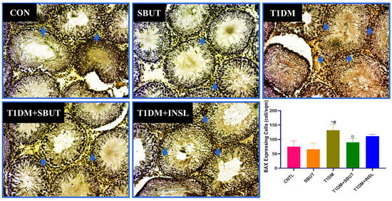
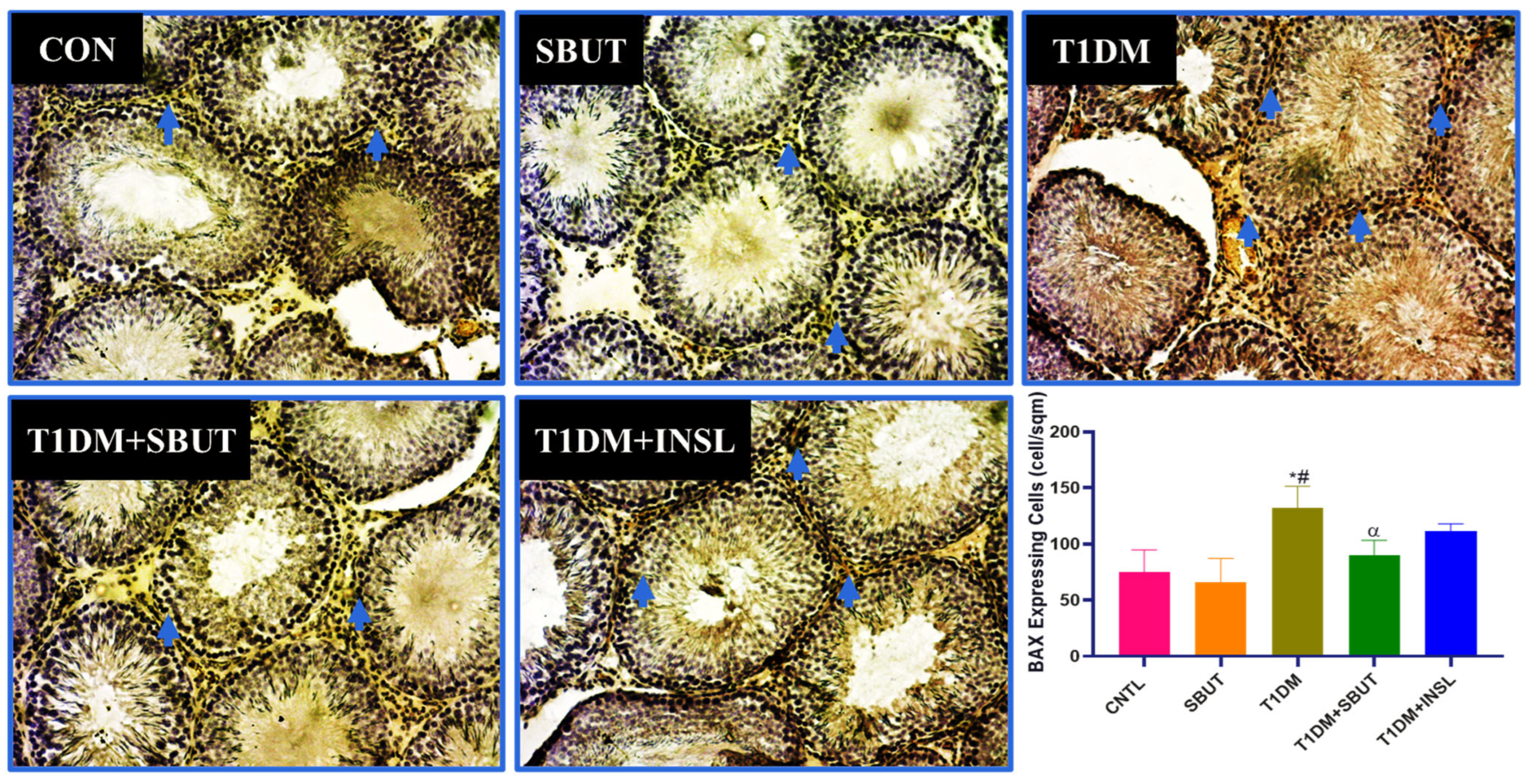
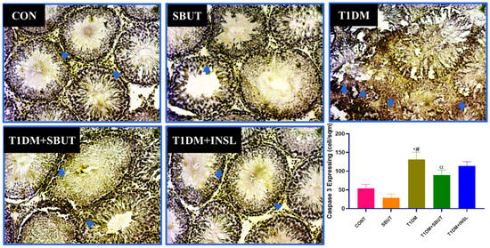
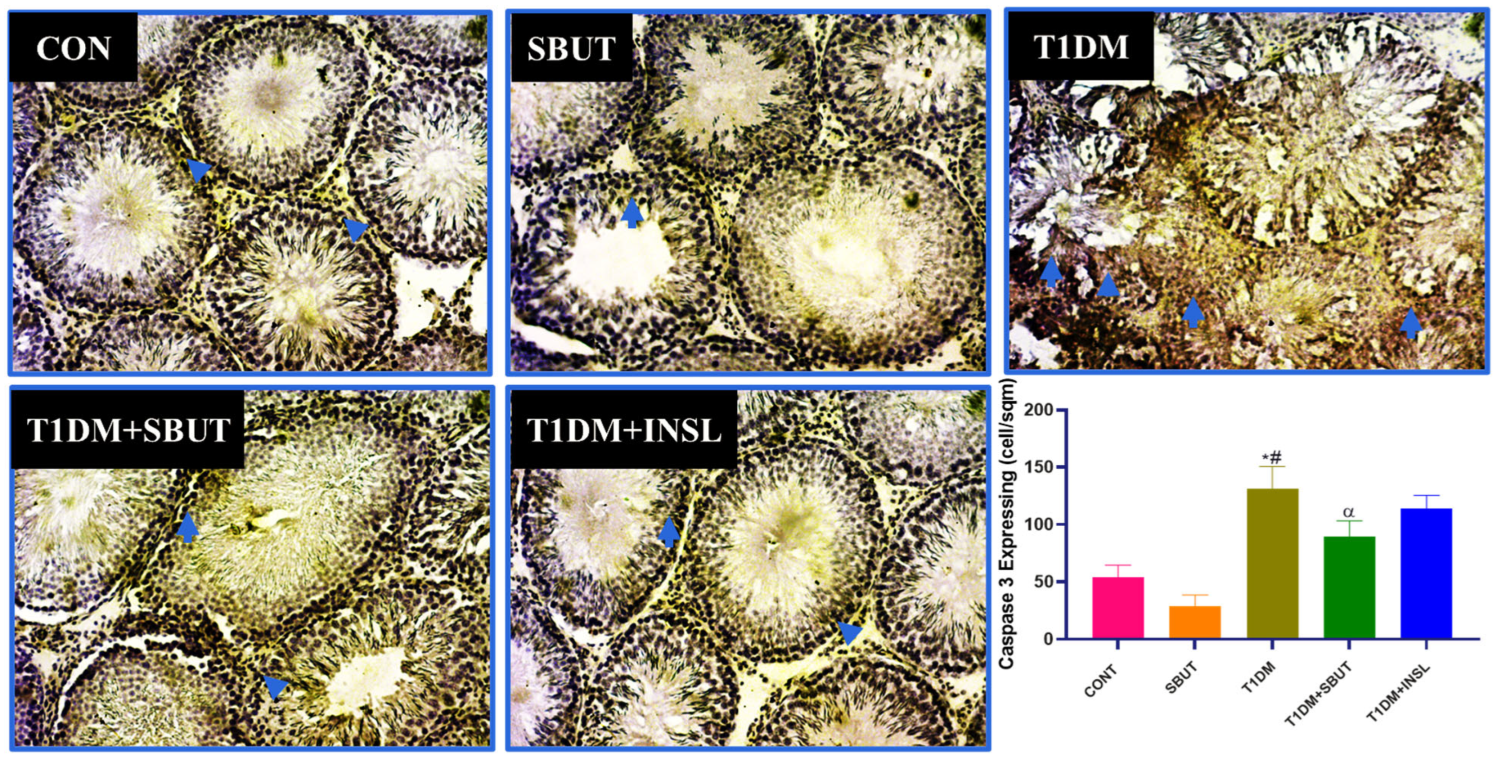
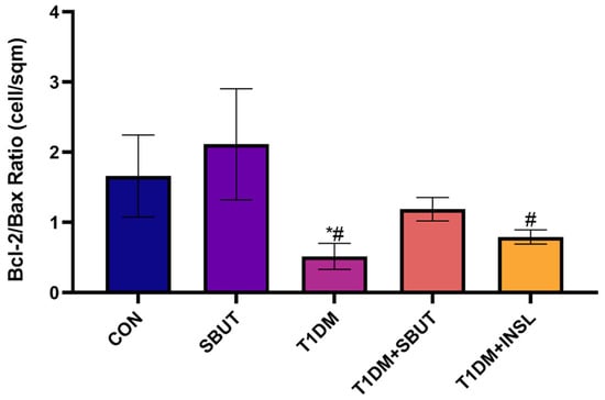
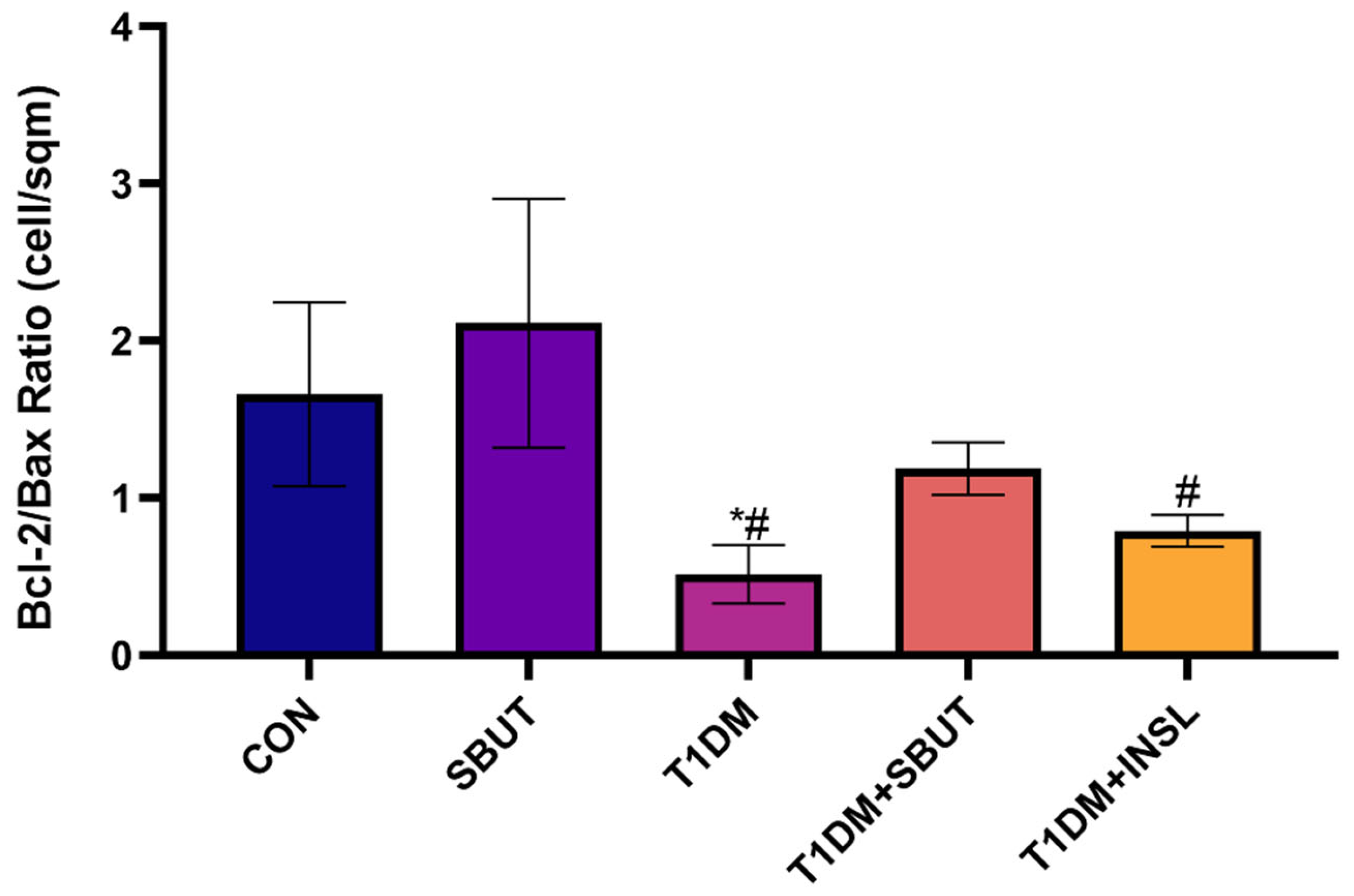
Figure 9, Figure 10, Figure 11 and Figure 12 show the effects of sodium butyrate in modulating testicular apoptosis in the T1DM group. The expression of anti-apoptotic B-cell lymphoma 2 (Bcl-2), pro-apoptotic Bcl2-associated X (BAX), and cysteine-aspartate-specific protease 3 (Caspase-3) were investigated with the immunohistochemical technique. The results show a significant decrease in Bcl-2 expression (Figure 9) and a significant increase in BAX (Figure 10) and Caspase-3 (Figure 11) expressions, with a marked decrease in the Bcl-2/Bax ratio (Figure 12) in the diabetic testis (T1DM) when compared with the CON group, indicating enhanced apoptosis. Treatment with SBUT and insulin after T1DM induction improved testicular expression of anti-apoptotic markers and a substantially restored Bcl-2/Bax ratio when compared to the T1DM group. This observed improvement was statistically significant only when treated with SBUT but not insulin. SBUT alone demonstrated the highest ratio, suggesting robust intrinsic anti-apoptotic potential.
Figure 9.
Photomicrograph and bar graph showing the expression of Bcl-2 at ×100M in the testis across experimental groups investigated with the anti-Bcl-2 immunohistochemistry technique. Blue arrows indicate immunoreactivity. (*, p < 0.05 vs. CON; #, p < 0.05 vs. SBUT; α, p < 0.05 vs. T1DM). Control (CON); sodium butyrate (SBUT); type 1 diabetes mellitus (T1DM); insulin (INSL).
Figure 10.
Photomicrograph and bar graph representing the expression of BAX at ×100M in the testis across experimental groups investigated with the anti-BAX immunohistochemistry technique. Blue arrows indicate immunoreactivity. (*, p < 0.05 vs. CON; #, p < 0.05 vs. SBUT; α, p < 0.05 vs. T1DM). Control (CON); sodium butyrate (SBUT); type 1 diabetes mellitus (T1DM); insulin (INSL).
Figure 11.
Photomicrograph and bar graph representing the expression of Caspase-3 at ×100M in the testis across experimental groups investigated with the anti-Caspase-3 immunohistochemistry technique. Blue arrows indicate immunoreactivity. (*, p < 0.05 vs. CON; #, p < 0.05 vs. SBUT; α, p < 0.05 vs. T1DM). Control (CON); sodium butyrate (SBUT); type 1 diabetes mellitus (T1DM); type 1 diabetes mellitus + sodium butyrate (T1DM + SBUT); type 1 diabetes mellitus + insulin (T1DM + INSL).
Figure 12.
Bar graph showing the morphometric analysis and quantification of Bcl-2/Bax ratio expression in the testis across experimental groups. (*, p < 0.05 vs. CON; #, p < 0.05 vs. SBUT). Control (CON); sodium butyrate (SBUT); type 1 diabetes mellitus (T1DM); insulin (INSL).
4. Discussion
The reproductive system, particularly the hypothalamic–pituitary–testicular axis (HPT), is altered when faced with the global health challenge of diabetes [23,41]. Chronic hyperglycemia, a major feature of this diabetic condition, ultimately induces oxidative stress and inflammation affecting the male reproductive cells, comprising Sertoli and Leydig cells’ function, causing germ cell apoptosis, impaired spermatogenesis leading to poor sperm quality, erectile/ejaculatory dysfunctions, and disruption of blood–testis barrier integrity, eventually impairing male fertility and sexual function [5,6,7,16,42].
The present study recorded hyperglycemia, elevated glycated hemoglobin, dyslipidemia, reduced body and testicular weight, decreased gonadosomatic index, decreased PCNA expression, and compromised sperm indices. There was also disrupted testicular histoarchitecture with increased accumulation of carbohydrate-rich substances, excessive collagen deposition, and aggravated apoptosis, all of which reveal diabetes-induced marked metabolic and reproductive dysregulation. However, administration of sodium butyrate to the diabetic animals improved glycemic control and lipid profile, restored sperm quality and apoptotic balance, and preserved the histoarchitecture of the testis.
The glycemic control observed in the SBUT-treated group is consistent with reports of significantly enhanced insulin levels, reduced plasma glucose and HbA1c levels, decreased pancreatic β-cell death, and overall glucose homeostasis in diabetic animals. This was attributed to histone deacetylase inhibition (HDACi) and enhanced histone acetylation [35,43], and modification of the p38/ERK MAPK and apoptotic signaling pathways [44]. Additionally, studies have reported that butyrate alleviates metabolic dysfunction by reducing adiposity and inflammation through GPR41/43 activation and HDACi, while also strengthening gut barrier integrity to limit endotoxemia and systemic metabolic inflammation [45,46,47].
In the present study, however, the standard drug insulin observed significantly improved glucose clearance, compared with the sodium butyrate group. This observation may be due to the distinct pathways and mechanisms of action, with insulin regulating glycemia by acting directly and selectively on its receptors in the target cells. On the other hand, butyrate indirectly affects glucose metabolism via gut microbiota regulation, systemic inflammation reduction, gut barrier function, signaling via SCFA receptors, and epigenetic modification of metabolic genes, as reported by Krauze et al. [43]. As a result, its impact may be more variable and dependent on the circumstances.
The reproductive dysregulation observed in this study corroborates a diabetic study by Soetan et al., uncovering hyperglycemia and reduced testosterone levels, alongside decreased sperm quality [23]. Low testosterone levels and impaired reproductive function are common features of diabetes-associated hypogonadism, which may result from central suppression of gonadotropin release (hypogonadotropic hypogonadism), direct testicular failure (primary hypogonadism), or a combination of both. Likewise, clinical studies of diabetic male patients have reported altered sperm parameters and reduced semen volume [48,49,50]. In the present study, SBUT and insulin treatments significantly increased testosterone levels, increased PCNA expression, and improved sperm count, motility, and viability relative to untreated diabetic rats, suggesting restoration of spermatogenic efficiency and sperm functional competence. In a reproductive performance study investigating the effects of dietary SBUT supplementation by Alhaj et al. in 2018, there was improved semen quality displayed by increasing semen volume, improved sperm indices, elevating serum testosterone levels, and enhancing testicular antioxidant capacity via increased GPX and SOD levels [51]. According to Liu et al., butyrate supplementation also improved sperm count, motility, morphology, and overall sperm quality in mice with high-fat-diet-induced spermatogenic and fertility impairments by restoring testicular metabolic homeostasis, especially through normalization of steroid and bile acid synthesis pathways [52]. These results imply that SBUT-mediated enhancement of sperm quality may require restoring testicular redox balance in addition to glycemic correction.
Testicular collagen deposition, aberrant carbohydrate accumulation, metabolic disruption, and increased extracellular matrix deposition are indicative of impaired spermatogenic support, compromised steroidogenic capacity, and loss of seminiferous tubule integrity, as evidenced by histopathological evaluation with PAS and Masson’s trichrome staining. This supports previous preclinical research utilizing these stains that reported diabetes-induced testicular injury [23,42,53,54,55]. On the other hand, sodium butyrate also reduced collagen deposition on Masson’s trichrome staining, indicating attenuation of diabetes-induced extracellular matrix remodeling and fibrotic changes, and normalized PAS staining patterns, indicating improved testicular glucose handling and metabolic homeostasis. This suggests that sodium butyrate guards against testicular damage brought on by diabetes.
Studies have also reported severe testicular apoptosis in a number of diabetes investigations [13,56,57,58,59]. When taken as a whole, in diabetic conditions, hyperglycemia induces excess reactive oxygen species production, leading to oxidative stress, which activates inflammation, mitochondrial dysfunction, and apoptosis. These changes, such as lipid, protein, and DNA damage, in the testis show a move toward the destabilization of the mitochondrial membrane, leading to the cytochrome c release, which activates the initiator and executioner caspases, which ultimately lead to the loss of germ cells and poor spermatogenesis. However, sodium butyrate was reported to critically regulate cellular proliferation, differentiation, apoptosis, energy metabolism, maintenance of epithelial junction integrity, and the pathogenesis of diabetes [35,60,61,62,63,64].
By significantly restoring apoptotic dysregulation in diabetic condition, this study confirmed the protective impact of SBUT. In this work, the restoration was demonstrated by elevated Bcl-2 expression, inhibition of pro-apoptotic markers, and a significant shift of the Bcl-2/BAX ratio toward cell survival. This change implies that excessive germ cell apoptosis is inhibited and mitochondrial integrity is preserved. Overall, sodium butyrate’s ability to maintain testicular cellular survival and structural integrity under diabetic stress is highlighted by its modulation of important apoptotic markers, making it a promising adjunct or alternative therapeutic approach for preventing diabetes-associated testicular degeneration. Additionally, while this present study focuses on the individual effects of sodium butyrate and insulin in type 1 diabetes, it is worth noting that insulin produced a greater improvement in HbA1c, whereas sodium butyrate produced greater improvements in sperm motility and a significant improvement in the sperm viability. Additionally, both treatments improved sperm count and abnormal sperm morphology relative to the diabetic control group. These findings suggest that the two interventions may act through partially distinct or complementary mechanisms, with insulin primarily improving glycemic control and sodium butyrate potentially providing additional benefits to the reproductive functions, which warrants further investigation.
5. Conclusions
Sodium butyrate ameliorated the metabolic and reproductive dysfunctions in this streptozotocin-induced diabetic model. Its histoprotective and anti-apoptotic effects were associated with restoration of sperm quality and preservation of testicular architecture. These protective effects appear to extend beyond glycemic regulation, suggesting additional antioxidant, anti-inflammatory, and epigenetic mechanisms within testicular tissue. These findings collectively position sodium butyrate as a promising adjunctive therapeutic candidate for mitigating diabetes-associated male reproductive impairment.
Author Contributions
Conceptualization, O.C.B., G.O.O. and B.O.I.; methodology, O.C.B. and B.O.I.; software, O.C.B.; validation, O.C.B., G.O.O. and B.O.I.; formal analysis, O.C.B., G.O.O. and B.O.I.; investigation, O.C.B.; resources, O.C.B., G.O.O. and B.O.I.; data curation, O.C.B., G.O.O. and B.O.I.; writing—original draft preparation, O.C.B.; writing—review and editing, O.C.B., G.O.O. and B.O.I.; visualization, O.C.B., G.O.O. and B.O.I.; supervision, G.O.O. and B.O.I.; project administration, O.C.B.; funding acquisition, O.C.B., G.O.O. and B.O.I. All authors have read and agreed to the published version of the manuscript.
Funding
This research received no external funding.
Institutional Review Board Statement
The study was conducted after obtaining ethical approval from the University of Lagos Animal Care and Use Research Ethics Committee (UNILAGACUREC), University of Lagos, Nigeria (Approval No. UNILAGACUREC/2024/05/AP02; date of approval: 30 May 2024).
Informed Consent Statement
Not applicable.
Data Availability Statement
The raw data supporting the conclusions of this article will be made available by the authors upon request.
Acknowledgments
The authors gratefully acknowledge the Department of Physiology, Babcock University, and University of Lagos. Special thanks to Arietarhire Leviticus O., Olaniyi Soetan O., Obekpa Matthew A., Agunloye Mary O., Sam Dashe, and Elum Israel I. for their support during the experimental procedure.
Conflicts of Interest
The authors declare no conflicts of interest.
References
- Njagi, P.; Groot, W.; Arsenijevic, J.; Dyer, S.; Mburu, G.; Kiarie, J. Financial costs of assisted reproductive technology for patients in low- and middle-income countries: A systemic review. Hum. Reprod. Open 2023, 2, hoad007. [Google Scholar] [CrossRef] [Scilit]
- Haung, B.; Wang, Z.; Kong, Y.; Jin, M.; Ma, L. Global, regional, and national burden of male infertility in 204 countries, and terrories between 1990 and 2019: An analysis of global burden of disease study. BMC Public Health 2023, 23, 2195. [Google Scholar] [CrossRef] [Scilit]
- Liang, Y.; Huang, J.; Zhao, Q.; Mo, H.; Su, Z.; Feng, S.; Li, S.; Ruan, X. Global, regional, and national prevalence and trends of infertility among individuals of reproductive age (15–49 years) from 1990 to 2021, with projections to 2040. Hum. Reprod. 2025, 40, 529–544. [Google Scholar] [CrossRef] [Scilit] [PubMed]
- Moronkeji, M.A.; Emokpae, M.A.; Ojo, T.A.; Moronkeji, R.E.; Ogundoju, L.T. The patterns and occupational distribution of hormonal abnormalities among men investigated for infertility in some centers in the southwest, Nigeria. J. Clin. Transl. Res. 2021, 7, 221–228. [Google Scholar] [CrossRef] [Scilit] [PubMed] [PubMed Central]
- Badejogbin, O.C.; Chijioke-Agu, O.E.; Olubiyi, M.V.; Agunloye, M.O. Pathogenesis of testicular dysfunction in diabetes: Exploring the mechanism and therapeutic interventions. J. Assist. Reprod. Genet. 2024, 42, 367–379. [Google Scholar] [CrossRef] [Scilit]
- Badejogbin, O.C.; Akano, O.P.; Julius, O.E.B.; Agunloye, M.O.; Olubiyi, M.V.; Chijioke-Agu, O.E.; Olaniyi, K.S. Potential Role of CREM in Diabetes-Associated Testicular Dysfunction: Current Evidence and Future Perspectives. Reprod. Med. Biol. 2025, 24, e12661. [Google Scholar] [CrossRef] [Scilit]
- Carrageta, D.F.; Pereira, S.C.; Ferreira, R.; Monteiro, M.P.; Oliveira, P.F.; Alves, M.G. Signatures of metabolic diseases on spermatogenesis and testicular metabolism. Nat. Rev. Urol. 2024, 21, 477–494. [Google Scholar] [CrossRef] [Scilit]
- Obajimi, G.O.; Kolade, C.O.; Aladejare, A. Is male factor infertility rising? Another side of the equation from an in vitro fertilization clinic southwestern Nigeria. Afr. J. Reprod. Gynaecol. Endocopy 2021, 6, 10–13. [Google Scholar] [CrossRef] [Scilit]
- Akang, E.N.; Opuwari, C.S.; Enyioma-Alozie, S.; Moungala, L.W.; Amatu, T.E.; Wada, I.; Ogbeche, R.O.; Ajayi, O.O.; Aderonmu, M.M.; Shote, O.B.; et al. Trends in semen parameters of infertile men in South Africa and Nigeria. Sci. Rep. 2023, 13, 6819. [Google Scholar] [CrossRef] [Scilit]
- Zeng, G.; Liu, L.; Wang, Y.; Yu, J.; Wang, H.; Li, F. Global, regional, and national burden and trends of reproductive-aged male and female infertility from 1990–2021. Front. Endocrinol. 2025, 16, 1506229. [Google Scholar] [CrossRef] [Scilit]
- Delbarba, A.; Anelli, V.; Bambini, F.; Buoso, C.; Facondo, P.; Gatta, E.; Girelli, A.; Cappelli, C.; Ferlin, A. Type 1 diabetes mellitus and sperm quality: A case-control study. Andrology 2025, 13, 208–216. [Google Scholar] [CrossRef] [Scilit]
- Rong, J.; Leng, X.; Jiang, K.; Tan, J.; Dong, M. Systemic impacts of diabetes on spermatogenesis and intervention strategies: Multilayered mechanism analysis and cutting-edge therapeutic approaches. Reprod. Biol. Endocrinol. 2025, 23, 122. [Google Scholar] [CrossRef] [Scilit]
- Aly, H.A.A. Mitochondria-Mediated Apoptosis Induced Testicular Dysfunction in Diabetic Rats: Ameliorative Effect of Resveratrol. Endocrinology 2021, 162, bqab018. [Google Scholar] [CrossRef] [Scilit] [PubMed]
- Aladaileh, S.H.; Abukhalil, M.H.; Alfwuaires, M.A.; Algefare, A.I.; Imran, M.R.; Anjum, S.; Alzahrani, S.; Alsubhi, W.A.; Karimulla, S.; Al-Shammari, A.H. Modulation of Oxidative Stress, Inflammation, and Apoptosis and Restoration of Sirt1/Nrf2/HO-1 Signaling by Diosmin Protect Against Diabetes-Induced Testicular Damage in Rats. Int. J. Mol. Sci. 2026, 27, 1268. [Google Scholar] [CrossRef] [Scilit]
- Hussain, T.; Kandeel, M.; Metwally, E.; Murtaza, G.; Kalhoro, D.H.; Yin, Y.; Tan, B.; Chughtai, M.I.; Yaseen, A.; Afzal, A.; et al. Unraveling the harmful effect of oxidative stress on male fertility: A mechanistic insight. Front. Endocrinol. 2023, 14, 1070692. [Google Scholar] [CrossRef] [Scilit]
- Ranjan, A.; Choubey, M.; Yada, T.; Krishna, A. Nesfatin-1 ameliorates type-2 diabetes-associated reproductive dysfunction in male mice. J. Endocrinol. Investig. 2020, 43, 515–528. [Google Scholar] [CrossRef] [Scilit]
- Bhatti, J.S.; Sehrawat, A.; Mishra, J.; Sidhu, I.S.; Navik, U.; Khullar, N.; Kumar, S.; Bhatti, G.K.; Reddy, P.H. Oxidative stress in the pathophysiology of type 2 diabetes and related complications: Current therapeutics strategies and future perspectives. Free Radic. Biol. Med. 2022, 184, 114–134. [Google Scholar] [CrossRef] [Scilit]
- Fan, Y.; Liu, Y.; Xue, K.; Gu, G.; Fan, W.; Xu, Y.; Ding, Z. Diet-induced obesity in male C57BL/6 mice decreases fertility as a consequence of disrupted blood-testis barrier. PLoS ONE 2015, 10, e0120775. [Google Scholar] [CrossRef] [Scilit]
- Ouvrier, A.; Alves, G.; Damon-Soubeyrand, C.; Marceau, G.; Cadet, R.; Janny, L.; Brugnon, F.; Kocer, A.; Pommier, A.; Lobaccaro, J.M.; et al. Dietary cholesterol-induced post-testicular infertility. PLoS ONE 2011, 6, e26966. [Google Scholar] [CrossRef] [Scilit]
- Kim, S.; Subramanian, S. Approach to Lipid Management in the Patient With Diabetes. J. Clin. Endocrinol. Metab. 2025, 110, 1740–1755. [Google Scholar] [CrossRef] [Scilit]
- Zhu, T.; Li, M.; Zhu, M.; Liu, X.; Huang, K.; Li, W.; Wang, S.X.; Yin, Y.; Li, P. Epigallocatechin-3-gallate alleviates type 2 diabetes mellitus via β-cell function improvement and insulin resistance reduction. Iran. J. Basic Med. Sci. 2022, 25, 483. [Google Scholar] [CrossRef] [Scilit]
- Anjorin, O.J.; Karigidi, K.O.; Agunloye, M.O.; Olaiya, C.O. Antidiabetic effects of fractions of methanol extract of Solanum macrocarpon Linn. On streptozotocin–induced diabetic male Wistar rats. Vegetos 2023, 37, 2122–2130. [Google Scholar] [CrossRef] [Scilit]
- Soetan, O.A.; Ajao, F.O.; Ajayi, A.F. Erythritol attenuates testicular dysfunction in diabetic rat via suppression of oxidative stress, inflammation and apoptosis. Biochem. Biophys. Res. Commun. 2024, 690, 149254. [Google Scholar] [CrossRef] [Scilit]
- Agunloye, M.O.; Owu, D.U.; Onaadepo, O.; Bisong, S.A.; Ogunyemi, O.M.; Ugwu, F.N. The Role of Avocado Plant and Its Derivatives in the Management of Diabetes Mellitus: A Natural Approach to Glycemic Control. J. Diabetes Metab. Disord. 2025, 24, 34. [Google Scholar] [CrossRef] [Scilit]
- Kaltsas, A.; Zachariou, A.; Markou, E.; Dimitriadis, F.; Sofikitis, N.; Pournaras, S. Microbial Dysbiosis and Male Infertility: Understanding the Impact and Exploring Therapeutic Interventions. J. Pers. Med. 2023, 13, 1491. [Google Scholar] [CrossRef] [Scilit]
- Magill, R.G.; MacDonald, S.M. Male infertility and the human microbiome. Front. Reprod. Health 2023, 5, 1166201. [Google Scholar] [CrossRef] [Scilit] [PubMed] [PubMed Central]
- Lv, S.; Huang, J.; Luo, Y.; Wen, Y.; Chen, B.; Qiu, H.; Chen, H.; Yue, T.; He, L.; Feng, B.; et al. Gut microbiota is involved in male reproductive function: A review. Front. Microbiol. 2024, 15, 1371667. [Google Scholar] [CrossRef] [Scilit]
- Buñay, J.; Gallardo, L.M.; Torres-Fuentes, J.L.; Aguirre-Arias, M.V.; Orellana, R.; Sepúlveda, N.; Moreno, R.D. A decrease of docosahexaenoic acid in testes of mice fed a high-fat diet is associated with impaired sperm acrosome reaction and fertility. Asian J. Androl. 2021, 23, 306–313. [Google Scholar] [CrossRef] [Scilit]
- Nabrdalik, K.; Krzyżak, K.; Hajzler, W.; Drożdż, K.; Kwiendacz, H.; Gumprecht, J.; Lip, G.Y.H. Fat, Sugar or Gut Microbiota in Reducing Cardiometabolic Risk: Does Diet Type Really Matter? Nutrients 2021, 13, 639. [Google Scholar] [CrossRef] [Scilit] [PubMed] [PubMed Central]
- Winter, H.G.; Rolnik, D.L.; Mol, B.W.J.; Torkel, S.; Alesi, S.; Mousa, A.; Habibi, N.; Silva, T.R.; Oi Cheung, T.; Thien Tay, C.; et al. Can Dietary Patterns Impact Fertility Outcomes? A Systematic Review and Meta-Analysis. Nutrients 2023, 15, 2589. [Google Scholar] [CrossRef] [Scilit]
- Akhtar, M.; Chen, Y.; Ma, Z.; Zhang, X.; Shi, D.; Khan, J.A.; Liu, H. Gut microbiota-derived short chain fatty acids are potential mediators in gut inflammation. Anim. Nutr. 2022, 8, 350–360. [Google Scholar] [CrossRef] [Scilit]
- Li, C.; Yao, J.; Yang, C.; Yu, S.; Yang, Z.; Wang, L.; He, N. Gut microbiota-derived short chain fatty acids act as mediators of the gut-liver-brain axis. Metab. Brain Dis. 2025, 40, 122. [Google Scholar] [CrossRef] [Scilit]
- González-Bosch, C.; Boorman, E.; Zunszain, P.A.; Mann, G.E. Short-chain fatty acids as modulators of redox signaling in health and disease. Redox Biol. 2021, 47, 102165. [Google Scholar] [CrossRef] [Scilit]
- Xiong, Y.; Ji, L.; Zhao, Y.; Liu, A.; Wu, D.; Qian, J. Sodium butyrate attenuates taurocholate-induced acute pancreatitis by maintaining colonic barrier and regulating gut microorganisms in mice. Front. Physiol. 2022, 13, 813735. [Google Scholar] [CrossRef] [Scilit]
- Khan, S.; Jena, G.B. Sodium butyrate reduces insulin-resistance, fat accumulation and dyslipidemia in type-2 diabetic rat: A comparative study with metformin. Chem. Biol. Interact. 2016, 254, 124–134. [Google Scholar] [CrossRef] [Scilit]
- Bridgeman, S.; Woo, H.C.; Newsholme, P.; Mamotte, C. Butyrate lowers cellular cholesterol through HDAC inhibition and impaired SREBP-2 signalling. Int. J. Mol. Sci. 2022, 23, 15506. [Google Scholar] [CrossRef] [Scilit]
- Sarlak, Z.; Naderi, N.; Amidi, B.; Ghorbanzadeh, V. Sodium Butyrate, a Gut Microbiota Derived Metabolite, in Type 2 Diabetes Mellitus and Cardiovascular Disease: A Review. Cardiovasc. Hematol. Agents Med. Chem. 2024, 23, 1–10. [Google Scholar] [CrossRef] [Scilit]
- Olaniyi, K.S.; Badejogbin, O.C.; Saliu, S.B.; Olatunji, L.A. Rescue effect of sodium acetate in diabetes mellitus-associated testicular dysfunction is accompanied by PCSK9 modulation. Biochimie 2021, 184, 52–62. [Google Scholar] [CrossRef] [Scilit]
- Tahoon, D.M.; Nasif, E.; Elkholy, R.A.; Elseady, W.S.; Hamama, M.G.; Saadawy, S.F.; ElKholy, R.A. Orlistat/gemfibrozil ameliorate testicular dysfunction in streptozotocin-induced diabetes in rats via targeting vegf/no/programmed cell death factor 4 signaling pathways. Bull. Pharm. Sci. Assiut Univ. 2023, 46, 597–614. [Google Scholar] [CrossRef] [Scilit]
- Olajide, O.J.; Gbadamosi, I.T.; Yawson, E.O.; Arogundade, T.; Lewu, F.S.; Ogunrinola, K.Y.; Adeniyi, P.A. Hippocampal degeneration and behavioral impairment during Alzheimer-like pathogenesis involves glutamate excitotoxicity. J. Mol. Neurosci. 2021, 71, 1205–1220. [Google Scholar] [CrossRef] [Scilit]
- Huang, R.; Chen, J.; Guo, B.; Jiang, C.; Sun, W. Diabetes-induced male infertility: Potential mechanisms and treatment options. Mol. Med. 2024, 30, 11. [Google Scholar] [CrossRef] [Scilit] [PubMed] [PubMed Central]
- Oludare, G.O.; Kannike, S.O.; Badejogbin, O.C.; Adesuyi, J.O.; Agboke, V.A. Berberine attenuates blood glucose, improves sperm functions and reduces oxidative stress in STZ-induced diabetic rats. J. Anat. Sci. 2025, 16, 121–131. [Google Scholar]
- Krauze, W.; Busz, N.; Pikuła, W.; Maternowska, M.; Prowans, P.; Maciejewska-Markiewicz, D. Effect of Sodium Butyrate Supplementation on Type 2 Diabetes-Literature Review. Nutrients 2025, 17, 1753. [Google Scholar] [CrossRef] [Scilit]
- Khan, S.; Jena, G.B. Protective role of sodium butyrate, a HDAC inhibitor on beta-cell proliferation, function and glucose homeostasis through modulation of p38/ERK MAPK and apoptotic pathways: Study in juvenile diabetic rat. Chem.-Biol. Interact. 2014, 213, 1–12. [Google Scholar] [CrossRef] [Scilit]
- Abildinova, G.Z.; Benberin, V.V.; Vochshenkova, T.A.; Afshar, A.; Mussin, N.M.; Kaliyev, A.A.; Zhussupova, Z.; Tamadon, A. Global trends and collaborative networks in gut microbiota-insulin resistance research: A comprehensive bibliometric analysis (2000–2024). Front. Med. 2024, 11, 1452227. [Google Scholar] [CrossRef] [Scilit]
- Lin, Z.; Sun, L. Research advances in the therapy of metabolic syndrome. Front. Pharmacol. 2024, 15, 1364881. [Google Scholar] [CrossRef] [Scilit]
- Maiuolo, J.; Bulotta, R.M.; Ruga, S.; Nucera, S.; Macrì, R.; Scarano, F.; Oppedisano, F.; Carresi, C.; Gliozzi, M.; Musolino, V.; et al. The postbiotic properties of butyrate in the modulation of the gut microbiota: The potential of its combination with polyphenols and dietary fibers. Int. J. Mol. Sci. 2024, 25, 6971. [Google Scholar] [CrossRef] [Scilit]
- Longo, M.; Caruso, P.; Varro, C.; Tomasuolo, M.; Cirillo, P.; Scappaticcio, L.; Romano, L.; Arcaniolo, D.; Maiorino, M.I.; Bellastella, G.; et al. Semen quality and metabolic profile in people with type 1 diabetes with and without erectile dysfunction: A cross-sectional study. J. Endocrinol. Investig. 2024, 47, 1787–1795. [Google Scholar] [CrossRef] [Scilit]
- Wang, R.; Zhang, Y.; Li, H.; Liu, W.; Xu, L.; Lu, M. Effect of type 2 diabetes mellitus on sperm quality and outcomes of assisted reproductive techniques in infertile male patients. Front. Endocrinol. 2025, 16, 1692746. [Google Scholar] [CrossRef] [Scilit]
- Facondo, P.; Di Lodovico, E.; Delbarba, A.; Anelli, V.; Pezzaioli, L.C.; Filippini, E.; Cappelli, C.; Corona, G.; Ferlin, A. The impact of diabetes mellitus type 1 on male fertility: Systematic review and meta-analysis. Andrology 2022, 10, 426–440. [Google Scholar] [CrossRef] [Scilit] [PubMed]
- Alhaj, H.W.; Li, Z.; Shan, T.; Dai, P.; Zhu, P.; Li, Y.; Li, C. Effects of dietary sodium butyrate on reproduction in adult breeder roosters. Anim. Reprod. Sci. 2018, 196, 111–119. [Google Scholar] [CrossRef] [Scilit]
- Liu, X.; Qi, Y.; Zhu, T.; Ding, X.; Zhou, D.; Han, C. Butyrate improves testicular spermatogenic dysfunction induced by a high-fat diet. Transl. Androl. Urol. 2025, 14, 627–636. [Google Scholar] [CrossRef] [Scilit]
- Sisman, A.R.; Kiray, M.; Camsari, U.M.; Evren, M.; Ates, M.; Baykara, B.; Aksu, I.; Guvendi, G.; Uysal, N. Potential novel biomarkers for diabetic testicular damage in streptozotocin-induced diabetic rats: Nerve growth factor Beta and vascular endothelial growth factor. Dis. Markers 2014, 2014, 108106. [Google Scholar] [CrossRef] [Scilit]
- Olasile, I.O.; Jegede, I.A.; Ugochukwu, O.; Ogedengbe, O.O.; Naidu, E.C.; Peter, I.A.; Azu, O.O. Histo-morphological and seminal evaluation of testicular parameters in diabetic rats under antiretroviral therapy: Interactions with Hypoxis hemerocallidea. Iran. J. Basic Med. Sci. 2018, 21, 1322. [Google Scholar] [CrossRef] [Scilit]
- Zheng, Y.C.; Feng, Y.L.; Wang, Y.H.; Kong, L.J.; Zhou, M.S.; Wu, M.M.; Liu, C.Y.; Weng, H.C.; Wang, H.W. Islet transplantation ameliorates diabetes-induced testicular interstitial fibrosis and is associated with inhibition of TGF-β1/Smad2 pathway in a rat model of type 1 diabetes. Mol. Med. Rep. 2021, 23, 376. [Google Scholar] [CrossRef] [Scilit]
- Zhao, L.; Gu, Q.; Xiang, L.; Dong, X.; Li, H.; Ni, J.; Wan, L.; Cai, G.; Chen, G. Curcumin inhibits apoptosis by modulating Bax/Bcl-2 expression and alleviates oxidative stress in testes of streptozotocin-induced diabetic rats. Ther. Clin. Risk Manag. 2017, 13, 1099–1105. [Google Scholar] [CrossRef] [Scilit]
- Lei, X.; Peng, H.; Wang, Y.; Xie, Y.; Shi, Q.; Tu, H.; Yao, J.; Mo, Z.; Zhang, S. Lycium barbarum polysaccharides improve testicular spermatogenic function in streptozotocin-induced diabetic rats. Front. Endocrinol. 2020, 11, 164. [Google Scholar] [CrossRef] [Scilit]
- He, W.; Liu, H.; Hu, L.; Wang, Y.; Huang, L.; Liang, A.; Wang, X.; Zhang, Q.; Chen, Y.; Cao, Y.; et al. Icariin improves testicular dysfunction via enhancing proliferation and inhibiting mitochondria-dependent apoptosis pathway in high-fat diet and streptozotocin-induced diabetic rats. Reprod. Biol. Endocrinol. 2021, 19, 168. [Google Scholar] [CrossRef] [Scilit]
- Gültekin, B.; Karabekir, S.Ç.; Ayan, I.Ç.; Savaş, H.B.; Cüce, G.; Kalkan, S.S. Effect of carvacrol on diabetes-induced oxidative stress, fibrosis and apoptosis in testicular tissues of adult rats. Physiol. Res. 2025, 74, 459–469. [Google Scholar] [CrossRef] [Scilit]
- Khan, S.; Jena, G. Sodium butyrate, a HDAC inhibitor ameliorates eNOS, iNOS and TGF-β1-induced fibrogenesis, apoptosis and DNA damage in the kidney of juvenile diabetic rats. Food Chem. Toxicol. 2014, 73, 127–139. [Google Scholar] [CrossRef] [Scilit]
- Chriett, S.; Zerzaihi, O.; Vidal, H.; Pirola, L. The histone deacetylase inhibitor sodium butyrate improves insulin signalling in palmitate-induced insulin resistance in L6 rat muscle cells through epigenetically-mediated up-regulation of Irs1. Mol. Cell. Endocrinol. 2017, 439, 224–232. [Google Scholar] [CrossRef] [Scilit]
- Dong, W.; Jia, Y.; Liu, X.; Zhang, H.; Li, T.; Huang, W.; Chen, X.; Wang, F.; Sun, W.; Wu, H. Sodium butyrate activates NRF2 to ameliorate diabetic nephropathy possibly via inhibition of HDAC. J. Endocrinol. 2017, 232, 71–83. [Google Scholar] [CrossRef] [Scilit]
- Zhang, L.; Du, J.; Yano, N.; Wang, H.; Zhao, Y.T.; Dubielecka, P.M.; Zhuang, S.; Chin, Y.E.; Qin, G.; Zhao, T.C. Sodium Butyrate Protects-Against High Fat Diet-Induced Cardiac Dysfunction and Metabolic Disorders in Type II Diabetic Mice. J. Cell. Biochem. 2017, 118, 2395–2408. [Google Scholar] [CrossRef] [Scilit]
- Moon, H.R.; Yun, J.M. Sodium butyrate inhibits high glucose-induced inflammation by controlling the acetylation of NF-κB p65 in human monocytes. Nutr. Res. Pract. 2023, 17, 164–173. [Google Scholar] [CrossRef] [Scilit]

Disclaimer/Publisher’s Note: The statements, opinions and data contained in all publications are solely those of the individual author(s) and contributor(s) and not of MDPI and/or the editor(s). MDPI and/or the editor(s) disclaim responsibility for any injury to people or property resulting from any ideas, methods, instructions or products referred to in the content. |
© 2026 by the authors. Licensee MDPI, Basel, Switzerland. This article is an open access article distributed under the terms and conditions of the Creative Commons Attribution (CC BY) license.