


Abstract
Background: Obesity is increasingly recognized as a complex neuroimmunometabolic disorder rather than simply a consequence of excessive caloric intake. Chronic low-grade inflammation, oxidative stress, gut microbiota dysbiosis, hypothalamic dysfunction, and alterations in reward-related neural circuits interact to promote persistent weight gain and metabolic deterioration. Despite substantial advances in pharmacological management, achieving long-term obesity control remains challenging because of treatment discontinuation, interindividual variability in treatment response, and the multifactorial nature of the disease. Consequently, there is growing interest in integrative therapeutic strategies that target the interconnected nervous, immune, and metabolic pathways underlying obesity. Methods: A narrative literature review was conducted to examine the neuroimmunometabolic mechanisms underlying obesity and contemporary and emerging therapeutic strategies. The review covered neuroendocrine, inflammatory, metabolic, adipose tissue-related, gut microbiota-related, and therapeutic perspectives. Relevant publications were identified using available biomedical and scientific databases. Publications were screened using the following keywords: “obesity”, “neuroimmunometabolic dysregulation”, “inflammation”, “oxidative stress”, “gut microbiota”, “reward system”, “hypothalamic–pituitary–adrenal axis”, “personalized therapy”, “GLP-1 receptor agonists”, and “emerging therapeutics”. The evidence reviewed included findings from experimental and preclinical studies, as well as clinical studies involving human participants. The review aimed to summarize current knowledge of obesity pathophysiology and established and emerging therapeutic approaches. Results and Conclusions: Current evidence demonstrates that obesity arises from complex interactions involving genetic and epigenetic susceptibility, adipose tissue inflammation, mitochondrial dysfunction, oxidative stress, gut microbiota alterations, HPA axis hyperactivation, and dysregulated dopaminergic and serotonergic signaling. These mechanisms contribute to insulin resistance, impaired appetite regulation, maladaptive eating behaviors, and progressive metabolic dysfunction. Core lifestyle interventions, approved incretin-based pharmacotherapies, and metabolic-bariatric surgery represent established clinical standards. Meanwhile, a range of novel strategies is under investigation. These emerging approaches include anti-inflammatory and nutrigenomic dietary patterns, next-generation probiotics (Akkermansia muciniphila, Hafnia alvei), metabolic adjuvants (melatonin, N-acetylcysteine), mindfulness-based interventions, neuromodulation techniques, and experimental molecular therapies (antisense oligonucleotides, iPSC platforms). These findings support a neuroimmunometabolic model of obesity. However, further longitudinal and translational studies are essential to validate emerging approaches before their routine clinical implementation.
1. Introduction
Obesity is a multifactorial chronic disease. The World Health Organization (WHO) defines obesity as a chronic disease characterized by excessive body fat accumulation that poses a risk to health [1]. In the ICD-11 classification, obesity is assigned the code 5B81 [2]. Obesity develops under the influence of environmental, neurobiological, genetic, psychosocial, oxidative-inflammatory, and hormonal factors, as well as alterations in the gut microbiota. Important neurobiological factors include disturbances in the neurohormonal regulation of appetite involving ghrelin and leptin, among other hormones. Alterations in neurotransmission and dysregulation of the brain reward system also appear to play significant roles [3]. Furthermore, growing evidence highlights the role of the gut microbiota, which may influence energy metabolism, inflammatory status, and glucose-lipid homeostasis [4]. Regardless of the primary cause, obesity ultimately develops in the setting of a positive energy balance, in which caloric intake exceeds energy expenditure [5]. Unhealthy dietary habits, particularly those associated with a Western dietary pattern, play a substantial role in the pathogenesis of obesity [6]. This dietary pattern is characterized by high intakes of sodium, fats, saturated and trans fats, sugar-rich foods, and fried products, together with low consumption of vegetables, fruits, nuts, and other natural foods [7]. Foods commonly consumed as part of this dietary pattern are often energy-dense and, when consumed inappropriately, contribute substantially to a positive energy balance. Obesity contributes to numerous diseases and increases the risk of others, including cardiovascular diseases, type 2 diabetes, neuromuscular disorders, and certain cancers [8]. It is also associated with the development of oxidative stress and chronic inflammation, which exacerbate each other [9]. Accumulating evidence indicates that obesity should be viewed as a neuroimmunometabolic disorder resulting from bidirectional interactions between the nervous, immune, and metabolic systems. Under physiological conditions, crosstalk among the sympathetic nervous system, adipose tissue, and immune cells helps regulate energy homeostasis. Disruption of these pathways, however, promotes chronic inflammation, metabolic dysfunction, and obesity progression [10]. Globally, more than 2.5 million people die each year from chronic diseases caused by or associated with obesity [11]. Obesity does not resolve spontaneously, and maintaining a healthy body weight requires ongoing intervention [8]. According to Global Burden of Disease Study data, 45,1% of adults aged 25 years and older worldwide were struggling with overweight or obesity [12].
According to the Public Opinion Research Center in Poland, 21% of adults are affected by obesity and 38% are overweight, while estimates from the National Health Fund suggest that by 2035, more than 35% of adult men and more than 25% of adult women in Poland will be affected by obesity [11,13]. Screening for obesity involves body weight assessment, including waist circumference (WC) and body mass index (BMI). A BMI in the range of 25.0–29.9 kg/m2 indicates overweight, while a BMI > 30.0 kg/m2 indicates obesity. However, BMI does not reliably assess nutritional status in individuals with excessive muscle mass, in pregnant women, in older adults, or in patients with sarcopenia. The waist-to-hip ratio (WHR) is also used to assess fat distribution and cardiometabolic risk [14]. Abdominal obesity is diagnosed when WHR > 0.85 in women and WHR > 0.9 in men. In the case of WC measurement, abdominal obesity is diagnosed at ≥94 cm in men and ≥80 cm in women [11]. The recent increase in obesity incidence has been associated with the COVID-19 pandemic, during which many individuals reduced physical activity and transitioned to remote work [15]. The relationship between obesity and mood disorders has also received increasing attention. Obesity is associated with an increased risk of anxiety disorders, depression, and reduced quality of life, which may promote the development of maladaptive eating behaviors [16,17]. Emotional eating (EE) has attracted growing attention in the literature. Rather than a distinct disease entity, EE describes a tendency to eat in response to emotions rather than physiological hunger [18]. EE frequently co-occurs with obesity and depression. A prospective study demonstrated that EE mediates the relationship between depression and increases in BMI and WC over a seven-year period [19]. Depression may enhance the tendency toward EE, which in turn leads to greater caloric intake and weight gain over time [20]. Obesity is increasingly recognized not merely as a consequence of unhealthy dietary habits but as a complex disorder closely linked to neuroimmunometabolic dysregulation. It is therefore important to synthesize the mechanisms underlying the development and progression of obesity and examine therapeutic strategies that complement currently available pharmacological treatment. Despite considerable advances in pharmacological management, additional therapeutic strategies remain essential because of contraindications, interindividual variability in treatment response, treatment discontinuation, and the need for personalized long-term management. This review integrates current knowledge on neuroimmunometabolic mechanisms of obesity with both established and emerging therapeutic strategies, highlighting novel pharmacological agents under clinical investigation, targeted biological therapies, and non-pharmacological interventions supported by recent mechanistic evidence.
2. Materials and Methods
This manuscript is a narrative review rather than a systematic review. Accordingly, it was not conducted or reported in accordance with the PRISMA guidelines, and no meta-analysis was performed. The review presents an overview of the neuroimmunometabolic mechanisms underlying obesity and current and emerging therapeutic approaches. Electronic searches were conducted in PubMed, MEDLINE, Scopus, Web of Science, and Google Scholar. Where relevant, the literature search was supplemented with peer-reviewed original research articles, including clinical studies, observational cohort studies, and case-control studies; systematic reviews; meta-analyses; high-quality narrative reviews; official clinical guidelines; and epidemiological reports published in English or Polish by recognized health organizations (e.g., WHO, GUS, NFZ). Table 1 presents the general criteria used to guide literature selection. The search covered publications from 1998 to July 2026. Publications were identified using the following keywords: “obesity”, “neuroimmunometabolic dysregulation”, “inflammation”, “oxidative stress”, “gut microbiota”, “reward system”, “hypothalamic–pituitary–adrenal axis”, “personalized therapy”, “GLP-1 receptor agonists”, and “emerging therapeutics”. Table 2 summarizes key primary studies relevant to the scope of the review, including their study design, target population (humans vs. animal models), key findings, and an adapted level-of-evidence classification (Levels 1b, 2b, 2c, 3b, 4, and 5). This classification was used to describe the methodological and translational context of the included studies; it does not constitute a formal assessment of study quality, risk of bias, or certainty of evidence. The classification was adapted to accommodate the heterogeneous evidence base addressed in this narrative review, which includes clinical, observational, genetic, animal, and in vitro studies. Studies published within the last five years were prioritized to capture recent mechanistic discoveries and clinical advances, including multireceptor agonists, next-generation probiotics, and novel neuromodulation approaches. Findings from original research, including prospective and retrospective cohort studies and randomized controlled trials, were presented to illustrate both established and emerging therapeutic approaches.
Table 1.
General criteria used to guide literature selection.
Table 2.
Pathomechanism-based therapeutic targets and emerging intervention strategies in obesity.
Adapted level of evidence classification:
Level 1—Evidence from human interventional studies, including randomized controlled trials (RCTs) and controlled clinical trials.
Level 2b—Evidence from prospective or observational human studies, including cohort studies, clinical interventional analyses, and prospective imaging studies.
Level 2c—Evidence from cross-sectional, descriptive, or population-based observational studies.
Level 3b—Evidence from family-based genetic studies, case-control genetic studies, or other studies involving specific affected families or genetically defined populations.
Level 4—Preclinical evidence derived primarily from animal studies or experimental models, including translational animal studies.
Level 5—Evidence derived from in vitro, cellular, or other laboratory-based experimental studies without direct clinical validation in humans.
3. Results
3.1. Pathomechanisms of Obesity Development
The pathogenesis of obesity cannot be understood as a collection of isolated components. Rather, it involves a highly integrated neuroimmunometabolic network driven by continuous, reciprocal interactions. Within this multidirectional framework, genetic and epigenetic predispositions establish susceptibility to metabolic dysregulation. This susceptibility interacts dynamically with chronic adipose tissue inflammation, mitochondrial strain, and altered gut microbiome composition. Peripheral metabolic dysfunction involves extensive crosstalk rather than a simple, unidirectional sequence of events. For example, gut barrier disruption and systemic endotoxemia exacerbate adipose inflammation, which in turn worsens mitochondrial dysfunction. Importantly, peripheral inflammatory mediators can affect the central nervous system through several mechanisms, including altered blood–brain barrier permeability and signaling via circumventricular organs, contributing to hypothalamic neuroinflammation. This central disruption dysregulates the hypothalamic–pituitary–adrenal (HPA) axis and alters dopaminergic reward circuitry, promoting persistent overeating through disrupted homeostatic and hedonic regulation. These interconnected feedback mechanisms reinforce the obesity phenotype. Mapping these interconnected pathways enables a dual therapeutic approach, targeting individual pathways while also modulating the multisystem network as a whole [21]. Table 2 presents the pathophysiology based therapeutic targets and new intervention strategies for obesity.
3.1.1. Genetically Determined Obesity
Genetic predisposition is an important biological factor contributing to obesity, accounting for 40–70% of body mass variation in the population. Although environmental changes have exacerbated the problem, genetic predisposition influences individual susceptibility to obesity [22]. This susceptibility arises from mutations, polymorphisms, and alterations in gene expression that contribute to differences in physiological responses to external factors [23]. Genetically determined obesity can be categorized into three main forms: monogenic, polygenic, and syndromic [24]. Monogenic obesity results from a mutation in one gene, most frequently within the leptin-melanocortin pathway, which plays a pivotal role in the regulation of appetite and energy homeostasis. This form is characterized by early onset and a severe clinical course [25]. An example is leptin deficiency (LEP mutation), which causes extreme hyperphagia and rapid weight gain starting in infancy, as well as impaired immunity and maturation [26]. Similar clinical presentations occur with mutations in the leptin receptor (LEPR) gene, which are accompanied by hormonal disorders [27]. The most common form of monogenic obesity is melanocortin 4 receptor (MC4R) deficiency, leading to increased appetite and gradual weight gain [28,29]. Less common forms of monogenic obesity include proopiomelanocortin (POMC) deficiency, which leads to HPA axis dysfunction, very early-onset obesity, and characteristic light skin and hair pigmentation [30]. Forms resulting from mutations in the PCSK1 gene manifest as obesity, postprandial hypoglycemia, hypogonadism, and malabsorption [31]. Polygenic obesity is the most common genetic form. It results from the cumulative effects of multiple gene variants (including FTO, TMEM18, and MC4R genes) [32]. In this form, genes do not inevitably determine disease development; gene-environment interactions are particularly important. Individuals carrying more risk alleles may exhibit greater sensitivity to environmental factors conducive to weight gain, such as a high-calorie diet, a sedentary lifestyle, or irregular meal patterns [33]. Understanding these mechanisms can improve risk prediction and inform the development of personalized prevention and treatment strategies that account for the patient’s individual genetic profile [34]. Syndromic obesity occurs as part of genetic syndromes characterized by excessive body mass together with developmental disorders, dysmorphic features, and other organ abnormalities [35]. The best-characterized syndrome associated with severe early-childhood obesity, hypotonia, and delayed development is Prader–Willi syndrome (loss of the functional paternal copy of the chromosome 15 region) [36]. It is accompanied by short stature, hypogonadism, and typical facial dysmorphic features [37]. Other examples include ciliopathies, such as Bardet-Biedl or Alström syndromes. These syndromes are associated with mutations in genes involved in cilia formation, resulting in ciliary dysfunction and multisystem clinical manifestations. In these cases, obesity coexists with visual and hearing impairments or renal dysfunction [38,39]. Obesity also appears in Carpenter, Cohen, Angelman, and Börjeson–Forssman–Lehmann syndromes, Albright’s hereditary osteodystrophy, as well as in rare disorders resulting from deletions or uniparental disomy of specific chromosomal regions [40,41]. In many of these conditions, excessive body mass develops secondary to neuroendocrine disorders, reduced physical activity, impaired appetite control, or global metabolic deficits [42].
3.1.2. Epigenetic Changes
Epigenetics encompasses changes in gene expression that do not alter the DNA sequence but can substantially influence the development and persistence of obesity. Epigenetic modifications arise from interactions with the environment and, in some cases, can be inherited. The principal epigenetic mechanisms include DNA methylation, histone modifications, and regulation by non-coding RNAs, particularly microRNAs (miRNAs). These changes influence the expression of genes involved in appetite control, adipocyte differentiation, energy homeostasis, and metabolic processes within adipose tissue and the central nervous system (CNS) [43]. DNA methylation is one of the best-characterized epigenetic mechanisms associated with obesity. Changes in the methylation of genes such as LEP, POMC, and ADIPOQ correlate with the obesity phenotype and can alter the metabolic response to diet and physical activity. DNA methylation patterns may change during the prenatal period or early childhood. Factors such as maternal nutritional status, environmental toxins, and psychosocial stress drive this process. These influences can lead to epigenetic programming that predisposes individuals to weight gain in later life [44]. Histone modifications, including acetylation, methylation, and ubiquitination, affect chromatin organization and the accessibility of genes to the transcriptional apparatus. They therefore play an important role in the epigenetic regulation of adipogenesis and obesity development. During adipocyte differentiation, dynamic changes in histone modifications occur in genes essential to this process, including C/EBPβ (enhancer-binding protein β), Pref-1 (preadipocyte factor-1), aP2 (adipocyte protein 2), PPARγ (peroxisome proliferator-activated receptor γ), and C/EBPα (enhancer-binding protein α). Furthermore, enzymes involved in histone modifications, including histone deacetylases (HDACs), regulate the expression of genes dependent on environmental and metabolic factors, highlighting their importance in the pathophysiology of obesity [45]. Certain non-coding RNAs, particularly miRNAs, have an established role in regulating gene expression through silencing and post-transcriptional modifications. MiRNAs play an essential role in the differentiation and proliferation of adipocytes. They are also linked to the chronic inflammation and insulin resistance observed in individuals with obesity. Changes in miRNA profiles, both in serum and in adipocytes, correlate with BMI values, body weight, and the metabolic response following dietary or surgical interventions, indicating their potential as biomarkers and possible therapeutic targets in the treatment of obesity [46]. Importantly, some epigenetic changes are reversible. Dietary interventions, physical activity, pharmacotherapy, or surgical methods can modify patterns of methylation and miRNA expression. These findings open possibilities for epigenome-targeted therapies. However, the efficacy and durability of such interventions depend on the patient’s age, the duration of exposure to risk factors, and genetic predispositions [47]. Evidence on epigenetic inheritance indicates that some changes can be transmitted across generations, which may partially explain the observed intergenerational increase in obesity prevalence in populations exposed to chronic malnutrition and stress. Nevertheless, transgenerational mechanisms remain under active investigation and require further validation [48].
3.1.3. Obesity and Inflammation
Obesity is recognized as a chronic disease closely associated with persistent low-grade inflammation in adipose tissue and systemic metabolic disturbances. As body mass increases, adipose tissue-particularly visceral fat-expands excessively, altering adipocyte function and the secretion of biologically active substances. Rather than serving solely as an energy reservoir, adipose tissue functions as an active endocrine organ, producing numerous substances that influence metabolism, appetite regulation, and the body’s immune response. Substances produced specifically by adipocytes are termed adipokines [49]. In individuals with excessive body mass, an imbalance between anti-inflammatory and pro-inflammatory adipokines contributes to persistent inflammation. Particularly important is the increased secretion of pro-inflammatory adipokines such as leptin, resistin, visfatin, TNF-α (tumor necrosis factor-α), and IL-6 (interleukin 6), accompanied by a simultaneous decrease in anti-inflammatory adiponectin, which, under physiological conditions, exhibits insulin-sensitizing effects [50]. Leptin levels rise with increasing adipose tissue mass. In addition to regulating appetite and energy balance, leptin exerts substantial immunomodulatory effects. It acts as a pro-inflammatory factor by stimulating the proliferation and activation of monocytes and macrophages, thereby enhancing the production of inflammatory cytokines. In obesity, leptin resistance develops in the CNS, whereas peripheral leptin activity remains preserved or may even increase, further exacerbating inflammatory processes in peripheral tissues. Resistin, secreted by adipocytes and macrophages infiltrating adipose tissue, also stimulates the production of pro-inflammatory cytokines. Additionally, it contributes to insulin resistance, especially in the liver, by stimulating gluconeogenesis enzymes and increasing glycogen breakdown. In addition to increasing inflammatory cytokine production, visfatin stimulates adipocyte maturation and influences glucose metabolism by modulating glucose uptake in fat and muscle cells, which may promote the development of metabolic dysfunctions [51]. Unlike the adipokines described above, adiponectin exhibits anti-inflammatory and protective effects, and its concentration decreases as adipose tissue mass accumulates. Under physiological conditions, adiponectin increases tissue insulin sensitivity, inhibits macrophage activation, and limits the production of pro-inflammatory cytokines, contributing to the maintenance of metabolic homeostasis. Reduced adiponectin levels in obesity therefore promote inflammation and insulin resistance, highlighting the importance of this protective factor in disease pathogenesis [52]. Adipose tissue is also a direct source of cytokines involved in the initiation and maintenance of inflammation, such as TGF-β (transforming growth factor beta), TNF-α, IL-1 (interleukin 1), and IL-6, as well as proteins affecting coagulation processes, including PAI-1 (plasminogen activator inhibitor) [53]. IL-6 has both pro-inflammatory and anti-inflammatory effects, depending on its concentration and the site and duration of cellular exposure. High concentrations of IL-6 can damage hepatocytes and impair insulin signaling within them, contributing to the development of insulin resistance. TNF-α can limit the activity of enzymes responsible for the esterification of fatty acids in adipose tissue while simultaneously stimulating lipolysis, leading to increased concentrations of free fatty acids and decreased glucose uptake. Furthermore, TNF-α impairs insulin signaling, exacerbating insulin resistance. TGF-β is synthesized in both subcutaneous and visceral adipose tissue, and its concentration correlates with the degree of obesity [54]. An important feature of obesity-associated inflammation is increased macrophage recruitment to adipose tissue, followed by activation and a phenotypic shift from the anti-inflammatory M2 to the pro-inflammatory M1 phenotype. Within the remodeled adipose tissue, macrophages aggregate around necrotic adipocytes to form characteristic “crown-like structures.” These structures indicate ongoing local inflammation and continuously release pro-inflammatory mediators. Cytokines produced by M1 macrophages, such as TNF-α and IL-6, and chemokines contribute to the maintenance of a chronic inflammatory response within the adipose tissue and affect insulin signaling in adipocytes and other tissues. Paracrine interactions between activated macrophages and adipocytes mutually reinforce the inflammatory response, promoting the progression of insulin resistance and metabolic dysfunction characteristic of obesity [55].
3.1.4. Oxidative Stress in Obesity
Oxidative stress is a major pathophysiological mechanism underlying the development and progression of obesity and its metabolic complications. Reactive oxygen species (ROS) include radicals and non-radical molecules formed during the metabolism of molecular oxygen. Examples include the superoxide anion radical (O2•−), the hydroxyl radical (•OH), and hydrogen peroxide (H2O2). These compounds are generated physiologically during cellular metabolism, primarily in the mitochondria during oxidative phosphorylation. Under normal conditions, ROS perform essential biological functions, participating in the regulation of signaling pathways, cellular proliferation, and the immune response [56]. Oxidative stress develops when the balance between the production of reactive oxygen species and the capacity for their neutralization by endogenous antioxidant mechanisms is disrupted. Antioxidant defense systems include enzymes such as superoxide dismutase, catalase, and glutathione peroxidase, as well as low-molecular-weight antioxidants, including glutathione, vitamins C and E, and melatonin. Excessive ROS production or weakened antioxidant defense leads to ROS accumulation and subsequent lipid peroxidation, oxidative protein modifications, and DNA damage, thereby disrupting cellular function and promoting the development of metabolic diseases [57]. Oxidative stress has a close, bidirectional relationship with obesity, serving as both a factor contributing to its development and a consequence of the progressive metabolic dysfunction accompanying excessive body mass. The excessive energy intake characteristic of obesity leads to an increased influx of free fatty acids and glucose into cells, resulting in the intensification of metabolic processes. Of particular importance is increased oxidative phosphorylation activity in the mitochondria, which is accompanied by functional disturbances in the electron transport chain. Consequently, ROS production increases because of the incomplete reduction of molecular oxygen in the respiratory chain [58]. Another important extramitochondrial source of ROS in obesity is increased NADPH oxidase activity in adipose tissue, especially in adipocytes and infiltrating immune system cells. The activation of these enzymes promotes increased ROS production and reinforces local oxidative stress and inflammation within the adipose tissue, contributing to the systemic metabolic disorders characteristic of obesity [59]. Additionally, chronic hyperglycemia and substrate overload activate several pro-oxidative pathways. These include glyceraldehyde autoxidation, protein kinase C (PKC) activation, and hyperactivation of the polyol and hexosamine pathways. These mechanisms increase NADPH consumption, disrupt redox balance, and promote secondary ROS overproduction, exacerbating oxidative stress and promoting further metabolic dysfunction. Individuals with obesity exhibit a more pronounced and prolonged postprandial increase in ROS production, particularly after the consumption of meals rich in fats and simple carbohydrates, which further increases the total oxidative burden of the organism [60]. Adipose tissue also plays a significant role in the pathogenesis of oxidative stress in obesity, as it undergoes progressive dysfunction under conditions of excessive expansion. Hypertrophic adipocytes exhibit increased metabolic and secretory activity, leading to the overproduction of pro-inflammatory adipokines such as TNF-α and IL-6. These mediators activate inflammatory pathways, for instance through the transcription factor NF-κB, while simultaneously intensifying the generation of ROS and perpetuating chronic low-grade inflammation. This creates a vicious cycle in which oxidative stress and the inflammatory process mutually reinforce one another, contributing to the progression of obesity [50]. From an endocrine perspective, the elevated leptin levels characteristic of obesity also increase oxidative stress. In addition to regulating appetite and energy expenditure, leptin exhibits pro-inflammatory properties and can stimulate the production of ROS, particularly in the presence of established leptin resistance. At the same time, impaired antioxidant defenses in obesity limit ROS neutralization, favoring the perpetuation of redox imbalances [61]. Oxidative stress and mitochondrial dysfunction remain closely linked to the development of insulin resistance, one of the key metabolic disorders accompanying obesity. ROS can disrupt insulin signaling by modifying proteins involved in signal transduction from the insulin receptor, including the inhibition of phosphatidylinositol 3-kinase activity and the intensification of serine phosphorylation of insulin receptor substrates. Consequently, impaired cellular glucose transport further exacerbates insulin resistance in peripheral tissues [62].
3.1.5. The Role of the Gut Microbiota in the Development of Obesity
The gut microbiota is a diverse community of microorganisms, including bacteria, viruses, fungi, and archaea, that inhabit the human gastrointestinal tract and play a key role in maintaining the host’s metabolic, immunological, and energy homeostasis. The composition and function of the microbiota develop from infancy and continue to change in response to environmental factors such as diet, lifestyle, and medication use [63]. The gut microbiota contributes to the breakdown of dietary components that are not digested in the upper gastrointestinal tract, including the fermentation of dietary fiber and the production of short-chain fatty acids (SCFAs), such as acetate, propionate, and butyrate. These products not only influence lipid and glucose metabolism but also modulate the integrity of the intestinal barrier and communication between the gut, brain, and adipose tissue, participating in the regulation of appetite and energy homeostasis [64]. Changes in microbiota composition are closely linked to obesity. Individuals with obesity often exhibit reduced bacterial diversity and shifts in the proportions of dominant microbial groups [65]. These changes may favor more efficient energy extraction from food, including undigested polysaccharides and dietary fiber, by intensifying fermentation into SCFAs. Increased SCFA production can heighten energy availability and support fat storage, which promotes the development of obesity, especially in the context of a high-calorie diet and metabolic predispositions [66]. Microbial metabolites, including SCFAs, influence host energy metabolism by activating specific receptors in the gut, adipose tissue, and nervous system. SCFAs such as acetate, propionate, and butyrate function not only as an energy source but also as modulators of gut hormone secretion, such as peptide YY (PYY) and glucagon-like peptide-1 (GLP-1), which regulate appetite and glucose metabolism. Furthermore, SCFAs participate in signaling through GPR41 and GPR43 receptors, influencing lipid metabolism, adipose tissue function, and communication within the gut-brain axis, overall contributing to the regulation of energy homeostasis [67]. In parallel, the microbiota serves as a key regulator of bile acid metabolism, transforming primary bile acids into secondary bile acids that act via receptors such as FXR and TGR5 to modulate energy expenditure and lipid metabolism [68]. Intestinal barrier permeability is an integral component of the gut–brain axis. Alterations in microbiota composition can impair intestinal barrier integrity, allowing the translocation of microbial components, including lipopolysaccharide (LPS) from the membrane of Gram-negative bacteria, into the systemic circulation. This phenomenon is termed metabolic endotoxemia and is characteristic of obesity and its associated metabolic conditions. LPS, as a component of the Gram-negative bacterial cell wall, exhibits potent pro-inflammatory properties. After entering the circulation, LPS binds to Toll-like receptors (TLRs), particularly TLR4, on immune system cells and adipocytes, activating inflammatory cascades and promoting the production of pro-inflammatory cytokines. Consequently, chronic inflammation develops, which exacerbates insulin resistance and the metabolic dysfunction typical of obesity [69]. Microbiota metabolites can also modulate neuroendocrine and immunological signaling within the gut-brain axis. Changes in the gut microbiota influence appetite-regulating signals, neurotransmitter release, and energy control centers in the brain, thereby affecting the host’s eating behaviors and energy balance. In obesity, disrupted gut-brain axis communication may contribute to increased food intake and impaired appetite control [70]. However, given the considerable interindividual variability, current evidence points toward a complex, bidirectional relationship, making it challenging to definitively delineate cause from effect in obesity development.
3.1.6. The Role of Stress and the HPA Axis
Stress is the body’s physiological response to factors that disrupt homeostasis. Depending on its duration and intensity, this response may be adaptive or may lead to functional impairment. Stress can be acute or chronic. Prolonged stress is particularly important in body mass regulation, which substantially modulates energy metabolism, primarily through the activation of the HPA axis. The HPA axis is a key component of the adaptive stress response. It is functionally linked to the limbic and autonomic nervous systems and participates in the regulation of emotions, appetite, and eating behaviors. In response to stressors, the hypothalamus secretes corticotropin-releasing hormone (CRH), which stimulates the anterior pituitary gland to release adrenocorticotropic hormone (ACTH) [71]. ACTH then stimulates the adrenal cortex to synthesize and release glucocorticoids, mainly cortisol. Simultaneously, the sympathetic nervous system is activated within the adrenal medulla, leading to increased secretion of catecholamines, including adrenaline and noradrenaline. These hormones mediate the classic “fight-or-flight” response, enabling rapid mobilization of energy resources by enhancing glycogenolysis, gluconeogenesis, and lipolysis. During chronic stress, catecholamines may also indirectly increase appetite and food intake, especially by influencing reward mechanisms [72]. Cortisol plays a crucial role in regulating the metabolism of carbohydrates, lipids, and proteins. Under physiological conditions, HPA axis activity is tightly regulated by negative feedback, whereby cortisol inhibits further secretion of CRH and ACTH. During chronic stress, however, sustained HPA axis activation can lead to dysregulation, disruption of the circadian rhythm of cortisol secretion, and reduced sensitivity to inhibitory feedback [73]. In obesity, chronically increased tissue exposure to cortisol, referred to as functional hypercortisolemia, is particularly important. Although serum cortisol levels are not consistently elevated in individuals with obesity, local glucocorticoid activity may be increased in peripheral tissues, especially in visceral adipose tissue. This phenomenon is associated with increased 11β-HSD1 activity. By converting inactive cortisone into active cortisol locally, the enzyme elevates glucocorticoid levels in adipose tissue and drives visceral fat accumulation [74]. Cortisol influences adipose tissue metabolism by stimulating lipogenesis, preadipocyte differentiation, and fat redistribution favoring deposition within the visceral compartment. Simultaneously, it can exacerbate insulin resistance by antagonizing insulin action in peripheral tissues, increasing hepatic gluconeogenesis, and disrupting insulin signaling. These mechanisms exacerbate the metabolic dysregulation characteristic of obesity, such as hyperglycemia, dyslipidemia, and chronic low-grade inflammation [75]. The effects of stress on food intake and appetite regulation depend on its nature, intensity, and duration. Acute, short-term high-intensity stress primarily leads to sympathetic nervous system activation and increased catecholamine secretion, which is associated with a transient inhibition of appetite and decreased interest in food. Under these conditions, the body directs its resources toward an immediate adaptive response, temporarily suppressing food intake [76]. Different mechanisms operate during chronic stress. Sustained HPA axis activation increases cortisol secretion by the adrenal cortex and is accompanied by persistent sympathetic arousal. Long-term exposure to cortisol and catecholamines promotes appetite stimulation, increased meal frequency, and the development of habitual overeating. This phenomenon is associated with reward system activation, adaptation of the stress response, and changes in neurotransmitters and neuropeptides involved in appetite regulation, such as neuropeptide Y and dopamine [77]. Studies indicate that individuals experiencing chronic stress consume more calories on stressful days, and elevated cortisol is associated with a preference for sweet and high-calorie products [78]. Additionally, sleep deprivation and circadian rhythm disturbances intensify HPA axis activity and raise evening cortisol levels, thereby promoting greater caloric intake and fat deposition [79].
3.2. Neurobiology of Appetite and Reward Regulation
In addition to the mechanisms of obesity development described above, the function of brain structures involved in the neurohormonal regulation of appetite, satiety, and reward plays a crucial role in maintaining a healthy body weight. While immune factors and the microbiota determine peripheral signals, a comprehensive understanding of the pathophysiology of obesity requires an analysis of the interaction between these structures within the CNS. CNS regulation of appetite and satiety involves two fundamental mechanisms: homeostatic and non-homeostatic. Homeostatic regulation adjusts food intake to the body’s current energy and metabolic needs. In contrast, non-homeostatic regulation involves the reward system, which promotes eating driven by palatability, particularly in the case of high-calorie foods rich in sugars and fats [80]. Neurohormonal regulation of food intake is complex and depends on interactions between the central and peripheral nervous systems. Two hypothalamic centers with opposing functions play a crucial role in the homeostatic control of food consumption. The first is the hunger center in the nuclei of the lateral hypothalamic area (LHA), and the second is the satiety center in the paraventricular nucleus (PVN). Peripheral hormonal signals act on the arcuate nucleus to enable coordinated control of eating behaviors. Ghrelin activates neurons that release orexigenic neuropeptide Y (NPY) and agouti-related peptide (AgRP) from the arcuate nucleus of the hypothalamus to the LHA. In the LHA, neurons produce orexin (ORX) and melanin-concentrating hormone (MCH), which increase appetite and decrease energy expenditure. Conversely, under the influence of cholecystokinin (CCK), PYY, and GLP-1, neurons are stimulated to secrete anorexigenic POMC, Cocaine- and Amphetamine-Regulated Transcript (CART), and α-melanocyte-stimulating hormone (α-MSH) from the arcuate nucleus of the hypothalamus to the PVN. In the PVN, neurons produce oxytocin (OT), CRH, and thyrotropin-releasing hormone (TRH), resulting in appetite reduction and increased energy expenditure. Additionally, a local network functions within the PVN, where POMC neurons are inhibited by GABAergic inputs originating from NPY and AgRP neurons [81]. The non-homeostatic mechanism describes abnormalities in the functioning of the brain’s reward and motivational system, encompassing the area from LHA to the ventral tegmental area (VTA) [82]. A key component of this circuit is the mesolimbic dopaminergic pathway. This pathway comprises dopaminergic neurons projecting through the VTA, the nucleus accumbens (NAc), and the prefrontal cortex (PFC) [83]. Dopamine is released from VTA dopaminergic neurons into the NAc, where it binds to D1R-expressing medium spiny neurons [84]. D1 and D2 receptors predominate in the dorsal and ventral striatum, defining two neuronal populations: D1-MSNs (forming the direct pathway, which facilitates movement and learning) and D2-MSNs (giving rise to the indirect pathway, which exerts inhibitory effects). D2 receptors are also expressed on dopaminergic axons [85]. The sight, smell, and taste of foods rich in fats and simple sugars strongly stimulate dopaminergic neurons to release dopamine, evoking a feeling of pleasure [86]. Simultaneously, reward system neurons express receptors sensitive to peripheral hormonal signals such as ghrelin, leptin, insulin, GLP-1, orexin, or melanocortins, enabling the integration of metabolic information with the motivational and emotional processes associated with eating [87]. Reward and appetite regulation also involves close interactions with intestinal and other peripheral hormones. Ghrelin enhances dopamine release, exerting a direct influence on reward system structures. It activates AgRP/NPY neurons in the hypothalamus, which increases the motivation to seek and consume food. This mechanism links metabolic hunger signals with the hedonic aspect of eating. Leptin and insulin physiologically inhibit reward system activity and reduce motivation for rewarding food. These hormones suppress orexigenic neurons and activate anorexigenic neurons. In obesity, leptin and insulin resistance impair this regulation, leading to the maintenance of a strong rewarding response to food stimuli despite an energy surplus [87].
3.2.1. Theories of Reward System Dysfunction in Obesity
Two theories of reward system dysfunction have been described: the hypo-responsivity and hyper-responsivity theories. The first theory attributes obesity to reduced reward system sensitivity to food stimuli and an increased threshold for experiencing reward. Individuals may repeatedly seek and consume larger quantities of high-calorie foods to compensate for the biochemical deficit and achieve the desired sense of pleasure. Weaker signaling drives the craving for a reward. Positron emission tomography (PET) studies have revealed lower dopamine D2 receptor availability in the striatum of obese individuals compared to those with normal body weight [88,89]. Under this model, individuals consume larger portions, resulting in weight gain. This model treats obesity identically to substance addiction (drug abuse) [90,91]. Similar relationships have been observed in animal models. The findings indicate that a blunted dopaminergic response may encourage compensatory consumption of more rewarding food products. The reward system can also become secondarily impaired by long-term consumption of high-energy food [92]. The hyper-responsivity theory posits that obesity results from hypersensitivity and an exaggerated response of the reward system to food cues. The mere sight of food triggers a strong urge to consume. As exposure to these types of products increases, the discrepancy between the reward system’s response to food-anticipating cues and its response to actual consumption widens. This results in the need to eat larger portions to achieve the expected rewarding effect. This approach is supported by neuroimaging studies, which have shown increased activation of the nucleus accumbens, midbrain structures, and the orbitofrontal cortex in obese individuals in response to visual food cues and the anticipation of consuming a high-calorie drink. This model is based on the concept of incentive sensitization, where the anticipation phase significantly dominates the consumption phase [93]. Table 3, comparing both theories of reward system dysfunction, is presented below.
Table 3.
Comparison of reward system dysfunction theories in obesity.
Although individuals with obesity may experience pleasure from eating, long-term consumption of products rich in fat or calories appears to weaken the response of the reward system to actual consumption, while maintaining a strong response to food-related cues themselves. This phenomenon is confirmed by studies in animal models, in which chronic administration of glucose led to a reduction in dopaminergic activity of the brain [94]. As abnormal eating patterns become established, a gradual shift in dominant neuronal activity is observed from a motivational system based on dopaminergic signaling toward structures responsible for habit formation. The capacity for conscious control is reduced, making subsequent behavioral modification more difficult [95]. Other neurotransmitters involved in processing pleasure, motivation, and impulse control also play a role in shaping eating behaviors in obesity. The endogenous opioid system is particularly important, as it enhances the sensation of pleasure associated with food consumption and is especially strongly activated during the intake of highly palatable foods rich in sugars and fats. The activation of this system reinforces the previously established, perceived attractiveness of food stimuli, favoring the perpetuation of habitual and compulsive eating patterns [96]. The interaction of the opioid system with the dopaminergic system, especially under conditions of stress and emotional burden, can lead to the formation of self-perpetuating cycles of excessive food consumption. Neuroimaging studies support this association: individuals scoring high on food addiction scales exhibited increased activity in structures such as the insula, cingulate gyrus, orbitofrontal cortex, and amygdala—areas responsible for integrating emotional stimuli, assessing reward value, and motivation to act. At the same time, a relative reduction in activity in structures involved in behavioral inhibition and cognitive control was observed, which favors impulsive eating [97]. Serotonin also plays an important role, as its disrupted signaling is associated with impaired impulse control, mood regulation disorders, and increased susceptibility to emotional eating [98]. The endocannabinoid system also plays an important role, which, through the activation of CB1 receptors, increases the motivation to consume high-calorie foods and strengthens the hedonic component of eating behaviors, further exacerbating reward axis dysregulation in obesity [99].
3.2.2. Dopaminergic and Serotonergic System
5-hydroxytryptamine (5-HT) is a key metabolite of tryptophan [100]. Approximately 90% of the total 5-HT pool in the body, including all plasma serotonin, is synthesized by enterochromaffin (EC) cells located in the gastrointestinal tract [86]. In addition to its functions in gut motility, 5-HT plays a vital role in maintaining energy balance [100]. The gut microbiota can stimulate 5-HT release from ECs through SCFA-mediated activation of free fatty acid receptor 2 (FFAR2) [101]. Central serotonin originates mainly in the brainstem raphe nuclei and exerts a potent anorexigenic (appetite-suppressing) effect. It integrates signals concerning the body’s energy status within the hypothalamus and brainstem structures. This mechanism relies on two processes within the arcuate nucleus of the hypothalamus (ARC): the stimulation of POMC neurons and the inhibition of AgRP and NPY neuronal activity. Central 5-HT also increases energy expenditure by stimulating thermogenesis in brown adipose tissue (BAT). Decreased availability of central serotonin receptors and the serotonin transporter (SERT) is associated with the disruption of this mechanism and the development of obesity. Resistance to metabolic feedback signals results in the reward system overriding the control of food intake, leading to hyperphagia (increased food consumption) that significantly exceeds actual metabolic demands. Peripheral 5-HT acts in an orexigenic and anabolic manner. Its primary role is to promote energy absorption and storage, as well as to participate in regulating systemic metabolism by stimulating adipogenesis, influencing insulin secretion, and maintaining glucose homeostasis [98]. Increased peripheral serotonin activity predisposes individuals to visceral fat accumulation, the development of insulin resistance, and the dysregulation of lipid metabolism [100]. Dopamine is a key neurotransmitter in the CNS. It contributes to reward, motivation, and motor control. It also influences energy homeostasis through close interactions with gastrointestinal signals [102]. Plasma dopamine levels are stimulated by postprandial glucose. DA acts in white adipose tissue (WAT) and is regulated by the gut [103]. Acting through D1 and D2 receptors, dopamine modulates metabolic processes involved in maintaining normal glycemia and regulating energy expenditure. Disturbances in the Trappc9 gene, which lead to abnormalities in the function of the Rab11 protein, may limit the formation of dopaminergic synapses. These changes are accompanied by reduced D2 receptor expression and a simultaneous increase in D1 receptor expression. This results in hyperglycemia, obesity, or non-alcoholic fatty liver disease (NAFLD) [100]. Food addiction is associated with reduced dopamine D2 receptor density, resulting from disturbances in the functioning of the signaling pathway [104]. Studies have shown that specific diets affect dopamine signaling in the brain. Consumption of foods rich in fats and sugars increases sensitivity to food-related cues and induces changes in reward system structures (the nucleus accumbens and the ventral tegmental area) [105]. Increased dopamine release in the nucleus accumbens suggests the availability of rewards and stimulates food-seeking behavior [106].
3.2.3. Hormones of Hunger and Satiety (Leptin, Ghrelin, Insulin, Cortisol, Amylin), FGF21
Leptin transmits a satiety signal to the hypothalamus, inhibiting appetite [100]. Its main function is to inform the brain about stored energy levels and regulate food intake. Leptin is secreted by adipose tissue. Leptin levels are highly correlated with BMI [86]. High leptin concentrations suppress hunger and promote increased energy expenditure by acting on AgRP/NPY and POMC/CART neurons in the hypothalamus. Low leptin levels, associated with reduced adipose tissue mass, increase food intake and reduce energy expenditure [86]. In obesity, chronically elevated leptin levels lead to the development of leptin resistance [107,108]. Ghrelin is a peripheral hormone with orexigenic effects. It acts on hypothalamic pathways controlling appetite. After crossing the blood-brain barrier, ghrelin directly targets NPY/AgRP neurons. Additionally, it binds to receptors on vagal afferent terminals, transmitting hunger signals to the brainstem that subsequently stimulate the same hypothalamic neurons [86]. It is synthesized and secreted within the gastrointestinal tract, specifically by neuroendocrine X/A-like cells in the gastric mucosa [109]. Plasma ghrelin concentrations rise before meals and decrease afterward. It reduces the inhibitory effect of insulin on gluconeogenesis enzymes, limits glucose-stimulated insulin secretion, and promotes hyperglycemia. It increases the expression of genes related to lipid storage, intracellular fat content, and body weight [86,109,110]. Insulin is secreted by pancreatic beta cells. It lowers blood glucose levels and stimulates glucose uptake in peripheral tissues. It inhibits glucose production in the liver, kidneys, and small intestine. Individuals with metabolically unhealthy obesity (MUO) are characterized by reduced insulin sensitivity, increased visceral fat deposition, dyslipidemia, and chronic inflammation compared to individuals with metabolically healthy obesity (MHO) [111]. In obesity, muscle and hepatic insulin resistance trigger compensatory hyperinsulinemia, which further exacerbates fat accumulation [112]. Cortisol is produced by the adrenal cortex, primarily in the zona fasciculata. It regulates homeostasis and influences metabolism, immunity, and the stress response [113]. Enzyme-dependent pre-receptor regulation is an important aspect of cortisol activity. The enzyme 11β-hydroxysteroid dehydrogenase 1 (11β-HSD1), present in adipose tissue, enhances the conversion of cortisone to cortisol in the liver, muscles, and adipose tissue. Increased adipose tissue volume enhances 11β-HSD1 activity, which leads to stimulation of glucocorticoid receptors (GR) and mineralocorticoid receptors (MR) [114]. Amylin is produced and secreted by pancreatic beta cells alongside insulin. It plays a key role in reducing food intake by suppressing appetite and accelerating energy expenditure. It induces satiety, thereby helping to reduce body weight. Amylin also acts on brain regions (e.g., the lateral hypothalamus). Furthermore, it can influence the hedonic aspects of appetite control, including reducing the rewarding value of food. For this reason, synthetic amylin analogues are currently being studied as potential obesity treatments [115]. Fibroblast growth factor 21 (FGF21) is a hormone-like endocrine factor. FGF21 is synthesized primarily in the liver and adipose tissue. Smaller amounts are also produced by cells in the central nervous system, muscle, pancreas, and heart. Under physiological conditions, basal FGF21 levels are low [116]. In individuals with obesity, FGF21 levels are increased. FGF21 regulates numerous metabolic processes. Its actions promote weight loss, increase energy expenditure, and improve insulin sensitivity. It also exerts a potent triglyceride-lowering effect in the liver and plasma, with reductions reaching approximately 70%. Increased hepatic production of FGF21 in response to high carbohydrate intake constitutes a negative feedback mechanism that limits further consumption of simple sugars. The metabolic effects of FGF21 may differ between species. The most pronounced interspecies differences concern its effects on body weight. Prolonged administration of FGF21 or its analogues results in substantial weight loss in rodents, whereas the effect in humans is more moderate. The mechanisms underlying FGF21-dependent weight loss, particularly those related to its actions in the CNS, have not yet been fully elucidated [117].
3.2.4. Bile-Acid/FXR/TGR5 Signaling
Bile acids (BAs) are synthesized from cholesterol exclusively in hepatocytes [118]. BAs play a primary role in lipid absorption in the gastrointestinal tract. In addition, they maintain glucose, lipid, and energy homeostasis [119,120]. Selected BA metabolites interact with immune-related transcription factors, modulating immune responses via anti-inflammatory pathways and the induction of tolerogenic cells. They also modulate intestinal motility and neuroendocrine signaling [121], exerting their effects through nuclear and membrane receptors [119]. Nuclear receptors, including farnesoid X receptor (FXR), primarily mediate transcription-dependent programs involved in epithelial protection, BA transport, detoxification, and metabolic adaptation [121]. FXR is highly expressed in the ileum and colon [122]. This receptor exhibits the highest affinity for dihydroxy bile acids, such as chenodeoxycholic acid [121]. FXR activation effectively downregulates the expression of pro-inflammatory cytokines, such as IL-1β, IL-6, and TNF-α, suppresses immune cell infiltration, and enhances the expression of tight junction (TJ) proteins, such as ZO-1 and occludin, thereby preserving intestinal barrier integrity [122]. Membrane-associated receptors, particularly Takeda G protein-coupled receptor 5 (TGR5), mediate rapid non-genomic responses that influence intestinal secretion, enteroendocrine signaling, immune cell activation, and motility. TGR5 stimulates energy expenditure in brown adipose tissue and GLP-1 secretion from intestinal enteroendocrine L-cells. TGR5 exhibits higher affinity for secondary bile acids, such as lithocholic acid and deoxycholic acid. TGR5 expression has been detected in colonic epithelial cells, dendritic cells, and group 3 innate lymphoid cells (ILC3s). Dysregulation of signaling pathways mediated by BA receptors and metabolites is increasingly linked to the pathogenesis of obesity as well as metabolic and inflammatory liver and intestinal disorders. In obesity, an altered gut microbiota composition limits the generation of secondary bile acids, which blunts TGR5-mediated GLP-1 secretion from L-cells and impairs insulin sensitivity. Concurrently, hepatic FXR activation is suppressed, preventing the inhibition of lipogenesis and the enhancement of fatty acid oxidation, thereby inducing hepatic steatosis [121].
3.2.5. Influence of Inflammatory Factors on Dopaminergic Neurotransmission
Inflammatory cytokines primarily act on reward pathways and the dopaminergic system in the brain, leading to reduced motivation and slowed motor activity [123]. Cytokines affect the function of the dopaminergic system in the basal ganglia. They may disrupt dopaminergic neurotransmission by reducing dopamine synthesis, release, and storage while increasing reuptake [124]. Cytokines and inflammation activate GTP cyclohydrolase I, a key enzyme in the synthesis of tetrahydrobiopterin (BH4), while simultaneously limiting its availability. BH4 is an essential cofactor for the conversion of phenylalanine to tyrosine and tyrosine to L-3,4-dihydroxyphenylalanine (L-DOPA), which are necessary for dopamine synthesis in the enzymatic pathway of dopamine production. Studies have shown that exposure of sympathetic neurons to IL-6 reduces BH4 levels. Inflammatory cytokines increase NO and ROS production and activate the indoleamine 2,3-dioxygenase (IDO) pathway and the kynurenine pathway. These changes contribute to oxidative stress. ROS and NO oxidize BH4. Cytokines may impair the function of vesicular monoamine transporter 2 (VMAT2) and increase the activity of the dopamine transporter (DAT), which raises cytosolic dopamine levels and promotes its autoxidation as well as the formation of neurotoxic metabolites. Furthermore, kynurenic acid, derived from kynurenine in astrocytes, inhibits glutamatergic neurotransmission, reducing dopamine release in the striatum. Excessive activation of glutamate and kynurenine metabolites may additionally potentiate oxidative stress and excitotoxicity [123,125,126].
3.2.6. Executive Dysfunction in Obesity: A Neurobehavioral Feedback Loop
Although homeostatic hypothalamic signals and hedonic impulses from the reward system generate primary drives to eat, the ultimate decision to initiate or refrain from eating depends on the efficiency of top-down cognitive control mechanisms. Executive functions play a central role in mediating these processes, modulating the interplay between metabolic needs and reward-driven desires. Executive functions comprise higher-order cognitive processes regulated primarily by the prefrontal cortex, which enable the flexible adaptation of behavior to environmental demands, the overcoming of habits, and the resistance to temptation. These functions include cognitive control, sustained and shifting attention, inhibition of inappropriate responses, action planning, and adaptive modification of behavior. In body mass regulation, these mechanisms help limit impulsive food intake and support decisions consistent with long-term nutritional goals [127]. Research indicates that individuals with obesity exhibit reduced executive function performance, particularly in the areas of impulse inhibition, working memory, and cognitive flexibility, compared to individuals with a healthy body mass. These deficits may manifest as difficulty resisting appealing, high-calorie foods and a limited capacity to implement planned intake-control actions. Consequently, reduced self-control promotes more frequent snacking, larger portion sizes, and compulsive eating, which increases the positive energy balance and the risk of developing obesity [128]. In individuals with excessive body mass, reduced activity and structural changes are observed in the prefrontal cortex, specifically in its dorsolateral prefrontal cortex (dlPFC), which is crucial for impulse regulation and the planning of nutrition-related actions. Impaired function in these regions correlates with greater impulsivity and difficulties in maintaining healthy eating habits. Neuroimaging studies indicate that exposure to food stimuli, such as images or smells of food, weakens prefrontal cortex activity, facilitating yielding to temptation even in states of satiety or during conscious attempts to limit intake. Executive dysfunction combined with impulsivity favors more frequent snacking, the selection of immediately rewarding products, and hinders the implementation of lasting changes in eating behaviors. Reduced dlPFC activity during tasks requiring impulse control correlates with a higher BMI and increased dietary impulsivity in individuals with obesity [129]. Researchers have suggested a bidirectional relationship between obesity and executive function. On the one hand, executive function deficits may predispose individuals to excessive food intake and weight gain. On the other hand, obesity itself may negatively impact brain function through biological mechanisms such as chronic inflammation, elevated lipid levels, or insulin resistance, which further impairs executive function performance. Consequently, a self-perpetuating cycle may develop in which cognitive deficits promote the development and persistence of obesity, while obesity exacerbates executive function impairment, increasing the difficulty. Figure 1 presents the neuroimmunometabolic drivers of obesity.
Figure 1.
Neuroimmunometabolic Drivers of Obesity.
3.3. Therapeutic Strategies and Future Directions
Contemporary approaches to obesity treatment increasingly move beyond the traditional model toward interventions targeting shared biological mechanisms. Obesity management requires a multipronged approach. Collaboration among the physician leading the team, a dietitian, a physiotherapist, and a psychologist is essential [130]. Modification of dietary habits is fundamental to treatment, although no single dietary pattern is universally recommended specifically for obesity therapy. Maintaining a negative energy balance remains crucial [11].
3.3.1. Anti-Inflammatory Diet and Nutrigenomics
Anti-inflammatory dietary patterns have recently received considerable attention. This dietary approach emphasizes natural foods and high intakes of omega-3 fatty acids (n-3 PUFA), zinc, polyphenols, and vitamins, particularly D and E [131]. An increasing number of studies indicate that gut microbiota composition correlates with metabolic disturbances associated with obesity, and such a dietary pattern may beneficially influence its composition and, consequently, mechanisms related to this disease [132]. Obesity is often associated with the development of chronic inflammation, which exacerbates insulin resistance and lipid metabolism disorders, and also promotes fat accumulation [133]. The effects of dietary strategies on adipose tissue inflammation extend beyond calorie reduction and may also reflect the biological activity of specific nutrients. For example, n-3 PUFAs act as natural ligands for GPR120 receptors, inhibiting inflammatory pathways in adipose tissue macrophages [134]. Reducing saturated fatty acid and simple sugar intake, in turn, leads to the suppression of NF-κB pathway activity, which is a key condition for restoring normal insulin sensitivity in tissues [135]. Thus, this dietary approach may contribute not only to weight reduction but also to the alleviation of systemic inflammation and, consequently, obesity-related complications [132]. An anti-inflammatory dietary profile has been shown to play an important role in modulating the gut microbiota, the dysbiosis of which is directly correlated with low-grade chronic inflammation [136]. Diets rich in dietary fiber, polyphenols, and n-3 PUFAs promote the growth of beneficial bacteria, such as Bifidobacterium and Lactobacillus, leading to increased SCFA production [137]. These acids, especially butyrate, exert immunomodulatory effects by strengthening the intestinal barrier and reducing the translocation of LPS into the bloodstream. This may limit TLR4 activation and reduce the systemic production of pro-inflammatory cytokines such as IL-6 and TNF-α [138]. Polyphenols, including quercetin and resveratrol, are important components of the anti-inflammatory diet [139,140]. Polyphenols reduce the activity of enzymes responsible for ROS production, thereby decreasing their formation and counteracting oxidative damage. Through these mechanisms, polyphenols may reduce cellular damage and improve cellular function [141]. Flavonoids, a group of polyphenols, exert both anti-inflammatory and immunomodulatory effects, as they influence the activity of immune cells (e.g., monocytes, macrophages) and may restore immune balance. Their actions include inhibiting the transcription factor NF-κB, which regulates pro-inflammatory genes, as well as modulating the STAT pathway. Flavonoids may also inhibit the pro-inflammatory enzyme cyclooxygenase-2 (COX-2) and modulate the NLRP3 inflammasome complex [142]. Flavonoids are derived from plant-based foods, and their presence in the diet is natural and safe [143]. An anti-inflammatory diet rich in these compounds may therefore support obesity treatment, although this area remains relatively new and requires further research. Nutrigenomic studies highlight the strong influence of individual genetic profiles on responses to dietary interventions. For example, in carriers of the PLIN1AA genotype, late meal consumption is associated with less weight loss [144]. Individuals with the IRS1 rs2943641 CC genotype may derive greater benefits in terms of weight loss and improvement of insulin resistance from a high-carbohydrate, low-fat diet [145]. Similarly, variation in the PPM1K region (rs1440581) was associated with differential responses to dietary fat, with carriers of the C allele showing less weight loss and smaller reductions in insulin and HOMA-IR in response to a high-fat diet [146]. The term anti-inflammatory diet encompasses several dietary patterns, including the Mediterranean diet. A meta-analysis found that adherence to an anti-inflammatory dietary pattern was associated with significant weight reduction. Specifically, the mean body weight reduction in the intervention group was 1.75 kg greater than in the control group, while the decrease in BMI was 0.57 kg/m2 greater. Furthermore, when the intervention included a caloric deficit, the mean reduction in body weight in the intervention group was 3.88 kg greater than that in the control group [147].
3.3.2. Probiotics
Recent evidence increasingly supports the potential benefits of probiotics in individuals with obesity. Probiotics are live microorganisms, including lactic acid bacteria, that can positively affect intestinal transit time and support gut microbiota development [148]. They may alter the composition and activity of intestinal microorganisms, influence hunger and satiety signals, and thereby indirectly affect caloric intake. Dysbiosis may promote inflammation, insulin resistance, and metabolic disturbances, whereas probiotics may help restore microbial balance [149]. A systematic review suggested that supplementation with probiotics or synbiotics may contribute to reductions in body weight or fat mass in individuals with overweight or obesity. Strains from the following genera were particularly promising: Lactobacillus and Bifidobacterium. However, in many studies, supplementation was combined with dietary or lifestyle modifications, making it difficult to isolate the effect of probiotics alone. This, together with substantial heterogeneity across studies, represents an important limitation [150]. Despite these limitations, research suggests that probiotics may be useful in the treatment of overweight and obesity, although the effects may vary depending on the bacterial strain, dose, diet, and individual microbiota composition [149,150]. Animal studies have also shown that probiotic administration reduces weight gain and fat accumulation. It may also improve metabolic parameters such as insulin sensitivity and reduce inflammation [151]. These findings also have limitations and require confirmation in human studies. Next-generation probiotics (NGPs) have recently attracted attention as potential preventive or therapeutic tools in obesity and related diseases. NGPs may help restore a beneficial gut microbiota composition and improve intestinal barrier function. They may also reduce inflammation and influence hormonal balance. Preliminary findings on NGPs are promising and suggest potential applications in obesity therapy. Among the most frequently studied bacteria in this context are Akkermansia muciniphila, Faecalibacterium prausnitzii, Bacteroides uniformis, Parabacteroides distasonis, Christensenella minuta, and Hafnia alvei [152]. One study in individuals with overweight or obesity investigated three months of supplementation with live Akkermansia muciniphila bacteria or their pasteurized form. The study was randomized, double-blind, and placebo-controlled. The results indicated that supplementation was safe and well tolerated throughout the study period. Participants showed significant improvements in insulin sensitivity, reductions in insulinemia, and decreased total cholesterol levels. Small reductions in body weight and fat mass were also observed, although not always statistically significant compared to placebo [153]. Another study evaluated whether supplementation with the probiotic Hafnia alvei HA4597® (manufactured by Biodis, Noyant, France) could enhance weight loss and improve satiety in individuals with overweight following a low-calorie diet. After 12 weeks, the proportion of participants who lost ≥3% of body weight was significantly higher in the probiotic group than in the placebo group (54.9% vs. 41.4%; p = 0.048). Additionally, glucose levels in the intervention group were significantly lower than in the placebo group (5.38 ± 0.3 mmol/L vs. 5.52 ± 0.53 mmol/L; p = 0.027). The mechanism of action of this probiotic is related to bacterial production of the ClpB peptide, which mimics a hormone involved in satiety regulation in the central nervous system [154]. Animal studies have also investigated Faecalibacterium prausnitzii. These studies examined whether newly isolated strains of this bacterium, derived from human feces, could alleviate symptoms of obesity and metabolic disturbances induced by a high-fat diet in mice. The results showed reduced weight gain, liver and adipose tissue mass, and total caloric intake. The treatment also reduced levels of pro-inflammatory cytokines and intestinal inflammation markers, including IL-1β, IL-6, and TNF-α. Furthermore, intestinal barrier function improved, as indicated by increased mucosal thickness and numbers of mucus-producing cells. Administration of F. prausnitzii also altered microbiota composition, including an increase in beneficial bacterial groups (e.g., Ruminococcus, bacteroides). These findings suggest that new F. prausnitzii strains may have potential as NGPs for obesity and metabolic disorders. However, broader investigation and, above all, more human studies are needed [155].
3.3.3. Supplementation
Melatonin supplementation is a relatively new area of interest in supportive obesity treatment. Melatonin is an indoleamine produced in the pineal gland. It regulates the circadian sleep-wake rhythm, improves sleep quality, and exhibits antioxidant and anti-inflammatory properties [156]. Melatonin may modulate white adipose tissue activity and lipid metabolism. It may also increase brown adipose tissue activity and volume. Melatonin may also influence mitochondrial function, meaning it can affect cellular energy efficiency, improve energy utilization, reduce ROS production, and thereby help regulate metabolism, which is crucial in the context of obesity. It also has beneficial effects on glucose homeostasis and insulin sensitivity [157]. A systematic review and meta-analysis of randomized clinical trials in adults showed that melatonin supplementation was associated with a statistically significant, although moderate, reduction in body weight [158]. Thus, melatonin exhibits several potentially beneficial biological effects that may support fat mass reduction, improve metabolism, and reduce the risk of obesity-related complications. However, further high-quality, methodologically comparable studies are needed before melatonin can be recommended as a therapeutic tool in obesity treatment. N-acetylcysteine (NAC) is another promising compound that may support the restoration of metabolic homeostasis in individuals with obesity. Studies indicate that NAC may improve adipose tissue function, modify cellular metabolism, and reduce oxidative stress and inflammation. Therefore, NAC has potential as an adjunct for metabolic health in obesity, but evidence remains insufficient to recommend its therapeutic use [159]. Modulation of glutamine metabolic pathways is an innovative but highly experimental therapeutic approach that has not yet reached clinical trials [160]. This strategy is closely linked to the chronic low-grade inflammatory state that characterizes obesity [161]. In obesity, macrophages undergo phenotypic polarization toward the pro-inflammatory M1 phenotype. This promotes the production of inflammatory cytokines, disrupts adipose tissue homeostasis, and contributes to insulin resistance. Glutamine metabolism is an important regulator of macrophage activation, as glutamine serves as an important energy source and provides metabolic intermediates required for immune cell function. Preclinical evidence suggests that modulation of glutamine metabolic pathways may influence macrophage function by promoting their polarization toward the anti-inflammatory M2 phenotype. Such metabolic reprogramming could reduce inflammation within adipose tissue and consequently alleviate obesity-associated metabolic disturbances [160]. To date, however, relatively few studies have investigated this mechanism, and the available evidence is derived predominantly from animal models. One such study reported promising effects of glutamine supplementation in obesity, showing that dietary glutamine supplementation attenuated the detrimental effects of obesity on the skeletal system in obese mice [162]. Nevertheless, this supplementation strategy appears to mitigate the consequences of obesity rather than address its underlying causes. Therefore, further long-term studies in animal models are warranted. If the preclinical findings remain favorable, subsequent clinical investigations in humans will be needed.
3.3.4. Pharmacological Treatment
Beyond nutritional approaches and supportive compounds, additional aspects of obesity treatment warrant consideration. One such approach is pharmacotherapy, which is used in specific cases in patients over 18 years of age with a baseline BMI ≥ 30 kg/m2 or ≥27 kg/m2 if at least one comorbidity related to abnormal body weight is present [11]. Surgical intervention is the final stage of treatment for grade II and III obesity; however, it does not guarantee a definitive cure. Patients who undergo surgery and do not adhere to dietary recommendations may experience weight regain [163]. Pharmacotherapy therefore represents a less invasive alternative, although its development is limited by the small number of available drugs, their moderate efficacy, and concerns regarding long-term safety [164]. Currently, five medicinal products authorised by the European Medicines Agency (EMA) for obesity treatment are available in the European Union: orlistat (Xenical®; CHEPLAPHARM Arzneimittel GmbH, Greifswald, Germany), a combination of naltrexone hydrochloride and bupropion hydrochloride (Mysimba®; Orexigen Therapeutics Ireland Limited, Dublin, Ireland), liraglutide (Saxenda®; Novo Nordisk A/S, Bagsværd, Denmark), semaglutide (Wegovy®; Novo Nordisk A/S, Bagsværd, Denmark), and tirzepatide (Mounjaro®; Eli Lilly Nederland B.V., Utrecht, The Netherlands) [165,166,167,168,169].
In Poland, a medicinal product containing phentermine and topiramate (Qsiva®; VIVUS B.V., Amsterdam, The Netherlands) is also available, although it has not been approved by the EMA [170]. In addition to established pharmacological agents for obesity, next-generation drugs have recently attracted increasing attention. One example is orforglipron, a novel oral GLP-1 receptor agonist. As a non-peptide small molecule, it has recently gained Food and Drug Administration (FDA) approval, although it awaits marketing authorization from the EMA. Unlike injectable peptides, its high oral bioavailability enables once-daily administration without dietary restrictions. Mechanistically, it enhances insulin secretion, delays gastric emptying, and reduces appetite. Clinical trials have reported dose-dependent weight loss of up to ~12%, together with improvements in blood pressure, lipid parameters, and hsCRP levels [171,172]. Although most other next-generation agents remain under clinical evaluation, increasing evidence indicates their substantial efficacy. One such agent is retatrutide, a triple agonist of the GIP, GLP-1, and glucagon receptors [173]. Its mechanisms of action include increased insulin sensitivity, delayed gastric emptying, reduced appetite, and enhanced adipose tissue metabolism. It may also reduce HbA1c levels [173,174]. Phase II clinical trials demonstrated body weight reductions of up to 24.2% in individuals with obesity. Retatrutide also improved cardiometabolic parameters, including blood pressure, lipid metabolism, and hepatic steatosis, with a reduction of 82% [175,176]. According to company-reported topline results from the phase III TRIUMPH-1 trial, retatrutide demonstrated a marked dose-dependent reduction in body weight. The trial lasted 80 weeks, with an extension to 104 weeks for a proportion of participants. At week 80, mean body weight reductions were approximately 19.0% with 4 mg, 25.9% with 9 mg, and 28.3% with 12 mg, compared with approximately 2.2% in the placebo group. These reductions corresponded to approximately 21–32 kg of weight loss. Improvements in several metabolic parameters were also reported. These included a reduction in waist circumference of approximately 24 cm, decreases in triglycerides and atherogenic cholesterol, and a reduction in systolic blood pressure of approximately 10–12 mmHg. Inflammatory markers such as hsCRP also decreased. Reported adverse events consisted predominantly of mild or moderate gastrointestinal symptoms [177]. These findings have not yet appeared in a peer-reviewed primary research article and should therefore be regarded as preliminary company-reported data. Despite these phase III results, further evidence is required. This should include publication of complete peer-reviewed data, longer-term follow-up, and, if regulatory approval is obtained, phase IV studies. These data will be important for evaluating long-term safety, durability of efficacy, and rare adverse effects in the general population. Survodutide is another agent currently undergoing phase III clinical evaluation. It acts as a dual agonist of the glucagon and GLP-1 receptors. It strongly activates the GLP-1 pathway, thereby reducing appetite, increasing satiety, and improving postprandial glycaemic control [178]. Additionally, it moderately activates the glucagon pathway, which acts primarily in the liver, where it enhances the breakdown of stored fat and increases its utilization as an energy source [179,180]. A randomized, double-blind, placebo-controlled phase III trial demonstrated that after 48 weeks of treatment, the mean body weight reduction was −12.2% in the treatment group compared with −1.0% in the placebo group. More than three-quarters of participants (79.1%) achieved a body weight reduction of ≥5% following treatment with survodutide, compared with 14.3% in the placebo group. Additionally, approximately 84% of patients treated with survodutide achieved a reduction in liver fat content of at least 30%, whereas this effect was observed in only approximately 24% of participants receiving placebo [181]. Studies evaluating survodutide indicate that this agent effectively reduces body weight and hepatic steatosis through the simultaneous activation of the GLP-1 and glucagon pathways, and the subsequent steps include full publication of the phase III data and regulatory evaluation by the FDA and EMA, which will determine its potential introduction into clinical practice. Combining amylin analogues with the GLP-1 receptor agonist semaglutide is another promising therapeutic approach to obesity. As previously discussed, amylin is a peptide secreted together with insulin by pancreatic β-cells [182]. Cagrilintide is a long-acting amylin analogue that exerts its effects through activation of amylin receptors (AMY1–AMY3). Cagrilintide delays gastric emptying, thereby prolonging postprandial satiety. It also suppresses postprandial glucagon secretion in the pancreas. In the brain, it activates neurons within the area postrema and nucleus tractus solitarius (NTS), which are involved in satiety signaling [183]. CagriSema, a fixed-dose combination of semaglutide and cagrilintide, has demonstrated clinically significant weight loss in phase III clinical evaluation. In the REDEFINE 1 randomized, double-blind, placebo-controlled phase 3a trial, coadministration of cagrilintide and semaglutide resulted in a mean body weight reduction of 20.4% after 68 weeks in adults with overweight or obesity without diabetes, compared with 3.0% with placebo [184]. Despite promising findings on the efficacy of cagrilintide and combination therapy, long-term studies are still required to evaluate the durability of treatment effects, safety, and tolerability of these agents in body weight reduction.
3.3.5. Neuromodulation Therapies
The alterations in DLPFC activity described above, together with their association with reduced executive control over eating, provide a rationale for targeting this region using emerging neuromodulation-based interventions. Preliminary evidence suggests that neuromodulation therapies may help reduce body weight in individuals with obesity. These approaches remain experimental. Nevertheless, neuromodulation has attracted substantial research interest as a possible adjunct to obesity treatment. The potential of deep brain stimulation (DBS), transcranial magnetic stimulation (TMS), and vagus nerve stimulation (VNS) is currently being explored [185,186]. In a randomized study, multiple sessions of repetitive transcranial magnetic stimulation (rTMS) were applied to the DLPFC, a region involved in impulse control and decision-making. Stimulation of the DLPFC may have enhanced its capacity to inhibit impulsive eating. By influencing the connections between the DLPFC and reward-related brain structures, rTMS led individuals to consume fewer calories per day compared to the control group [187]. One study suggests that VNS and DBS may modulate signaling between the gastrointestinal tract and the brain, as well as the activity of reward centers and the hypothalamus, thereby influencing mechanisms of satiety and appetite [188]. A review based on 23 publications examining the effects of DBS in patients with anorexia nervosa and a small number of individuals with obesity reported a modest reduction in BMI over a mean follow-up period of 17.2 months, with an average decrease of 3.97%. However, the authors emphasized that evidence quality was low because of the small number of participants and the predominance of case reports [189]. The available data remain insufficient, and further well-designed studies are required.
3.3.6. Targeted Therapies
An emerging research direction involves targeted therapies acting directly on regulatory enzymes, such as 11β-hydroxysteroid dehydrogenase type 1 (11β-HSD1). This enzyme converts inactive cortisone into active cortisol in adipose tissue, enhancing the local effects of the hormone [190]. Animal studies have shown that blocking the gene encoding this enzyme protects against visceral fat accumulation. In one study, 11β-HSD1-deficient mice exhibited approximately 40–50% lower body weight gain than wild-type controls. They also showed a marked reduction in visceral adiposity of up to 60%, together with improved glucose tolerance and insulin sensitivity [191]. Despite promising findings in animal models, a study conducted in individuals with obesity demonstrated that treatment with the 11β-HSD1 inhibitor BI 187004, administered either as monotherapy or in combination with metformin, did not result in a clinically significant reduction in body weight [192]. As noted above, MC4R is a key regulator of appetite. This receptor plays a central role in both monogenic and polygenic forms of obesity [28]. MC4R has recently attracted growing attention as a potential therapeutic target for obesity. A clinical report described two patients with POMC deficiency, a rare genetic disorder characterized, among other features, by severe obesity, who were treated with the long-acting MC4R agonist setmelanotide. During therapy, adverse effects such as skin hyperpigmentation were observed. Clinically beneficial outcomes were also reported, including body weight reduction and decreased hunger, assessed using standardized scales. In one patient, a body weight reduction of 35.8% was observed, while in the second patient the reduction reached 47.5% [193]. Although derived from a limited clinical report, these findings provide promising evidence supporting the development of targeted therapeutic strategies for patients with genetically determined obesity. A systematic review also evaluated the effects of setmelanotide in patients with rare genetic obesity syndromes. Approximately 80% of individuals with POMC or PCSK1 deficiency achieved at least a 10% reduction in body weight after approximately one year of treatment [194]. Ongoing research into the effects of this drug on body weight reduction and hyperphagia in patients with genetic forms of obesity is crucial, as it represents a targeted therapy directed at a specific signaling pathway that is disrupted in these conditions. The therapeutic use of monoclonal antibodies in obesity has also received increasing attention. One such agent is bimagrumab, a human monoclonal antibody directed against activin type II receptors (ActRII). It blocks the activity of myostatin and activin, thereby limiting their inhibitory effects on muscle growth. Its effects also include reducing adipose tissue mass and increasing lean body mass [195]. A systematic review and meta-analysis showed significant improvements in body composition and metabolic parameters, including insulin sensitivity. Compared with placebo, bimagrumab treatment was associated with a 4.55 kg greater reduction in fat mass. It also resulted in a 1.90 kg greater increase in lean body mass and a 5.29% greater increase in thigh muscle volume [196]. One study compared bimagrumab, semaglutide, and their combination with placebo over 72 weeks. Mean body weight reductions were -10.8% with bimagrumab, −15.7% with semaglutide, and −22.1% with combination therapy. This corresponded to a mean body weight loss of 24.2 kg in the group receiving both agents. Additionally, total fat mass was reduced by −28.5% with bimagrumab monotherapy and by as much as −45.7% with the combination of bimagrumab and semaglutide. Notably, adding bimagrumab significantly limited muscle loss, which may occur during treatment with semaglutide alone (−7.4%), compared with −2.9% observed with the combination therapy [197]. However, the authors emphasized the need for further long-term studies of this antibody in obesity treatment, as phase II clinical trials are currently ongoing. Monlunabant is another agent currently in phase II clinical trials. Its mechanism of action involves the endocannabinoid system described earlier. It is a second-generation inverse agonist of the CB1 receptor [198]. It is intended to exert its effects predominantly in peripheral tissues, which may contribute to the elimination of central psychiatric adverse effects observed with first-generation CB1 antagonists [199]. After the 16-week randomized phase, a marked dose-dependent reduction in body weight was observed, with mean reductions of 6.4 kg for 10 mg of monlunabant, 6.9 kg for 20 mg, and 8 kg for 50 mg [200]. During the extension phase (weeks 16–52), mean body weight loss ranged from 3.3 to 9.9 kg, depending on the dose. Over the entire 52-week study period, participants lost a mean of 10.6–14.4 kg, corresponding to approximately 9.5–12.6% of body weight. Additionally, 24–34% of participants achieved a body weight reduction of ≥15% [198]. The authors acknowledged study limitations and emphasized the need for further long-term randomized clinical trials, with particular emphasis on adverse effects characteristic of the first generation of drugs in this class.
3.3.7. Psychological Treatment and Lifestyle Modification
Lifestyle modification and psychological interventions remain the cornerstone of obesity management. Recent evidence has broadened our understanding of their mechanisms of action. Their benefits extend beyond behavioral change to the modulation of neuroendocrine, inflammatory, and metabolic pathways involved in obesity. A meta-analysis found that cognitive behavioral therapy (CBT) can support weight loss in patients with obesity. The mean weight reduction among individuals receiving behavioral therapies was approximately 2.39 kg after 12–18 months of follow-up. Moreover, participants in programs that included psychological support were about 1.94 times more likely to achieve clinically significant weight loss, defined as a reduction of at least 5% of initial body weight [201]. One study found that the greatest weight loss occurred among individuals who received psychotherapeutic support in addition to dietary intervention. Psychotherapy can help patients understand the mechanisms underlying obesity. It may also help them recognize that eating can serve as a coping strategy for difficult emotions. It may also support the development of self-acceptance and self-esteem [202]. One advantage of CBT is its individualized approach. With intensive CBT-OB (personalized cognitive behavioral therapy for obesity), patients with severe obesity lost an average of 15% of body weight after 12 months. In outpatient group therapy settings, the average weight loss was 9.9% after 18 months [203]. CBT may also help reduce dysfunctional behaviors such as EE. Researchers highlight the importance of integrating CBT with other treatment components, such as diet and physical activity, within a multidisciplinary approach [204]. Interventions such as optimizing sleep hygiene and using relaxation techniques, including mindfulness-based stress reduction (MBSR), may modulate the HPA axis. This can reduce cortisol levels and inflammatory mediators such as IL-6 and TNF-α, thereby improving glucose metabolism [205,206]. Regular physical activity is also an important non-pharmacological intervention that contributes to reducing pro-inflammatory cytokines and increasing anti-inflammatory mediators, such as IL-10. This effect is partly attributable to increased myokine production by skeletal muscles [207].
Smoking cessation may also reduce oxidative stress and inhibit the pro-inflammatory NF-κB pathway. This can lower hs-CRP levels and potentially enhance the effectiveness of weight-loss interventions [206].
3.3.8. Innovative Research Directions
Innovative approaches involving genetic engineering are also under development. Induced pluripotent stem cells (iPSCs) enable in vitro disease modeling and the analysis of hypothalamic neurons [208]. One promising approach is CRISPR-Cas9 technology, which can be used to correct defective genes in monogenic obesity, for example, to restore leptin production [209]. Another method involves antisense oligonucleotides (ASOs), which block the production of proteins from mutated alleles. Studies in mice have shown that ASOs targeting the CerS6 gene slow weight gain and improve glucose metabolism [210]. Another approach involves gene therapy using adeno-associated viral (AAV) vectors, such as those used to introduce the fat-1 gene. This approach increases the proportion of n-3 PUFA at the expense of n-6 PUFA, thereby improving metabolic function [211]. The gut microbiota and its metabolites remain important research targets, as they may influence the expression of the microRNA miR-181 family in white adipocytes. Dysregulation of this axis has been implicated in the development of insulin resistance and inflammation. Therefore, the miR-181 family may represent a potential target for new therapeutic strategies in obesity [212]. However, these approaches remain experimental, and most of the available evidence originates from preclinical studies or early-phase research. They therefore cannot yet be considered standard therapeutic options. With further scientific advances and clinical validation, these novel strategies may become an important component of obesity treatment. Emerging research directions in obesity therapy focus on neurobiological mechanisms, modulation of the gut microbiota, reduction of oxidative stress and inflammation, and strategies for managing emotions. Current findings indicate that obesity treatment should be multifaceted, combining psychotherapy, education, lifestyle modification, pharmacotherapy, and—according to recent research—targeting chronic inflammation and regulation of the HPA axis. Further studies, particularly in the field of psychoneuroimmunology, are necessary to better understand the underlying mechanisms of obesity and to develop innovative therapeutic approaches. Table S1 presents the primary studies cited in the present review. Table S2 in the Supplementary materials presents the clinical maturity, mechanisms, evidence, and therapeutic effects of current and emerging interventions for obesity.
4. Discussion
Current research on obesity pathogenesis extends beyond a model based solely on positive energy balance. The mechanisms underlying this metabolic disorder extend beyond traditional endocrine-metabolic frameworks and involve a neuroimmunometabolic network. The endocrine-metabolic model of obesity accounts for widespread hormonal dysregulation. The neuroimmunometabolic framework adds an important conceptual perspective: it shows that central reward and cognitive circuits, immune system activation, and metabolic pathways do not merely coexist, but operate via bidirectional positive feedback loops. In this model, low-grade peripheral inflammation-for example, inflammation triggered by macrophage polarization in adipose tissue-directly affects the CNS. It modifies synaptic plasticity in the reward system and hypothalamic centers, leading to HPA axis dysfunction, metabolic adaptation, and emotional eating. Weight regain often occurs after obesity treatment or lifestyle modification because interventions targeting only one component may be counteracted by physiological mechanisms that favor a return to the previous body weight. Clinical translation of this conceptual model requires distinguishing established therapeutic strategies from hypothetical mechanisms. The gut-brain metabolic axis represents a fully validated therapeutic target. Incretin-based therapies directly leverage the neuroimmunometabolic network by restoring central satiety signaling, delaying gastric emptying, and reducing systemic inflammation. Bariatric surgery enables rapid improvement in metabolic and neurohormonal parameters. However, prevention of weight regain requires ongoing psychological and behavioral support and regular medical monitoring. Conversely, targeting low-grade peripheral inflammation and cognitive-reward dysregulation via supportive interventions remains largely hypothetical and requires further rigorous verification. Strategies such as anti-inflammatory diets, next-generation probiotics aimed at restoring gut barrier integrity, non-invasive neuromodulation, and stress reduction offer promising avenues for disrupting specific neuroimmunological-behavioral feedback loops. Their long-term efficacy, whether used alone or in combination, requires confirmation in large-scale controlled clinical trials. A neuroimmunometabolic perspective does not replace lifestyle interventions, surgery, or pharmacotherapy. Rather, it provides the foundation for developing multi-target strategies. Identifying patient-specific biological and behavioral factors will be crucial for breaking treatment resistance and achieving long-term health maintenance.
5. Limitations of the Review
This review provides an overview of current knowledge of the neuroimmunometabolic mechanisms underlying obesity and contemporary therapeutic strategies. Several limitations should be considered when interpreting the evidence presented. Because this is a narrative rather than a systematic review, it was not conducted in accordance with the PRISMA guidelines, which may involve some risk of selection bias and selective reporting of findings. The absence of a quantitative meta-analysis precludes direct comparisons of the efficacy of the therapeutic approaches discussed. Substantial heterogeneity in the evidence presented, including differences in sample size, patient populations, and study designs (experimental, cohort, and clinical studies), limits the comparability of findings across individual therapeutic categories. Methodological differences between human studies and animal models represent another limitation. Therapeutic effects observed in animal studies (FGF21), including body weight reduction or adipose tissue remodeling, do not necessarily translate into comparable outcomes or safety profiles in clinical practice in humans. Prolonged administration of FGF21 or its analogues induces a marked decrease in body weight in rodents, but more modest effects on body weight in humans. Some therapies discussed remain experimental and are limited to laboratory research or early clinical development. Several approaches (neuromodulation techniques, CRISPR-Cas9-based platforms) have not yet undergone clinical validation. For next-generation pharmacological agents, the available evidence is largely limited to relatively short follow-up periods in clinical trials. Consequently, uncertainty remains regarding the durability of the observed effects and their maintenance after treatment discontinuation. Many next-generation multireceptor agonists and targeted therapies are newly developed agents undergoing phase II and III clinical trials. Thus, rare or long-term adverse effects remain insufficiently characterized. Among the FGF21 analogues evaluated in clinical studies, one was discontinued during development after increases in heart rate and blood pressure were observed. Publication bias should also be considered, as studies demonstrating significant weight loss are more likely to be published than studies reporting neutral or negative findings.
6. Conclusions
Contemporary approaches recognize obesity as a chronic, multifactorial disease whose pathophysiology involves complex interactions between the central nervous system, the immune system, and metabolic regulation. This neuroimmunometabolic perspective highlights the complex mechanisms underlying the development and persistence of obesity, extending beyond the traditional view of obesity as merely a consequence of excessive caloric intake. The chronic, relapsing nature of obesity requires individualized, long-term treatment planning. Clinically validated treatments, including lifestyle modification, established pharmacotherapy, and bariatric surgery, remain the cornerstone of clinical management. A multidisciplinary approach incorporating behavioral and psychological interventions further supports the long-term maintenance of treatment outcomes. Importantly, novel therapeutic targets, including inflammatory pathways, gut microbiota modulation, and neuromodulation, remain investigational. Their efficacy and safety require confirmation in further long-term clinical trials. Ultimately, successful obesity management requires integrating clinically validated interventions with emerging biological insights to support safe, effective, and personalized patient care.
Supplementary Materials
The following supporting information can be downloaded at: https://www.mdpi.com/article/10.3390/endocrines7040073/s1, Table S1. Primary studies cited in the present review. Table S2. Clinical maturity, mechanisms, evidence, and therapeutic effects of current and emerging interventions for obesity.
Author Contributions
Conceptualization, A.L., P.M., K.K. and E.R.; investigation, A.L., P.M., K.K. and E.R.; resources, A.L., P.M., K.K. and E.R.; writing—original draft preparation, A.L., P.M. and K.K.; writing—review and editing, E.R. All authors have read and agreed to the published version of the manuscript.
Funding
This work was funded by the Medical University of Silesia grant no. BNW-1-077/K/5/O.
Institutional Review Board Statement
Not applicable.
Informed Consent Statement
Not applicable.
Data Availability Statement
No new data were created or analyzed in this study. Data sharing is not applicable to this article.
Acknowledgments
No additional acknowledgments.
Conflicts of Interest
The authors declare that there are no conflicts of interest regarding the publication of this paper.
References
- World Health Organization. Obesity and Overweight. 1 March 2024. Available online: https://www.who.int/news-room/fact-sheets/detail/obesity-and-overweight (accessed on 26 September 2026).
- World Health Organization. ICD-11 for Mortality and Morbidity Statistics. Available online: https://icd.who.int/browse/2025-01/mms/en#149403041 (accessed on 2 March 2026).
- Becetti, I.; Bwenyi, E.L.; de Araujo, I.E.; Ard, J.; Cryan, J.F.; Farooqi, I.S.; Ferrario, C.R.; Gluck, M.E.; Holsen, L.M.; Kenny, P.J.; et al. The Neurobiology of Eating Behavior in Obesity: Mechanisms and Therapeutic Targets: A Report from the 23rd Annual Harvard Nutrition Obesity Symposium. Am. J. Clin. Nutr. 2023, 118, 314–328. [Google Scholar] [CrossRef] [Scilit] [PubMed]
- Negm, S.H. Gut Microbiota and Obesity. Gut Microbiota Health Dis. 2023, 87–97. [Google Scholar] [CrossRef] [Scilit]
- Romieu, I.; Dossus, L.; Barquera, S.; Blottière, H.M.; Franks, P.W.; Gunter, M.; Hwalla, N.; Hursting, S.D.; Leitzmann, M.; Margetts, B.; et al. Energy Balance and Obesity: What Are the Main Drivers? Cancer Causes Control 2017, 28, 247–258. [Google Scholar] [CrossRef] [Scilit] [PubMed]
- Lathigara, D.; Kaushal, D.; Wilson, R.B. Molecular Mechanisms of Western Diet-Induced Obesity and Obesity-Related Carcinogenesis-A Narrative Review. Metabolites 2023, 13, 675. [Google Scholar] [CrossRef] [Scilit] [PubMed]
- Azzam, A.; Azzeddine, A. Is the World Converging to a ‘Western Diet’? Public Health Nutr. 2021, 24, 309–317. [Google Scholar] [CrossRef] [Scilit] [PubMed]
- Bogdański, P.; Balsam, P.; Ciudin, A.; Dzida, G.; Jaguszewski, M.J.; Jasik, M.; Kalarus, Z.; Mamcarz, A.; Mastalerz-Migas, A.; Myśliwiec, M.; et al. Interdyscyplinarne stanowisko w sprawie rozpoznawania i leczenia otyłości. Forum Med. Rodz. 2020, 14, 143–158. [Google Scholar]
- Nuszkiewicz, J.; Kwiatkowska, A.; Majko, K.; Wesołowski, R.; Szewczyk-Golec, K. Stres oksydacyjny i stan zapalny a rozwój otyłości: Protekcyjne działanie melatoniny. Probl. Hig. Epidemiol. 2017, 98, 226–232. [Google Scholar]
- Larabee, C.M.; Neely, O.C.; Domingos, A.I. Obesity: A Neuroimmunometabolic Perspective. Nat. Rev. Endocrinol. 2020, 16, 30–43. [Google Scholar] [CrossRef] [Scilit] [PubMed]
- Polskie Towarzystwo Leczenia Otyłości. Zalecenia kliniczne dotyczące postępowania u chorych na otyłość. 2022. Stanowisko Polskiego Towarzystwa Leczenia Otyłości. Med. Prakt. 2024, 10–21. [Google Scholar]
- Ng, M.; Gakidou, E.; Lo, J.; Abate, Y.H.; Abbafati, C.; Abbas, N.; Abbasian, M.; Abd ElHafeez, S.; Abdel-Rahman, W.M.; Abd-Elsalam, S.; et al. Global, Regional, and National Prevalence of Adult Overweight and Obesity, 1990–2021, with Forecasts to 2050: A Forecasting Study for the Global Burden of Disease Study 2021. Lancet 2025, 405, 813–838. [Google Scholar] [CrossRef] [Scilit] [PubMed]
- Narodowy Fundusz Zdrowia. Europejski Dzień Walki z Otyłością–Nowy Raport NFZ: Otyłość i Jej Konsekwencje. Available online: https://www.nfz.gov.pl/aktualnosci/aktualnosci-centrali/europejski-dzien-walki-z-otyloscia-nowy-raport-nfz-otylosc-i-jej-konsekwencje,8611.html (accessed on 2 March 2026).
- Huxley, R.; Mendis, S.; Zheleznyakov, E.; Reddy, S.; Chan, J. Body Mass Index, Waist Circumference and Waist: Hip Ratio as Predictors of Cardiovascular Risk—A Review of the Literature. Eur. J. Clin. Nutr. 2010, 64, 16–22. [Google Scholar] [CrossRef] [Scilit] [PubMed]
- Yu, W.; Rohli, K.E.; Yang, S.; Jia, P. Impact of Obesity on COVID-19 Patients. J. Diabetes Complicat. 2021, 35, 107817. [Google Scholar] [CrossRef] [Scilit] [PubMed]
- Juruć, A.; Wierusz-Wysocka, B.; Bogdański, P. Psychological Aspects of Food and Overweight [Psychologiczne aspekty jedzenia i nadmiernej masy ciała]. Farm. Współ. 2011, 4, 119–126. [Google Scholar]
- Moradi, M.; Mozaffari, H.; Askari, M.; Azadbakht, L. Association between Overweight/Obesity with Depression, Anxiety, Low Self-Esteem, and Body Dissatisfaction in Children and Adolescents: A Systematic Review and Meta-Analysis of Observational Studies. Crit. Rev. Food Sci. Nutr. 2021, 62, 555–570. [Google Scholar] [CrossRef] [Scilit] [PubMed]
- Adriaanse, M.A.; de Ridder, D.T.D.; Evers, C. Emotional Eating: Eating When Emotional or Emotional about Eating? Psychol. Health 2011, 26, 23–39. [Google Scholar] [PubMed]
- Konttinen, H.; van Strien, T.; Männistö, S.; Jousilahti, P.; Haukkala, A. Depression, emotional eating and long-term weight changes: A population-based prospective study. Int. J. Behav. Nutr. Phys. Act. 2019, 16, 28. [Google Scholar] [CrossRef] [Scilit] [PubMed]
- Konttinen, H. Emotional Eating and Obesity in Adults: The Role of Depression, Sleep and Genes. Proc. Nutr. Soc. 2020, 79, 283–289. [Google Scholar] [CrossRef] [Scilit] [PubMed]
- Masood, B.; Moorthy, M. Causes of obesity: A review. Clin. Med. 2023, 23, 284–291. [Google Scholar] [CrossRef] [Scilit] [PubMed]
- Wu, Y.; Duan, H.; Tian, X.; Xu, C.; Wang, W.; Jiang, W.; Pang, Z.; Zhang, D.; Tan, Q. Genetics of Obesity Traits: A Bivariate Genome-Wide Association Analysis. Front. Genet. 2018, 9, 179. [Google Scholar] [CrossRef] [Scilit] [PubMed]
- Bouchard, C. Genetics of obesity: What we have learned over decades of research. Obesity 2021, 29, 802–820. [Google Scholar] [CrossRef] [Scilit] [PubMed]
- Dubern, B.; Mosbah, H.; Pigeyre, M.; Clément, K.; Poitou, C. Rare genetic causes of obesity: Diagnosis and management in clinical care. Ann. Endocrinol. 2022, 83, 63–72. [Google Scholar] [CrossRef] [Scilit] [PubMed]
- Loos, R.J.F.; Yeo, G.S.H. The genetics of obesity: From discovery to biology. Nat. Rev. Genet. 2022, 23, 120–133. [Google Scholar] [CrossRef] [Scilit] [PubMed]
- Sohn, Y.B. Genetic obesity: An update with emerging therapeutic approaches. Ann. Pediatr. Endocrinol. Metab. 2022, 27, 169–175. [Google Scholar] [CrossRef] [Scilit] [PubMed]
- Farooqi, I.S.; Wangensteen, T.; Collins, S.; Kimber, W.; Matarese, G.; Keogh, J.M.; Lank, E.; Bottomley, B.; Lopez-Fernandez, J.; Ferraz-Amaro, I.; et al. Clinical and molecular genetic spectrum of congenital deficiency of the leptin receptor. N. Engl. J. Med. 2007, 356, 237–247. [Google Scholar] [CrossRef] [Scilit] [PubMed]
- Yeo, G.S.; Farooqi, I.S.; Aminian, S.; Halsall, D.J.; Stanhope, R.G.; O’Rahilly, S. A frameshift mutation in MC4R associated with dominantly inherited human obesity. Nat. Genet. 1998, 20, 111–112. [Google Scholar] [CrossRef] [Scilit] [PubMed]
- Dubern, B.; Bisbis, S.; Talbaoui, H.; Le Beyec, J.; Tounian, P.; Lacorte, J.M.; Clément, K. Homozygous null mutation of the melanocortin-4 receptor and severe early-onset obesity. J. Pediatr. 2007, 150, 613–617.e1. [Google Scholar] [CrossRef] [Scilit] [PubMed]
- Gregoric, N.; Groselj, U.; Bratina, N.; Debeljak, M.; Zerjav Tansek, M.; Suput Omladic, J.; Kovac, J.; Battelino, T.; Kotnik, P.; Avbelj Stefanija, M. Two Cases With an Early Presented Proopiomelanocortin Deficiency-A Long-Term Follow-Up and Systematic Literature Review. Front. Endocrinol. 2021, 12, 689387. [Google Scholar] [CrossRef] [Scilit] [PubMed]
- Concepción-Zavaleta, M.J.; Quiroz-Aldave, J.E.; Durand-Vásquez, M.C.; Gamarra-Osorio, E.R.; Valencia de la Cruz, J.C.; Barrueto-Callirgos, C.M.; Puelles-León, S.L.; Alvarado-León, E.J.; Leiva-Cabrera, F.; Zavaleta-Gutiérrez, F.E.; et al. A comprehensive review of genetic causes of obesity. World J. Pediatr. 2024, 20, 26–39. [Google Scholar] [CrossRef] [Scilit] [PubMed]
- Mahmoud, R.; Kimonis, V.; Butler, M.G. Genetics of Obesity in Humans: A Clinical Review. Int. J. Mol. Sci. 2022, 23, 11005. [Google Scholar] [CrossRef] [Scilit] [PubMed]
- Genetic Investigation of Anthropometric Traits (GIANT) Consortium. Genetic studies of body mass index yield new insights for obesity biology. Nature 2015, 518, 197–206. [Google Scholar] [CrossRef] [Scilit] [PubMed]
- Frayling, T.M.; Timpson, N.J.; Weedon, M.N.; Zeggini, E.; Freathy, R.M.; Lindgren, C.M.; Perry, J.R.B.; Elliott, K.S.; Lango, H.; Rayner, N.W.; et al. A common variant in the FTO gene is associated with body mass index and predisposes to childhood and adult obesity. Science 2007, 316, 889–894. [Google Scholar] [CrossRef] [Scilit] [PubMed]
- Duis, J.; Butler, M.G. Syndromic and nonsyndromic obesity: Underlying genetic causes in humans. Adv. Biol. 2022, 6, 2101154. [Google Scholar] [CrossRef] [Scilit] [PubMed]
- Bielinska, B.; Blaydes, S.M.; Buiting, K.; Yang, T.; Krajewska-Walasek, M.; Horsthemke, B.; Brannan, C.I. De novo deletions of SNRPN exon 1 in early human and mouse embryos result in a paternal to maternal imprint switch. Nat. Genet. 2000, 25, 74–78, Erratum in Nat. Genet. 2000, 25, 241. [Google Scholar] [CrossRef] [Scilit] [PubMed]
- Carvalho, L.M.L.; Jorge, A.A.L.; Bertola, D.R.; Krepischi, A.C.V.; Rosenberg, C. A comprehensive review of syndromic forms of obesity: Genetic etiology, clinical features and molecular diagnosis. Curr. Obes. Rep. 2024, 13, 313–337. [Google Scholar] [CrossRef] [Scilit] [PubMed]
- Florea, L.; Caba, L.; Gorduza, E.V. Bardet–Biedl syndrome—Multiple kaleidoscope images: Insight into mechanisms of genotype–phenotype correlations. Genes 2021, 12, 1353. [Google Scholar] [CrossRef] [Scilit] [PubMed]
- Choudhury, A.R.; Munonye, I.; Sanu, K.P.; Islam, N.; Gadaga, C. A review of Alström syndrome: A rare monogenic ciliopathy. Intractable Rare Dis. Res. 2021, 10, 257–262. [Google Scholar] [CrossRef] [Scilit] [PubMed]
- Jenkins, D.; Seelow, D.; Jehee, F.S.; Perlyn, C.A.; Alonso, L.G.; Bueno, D.F.; Donnai, D.; Josifova, D.; Mathijssen, I.M.J.; Morton, J.E.V.; et al. RAB23 mutations in Carpenter syndrome imply an unexpected role for hedgehog signaling in cranial-suture development and obesity. Am. J. Hum. Genet. 2007, 80, 1162–1170. [Google Scholar] [CrossRef] [Scilit] [PubMed]
- Farzand, A.; Rohin, M.A.K.; Awan, S.J.; Sharif, Z.; Yaseen, A.; Ahmad, A.M.R. A review of syndromic forms of obesity: Genetic etiology, clinical features, and molecular diagnosis. Curr. Issues Mol. Biol. 2025, 47, 718. [Google Scholar] [CrossRef] [Scilit] [PubMed]
- D’Angelo, C.S.; Varela, M.C.; de Castro, C.I.E.; Otto, P.A.; Perez, A.B.A.; Lourenço, C.M.; Kim, C.A.; Bertola, D.R.; Kok, F.; Garcia-Alonso, L.; et al. Chromosomal microarray analysis in the genetic evaluation of 279 patients with syndromic obesity. Mol. Cytogenet. 2018, 11, 14. [Google Scholar] [CrossRef] [Scilit] [PubMed]
- Nohesara, S.; Abdolmaleky, H.M.; Pirani, A.; Pettinato, G.; Thiagalingam, S. The obesity–epigenetics–microbiome axis: Strategies for therapeutic intervention. Nutrients 2025, 17, 1564. [Google Scholar] [CrossRef] [Scilit] [PubMed]
- Mahmoud, A.M. An overview of epigenetics in obesity: The role of lifestyle and therapeutic interventions. Int. J. Mol. Sci. 2022, 23, 1341. [Google Scholar] [CrossRef] [Scilit] [PubMed]
- Chen, P.; Wang, Y.; Chen, F.; Zhou, B. Epigenetics in obesity: Mechanisms and advances in therapies based on natural products. Pharmacol. Res. Perspect. 2024, 12, e1171. [Google Scholar] [CrossRef] [Scilit] [PubMed]
- Engin, A.B.; Engin, A. MicroRNAs as epigenetic regulators of obesity. Adv. Exp. Med. Biol. 2024, 1460, 595–627. [Google Scholar] [CrossRef] [Scilit] [PubMed]
- Talukdar, F.R.; Escobar Marcillo, D.I.; Laskar, R.S.; Novoloaca, A.; Cuenin, C.; Sbraccia, P.; Nisticò, L.; Guglielmi, V.; Gheit, T.; Tommasino, M.; et al. Bariatric surgery-induced weight loss and associated genome-wide DNA-methylation alterations in obese individuals. Clin. Epigenet. 2022, 14, 176. [Google Scholar] [CrossRef] [Scilit] [PubMed]
- Soubry, A.; Hoyo, C.; Jirtle, R.L.; Murphy, S.K. A paternal environmental legacy: Evidence for epigenetic inheritance through the male germ line. BioEssays 2014, 36, 359–371. [Google Scholar] [CrossRef] [Scilit] [PubMed]
- Clemente-Suárez, V.J.; Redondo-Flórez, L.; Beltrán-Velasco, A.I.; Martín-Rodríguez, A.; Martínez-Guardado, I.; Navarro-Jiménez, E.; Laborde-Cárdenas, C.C.; Tornero-Aguilera, J.F. The role of adipokines in health and disease. Biomedicines 2023, 11, 1290. [Google Scholar] [CrossRef] [Scilit] [PubMed]
- Pedro, W.J.S.; Barbosa Júnior, F.V.; Alves, F.N.B.R.; Braga, L.V.; Alves, L.R.; Afonso, J.P.R.; Silva, I.O.; Silva, C.H.M.; Vencio, S.; Capodaglio, P.; et al. Role of adipokines chemerin, visfatin, and omentin in obesity and their inflammatory and metabolic implications. Biomedicines 2025, 13, 2321. [Google Scholar] [CrossRef] [Scilit] [PubMed]
- Kirichenko, T.V.; Markina, Y.V.; Bogatyreva, A.I.; Tolstik, T.V.; Varaeva, Y.R.; Starodubova, A.V. The role of adipokines in inflammatory mechanisms of obesity. Int. J. Mol. Sci. 2022, 23, 14982. [Google Scholar] [CrossRef] [Scilit] [PubMed]
- Taylor, E.B. The complex role of adipokines in obesity, inflammation, and autoimmunity. Clin. Sci. 2021, 135, 731–752. [Google Scholar] [CrossRef] [Scilit] [PubMed]
- Aruwa, C.E.; Sabiu, S. Adipose tissue inflammation linked to obesity: A review of current understanding, therapies and relevance of phyto-therapeutics. Heliyon 2024, 10, e23114. [Google Scholar] [CrossRef] [Scilit] [PubMed]
- Pestel, J.; Blangero, F.; Watson, J.; Pirola, L.; Eljaafari, A. Adipokines in obesity and metabolic-related-diseases. Biochimie 2023, 212, 48–59. [Google Scholar] [CrossRef] [Scilit] [PubMed]
- Soedono, S.; Julietta, V.; Nawaz, H.; Cho, K.W. Dynamic roles and expanding diversity of adipose tissue macrophages in obesity. J. Obes. Metab. Syndr. 2024, 33, 193–212. [Google Scholar] [CrossRef] [Scilit] [PubMed]
- Kıran, T.R.; Otlu, O.; Karabulut, A.B. Oxidative stress and antioxidants in health and disease. J. Lab. Med. 2023, 47, 1–11. [Google Scholar] [CrossRef] [Scilit]
- Jomova, K.; Raptova, R.; Alomar, S.Y.; Alwasel, S.H.; Nepovimova, E.; Kuca, K.; Valko, M. Reactive oxygen species, toxicity, oxidative stress, and antioxidants: Chronic diseases and aging. Arch. Toxicol. 2023, 97, 2499–2574. [Google Scholar] [CrossRef] [Scilit] [PubMed]
- Martínez-Martínez, E.; Cachofeiro, V. Oxidative stress in obesity. Antioxidants 2022, 11, 639. [Google Scholar] [CrossRef] [Scilit] [PubMed]
- Furukawa, S.; Fujita, T.; Shimabukuro, M.; Iwaki, M.; Yamada, Y.; Nakajima, Y.; Nakayama, O.; Makishima, M.; Matsuda, M.; Shimomura, I. Increased oxidative stress in obesity and its impact on metabolic syndrome. J. Clin. Investig. 2004, 114, 1752–1761. [Google Scholar] [CrossRef] [PubMed]
- González, P.; Lozano, P.; Ros, G.; Solano, F. Hyperglycemia and oxidative stress: An integral, updated and critical overview of their metabolic interconnections. Int. J. Mol. Sci. 2023, 24, 9352. [Google Scholar] [CrossRef] [Scilit] [PubMed]
- Di Domenico, M.; Pinto, F.; Quagliuolo, L.; Contaldo, M.; Settembre, G.; Romano, A.; Coppola, M.; Ferati, K.; Bexheti-Ferati, A.; Sciarra, A.; et al. The role of oxidative stress and hormones in controlling obesity. Front. Endocrinol. 2019, 10, 540, Erratum in Front. Endocrinol. 2019, 10, 69. [Google Scholar] [CrossRef] [Scilit] [PubMed]
- Raut, S.K.; Khullar, M. Oxidative stress in metabolic diseases: Current scenario and therapeutic relevance. Mol. Cell. Biochem. 2023, 478, 185–196. [Google Scholar] [CrossRef] [Scilit] [PubMed]
- Zsálig, D.; Berta, A.; Tóth, V.; Szabó, Z.; Simon, K.; Figler, M.; Pusztafalvi, H.; Polyák, É. A review of the relationship between gut microbiome and obesity. Appl. Sci. 2023, 13, 610. [Google Scholar] [CrossRef] [Scilit]
- Afzaal, M.; Saeed, F.; Shah, Y.A.; Hussain, M.; Rabail, R.; Socol, C.T.; Hassoun, A.; Pateiro, M.; Lorenzo, J.M.; Rusu, A.V.; et al. Human gut microbiota in health and disease: Unveiling the relationship. Front. Microbiol. 2022, 13, 999001. [Google Scholar] [CrossRef] [Scilit] [PubMed]
- Jin, L.; Zhu, Y.; Ying, Y.; Yan, P.; Jin, H.; Chen, Z. Precision nutrition targeting the gut microbiota for weight management: Mechanisms and applications. Front. Microbiol. 2026, 17, 1806152. [Google Scholar] [CrossRef] [Scilit] [PubMed]
- Turnbaugh, P.J.; Ley, R.E.; Mahowald, M.A.; Magrini, V.; Mardis, E.R.; Gordon, J.I. An obesity-associated gut microbiome with increased capacity for energy harvest. Nature 2006, 444, 1027–1031. [Google Scholar] [CrossRef] [Scilit] [PubMed]
- Liu, S. Mechanisms of gut microbiota in host fat deposition: Metabolites, signaling pathways, and translational applications. Front. Microbiol. 2025, 16, 1675155. [Google Scholar] [CrossRef] [Scilit] [PubMed]
- Bäckhed, F.; Ding, H.; Wang, T.; Hooper, L.V.; Koh, G.Y.; Nagy, A.; Semenkovich, C.F.; Gordon, J.I. The gut microbiota as an environmental factor that regulates fat storage. Proc. Natl. Acad. Sci. USA 2004, 101, 15718–15723. [Google Scholar] [CrossRef] [Scilit] [PubMed]
- Cani, P.D.; Amar, J.; Iglesias, M.A.; Poggi, M.; Knauf, C.; Bastelica, D.; Neyrinck, A.M.; Fava, F.; Tuohy, K.M.; Chabo, C.; et al. Metabolic endotoxemia initiates obesity and insulin resistance. Diabetes 2007, 56, 1761–1772. [Google Scholar] [CrossRef] [Scilit] [PubMed]
- Song, X.; Wang, L.; Liu, Y.; Zhang, X.; Weng, P.; Liu, L.; Zhang, R.; Wu, Z. The gut microbiota–brain axis: Role of the gut microbial metabolites of dietary food in obesity. Food Res. Int. 2022, 153, 110971. [Google Scholar] [CrossRef] [Scilit] [PubMed]
- Chu, B.; Marwaha, K.; Sanvictores, T.; Awosika, A.O.; Ayers, D. Physiology, stress reaction. In StatPearls; StatPearlsPublishing: Treasure Island, FL, USA, 2024. [Google Scholar]
- James, K.A.; Stromin, J.I.; Steenkamp, N.; Combrinck, M.I. Understanding the relationships between physiological and psychosocial stress, cortisol and cognition. Front. Endocrinol. 2023, 14, 1085950. [Google Scholar] [CrossRef] [Scilit] [PubMed]
- Ring, M. An integrative approach to HPA axis dysfunction: From recognition to recovery. Am. J. Med. 2025, 138, 1451–1463. [Google Scholar] [CrossRef] [Scilit] [PubMed]
- Lengton, R.; Schoenmakers, M.; Penninx, B.W.J.H.; Boon, M.R.; van Rossum, E.F.C. Glucocorticoids and HPA axis regulation in the stress–obesity connection: A comprehensive overview of biological, physiological and behaviouraldimensions. Clin. Obes. 2025, 15, e12725. [Google Scholar] [CrossRef] [Scilit] [PubMed]
- Beaupere, C.; Liboz, A.; Fève, B.; Blondeau, B.; Guillemain, G. Molecular mechanisms of glucocorticoid-induced insulin resistance. Int. J. Mol. Sci. 2021, 22, 623. [Google Scholar] [CrossRef] [Scilit] [PubMed]
- Goens, D.; Virzi, N.E.; Jung, S.E.; Rutledge, T.R.; Zarrinpar, A. Obesity, chronic stress, and stress reduction. Gastroenterol. Clin. N. Am. 2023, 52, 347–362. [Google Scholar] [CrossRef] [Scilit] [PubMed]
- Dallman, M.F.; Pecoraro, N.; Akana, S.F.; La Fleur, S.E.; Gomez, F.; Houshyar, H.; Bell, M.E.; Bhatnagar, S.; Laugero, K.D.; Manalo, S. Chronic stress and obesity: A new view of “comfort food”. Proc. Natl. Acad. Sci. USA 2003, 100, 11696–11701. [Google Scholar] [CrossRef] [Scilit] [PubMed]
- Di Polito, N.; Stylianakis, A.A.; Richardson, R.; Baker, K.D. Real-world intake of dietary sugars is associated with reduced cortisol reactivity following an acute physiological stressor. Nutrients 2023, 15, 209. [Google Scholar] [CrossRef] [Scilit] [PubMed]
- Taheri, S.; Lin, L.; Austin, D.; Young, T.; Mignot, E. Short sleep duration is associated with reduced leptin, elevated ghrelin, and increased body mass index. PLoS Med. 2004, 1, e62. [Google Scholar] [CrossRef] [Scilit] [PubMed]
- Li, Y.; Jiang, Q.; Wang, L. Appetite Regulation of TLR4-Induced Inflammatory Signaling. Front. Endocrinol. 2021, 12, 777997. [Google Scholar] [CrossRef] [Scilit] [PubMed]
- Choo, Y.N.; Narayanan, R.; Subramaniyan, V. Hypothalamic regulation of obesity: Revealing the therapeutic potential of a novel anti-obesity peptide. Vasc. Pharmacol. 2025, 161, 107553. [Google Scholar] [CrossRef] [Scilit] [PubMed]
- Chen, X.; Wang, Y.; Fu, S.; Wan, Y.; Mao, J.; Cui, K.; Jiang, H. The Integrated Function of the Lateral Hypothalamus in Energy Homeostasis. Cells 2025, 14, 1042. [Google Scholar] [CrossRef] [Scilit] [PubMed]
- Zuo, Y.; Mo, Q.; Wu, X.-M.; Li, L.-J.; Linghu, J.-R.; Yang, S.; Li, X.-Y. New perspective on acupuncture treatment for obesity: Mechanisms in the brain based on multidimensional neural circuits. Am. J. Transl. Res. 2026, 18, 3676–3688. [Google Scholar] [CrossRef] [Scilit] [PubMed]
- Stuber, G.D.; Schwitzgebel, V.M.; Lüscher, C. The neurobiology of overeating. Neuron 2025, 113, 1680–1693. [Google Scholar] [CrossRef] [Scilit] [PubMed]
- Liu, C.; Goel, P.; Kaeser, P.S. Spatial and temporal scales of dopamine transmission. Nat. Rev. Neurosci. 2021, 22, 345–358. [Google Scholar] [CrossRef] [Scilit] [PubMed]
- Yu, M.; Yu, B.; Chen, D. The effects of gut microbiota on appetite regulation and the underlying mechanisms. Gut Microbes 2024, 16, 2414796. [Google Scholar] [CrossRef] [Scilit] [PubMed]
- Guillén-Medina, L.Y.; Rodriguez-Rocha, N.P.; Altamirano-Martínez, M.B.; Maldonado-Ulloa, G.; Mora-López, G.V.; Macedo-Ojeda, G. Emotional Eating and Abdominal Obesity: A Narrative Review of the Potential Mechanisms Underlying Their Relationship and Emerging Interventions for Their Management. Nutrients 2026, 18, 1767. [Google Scholar] [CrossRef] [Scilit] [PubMed]
- Dunn, J.P.; Kessler, R.M.; Feurer, I.D.; Volkow, N.D.; Patterson, B.W.; Ansari, M.S.; Li, R.; Marks-Shulman, P.A.; Abumrad, N.N. Relationship of dopamine type 2 receptor binding potential with fasting neuroendocrine hormones and insulin sensitivity in human obesity. Diabetes Care 2012, 35, 1105–1111. [Google Scholar] [CrossRef] [Scilit] [PubMed]
- Dunn, J.P.; Cowan, R.L.; Volkow, N.D.; Feurer, I.D.; Li, R.; Williams, D.B.; Kessler, R.M.; Abumrad, N.N. Decreased dopamine type 2 receptor availability after bariatric surgery: Preliminary findings. Brain Res. 2010, 1350, 123–130. [Google Scholar] [CrossRef] [Scilit] [PubMed]
- Michaelides, M.; Thanos, P.K.; Kim, R.; Cho, J.; Ananth, M.; Wang, G.J.; Volkow, N.D. PET imaging predicts future body weight and cocaine preference. Neuroimage 2012, 59, 1508–1513. [Google Scholar] [CrossRef] [Scilit] [PubMed]
- Volkow, N.D.; Wang, G.J.; Maynard, L.; Jayne, M.; Fowler, J.S.; Zhu, W.; Logan, J.; Gatley, S.J.; Ding, Y.S.; Wong, C.; et al. Brain dopamine is associated with eating behaviors in humans. Int. J. Eat. Disord. 2003, 33, 136–142. [Google Scholar] [CrossRef] [Scilit] [PubMed]
- Johnson, P.M.; Kenny, P.J. Dopamine D2 receptors in addiction-like reward dysfunction and compulsive eating in obese rats. Nat. Neurosci. 2010, 13, 635–641, Corrigendum in Nat. Neurosci. 2010, 13, 1033. [Google Scholar] [CrossRef] [Scilit] [PubMed]
- Rothemund, Y.; Preuschhof, C.; Bohner, G.; Bauknecht, H.C.; Klingebiel, R.; Flor, H.; Klapp, B.F. Differential activation of the dorsal striatum by high-calorie visual food stimuli in obese individuals. Neuroimage 2007, 37, 410–421. [Google Scholar] [CrossRef] [Scilit] [PubMed]
- Colantuoni, C.; Rada, P.; McCarthy, J.; Patten, C.; Avena, N.M.; Chadeayne, A.; Hoebel, B.G. Evidence that intermittent, excessive sugar intake causes endogenous opioid dependence. Obes. Res. 2002, 10, 478–488. [Google Scholar] [CrossRef] [Scilit] [PubMed]
- Everitt, B.J.; Robbins, T.W. Neural systems of reinforcement for drug addiction: From actions to habits to compulsion. Nat. Neurosci. 2005, 8, 1481–1489, Erratum in Nat. Neurosci. 2006, 9, 979. [Google Scholar] [CrossRef] [Scilit] [PubMed]
- Hebebrand, J.; Albayrak, Ö.; Adan, R.; Antel, J.; Dieguez, C.; de Jong, J.; Leng, G.; Menzies, J.; Mercer, J.G.; Murphy, M.; et al. “Eating addiction”, rather than “food addiction”, better captures addictive-like eating behavior. Neurosci. Biobehav. Rev. 2014, 47, 295–306. [Google Scholar] [CrossRef] [Scilit] [PubMed]
- Gearhardt, A.N.; Yokum, S.; Orr, P.T.; Stice, E.; Corbin, W.R.; Brownell, K.D. Neural correlates offood addiction. Arch. Gen. Psychiatry 2011, 68, 808–816. [Google Scholar] [CrossRef] [Scilit] [PubMed]
- van Galen, K.A.; Ter Horst, K.W.; Serlie, M.J. Serotonin, food intake, and obesity. Obes. Rev. 2021, 22, e13210. [Google Scholar] [CrossRef] [Scilit] [PubMed]
- Kirkham, T.C. Endocannabinoids in the regulation of appetite and body weight. Behav. Pharmacol. 2005, 16, 297–313. [Google Scholar] [CrossRef] [Scilit] [PubMed]
- Wang, S.Y.; Zhang, M.Z.; Chen, Z.M.; Xie, C.Y.; Yang, G.H.; Xu, B.; Xu, T.C. Intestinal-related substances in obesity regulation: A comprehensive review. World J. Gastrointest. Pharmacol. Ther. 2025, 16, 111082. [Google Scholar] [CrossRef] [Scilit] [PubMed]
- Kwiecień, S.; Śliwowski, Z.; Pajdo, R.; Ptak-Belowska, A.; Brzozowski, T. Role of brain-gut axis in mechanism of gastrointestinal defence. J. Physiol. Pharmacol. 2025, 76, 487–499. [Google Scholar] [CrossRef] [Scilit] [PubMed]
- Espes, D.; Liljebäck, H.; Hill, H.; Elksnis, A.; Caballero-Corbalan, J.; Carlsson, P.O. GABA induces a hormonal counter-regulatory response in subjects with long-standing type 1 diabetes. BMJ Open Diabetes Res. Care 2021, 9, e002442. [Google Scholar] [CrossRef] [Scilit] [PubMed]
- Tavares, G.; Rosendo-Silva, D.; Simões, F.; Eickhoff, H.; Marques, D.; Sacramento, J.F.; Capucho, A.M.; Seiça, R.; Conde, S.V.; Matafome, P. Circulating Dopamine Is Regulated by Dietary Glucose and Controls Glucagon-like 1 Peptide Action in White Adipose Tissue. Int. J. Mol. Sci. 2023, 24, 2464. [Google Scholar] [CrossRef] [Scilit] [PubMed]
- Lapo Pais, M.; Crisostomo, J.; Abrunhosa, A.; Castelo-Branco, M. PET/fMRI demonstrates that bariatric surgery may reverse striatal dopaminergic dysfunction in women with obesity. Commun. Med. 2025, 5, 375. [Google Scholar] [CrossRef] [Scilit] [PubMed]
- Lommi, G.; Klein, L.; Lutz, T.A.; Boyle, C.N. Exposure to high-energy diet reduces cue-evoked dopamine in obesity-resistant but not obesity-prone female rats. Physiol. Behav. 2025, 301, 115060. [Google Scholar] [CrossRef] [Scilit] [PubMed]
- Potter, L.E.; Toth, B.A.; Manna, J.; Lyons, H.C.; Evans, J.R.; Burgess, C.R. Bidirectional modulation of accumbens dopamine by MCH neurons during learning and consummatory behavior. bioRxiv 2025. [Google Scholar] [CrossRef] [Scilit] [PubMed]
- Chan, J.L.; Heist, K.; DePaoli, A.M.; Veldhuis, J.D.; Mantzoros, C.S. The role of falling leptin levels in the neuroendocrine and metabolic adaptation to short-term starvation in healthy men. J. Clin. Investig. 2003, 111, 1409–1421. [Google Scholar] [CrossRef] [Scilit]
- Purnell, J.Q.; Le Roux, C.W. Metabolic and appetitive regulation of adipocyte mass during treatment of obesity. J. Intern. Med. 2026, 299, 66–78. [Google Scholar] [CrossRef] [Scilit] [PubMed]
- López, M.; Nogueiras, R. Ghrelin. Curr. Biol. 2023, 33, R1133–R1135. [Google Scholar] [CrossRef] [Scilit] [PubMed]
- Murata, M.; Okimura, Y.; Iida, K.; Matsumoto, M.; Sowa, H.; Kaji, H.; Kojima, M.; Kangawa, K.; Chihara, K. Ghrelin modulates the downstream molecules of insulin signaling in hepatoma cells. J. Biol. Chem. 2002, 277, 5667–5674. [Google Scholar] [CrossRef] [Scilit] [PubMed]
- Tong, Y.; Xu, S.; Huang, L.; Chen, C. Obesity and insulin resistance: Pathophysiology and treatment. Drug Discov. Today 2022, 27, 822–830. [Google Scholar] [CrossRef] [Scilit] [PubMed]
- Kashlan, D.; Chaudhry, Z. Weight Gain and Obesity Management in People With HIV: Pathophysiology, Antiretroviral Therapy Effects, and Clinical Strategies. Top. Antivir. Med. 2026, 34, 533–544. [Google Scholar] [CrossRef] [Scilit]
- Erceg, N.; Micic, M.; Forouzan, E.; Knezevic, N.N. The Role of Cortisol and Dehydroepiandrosterone in Obesity, Pain, and Aging. Diseases 2025, 13, 42. [Google Scholar] [CrossRef] [Scilit] [PubMed]
- Golec, S.; Viadya, A.; Pitt, B.; Jaffe, I.Z. Mechanisms for Mineralocorticoid-driven Age-related Hypertension: Potential Therapeutic Role of Mineralocorticoid Receptor Antagonists and Aldosterone Synthase Inhibitors. Circ. Res. 2026, 139, e327593. [Google Scholar] [CrossRef] [Scilit] [PubMed]
- Mietlicki-Baase, E.G. Brain amylin signaling, feeding, and reward. Compr. Physiol. 2026, 16, e70120. [Google Scholar] [CrossRef] [Scilit] [PubMed]
- Lyu, G.; Lu, X.; Meng, Q.; Liu, Y.; Liu, S.; Liu, J.; Dong, P. Correlation analysis of cognitive dysfunction and fluctuations in serum fibroblast growth factor 21 (FGF21) levels in the overweight group. J. Med. Biochem. 2026, 45, 323–336. [Google Scholar] [CrossRef] [Scilit] [PubMed]
- Flippo, K.H.; Potthoff, M.J. Metabolic Messengers: FGF21. Nat. Metab. 2021, 3, 309–317. [Google Scholar] [CrossRef] [Scilit] [PubMed]
- Lefebvre, P.; Cariou, B.; Lien, F.; Kuipers, F.; Staels, B. Role of Bile Acids and Bile Acid Receptors in Metabolic Regulation. Physiol. Rev. 2009, 89, 147–191. [Google Scholar] [CrossRef] [Scilit] [PubMed]
- Zhang, B.; Li, Y.; Fan, P.; Wang, S. Quercetin Ameliorates Glucose Metabolism Dysregulation in CUMS Model Rats via the Bile Acid-FXR/TGR5 Axis. Biol. Pharm. Bull. 2026, 49, 179–195. [Google Scholar] [CrossRef] [Scilit] [PubMed]
- Deng, B.; Li, X.; Li, Y.; Han, L.; Zou, Z.; Zhang, Y.; Luo, J.; Tang, X.; Kong, H.; Shi, C.; et al. Bile acids as a “new hormone” in aging-related liver disease. Life Med. 2026, 5, lnag030. [Google Scholar] [CrossRef] [Scilit] [PubMed]
- Cheng, Y.; Zhang, L.; Zhang, M.; Yu, J. Advances in research on the effects of bile acids and their receptors on intestinal function. Front. Nutr. 2026, 13, 1821418. [Google Scholar] [CrossRef] [Scilit] [PubMed]
- Kang, L.; Feng, C.; Tang, Z.; Gu, W.; Wang, Y.; Pei, A.; Piao, M. Pueraria flavones attenuate DSS-induced colitis by regulating the microbiota-bile acid-FXR/TGR5 axis and suppressing mtDNA-cGAS-STING signaling. iScience 2026, 29, 115275. [Google Scholar] [CrossRef] [Scilit] [PubMed]
- Felger, J.C. The Role of Dopamine in Inflammation-Associated Depression: Mechanisms and Therapeutic Implications. Curr. Top. Behav. Neurosci. 2017, 31, 199–219. [Google Scholar] [CrossRef] [Scilit] [PubMed]
- Mancini, M.; Natoli, S.; Gardoni, F.; Di Luca, M.; Pisani, A. Dopamine Transmission Imbalance in Neuroinflammation: Perspectives on Long-Term COVID-19. Int. J. Mol. Sci. 2023, 24, 5618. [Google Scholar] [CrossRef] [Scilit] [PubMed]
- Li, W.; Knowlton, D.; Woodward, W.R.; Habecker, B.A. Regulation of noradrenergic function by inflammatory cytokines and depolarization. J. Neurochem. 2003, 86, 774–783. [Google Scholar] [CrossRef] [Scilit] [PubMed]
- Liu, C.; Kaeser, P.S. Mechanisms and regulation of dopamine release. Curr. Opin. Neurobiol. 2019, 57, 46–53. [Google Scholar] [CrossRef] [Scilit] [PubMed]
- Cserjési, R.; Luminet, O.; Poncelet, A.S.; Lénárd, L. Altered executive function in obesity. Exploration of the role of affective states on cognitive abilities. Appetite 2009, 52, 535–539. [Google Scholar] [CrossRef] [Scilit] [PubMed]
- Yang, Y.; Shields, G.S.; Guo, C.; Liu, Y. Executive function performance in obesity and overweight individuals: A meta-analysis and review. Neurosci. Biobehav. Rev. 2018, 84, 225–244. [Google Scholar] [CrossRef] [Scilit] [PubMed]
- Liu, X.; Turel, O.; Xiao, Z.; He, J.; He, Q. Impulsivity and neural mechanisms that mediate preference for immediate food rewards in people with vs without excess weight. Appetite 2022, 169, 105798. [Google Scholar] [CrossRef] [Scilit] [PubMed]
- Kłoda, K. Leczenie otyłości przez lekarza rodzinnego. Komentarze ekspertów do Kanadyjskich wytycznych praktyki klinicznej w otyłości u dorosłych-wpływ na polską praktykę. Gen. Pract./Lek. POZ 2020, 6, 233. [Google Scholar]
- Ricker, M.A.; Haas, W.C. Anti-inflammatory diet in clinical practice: A review. Nutr. Clin. Pract. 2017, 32, 318–325. [Google Scholar] [CrossRef] [Scilit] [PubMed]
- Bagheri, S.; Zolghadri, S.; Stanek, A. Beneficial effects of anti-inflammatory diet in modulating gut microbiota and controlling obesity. Nutrients 2022, 14, 3985. [Google Scholar] [CrossRef] [Scilit] [PubMed]
- Dróżdż, J. OTYŁOŚĆ JAKO RODZAJ PRZEWLEKŁEGO ZAPALENIA. Wszechświat 2011, 112, 264–272. [Google Scholar]
- Oh, D.Y.; Talukdar, S.; Bae, E.J.; Imamura, T.; Morinaga, H.; Fan, W.; Li, P.; Lu, W.J.; Watkins, S.M.; Olefsky, J.M. GPR120 is an omega-3 fatty acid receptor mediating potent anti-inflammatory and insulin-sensitizing effects. Cell 2010, 142, 687–698. [Google Scholar] [CrossRef] [Scilit] [PubMed]
- Hotamisligil, G.S. Inflammation, Metaflammation and Immunometabolic Disorders. Nature 2017, 542, 177–185. [Google Scholar] [CrossRef] [Scilit] [PubMed]
- Lozano, C.P.; Wilkens, L.R.; Shvetsov, Y.B.; Maskarinec, G.; Park, S.-Y.; Shepherd, J.A.; Boushey, C.J.; Hebert, J.R.; Wirth, M.D.; Ernst, T.; et al. Associations of the Dietary Inflammatory Index with Total Adiposity and Ectopic Fat Through the Gut Microbiota, LPS, and C-Reactive Protein in the Multiethnic Cohort–Adiposity Phenotype Study. Am. J. Clin. Nutr. 2022, 115, 1344–1356. [Google Scholar] [CrossRef] [Scilit] [PubMed]
- Koh, A.; De Vadder, F.; Kovatcheva-Datchary, P.; Bäckhed, F. From dietary fiber to host physiology: Short-chain fatty acids as key bacterial metabolites. Cell 2016, 165, 1332–1345. [Google Scholar] [CrossRef] [Scilit] [PubMed]
- Rogero, M.M.; Calder, P.C. Obesity, inflammation, toll-like receptor 4 and fatty acids. Nutrients 2018, 10, 432. [Google Scholar] [CrossRef] [Scilit] [PubMed]
- Constantinescu, T.; Mihis, A.G. Resveratrol as a Privileged Molecule with Antioxidant Activity. Food Chem. Adv. 2023, 3, 100539. [Google Scholar] [CrossRef] [Scilit]
- Azeem, M.; Hanif, M.; Mahmood, K.; Ameer, N.; Chughtai, F.R.S.; Abid, U. An Insight into Anticancer, Antioxidant, Antimicrobial, Antidiabetic and Anti-Inflammatory Effects of Quercetin: A Review. Polym. Bull. 2023, 80, 241–262. [Google Scholar] [CrossRef] [Scilit] [PubMed]
- Nani, A.; Murtaza, B.; Khan, A.S.; Khan, N.A.; Hichami, A. Antioxidant and anti-inflammatory potential of polyphenols contained in Mediterranean diet in obesity: Molecular mechanisms. Molecules 2021, 26, 985. [Google Scholar] [CrossRef] [Scilit] [PubMed]
- Kariagina, A.; Doseff, A.I. Anti-inflammatory mechanisms of dietary flavones: Tapping into nature to control chronic inflammation in obesity and cancer. Int. J. Mol. Sci. 2022, 23, 15753. [Google Scholar] [CrossRef] [Scilit] [PubMed]
- Shamsudin, N.F.; Ahmed, Q.U.; Mahmood, S.; Shah, S.A.A.; Sarian, M.N.; Khattak, M.M.A.K.; Khatib, A.; Sabere, A.S.M.; Yusoff, Y.M.; Latip, J. Flavonoids as Antidiabetic and Anti-Inflammatory Agents: A Review on Structural Activity Relationship-Based Studies and Meta-Analysis. Int. J. Mol. Sci. 2022, 23, 12605. [Google Scholar] [CrossRef] [Scilit] [PubMed]
- Garaulet, M.; Vera, B.; Bonnet-Rubio, G.; Gómez-Abellán, P.; Lee, Y.C.; Ordovás, J.M. Lunch eating predicts weight-loss effectiveness in carriers of the common allele at PERILIPIN1: The ONTIME (Obesity, Nutrigenetics, Timing, Mediterranean) study. Am. J. Clin. Nutr. 2016, 104, 1160–1166. [Google Scholar] [CrossRef] [Scilit] [PubMed]
- Qi, Q.; Bray, G.A.; Smith, S.R.; Hu, F.B.; Sacks, F.M.; Qi, L. Insulin receptor substrate 1 gene variation modifies insulin resistance response to weight-loss diets in a 2-year randomized trial: The Preventing Overweight Using Novel Dietary Strategies (POUNDS LOST) trial. Circulation 2011, 124, 563–571. [Google Scholar] [PubMed]
- Xu, M.; Qi, Q.; Liang, J.; Bray, G.A.; Hu, F.B.; Sacks, F.M.; Qi, L. Genetic determinant for amino acid metabolites and changes in body weight and insulin resistance in response to weight-loss diets: The Preventing Overweight Using Novel Dietary Strategies (POUNDS LOST) trial. Circulation 2013, 127, 1283–1289. [Google Scholar] [PubMed]
- Esposito, K.; Kastorini, C.M.; Panagiotakos, D.B.; Giugliano, D. Mediterranean diet and weight loss: Meta-analysis of randomized controlled trials. Metab. Syndr. Relat. Disord. 2011, 9, 1–12. [Google Scholar] [CrossRef] [Scilit] [PubMed]
- Sanders, M.E.; Merenstein, D.J.; Reid, G.; Gibson, G.R.; Rastall, R.A. Probiotics and Prebiotics in Intestinal Health and Disease: From Biology to the Clinic. Nat. Rev. Gastroenterol. Hepatol. 2019, 16, 605–616, Correction in Nat. Rev. Gastroenterol. Hepatol. 2019, 16, 642. [Google Scholar] [CrossRef] [Scilit] [PubMed]
- Torres, B.; Sánchez, M.C.; Virto, L.; Llama-Palacios, A.; Ciudad, M.J.; Collado, L. Use of probiotics in preventing and treating excess weight and obesity. A systematic review. Obes. Sci. Pract. 2024, 10, e759. [Google Scholar] [CrossRef] [Scilit] [PubMed]
- Álvarez-Arraño, V.; Martín-Peláez, S. Effects of probiotics and synbiotics on weight loss in subjects with overweight or obesity: A systematic review. Nutrients 2021, 13, 3627. [Google Scholar] [CrossRef] [Scilit] [PubMed]
- Tang, C.; Kong, L.; Shan, M.; Lu, Z.; Lu, Y. Protective and ameliorating effects of probiotics against diet-induced obesity: A review. Food Res. Int. 2021, 147, 110490. [Google Scholar] [CrossRef] [Scilit] [PubMed]
- Vallianou, N.G.; Kounatidis, D.; Tsilingiris, D.; Panagopoulos, F.; Christodoulatos, G.S.; Evangelopoulos, A.; Karampela, I.; Dalamaga, M. The role of next-generation probiotics in obesity and obesity-associated disorders: Current knowledge and future perspectives. Int. J. Mol. Sci. 2023, 24, 6755. [Google Scholar] [CrossRef] [Scilit] [PubMed]
- Depommier, C.; Everard, A.; Druart, C.; Plovier, H.; Van Hul, M.; Vieira-Silva, S.; Falony, G.; Raes, J.; Maiter, D.; Delzenne, N.M.; et al. Supplementation with Akkermansia muciniphila in overweight and obese human volunteers: A proof-of-concept exploratory study. Nat. Med. 2019, 25, 1096–1103. [Google Scholar] [CrossRef] [Scilit] [PubMed]
- Déchelotte, P.; Breton, J.; Trotin-Picolo, C.; Grube, B.; Erlenbeck, C.; Bothe, G.; Fetissov, S.O.; Lambert, G. The probiotic strain H. alvei HA4597® improves weight loss in overweight subjects under moderate hypocaloric diet: A proof-of-concept, multicenter randomized, double-blind placebo-controlled study. Nutrients 2021, 13, 1902. [Google Scholar] [CrossRef] [Scilit] [PubMed]
- Yang, M.; Wang, J.-H.; Shin, J.-H.; Lee, D.; Lee, S.-N.; Seo, J.-G.; Shin, J.-H.; Nam, Y.-D.; Kim, H.; Sun, X. Pharmaceutical efficacy of novel human-origin Faecalibacterium prausnitzii strains on high-fat-diet-induced obesity and associated metabolic disorders in mice. Front. Endocrinol. 2023, 14, 1220044. [Google Scholar] [CrossRef] [Scilit] [PubMed]
- Ahmad, S.B.; Ali, A.; Bilal, M.; Rashid, S.M.; Wani, A.B.; Bhat, R.R.; Rehman, M.U. Melatonin and Health: Insights of Melatonin Action, Biological Functions, and Associated Disorders. Cell. Mol. Neurobiol. 2023, 43, 2437–2458. [Google Scholar] [CrossRef] [Scilit] [PubMed]
- Genario, R.; Cipolla-Neto, J.; Bueno, A.A.; Santos, H.O. Melatonin supplementation in the management of obesity and obesity-associated disorders: A review of physiological mechanisms and clinical applications. Pharmacol. Res. 2021, 163, 105254. [Google Scholar] [CrossRef] [Scilit] [PubMed]
- Delpino, F.M.; Figueiredo, L.M. Melatonin supplementation and anthropometric indicators of obesity: A systematic review and meta-analysis. Nutrition 2021, 91, 111399. [Google Scholar] [CrossRef] [Scilit] [PubMed]
- Radomska-Leśniewska, D.M.; Niderla-Bielińska, J.; Kujawa, M.; Jankowska-Steifer, E. Targeting Metabolic Dysregulation in Obesity and Metabolic Syndrome: The Emerging Role of N-Acetylcysteine. Metabolites 2025, 15, 645. [Google Scholar] [CrossRef] [Scilit] [PubMed]
- Ren, W.; Xia, Y.; Chen, S.; Wu, G.; Bazer, F.W.; Zhou, B.; Tan, B.; Zhu, G.; Deng, J.; Yin, Y. GlutamineMetabolism in Macrophages: A Novel Target for Obesity/Type 2 Diabetes. Adv. Nutr. 2019, 10, 321–330. [Google Scholar] [PubMed]
- Karczewski, J.; Śledzińska, E.; Baturo, A.; Jończyk, I.; Maleszko, A.; Samborski, P.; Begier-Krasińska, B.; Dobrowolska, A. Obesity and Inflammation. Eur. Cytokine Netw. 2018, 29, 83–94. [Google Scholar] [CrossRef] [Scilit] [PubMed]
- Dzubanova, M.; Ferencakova, M.; Nguyen, D.K.; Bardova, K.; Golovina, E.; Fehervary, H.; Benova, A.; Odabaşı, Y.; Naraine, R.; Sindelka, R.; et al. Glutamine: A Novel Player in Maintaining Skeletal Strength and Body Fitness in Obese Mice. Clin. Nutr. 2025, 54, 162–176. [Google Scholar] [PubMed]
- Yumuk, V.; Tsigos, C.; Fried, M.; Schindler, K.; Busetto, L.; Micic, D.; Toplak, H. European Guidelines for Obesity Management in Adults. Obes. Facts 2015, 8, 402–424, Erratum in Obes. Facts 2016, 9, 64. [Google Scholar] [CrossRef] [Scilit] [PubMed]
- Billes, S.K.; Sinnayah, P.; Cowley, M.A. Naltrexone/bupropion for obesity: An investigational combination pharmacotherapy for weight loss. Pharmacol. Res. 2014, 84, 1–11. [Google Scholar] [CrossRef] [Scilit] [PubMed]
- European Medicines Agency (EMA). Mounjaro. Available online: https://www.ema.europa.eu/en/medicines/human/EPAR/mounjaro (accessed on 3 November 2026).
- European Medicines Agency (EMA). Wegovy. Available online: https://www.ema.europa.eu/en/medicines/human/EPAR/wegovy (accessed on 3 November 2026).
- European Medicines Agency (EMA). Xenical. Available online: https://www.ema.europa.eu/en/medicines/human/EPAR/xenical (accessed on 5 April 2026).
- European Medicines Agency (EMA). Mysimba. Available online: https://www.ema.europa.eu/en/medicines/human/EPAR/mysimba (accessed on 5 April 2026).
- European Medicines Agency (EMA). Saxenda. Available online: https://www.ema.europa.eu/en/medicines/human/EPAR/saxenda (accessed on 5 April 2026).
- Qsiva 3.75 mg + 23 mg, 7.5 mg + 46 mg, 11.25 mg + 69 mg, 15 mg + 92 mg, Hard Modified-release Capsules: Summary of Product Characteristics (SmPC) [SmPC for Qsiva]; VIVUS BV: Amsterdam, Netherlands, June 19, 2025. Available from Urząd Rejestracji Produktów Leczniczych, Wyrobów Medycznych i Produktów Biobójczych. Available online: http://www.urpl.gov.pl/pl (accessed on 3 November 2026).
- Frias, J.P.; Hsia, S.; Eyde, S.; Liu, R.; Ma, X.; Konig, M.; Kazda, C.; Mather, K.J.; Haupt, A.; Pratt, E.; et al. Efficacy and safety of oral orforglipron in patients with type 2 diabetes: A multicentre, randomised, dose-response, phase 2 study. Lancet 2023, 402, 472–483, Correction in Lancet 2023, 402, 774. Correction in Lancet 2024, 403, 2786. [Google Scholar] [CrossRef] [Scilit] [PubMed]
- Wharton, S.; Blevins, T.; Connery, L.; Rosenstock, J.; Raha, S.; Liu, R.; Ma, X.; Mather, K.J.; Haupt, A.; Robins, D.; et al. Daily Oral GLP-1 Receptor Agonist Orforglipron for Adults with Obesity. N. Engl. J. Med. 2023, 389, 877–888. [Google Scholar] [CrossRef] [Scilit] [PubMed]
- Katsi, V.; Koutsopoulos, G.; Fragoulis, C.; Dimitriadis, K.; Tsioufis, K. Retatrutide-a Game Changer in Obesity Pharmacotherapy. Biomolecules 2025, 15, 796. [Google Scholar] [CrossRef] [Scilit] [PubMed]
- Rosenstock, J.; Frias, J.; Jastreboff, A.M.; Du, Y.; Lou, J.; Gurbuz, S.; Thomas, M.K.; Hartman, M.L.; Haupt, A.; Milicevic, Z.; et al. Retatrutide, a GIP, GLP-1 and Glucagon Receptor Agonist, for People with Type 2 Diabetes: A Randomised, Double-Blind, Placebo and Active-Controlled, Parallel-Group, Phase 2 Trial Conducted in the USA. Lancet 2023, 402, 529–544. [Google Scholar] [CrossRef] [Scilit] [PubMed]
- Jastreboff, A.M.; Kaplan, L.M.; Frías, J.P.; Wu, Q.; Du, Y.; Gurbuz, S.; Coskun, T.; Haupt, A.; Milicevic, Z.; Hartman, M.L. Triple-Hormone-Receptor Agonist Retatrutide for Obesity-A Phase 2 Trial. N. Engl. J. Med. 2023, 389, 514–526. [Google Scholar] [PubMed]
- Heerspink, H.J.L.; Lu, Z.; Du, Y.; Duffin, K.L.; Coskun, T.; Haupt, A.; Hartman, M.L. The Effect of Retatrutideon Kidney Parameters in Participants with Type 2 Diabetes Mellitus and/or Obesity. Kidney Int. Rep. 2025, 10, 1980–1992. [Google Scholar] [CrossRef] [Scilit] [PubMed]
- Eli Lilly and Company. Lilly’s Triple Agonist, Retatrutide, Delivered Powerful Weight Loss in Pivotal Phase 3 Obesity Trial. Available online: https://investor.lilly.com/news-releases/news-release-details/lillys-triple-agonist-retatrutide-delivered-powerful-weight-loss (accessed on 1 July 2026).
- Almandoz, J.P.; Miras, A.D.; Seufert, J.; Paul, E. How Does Survodutide Work? Plain Language Review of a Potential New Medication for Obesity and Liver Disease. Ther. Adv. Gastroenterol. 2026, 19, 17562848261457269. [Google Scholar] [CrossRef] [Scilit] [PubMed]
- Kaya, E.; Yilmaz, Y.; Alkhouri, N. Survodutide in MASH: Bridging the gap between hepatic and systemic metabolicdysfunction. Expert Opin. Investig. Drugs 2024, 33, 1167–1176. [Google Scholar] [PubMed]
- Zimmermann, T.; Bleymehl, K.; Haebel, P.; Perens, J.; Roostalu, U.; Hecksher-Sørensen, J.; Doerr, J.; Jarosch, S.; Lam, D.; Klein, H.; et al. Survodutide Acts Through Circumventricular Organs in the Brain and Activates Neuronal Regions Associated with Appetite Regulation. Mol. Metab. 2026, 105, 102326. [Google Scholar] [CrossRef] [Scilit] [PubMed]
- Kaplan, L.M.; Startseva, E.; Le Roux, C.W.; Wharton, S.; Bozkurt, B.; Mazo, D.F.; von Schlippenbach, J.; González Maldonado, S.; Hussain, S.A.; Neff, G.W.; et al. Survodutide in Adults with Obesity and Metabolic Dysfunction-Associated Steatotic Liver Disease: SYNCHRONIZE-MASLD, a Randomized, Double-Blind, Placebo-Controlled Phase 3 Trial. Nat. Med. 2026, 32, 2948–2958, Correction in Nat. Med. 2026, 32, 3913. https://doi.org/10.1038/s41591-026-04650-w. [Google Scholar] [CrossRef] [Scilit] [PubMed]
- Dehestani, B.; Stratford, N.R.; Le Roux, C.W. Amylin as a future obesity treatment. J. Obes. Metab. Syndr. 2021, 30, 320. [Google Scholar] [CrossRef] [Scilit] [PubMed]
- D’Ascanio, A.M.; Mullally, J.A.; Frishman, W.H. Cagrilintide: A long-acting amylin analog for the treatment of obesity. Cardiol. Rev. 2024, 32, 83–90. [Google Scholar] [CrossRef] [Scilit] [PubMed]
- Garvey, W.T.; Blüher, M.; Contreras, C.K.O.; Davies, M.J.; Lehmann, E.W.; Pietiläinen, K.H.; Rubino, D.; Sbraccia, P.; Wadden, T.; Zeuthen, N.; et al. REDEFINE 1 Study Group. Coadministered cagrilintide and semaglutide in adults with overweight or obesity. N. Engl. J. Med. 2025, 393, 635–647. [Google Scholar] [PubMed]
- Gouveia, F.V.; Silk, E.; Davidson, B.; Pople, C.B.; Abrahao, A.; Hamilton, J.; Ibrahim, G.M.; Müller, D.J.; Giacobbe, P.; Lipsman, N.; et al. A systematic review on neuromodulation therapies for reducing body weight in patients with obesity. Obes. Rev. 2021, 22, e13309. [Google Scholar] [CrossRef] [Scilit] [PubMed]
- Jauregui-Lobera, I.; Martinez-Quinones, J.V. Neuromodulation in eating disorders and obesity: A promising way of treatment? Neuropsychiatr. Dis. Treat. 2018, 14, 2817–2835. [Google Scholar] [CrossRef] [Scilit] [PubMed]
- Kim, S.-H.; Chung, J.-H.; Kim, T.-H.; Lim, S.H.; Kim, Y.; Lee, Y.-A.; Song, S.-W. The effects of repetitive transcranial magnetic stimulation on eating behaviors and body weight in obesity: A randomized controlled study. Brain Stimul. 2018, 11, 528–535. [Google Scholar] [CrossRef] [Scilit] [PubMed]
- Göbel, C.H.; Tronnier, V.M.; Münte, T.F. Brain stimulation in obesity. Int. J. Obes. 2017, 41, 1721–1727. [Google Scholar] [CrossRef] [Scilit] [PubMed]
- Hsu, T.I.; Nguyen, A.; Gupta, N.; Godbole, N.; Perisetla, N.; Hatter, M.J.; Beyer, R.S.; Bui, N.E.; Jagan, J.; Yang, C.; et al. Effectiveness of Deep Brain Stimulation in Treatment of Anorexia Nervosa and Obesity: A Systematic Review. World Neurosurg. 2022, 168, 179–189. [Google Scholar] [PubMed]
- Baudrand, R.; Carvajal, C.A.; Riquelme, A.; Morales, M.; Solis, N.; Pizarro, M.; Escalona, A.; Boza, C.; Pérez, G.; Domínguez, A.; et al. Overexpression of 11beta-hydroxysteroid dehydrogenase type 1 in hepatic and visceral adipose tissue is associated with metabolic disorders in morbidly obese patients. Obes. Surg. 2010, 20, 77–83. [Google Scholar] [CrossRef] [Scilit] [PubMed]
- Morton, N.M.; Paterson, J.M.; Masuzaki, H.; Holmes, M.C.; Staels, B.; Fievet, C.; Walker, B.R.; Flier, J.S.; Mullins, J.J.; Seckl, J.R. Novel adipose tissue-mediated resistance to diet-induced visceral obesity in 11 beta-hydroxysteroid dehydrogenase type 1-deficient mice. Diabetes 2004, 53, 931–938. [Google Scholar] [CrossRef] [Scilit] [PubMed]
- Bianzano, S.; Nordaby, M.; Plum-Mörschel, L.; Peil, B.; Heise, T. Safety, Tolerability, Pharmacodynamics and Pharmacokinetics Following Once-Daily Doses of BI 187004, an Inhibitor of 11 Beta-Hydroxysteroid Dehydrogenase-1, over 28 Days in Patients with Type 2 Diabetes Mellitus and Overweight or Obesity. Diabetes Obes. Metab. 2023, 25, 832–843. [Google Scholar] [CrossRef] [Scilit] [PubMed]
- Kühnen, P.; Clément, K. Long-Term MC4R Agonist Treatment in POMC-Deficient Patients. N. Engl. J. Med. 2022, 387, 852–854. [Google Scholar] [CrossRef] [Scilit] [PubMed]
- Trapp, C.M.; Censani, M. Setmelanotide: A Promising Advancement for Pediatric Patients with Rare Forms of Genetic Obesity. Curr. Opin. Endocrinol. Diabetes Obes. 2023, 30, 136–140. [Google Scholar] [CrossRef] [Scilit] [PubMed]
- Ryan, D.H. Next Generation Antiobesity Medications: Setmelanotide, Semaglutide, Tirzepatide and Bimagrumab: What Do They Mean for Clinical Practice? J. Obes. Metab. Syndr. 2021, 30, 196. [Google Scholar] [CrossRef] [Scilit] [PubMed]
- Kanbay, M.; Siriopol, D.; Copur, S.; Hasbal, N.B.; Güldan, M.; Kalantar-Zadeh, K.; Garfias-Veitl, T.; von Haehling, S. Effect of Bimagrumab on Body Composition: A Systematic Review and Meta-Analysis. Aging Clin. Exp. Res. 2024, 36, 1. [Google Scholar] [CrossRef] [Scilit] [PubMed]
- Heymsfield, S.B.; Aronne, L.J.; Montgomery, P.; Klickstein, L.B.; Coleman, L.A.; Dole, K.; Mindeholm, L.; Spruill, S.; Li, X.; Attie, K.M. Bimagrumab Plus Semaglutide Alone or in Combination for the Treatment of Obesity: A Randomized Phase 2 Trial. Nat. Med. 2026, 32, 869–882. [Google Scholar] [CrossRef] [Scilit] [PubMed]
- Knop, F.K.; Kunos, G.; Dicker, D.; Paquette, J.-S.; Aronne, L.; Birkhan, O.; Frenkel, O.; Holst-Hansen, T.; Lalonde, K.; Crater, G. Monlunabant in Adults with Obesity and Metabolic Syndrome: Open-Label Extension of a Phase 2a Trial. Diabetes Obes. Metab. 2026, 28, 4366–4371. [Google Scholar] [CrossRef] [Scilit] [PubMed]
- Ncube, K.N.; Moloi, N.L.; Malange, T.D. The Evolution of Obesity Pharmacotherapy from Sympathomimetics to Incretin-Based Therapies. SA Pharm. J. 2026, 93, 43f–43i. [Google Scholar] [CrossRef] [Scilit]
- Knop, F.K.; Kunos, G.; Dicker, D.; Paquette, J.-S.; Aronne, L.; Frenkel, O.; Holst-Hansen, T.; Lalonde, K.; Lee, J.; Crater, G. Efficacy and Safety of Monlunabant in Adults with Obesity and Metabolic Syndrome: A Double-Blind, Randomised, Placebo-Controlled, Phase 2a Trial. Lancet Diabetes Endocrinol. 2025, 13, 911–923. [Google Scholar] [CrossRef] [Scilit] [PubMed]
- LeBlanc, E.S.; Patnode, C.D.; Webber, E.M.; Redmond, N.; Rushkin, M.; O’Connor, A. Behavioral and pharmacotherapy weight loss interventions to prevent obesity-related morbidity and mortality in adults: Updated evidence report and systematic review for the US Preventive Services Task Force. JAMA 2018, 320, 1172–1191. [Google Scholar] [PubMed]
- Juchacz, K.; Kłos, P.; Dziedziejko, V.; Wójciak, R.W. The effectiveness of supportive psychotherapy in weight loss in a group of young overweight and obese women. Nutrients 2021, 13, 532. [Google Scholar] [CrossRef] [Scilit] [PubMed]
- Dalle Grave, R.; Sartirana, M.; Calugi, S. Personalized cognitive-behavioural therapy for obesity (CBT-OB): Theory, strategies and procedures. BioPsychoSoc. Med. 2020, 14, 5. [Google Scholar] [CrossRef] [Scilit] [PubMed]
- Castelnuovo, G.; Pietrabissa, G.; Manzoni, G.M.; Cattivelli, R.; Rossi, A.; Novelli, M.; Varallo, G.; Molinari, E. Cognitive behavioral therapy to aid weight loss in obese patients: Current perspectives. Psychol. Res. Behav. Manag. 2017, 10, 165–173. [Google Scholar] [CrossRef] [Scilit] [PubMed]
- Margină, D.; Ungurianu, A.; Purdel, C.; Tsoukalas, D.; Sarandi, E.; Thanasoula, M.; Tekos, F.; Mesnage, R.; Kouretas, D.; Tsatsakis, A. Chronic Inflammation in the Context of Everyday Life: Dietary Changes as Mitigating Factors. Int. J. Environ. Res. Public Health 2020, 17, 4135. [Google Scholar] [CrossRef] [Scilit] [PubMed]
- Kantor, E.D.; Lampe, J.W.; Kratz, M.; White, E. Lifestyle Factors and Inflammation: Associations by Body Mass Index. PLoS ONE 2013, 8, e67833. [Google Scholar] [CrossRef] [Scilit] [PubMed]
- Rahimi, G.R.M.; Yousefabadi, H.A.; Niyazi, A.; Rahimi, N.M.; Alikhajeh, Y. Effects of Lifestyle Intervention on Inflammatory Markers and Waist Circumference in Overweight/Obese Adults with Metabolic Syndrome: A Systematic Review and Meta-Analysis of Randomized Controlled Trials. Biol. Res. Nurs. 2022, 24, 94–105. [Google Scholar] [CrossRef] [Scilit] [PubMed]
- Rajamani, U.; Gross, A.R.; Hjelm, B.E.; Sequeira, A.; Vawter, M.P.; Tang, J.; Gangalapudi, V.; Wang, Y.; Andres, A.M.; Gottlieb, R.A.; et al. Super-obese patient-derived iPSC hypothalamic neurons exhibit obesogenic signatures and hormone responses. Cell Stem Cell 2018, 22, 698–712.e9. [Google Scholar] [CrossRef] [Scilit] [PubMed]
- Zhu, L.; Yang, X.; Li, J.; Jia, X.; Bai, X.; Zhao, Y.; Cheng, W.; Shu, M.; Zhu, Y.; Jin, S. Leptin gene-targeted editing in ob/ob mouse adipose tissue based on the CRISPR/Cas9 system. J. Genet. Genom. 2021, 48, 134–146. [Google Scholar] [CrossRef] [Scilit] [PubMed]
- Raichur, S.; Brunner, B.; Bielohuby, M.; Hansen, G.; Pfenninger, A.; Wang, B.; Bruning, J.C.; Larsen, P.J.; Tennagels, I.N. The Role of C16: 0 Ceramide in the Development of Obesity and Type 2 Diabetes: CerS6 Inhibition as a Novel Therapeutic Approach. Mol. Metab. 2019, 21, 36–50. [Google Scholar] [CrossRef] [Scilit] [PubMed]
- Tang, R.; Harasymowicz, N.S.; Wu, C.-L.; Choi, Y.-R.; Lenz, K.; Oswald, S.J.; Guilak, F. Gene therapy for fat-1 prevents obesity-induced metabolic dysfunction, cellular senescence, and osteoarthritis. Proc. Natl. Acad. Sci. USA 2024, 121, e2402954121. [Google Scholar] [CrossRef] [Scilit] [PubMed]
- Virtue, A.T.; McCright, S.J.; Wright, J.M.; Jimenez, M.T.; Mowel, W.K.; Kotzin, J.J.; Joannas, L.; Basavappa, M.G.; Spencer, S.P.; Clark, M.L.; et al. The gut microbiota regulates white adipose tissue inflammation and obesity via a family of microRNAs. Sci. Transl. Med. 2019, 11, eaav1892. [Google Scholar] [CrossRef] [Scilit] [PubMed]
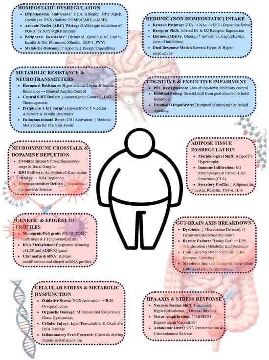
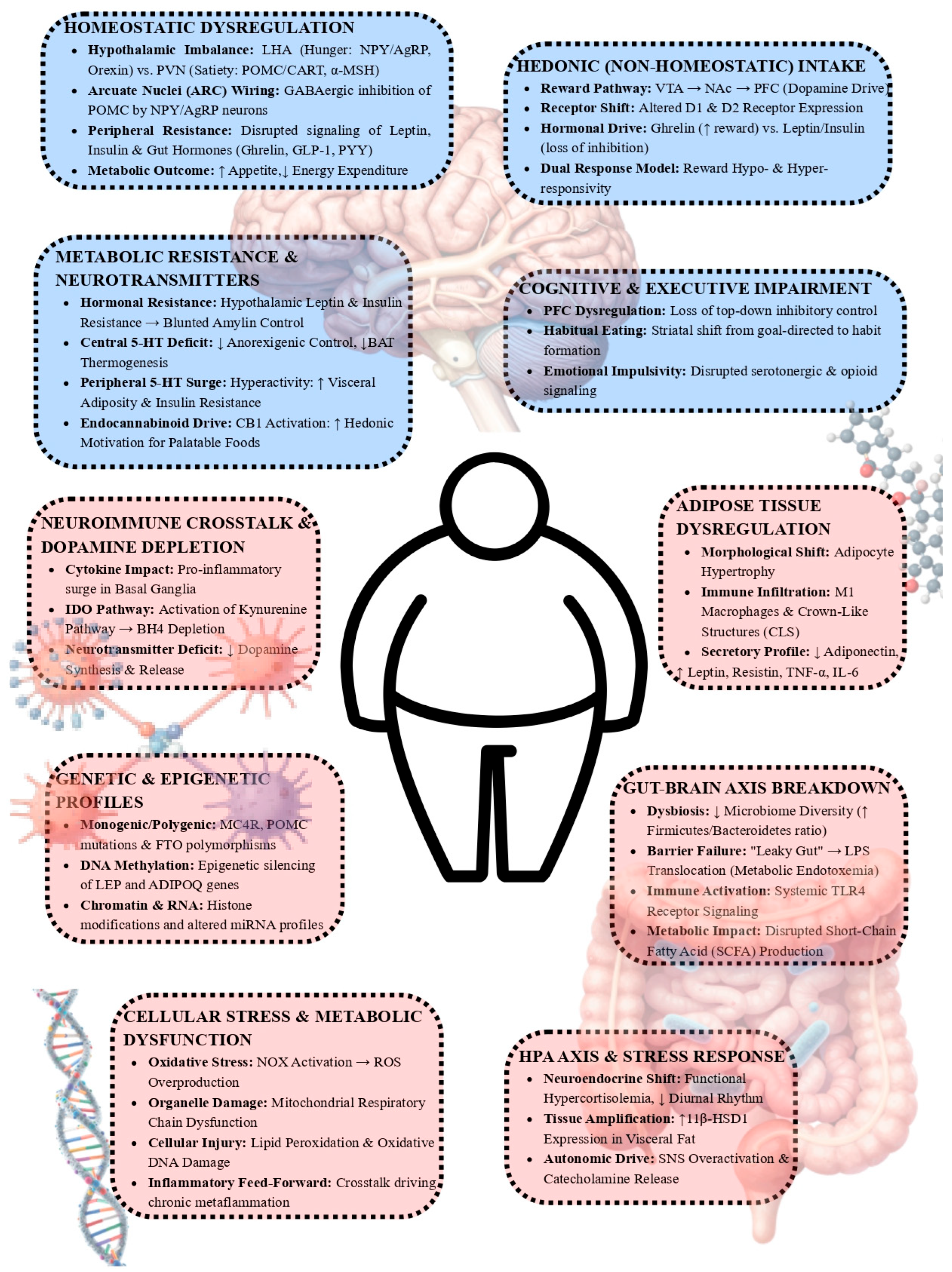

Disclaimer/Publisher’s Note: The statements, opinions and data contained in all publications are solely those of the individual author(s) and contributor(s) and not of MDPI and/or the editor(s). MDPI and/or the editor(s) disclaim responsibility for any injury to people or property resulting from any ideas, methods, instructions or products referred to in the content. |
© 2026 by the authors. Licensee MDPI, Basel, Switzerland. This article is an open access article distributed under the terms and conditions of the Creative Commons Attribution (CC BY) license.