


Abstract
Background/Objectives: Renal scintigraphy with 99mTc-MAG3 is a non-invasive tool for assessing early post-kidney-transplant function and detecting complications. While its utility in predicting delayed graft function (DGF) is established, evidence regarding its capacity to predict long-term graft survival remains limited. This study aimed to evaluate whether early post-transplant scintigraphy provides independent prognostic information for long-term graft survival. Methods: We conducted a retrospective cohort study of kidney transplantations performed at a single tertiary-care academic institution (2015–2019). Patients undergoing simultaneous multi-organ transplantation or experiencing complications precluding early scintigraphy were excluded. All included patients underwent 99mTc-MAG3 scintigraphy within 48 h post-transplantation. Renogram curves were categorized using the Heaf and Iversen classification (Grades 1–4). Univariate and multivariate Cox proportional hazards regression analyses were performed to assess death-censored graft survival. The study followed STROBE reporting guidelines. Results: Among the 317 included patients, renogram curves were distributed as follows: Grade 1 (n = 31, 9.8%), Grade 2 (n = 69, 21.8%), Grade 3 (n = 92, 29.0%), and Grade 4 (n = 125, 39.4%). The overall DGF incidence was 25.9%, with rates progressively increasing across the grades: 0% (Grade 1), 4.3% (Grade 2), 16.3% (Grade 3), and 51.2% (Grade 4) (p < 0.001). On multivariate analysis adjusting for recipient BMI, donation technique, Kidney Donor Risk Index (KDRI), and DGF, grafts with reduced uptake (Grade 4) demonstrated a significantly higher risk of graft loss compared to those with normal uptake (Grades 1–3 combined) (HR: 3.15; 95% CI: 1.34–7.40; p = 0.008). The mean follow-up was 45.6 months (IQR: 34.5–60). Conclusions: 99mTc-MAG3 scintigraphy performed within 48 h of kidney transplantation provides independent prognostic information for long-term graft survival. The Grade 4 renogram pattern identifies a high-risk subgroup with over threefold increased risk of subsequent graft loss. These findings support the integration of early scintigraphy into post-transplant risk stratification protocols, though prospective validation is warranted.
1. Introduction
Kidney transplantation represents the best treatment for patients with end-stage kidney disease (ESRD), providing better survival rates and quality of life than long-term dialysis [1]. Unfortunately, about 20–55% of all transplant recipients will experience delayed graft function (DGF), defined as the need for dialysis within the first week post-transplantation [2,3,4]. The development of DGF is the result of an ischemic/reperfusion injury resulting from complex interactions between the donor and recipient [2,3,4,5]. While many studies have yielded conflicting results regarding whether it has a long-term impact on the outcome of renal transplants, meta-analyses indicate a significantly greater relative risk (RR: 1.41) of renal graft loss among kidney transplant patients with DGF [3,4,5,6].
The observed prognostic variability associated with delayed graft function (DGF) is likely attributable to inherent differences in the viable nephron mass and intrinsic reparative capabilities of individual renal allografts at the time of transplantation [2,7]. Transient cellular injury in allografts procured from standard criteria donors (SCDs) may permit subsequent functional recovery. Conversely, DGF manifesting in grafts from expanded criteria donors (ECDs) has the potential to hasten chronic fibrotic processes, ultimately culminating in irreversible nephron loss [7,8].
Renal scintigraphy using technetium-99m-labeled mercaptoacetyltriglycine (99mTc-MAG3) provides a non-invasive, dynamic assessment of immediate post-transplant renal function [9]. MAG3 is favored over alternative tracers due to its superior excretory characteristics and image quality [10]. Previous studies have established its utility in predicting DGF duration and hospital length of stay [11,12,13]. The Heaf and Iversen grading system, based on renogram curve morphology, enables semi-quantitative assessment of graft function, categorizing scans from normal uptake with reduced excretion (Grade 1) to reduced absolute uptake without excretion (Grade 4) [13].
Currently, no substantial evidence is available to support a correlation between early-phase functional imaging tests and long-term kidney transplant outcomes [1,4,5,6,7]. If there were a strong correlation between functional imaging results at early stages and the long-term success (or failure) of an organ transplant, it could greatly enhance patient risk assessment and allow for increased tailoring of post-operative management strategies.
Study Objectives: The principal aim of this study was to investigate whether 99mTc-MAG3 scintigraphy performed within 48 h of kidney transplantation provides independent prognostic information for long-term death-censored graft survival. The secondary aim was to characterize the relationships between early scintigraphic patterns, DGF incidence, and graft outcomes across different donor and recipient subgroups.
2. Materials and Methods
2.1. Study Design and Reporting
This retrospective cohort study was conducted at a single tertiary-care academic institution and is reported in accordance with the Strengthening the Reporting of Observational Studies in Epidemiology (STROBE) statement (Supplementary File S1). The study protocol was approved by the institutional ethics committee (CEI) at Cordoba (approval ref. N298-4365, 19 December 2019). All procedures followed the ethical standards of the Declaration of Helsinki.
2.2. Study Population
All adult patients (≥18 years) undergoing kidney transplantation at our institution between 1 January 2015 and 31 December 2019 were screened for eligibility. The exclusion criteria were: (1) simultaneous multi-organ transplantation; (2) surgical or clinical complications at the time of scheduled scintigraphy precluding image acquisition; and (3) incomplete follow-up data.
Informed consent for kidney donation, transplantation, and data registry was obtained from all patients prior to inclusion. Donors included both donation after brain death (DBD) and donation after circulatory death (DCD) donors, with DCD further classified as rapid recovery (RR) or normothermic regional perfusion (NRP) cases.
2.3. Scintigraphy Protocol and Image Analysis
All patients underwent renal scintigraphy within 48 h of transplantation. Studies were performed using a General Electric gamma camera (Optima NM/CT 640 or Millenium MPR, GE Medical Systems, 4 Hayozma St., Tirat Carmel, Israel.) equipped with a high-resolution parallel-hole collimator (LEHR) with a 20% window centered on the 140 KeV photopeak of 99mTc. Image matrices were set at 128 × 128 pixels.
Following intravenous bolus injection of 166.5 MBq (4.5 mCi) 99mTc-MAG3 in 20 mL normal saline, patients were positioned supine with the detector field centered over the renal graft and bladder. A two-phase dynamic acquisition protocol was employed: (1) angiographic phase: 1 frame/2 s for 60 s (30 frames); (2) dynamic phase: 1 frame/minute for 19 min (19 frames). A 60 s static image was acquired immediately following the dynamic study.
Regions of interest (ROIs) were drawn manually by a single experienced nuclear medicine physician to ensure consistency. The renal ROI encompassed the entire renal cortex, while the background ROI was placed in a half-moon configuration inferolateral to the kidney. Background-corrected activity–time renogram curves were generated from the start of acquisition to 20 min.
Renogram curves were classified according to the Heaf and Iversen grading scale [13]:
- Grade 1: Normal uptake with reduced excretion;
- Grade 2: Normal uptake with flat excretion curve;
- Grade 3: Rising curve without excretion phase;
- Grade 4: Reduced absolute uptake without excretion phase.
No patients in this cohort exhibited Grade 0 (normal) renograms, indicating that all grafts demonstrated some degree of functional impairment.
2.4. Data Collection and Variables
Data were extracted from electronic medical records and the institutional transplant database. Variables were categorized into three domains:
- -
- Recipient-related: Age, gender, body mass index (BMI), diabetes mellitus (DM), hypertension, peripheral artery disease, cardiac disease, stroke, pre-transplant renal replacement therapy modality (none, CAPD, hemodialysis), time on dialysis, hyperimmunization status (panel-reactive antibody ≥85%), and history of previous kidney transplantation.
- -
- Donor-related: Age, gender, BMI, DM, hypertension, peripheral artery disease, cardiac disease, donation technique (DBD, DCD-RR, DCD-NRP), cause of death (stroke, anoxia, head trauma, other), terminal serum creatinine, and Kidney Donor Risk Index (KDRI).
- -
- Transplant-related: Cold ischemia time (hours), HLA mismatches (number), acute rejection episodes (biopsy-proven), DGF (defined as need for dialysis within first 7 days post-transplant), and follow-up duration (months). The primary outcome was death-censored graft survival, defined as the time from transplantation to a return to chronic dialysis or re-transplantation, with patients censored at death with a functioning graft or last follow-up.
2.5. Statistical Analysis
Descriptive statistics are presented as absolute frequencies (n) and relative frequencies (%) for categorical variables, and as the mean ± standard deviation (SD) or median with interquartile range (IQR) for continuous variables, depending on distribution normality (assessed using the Shapiro–Wilk test).
Between-group comparisons were performed using the Chi-squared test or Fisher’s exact test for categorical variables, and ANOVA or the Kruskal–Wallis test for continuous variables, as appropriate. Kaplan–Meier survival curves were constructed to visualize death-censored graft survival stratified by scintigraphic grade, with between-group comparisons performed using the log-rank test.
Univariate and multivariate Cox proportional hazards regression models were used to assess factors associated with graft loss. The multivariate model included clinically relevant variables and those showing associations at p < 0.10 in the univariate analysis: recipient BMI, donation technique, KDRI, DGF, and scintigraphic grade. Scintigraphic grade was analyzed both as a four-level categorical variable and as a dichotomized variable (Grades 1–3 [preserved uptake] vs. Grade 4 [reduced uptake]), based on the physiological distinction between preserved and reduced radiopharmaceutical uptake.
Results are presented as hazard ratios (HRs) with 95% confidence intervals (CIs). A two-sided p-value < 0.05 was considered statistically significant. All analyses were performed using IBM SPSS Statistics for Windows, version 25.0 (IBM Corp., Armonk, NY, USA).
3. Results
3.1. Study Population
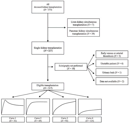
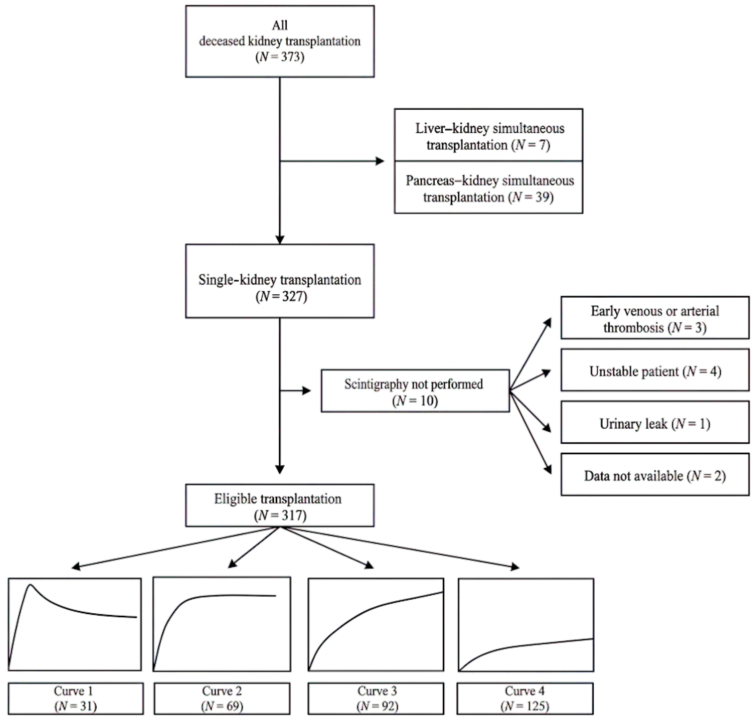
During the five-year study period, 373 kidney transplants were performed at our institution. After the exclusion criteria were applied (56 patients excluded: 23 multi-organ transplants, 33 with complications precluding early scintigraphy), 317 patients were included in the final analysis (Figure 1). The median follow-up was 45.6 months (IQR: 34.5–60 months), providing robust long-term outcome data.
Figure 1.
Flowchart depicting the cohort distribution.
3.2. Baseline Characteristics
Recipient characteristics: The cohort had a mean age of 55.2 ± 13.1 years, with male predominance (65.6%). Diabetes mellitus and hypertension were prevalent in 22.7% and 82.7%, respectively. The majority (80.8%) were receiving hemodialysis pre-transplant, and 13.9% were hyperimmunized. Prior kidney transplantation was documented in 17.4% of recipients.
When stratified by scintigraphic grade, recipient characteristics were largely similar across the groups, with the exception of BMI. Recipients with Grades 3 and 4 had significantly higher BMI compared to those with Grades 1 and 2 (p = 0.005) (Table 1).
Table 1.
Recipient-related characteristics.
Donor characteristics: The mean donor age was 56.4 ± 14.7 years, with 62.5% male donors. Donors in Grade 1 were significantly younger compared to those in other groups (p = 0.04). The prevalence of donor DM and hypertension differed significantly across the groups, being higher in donors associated with Grades 3 and 4 (p = 0.01 and p = 0.02, respectively).
DBD was the predominant donation technique (79% overall), but its proportion decreased progressively from Grade 1 (97%) to Grade 4 (72%) (p = 0.009). Conversely, DCD-RR increased from 3.2% in Grade 1 to 23.2% in Grade 4. KDRI values increased progressively from Grade 1 (1.1) to Grade 4 (1.3) (p = 0.006), indicating higher donor risk in grafts with poorer early function (Table 2).
Table 2.
Donor-related characteristics.
Transplant characteristics: The mean cold ischemia time was 11.6 ± 4.1 h, with no significant differences across the groups (p = 0.7). HLA mismatches and acute rejection rates were similar across the groups.
The overall DGF incidence was 25.9%, with a striking progressive increase across the scintigraphic grades: 0% (Grade 1), 4.3% (Grade 2), 16.3% (Grade 3), and 51.2% (Grade 4) (p < 0.001). The follow-up duration differed significantly across the groups (p = 0.01), with Grade 4 patients having the shortest follow-up (mean 41.5 months), potentially reflecting earlier graft losses (Table 3).
Table 3.
Transplant-related characteristics.
3.3. Graft Survival Analysis
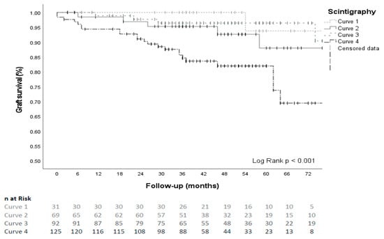
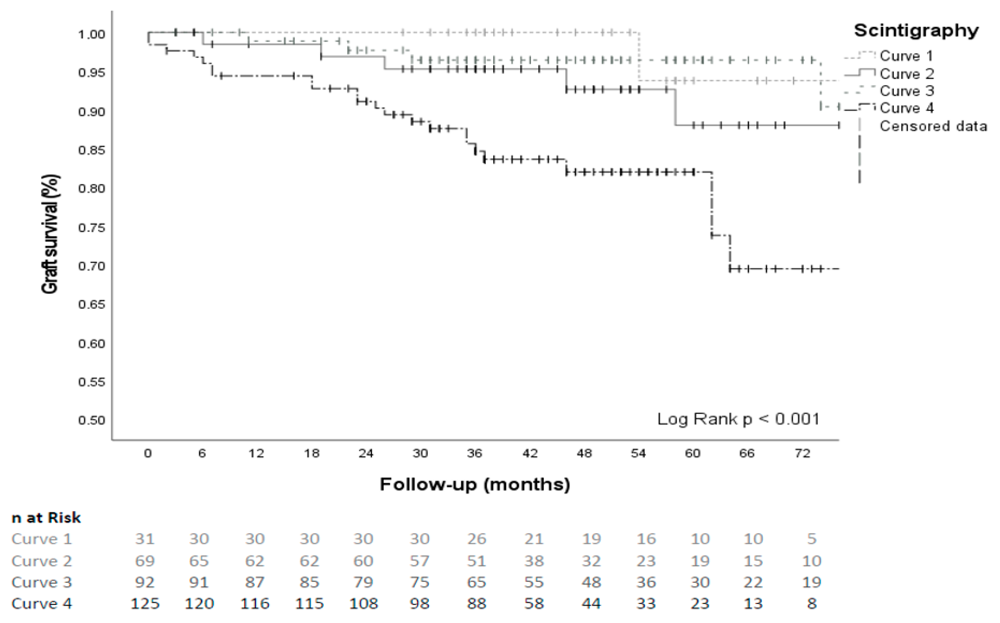
Kaplan–Meier survival analysis demonstrated significantly different graft survival trajectories according to the scintigraphic grade (log-rank p < 0.001). Grade 4 grafts exhibited markedly inferior survival compared to Grades 1–3, which showed comparable outcomes (Figure 2).
Figure 2.
Graft survival analysis: Kaplan–Meier curves stratified by scintigraphic classifications.
3.4. Graft Loss Causes
During follow-up, 42 patients (13.2%) experienced death-censored graft loss. The most frequent underlying causes were chronic allograft dysfunction (n = 19, 45.2%), acute rejection (n = 10, 23.8%), and primary non-function (PNF) (n = 5, 11.9%). Primary non-function occurred exclusively in the Curve 4 group (five of 125 patients, 4.0%). Other causes included recurrent glomerular disease (n = 4), vascular thrombosis (n = 2), and infection-related graft loss (n = 2).
3.5. Multivariate Cox Regression Analysis
Univariate Cox regression identified several factors associated with graft loss. In multivariate analysis (Table 4), after adjustments for recipient BMI, donation technique, KDRI, and DGF, scintigraphic grade remained an independent predictor of graft survival.
Table 4.
Multivariate Cox regression analysis for graft survival.
When analyzed as a dichotomized variable based on the presence of preserved radiopharmaceutical uptake, grafts with reduced uptake (Grade 4) demonstrated a significantly higher risk of graft loss compared to those with preserved uptake (Grades 1–3 combined) (HR: 3.15; 95% CI: 1.34–7.40; p = 0.008). DGF also independently predicted graft loss (HR: 3.58; 95% CI: 1.55–8.26; p = 0.003). Higher recipient BMI was paradoxically associated with a lower risk of graft loss (HR: 0.90; 95% CI: 0.83–0.98; p = 0.01)
The point estimates indicated gradually increasing hazard when scintigraphic grade was entered as a four-level categorical variable with Grade 1 as the reference. However, the association did not reach statistical significance for individual grades, and confidence intervals were wide because there were few events in lower-grade groups.
4. Discussion
4.1. Principal Findings
This retrospective, single-center cohort study illustrates how 99mTc-MAG3 scintigraphy performed within 48 h of kidney transplantation adds independent prognostic value for long-term graft survival. The key finding is that renal allograft patients that have a Grade 4 renogram (very low or absent radiopharmaceutical uptake and absent excretion) are three times more likely to lose the graft than those with radiopharmaceutical uptake (Grades 1–3), even when adjusted for DGF, donor risk indices, and other potential confounders. These findings expand on earlier studies that correlated early scintigraphy results with short-term outcomes [14,15,16,17] by demonstrating a clear association with medium- to long-term graft survival.
4.2. Interpretation and Pathophysiological Insights
The distinction between preserved uptake (Grades 1–3) and reduced uptake (Grade 4) appears physiologically meaningful. Preserved 99mTc-MAG3 uptake reflects functioning nephron mass and intact tubular extraction capacity [13], suggesting that even when excretion is impaired (as in Grades 1–3), the underlying parenchyma retains viability. However, in a Grade 4 state, there is more extensive injury to the parenchyma, which may reflect varying degrees of ischemic–reperfusion injury, insufficient functional reserve, and reduced nephron capacity for tolerating stress [2,18].
DGF rates demonstrated a clear biological gradient by gradually increasing from 0% at Grade 1 to 51.2% at Grade 4. Furthermore, the lack of DGF among patients who were first evaluated at the Grade 1 level demonstrates that normal early uptake is a strong predictor of later DGF development and may assist with the planning of early hospital discharge and the allocation of resources.
Adverse outcomes associated with the Grade 4 pattern could represent irreversible tubular injury that occurs via cell death regulatory mechanisms, including necroptosis [19]. The pro-inflammatory immune response to necroptosis (as opposed to apoptosis) may increase the risk for rejection and contribute to nephron loss secondary to fibrosis [18]. Identifying this phenotype early would allow for the implementation of targeted interventions, but the effectiveness of such interventions at altering the course of disease is not yet known.
4.3. Methodological Strengths
This study has several methodological strengths. First, the consistent cold ischemia time across all groups (mean 11.6 h, p = NS) eliminates this important variable as a confounder in the observed outcome differences. Second, the substantial follow-up period (median 45.6 months) provides confidence in the long-term validity of the findings. Third, by manually drawing all ROIs with a single operator, we minimized inter-observer variability in the renogram classification. Fourth, the comprehensive multivariate analysis adjusted for key confounders including the KDRI, the donation technique, and DGF, strengthening causal inferences.
4.4. Limitations
A few significant limitations should be considered:
Study design: A single-center, retrospective design creates potential selection bias and restricts generalizability. We may have underestimated the true prognostic gradient by excluding sicker patients who were unable to receive imaging due to the requirement for early scintigraphy, even though we included consecutive individuals.
Selection and indication bias: Not every transplant recipient had scintigraphy done in accordance with a rigorous methodology during the study period. Clinical circumstances occasionally prevented or delayed scanning, despite the institution’s goal of performing early imaging on every patient. We cannot rule out the potential that imaging was obtained preferentially in individuals who were thought to be at higher risk due to the lack of a systematic strategy, which could have biased associations. Additionally, clinicians were not blinded to the scintigraphy data, and these results might have affected management choices later, possibly leading to a self-fulfilling prediction for the results.
Timing variability: The precise time after transplant varied, even though all scans were completed within 48 h. This fluctuation may have added measurement error since graft performance can improve quickly in the early post-operative period. Nonetheless, the cohort’s persistent association indicates that the signal is resilient to this fluctuation.
Technical considerations: Despite being done by a single operator to guarantee consistency, manual ROI drawing is still subjective by nature. Reproducibility in multicenter situations may be impacted by inter-operator variability. Furthermore, certain individuals may be misclassified due to patient variables that affect picture quality and quantitative accuracy, such as obesity, overlaying intestinal gas, or inadequate bolus injection.
Sample size and subgroup analyses: Even though there were 317 patients, there were few graft loss events, especially in the lower-grade groups, which led to large confidence intervals when the grade was analyzed as a four-level categorical variable. This required dichotomization for the main multivariate model and prevented in-depth subgroup analysis (such as interaction with donor type).
Causal inference: Even though we controlled measurable confounders, residual confounding from unmeasured factors (such as adherence, subclinical rejection, and specific immunosuppressive procedures) cannot be ruled out. It is not possible to draw firm conclusions on causality due to the observational design.
4.5. Perspectives for Clinical Practice
Although prospective validation is necessary prior to widespread use, the results of this study have numerous potential implications for clinical practice:
Risk stratification: A high-risk subgroup (Grade 4) may be found by early scintigraphy, requiring increased surveillance, closer monitoring, and possibly adjusted immunosuppression. On the other hand, quicker discharge pathways may be appropriate for Grade 1 patients who have normal uptake and no DGF.
Integration with existing tools: In addition to clinical endpoints (DGF) and conventional risk indices (KDRI), the scintigraphic grade offers information. Grade 4 and DGF both independently predicted graft loss in our multivariate model, indicating that early functional imaging captures features of graft health not entirely reflected in clinical DGF status alone.
Cost-effectiveness considerations: The expenses and resource usage of routine early scintigraphy must be compared against its incremental value. The approach might be economical if Grade 4 detection allows for focused therapies that stop graft loss. On the other hand, routine use might not be warranted if scintigraphy results have little effect on management. Cost-effectiveness assessments must be done formally.
Nursing and multidisciplinary care: Early detection of high-risk grafts has consequences for nursing care, including increased fluid balance monitoring, assistance with medication adherence, and early detection of problems. Shared decision-making with patients and families may be improved by including scintigraphic results in multidisciplinary team talks [20].
Technological considerations: Reproducibility and prognostic accuracy may be improved by developments in quantitative analysis, such as automated ROI construction and machine learning techniques for curve interpretation. Risk prediction may be further improved by integration with new transplant injury biomarkers [21].
4.6. Comparison with Previous Studies
The results of the current study are consistent with and build upon the previous research conducted in this area. Stevens and colleagues showed that at 48 h post-transplantation, quantitative measurements from renography were predictive of 5-year graft function [16]. Ngai and Demetriades found that day 3 MAG3 measurements correlated with 1-year graft function [15], while Park and coworkers showed a relationship between early scan results and 5-year outcomes [14]. This study extends upon existing studies by incorporating the Heaf and Iversen clinical grading system, adjusting for contemporary risk criteria (KDRI), and demonstrating independence from DGF in multivariate analyses.
When compared to excretory patterns (Grades 1–3), the higher prognostic signal linked to decreased uptake (Grade 4) indicates that long-term risk is driven by parenchymal injury rather than delayed function per se. This is consistent with previous research demonstrating that the effect of DGF is dependent on the quality of the donor [7,8] and that permanent damage mechanisms may be fundamentally different from reversible dysfunction [18,19].
4.7. Unanswered Questions and Future Research
A number of significant questions are still unanswered and call for further investigation. A multicenter prospective cohort with standardized imaging, blinded outcomes, and sample size calculations based on current effect sizes should first validate the findings. To determine whether responding to scintigraphic findings—for example, stepping up monitoring or interventions in Grade 4 patients—actually improves graft survival or related composite outcomes, a randomized trial is required after validation. While health economic analyses are required to ascertain whether early routine scintigraphy is cost-effective across various healthcare systems, mechanistic studies combining scintigraphy with biomarkers (such as urinary tubular injury markers, donor-derived cell-free DNA) could shed light on the biological pathways linking early functional impairment with later graft loss.
Innovation and methodological improvements should be pursued concurrently. The best time to perform imaging for prognostication (24, 48, or 72 h after transplant) should be determined by comparative effectiveness studies. Lastly, using machine learning to combine scintigraphic data with clinical, donor, and recipient variables may result in more potent predictive models than using any one parameter by itself, aiding in post-transplant management and risk stratification personalization.
5. Conclusions
This retrospective cohort study demonstrated that 99mTc-MAG3 scintigraphy performed within 48 h of kidney transplantation provides independent prognostic information for long-term graft survival. The Grade 4 renogram pattern, characterized by reduced radiopharmaceutical uptake without an excretory phase, identifies a high-risk subgroup with over threefold increased risk of subsequent graft loss, independent of both DGF and conventional donor risk factors. This could allow for customized post-transplant care, with accelerated protocols for low-risk patients and increased monitoring for high-risk grafts.
Due to this study’s limitations, definitive clinical recommendations for early scintigraphy adoption are currently premature; therefore, the findings should generate hypotheses rather than alter practice. Before routine use, prospective multicenter validation using standardized protocols is crucial, along with formal cost-effectiveness analyses to support medical costs across healthcare systems.
Supplementary Materials
The following supporting information can be downloaded at https://www.mdpi.com/article/10.3390/transplantology7020011/s1, Supplementary File S1: STROBE statement checklist for cohort studies.
Author Contributions
Conceptualization, J.S. and J.P.C.; Data curation, J.S., E.G.-G. and J.P.C.; Formal analysis, L.C.; Investigation, J.A.C.; Methodology, J.S. and E.G.-G.; Resources, J.M. and J.A.V.; Supervision, J.P.C. and J.E.R.; Validation, J.M.; Visualization, J.A.C.; Writing—original draft, J.S., L.C. and J.A.V.; Writing—review and editing, J.E.R. All authors have read and agreed to the published version of the manuscript.
Funding
This research received no external funding.
Institutional Review Board Statement
This study was conducted in accordance with the Declaration of Helsinki and approved by the Ethics Committee of Cordoba (TRASUROL Ref. N298-4365, 19 December 2019).
Informed Consent Statement
Informed consent for kidney donation, transplantation, and database registry was obtained from all patients.
Data Availability Statement
The data presented in this study are available on request from the corresponding author. The data are not publicly available due to privacy or ethical restrictions.
Conflicts of Interest
The authors declare no conflicts of interest.
Abbreviations
The following abbreviations are used in this manuscript:
| 99mTc-MAG3 | technetium-99m-mercaptoacetyltriglycine |
| BMI | body mass index |
| CI | confidence interval |
| DBCD | donation after brain death |
| DCD | donation after circulatory death |
| DGF | delayed graft function |
| DM | diabetes mellitus |
| ESRD | end-stage renal disease |
| HLA | human leukocyte antigen |
| HR | hazard ratio |
| IQR | interquartile range |
| KDRI | Kidney Donor Risk Index |
| NRP | normothermic regional perfusion |
| ROI | region of interest |
| RR | rapid recovery |
| SD | standard deviation |
| STROBE | Strengthening the Reporting of Observational Studies in Epidemiology |
References
- Wolfe, R.A.; Ashby, V.B.; Milford, E.L.; Ojo, A.O.; Ettenger, R.E.; Agodoa, L.Y.C.; Held, P.J.; Port, F.K. Comparison of mortality in all patients on dialysis, patients on dialysis awaiting transplantation, and recipients of a first cadaveric transplant. N. Engl. J. Med. 1999, 341, 1725–1730. [Google Scholar] [CrossRef] [Scilit]
- Cavaille-Coll, M.; Bala, S.; Velideeoglu, E.; Hernandez, A.; Archdeacon, P.; Gonzalez, G.; Neuland, C.; Meyer, J.; Albrecht, R. Summary of FDA workshop on ischemia reperfusion injury in kidney transplantation. Am. J. Transplant. 2013, 13, 1134–1148. [Google Scholar] [CrossRef] [Scilit] [PubMed]
- Yarlagadda, S.G.; Coca, S.G.; Formica, R.N.J.; Poggio, E.D.; Parikh, C.R. Association between delayed graft function and allograft and patient survival: A systematic review and meta-analysis. Nephrol. Dial. Transplant. 2009, 24, 1039–1047. [Google Scholar] [CrossRef] [Scilit]
- Salguero, J.; Chamorro, L.; Gomez-Gomez, E.; Robles, J.E.; Campos, J.P. Midterm Outcomes of Kidney Transplantation from Expanded Criteria Donors After Circulatory Death: A Single-Center Retrospective Cohort Study. Exp. Clin. Transplant. 2023, 21, 481–486. [Google Scholar] [CrossRef] [Scilit]
- Salguero, J.; Chamorro, L.; Gomez-Gomez, E.; Robles, J.E.; Campos, J.P. Graft survival and delayed graft function with normothermic regional perfusion and rapid recovery after circulatory death in kidney transplantation: A propensity score matching study. Minerva Urol. Nephrol. 2024, 76, 60–67. [Google Scholar] [CrossRef] [Scilit]
- Li, M.T.; Ramakrishnan, A.; Yu, M.; Daniel, E.; Sandra, V.; Sanichar, N.; King, K.L.; Stevens, J.S.; Husain, S.A.; Mohan, S. Effects of Delayed Graft Function on Transplant Outcomes: A Meta-analysis. Transplant. Direct. 2023, 9, e1433. [Google Scholar] [CrossRef] [Scilit]
- Salguero, J.; Chamorro, L.; Gomez-Gomez, E.; de Benito, P.; Robles, J.E.; Campos, J.P. Kidney Survival Impact of Delayed Graft Function Depends on Kidney Donor Risk Index: A Single-Center Cohort Study. J. Clin. Med. 2023, 12, 6397. [Google Scholar] [CrossRef] [Scilit]
- Han, F.; Lin, M.Z.; Zhou, H.L.; Sun, Q.-P.; Huang, Z.-Y.; Hong, L.-Q.; Wang, G.; Cai, R.-M.; Sun, Q.-Q. Delayed graft function is correlated with graft loss in recipients of expanded-criteria rather than standard-criteria donor kidneys: A retrospective, multicenter, observation cohort study. Chin. Med. J. 2020, 133, 561–570. [Google Scholar] [CrossRef] [Scilit]
- Benjamens, S.; Berger, S.P.; Glaudemans, A.W.J.M.; Sanders, I.S.F.; Pol, R.A.; Slart, R.H.J.A. Renal scintigraphy for post-transplant monitoring after kidney transplantation. Transplant. Rev. 2018, 32, 102–109. [Google Scholar] [CrossRef] [Scilit] [PubMed]
- Russell, C.D.; Thorstad, B.L.; Stutzman, M.E.; Yester, M.V.; Fowler, D.; Dubovsky, E.V. The kidney: Imaging with Tc-99m mercaptoacetyltriglycine, a technetium-labeled analog of iodohippurate. Radiology 1989, 172, 427–430. [Google Scholar] [CrossRef] [Scilit] [PubMed]
- Benjamens, S.; Pol, R.A.; Berger, S.P.; Glaudemans, A.W.J.M.; Dibbets-Schneider, P.; Slart, R.H.J.A.; de Geus-Oei, L.-F. Limited clinical value of two consecutive post-transplant renal scintigraphy procedures. Eur. Radiol. 2020, 30, 452–460. [Google Scholar] [CrossRef] [Scilit] [PubMed]
- Tulchinsky, M.; Dietrich, T.J.; Eggli, D.F.; Yang, H.C. Technetium-99m-MAG3 scintigraphy in acute renal failure after transplantation: A marker of viability and prognosis. J. Nucl. Med. 1997, 38, 475–478. [Google Scholar] [PubMed]
- Heaf, J.G.; Iversen, J. Uses and limitations of renal scintigraphy in renal transplantation monitoring. Eur. J. Nucl. Med. 2000, 27, 871–879. [Google Scholar] [CrossRef] [Scilit]
- Park, U.J.; Kim, H.T.; Cho, W.H.; Kim, M.-Y.; Hwang, E.-A.; Han, S.-Y.; Park, S.-B.; Kim, H.-C.; Zeon, S.-K. Use of early postoperative MAG3 renal scan to predict long-term outcomes of renal transplants. Exp. Clin. Transplant. 2013, 11, 118–121. [Google Scholar] [CrossRef] [Scilit]
- Ngai, S.; Demetriades, T. Prediction of long-term renal transplant allograft function from day 3 post-transplant Tc-99m MAG3 scintigraphy. Clin. Nucl. Med. 2008, 33, 102–105. [Google Scholar] [CrossRef] [Scilit]
- Stevens, H.; de Klerk, J.M.; Mertens, I.J.; van Rijk, P.P.; Hené, R.J. Quantitative baseline renography 48 hours after renal transplantation predicts long-term graft survival. Eur. J. Nucl. Med. 2001, 28, 1677–1681. [Google Scholar] [CrossRef] [Scilit] [PubMed]
- Barba, J.F.; Romero, L.; Tolosa, E.; Algarra, R.; Rosell, D.; Robles, J.; Zudaire, J.; Berian, J.; Richter, J.; Pascual, J. Study on the findings of an immediate renal gammagraphy and its effect on the survival of a kidney graft. Actas Urol. Esp. 2011, 35, 218–224. [Google Scholar] [CrossRef] [Scilit]
- Maremonti, F.; Meyer, C.; Linkermann, A. Mechanisms and Models of Kidney Tubular Necrosis and Nephron Loss. J. Am. Soc. Nephrol. 2022, 33, 472–486. [Google Scholar] [CrossRef] [Scilit]
- Pefanis, A.; Ierino, F.L.; Murphy, J.M.; Cowan, P.J. Regulated necrosis in kidney ischemia-reperfusion injury. Kidney Int. 2019, 96, 291–301. [Google Scholar] [CrossRef] [Scilit] [PubMed]
- Mancin, S.; Morales Palomares, S.; Sguanci, M.; Palmisano, A.; Gazineo, D.; Parozzi, M.; Ricco, M.; Savini, S.; Ferrara, G.; Anastasi, G.; et al. Relational skills of nephrology and dialysis nurses in clinical care settings: A scoping review and stakeholder consultation. Nurse Educ. Pract. 2025, 82, 104229. [Google Scholar] [CrossRef] [Scilit]
- Wieringa, F.P.; Suran, S.; Søndergaard, H.; Ash, S.; Cummins, C.; Chaudhuri, A.R.; Irmak, T.; Gerritsen, K.; Vollenbroek, J. The future of technology-based kidney replacement therapies: An update on portable, wearable, and implantable artificial kidneys. Am. J. Kidney Dis. 2025, 85, 787–796. [Google Scholar] [CrossRef] [Scilit] [PubMed]

Disclaimer/Publisher’s Note: The statements, opinions and data contained in all publications are solely those of the individual author(s) and contributor(s) and not of MDPI and/or the editor(s). MDPI and/or the editor(s) disclaim responsibility for any injury to people or property resulting from any ideas, methods, instructions or products referred to in the content. |
© 2026 by the authors. Licensee MDPI, Basel, Switzerland. This article is an open access article distributed under the terms and conditions of the Creative Commons Attribution (CC BY) license.