


Abstract
Background: Total hip arthroplasty (THA) is an effective treatment for advanced hip osteoarthritis, yet the influence of the surgical approach on early biomechanical and neuromuscular recovery remains debated. The direct anterior approach (DAA) is considered muscle-sparing, whereas the lateral approach (LA) involves partial abductor detachment and may influence postoperative gait and muscle activation. As a first step, this pilot study was designed primarily to assess the feasibility of an integrated, multidomain assessment protocol to combine clinical and patient-reported outcomes, instrumented gait analysis, hip biomechanics, and surface electromyography (sEMG), and to delineate preliminary trajectories of recovery after DAA versus LA THA to inform a future, adequately powered trial. Methods: In this prospective, non-randomized pilot study, 12 patients undergoing primary unilateral THA were evaluated and allocated according to surgical approach (DAA, n = 6; LA, n = 6). Assessments were performed at three months (T3M) and six months (T6M) postoperatively. Outcomes included pain intensity, SF-12, the Hip disability and Osteoarthritis Outcome Score (HOOS), instrumented gait analysis, hip kinematics and kinetics, and sEMG activity of the gluteus medius, semitendinosus, and rectus femoris of the operated limb. Given the small sample, between-group differences, within-group changes, and change scores from T3M to T6M were analyzed using non-parametric tests, and all results were treated as exploratory and hypothesis-generating. Results: Recruitment, retention, and completion of the full multidomain protocol were feasible, with no missing primary outcomes. Preliminary findings suggested that at three months, the DAA group showed lower pain and higher PCS-12, HOOS Total and HOOS Function scores, together with faster gait speed, longer normalized stride length, and shorter cycle and stance times. At six months, spatiotemporal gait parameters no longer appeared to differ between groups, suggesting the convergence of global gait performance, whereas the DAA group tended to maintain higher HOOS Total and Function scores and lower pain. The LA group showed greater late-stance hip extension and higher semitendinosus and rectus femoris sEMG amplitudes at T6M. Change-score analysis suggested a possible catch-up phenomenon in the LA group for gait speed, stride length, stance time, cycle time, and hip flexion range of motion, whereas semitendinosus activation tended to decrease in the DAA group and increase in the LA group. Given the limited sample, these observations are preliminary and require confirmation. Conclusions: This pilot study supports the feasibility of an integrated protocol combining patient-reported measures, instrumented gait analysis, and sEMG to characterize early recovery after THA. Preliminary, hypothesis-generating data suggest that DAA may be associated with a more favorable early postoperative clinical and spatiotemporal gait profile, with global gait performance tending to converge between approaches by six months, while persistent differences in hip-specific patient-reported outcomes and sEMG activation patterns hint that a similar gait performance may be achieved through different neuromuscular strategies. These preliminary findings should be interpreted with caution and used primarily to inform the design of larger, adequately powered studies with longer follow-up.
1. Introduction
Total hip arthroplasty (THA) is widely regarded as one of the most successful surgical procedures of the past decades, originally driven by advances in implant design, biomaterials, and fixation strategies [1]. More recently, attention has increasingly shifted toward “minimally invasive” techniques; however, there is still no universal agreement on what “minimally invasive” precisely means. Some authors emphasize a shorter skin incision, whereas others reserve the term for approaches that minimize soft tissues and muscle disruption, underscoring the need to distinguish minimal-incision methods from muscle-sparing techniques [2].
The clinical rationale to minimize soft-tissue trauma is the expectation of earlier mobilization and faster rehabilitation, particularly during the immediate postoperative period [3]. Several studies have associated these techniques with reduced blood loss and less pain compared with standard approaches [4]. Nonetheless, objective evidence demonstrating faster recovery, especially regarding walking ability and gait quality, remains relatively limited [5].
Among the commonly used approaches, the lateral approach (LA) is widely adopted; it requires release of part of the gluteus medius, raising concerns about potential alterations in gait mechanics and delayed postoperative recovery [6]. The direct anterior approach (DAA) has gained popularity as an alternative because it uses an internervous plane between the tensor fascia latae and sartorius, reducing soft-tissue dissection and preserving periarticular musculature; it is often described as minimally invasive and is associated with a shorter incision [7]. Importantly, THA outcomes are not determined by the surgical approach alone, but also by multiple factors including implant-related and technical variables [8].
Restoring a near-normal gait is a key goal after THA, particularly within the first postoperative year [9]. Gait analysis provides objective quantification of walking performance and has been used to evaluate postoperative hip function and gait quality after hip replacement [10,11]. More generally, gait analysis is applied to individuals with conditions affecting their ability to walk, and walking performance is commonly characterized through spatiotemporal gait parameters [12]. Spatiotemporal assessment is one of the principal components of gait analysis, together with kinematic and kinetic domains [13]. Methods span a continuum from low-cost clinical approaches (e.g., visual observation and simple timing tools) to complex instrumented systems used in research or specialized facilities [14]. Advanced gait laboratories may differ substantially in the platform and instrumentation used: conventional analyses have historically relied on force plates in overground settings, whereas newer systems increasingly include instrumented treadmills, at times enabling self-selected walking speed with the potential to capture multiple consecutive strides in limited space [15,16]. Despite acknowledged practical limitations, three-dimensional optoelectronic motion-capture systems are often considered the reference standard for detailed gait assessment [17]. These systems use retroreflective skin markers placed on anatomical landmarks to build biomechanical models and derive spatiotemporal, kinematic, and kinetic parameters [18]. Spatiotemporal outcomes typically include distance-related measures such as step and stride length and time-related measures such as stride time and walking speed, whereas kinematic analysis focuses on joint and segment angular motion and kinetic analysis quantifies movement-related forces [19].
From a broader perspective, THA should not be viewed solely as a structural reconstruction, but also as a substantial biomechanical and neuromuscular transition [20]. In this framework, electromyography (EMG) has become an important tool to investigate neuromuscular adaptations after THA, as it captures the electrical activity produced by skeletal muscles and their motor units during activation, enabling the direct evaluation of muscle activation and neuromuscular control [21]. Needle EMG allows for a detailed assessment of motor unit integrity and can identify denervation and reinnervation processes [22]. Early postoperative needle-EMG investigations reported impaired voluntary activation and reduced recruitment capacity of the hip abductors [23]. Subsequently, surface EMG has been used to describe muscle activation patterns during movement, providing quantitative information on timing, amplitude, and intermuscular coordination during functional tasks [24]. EMG studies suggest that atypical activation patterns may persist for months after surgery and may involve not only the abductors, but also other hip-related muscles during gait and load-bearing activities [25]. More recent high-density surface EMG and multimodal approaches indicate that neuromuscular reorganization can extend beyond the hip, involving coordinated adaptations across the limb and trunk, and may remain detectable up to one year or longer after surgery, even after pain resolution and basic functional recovery [26,27]. This apparent dissociation between biomechanical improvement and neuromuscular reorganization highlights a clinically relevant gap in current postoperative evaluation models [28].
Despite the growing interest in muscle-sparing approaches and the increasing use of objective measures to study recovery after THA, the available evidence remains fragmented: clinical outcomes are often reported separately from biomechanical performance, and neuromuscular adaptations may not align temporally with pain resolution or basic functional gains [5,27,28]. Moreover, while gait analysis and EMG have each been used to investigate postoperative recovery, an integrated, multidomain characterization capable of capturing compensatory motor strategies early after surgery is still lacking [5,12,24].
Therefore, the aim of this pilot prospective study was to perform a multidomain comparison between anterior and lateral THA by integrating patient-reported and clinical outcomes with objective gait analysis and surface electromyography (sEMG) [10,11,12,24]. We specifically sought to determine whether approach-related differences in early recovery are detectable through gait- and EMG-derived metrics that reflect compensatory activation patterns, and whether these metrics provide complementary information beyond conventional clinical assessment [19,25,28].
We hypothesized that compared with the lateral approach, the anterior approach would be associated with a faster normalization of functional outcomes and spatiotemporal gait parameters, alongside a more physiological sEMG activation profile during walking, indicating reduced compensatory neuromuscular strategies in the early postoperative phase [23,25,29,30,31].
2. Materials and Methods
2.1. Study Design and Ethics
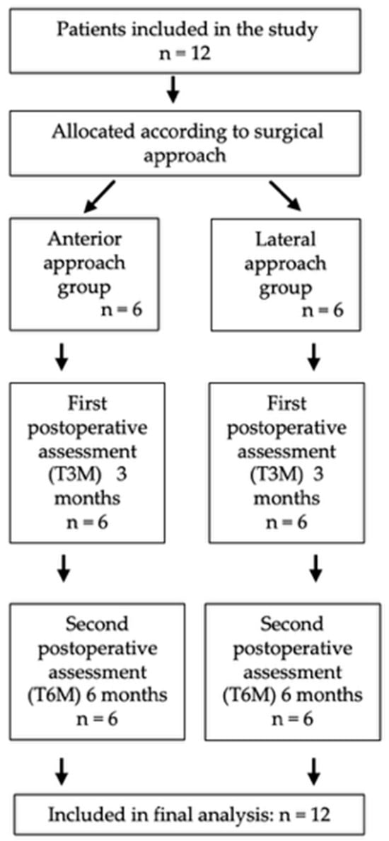
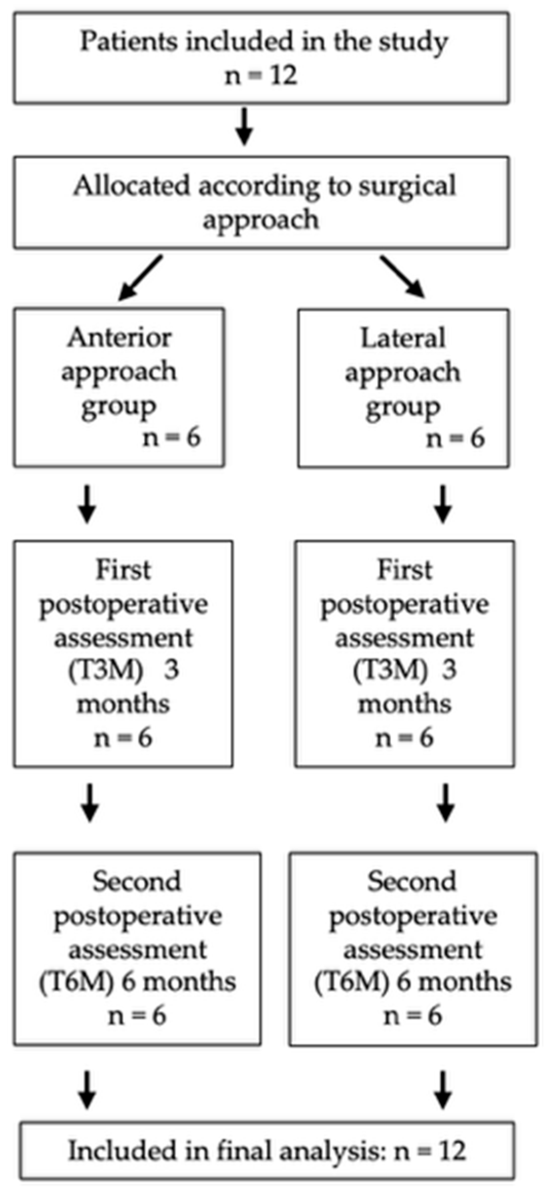
This prospective, two-arm observational study compared clinical outcomes, spatiotemporal and biomechanical gait parameters, and surface electromyographic (sEMG) recovery following direct anterior approach (DAA) versus lateral approach (LA) total hip arthroplasty (THA). A total of 12 patients were included in the analysis: 6 in the anterior approach group and 6 in the lateral approach group. Patients were screened and data were collected between June 2024 and December 2025 at Azienda Ospedaliero-Universitaria Sant’Andrea, Rome, Italy. The study was conducted in accordance with the Declaration of Helsinki and approved by La Sapienza University’s Institutional Review Board (Prot. 0495/2024—Approval Date: 29 May 2024). All participants provided written informed consent prior to the study procedures. No clinical trial registration was performed. According to the ICMJE definition, this study does not qualify as a clinical trial because it was observational: participants were not prospectively assigned by the investigators to a surgical intervention according to a study protocol, but were allocated to the anterior or lateral approach as part of routine clinical practice. Data were collected prospectively for observational purposes only. A flow diagram of this study is shown in Figure 1.
Figure 1.
Flow diagram of the study.
2.2. Participants
Eligible participants were adults undergoing primary unilateral THA for hip osteoarthritis and able to safely perform instrumented gait testing at follow-up. To reduce confounding factors affecting gait and neuromuscular activation, participants were not enrolled in the presence of conditions expected to significantly alter walking performance (e.g., major neurologic disorders, severe contralateral lower-limb impairment, or inability to ambulate independently at the scheduled evaluation). Patients were assigned to groups according to the surgical approach performed (DAA vs. LA) as part of routine clinical practice (non-randomized observational allocation). Baseline demographic and anthropometric data were recorded, including age, sex, weight, height, body mass index (BMI), and physical activity level.
2.3. Surgical Procedure
All procedures were performed by a single senior surgeon with extensive experience in both surgical approaches, to minimize technique-related variability. The same implant system was used in all cases to ensure consistency across groups. All patients received an uncemented acetabular component (Trinity™ cup, Corin Ltd., Cirencester, UK) combined with a cementless tapered wedge femoral stem (TriFit TS™, Corin Ltd., Cirencester, UK).
Direct anterior approach (DAA): In the anterior approach group, patients were positioned supine on a radiolucent operating table. The hip was approached through a minimally invasive direct anterior technique utilizing an intermuscular and internervous interval. A skin incision was performed approximately 3 cm lateral and 3 cm distal to the anterior superior iliac spine, oriented along the axis of the tensor fasciae latae (TFL). The intermuscular plane between the TFL and sartorius was developed proximally and continued distally between the TFL and rectus femoris. Care was taken to remain within the anatomical interval to minimize the risk of traction or irritation of the lateral femoral cutaneous nerve. Capsular access was obtained through a longitudinal capsulotomy. Femoral exposure was achieved through controlled release of the anterior and superior capsule to allow adequate hip extension and external rotation, without detachment of the abductor tendons [32,33].
Lateral approach (LA): In the lateral approach group, patients were positioned in the lateral decubitus position. A longitudinal incision centered over the greater trochanter was performed. After splitting the fascia lata, exposure was obtained through a transgluteal approach consistent with the Hardinge technique. Partial detachment of the anterior fibers of the gluteus medius and minimus was performed to allow capsular visualization and joint access, avoiding excessive proximal dissection to preserve the superior gluteal nerve. Following component implantation, meticulous reattachment of the abductor mechanism was performed to restore anatomical tension and footprint [34].
Perioperative care was standardized for all patients. Spinal anesthesia combined with loco-regional analgesia was administered in all cases. Antibiotic prophylaxis and thromboprophylaxis protocols were identical between groups and followed institutional guidelines. No systematic differences in perioperative management were present between the two surgical approaches.
2.4. Postoperative Rehabilitation
Postoperative management followed a standardized rehabilitation pathway applied consistently to both surgical approach groups. Early mobilization was initiated as soon as clinically feasible, according to the patient’s general clinical status, pain status, and postoperative precautions. Ambulation was progressively introduced under physiotherapist supervision, with gait re-education focused on safe weight bearing, step symmetry, balance control, and progressive recovery of walking independence. Assistive devices were used during the early postoperative phase, and were gradually reduced according to pain, balance, walking safety, and neuromuscular control.
The rehabilitation program included progressive functional exercises aimed at restoring hip and pelvic stability, lower-limb strength, and tolerance to daily activities. Exercises were individualized according to clinical evaluation and generally included active and active-assisted hip mobility exercises within the permitted range, isometric and progressive strengthening exercises for the hip and lower-limb muscles, balance and transfer training, stair training when appropriate, and progressive walking practice. Exercise intensity and functional progression were advanced according to pain, fatigue, movement quality, and patient tolerance.
All patients received standardized instructions for home-based activity progression and were referred to outpatient physiotherapy when clinically indicated. The same rehabilitation principles were applied to both groups, and no group-specific rehabilitation protocol was prescribed according to surgical approach. This approach is consistent with enhanced-recovery and postoperative rehabilitation principles after THA, which emphasize early mobilization, progressive functional recovery, gait training, strengthening, and individualized progression according to patient status and tolerance [4,35,36].
2.5. Assessment Timeline
Participants were enrolled consecutively during the study period (June 2024 and December 2025) and were followed according to a predefined postoperative assessment schedule aimed at capturing early recovery and mid-term functional stabilization. For each patient, the evaluation pathway included two standardized postoperative follow-up visits, planned at three months (T3M) and six months (T6M) after surgery. These timepoints were selected to reflect two clinically meaningful phases: the transition from initial recovery to autonomous gait reorganization (around 3 months), and the subsequent consolidation of walking function and neuromuscular strategies (around 6 months) [37].
At both T3M and T6M, participants underwent the same sequence of assessments on the same day whenever feasible, starting with the completion of patient-reported outcome measures and pain rating, followed by the instrumented gait analysis session with synchronized sEMG recording. Preoperative pain intensity data were available in the clinical records, therefore baseline VAS scores were extracted to better characterize the presurgical status and to contextualize postoperative pain trajectories. All outcomes were recorded using identical procedures at both postoperative evaluations.
2.6. Clinical and Patient-Reported Outcomes
At each postoperative assessment, pain and function were evaluated using a standardized set of validated measures. Pain intensity at the time of evaluation was quantified through a Visual Analog Scale (VAS) [38]. Generic health status was assessed using the 12-Item Short Form Survey (SF-12), with computation of the Physical Component Summary (PCS-12) and Mental Component Summary (MCS-12) scores [39]. Hip-specific symptoms and disability were captured using the Hip disability and Osteoarthritis Outcome Score (HOOS), including both the total score and domain-level scores (Pain, Function, and Quality of Life) [40]. All questionnaires were administered in the same modality at both timepoints, and scores were calculated according to the respective scoring manuals to ensure comparability across visits.
2.7. Instrumented Gait Analysis and Surface Electromyography
Instrumented gait analysis with synchronized surface electromyography (sEMG) was performed at both postoperative timepoints (T3M, 3 months; T6M, 6 months) to objectively characterize locomotor recovery after total hip arthroplasty performed through an anterior versus lateral approach. Gait data were acquired using a three-dimensional optoelectronic motion capture system (Smart D500; BTS Bioengineering, Milan, Italy) integrated with a force platform (INFINI-T-Force Plate BTS Bioengineering, Milan, Italy) and two video camera systems (BTS Bioengineering). Marker trajectories were sampled at 100 fps and ground reaction forces at 500 Hz. Prior to each session, system calibration was performed according to the manufacturer’s procedures. Reflective markers were placed on anatomical landmarks according to the protocol described by Davis et al. [41]. Participants walked barefoot along a 8–10 meter walkway at a self-selected comfortable speed. After a familiarization trial, each participant performed repeated walking trials at a self-selected speed. For each patient, three valid gait trials were recorded and analyzed. A trial was considered valid when the participant walked at a self-selected speed without interruption and with complete marker trajectories and force-plate acquisition [42,43].
Spatiotemporal outcomes included mean gait speed, cadence, cycle time, stance time of the operated limb, and stride length normalized to height [44]. Hip kinematics focused on sagittal-plane measures, including minimum hip flexion (as a surrogate of late-stance hip extension) and hip flexion range of motion (ROM) [45]. Hip kinetics were summarized using hip flexion–extension moment descriptors (peak and minimum values during gait) derived from synchronized kinematic and ground-reaction force data [46,47,48].
sEMG signals were recorded concurrently from the gluteus medius, rectus femoris, and semitendinosus of the operated limb with a sampling frequency of 1000 Hz. Skin was prepared (shaving, abrasion, alcohol) to reduce impedance, and bipolar surface electrodes were placed according to standardized recommendations with an inter-electrode distance of 20 mm [49,50]. Raw EMG signals were band-pass filtered (lower and upper cut-off frequencies of the Hamming filter were 10 and 400 Hz), full-wave rectified, and subsequently low-pass filtered at 5 Hz to obtain the linear envelope [51]. The amplitude values used for analysis therefore corresponded to processed sEMG envelope amplitudes expressed in microvolts (µV). No normalization to maximal voluntary contraction or to a dynamic reference task was performed.
2.8. Sample Size and Statistical Analysis
Given the pilot design, sample size estimation was informed by the large between-group effect observed in Varin et al. [52] for postoperative contralateral hip flexion/extension ROM (assumed Cohen’s d = 1.8), resulting in a minimum target of 6 participants per group (α = 0.05; power = 0.85).
Statistical analyses were performed using IBM SPSS Statistics (version 29.0; IBM Corp., Armonk, NY, USA). Continuous variables are presented as mean ± standard deviation (SD), and categorical variables as counts and percentages. Data distribution was explored using Shapiro–Wilk tests and visual inspection; given the pilot nature of the study and the distribution of several outcomes, non-parametric tests were used for the main comparisons.
Between-group comparisons (DAA vs. LA) at each postoperative timepoint (T3M and T6M) were performed using the Mann–Whitney U test. Within-group changes from T3M to T6M were assessed using the Wilcoxon signed-rank test. To compare recovery progression between approaches, change scores were computed as Δ = T6M − T3M and compared between groups using the Mann–Whitney U test. Effect sizes for between-group comparisons were calculated as rank-biserial correlation (r_rb). Statistical significance was set at p < 0.05 (two-tailed). Given the pilot and exploratory nature of the study and the multidomain assessment performed, no adjustment for multiple comparisons was applied. Therefore, all p values should be interpreted as exploratory and hypothesis-generating rather than confirmatory.
3. Results
3.1. T3M, 3-Month Postoperative Assessment Characteristics
At the first study assessment (T3M, three months postoperatively), no significant between-group differences were found in demographic or clinical characteristics (all p > 0.05) (Table 1). The two groups were comparable in terms of age, body weight, height, body mass index (BMI), and preoperative pain intensity (VAS). Similarly, the distribution of sex and physical activity level did not differ significantly between groups. These findings support the comparability of the sample, suggesting that subsequent between-group comparisons are less likely to be explained by baseline imbalances.
Table 1.
Characteristics at first postoperative assessment (T3M, 3 months).
3.2. Between-Group Comparison at Three Months Postoperatively (T3M)
At three months postoperatively (T3M), Mann–Whitney U tests were performed to compare the two groups across clinical outcomes and operated-limb gait, kinematic, kinetic, and electromyographic parameters, and significant between-group differences are summarized in Table 2.
Table 2.
Significant between-group differences at three months postoperatively (T3M).
At T3M, the anterior approach group (Group 1) exhibited significantly better clinical and patient-reported outcomes. Specifically, VAS pain was significantly lower in the anterior group (3.00 ± 0.63 vs. 4.17 ± 0.75; p = 0.026), a difference exceeding the published MCID threshold for postoperative pain measured using the VAS [53,54]. PCS-12 was also significantly higher in the anterior group (35.03 ± 3.43 vs. 28.08 ± 2.53; p = 0.009), with a between-group difference meeting the published MCID thresholds reported for the SF-12 Physical Component in orthopedic populations [55].
HOOS Total (68.20 ± 4.54 vs. 61.69 ± 1.58; p = 0.026), and HOOS Function (73.28 ± 5.55 vs. 67.39 ± 1.67; p = 0.009) were also significantly higher in the anterior group, with between-group differences approaching lower published MCID thresholds for HOOS-derived measures after total hip replacement [56].
Overall, these findings indicate better perceived physical health, better hip-related outcome, and lower pain intensity in the anterior group at the first postoperative assessment. MCS-12 (p = 0.093), HOOS Pain (p = 0.485), and HOOS Quality of Life (p = 0.180) did not differ significantly between groups.
In the gait analysis, mean gait speed (% height/s) was significantly higher in the anterior group than in the lateral group (58.8 ± 5 vs. 44.7 ± 4.6; p = 0.002), and the magnitude of this difference exceeded commonly reported thresholds for clinically meaningful differences in gait speed [57] and longer normalized stride length (68.02 ± 5.61% height vs. 59.87 ± 5.90% height; p = 0.041), together with shorter cycle (1.14 ± 0.09 s vs. 1.33 ± 0.14 s; p = 0.026) and stance times (0.68 ± 0.08 s vs. 0.83 ± 0.07 s; p = 0.026).
These findings are consistent with a faster gait pattern characterized by higher walking speed, longer stride length, and shorter cycle and stance times in the anterior group. Cadence tended to be higher in the anterior group (104.75 ± 8.74 vs. 90.97 ± 12.29 steps/min; p = 0.093), although this difference did not reach significance. No significant differences were detected in swing time, stance and swing phase proportions, single- and double-support durations, or step width (all p > 0.10). The kinematic analysis revealed comparable hip joint ranges of motion between groups for flexion, abduction, and rotation (all p > 0.10). Similarly, kinetic parameters (peak and minimum hip flexion–extension moments) and electromyographic amplitudes of the gluteus medius, semitendinosus, and rectus femoris muscles on the operated side were not significantly different (all p > 0.10). All significant between-group differences showed large effect sizes (|r(rb)| > 0.5).
3.3. Between-Group Comparison at Six Months Postoperatively (T6M)
At six months postoperatively (T6M), Mann–Whitney U tests were performed to compare the anterior and lateral approach groups on clinical, gait, kinematic, and electromyographic variables of the operated limb; significant between-group differences are summarized in Table 3. No significant differences were observed in VAS pain (p = 0.60), PCS-12 (p = 0.42), or MCS-12 (p = 0.87), indicating comparable levels of perceived pain intensity and general health status between groups at this stage. Regarding HOOS scores, the anterior approach group continued to demonstrate superior self-reported outcomes. HOOS Total (81.37 ± 2.64 vs. 73.87 ± 1.60; p = 0.004), HOOS Pain (85.50 ± 1.87 vs. 77.39 ± 3.16; p = 0.004), and HOOS Function (85.57 ± 2.02 vs. 81.51 ± 1.27; p = 0.010). The HOOS Quality of Life subscale (p = 0.23) showed no significant difference.
Table 3.
Significant between-group differences at six months postoperatively (T6M).
In the gait analysis, no significant differences were detected between groups in cycle time, stance time, swing time, stance/swing phase proportions, double support phase, mean gait speed, cadence, stride length, step length, or step width (all p > 0.33). No between-group differences were observed in spatiotemporal gait parameters at T6M, suggesting a convergence of gait performance by six months postoperatively.
The kinematic assessment showed similar ranges of motion for hip flexion, abduction, and rotation (all p > 0.33). Minimum hip flexion differed between groups (p = 0.037), with the lateral group reaching a more extended hip position during late stance (−8.42 ± 6.49°) compared with the anterior group (1.87 ± 7.89°). All other kinematic variables (maximum angles and ROMs) were comparable (p > 0.33). Regarding hip joint kinetics, neither the peak hip flexion–extension moment (0.48 ± 0.22 vs. 0.28 ± 0.10; p = 0.26) nor the minimum hip moment (−0.12 ± 0.52 vs. −0.58 ± 0.15; p = 0.20) differed significantly, indicating similar dynamic joint loading across surgical approaches.
For electromyographic (EMG) activity, gluteus medius amplitude was not significantly different (p = 0.18). However, semitendinosus activation was significantly higher in the lateral approach group (116.96 ± 26.03 vs. 55.16 ± 30.04; p = 0.004), and rectus femoris amplitude showed a significant difference (48.22 ± 14.10 vs. 36.08 ± 9.24; p = 0.04); both were higher in the lateral group. All significant between-group differences showed large effect sizes (|r_rb| > 0.5), supporting a relevant between-group difference at T6M.
3.4. Between-Group Comparison of Changes (Δ = T6M – T3M)
Mann–Whitney U tests were performed to compare postoperative changes (Δ = T6M – T3M) between the anterior and lateral approach groups, assessing functional progression from three to six months after surgery. Significant between-group differences were found for HOOS Pain, cycle time, stance time, mean normalized gait speed, stride length normalized to height, hip flexion ROM, and semitendinosus activation. The HOOS Pain score improved more in the anterior approach group (Δ = 20.86 ± 2.74) than in the lateral group (Δ = 15.40 ± 4.41; p = 0.025), indicating greater perceived pain relief. For gait temporal parameters, the anterior approach showed a smaller decrease in cycle time (Δ = −0.005 ± 0.12 vs. −0.19 ± 0.41; p = 0.041) and stance time (Δ = −0.007 ± 0.12 vs. −0.16 ± 0.09; p = 0.045), whereas the lateral approach exhibited a larger reduction in both measures. The normalized gait speed increased in both groups, with a greater increase in the lateral group (Δ = 4.8 ± 8.4 vs. 20.1 ± 13.2; p = 0.040). Regarding spatiotemporal gait parameters, stride length normalized to height increased in both groups, with a greater increase in the lateral group (Δ = 2.25 ± 4.2 vs. 12.2 ± 7.5; p = 0.025). Similarly, hip flexion ROM increased, again with a greater increase in the lateral group (Δ = 0.9 ± 5.08 vs. 11.3 ± 7.7; p = 0.042). Finally, semitendinosus EMG amplitude decreased in the anterior group (Δ = −54.93 ± 107.6) and increased in the lateral group (Δ = 38.74 ± 33.49), showing a significant between-group difference (p = 0.026). This pattern may suggest a trend toward neuromuscular normalization in the anterior approach, whereas the lateral group showed increased semitendinosus recruitment over time. All significant variables (Table 4) showed large effect sizes (|r(rb)| > 0.5).
Table 4.
Significant between-group differences in clinical and operated-limb changes from T3M to T6M.
4. Discussion
In this prospective observational pilot study comparing total hip arthroplasty performed through an anterior versus a lateral approach, several clinically and biomechanically relevant patterns emerged between 3 and 6 months postoperatively. At the first assessment time point (T3M, 3 months), patients treated with the anterior approach reported a better clinical status, with lower pain and better perceived physical function, and also demonstrated a more favorable spatiotemporal gait profile, characterized by higher walking speed, longer normalized stride length, and shorter cycle and stance times. Notably, the observed between-group difference in VAS pain, Hoos Total, Hoos Function, and PCS-12 approached the published MCID threshold for postoperative pain in this clinical context, suggesting that this early difference may have potential clinical relevance [53,54,55,56]. At 6 months (T6M), between-group differences in most spatiotemporal metrics were no longer detectable, suggesting a convergence of overall gait performance across the two approaches. However, the anterior approach maintained higher hip-specific patient-reported outcomes, with consistently better HOOS Total, Pain, and Function scores at T6M. Notably, at T6M, we observed neuromuscular and kinematic differences, including greater hip extension during late stance in the lateral group (more negative minimum hip flexion) and higher EMG amplitudes of the semitendinosus and rectus femoris compared with the anterior group.
However, no preoperative gait or sEMG assessment was performed; therefore, pre-existing biomechanical or neuromuscular differences between groups may have influenced the postoperative patterns observed in this study and cannot be excluded as potential contributors to the between-group differences. Change-score analysis (Δ = T6M − T3M) suggested a possible “catch-up” pattern in the lateral group, which showed larger improvements in spatiotemporal and kinematic measures despite greater HOOS Pain improvement in the anterior group [58].
Regarding surface electromyography, semitendinosus activity decreased over time in the anterior group but increased in the lateral group, suggesting that similar global spatiotemporal gait performance at 6 months may be achieved through different underlying neuromuscular activation patterns. This interpretation is consistent with previous EMG studies after THA showing that improvements in time–distance gait parameters do not necessarily coincide with the complete normalization of muscle activation patterns [59,60]. Horstmann et al. reported persistent postoperative EMG differences at 6 months after THA, including increased semitendinosus activation and altered activation patterns compared with healthy controls [59]. Similarly, posterior-chain overactivity and altered neuromuscular activation patterns may persist after THA despite clinical and functional improvement [60]. In our cohort, the increase in semitendinosus activity in the lateral group may be compatible with a compensatory stabilization strategy during gait, possibly related to the transition from an initially more protected walking pattern to a more demanding gait pattern at 6 months. However, given the small sample size, the exploratory nature of the sEMG analysis, the use of non-normalized sEMG amplitudes, and the absence of preoperative EMG data, these findings should be interpreted cautiously as preliminary indicators of different motor-control strategies rather than as evidence of persistent neuromuscular impairment. Future studies using time-domain EMG analyses, co-contraction indices, and integrated kinematic, kinetic, and patient-reported outcomes are needed to clarify whether these activation patterns reflect adaptive stabilization strategies, residual motor-control alterations, or clinically relevant discomfort during gait, given the established role of antagonist muscle coactivation in contributing to joint stability [61].
Our findings should also be considered in the context of recent developments in objective gait assessment after THA. Wearable sensors and inertial measurement units have increasingly been proposed as scalable tools to assess gait recovery outside the traditional motion-analysis laboratory, and validation studies in THA populations suggest that IMU-derived parameters may provide clinically useful information on gait and hip motion [62,63]. In parallel, machine-learning approaches applied to wearable or smartphone-derived mobility data are emerging as promising methods to classify gait patterns, predict recovery trajectories, and identify patients at risk of suboptimal functional recovery [64,65]. However, these approaches generally provide limited direct information on the neuromuscular strategies underlying movement. Conversely, advanced EMG methods, including normalized amplitude analysis, timing metrics, co-contraction indices, and high-density sEMG, may improve the characterization of postoperative motor-control adaptations after THA [60]. In this context, the present pilot study contributes by integrating patient-reported outcomes, instrumented gait analysis, and sEMG, providing a multidomain characterization of early recovery after anterior versus lateral THA. Future studies should combine laboratory-based biomechanical and neuromuscular assessments with wearable and machine-learning approaches to improve both mechanistic interpretation and real-world monitoring of postoperative recovery.
The anterior approach maintained higher HOOS scores at 6 months (Total, Pain, and Function) despite the absence of between-group differences at the same time point for generic or less joint-specific measures such as PCS-12 and VAS pain. Our results are in line with a recent prospective randomized study comparing the direct anterior and lateral approaches, reporting earlier functional advantages for the anterior approach, particularly walking speed and clinical scores, followed by the convergence of outcomes between the two approaches [66]. One possible explanation is a dissociation between patient-reported outcome measures and performance: at 6 months, spatiotemporal gait parameters converge between groups, yet hip-specific perceived outcomes remain different. Available evidence indeed indicates that relationships between patient-reported outcome measures and objective gait-related biomechanical factors after THA are often only partial, suggesting that the way patients walk does not always align with how they perceive their well-being [67].
From a clinical standpoint, these findings support the idea that in the mid-term postoperative window (3–6 months), hip-specific patient-reported outcome measures such as HOOS may be more sensitive than VAS or generic health scores in detecting between-group differences in perceived recovery, even when objective spatiotemporal gait measures appear similar [68].
Moreover, the anterior approach showed a more evident functional advantage at 3 months, with higher walking speed, longer normalized stride length, and shorter cycle and stance times. Notably, the magnitude of the between-group difference in walking speed reached commonly reported thresholds for clinically meaningful change [57]. Although validated MCID thresholds for normalized stride length and temporal gait parameters are not yet fully established, the overall spatiotemporal pattern supports a more efficient gait profile in the anterior group, in line with previous evidence of faster early functional recovery after THA with the direct anterior approach [69]. It should also be considered that gait speed may influence both EMG amplitudes and kinetic variables [70,71]. Therefore, part of the observed between-group differences in muscle activation or joint moments may reflect differences in walking speed rather than surgical approach alone.
At 6 months, however, in our patients, the most basic spatiotemporal parameters no longer differed between groups, indicating a macroscopic convergence of gait performance. This convergence of outcomes between groups is consistent with the literature describing substantial improvements in gait biomechanics in the first 6–12 postoperative months, with many spatiotemporal variables tending to improve substantially by around 6 months after surgery across different cohorts and reviews [37,72].
Convergence of spatiotemporal metrics should not be interpreted as the absence of underlying physiological differences; spatiotemporal variables are relatively coarse descriptors of gait output and may normalize, while more subtle features, such as joint-level kinematics/kinetics or neuromuscular control strategies, remain distinct or recover more slowly [73,74,75]. Therefore, the matching of results between the two groups observed at 6 months likely reflects a global functional recovery at the level of gait performance, rather than a complete normalization of the mechanisms used to achieve it [37].
The higher Δ values observed in the lateral group for stride length, normalized walking speed, and hip flexion ROM are plausibly explained by a “catch-up” effect [76], as this group started from less favorable spatiotemporal values at T3M and therefore had greater room for improvement. Change scores are intrinsically linked to baseline values and can be amplified by regression to the mean and ceiling effects; therefore, a larger Δ does not necessarily imply a better final outcome when absolute values converge at follow-up [58].
From a methodological standpoint, future adequately powered studies should prioritize baseline-adjusted analyses (e.g., ANCOVA on follow-up outcomes or mixed models with baseline as a covariate) to better isolate approach-related differences in recovery trajectories and reduce bias associated with initial imbalances [77].
At T6M, the only kinematic difference between groups was minimum hip flexion, with the lateral group reaching greater hip extension in late stance (more negative values). Although a validated MCID for this parameter after THA is not available, the between-group difference of approximately 10° exceeded commonly reported minimal detectable change thresholds for lower-limb gait kinematics [78]. This finding underscores that joint mechanics in the final phases of stance can remain different even when many spatiotemporal gait parameters have already converged [79,80,81]. Reduced hip extension during late stance is a well-recognized residual gait feature after THA and is closely related to step mechanics and compensatory strategies [37,82]; accordingly, differences in minimum hip flexion may persist between surgical approaches even when spatiotemporal gait parameters have already converged [83].
In parallel, late neuromuscular differences emerged: at 3 months, there were no EMG differences, whereas at 6 months, semitendinosus and rectus femoris activity was higher in the lateral group; moreover, semitendinosus increased over time in the lateral group and decreased in the anterior group. This pattern is compatible with previous studies showing that when patients transition from an initially more protected gait to higher-demand walking, different motor-control strategies may emerge even in the presence of similar spatiotemporal output—a “same result, different control” phenomenon already discussed in the post-THA EMG literature [59]. Future studies should more thoroughly analyze the neuromuscular activation pattern during gait, also including symmetry indices and tasks with different demands (fast walking, stairs/step, dual-task), to clarify whether the increased recruitment of semitendinosus and rectus femoris in the lateral group represents a transient compensatory strategy or a persistent adaptation. In addition, larger samples and analyses adjusted for baseline values should verify whether these EMG markers can identify subgroups of patients at risk of still-suboptimal motor control, thereby guiding more targeted rehabilitation interventions.
5. Limitations
This study has several limitations. First, the sample size was small, consistent with the pilot nature of the study, which limits statistical power and the generalizability of the findings. Second, the observational and non-randomized design does not allow for causal inference. Patients were assigned to the surgical approach according to routine clinical practice rather than randomization, hence selection bias and unmeasured confounding cannot be excluded. Therefore, some of the observed between-group differences may reflect pre-existing patient or clinical factors rather than the effects of surgical approach alone. Third, instrumented gait analysis and sEMG were not collected preoperatively or during the immediate early postoperative phase; therefore, recovery trajectories were described from the first postoperative assessment at 3 months onward, rather than from a pre-surgical baseline. The sEMG analysis was limited to three selected muscles and relied on a single amplitude-based measure. Therefore, it did not capture activation timing, onset/offset patterns, co-contraction indices, symmetry measures, or broader lower-limb muscle coordination. This choice is consistent with the pilot nature of the study, which was intended to provide an initial exploratory characterization of postoperative neuromuscular recovery and to inform future, more comprehensive investigations. Future studies should include a larger muscle set and more detailed time-domain, symmetry, and coordination analyses to better characterize postoperative motor-control adaptations after THA. Fifth, although sEMG data were not analyzed as raw signals but as processed linear-envelope amplitudes, the values were not normalized to maximal voluntary contraction or to another reference task. Therefore, between-subject and between-session comparisons may still be influenced by electrode placement, skin impedance, subcutaneous tissue thickness, and other patient-specific factors. For this reason, the sEMG findings should be interpreted cautiously and considered exploratory. Finally, the follow-up period was limited to 6 months. Therefore, the present findings should be interpreted as reflecting early-to-mid postoperative recovery rather than long-term biomechanical or neuromuscular adaptation after THA. Longer follow-up assessments, ideally at 12–24 months, are needed to determine whether the observed kinematic and sEMG differences persist, resolve, or evolve over time, and to clarify their long-term clinical relevance.
6. Conclusions
In patients undergoing total hip arthroplasty for osteoarthritis, both the anterior and lateral approaches were associated with progressive postoperative improvement in clinical status, with spatiotemporal gait performance tending to converge between groups by six months after surgery.
The anterior approach was associated with a more favorable early postoperative profile at 3 months, whereas the lateral approach showed higher semitendinosus and rectus femoris activation at six months, possibly suggesting different neuromuscular activation patterns during gait. These preliminary findings suggest that the two surgical techniques may lead to similar spatiotemporal gait performance by the mid-term postoperative phase, while still differing in hip-specific patient-reported outcomes and underlying neuromuscular control strategies.
The integration of patient-reported measures, instrumented gait analysis, and surface electromyography may provide a multidomain, quantitative characterization of postoperative recovery. This combined framework may support the use of biomechanical and wearable sensor-based technologies as objective tools to monitor motor adaptation and guide tailored rehabilitation programs.
Future research with larger samples and longer follow-up is warranted to confirm these preliminary findings and to determine whether early neuromuscular activation patterns can predict long-term functional outcomes and rehabilitation trajectories across different surgical approaches.
Author Contributions
Conceptualization, N.M., R.I., F.C.; methodology, N.M., M.M. (Massimiliano Mangone); validation, V.S., F.A.; investigation, M.V.; data curation, G.F., M.M. (Massimiliano Mangone); writing—original draft preparation N.M., G.S.; writing—review and editing, N.M., G.S., M.M. (Massimiliano Murgia); supervision, M.P. All authors have read and agreed to the published version of the manuscript.
Funding
This work was partially supported by funding from the Italian Ministry of Health [Ricerca corrente].
Institutional Review Board Statement
The study was conducted in accordance with the Declaration of Helsinki. Ethics approval for this study was obtained from the Ethics Committee of La Sapienza University’s Institutional Review Board (protocol code RS Prot. 0495/2024 and date of approval 29 May 2024).
Informed Consent Statement
Informed consent was obtained from all subjects involved in the study. Written informed consent has been obtained from the patients to publish this paper.
Data Availability Statement
The datasets used and data analyzed during the current study will be made available upon reasonable request to the corresponding authors (G.S., V.S.).
Conflicts of Interest
The authors declare no conflicts of interest.
References
- Woolson, S.T.; Mow, C.S.; Syquia, J.F.; Lannin, J.V.; Schurman, D.J. Comparison of primary total hip replacements performed with a standard incision or a mini-incision. J. Bone Jt. Surg. 2004, 86, 1353–1358. [Google Scholar] [CrossRef] [Scilit]
- Goldstein, W.M.; Branson, J.J.; Berland, K.A.; Gordon, A.C. Minimal-incision total hip arthroplasty. J. Bone Jt. Surg. Am. 2003, 85, 33–38. [Google Scholar] [CrossRef] [Scilit]
- Chimento, G.F.; Pavone, V.; Sharrock, N.; Kahn, B.; Cahill, J.; Sculco, T.P. Minimally invasive total hip arthroplasty: A prospective randomized study. J. Arthroplast. 2005, 20, 139–144. [Google Scholar] [CrossRef] [Scilit]
- Wohlrab, D.; Hagel, A.; Hein, W. Advantages of minimally invasive total hip replacement in the early phase of rehabilitation. Z. Orthop. Ihre Grenzgeb. 2004, 142, 685–690. (In German) [Google Scholar] [PubMed]
- Murphy, S.B.; Tannast, M. Conventional vs minimally invasive total hip arthro-plasty. A prospective study of rehabilitation and complications. Orthopade 2006, 35, 761–768. (In German) [Google Scholar] [CrossRef] [Scilit] [PubMed]
- Wang, Z.; Bao, H.W.; Hou, J.Z. Direct anterior versus lateral approaches for clinical outcomes after total hip arthroplasty: A meta-analysis. J. Orthop. Surg. Res. 2019, 14, 63. [Google Scholar] [CrossRef] [Scilit] [PubMed]
- Petis, S.; Howard, J.; Lanting, B.; Jones, I.; Birmingham, T.; Vasarhelyi, E. Comparing the anterior, posterior and lateral approach: Gait analysis in total hip arthroplasty. Can. J. Surg. 2018, 61, 50–57. [Google Scholar] [PubMed]
- Yue, C.; Kang, P.; Pei, F. Comparison of direct anterior and lateral approaches in total hip arthroplasty: A systematic review andmeta-analysis (PRISMA). Medicine 2015, 94, e2126. [Google Scholar] [CrossRef] [Scilit] [PubMed]
- Chechik, O.; Khashan, M.; Lador, R.; Salai, M.; Amar, E. Surgical approach and prosthesis fixation in hip arthroplasty world wide. Arch. Orthop. Trauma Surg. 2013, 133, 1595–1600. [Google Scholar] [CrossRef] [Scilit] [PubMed]
- Martin, C.T.; Pugely, A.J.; Gao, Y.; Clark, C.R. A comparison of hospital length of stay and short-term morbidity between the anterior and the posterior approaches to total hip arthroplasty. J. Arthroplast. 2013, 28, 849–854. [Google Scholar] [CrossRef] [Scilit]
- Bennett, D.; Ogonda, L.; Elliott, D.; Humphreys, L.; Beverland, D.E. Comparison of gait kinematics in patients receiving minimally invasive and traditional hip replacement surgery: A prospective blinded study. Gait Posture 2006, 23, 374–382. [Google Scholar] [CrossRef] [Scilit] [PubMed]
- Pegg, D.; Price, A.; Prieto-Alhambra, D.; Reed, M.; Rees, J.; Royall, M.; Sayers, A.; Stonadge, J.; Swanson, M.; Taylor, D.; et al. National Joint Registry 18th Annual Report; National Joint Registry: London, UK, 2021. [Google Scholar]
- Withers, T.M.; Lister, S.; Sackley, C.; Clark, A.; Smith, T.O. Is there a difference in physical activity levels in patients before and up to one year after unilateral total hip replacement? A systematic review and meta-analysis. Clin. Rehabil. 2017, 31, 639–650. [Google Scholar] [CrossRef] [Scilit] [PubMed]
- Moyer, R.; Lanting, B.; Marsh, J.; Al-Jurayyan, A.; Churchill, L.; Howard, J.; Somerville, L. Postoperative gait mechanics after total hip arthroplasty: A systematic review and meta-analysis. JBJS Rev. 2018, 6, e1. [Google Scholar] [CrossRef] [Scilit] [PubMed]
- Digioia, A.M.; Jaramaz, B.; Nikou, C.; Labarca, R.S.; Moody, J.E.; Colgan, B.D. Surgical navigation for total hip replacement with the use of hipnav. Oper. Tech. Orthop. 2000, 10, 3–8. [Google Scholar] [CrossRef] [Scilit]
- Marsh, M.; Newman, S. Trends and developments in hip and knee arthroplasty technology. J. Rehabil. Assist. Technol. Eng. 2021, 8, 2055668320952043. [Google Scholar] [CrossRef] [Scilit] [PubMed]
- Kalan, S.; Chauhan, S.; Coelho, R.F.; Orvieto, M.A.; Camacho, I.R.; Palmer, K.J.; Patel, V.R. History of robotic surgery. J. Robot. Surg. 2010, 4, 141–147. [Google Scholar] [CrossRef] [Scilit] [PubMed]
- Huppertz, A.; Radmer, S.; Asbach, P.; Juran, R.; Schwenke, C.; Diederichs, G.; Hamm, B.; Sparmann, M. Computed tomography for preoperative planning in minimal-invasive total hip arthroplasty: Radiation exposure and cost analysis. Eur. J. Radiol. 2011, 78, 406–413. [Google Scholar] [CrossRef] [Scilit] [PubMed]
- Matta, J.M.; Ferguson, T.A.J.O. The anterior approach for hip replacement. Orthopedics 2005, 28, 927–928. [Google Scholar] [CrossRef] [Scilit] [PubMed]
- Fan, Z.; Yan, L.; Liu, H.; Li, X.; Fan, K.; Liu, Q.; Li, J.J.; Wang, B. The Prevalence of Hip Osteoarthritis: A Systematic Review and Meta-Analysis. Arthritis Res. Ther. 2023, 25, 51. [Google Scholar] [CrossRef] [Scilit] [PubMed]
- Gong, J.; Xiang, C.; Zheng, S. Global, Regional, and National Burden of Hip Osteoarthritis (1992–2021) and Predictions of Future Disease Burden Trends: A Systematic Analysis of the Global Burden of Disease Study 2021. BMC Musculoskelet. Disord. 2025, 26, 908. [Google Scholar] [CrossRef] [Scilit] [PubMed]
- Nugent, M.; Young, S.W.; Frampton, C.M.; Hooper, G.J. The Lifetime Risk of Revision Following Total Hip Arthroplasty: A New Zealand Joint Registry Study. Bone It. J. 2021, 103, 479–485. [Google Scholar] [CrossRef] [Scilit]
- Learmonth, I.D.; Young, C.; Rorabeck, C. The Operation of the Century: Total Hip Replacement. Lancet 2007, 370, 1508–1519. [Google Scholar] [CrossRef] [Scilit] [PubMed]
- Nilsdotter, A.-K.; Petersson, I.F.; Roos, E.M.; Lohmander, L.S. Predictors of Patient Relevant Outcome after Total Hip Replacement for Osteoarthritis: A Prospective Study. Ann. Rheum. Dis. 2003, 62, 923–930. [Google Scholar] [CrossRef] [Scilit] [PubMed]
- Katz, J.N.; Arant, K.R.; Loeser, R.F. Diagnosis and Treatment of Hip and Knee Osteoarthritis: A Review. JAMA 2021, 325, 568–578. [Google Scholar] [CrossRef] [Scilit] [PubMed]
- Thejeel, B.; Endo, Y. Imaging of Total Hip Arthroplasty: Part I—Implant Design, Imaging Techniques, and Imaging of Component Wear and Fracture. Clin. Imaging 2023, 98, 74–85. [Google Scholar] [CrossRef] [Scilit] [PubMed]
- Rasch, A.; Dalén, N.; Berg, H.E. Muscle Strength, Gait, and Balance in 20 Patients with Hip Osteoarthritis Followed for 2 Years after THA. Acta Orthop. 2010, 81, 183–188. [Google Scholar] [CrossRef] [Scilit] [PubMed]
- Mendis, M.D.; Wilson, S.J.; Hayes, D.A.; Hides, J.A. Hip Muscle Atrophy in Patients with Acetabular Labral Joint Pathology. Clin. Anat. 2020, 33, 538–544. [Google Scholar] [PubMed]
- Klausmeier, V.; Lugade, V.; Jewett, B.A.; Collis, D.K.; Chou, L.S. Is there faster recovery with an anterior or anterolateral THA? A pilot study. Clin. Orthop. Relat. Res. 2010, 468, 533–541. [Google Scholar] [CrossRef] [Scilit] [PubMed]
- Skowronek, P.; Jankowski, P.; Czarzasta, K.; Kawka, M. Patient-Reported Outcomes and Preferences for Total Hip Arthroplasty Approach-Crossover Cohort Study. J. Clin. Med. 2026, 15, 1127. [Google Scholar] [CrossRef] [Scilit] [PubMed]
- Dorr, L.D.; Maheshwari, A.V.; Long, W.T.; Wan, Z.; Sirianni, L.E. Early pain relief and function after posterior minimally invasive and conventional total hip arthroplasty. A prospective, randomized, blinded study. J. Bone Jt. Surg. Am. 2007, 89, 1153–1160. [Google Scholar] [CrossRef] [Scilit]
- Iorio, R.; Corsetti, F.; Fenucci, S.; Viglietta, E.; Gugliotta, Y.; Migliorini, F.; Maffulli, N. A modified direct anterior approach for primary total hip arthroplasty: Surgical technique. J. Orthop. Surg. Res. 2025, 20, 919. [Google Scholar] [CrossRef] [Scilit] [PubMed]
- Smith-Petersen, M. A new supra-articular subperiosteal approach to the hip joint. I Am. J. Orthop. Surg. 1917, 15, 593. [Google Scholar]
- Matta, J.M.; Shahrdar, C.; Ferguson, T. Single-incision anterior approach for total hip arthroplasty on an orthopaedic table. Clin. Orthop. Relat. Res. 2005, 441, 115–124. [Google Scholar] [CrossRef] [Scilit] [PubMed]
- Konnyu, K.J.; Pinto, D.; Cao, W.; Aaron, R.K.; Panagiotou, O.A.; Bhuma, M.R.; Adam, G.P.; Balk, E.M.; Thoma, L.M. Rehabilitation for Total Hip Arthroplasty: A Systematic Review. Am. J. Phys. Med. Rehabil. 2023, 102, 11–18. [Google Scholar] [CrossRef] [Scilit] [PubMed]
- Wainwright, T.W.; Gill, M.; McDonald, D.A.; Middleton, R.G.; Reed, M.; Sahota, O.; Yates, P.; Ljungqvist, O. Consensus statement for perioperative care in total hip replacement and total knee replacement surgery: Enhanced Recovery After Surgery (ERAS®) Society recommendations. Acta Orthop. 2020, 91, 3–19. [Google Scholar] [CrossRef] [Scilit] [PubMed]
- Bahl, J.S.; Nelson, M.J.; Taylor, M.; Solomon, L.B.; Arnold, J.B.; Thewlis, D. Biomechanical changes and recovery of gait function after total hip arthroplasty for osteoarthritis: A systematic review and meta-analysis. Osteoarthr. Cartil. 2018, 26, 847–863. [Google Scholar] [CrossRef] [Scilit] [PubMed]
- Heller, G.Z.; Manuguerra, M.; Chow, R. How to analyze the Visual Analogue Scale: Myths, truths and clinical relevance. Scand. J. Pain 2016, 13, 67–75. [Google Scholar] [CrossRef] [Scilit] [PubMed]
- Ware, J., Jr.; Kosinski, M.; Keller, S.D. A 12-Item Short-Form Health Survey: Construction of scales and preliminary tests of reliability and validity. Med. Care 1996, 34, 220–233. [Google Scholar] [CrossRef] [Scilit] [PubMed]
- Nilsdotter, A.K.; Lohmander, L.S.; Klässbo, M.; Roos, E.M. Hip disability and osteoarthritis outcome score (HOOS)--validity and responsiveness in total hip replacement. BMC Musculoskelet. Disord. 2003, 4, 10. [Google Scholar] [CrossRef] [Scilit] [PubMed]
- Davis, R.B., III; Õunpuu, S.; Tyburski, D.; Gage, J.R. A gait analysis data collection and reduction technique. Hum. Mov. Sci. 1991, 10, 575–587. [Google Scholar] [CrossRef] [Scilit]
- Reed, L.F.; Urry, S.R.; Wearing, S.C. Reliability of spatiotemporal and kinetic gait parameters determined by a new instrumented treadmill system. BMC Musculoskelet. Disord. 2013, 14, 249. [Google Scholar] [CrossRef] [Scilit] [PubMed]
- Campagnini, S.; Pasquini, G.; Schlechtriem, F.; Fransvea, G.; Simoni, L.; Gerli, F.; Magaldi, F.; Cristella, G.; Riener, R.; Carrozza, M.C.; et al. Estimation of Spatiotemporal Gait Parameters in Walking on a Photoelectric System: Validation on Healthy Children by Standard Gait Analysis. Sensors 2023, 23, 6059. [Google Scholar] [CrossRef] [Scilit] [PubMed]
- Hollman, J.H.; McDade, E.M.; Petersen, R.C. Normative spatiotemporal gait parameters in older adults. Gait Posture 2011, 34, 111–118. [Google Scholar] [CrossRef] [Scilit] [PubMed]
- Bernthal, N.M.; Gamradt, S.C.; Kay, R.M.; Wren, T.A.; Cuomo, A.V.; Reid, J.; Bales, J.; Otsuka, N.Y. Static and dynamic gait parameters before and after multilevel soft tissue surgery in ambulating children with cerebral palsy. J. Pediatr. Orthop. 2010, 30, 174–179. [Google Scholar] [CrossRef] [Scilit] [PubMed]
- Shi, H.; Huang, H.; Yu, Y.; Liang, Z.; Zhang, S.; Yu, B.; Liu, H.; Ao, Y. Effect of dual task on gait asymmetry in patients after anterior cruciate ligament reconstruction. Sci. Rep. 2018, 8, 12057. [Google Scholar] [CrossRef] [Scilit] [PubMed]
- Bahadori, S.; Williams, J.M.; Wainwright, T.W. Lower limb kinematic, kinetic and spatial-temporal gait data for healthy adults using a self-paced treadmill. Data Brief 2020, 34, 106613. [Google Scholar] [CrossRef] [Scilit] [PubMed]
- Molina-Rueda, F.; Fernández-Vázquez, D.; Navarro-López, V.; Miangolarra-Page, J.C.; Carratalá-Tejada, M. The Timing of Kinematic and Kinetic Parameters during Gait Cycle as a Marker of Early Gait Deterioration in Multiple Sclerosis Subjects with Mild Disability. J. Clin. Med. 2022, 11, 1892. [Google Scholar] [CrossRef] [Scilit] [PubMed]
- Hermens, H.J.; Freriks, B.; Disselhorst-Klug, C.; Rau, G. Development of recommendations for SEMG sensors and sensor placement procedures. J. Electromyogr. Kinesiol. 2000, 10, 361–374. [Google Scholar] [CrossRef] [Scilit] [PubMed]
- Žargi, T.; Drobnič, M.; Stražar, K.; Kacin, A. Short–Term Preconditioning with Blood Flow Restricted Exercise Preserves Quadriceps Muscle Endurance in Patients After Anterior Cruciate Ligament Reconstruction. Front. Physiol. 2018, 9, 1150. [Google Scholar] [CrossRef] [Scilit] [PubMed]
- Ranavolo, A.; Mari, S.; Conte, C.; Serrao, M.; Silvetti, A.; Iavicoli, S.; Draicchio, F. A new muscle co-activation index for biomechanical load evaluation in work activities. Ergonomics 2015, 58, 966–979. [Google Scholar] [CrossRef] [Scilit] [PubMed]
- Varin, D.; Lamontagne, M.; Beaulé, P.E. Does the anterior approach for THA provide closer-to-normal lower-limb motion? J. Arthroplast. 2013, 28, 1401–1407. [Google Scholar] [CrossRef] [Scilit] [PubMed]
- Myles, P.S.; Myles, D.B.; Galagher, W.; Boyd, D.; Chew, C.; MacDonald, N.; Dennis, A. Measuring acute postoperative pain using the visual analog scale: The minimal clinically important difference and patient acceptable symptom state. Br. J. Anaesth. 2017, 118, 424–429. [Google Scholar] [CrossRef] [Scilit] [PubMed]
- Salaffi, F.; Stancati, A.; Silvestri, C.A.; Ciapetti, A.; Grassi, W. Minimal clinically important changes in chronic musculoskeletal pain intensity measured on a numerical rating scale. Eur. J. Pain 2004, 8, 283–291. [Google Scholar] [CrossRef] [Scilit] [PubMed]
- Jacquet, C.; Pioger, C.; Khakha, R.; Steltzlen, C.; Kley, K.; Pujol, N.; Ollivier, M. Evaluation of the “Minimal Clinically Important Difference” (MCID) of the KOOS, KSS and SF-12 scores after open-wedge high tibial osteotomy. Knee Surg. Sports Traumatol. Arthrosc. 2021, 29, 820–826. [Google Scholar] [CrossRef] [Scilit] [PubMed]
- Lyman, S.; Lee, Y.Y.; McLawhorn, A.S.; Islam, W.; MacLean, C.H. What Are the Minimal and Substantial Improvements in the HOOS and KOOS and JR Versions After Total Joint Replacement? Clin. Orthop. Relat. Res. 2018, 476, 2432–2441. [Google Scholar] [CrossRef] [Scilit] [PubMed]
- Perera, S.; Mody, S.H.; Woodman, R.C.; Studenski, S.A. Meaningful change and responsiveness in common physical performance measures in older adults. J. Am. Geriatr. Soc. 2006, 54, 743–749. [Google Scholar] [CrossRef] [Scilit] [PubMed]
- Fu, R.; Holmer, H.K. Change score or follow-up score? Choice of mean difference estimates could impact meta-analysis conclusions. J. Clin. Epidemiol. 2016, 76, 108–117. [Google Scholar] [CrossRef] [Scilit] [PubMed]
- Horstmann, T.; Listringhaus, R.; Haase, G.B.; Grau, S.; Mündermann, A. Changes in gait patterns and muscle activity following total hip arthroplasty: A six-month follow-up. Clin. Biomech. 2013, 28, 762–769. [Google Scholar] [CrossRef] [Scilit] [PubMed]
- May, M.C.; Zanirato, A.; Puce, L.; Giannarelli, E.; Trompetto, C.; Marinelli, L.; Formica, M. Electromyography After Total Hip Arthroplasty: A Systematic Review of Neuromuscular Alterations and Functional Movement Patterns. J. Clin. Med. 2026, 15, 400. [Google Scholar] [CrossRef] [Scilit] [PubMed]
- Baratta, R.; Solomonow, M.; Zhou, B.H.; Letson, D.; Chuinard, R.; D’Ambrosia, R. Muscular coactivation. The role of the antagonist musculature in maintaining knee stability. Am. J. Sports Med. 1988, 16, 113–122. [Google Scholar] [CrossRef] [Scilit] [PubMed]
- Zügner, R.; Tranberg, R.; Timperley, J.; Hodgins, D.; Mohaddes, M.; Kärrholm, J. Validation of inertial measurement units with optical tracking system in patients operated with Total hip arthroplasty. BMC Musculoskelet. Disord. 2019, 20, 52. [Google Scholar] [CrossRef] [Scilit] [PubMed]
- Atelšek, L.; Sašek, M.; Kozinc, Ž. Analysis of Gait Biomechanics in Patients After Total Hip and Knee Arthroplasty Using Low-Cost Sensors: An Observational Repeated-Measures Study. Sensors 2026, 26, 2731. [Google Scholar] [CrossRef] [Scilit] [PubMed]
- Teufl, W.; Taetz, B.; Miezal, M.; Lorenz, M.; Pietschmann, J.; Jöllenbeck, T.; Fröhlich, M.; Bleser, G. Towards an Inertial Sensor-Based Wearable Feedback System for Patients after Total Hip Arthroplasty: Validity and Applicability for Gait Classification with Gait Kinematics-Based Features. Sensors 2019, 19, 5006. [Google Scholar] [CrossRef] [Scilit] [PubMed]
- Surmacz, K.; Redfern, R.E.; Van Andel, D.C.; Kamath, A.F. Machine learning model identifies patient gait speed throughout the episode of care, generating notifications for clinician evaluation. Gait Posture 2024, 114, 62–68. [Google Scholar] [CrossRef] [Scilit] [PubMed]
- Samir, K.; Mahmoud, A.N.; Ibraheem, A.; Eid, A.S.; Ahmed, M.A.; Mahmoud, A.K.; Gameel, S. Gait analysis and clinical outcomes of anterior and lateral approach total hip arthroplasty: A prospective randomized study. J. Musculoskelet. Surg. Res. 2025, 9, 95–103. [Google Scholar] [CrossRef] [Scilit]
- Kurihara, Y.; Ohsugi, H.; Matsuda, T.; Tosaka, T.; Endo, Y.; Tsuneizumi, Y.; Tsukeoka, T. Early postoperative relationship between patient-reported outcome measures and gait biomechanical factors after total hip arthroplasty. Gait Posture 2022, 91, 14–18. [Google Scholar] [CrossRef] [Scilit] [PubMed]
- Gandek, B.; Roos, E.M.; Franklin, P.D.; Ware, J.E., Jr. A 12-item short form of the Hip disability and Osteoarthritis Outcome Score (HOOS-12): Tests of reliability, validity and responsiveness. Osteoarthr. Cartil. 2019, 27, 754–761. [Google Scholar] [CrossRef] [Scilit] [PubMed]
- Zhao, H.Y.; Kang, P.D.; Xia, Y.Y.; Shi, X.J.; Nie, Y.; Pei, F.X. Comparison of Early Functional Recovery After Total Hip Arthroplasty Using a Direct Anterior or Posterolateral Approach: A Randomized Controlled Trial. J. Arthroplast. 2017, 32, 3421–3428. [Google Scholar] [CrossRef] [Scilit] [PubMed]
- Schmitz, A.; Silder, A.; Heiderscheit, B.; Mahoney, J.; Thelen, D.G. Differences in lower-extremity muscular activation during walking between healthy older and young adults. J. Electromyogr. Kinesiol. 2009, 19, 1085–1091. [Google Scholar] [CrossRef] [Scilit] [PubMed]
- Fukuchi, C.A.; Fukuchi, R.K.; Duarte, M. Effects of walking speed on gait biomechanics in healthy participants: A systematic review and meta-analysis. Syst. Rev. 2019, 8, 153. [Google Scholar] [CrossRef] [Scilit] [PubMed]
- Mayr, E.; Nogler, M.; Benedetti, M.G.; Kessler, O.; Reinthaler, A.; Krismer, M.; Leardini, A. A prospective randomized assessment of earlier functional recovery in THA patients treated by minimally invasive direct anterior approach: A gait analysis study. Clin. Biomech. 2009, 24, 812–818. [Google Scholar] [CrossRef] [Scilit]
- Martino, G.; Ivanenko, Y.P.; D’Avella, A.; Serrao, M.; Ranavolo, A.; Draicchio, F.; Cappellini, G.; Casali, C.; Lacquaniti, F. Neuromuscular adjustments of gait associated with unstable conditions. J. Neurophysiol. 2015, 114, 2867–2882. [Google Scholar] [CrossRef] [Scilit] [PubMed]
- Stasi, S.; Papagiannis, G.; Triantafyllou, A.; Papagelopoulos, P.; Koulouvaris, P. Post-Arthroplasty Spatiotemporal Gait Parameters in Patients with Hip Osteoarthritis or Developmental Dysplasia of the Hip: An Observational Study. J. Funct. Morphol. Kinesiol. 2024, 9, 110. [Google Scholar] [CrossRef] [Scilit] [PubMed]
- Martinez, L.; Noé, N.; Beldame, J.; Matsoukis, J.; Poirier, T.; Brunel, H.; Van Driessche, S.; Lalevée, M.; Billuart, F. Quantitative gait analysis after total hip arthroplasty through a minimally invasive direct anterior approach: A case control study. Orthop. Traumatol. Surg. Res. 2022, 108, 103214. [Google Scholar] [CrossRef] [Scilit] [PubMed]
- Tennant, P.W.G.; Arnold, K.F.; Ellison, G.T.H.; Gilthorpe, M.S. Analyses of ‘change scores’ do not estimate causal effects in observational data. Int. J. Epidemiol. 2022, 51, 1604–1615. [Google Scholar] [CrossRef] [Scilit] [PubMed]
- Vickers, A.J.; Altman, D.G. Analysing controlled trials with baseline and follow up measurements. BMJ 2001, 323, 1123–1124. [Google Scholar] [CrossRef] [Scilit] [PubMed]
- Wilken, J.M.; Rodriguez, K.M.; Brawner, M.; Darter, B.J. Reliability and Minimal Detectible Change values for gait kinematics and kinetics in healthy adults. Gait Posture 2012, 35, 301–307. [Google Scholar] [CrossRef] [Scilit] [PubMed]
- Queen, R.M.; Campbell, J.C.; Schmitt, D. Gait Analysis Reveals that Total Hip Arthroplasty Increases Power Production in the Hip During Level Walking and Stair Climbing. Clin. Orthop. Relat. Res. 2019, 477, 1839–1847. [Google Scholar] [CrossRef] [Scilit] [PubMed]
- Kim, C.M.; Eng, J.J. Magnitude and pattern of 3D kinematic and kinetic gait profiles in persons with stroke: Relationship to walking speed. Gait Posture 2004, 20, 140–146. [Google Scholar] [CrossRef] [Scilit] [PubMed]
- Zhong, L.; Kwong, P.W.; Zhang, J.J.; Sidarta, A.; Yu, C.C. Gait kinetics before and after total hip arthroplasty in people with unilateral hip osteoarthritis. PLoS ONE 2025, 20, e0326502. [Google Scholar] [CrossRef] [Scilit] [PubMed]
- Kiss, R.M.; Illyés, Á. Comparison of gait parameters in patients following total hip arthroplasty with a direct-lateral or antero-lateral surgical approach. Hum. Mov. Sci. 2012, 31, 1302–1316. [Google Scholar] [CrossRef] [Scilit] [PubMed]
- Yoo, J.I.; Cha, Y.H.; Kim, K.J.; Kim, H.-Y.; Choy, W.-S.; Hwang, S.-C. Gait analysis after total hip arthroplasty using direct anterior approach versus anterolateral approach: A systematic review and meta-analysis. BMC Musculoskelet. Disord. 2019, 20, 63. [Google Scholar] [CrossRef] [Scilit] [PubMed]

Disclaimer/Publisher’s Note: The statements, opinions and data contained in all publications are solely those of the individual author(s) and contributor(s) and not of MDPI and/or the editor(s). MDPI and/or the editor(s) disclaim responsibility for any injury to people or property resulting from any ideas, methods, instructions or products referred to in the content. |
© 2026 by the authors. Licensee MDPI, Basel, Switzerland. This article is an open access article distributed under the terms and conditions of the Creative Commons Attribution (CC BY) license.