


Abstract
Background/Objectives: Alveolar ridge atrophy following tooth extraction complicates subsequent prosthetic rehabilitation and implant placement. This systematic review seeks to provide a comprehensive evaluation of existing evidence regarding the effectiveness of xenogeneic bone graft materials in socket preservation, particularly emphasizing their influence on the adjacent soft tissues. Methods: A systematic literature search was carried out across the CENTRAL, Embase, MEDLINE, PubMed, and Scopus databases. The search targeted randomized controlled trials (RCTs) published in English between January 2015 and September 2025 that examined xenogenic bone grafts used for ridge preservation in comparison either to spontaneous socket healing or to other types of grafting materials. The primary outcomes of interest were bone regeneration and alterations in soft tissues. Multiple independent reviewers performed study screening, data extraction, and risk of bias evaluation using the RoB 2 tool. Results: From 2242 initial records, 4 RCTs (138 patients) met the inclusion criteria. Studies compared xenografts (deproteinized bovine bone mineral with/without collagen), often combined with membranes, to unassisted healing. Augmentation techniques consistently showed a trend toward reduced horizontal and vertical ridge contraction compared to controls, though differences often lacked statistical significance (p > 0.05). Histological analysis revealed significantly less vital bone formation and residual graft particles in xenograft sites versus controls, suggesting volume maintenance is largely graft-dependent. Soft tissue contour changes were evaluated using 3D model scanning. Risk of bias varied, with concerns regarding randomization and reported outcomes in some studies. Conclusions: Xenogenic materials demonstrate comparable clinical effectiveness in preserving alveolar ridge dimensions, though radiographic volume stability may be partially attributed to the slow resorption of the graft material itself rather than new bone formation.
1. Introduction
One of the most common delayed complications following tooth extraction is alveolar ridge atrophy. This process begins as early as the first week after extraction [1]. Clinical and morphometric evidence indicates that roughly half of the original alveolar ridge volume disappears during the initial six months post-extraction [2]. Bone tissue resorption occurs in both vertical and horizontal planes. However, bone loss is most frequently observed in the horizontal dimension [3,4].
The pathogenesis of this process is associated with rupture of the periodontal ligament, which compromises local blood supply, while the absence of functional loading reduces mechanical stimulation of the bone. Consequently, tissue perfusion and nutrition are impaired, physiological remodeling is slowed, and osteoclasts are activated, accelerating resorption [5,6]. These processes are most intense during the first three months [1]. During this period, the primary architecture of the post-extraction socket is established. It consists of cancellous bone, which is less dense and more susceptible to resorption. The bone tissue gradually remodels, leading to additional volume loss [7]. The buccal cortical plate undergoes the greatest resorption. This is attributed to its reduced thickness and vascularization compared to the palatal or lingual plates, predisposing to predominant resorption in the bucco-lingual dimension [8,9].
The clinical significance of alveolar ridge atrophy lies in the substantial complication of prosthodontic treatment, including both removable and fixed prosthodontics. Disruption of the bony ridge architecture in the planned implantation site reduces primary implant stability, increases the risk of intra- and postoperative complications, and adversely affects the aesthetics of the future restoration [10,11].
Bone tissue deficiency is particularly critical in aesthetically significant zones. In the region of the maxillary anterior teeth, atrophy leads to soft tissue recession, complicating the creation of a natural prosthetic restoration. Due to ridge height loss, a pathological gap forms between the crown and the gingiva, which progresses over time. Alveolar ridge resorption causes apical migration of the mucosa and exposure of unaesthetic restoration components, such as the abutment or metal framework [12]. This poses significant challenges for establishing an aesthetic gingival contour and preserving natural gingival zeniths [13].
Bone volume deficiency in the region of the maxillary molars and premolars often necessitates maxillary sinus augmentation, which increases treatment duration and cost and is associated with additional surgical risks [14,15,16]. Thus, alveolar ridge atrophy entails more invasive, costly, and less predictable rehabilitation protocols.
To prevent resorption of bone structures, alveolar ridge preservation methods (socket preservation) are widely used in clinical practice, involving filling the post-extraction socket with various bone grafting materials. Their primary objective is to minimize bone volume loss and provide an osteoconductive scaffold. Thus, favorable conditions are created for subsequent implant osseointegration [17,18].
Among bone grafting materials, classified by origin into autogenous, allogeneic, xenogeneic, and synthetic, xenogeneic materials are of particular interest. They consist of demineralized or heat-treated matrices devoid of organic components (typically of bovine or porcine origin). Studies have demonstrated that they provide an osteoconductive matrix, support neoangiogenesis, and activate osteoblasts, which allows the material to be gradually replaced by newly formed bone [19]. The degree of resorption and volume retention depend on the material-processing method: high-temperature processing enhances volume stability but slows remodeling, whereas low-temperature processing promotes more active bone replacement [20]. This fundamental difference in properties directly dictates clinical application strategy: high-temperature materials are preferred in situations requiring long-term implant stability, while low-temperature materials may be selected to accelerate native bone formation.
Due to their structural similarity to human bone and high biocompatibility, xenogeneic materials, according to multiple studies, demonstrate efficacy in maintaining stable alveolar ridge volume [13,21,22,23]. An important advantage is also the absence of additional surgical trauma, which is inevitable during autogenous graft harvesting [24,25].
Despite an extensive evidence base regarding the osteoinductive and osteoconductive properties of grafting materials, scientific attention often remains focused solely on the bone component of regeneration. Meanwhile, accumulated clinical data convincingly indicate that the long-term success of bone augmentation is inextricably linked to the condition of soft tissues [1,2,4]. Soft tissues perform a critically important barrier function, ensure vascularization of the regeneration zone, and shape the aesthetic profile [26,27].
Bone and gingival soft tissues are interconnected. The bone regeneration process directly influences soft tissues: the restored bone contour serves as the foundation for forming a stable gingival profile, and signaling molecules released during osteogenesis induce fibroblast proliferation and formation of the attachment zone [14,17]. The reverse relationship is equally significant: inadequate soft tissue coverage (thin biotype, deficiency of keratinized mucosa) is a frequent cause of complications such as membrane exposure, graft infection, and subsequent recession, which ultimately nullifies the results of augmentation [11,28].
This review was designed as a focused, high-level synthesis restricted to randomized controlled trials evaluating xenogenic grafts for alveolar ridge preservation with both hard and soft tissue outcomes. Since only a small number of RCTs met these strict criteria, this review is not an exhaustive overview of all available ARP therapy treatments.
2. Materials and Methods
This systematic review was carried out following the guidelines outlined in the Cochrane Risk of Bias Tool for Randomized Trials as well as the Preferred Reporting Items for Systematic Reviews and Meta-Analyses (PRISMA) Statement [29]. The review was registered with the International Prospective Register of Systematic Reviews (PROSPERO; registration number: CRD420251138995).
2.1. Inclusion and Exclusion Criteria
The search strategy for this systematic review was organized based on the PICOS framework(Patient/Population, Intervention, Comparison, Outcome, Study design).
P—Patient: Patients aged 18 years or older undergoing tooth extraction.
I—Intervention: Application of alveolar ridge preservation techniques utilizing xenogeneic bone grafting materials.
C—Comparison: Spontaneous healing (unfilled socket) and healing with other types of bone grafts (autografts, allografts, or alloplastic materials).
O—Outcomes: Parameters assessing bone regeneration and soft tissue healing.
S—Study design: Only randomized controlled trials (RCTs) published in English between 1 January 2015, and 1 September 2025.
2.2. Sources of Information
The following electronic databases were searched for relevant literature: the Cochrane Central Register of Controlled Trials (CENTRAL), Embase (via Ovid), MEDLINE, PubMed, and Scopus.
2.3. Search Strategy
The search terms were designed to address the PECO components:
- P (Patients): Patients following tooth extraction.
- E (Exposure): Patients undergoing alveolar ridge preservation procedures with the use of xenogeneic bone grafting materials following tooth extraction.
- C (Comparison): Spontaneous healing (i.e., unassisted healing of the extraction socket without graft placement) or the use of alternative bone grafting materials.
- (Outcomes): Changes in alveolar ridge dimensions (width and height, measured in millimeters), the percentage of newly formed bone as assessed by histomorphometric analysis (%), and volumetric and linear changes in soft tissue architecture.
Inclusion criteria:
- -
- Randomized controlled trials (RCTs);
- -
- Adult patients (aged 18 years or older);
- -
- Application of alveolar ridge augmentation techniques;
- -
- Socket grafting with xenogeneic bone grafting materials following tooth extraction;
- -
- Minimum follow-up period of 3 months.
Exclusion criteria:
- -
- Study designs other than RCTs;
- -
- Articles with incomplete or unavailable full texts;
- -
- Adolescent patients (aged under 18 years);
- -
- Immediate implant placement combined with guided bone regeneration;
- -
- Follow-up period of less than 3 months.
Filters were applied to restrict results to full-text availability, randomized controlled trial study design, and publication dates ranging from January 2015 to September 2025. The search was performed using the search algorithms of the Cochrane Library, PubMed, and eLibrary databases. The following search terms were employed: “alveolar ridge augmentation”, “Tooth Socket”, “Alveolar Process”, “tissue conditioning, dental”, “tissue expansion”, “bone substitutes”, “xenograft”, and “demineralized bone matrix”. The final search was performed on 1 September 2025.
Search strategy for PubMed/Cochrane Library/eLibrary:
(“alveolar ridge augmentation” OR “Tooth Socket” OR “Alveolar Process” OR “tissue conditioning, dental” OR “tissue expansion”) AND (“bone substitutes” OR “xenograft” OR “demineralized bone matrix”)
2.4. Selection of Studies
Studies meeting the inclusion and exclusion criteria were included in the review. Article selection was performed independently by five researchers.
2.5. Data Collection Process and Items
For every eligible study, we extracted the following key information: publication details (author names, year, and journal), participant characteristics (number of subjects and their ages), and outcome parameters that measured how successfully alveolar ridge augmentation worked following tooth extraction. Particular emphasis was placed on which bone grafting materials were selected and how they were applied clinically during socket augmentation, along with the subsequent assessment of soft tissue status.
Data extraction and collection were carried out independently by three reviewers. Any discrepancies identified during this phase were eventually settled through discussion among the authors. Summary of search strategy and study selection criteria is presented in Table 1.
Table 1.
Summary of search strategy and study selection criteria.
2.6. Assessment of the Risk of Systematic Errors (Bias) in Studies
The Cochrane risk-of-bias tool for randomized trials (RoB 2) was used to assess the risk of bias:
- -
- Randomization process;
- -
- Deviations from intended interventions;
- -
- Missing outcome data;
- -
- Measurement of the outcome;
- -
- Selection of the reported result.
The risk of bias was assessed independently by three reviewers employing a standardized tool. The assigned judgments fell into three categories: “low risk”, “high risk”, or “some concerns”. Whenever discrepancies arose during the assessment process, they were resolved through discussion among the authors. All assessed items were duly documented in the risk of bias table.
2.7. Data Synthesis
We restricted the review to randomized controlled trials in order to minimize selection bias and confounding, even though this approach inevitably reduced the number of eligible studies.
Owing to the limited number of trials and substantial heterogeneity in outcome definitions and measurement techniques, we did not perform a quantitative meta-analysis and opted for a narrative synthesis.
3. Results
3.1. Study Selection
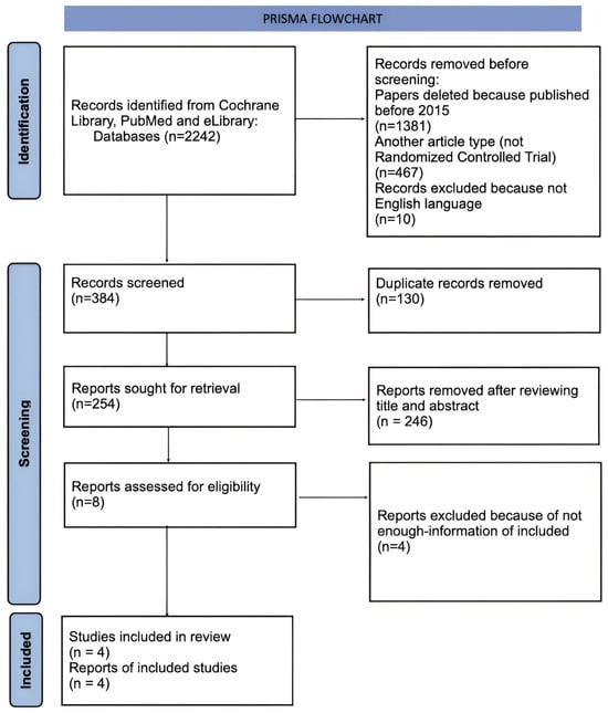
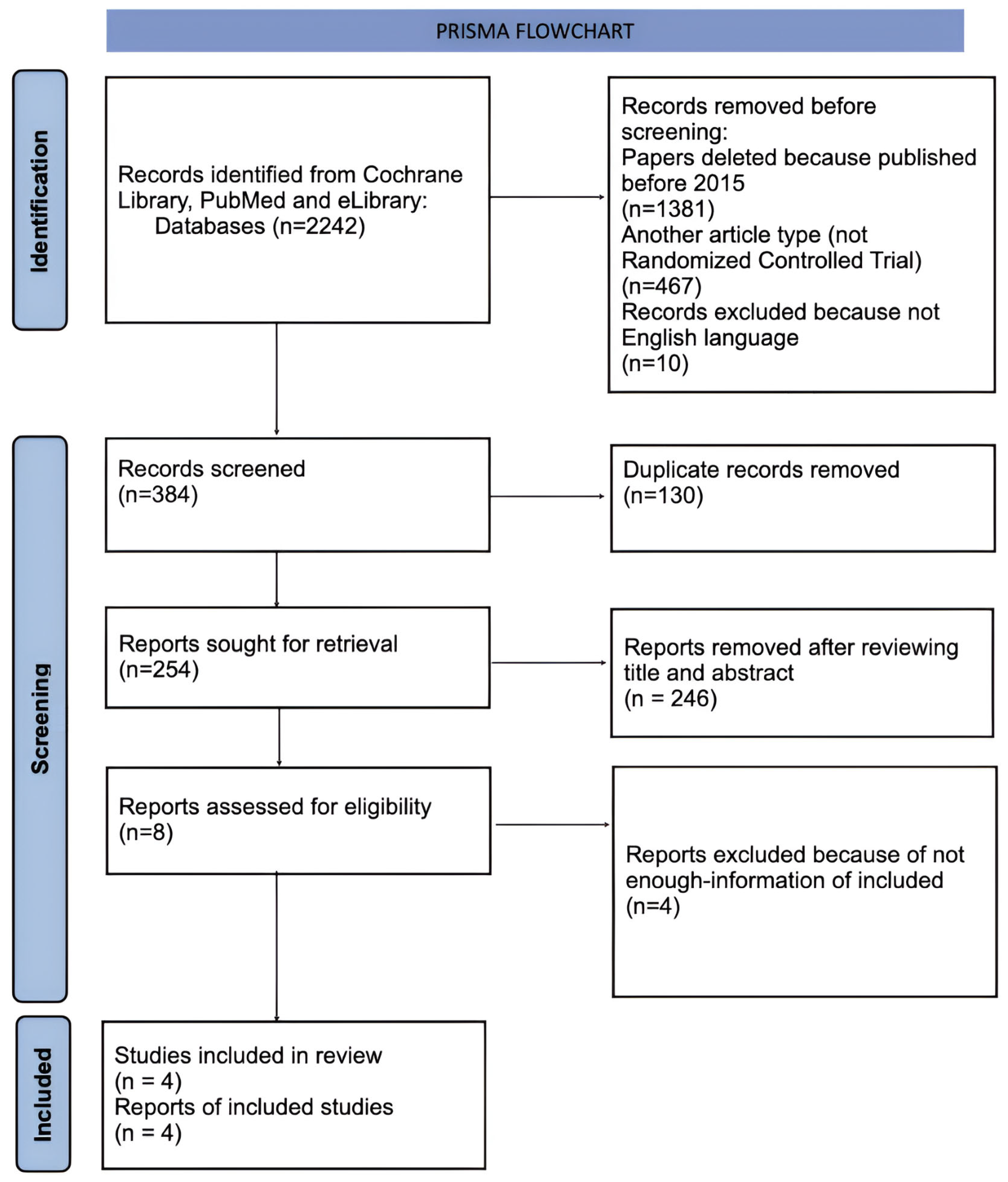
The preliminary search of electronic databases (PubMed, Cochrane Library, and eLibrary) yielded 2242 publications.
After applying filters (publication period: 2015–2025; study design: randomized controlled trials; language: English), five researchers independently screened and selected 384 publications across the various electronic databases.
After eliminating 130 duplicate records, 254 studies were retained for further evaluation. Screening of titles and abstracts resulted in the exclusion of 246 publications, after which the full texts of the remaining 8 articles were analyzed in detail. After full-text analysis, 3 additional publications were excluded due to the absence of relevant data. Furthermore, one study could not be retrieved for full-text evaluation [30], as the authors did not respond to the request; consequently, this work was excluded. Ultimately, a total of 4 publications were included in the final systematic review (Figure 1).
Figure 1.
Flow diagram describing the selection process according to the Preferred Reporting Items for Systematic Reviews and Meta-Analyses (PRISMA) recommendations.
3.2. Characteristics of Included Studies
All four included studies [31,32,33,34] were randomized controlled trials (RCTs) published in English between 2015 and 2025. The total number of participants across all studies was 138 (Table 2). A key inclusion criterion for this review was the absence of immediate implant placement and the availability of assessments of soft tissue changes following tooth extraction. Dental implantations were excluded from the study, as the aim of the study was to investigate the ARP method for preventing tissue atrophy after tooth extraction in the absence of immediate implantation.
Table 2.
Characteristics of the included randomized controlled trials.
The studies compared different ridge preservation techniques with a control group undergoing spontaneous socket healing. As bone grafting materials, xenogeneic materials derived from bovine bone were used: deproteinized bovine bone mineral (DBBM) [34] and deproteinized bovine bone mineral with collagen (DBBM-C) [31,32,34]. In three studies [31,32,33], the xenograft was used in combination with a collagen membrane; additionally, one study [33] compared a membrane with a free gingival graft harvested from the palatal donor site.
In one study [31], primary data on dimensional changes were obtained through clinical measurements using a customized stent. In two studies [33,34], three-dimensional (3D) scanning of casts was employed to assess volumetric changes in the soft tissue contour, whereas in the study by Zampara et al. [32], the main focus was placed on histological and histomorphometric analysis of bone tissue from the augmentation site at 3 months postoperatively.
In three studies [31,33,34], the results showed a decrease in the horizontal width of the alveolar process when ARP was used compared to the control group over a period of 3–6 months. However, the observed differences were not statistically significant (p > 0.05). However, the observed differences were not statistically significant (p > 0.05). Nevertheless, a clinically relevant observation [33] was that contour losses in the control groups were greater and exhibited higher variability. A similar trend toward better volume preservation with augmentation, albeit without statistical significance, was also observed for vertical changes [31,33].
Histological findings from two studies [31,32] indicated that, at 3–6 months, sockets filled with xenograft exhibited a lower amount of vital bone formation compared with the control group. In Zampara et al. [32], this difference was statistically significant (p < 0.001), and xenograft also showed significantly less vital bone than alloplastic or allogeneic grafts, while a substantial quantity of slowly resorbing graft particles remained. In Gabay et al. [31], the difference was not statistically significant (p = 0.11). These findings suggest that, in Zampara et al. [32], volume preservation in the xenograft group was primarily attributable to the graft material itself rather than to newly formed bone, whereas in Gabay et al. [31] this was less evident.
3.3. Risk of Systematic Errors (Bias) in Studies
The risk of bias in all four included studies was assessed using the Cochrane Risk-of-Bias Tool for Randomized Trials (RoB 2) (Table 3).
Table 3.
Risk of bias of randomized clinical trials, assessed through the ROB2 tool.
The conducted risk-of-bias assessment (RoB 2) identified domains requiring caution. In the study by Zampara et al. [32], concerns were noted regarding the risk-of-bias assessment, specifically related to the selection of reported results, due to the absence of a pre-registered study protocol.
In the study conducted by Flügge et al. [34], a high risk of bias was observed in the “randomization process” domain, while additional concerns were noted regarding the assessment of outcomes. Details on the randomization mechanism and allocation concealment were not reported, which could have influenced participant allocation. Additionally, the statistical analysis of different tooth groups and the varying numbers of extracted teeth raised concerns. It is important to note that the authors themselves acknowledged the limitations of their study, stating that its primary objective was to refine the measurement methodology and to calculate the sample size for future investigations.
4. Discussion
Tooth extraction leads to a defect of the alveolar process and bone tissue resorption. To minimize post-extraction complications and facilitate subsequent implant placement, augmentation techniques are widely used, including xenogeneic osteoconductive materials with predictable biocompatibility and no need for donor-site harvesting [6,26,27,35,36]. Modern clinical data indicate that they are non-inferior to the results achieved with allogeneic or autogenous materials [5,6,15,37]. In this review, we focused specifically on randomized controlled trials evaluating xenogenic materials not only for ridge dimension preservation, but also for their influence on soft tissue stability and histologic tissue composition.
Currently, the main sources of xenogeneic material are pigs (porcine material) and cattle (bovine material), the bone structure of which is similar to that of humans. However, the choice between them often presents difficulties, primarily due to the potential risk of prion disease transmission associated with bovine materials [9]. According to recent research, there is no difference in new bone gain between them [9,18]. Thus, material selection may depend on handling properties, availability, and surgeon preference.
In addition to the source, a critically important parameter is the type of thermal processing of the xenomaterial, which directly affects the regenerative potential. According to study results, materials subjected to high-temperature processing are characterized by slower resorption [32], which may help preserve ridge volume during the early healing period [23,32]. However, reduced resorption speed simultaneously slows the process of complete remodeling and replacement of the graft with mature bone tissue and increases the risks of material encapsulation. According to the work by Couso-Queiruga et al., longer healing was associated with a higher degree of mineralization and better graft integration [2]. Thus, careful assessment of the bone status is necessary before implant placement [22,32].
The macro- and microstructure of xenografts, including the degree of decalcification and preservation of the organic matrix, may influence cell infiltration and vascularization in the regeneration zone [7,14]. These properties may partly explain differences in remodeling speed and histologic outcomes reported for different xenogenic materials.
An important advantage of xenomaterials is their good compatibility with barrier membranes for guided bone regeneration, which allows for effective control of the healing process and prevents soft tissue migration into the defect area [1,28,37]. In the four included RCTs [31,32,33,34], the use of collagen membranes or free gingival punches was generally associated with numerically better preservation of the alveolar ridge contour than spontaneous healing in the short term (up to 6 months), although the magnitude of these differences varied across studies and in most cases did not reach statistical significance for the primary dimensional outcomes. One trial [34] reported that the benefit of grafting was most pronounced during the first 4–6 weeks, with comparable resorption in both groups by 12 weeks. These findings suggest that soft tissue management may be an important cofactor influencing the clinical performance of xenogenic grafts, but the available evidence remains limited because of heterogeneity and small sample sizes.
Interpretation of the findings is limited by substantial clinical and methodological heterogeneity across the included trials. The studies differed in graft composition, adjunctive procedures, follow-up duration, and outcome definitions. Dimensional outcomes were measured using different approaches, including customized stents, direct clinical measurements, and 3D cast scanning, whereas histological endpoints were reported in only a subset of studies. These differences reduce comparability and preclude quantitative meta-analysis.
4.1. Discussion of Included Studies
The resorption rate of bone material directly affects dental implantation planning. According to data from Flügge et al. [34], socket augmentation allows slowing the resorption process to 12 weeks. At the same time, the resorption period without socket augmentation is observed up to 6 weeks. However, at 3 months, no statistical difference is observed between the groups. Kai R. Fischer and his co-authors came to the same conclusion [33]. Notably, in the study by Flügge et al. [34], attention was drawn to the minimal variability of resorption within the augmentation group compared to the expressed resorption in the control group. At the same time, it is necessary to consider the fact that the study assessed bone tissue status based on volumetric changes in soft tissues. Thus, the authors emphasize that socket augmentation creates favorable conditions for early dental implant placement. Available randomized evidence suggests that xenogenic materials may achieve short-term ridge preservation outcomes broadly similar to those reported for other graft types, although direct head-to-head comparisons are scarce and underpowered.
Similar results were observed in the study by Zampara et al. [32]. The authors emphasize that the type of osteoplastic material has a significant influence on outcomes. The study confirmed the osteoconductive properties of various materials. However, it also revealed that the amount of newly formed bone tissue when using an allograft or synthetic material exceeded the corresponding indicator for xenogeneic material by more than two times. In this regard, the authors recommend performing implantation at later times when using xenogeneic grafts—over 3 months. However, these findings should be interpreted with caution because all included trials enrolled small samples, reported only short-term outcomes (3–6 months), and used non-standardized measurement methods. This conclusion is confirmed in the systematic review by De Risi et al. [38], where maximum bone gain in the allogeneic material group was registered at 3 months, while for xenogeneic materials, the indicators remained the lowest even at 5 months. An additional factor slowing remodeling is the high percentage of residual xenogeneic material (over 35% versus 21% for allogeneic material).
The biological significance of persistent xenogenic particles remains an important issue, especially in relation to long-term peri-implant tissue behavior. Fischer et al. [33] reported encapsulation and Type III bone formation in 60% of cases treated with xenogenic materials, which suggests that a substantial proportion of the graft may remain isolated from active remodeling rather than being fully replaced by mature bone. Similar concerns were raised by Milani et al. [14], who described the remodeling pattern of deproteinized bovine bone in extraction sockets and demonstrated that residual particles can persist for an extended period while new bone formation proceeds around them. In addition, the systematic review and meta-analysis showed that xenogenic materials are associated with slower remodeling and a higher proportion of residual graft compared with allogeneic materials, reinforcing the view that dimensional stability may partly reflect the physical persistence of the graft [38]. Taken together, these data indicate that radiographic volume maintenance should not be interpreted as equivalent to complete biological regeneration, and long-term clinical monitoring remains necessary when xenogenic grafts are used for ridge preservation.
These findings should be interpreted by distinguishing radiographic or volumetric stability from the histological quality of the regenerated site. A socket may appear dimensionally preserved on clinical or radiographic assessment, yet still contain a relatively low percentage of vital bone and a substantial amount of residual xenogenic particles. Therefore, xenografts may contribute to ridge maintenance mainly by space preservation and slow resorption rather than by rapid replacement with mature bone. This may have implications for secondary stability and implant bed preparation, while the long-term biological significance of residual particles and fibrous tissue remains uncertain.
A high level of residual material correlates with the risk of its encapsulation. According to Fischer et al. [33], complications such as material encapsulation and Type III bone formation were observed in 60% of cases when using xenomaterials. Taken together, these findings support the notion that xenogenic grafts often act primarily as space-maintaining materials that stabilize the ridge contour rather than as substitutes that are rapidly replaced by vital bone. In other words, the apparent radiographic and volumetric stability observed in xenograft-treated sites may not necessarily reflect complete regeneration of high-quality bone but rather the persistence of non-resorbed particles and fibrous tissue. This distinction is clinically relevant, as it may influence secondary implant stability, the risk of fibrous encapsulation, and the susceptibility of peri-implant tissues to inflammatory breakdown over time, and it should therefore be carefully considered when determining the timing of implant placement and planning long-term maintenance. However, long-term patient monitoring and subsequent treatment with dental implants are necessary to confirm this. One of the key pathogenetic mechanisms of encapsulation is the proliferation of fibroblasts and epithelial cells into the defect area, which prevents the migration and proliferation of osteogenic cells [39]. This creates a clinical problem, since the long-term influence of encapsulated material on peri-implant tissues remains a subject of discussion and is insufficiently studied [33], which necessitated a separate analysis of soft tissue status within this review.
In clinical practice, various barrier systems are used for successful guided bone regeneration. Their task is to prevent connective tissue ingrowth into the defect area, slow bone resorption, and ensure soft tissue preservation in the intervention area [40]. The use of a combined regeneration method leads to a more predictable aesthetic result. Most often, collagen membranes or an autogenous free graft from the maxillary tuberosity or hard palate area are used for this purpose.
A comparison of two barrier systems was conducted in the studies included in our systematic review. Thus, Kai R. Fischer and his co-authors [33] note that when using a palatal graft, soft tissue shrinkage was the least. When using a collagen matrix or its absence, a thin gingival biotype was observed in the area of planned subsequent dental implantation. Also, Gabay et al. [31] noted that the use of bone material with a collagen membrane better preserves vertical and horizontal gingival dimensions compared to their absence. However, the result did not reach statistical difference. The authors explain this point by the limited sample size, high individual data variability, and the inclusion of predominantly intact sockets with minimal defects, where the potential for natural healing is initially high.
According to new scientific studies, collagen matrices demonstrate comparable efficacy in increasing tissue thickness in the short term and can be considered as a clinical alternative, especially in cases where minimizing invasiveness and patient postoperative morbidity is a priority [41]. At the same time, the bone material type itself is a factor influencing soft tissue behavior. According to the histological results of the study by Zampara et al. [32], soft tissue proliferation into the defect area in the xenograft group was the most significant (up to 54.5%) compared to alloplastic, synthetic material, and its absence. Despite the fact that primary implant stability in the studies by Zampara et al. [32] and Gabay et al. [31] did not depend on the material type, the pronounced fibrous reaction associated with xenomaterials requires attention when planning implantation timing and predicting long-term outcomes. Given the limited number of RCTs and the absence of long-term follow-up, any apparent advantages of xenografts over allogeneic or autogenous materials remain speculative.
Recent comparative evidence suggests that bovine- and porcine-derived xenografts provide broadly similar clinical and histomorphometric performance in alveolar ridge preservation. In a randomized controlled clinical trial [9]. found no statistically significant differences between bovine and porcine xenografts with respect to vital bone formation, residual graft material, connective tissue proportion, or dimensional stability, although additional grafting at implant placement was required more frequently in the porcine group. Comparable biological and dimensional outcomes for bovine- and porcine-derived xenografts are reported in both horizontal ridge augmentation and extraction socket preservation, supporting the view that clinical selection may be guided more by handling characteristics, procedural preferences, and case-specific considerations than by major differences in regenerative potential [18]. In this context, the present review, which was limited to bovine-derived materials, should be interpreted as reflecting the performance of a specific xenograft subgroup rather than xenogenic materials as a whole.
4.2. Limitations
In our study, we reviewed articles with various assessment parameters; thus, comparison between these works presents difficulties. We did not focus on a specific type of xenogeneic material, while the efficacy of using different types may differ significantly. However, all studies considered xenogeneic materials in granular form. According to a recent study [3], the use of pre-formed material leads to results similar to granular material according to radiological and histological analysis. Soft tissue assessment in the studies was performed using various methods, resulting in difficulties in generalizing the results. Also, there is no assessment of non-resorbable membranes for more accurate study.
Moreover, all included RCTs reported only short-term outcomes (3–6 months) and did not evaluate long-term complications such as late infection, incomplete graft resorption, or implant survival. As a result, the long-term clinical impact of persistent xenogenic particles and possible fibrous encapsulation could not be assessed within the scope of this review. In addition, the included studies were heterogeneous in several methodological aspects, which precluded a reliable quantitative synthesis. The trials used different outcome assessment methods, including customized stents, clinical linear measurements, 3D cast scanning, and histologic or histomorphometric analysis, and they also differed in follow-up duration (3 months versus 6 months). These differences in measurement technique and timing influenced the interpretation of ridge dimensional changes, soft tissue contour outcomes, and histologic maturation. This made it difficult to directly compare the results and prevented a reliable meta-analysis.
Furthermore, numerous non-randomized and observational alveolar ridge preservation studies were excluded by design; thus, our conclusions apply only to a small subset of RCTs and should not be extrapolated to all alveolar ridge preservation techniques or study designs. We did not search trial registries or grey literature, and our electronic search strategy was based on a limited set of keywords, which may have resulted in the omission of additional relevant studies. Therefore, despite these limitations, the results may serve as a basis for subsequent targeted systematic reviews and meta-analyses.
Only four RCTs fulfilled our strict inclusion criteria, which limits the generalizability of the findings and prevents firm clinical recommendations. The review should therefore be interpreted as a focused qualitative synthesis rather than a definitive evidence summary for all ARP approaches.
5. Conclusions
Within the limitations of the available evidence, the included RCTs suggest that xenogenic materials may demonstrate clinical effectiveness. However, the included RCTs showed only modest benefits, limited vital bone formation, and frequent residual graft particles.
These findings suggest that xenografts may maintain ridge volume partly through the persistence of graft material rather than through complete bone regeneration. Soft tissue outcomes also favored augmented sites, but the evidence was heterogeneous and based on short follow-up periods.
Overall, xenogenic grafts may be a useful option for alveolar ridge preservation, but their long-term biological and clinical advantages remain uncertain. Further randomized trials with standardized outcome measures and longer follow-up are needed.
Supplementary Materials
The following supporting information can be downloaded at: https://www.mdpi.com/article/10.3390/prosthesis8070068/s1, Supplementary Material S1: PRISMA 2020 Checklist. Reference [42] is cited in the Supplementary Material.
Author Contributions
Conceptualization, E.D.; methodology, M.S. (Marina Skachkova) and S.D.; software, M.S. (Mikhail Stepanov); validation, Y.E. and Y.V.; formal analysis, H.D. and M.S. (Mikhail Stepanov); investigation, M.S. (Marina Skachkova), A.K., D.S., M.A. and M.K.; data curation, A.K. and K.B.; writing—original draft preparation, M.S. (Marina Skachkova), D.S. and M.A.; writing—review and editing, M.S. (Marina Skachkova), A.K. and D.S.; visualization, K.B.; supervision, S.T.; project administration, S.D. All authors have read and agreed to the published version of the manuscript.
Funding
This research received no external funding.
Institutional Review Board Statement
Not applicable.
Informed Consent Statement
Not applicable.
Data Availability Statement
The original contributions presented in this study are included in the article/Supplementary Material. Further inquiries can be directed to the corresponding author.
Conflicts of Interest
The authors declare no conflicts of interest.
Abbreviations
The following abbreviations are used in this manuscript:
| RCTs | Randomized Controlled Trials |
| PoB 2 | Revised Cochrane Risk-of-Bias 2 |
| PRISMA | Preferred Reporting Items for Systematic Reviews and Meta-Analyses |
| PROSPERO | Prospective Register of Systematic Reviews |
| PICOS | Patient/Population, Intervention, Comparison, Outcome, Study design |
| DBBM | Deproteinized bovine bone mineral |
| DBBM-C | Deproteinized bovine bone mineral with collagen |
References
- El-Sioufi, I.; Oikonomou, I.; Koletski, D.; Bobetsis, Y.A.; Madianos, P.N.; Vassilopoulos, S. Clinical evaluation of different alveolar ridge preservation techniques after tooth extraction: A randomized clinical trial. Clin. Oral Investig. 2023, 27, 4471–4480. [Google Scholar] [CrossRef] [Scilit] [PubMed]
- Couso-Queiruga, E.; Weber, H.A.; Garaicoa-Pazmino, C.; Barwacz, C.; Kalleme, M.; Galindo-Moreno, P.; Avila-Ortiz, G. Influence of healing time on the outcomes of alveolar ridge preservation using a collagenated bovine bone xenograft: A randomized clinical trial. J. Clin. Periodontol. 2023, 50, 132–146. [Google Scholar] [CrossRef] [Scilit] [PubMed]
- Choi, Y.; Cheong, J.; Song, Y.; Kim, J.; Kang, D.Y.; Ahn, H.S.; Park, S.; Esposito, M.; Shin, H.S.; Park, W. Moldable and Particulate Bone Material in Alveolar Ridge Preservation: A Multicenter Randomized Controlled Trial. Clin. Implant Dent. Relat. Res. 2025, 27, e70011. [Google Scholar] [CrossRef] [Scilit] [PubMed]
- Kim, H.; Han, H.S.; Ghanaati, S.; Zadeh, H.H.; Kim, S.; Cho, Y.D. Alveolar Ridge Preservation Using a Collagenated Xenograft: A Randomized Clinical Trial. Int. Dent. J. 2025, 75, 1155–1164. [Google Scholar] [CrossRef] [Scilit] [PubMed]
- Assem, N.Z.; Pazmiño, V.F.C.; Rodas, M.A.R.; Caliente, E.A.; da Silva Dalben, G.; Soares, S.; Santiago, J.F.; de Almeida, A.L.P.F. Bone Substitutes Graft for Regeneration of the Anterior Maxillary Alveolar Process: A Systematic Review. J. Oral Implantol. 2023, 49, 102–113. [Google Scholar] [CrossRef] [Scilit] [PubMed]
- Ouyang, L.; Li, J.; Dong, Y.; Li, J.; Jin, F.; Luo, Y.; Wang, R.; Wang, S. Comparison of clinical efficacy between autologous partially demineralized dentin matrix and deproteinized bovine bone mineral for bone augmentation in orthodontic patients with alveolar bone deficiency: A randomized controlled clinical trial. BMC Oral Health 2024, 24, 984. [Google Scholar] [CrossRef] [Scilit] [PubMed]
- Munhoz, E.A.; Bodanezi, A.; Cestari Biol, T.M.; Zardin Graeff, M.S.; Junior, O.F.; de Carvalho, P.S.P.; Taga, R. Impact of Inorganic Xenograft on Bone Healing and Osseointegration: An Experimental Study in Rabbits. Implant Dent. 2017, 26, 875–881. [Google Scholar] [CrossRef] [Scilit] [PubMed]
- Mendoza-Azpur, G.; de la Fuente, A.; Chavez, E.; Valdivia, E.; Khouly, I. Horizontal ridge augmentation with guided bone regeneration using particulate xenogenic bone substitutes with or without autogenous block grafts: A randomized controlled trial. Clin. Implant Dent. Relat. Res. 2019, 21, 521–530. [Google Scholar] [CrossRef] [Scilit] [PubMed]
- Lai, V.J.; Michalek, J.E.; Liu, Q.; Mealey, B.L. Ridge preservation following tooth extraction using bovine xenograft compared with porcine xenograft: A randomized controlled clinical trial. J. Periodontol. 2020, 91, 361–368. [Google Scholar] [CrossRef] [Scilit] [PubMed]
- Gao, L.; Zhu, H.; Kou, Y.; Hu, H.; Wu, Y.; Tang, Z.; Li, Q. The effects of β-TCP and/or xenogeneic bone substitute on alveolar ridge preservation: A randomized clinical trial. Clin. Oral Investig. 2025, 29, 200. [Google Scholar] [CrossRef] [Scilit] [PubMed]
- Stumbras, A.; Galindo-Moreno, P.; Januzis, G.; Juodzbalys, G. Three-dimensional analysis of dimensional changes after alveolar ridge preservation with bone substitutes or plasma rich in growth factors: Randomized and controlled clinical trial. Clin. Implant Dent. Relat. Res. 2021, 23, 96–106. [Google Scholar] [CrossRef] [Scilit] [PubMed]
- Issa, D.R.; Elamrousy, W.; Gamal, A.Y. Alveolar ridge splitting and simvastatin loaded xenograft for guided bone regeneration and simultaneous implant placement: Randomized controlled clinical trial. Clin. Oral Investig. 2024, 28, 71. [Google Scholar] [CrossRef] [Scilit] [PubMed]
- Oguić, M.; Čandrlić, M.; Tomas, M.; Vidaković, B.; Blašković, M.; Jerbić Radetić, A.T.; Zoričić Cvek, S.; Kuiš, D.; Cvijanović Peloza, O. Osteogenic Potential of Autologous Dentin Graft Compared with Bovine Xenograft Mixed with Autologous Bone in the Esthetic Zone: Radiographic, Histologic and Immunohistochemical Evaluation. Int. J. Mol. Sci. 2023, 24, 6440. [Google Scholar] [CrossRef] [Scilit] [PubMed]
- Milani, S.; Dal Pozzo, L.; Rasperini, G.; Sforza, C.; Dellavia, C. Deproteinized bovine bone remodeling pattern in alveolar socket: A clinical immunohistological evaluation. Clin. Oral Implant. Res. 2016, 27, 295–302. [Google Scholar] [CrossRef] [Scilit] [PubMed]
- Abellán, D.; Barallat, L.; Vilarrasa, J.; Cabezas, M.; Pascual La Rocca, A.; Valles, C.; Nart, J. Ridge preservation in molar sites comparing xenograft versus mineralized freeze-dried bone allograft: A randomized clinical trial. Clin. Oral Implant. Res. 2022, 33, 511–523. [Google Scholar] [CrossRef] [Scilit] [PubMed]
- Al Qabbani, A.; Al Kawas, S.; ARazak, N.H.; Al Bayatti, S.W.; Enezei, H.H.; Samsudin, A.R.; Sheikh Ab Hamid, S. Three-Dimensional Radiological Assessment of Alveolar Bone Volume Preservation Using Bovine Bone Xenograft. J. Craniofac. Surg. 2018, 29, e203–e209. [Google Scholar] [CrossRef] [Scilit] [PubMed]
- Mercado, F.; Vaquette, C.; Hamlet, S.; Ivanovski, S. Enamel matrix derivative promotes new bone formation in xenograft assisted maxillary anterior ridge preservation-A randomized controlled clinical trial. Clin. Oral Implant. Res. 2021, 32, 732–744. [Google Scholar] [CrossRef] [Scilit] [PubMed]
- Bhatavadekar, N.; Gandhi, Y.; Padhye, N. Comparative Assessment of Bovine Versus Porcine Xenograft for Augmentation: A Randomized Prospective Cohort Study. Int. J. Periodontics Restor. Dent. 2022, 42, 789–796. [Google Scholar] [CrossRef] [Scilit] [PubMed]
- Simonffy, L.; Minya, F.; Trimmel, B.; Lacza, Z.; Dobo-Nagy, C. Albumin-Impregnated Allograft Filling of Surgical Extraction Sockets Achieves Better Bone Remodeling Than Filling with Either Blood Clot or Bovine Xenograft. Int. J. Oral Maxillofac. Implant. 2020, 35, 297–304. [Google Scholar] [CrossRef] [Scilit] [PubMed]
- Santos, A.; Botelho, J.; Machado, V.; Borrecho, G.; Proença, L.; Mendes, J.J.; Mascarenhas, P.; Alcoforado, G. Autogenous Mineralized Dentin versus Xenograft granules in Ridge Preservation for Delayed Implantation in Post-extraction Sites: A Randomized controlled clinical trial with an 18 months follow-up. Clin. Oral Implant. Res. 2021, 32, 905–915. [Google Scholar] [CrossRef] [Scilit] [PubMed]
- Casarez-Quintana, A.; Mealey, B.L.; Kotsakis, G.; Palaiologou, A. Comparing the histological assessment following ridge preservation using a composite bovine-derived xenograft versus an alloplast hydroxyapatite-sugar cross-linked collagen matrix. J. Periodontol. 2022, 93, 1691–1700. [Google Scholar] [CrossRef] [Scilit] [PubMed]
- Serrano Méndez, C.A.; Lang, N.P.; Caneva, M.; Ramírez Lemus, G.; Mora Solano, G.; Botticelli, D. Comparison of allografts and xenografts used for alveolar ridge preservation. A clinical and histomorphometric RCT in humans. Clin. Implant Dent. Relat. Res. 2017, 19, 608–615. [Google Scholar] [CrossRef] [Scilit] [PubMed]
- Shakibaie, B.; Blatz, M.B.; Sabri, H.; Jamnani, E.D.; Barootchi, S. Effectiveness of Two Differently Processed Bovine-Derived Xenografts for Alveolar Ridge Preservation with a Minimally Invasive Tooth Extraction Approach: A Feasibility Clinical Trial. Int. J. Periodontics Restor. Dent. 2023, 43, 540–549. [Google Scholar] [CrossRef] [Scilit] [PubMed]
- Thoma, D.S.; Bienz, S.P.; Payer, M.; Hüsler, J.; Schmidlin, P.R.; Hämmerle, C.H.F.; Jakse, N.; Jung, R.E. Randomized clinical study using xenograft blocks loaded with bone morphogenetic protein-2 or autogenous bone blocks for ridge augmentation—A three-dimensional analysis. Clin. Oral Implant. Res. 2019, 30, 872–881. [Google Scholar] [CrossRef] [Scilit] [PubMed]
- Temmerman, A.; Cortellini, S.; Van Dessel, J.; De Greef, A.; Jacobs, R.; Dhondt, R.; Teughels, W.; Quirynen, M. Bovine-derived xenograft in combination with autogenous bone chips versus xenograft alone for the augmentation of bony dehiscences around oral implants: A randomized, controlled, split-mouth clinical trial. J. Clin. Periodontol. 2020, 47, 110–119. [Google Scholar] [CrossRef] [Scilit] [PubMed]
- Işık, G.; Özden Yüce, M.; Koçak-Topbaş, N.; Günbay, T. Guided bone regeneration simultaneous with implant placement using bovine-derived xenograft with and without liquid platelet-rich fibrin: A randomized controlled clinical trial. Clin. Oral Investig. 2021, 25, 5563–5575. [Google Scholar] [CrossRef] [Scilit] [PubMed]
- Iorio-Siciliano, V.; Ramaglia, L.; Blasi, A.; Bucci, P.; Nuzzolo, P.; Riccitiello, F.; Nicolò, M. Dimensional changes following alveolar ridge preservation in the posterior area using bovine-derived xenografts and collagen membrane compared to spontaneous healing: A 6-month randomized controlled clinical trial. Clin. Oral Investig. 2020, 24, 1013–1023. [Google Scholar] [CrossRef] [Scilit] [PubMed]
- Abaza, G.; Abdel Gaber, H.K.; Afifi, N.S.; Adel-Khattab, D. Injectable platelet rich fibrin versus hyaluronic acid with bovine derived xenograft for alveolar ridge preservation. A randomized controlled clinical trial with histomorphometric analysis. Clin. Implant Dent. Relat. Res. 2024, 26, 88–102. [Google Scholar] [CrossRef] [Scilit] [PubMed]
- Page, M.J.; McKenzie, J.E.; Bossuyt, P.M.; Boutron, I.; Hoffmann, T.C.; Mulrow, C.D.; Shamseer, L.; Tetzlaff, J.M.; Akl, E.A.; Brennan, S.E.; et al. The PRISMA 2020 Statement: An Updated Guideline for Reporting Systematic Reviews. PLoS Med. 2021, 18, e1003583. [Google Scholar] [CrossRef] [PubMed]
- Kadkhodazadeh, M.; Sohrabi, Z.; Abrishami, M.R.; Amid, R.; Mashhadiabbas, F.; Shojaei, N.; Dehghan, M.M.; Faghihi, F. Histologic and Histomorphometric Assessment of Xenograft Bone Substitute versus Synthetic Nonceramic Hydroxyapatite for Canine Tooth-Socket Preservation. J. Long-Term Eff. Med. Implants 2019, 29, 281–288. [Google Scholar] [CrossRef] [Scilit] [PubMed]
- Gabay, E.; Katorza, A.; Zigdon-Giladi, H.; Horwitz, J.; Machtei, E.E. Histological and dimensional changes of the alveolar ridge following tooth extraction when using collagen matrix and collagen-embedded xenogenic bone substitute: A randomized clinical trial. Clin. Implant Dent. Relat. Res. 2022, 24, 382–390. [Google Scholar] [CrossRef] [Scilit] [PubMed]
- Zampara, E.; Alshammari, M.; De Bortoli, J.; Mullings, O.; Gkisakis, I.G.; Benalcázar Jalkh, E.B.; Tovar, N.; Coelho, P.G.; Witek, L. A Histologic and Histomorphometric Evaluation of an Allograft, Xenograft, and Alloplast Graft for Alveolar Ridge Preservation in Humans: A Randomized Controlled Clinical Trial. J. Oral Implantol. 2022, 48, 541–549. [Google Scholar] [CrossRef] [Scilit] [PubMed]
- Fischer, K.R.; Mühlemann, S.; Jung, R.E.; Friedmann, A.; Fickl, S. Dimensional Evaluation of Different Ridge Preservation Techniques with a Bovine Xenograft: A Randomized Controlled Clinical Trial. Int. J. Periodontics Restor. Dent. 2018, 38, 549–556. [Google Scholar] [CrossRef] [Scilit] [PubMed]
- Flügge, T.; Nelson, K.; Nack, C.; Stricker, A.; Nahles, S. 2-Dimensional changes of the soft tissue profile of augmented and non-augmented human extraction sockets: A randomized pilot study. J. Clin. Periodontol. 2015, 42, 390–397. [Google Scholar] [CrossRef] [Scilit] [PubMed]
- Iorio-Siciliano, V.; Blasi, A.; Nicolò, M.; Iorio-Siciliano, A.; Riccitiello, F.; Ramaglia, L. Clinical Outcomes of Socket Preservation Using Bovine-Derived Xenograft Collagen and Collagen Membrane Post-Tooth Extraction: A 6-Month Randomized Controlled Clinical Trial. Int. J. Periodontics Restor. Dent. 2017, 37, e290–e296. [Google Scholar] [CrossRef] [Scilit] [PubMed]
- Cardaropoli, D.; Tamagnone, L.; Roffredo, A.; Gaveglio, L.; Cardaropoli, G. Socket preservation using bovine bone mineral and collagen membrane: A randomized controlled clinical trial with histologic analysis. Int. J. Periodontics Restor. Dent. 2012, 32, 421–430. [Google Scholar] [PubMed]
- Bayat, N.; Ghavimi, M.A.; Rahimipour, K.; Razi, S.; Esmaeili, F. Radiographic texture analysis of the hard tissue changes following socket preservation with allograft and xenograft materials for dental implantation: A randomized clinical trial. Oral Maxillofac. Surg. 2024, 28, 705–713. [Google Scholar] [CrossRef] [Scilit] [PubMed]
- De Risi, V.; Clementini, M.; Vittorini, G.; Mannocci, A.; De Sanctis, M. Alveolar ridge preservation techniques: A systematic review and meta-analysis of histological and histomorphometrical data. Clin. Oral Implant. Res. 2015, 26, 50–68. [Google Scholar]
- Lau, C.S.; Chua, J.; Prasadh, S.; Lim, J.; Saigo, L.; Goh, B.T. Alveolar Ridge Augmentation with a Novel Combination of 3D-Printed Scaffolds and Adipose-Derived Mesenchymal Stem Cells-A Pilot Study in Pigs. Biomedicines 2023, 11, 2274. [Google Scholar] [CrossRef] [Scilit] [PubMed]
- Dimitriou, R.; Mataliotakis, G.I.; Calori, G.M.; Giannoudis, P.V. The role of barrier membranes for guided bone regeneration and restoration of large bone defects: Current experimental and clinical evidence. BMC Med. 2012, 10, 81. [Google Scholar] [CrossRef] [Scilit] [PubMed]
- Tommasato, G.; Del Fabbro, M.; Oliva, N.; Khijmatgar, S.; Grusovin, M.G.; Sculean, A.; Canullo, L. Autogenous graft versus collagen matrices for peri-implant soft tissue augmentation. A systematic review and network meta-analysis. Clin. Oral Investig. 2024, 28, 300. [Google Scholar] [CrossRef] [Scilit] [PubMed]
- Page, M.J.; McKenzie, J.E.; Bossuyt, P.M.; Boutron, I.; Hoffmann, T.C.; Mulrow, C.D.; Shamseer, L.; Tetzlaff, J.M.; Akl, E.A.; Brennan, S.E.; et al. The PRISMA 2020 statement: An updated guideline for reporting systematic reviews. BMJ 2021, 372, n71. [Google Scholar] [CrossRef] [Scilit] [PubMed]

Disclaimer/Publisher’s Note: The statements, opinions and data contained in all publications are solely those of the individual author(s) and contributor(s) and not of MDPI and/or the editor(s). MDPI and/or the editor(s) disclaim responsibility for any injury to people or property resulting from any ideas, methods, instructions or products referred to in the content. |
© 2026 by the authors. Licensee MDPI, Basel, Switzerland. This article is an open access article distributed under the terms and conditions of the Creative Commons Attribution (CC BY) license.