


Abstract
Objective: This study aimed to quantitatively evaluate five opportunistic respiratory pathogens among individuals, with or without dentures, who have either healthy or diseased periodontal conditions. Methods: Saliva samples were obtained from 24 older adults. DNA extraction was performed, followed by a quantitative Polymerase Chain Reaction targeting five opportunistic respiratory pathogens, namely Staphylococcus aureus, Pseudomonas aeruginosa, Streptococcus pyogenes, Klebsiella pneumoniae, and Streptococcus agalactiae. The abundance of opportunistic pathogens among the four groups [denture-wearing with active periodontal disease (DAP), non-denture-wearing with active periodontal disease (NDAP), denture-wearing with stable periodontal health (DSP), and non-denture-wearing with stable periodontal health (NDSP)] were compared. Results: Saliva samples of all participants (100%) carried at least one opportunistic respiratory pathogen. Of the five pathogens, S. aureus, P. aeruginosa, K. pneumoniae, and S. agalactiae were present in more than 50% of the samples. A significant difference in the bacterial load of K. pneumoniae and P. aeruginosa was observed (p < 0.05). The quantity of P. aeruginosa in the NDAP group was significantly lower than the DSP group. However, for K. pneumoniae, no significant differences between the groups were detected. Conclusions: In this small pilot cohort, a relatively high prevalence and substantial microbial load of opportunistic respiratory pathogens were identified in the saliva of older adults. On the basis of these preliminary findings, optimal oral and denture hygiene care is suggested as a potential approach to reduce the presence and burden of respiratory pathogens in this population, although larger studies are required to confirm these associations and determine their clinical significance.
1. Introduction
Hong Kong is currently experiencing a trend of rapid population ageing. This ageing population is expected to continue, with projections estimating that the total number of older adults (65 years old and above) is anticipated to increase from 1.45 million in 2021 (which constitutes 20.5% of the total population) to 2.74 million (36% of the total population) by the year 2046 [1]. A most recent oral health survey found that 33.3% of non-institutionalised older adults had less than 10 occluding pairs of teeth and 50.7% of them use dental prostheses to replace their missing teeth [2]. Unsurprisingly, periodontal disease and dental caries are the main causes of tooth loss in this population. Currently, the FDI World Dental Federation has identified four main oral health challenges facing older adults: caries, periodontitis, tooth loss, and oral hypofunction [3].
Denture surfaces have been considered as reservoirs for pathogenic bacteria by supporting the growth of biofilms composed of bacteria and fungi [4,5]. These pathogenic microorganisms contribute to both oral and systemic diseases [6,7]. Poor denture hygiene is associated with a range of oral diseases, including dental caries, periodontal disease, denture stomatitis, and halitosis [8,9]. Older adults may be particularly susceptible to these diseases due to inadequate oral hygiene and poor denture care, which can be affected by quality of denture, socioeconomic status, age, education level, systemic diseases, and smoking [10,11,12]. In addition, oral pathogens present in denture biofilms have also been linked to the development of various systemic diseases such as cardiovascular diseases, respiratory diseases, and diabetes mellitus [7,8]. Nevertheless, the negative impact of removable partial denture (RPD) use on periodontal health has been well documented, particularly with respect to increased plaque accumulation and gingivitis [13]. Subgingival biofilm communities, comprising bacteria, archaea, fungi, and viruses, are a key contributor to the etiopathogenesis of periodontitis [14]. Notably, periodontitis is also recognised for its complex relationships with diabetes mellitus, atherosclerotic cardiovascular disease, adverse pregnancy outcomes, metabolic syndrome, and other chronic systemic diseases [15]. Although the exact mechanisms by which oral microbiota contribute to systemic diseases remain unclear, it is believed that dysbiosis leads to the proliferation of pathogenic bacteria, which may disrupt the immune system and inflammatory responses. Non-resolving local inflammation triggered by these pathogens can result in chronic inflammatory states, which have been strongly linked to both oral and systemic diseases [6,15]. Therefore, the complex interplay among RPDs, periodontal health, and systemic health merits further investigation and should not be underestimated in clinical and research contexts.
The potential association of denture-wearing and periodontitis with the development of respiratory diseases has been highlighted in reviews [16,17,18]. A bi-directional association between oral and lung microbiomes has been demonstrated by Pu et al. [19] by reporting that oral and respiratory infections have a common pathogenic mechanism. The mechanisms by which the oral microbiome contributes to respiratory infections remain unclear. Despite ongoing uncertainty, the potential roles of oral pathogens include micro- or macro-aspiration of pathogenic bacteria; the presence of enzymes associated with periodontal disease in saliva that change mucosal tissues, facilitating bacterial attachment; destruction of protective salivary pellicles, permitting pathogens to avoid clearance; and alterations in the respiratory epithelium mediated by cytokines [20]. The colonisation of respiratory pathogens in denture biofilm and their potential implications have been investigated in several clinical studies [21,22,23], with reported prevalence rates ranging from 46% to 83.5%. Lim et al. [24] also identified Staphylococcus aureus, Staphylococcus epidermidis, and Klebsiella pneumoniae as the three most common respiratory pathogens residing on removable dentures. The roles of denture-wearing and periodontal disease have previously been studied using high-throughput metagenomic sequencing to characterise species-level microbial composition. This study demonstrated a high abundance of opportunistic respiratory pathogens in the saliva of older adults, particularly Streptococcus agalactiae and Streptococcus pneumoniae [25]. Respiratory disease has also been associated with periodontal pathogens such as Porphyromonas gingivalis, Treponema denticola, Fusobacterium nucleatum, Prevotella intermedia, and Aggregatibacter actinomycetemcomitans [26].
The existing literature has explored the microbial profile of dental and denture biofilm, and the changes in composition that result from local and systemic factors. However, a quantitative comparison of opportunistic respiratory pathogens in RPD-wearing and non-RPD-wearing older adults with healthy and diseased periodontal conditions has not been explored. Therefore, the present clinical study quantitatively assessed both the bacterial load and prevalence of five opportunistic respiratory pathogens (S. aureus, Pseudomonas aeruginosa, Streptococcus pyogenes, K. pneumoniae, and S. agalactiae), using quantitative Polymerase Chain Reaction (qPCR).
2. Materials and Methods
2.1. Participant Recruitment
Saliva samples were obtained from 24 participants at the clinical research centre of a dental school. No sample size calculation was conducted, as the present cross-sectional study was regarded as a pilot clinical study. Ethical approval was obtained, and written consent was collected. A purposive sampling method was performed by recruiting participants who met the inclusion criteria for one of four groups: denture-wearing with active periodontal disease (DAP), non-denture-wearing with active periodontal disease (NDAP), denture-wearing with stable periodontal health (DSP), and non-denture-wearing with stable periodontal health (NDSP). Assessment of periodontal status was performed by three dental students. The examinations and diagnoses were verified by clinical tutors in the periodontal clinic as part of the examiners’ routine clinical training.
For inclusion criteria, participants in the periodontal disease groups (DAP and NDAP) should have had no history of periodontal therapy or signs of clinical improvement. If participants had previously received periodontal therapy, they must exhibit the persistence of one or more periodontal pockets. Probing depths of ≥5 mm, or probing depths of ≥4 mm accompanied by bleeding on probing, should be documented. Participants with healthy periodontium (DSP and NDSP) should exhibit no evidence of active or historical periodontal disease, specifically including the absence of clinical attachment loss. Participants in the removable denture groups included partially dentate individuals with maxillary or mandibular partial denture bases made of acrylic resins. Each participant was required to have good denture quality (Kapur index; retention ≥ 2 and stability ≥ 1). The denture cleanliness index of recruited participants should have a minimum score of 1. In addition, the recruited participants with systemic diseases must be managed by medications. Participants were not included in this study if they had received antibiotics, antifungals, antivirals, anti-inflammatories, bisphosphonates, or phenytoin within one month prior to sample collection; had used mouthwash before sample collection; had used denture adhesive to retain the denture; had an acrylic denture relined; were current smokers; or had quit smoking less than six months before sample collection.
2.2. Saliva Sampling and DNA Extraction
Saliva samples of participants were collected according to protocols recommended by a study [27]. Participants were asked not to consume any food or beverages one hour prior to sample collection. These participants were tasked to rinse with 10 mL of 0.9% (w/v) saline solution for 1 min and expelled the solution into a sterile Falcon tube (50 mL). The protocol of centrifugation was 9880 rpm (14,000× g) for 10 min. Denture wearers were asked to rinse with a saline solution while wearing their dentures. Subsequently the remaining plaque pellets were resuspended into a mixed solution (180 µL of 20 mM Tris-HCl; 2 mM EDTA; 1.2% Triton with 20 µL of 20 mg/mL lysozyme) and subjected to overnight incubation at 37 °C. Proteinase K extraction buffer (20 µL) was incorporated into the solution, homogenised and vortexed. The samples were first incubated at 56 °C for 2 h, followed by incubation at 95 °C for 15 min. Afterwards, 200 µL of ethanol was added to each sample and mixed by pulse-vortexing for 15 s. DNA extraction was carried out according to the manufacturer’s instructions, provided with the QIAmp Mini DNA Extraction Kit (Qiagen GmbH, Hilden, Germany). The concentration of extracted DNA was then quantified using the Qubit 2.0 Fluorometer (Life Technologies, Carlsbad, CA, USA).
2.3. Bacterial Transformation with Plasmids and DNA Extraction
A targeted strategy was employed to identify opportunistic respiratory pathogens. Reference bacterial strains for five species associated with respiratory infections were utilised, and the absolute abundance of each species was determined using the 16S rRNA gene isolated from plasmids (Invitrogen, Thermo Fisher Scientific). These were genetically expressed through Escherichia coli DH5ɑ cells. The number of plasmids was quantified through bacterial copies (PCN) for each bacterium. PCN = (Copy number of plasmid gene)/(Copy number of chromosomal gene). DNA was extracted and the concentration and purity were then assessed by Nanodrop (Thermo Fisher Scientific, Waltham, MA, USA).
2.4. Quantitative Polymerase Chain Reaction
The primers used were taken from the published literature. The oligonucleotide primers of bacteria used in this study are listed in Table 1. Following the manufacturer’s instructions, 2 µL of DNA was incorporated into 18 µL of mastermix [TB Green Advantage qPCR Premix (10 µL), double-distilled water (6 µL), forward primer (1 µL), and reverse primer (1 µL)]. The primer (Table 1) specific to each bacterial species binds to the target DNA, which allows for amplification and quantification of the bacteria species in each sample. The StepOne platform facilitated qPCR, while data analysis was performed using its corresponding software (Thermo Fisher Scientific, Waltham, MA, USA). The baseline threshold values for each sample were calibrated to align with the corresponding standard curve. The Ct values were converted to colony-forming unit (CFU) equivalents using standard curves from 10-fold serial dilutions of purified plasmid DNA containing the single-copy target gene (R2 ≥ 0.95 ± 0.03). The conversion assumes: (i) a linear Ct-to-log gene copy relationship; (ii) one gene copy = one genome = one CFU equivalent; (iii) consistent DNA extraction efficiency; and (iv) complete bacterial lysis. CFU equivalents were calculated by interpolating sample Ct values within these curves. All generated CFU values fall within the calibrated range of their respective standard curves, and no values were extrapolated below the lowest standard point. Notably, qPCR quantified total DNA regardless of viability. In addition, inter-run variability was controlled by including fresh standard curves in every qPCR run and accepting only runs with R2 ≥ 0.95 and amplification efficiency between 90% and 110%.
Table 1.
Primer sequence.
2.5. Statistical Analysis
The Shapiro–Wilk test was used to check for normality, and a non-normal distribution was identified. An independent-samples Kruskal–Wallis test was utilised for comparison of microbial load of bacteria between the four groups (DAP, NDAP, DSP, NDSP), and a Bonferroni post hoc test was subsequently performed. Due to the small sample size, effect sizes were estimated by performing a one-way ANOVA on rank-transformed data and calculating η2, which is equivalent to the epsilon-squared (ε2) statistic for the Kruskal–Wallis test. Statistical analysis was performed using SPSS 28.0 (IBM Statistics, Armonk, NY, USA). Statistical significance was defined as a p-value less than 0.05. Statistical testing was performed for the exploratory comparison of bacterial load among the four groups.
3. Results
3.1. Participants
Twelve female and twelve male participants, with ages ranging from 60 to 82 years (mean age = 69.46 ± 6.41), were recruited for this cross-sectional study. Twelve participants wore removable maxillary or mandibular dentures and had at least two remaining teeth, while the other twelve did not wear dentures. All denture wearers cleaned their dentures 1–2 times per day. Six participants used both mechanical and chemical cleaning methods, while the remaining participants used either method alone. None of the participants were current smokers. Half of the participants had a history of periodontal disease, and the other half presented with active periodontal disease.
3.2. Quantifying Opportunistic Pathogens
An abundance of opportunistic pathogens was found in all patients. Saliva samples of all participants (100%) were found to carry opportunistic respiratory pathogens. The following five respiratory pathogens were examined, including S. aureus, P. aeruginosa, S. pyogenes, K. pneumoniae and S. agalactiae (Table 2). Of the five identified pathogens, S. agalactiae was present in all saliva samples. The detection rates of other pathogens, in descending order, were S. aureus (91.7%), K. pneumoniae (83.3%), P. aeruginosa (79.2%), and S. pyogenes (41.7%). The R2 values varied between 0.95 and 0.99. The quantity of each bacterium is presented in Table 2. Of the five pathogenic bacteria, S. aureus was found to have the highest quantity, with a total CFU count of 4.81 × 10⁸, followed by P. aeruginosa with a CFU count of 2.24 × 10⁵. S. agalactiae, K. pneumoniae, and S. pyogenes were found to have lower bacterial loads, in descending order. The total number of CFUs based on the 16S gene ranged from 3.06 × 101 to 7.86 × 10⁹. The average 16S CFU total for all samples was 2.93 × 10¹⁰.
Table 2.
Prevalence and bacterial load of respiratory pathogens in saliva samples.
3.3. Group Comparison
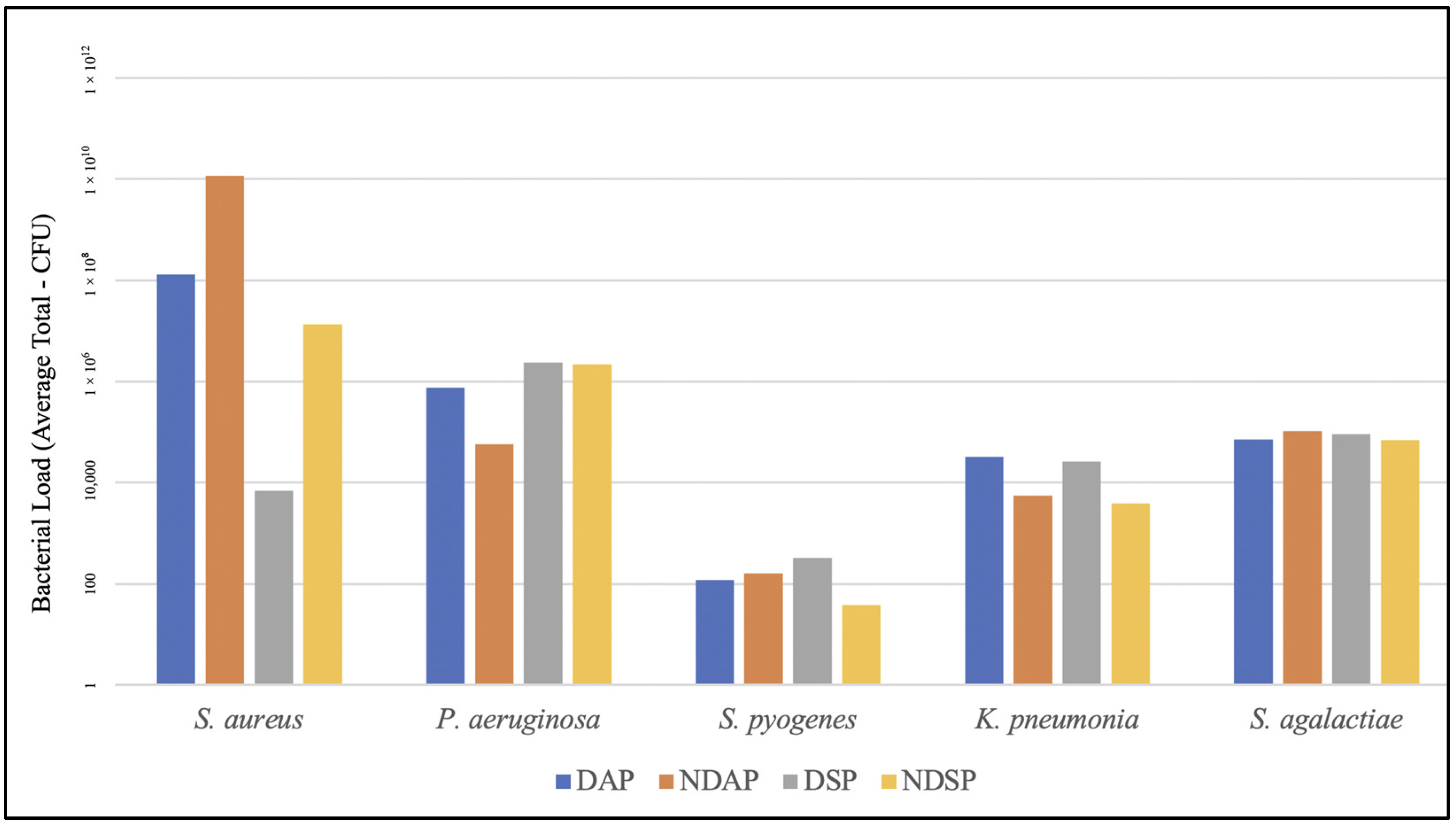
The relationship between denture use, periodontal disease, and respiratory pathogens was explored. The microbial loads of each pathogen in the four groups are presented in Figure 1. Significant differences were noted for K. pneumoniae (p = 0.04) and P. aeruginosa (p = 0.022) when comparing the four groups. A Bonferroni post hoc analysis revealed no statistically significant differences between groups for K. pneumoniae. However, this lack of significance does not preclude potential biological relevance and may reflect a Type II error due to the limited sample size and statistical power. The effect size, quantified using partial eta squared (η2), was 0.420, 0.361, 0.281, 0.275, and 0.079 for P. aeruginosa, K. pneumoniae, S. pyogenes, S. aureus, and S. agalactiae, respectively. Within the context of the stable periodontal condition, the denture-wearing group appeared to harbour more K. pneumoniae compared to the non-denture group, with a p-value approaching significance (p = 0.068). In contrast, a significantly lower quantity of P. aeruginosa was noted in the NDAP group compared to the DSP group (p = 0.033) (Table 3).
Figure 1.
Pathogen quantity comparison between the four groups. DAP, denture-wearing with active periodontal disease; NDAP, non-denture-wearing with active periodontal disease; DSP, denture-wearing with stable periodontal health; NDSP, non-denture-wearing with stable periodontal health.
Table 3.
Bonferroni correction for multiple pairwise comparisons of groups for K. pneumoniae and P. aeruginosa.
4. Discussion
The oral microbiome is home to over a thousand bacterial species that may be pathogenic or innocuous in nature [5]. Alterations in the oral microbiome have been linked to systemic and local factors such as systemic diseases, oral diseases, number of natural dentitions, oral hygiene habits, and denture cleanliness [28,29,30]. Older adults, aged 65 and older, are more susceptible to pathogenic bacterial proliferation, given that systemic diseases, oral diseases, edentulism, and denture use are characteristic of their cohort. As the oral microbiome consists of a multitude of species, a targeted approach using qPCR was adopted to identify the microbial profile of a few opportunistic respiratory pathogens. Through the use of qPCR, this study demonstrated that all participants presented with significant quantities of pathogenic bacteria associated with respiratory diseases. However, CFU equivalents reflect the total genomic DNA load rather than culturable cell counts and may therefore overestimate the number of viable bacteria, as DNA from non-viable cells is also detected. Nonetheless, at baseline, the measurements are likely to be clinically representative, because the samples mainly contain active biofilms with a high proportion of viable bacteria and no prior intervention that would generate large amounts of residual DNA. For clinical interpretation, genomic quantification offers important advantages, including faster turnaround, the ability to detect unculturable species, and a standardised, highly reproducible methodology. The two most abundant pathogenic bacteria found in this study were S. aureus and P. aeruginosa, which is supported by the findings of O’Donnell et al. [21]. In contrast, the prevalence rates of certain bacteria were higher than those reported in previous studies, possibly due to saliva samples being collected rather than denture biofilm samples, as analysed in earlier studies, and the inclusion of participants with active periodontal disease [21,22].
P. aeruginosa was detected in 79.2% of the saliva samples. The high prevalence of this pathogen may be attributed to denture use and periodontal disease. Although, P. aeruginosa is not commonly identified as a periodontal pathogen, a previous study observed that there was an increased number of this pathogen in the oral epithelial cells of individuals with periodontal disease [31]. Furthermore, P. aeruginosa is considered one of the most frequent colonisers of denture biofilm [32]. P. aeruginosa, an opportunistic respiratory pathogen, was also reported to be related to the pathogenesis of nosocomial pneumonia and cystic fibrosis [33,34]. Souto et al. [33] discussed the numerous virulence properties of this pathogen, which include its capacity to generate extracellular enzymes and toxins, adhere and lead to biofilm formation, and possess antibiotic resistance. In this study, the significantly higher quantity of P. aeruginosa noted in the DSP group may be attributed to one participant, who used an implant-supported mandibular overdenture, showing a particularly high quantity of this bacterium, which could represent an outlier in the data. Notably, because group comparisons were conducted using the Kruskal–Wallis test, which is based on ranked data and is relatively robust to outliers, this observation was retained and no sensitivity analysis excluding it was performed. In addition, given the small sample size and the pilot nature of this study, the results should be interpreted with caution. The presence of P. aeruginosa in individuals with peri-implant diseases has more recently been investigated. The colonisation of this pathogen in peri-implant plaque was similarly observed in a study by Sá et al. [35], who reported a higher abundance in peri-implant biofilm compared to subgingival biofilm. Nevertheless, this finding is in agreement with previous studies [29,36], which identified that a higher abundance of P. aeruginosa was found on removable denture surfaces.
K. pneumoniae is an opportunistic Gram-negative bacteria regarded as a common etiological agent of pneumonia in immunocompromised and hospitalised individuals [37]. In this study, a larger quantity was observed in the denture-wearing groups (DAP, DSP). This result is in agreement with the existing literature that has found a higher prevalence of K. pneumoniae in denture wearers compared to non-denture wearers [23,36]. K. pneumoniae has been identified as a causative agent of aspiration pneumonia, as the presence of this pathogen in denture biofilm poses the risk of aspiration pneumonia due to dentures being in close proximity to the respiratory tract [24,38]. In addition, denture-wearing can also be a risk factor for pneumonia incidence among the elderly population [17]. Bannwart et al. [39] reported the presence of respiratory pathogens on the dentures of 15 patients in the intensive care unit and described dentures as potential sources of pathology. This study discussed that, coupled with the immunocompromised status of these patients, the risk of development of respiratory diseases such as hospital-acquired pneumonia was increased.
This study also found a higher abundance of S. aureus in participants without dentures compared to those with dentures. S. aureus was also in abundance in participants with active periodontal disease compared to those with stable periodontal health condition. This finding is in agreement with a study by Veras et al. [40]. They found a higher quantity of S. aureus in individuals with periodontitis compared to healthy participants. Similarly, Fritschi et al. [41] highlighted the role of S. aureus in periodontitis, as high levels of S. aureus were found in individuals with aggressive periodontitis. S. pyogenes is a human-specific bacterial pathogen, and patients with asthma are more susceptible to S. pyogenes upper respiratory tract infections [42]. Notably, although this pathogen was present at the lowest abundance in samples from older adults in the present study, this population is still considered to be at high risk for S. pyogenes infections [43]. However, the interaction of bacteria within mixed biofilms is also important; for example, Streptococcus oralis, a common bacterium found in denture biofilm [5], has been reported to protect epithelial host cells from S. pyogenes-induced damage [44]. Comparatively, S. agalactiae has increasingly been detected in a substantial number of respiratory samples from individuals diagnosed with cystic fibrosis [45]. Despite not being a native coloniser of the oral cavity, this pathogen was also found in denture biofilm in a previous study [22].
Respiratory pathogens have long been established as causative agents of respiratory diseases. The two-way relationship between oral and respiratory microbiomes described by Pu et al. [19] indicated that these opportunistic organisms are prevalent and may play a role in the development of pneumonia, chronic obstructive pulmonary disease, and cystic fibrosis [46]. On the other hand, there is also increasing evidence suggesting that periodontal disease may facilitate respiratory diseases including pneumonia and chronic obstructive pulmonary disease [16]. Aspiration of periodontal pathogens can result in aspiration pneumonia and also has the potential to cause nosocomial infections [47]. Additionally, Twigg et al. [48] reported that there was a significantly high abundance of putative respiratory pathogens in the denture-associated microbiota for denture wearers diagnosed with pneumonia. The significant differences in the diversity and richness of bacterial communities among these patients suggests a possible association with the development of respiratory disease [22,48] and highlights the significance of oral and denture hygiene care in older adults [49].
The limitations of this study include the small sample size, the restricted range of pathogens examined, the lack of detailed periodontal parameters, and the choice of saliva as the sole sample type. The small sample size may have reduced statistical power and affected the ability to detect significant differences. Examining only a limited number of pathogens provides an incomplete picture of the salivary microbiota in older adults. Additional periodontal parameters, such as mean probing depth and bleeding on probing in each group, might have allowed a more detailed characterisation of clinical differences. Furthermore, although saliva is an appropriate sample for providing an overall picture of a participant’s microbial profile, particularly in this pilot study, which aims to explore respiratory pathogen profiles in older adults, relying on saliva alone makes it difficult to determine whether the detected pathogens originated from denture biofilm, other oral surfaces, or the respiratory tract. Given these constraints, the findings of this study should be considered exploratory rather than confirmatory. Future research with a larger sample size, incorporation of multivariable modelling, and more extensive sampling (including dental and denture biofilm) is recommended to validate and extend these results and to better define microbial profiles in this population.
5. Conclusions
Based on this limited pilot cohort, a high prevalence and high abundance of the examined opportunistic respiratory pathogens were observed in the saliva of older adults. These findings indicate that saliva may act as a reservoir for opportunistic respiratory pathogens within the oral cavity, and that their presence and levels may be associated with factors such as denture use and periodontal status. However, larger longitudinal studies are needed to confirm these observations and clarify these relationships. In this context, the results point to the potential relevance of optimal oral and denture hygiene practices in relation to the presence of respiratory pathogens in older adults and suggest that enhanced protective strategies for individuals who wear dentures or are diagnosed with periodontal disease may warrant further investigation.
Author Contributions
Conceptualization, A.P. and T.W.L.; Methodology, G.B.C.B., Y.Y.W., A.P. and T.W.L.; Software, G.B.C.B.; Validation, G.B.C.B., Y.Y.W., A.P. and T.W.L.; Formal analysis, G.B.C.B., Y.Y.W. and A.P.; Investigation, G.B.C.B., Y.Y.W. and A.P.; Resources, A.P.; Data curation, G.B.C.B., Y.Y.W. and A.P.; Writing—original draft, G.B.C.B. and Y.Y.W.; Writing—review & editing, A.P. and T.W.L.; Visualization, G.B.C.B. and A.P.; Supervision, A.P. and T.W.L.; Project administration, T.W.L.; Funding acquisition, T.W.L. All authors have read and agreed to the published version of the manuscript.
Funding
This research was funded by Seed Fund for Basic Research, The University of Hong Kong, grant number [109002230].
Institutional Review Board Statement
The study was conducted in accordance with the Declaration of Helsinki, and the protocol was approved by the Ethics Committee of The University of Hong Kong/Hospital Authority Hong Kong West Cluster (IRB Number: UW25-027, approve date: 20 February 2025).
Informed Consent Statement
Informed consent for participation was obtained from all subjects involved in the study. Written informed consent has been obtained from the patients to publish this paper.
Data Availability Statement
The raw data supporting the conclusions of this article will be made available by the authors on request.
Acknowledgments
The authors would like to thank the dental students, research assistants, and staff at the Central Research Laboratories, Faculty of Dentistry, The University of Hong Kong.
Conflicts of Interest
The authors declare no conflicts of interest.
References
- The Government of the Hong Kong Special Administrative Region. Hong Kong Population Projections for 2022–2046 Released. Available online: https://www.info.gov.hk/gia/general/202308/15/P2023081500308.htm (accessed on 25 November 2025).
- The Government of the Hong Kong Special Administrative Region, Department of Health. Oral Health Survey 2021 Report. Available online: https://www.toothclub.gov.hk/en/en_pdf/Oral_Health_Survey_2021/Oral_Health_Survey_2021_Report_EN.pdf (accessed on 25 November 2025).
- FDI World Dental Federation. Oral Health of Older Adults. Available online: https://www.fdiworlddental.org/sites/default/files/2025-06/FDI%20Oral%20Health%20of%20Older%20Adults%20Fact%20Sheet.pdf (accessed on 25 November 2025).
- Derafshi, R.; Bazargani, A.; Ghapanchi, J.; Izadi, Y.; Khorshidi, H. Isolation and identification of nonoral pathogenic bacteria in the oral cavity of patients with removable dentures. J. Int. Soc. Prev. Community Dent. 2017, 7, 197–201. [Google Scholar] [CrossRef] [Scilit]
- Lim, T.W.; Huang, S.; Jiang, Y.; Zhang, Y.; Burrow, M.F.; McGrath, C. Characterization of pathogenic microbiome on removable prostheses with different levels of cleanliness using 2bRAD-M metagenomic sequencing. J. Oral Microbiol. 2024, 16, 2317059. [Google Scholar] [CrossRef] [Scilit]
- Lee, Y.H.; Chung, S.W.; Auh, Q.S.; Hong, S.J.; Lee, Y.A.; Jung, J.; Lee, G.J.; Park, H.J.; Shin, S.I. Progress in oral microbiome related to oral and systemic diseases: An update. Diagnostics 2021, 11, 1283. [Google Scholar] [CrossRef] [Scilit]
- Lim, T.W.; Li, K.J.; Wang, J.; Yu, D.S.; Burrow, M.F.; McGrath, C. Association between removable denture-wearing and systemic diseases: A scoping review. J. Dent. 2025, 161, 105977. [Google Scholar] [CrossRef] [Scilit]
- Coulthwaite, L.; Verran, J. Potential pathogenic aspects of denture plaque. Br. J. Biomed. Sci. 2007, 64, 180–189. [Google Scholar] [CrossRef] [Scilit] [PubMed]
- Mylonas, P.; Milward, P.; McAndrew, R. Denture cleanliness and hygiene: An overview. Br. Dent. J. 2022, 233, 20–26. [Google Scholar] [CrossRef] [Scilit] [PubMed]
- Cankaya, Z.T.; Yurdakos, A.; Kalabay, P.G. The association between denture care and oral hygiene habits, oral hygiene knowledge and periodontal status of geriatric patients wearing removable partial dentures. Eur. Oral Res. 2020, 54, 9–15. [Google Scholar] [CrossRef] [Scilit]
- Lim, T.W.; Burrow, M.F.; McGrath, C. Evaluating risk factors associated with poor removable prosthesis hygiene in community-dwelling elders: A cross-sectional study. J. Prosthet. Dent. 2025, 134, 723–729. [Google Scholar] [CrossRef] [Scilit] [PubMed]
- Ng, J.Y.; Lim, T.W.; Tarib, N.; Ho, T.K. Effect of educational progressive web application on patient’s oral and denture knowledge and hygiene: A randomised controlled trial. Health Inform. J. 2021, 27, 14604582211035821. [Google Scholar] [CrossRef] [Scilit]
- Preshaw, P.; Walls, A.; Jakubovics, N.; Moynihan, P.J.; Jepson, N.; Loewy, Z. Association of removable partial denture use with oral and systemic health. J. Dent. 2011, 39, 711–719. [Google Scholar] [CrossRef] [Scilit]
- Curtis, M.A.; Diaz, P.I.; Van Dyke, T.E. The role of the microbiota in periodontal disease. Periodontology 2000 2020, 83, 14–25. [Google Scholar] [CrossRef] [Scilit]
- Genco, R.J.; Sanz, M. Clinical and public health implications of periodontal and systemic diseases: An overview. Periodontology 2000 2020, 83, 7–13. [Google Scholar] [CrossRef] [Scilit]
- Bansal, M.; Khatri, M.; Taneja, V. Potential role of periodontal infection in respiratory diseases-a review. J. Med. Life 2013, 6, 244–248. [Google Scholar] [PubMed]
- Lim, T.W.; Li, K.Y.; Burrow, M.F.; McGrath, C. Association between removable prosthesis-wearing and pneumonia: A systematic review and meta-analysis. BMC Oral Health 2024, 24, 1061. [Google Scholar] [CrossRef] [Scilit]
- Molina, A.; Huck, O.; Herrera, D.; Montero, E. The association between respiratory diseases and periodontitis: A systematic review and meta-analysis. J. Clin. Periodontol. 2023, 50, 842–887. [Google Scholar] [CrossRef] [Scilit] [PubMed]
- Pu, C.Y.; Seshadri, M.; Manuballa, S.; Yendamuri, S. The oral microbiome and lung diseases. Curr. Oral Health Rep. 2020, 7, 79–86. [Google Scholar] [CrossRef] [Scilit]
- Scannapieco, F.A. Relationships between periodontal disease and bacterial pneumonia. J. Periodontol. 1996, 67, 1114–1122. [Google Scholar] [CrossRef] [Scilit] [PubMed]
- O’Donnell, L.E.; Smith, K.; Williams, C.; Nile, C.J.; Lappin, D.F.; Bradshaw, D.; Lambert, M.; Robertson, D.P.; Bragg, J.; Hannah, V.; et al. Dentures are a Reservoir for Respiratory Pathogens. J. Prosthodont. 2016, 25, 99–104. [Google Scholar] [CrossRef] [Scilit]
- Lim, T.W.; Huang, S.; Zhang, Y.; Burrow, M.F.; McGrath, C. A comparison of the prevalence of respiratory pathogens and opportunistic respiratory pathogenic profile of ‘clean’ and ‘unclean’ removable dental prostheses. J. Dent. 2024, 145, 104968. [Google Scholar] [CrossRef] [Scilit]
- Sumi, Y.; Miura, H.; Sunakawa, M.; Michiwaki, Y.; Sakagami, N. Colonization of denture plaque by respiratory pathogens in dependent elderly. Gerodontology 2002, 19, 25–29. [Google Scholar] [CrossRef] [Scilit]
- Lim, T.W.; Li, K.Y.; Burrow, M.F.; McGrath, C. Prevalence of respiratory pathogens colonizing on removable dental prostheses in healthy older adults: A systematic review and meta-analysis. J. Prosthodont. 2024, 33, 417–426. [Google Scholar] [CrossRef] [Scilit]
- Wong, H.H.; Hung, C.H.; Yip, J.; Lim, T.W. Metagenomic characterization and comparative analysis of removable denture-wearing and non-denture-wearing individuals in healthy and diseased periodontal conditions. Microorganisms 2024, 12, 1197. [Google Scholar] [CrossRef] [Scilit]
- Zhang, Z.; Wen, S.; Liu, J.; Ouyang, Y.; Su, Z.; Chen, D.; Liang, Z.; Wang, Y.; Luo, T.; Jiang, Q.; et al. Advances in the relationship between periodontopathogens and respiratory diseases. Mol. Med. Rep. 2024, 29, 42. [Google Scholar] [CrossRef] [Scilit] [PubMed]
- Lim, Y.; Totsika, M.; Morrison, M.; Punyadeera, C. The saliva microbiome profiles are minimally affected by collection method or DNA extraction protocols. Sci. Rep. 2017, 7, 8523. [Google Scholar] [CrossRef] [Scilit] [PubMed]
- Rajasekaran, J.J.; Krishnamurthy, H.K.; Bosco, J.; Jayaraman, V.; Krishna, K.; Wang, T.; Bei, K. Oral microbiome: A review of its impact on oral and systemic health. Microorganisms 2024, 12, 1797. [Google Scholar] [CrossRef] [Scilit]
- Lim, T.W.; Huang, S.; Burrow, M.F.; McGrath, C. A randomised crossover clinical trial of the efficacy of an ultrasonic cleaner combined with a denture cleanser on the microbiome on removable dentures among community-dwelling older adults. J. Dent. 2025, 156, 105709. [Google Scholar] [CrossRef] [Scilit] [PubMed]
- O’Donnell, L.E.; Robertson, D.; Nile, C.J.; Cross, L.J.; Riggio, M.; Sherriff, A.; Bradshaw, D.; Lambert, M.; Malcolm, J.; Bujis, M.J.; et al. The oral microbiome of denture wearers is influenced by levels of natural dentition. PLoS ONE 2015, 10, e0137717. [Google Scholar] [CrossRef] [Scilit]
- Tag ElDein, M.A.; Yassin, A.S.; El-Tayeb, O.; Kashef, M.T. Chlorhexidine leads to the evolution of antibiotic-resistant Pseudomonas aeruginosa. Eur. J. Clin. Microbiol. Infect. Dis. 2021, 40, 2349–2361. [Google Scholar] [CrossRef] [Scilit]
- Thi, M.T.T.; Wibowo, D.; Rehm, B.H.A. Pseudomonas aeruginosa biofilms. Int. J. Mol. Sci. 2020, 21, 8671. [Google Scholar] [CrossRef] [Scilit]
- Souto, R.; Silva-Boghossian, C.M.; Colombo, A.P.V. Prevalence of Pseudomonas aeruginosa and Acinetobacter spp. in subgingival biofilm and saliva of subjects with chronic periodontal infection. Braz. J. Microbiol. 2014, 45, 495–501. [Google Scholar] [CrossRef] [Scilit]
- Pihl, M.; Chávez de Paz, L.E.; Schmidtchen, A.; Svensäter, G.; Davies, J.R. Effects of clinical isolates of Pseudomonas aeruginosa on Staphylococcus epidermidis biofilm formation. FEMS Immunol. Med. Microbiol. 2010, 59, 504–512. [Google Scholar] [CrossRef] [Scilit]
- Sá, A.M.; Mendes, J.M.; Silva, A.S.; Gonçalves, M.P.; Cardoso, M.; Coelho, C. Opportunistic pathogens isolated from peri-implant and periodontal subgingival plaque from adjacent teeth. Appl. Sci. 2023, 13, 9078. [Google Scholar] [CrossRef] [Scilit]
- Al-Shami, I.Z.; Alrubaidi, Y.A.; AL-Haddad, K.A.; Al-Najhi, M.M.; Al-Shamahy, H.A.; Al-Kibsi, T.A.; Al-Kholani, A.I. Effect of removable dentures on colonization of aerobic bacteria in the oral cavity and antibiotic pattern of the common isolated bacteria. Univers. J. Pharm. Res. 2022, 7, 1–9. [Google Scholar] [CrossRef] [Scilit]
- Abbas, R.; Chakkour, M.; Zein El Dine, H.; Obaseki, E.F.; Obeid, S.T.; Jezzini, A.; Ghssein, G.; Ezzeddine, Z. General overview of Klebsiella pneumonia: Epidemiology and the role of siderophores in its pathogenicity. Biology 2024, 13, 78. [Google Scholar] [CrossRef] [Scilit]
- Noguchi, S.; Akata, K.; Mukae, H.; Yatera, K. Microbiological etiology of aspiration pneumonia in Japan: Insights from a systematic review and meta-analysis. Respir. Investig. 2025, 63, 510–516. [Google Scholar] [CrossRef] [Scilit] [PubMed]
- Bannwart, L.C.; de Moraes Melo Neto, C.L.; dos Santos, D.M.; Moreno, A.L.; Pesqueira, A.A.; Goiato, M.C.; de Magalhães Bertoz, A.P. Dentistry and Intensive Care Unit: A brief report. Eur. J. Dent. 2022, 16, 449–453. [Google Scholar] [CrossRef] [Scilit]
- Veras, E.L.; Castro dos Santos, N.; Souza, J.G.; Figueiredo, L.C.; Retamal-Valdes, B.; Barão, V.A.; Shibli, J.; Bertolini, M.; Faveri, M.; Teles, F.; et al. Newly identified pathogens in periodontitis: Evidence from an association and an elimination study. J. Oral Microbiol. 2023, 15, 2213111. [Google Scholar] [CrossRef] [Scilit]
- Fritschi, B.Z.; Albert-Kiszely, A.; Persson, G.R. Staphylococcus aureus and other bacteria in untreated periodontitis. J. Dent. Res. 2008, 87, 589–593. [Google Scholar] [CrossRef] [Scilit] [PubMed]
- Frey, D.; Jacobson, R.; Poland, G.; Li, X.; Juhn, Y. Assessment of the association between pediatric asthma and Streptococcus pyogenes upper respiratory infection. Allergy Asthma Proc. 2009, 30, 540–545. [Google Scholar] [CrossRef] [Scilit]
- Avire, N.J.; Whiley, H.; Ross, K. A review of Streptococcus pyogenes: Public health risk factors, prevention and Control. Pathogens 2021, 10, 248. [Google Scholar] [CrossRef] [Scilit]
- Fiedler, T.; Riani, C.; Koczan, D.; Standar, K.; Kreikemeyer, B.; Podbielski, A. Protective mechanisms of respiratory tract streptococci against Streptococcus pyogenes biofilm formation and epithelial cell infection. Appl. Environ. Microbiol. 2013, 79, 1265–1276. [Google Scholar] [CrossRef] [Scilit]
- Eickel, V.; Kahl, B.; Reinisch, B.; Dübbers, A.; Küster, P.; Brandt, C.; Spellerberg, B. Emergence of respiratory Streptococcus agalactiae isolates in cystic fibrosis patients. PLoS ONE 2009, 4, e4650. [Google Scholar] [CrossRef] [Scilit]
- Gomes-Filho, I.S.; Passos, J.S.; Seixas da Cruz, S. Respiratory disease and the role of oral bacteria. J. Oral Microbiol. 2010, 2, 5811. [Google Scholar] [CrossRef] [Scilit]
- Jerônimo, L.S.; Abreu, L.G.; Cunha, F.A.; Lima, R.P.E. Association Between Periodontitis and Nosocomial Pneumonia: A Systematic Review and Meta-analysis of Observational Studies. Oral Health Prev. Dent. 2020, 18, 11–17. [Google Scholar] [PubMed]
- Twigg, J.A.; Smith, A.; Haury, C.; Wilson, M.J.; Lees, J.; Waters, M.; Williams, D.W. Compositional shifts within the denture-associated bacteriome in pneumonia—An analytical cross-sectional study. J. Med. Microbiol. 2023, 72, 001702. [Google Scholar] [CrossRef] [Scilit] [PubMed]
- Lim, T.W.; Pan, H.; Pan, M.; Burrow, M.F.; McGrath, C. Agreement in quantification of removable prosthesis plaque area coverage using a semi-automated planimetric assessment method. J. Dent. 2023, 138, 104721. [Google Scholar] [CrossRef] [Scilit] [PubMed]

Disclaimer/Publisher’s Note: The statements, opinions and data contained in all publications are solely those of the individual author(s) and contributor(s) and not of MDPI and/or the editor(s). MDPI and/or the editor(s) disclaim responsibility for any injury to people or property resulting from any ideas, methods, instructions or products referred to in the content. |
© 2026 by the authors. Licensee MDPI, Basel, Switzerland. This article is an open access article distributed under the terms and conditions of the Creative Commons Attribution (CC BY) license.