


Abstract
Background: Targeted muscle reinnervation (TMR) is increasingly used to enhance prosthetic control and to reduce post-amputation pain. Its implementation across new centers raises questions about the reproducibility of outcomes and the impact of surgical experience. Methods: We compared the first three TMR patients treated in a newly established center in Nantes, France, with three patients treated in a high-volume center in Göttingen, Germany. Functional outcomes were measured using the Box and Block test (BBT), and operative time was recorded. Two French cases were performed with the assistance of a Göttingen-based surgeon. Conclusions: The functional outcomes showed a similar trend in both groups. The mean BBT scores were equivalent, suggesting reliable reinnervation and prosthetic integration even in early cases. Operative times were longer in Nantes, but did not impact outcomes. TMR appears not to have a pronounced learning curve, particularly regarding functional success in early cases under guided protocols. Factors such as assistance from experienced surgeons and favorable donor-to-recipient nerve ratios likely contribute to consistent outcomes. These findings support the reproducibility of TMR across institutions. Results: Within the first two years of rehabilitation we observed improvements in both functional performance and patient-reported quality of life. All six patients across both centers in-creased in BBT scores. All the patients reported an increase in social relationships and psychological health, and two of three patients reported an increase in physical health. Importantly, all six patients discontinued the use of pain medication at 2 years fol-lowing TMR. Furthermore, the French patients reported a decrease from 65–82 mm to 0–31 mm across the patients’ Visual Analog Scale (VAS) pain scores.
1. Introduction
Targeted muscle reinnervation (TMR) has emerged as an effective surgical technique for improving prosthetic control and reducing neuroma-related and phantom limb pain in people with amputation. Originally developed by Kuiken et al. [1], TMR involves the transfer of transected peripheral nerves to redundant motor branches, allowing for more intuitive control of myoelectric prostheses while simultaneously mitigating the formation of symptomatic neuromas [2,3]. Multiple studies have since confirmed the efficacy of TMR in patients with both upper and lower limb amputation, with significant reductions in residual limb pain and improved prosthetic functionality [2,4,5].
As the technique gains global acceptance, many surgical centers are in the process of implementing TMR programs. This expansion raises important questions regarding the reproducibility of outcomes, the impact of institutional experience, and the presence of a learning curve. While multicenter analyses and systematic reviews have supported the generalizability of TMR [6,7], there is a paucity of data specifically evaluating the outcomes in newly established centers during the early phase of implementation.
This study compares the first three patients who underwent TMR in a newly established program in Nantes, France, with three patients treated in a high-volume, experienced center in Göttingen, Germany. Notably, two of the French procedures were performed with intraoperative assistance from a Göttingen-based surgeon. We aimed to assess functional outcomes and operative metrics to evaluate the consistency of early results in a developing center compared to those of an established institution.
2. Materials and Methods
We conducted a retrospective, observational cohort study of six patients with a unilateral upper-limb amputation treated with TMR between 2014 and 2024 at two centers following harmonized clinical pathways.
Patients were included if the level of amputation was transhumeral (TH) or further proximally (glenohumeral, shoulder disarticulation) and had participated in a multidisciplinary consultation, including a psychologist, to validate the patient’s motivation and cognitive ability to ensure full compliance with the postoperative rehabilitation protocol. Patients were both male (n = 5) and female (n = 1) and aged between 19 and 45 years (see Table 1).
Table 1.
Baseline characteristics table. Summary of basic demographics and clinical characteristics.
The patient’s clinical examination pre-surgery focused on (1) the trauma context of the injury (anamnesis, risk factors, and comorbidities), (2) the patient’s skin condition (quality of the skin covering, e.g., skin grafts and adhesions), (3) the residual limb volume, (4) a focused assessment for neuroma- or phantom-limb pain, and (5) shoulder mobility. Exclusion criteria included (1) unfavorable anatomical conditions (e.g., muscle involution or meningoceles), (2) ongoing treatment for osteomyelitis, (3) pre-existing psychiatric conditions, or (4) non-adherence to the 24-month rehabilitation protocol.
Before surgery, the rehabilitation process focused on maintaining shoulder joint mobility, optimizing stump trophicity, proprioception, coordination, and balance, while also ensuring daily prosthesis use, conducting psychological assessments, and implementing both home-based and occupational therapy-guided phantom limb stimulation, alongside preoperative testing in occupational therapy.
The procedures in Nantes were guided by a surgical roadmap described by Aszmann et al. and Gart et al. [8,9], with intraoperative assistance from a Göttingen surgeon in two cases. The cases treated in Göttingen followed established protocols [2,5].
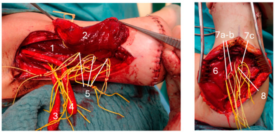
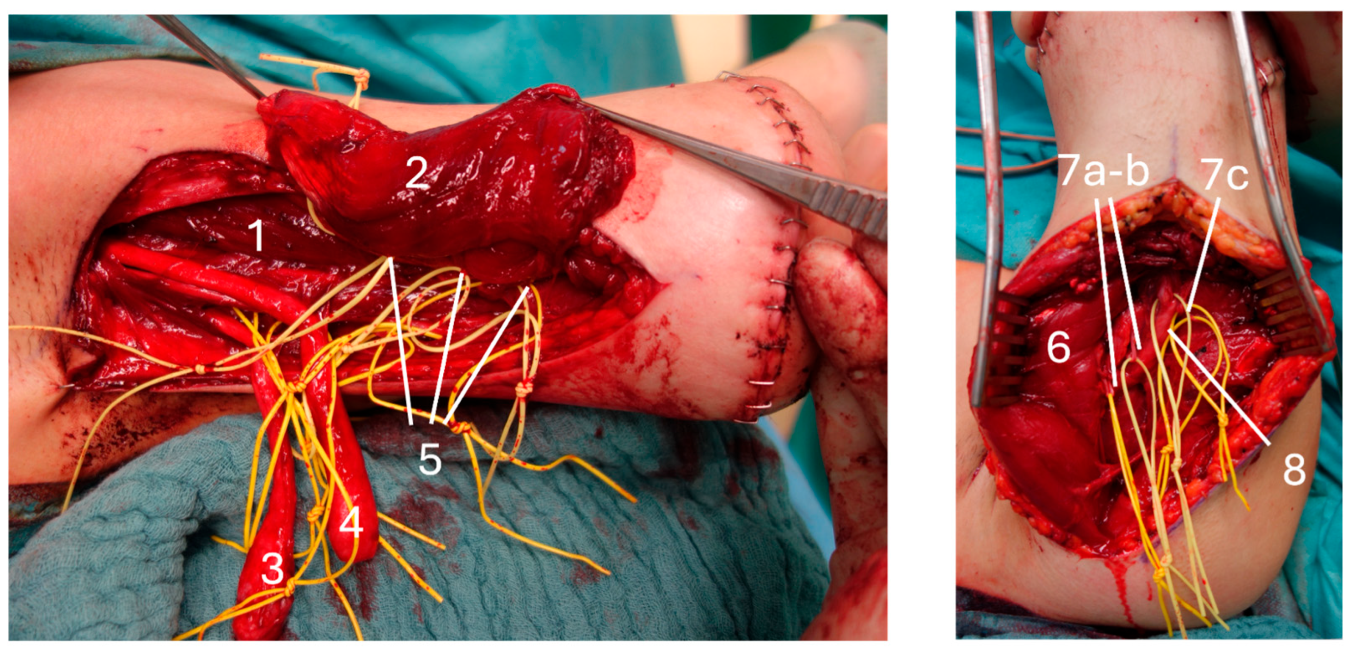
In brief, for people with transhumeral amputation level, surgical exposure was achieved through two skin incisions: one medial along the brachial canal to access the biceps brachii, and one posteriorly to access the triceps brachii (see Figure 1). After successful identification and preparation of donor nerves and target muscle fascicles using intraoperative neurostimulation, residual peripheral nerves were transferred to nearby target muscles following standardized anatomical principles (see Table 2) while adapting the operative procedure to the individual anatomical situation.
Figure 1.
Intraoperative photography from patient with transhumeral amputation. Anatomic view: (Left) panel 1: brachialis muscle; 2: long head of biceps dissected; 3: ulnar nerve; 4: median nerve; 5: Nn proprii M. brachialis brachii. (Right) panel dorsomedial access to triceps muscle; 6: long head of triceps muscle; 7a and b: branches of radial nerve to long head of triceps; 7c: radial nerve branches to lateral head of triceps muscle; 8: radial nerve.
Table 2.
Surgery road map for all patients. Detailed nerve matrix for transhumeral, glenohumeral, and upper limb (UL) exarticulation amputation.
Donor-to-target nerve matching followed a structured decision-making algorithm:
- (1)
- Preoperative mapping using Hoffmann–Tinel stimulation and sensory response testing was used to identify donor nerve projection areas and potential target regions.
- (2)
- After neuroma excision, the available length and mobility of each donor nerve were assessed. Nerves with sufficient length and gliding capacity were eligible for transfer to more distal target muscles.
- (3)
- In cases of severe scarring, fibrosis, or short residual nerve segments requiring resection, transfer to a more proximal target muscle was preferred to ensure a tension-free coaptation and avoid traction on the repair site.
- (4)
- Target muscle selection followed standardized anatomical principles prioritizing muscles with preserved volume, accessible motor entry points, and favorable positioning for future myoelectric signal generation and prosthetic control.
This stepwise approach resulted in individualized nerve transfer configurations, as injury patterns and donor nerve quality varied considerably between patients, while maintaining adherence to the established surgical roadmaps described by Aszmann et al. and Gart et al. [8,9].
Donor nerves were major motor or mixed nerves, and recipient nerves were terminal branches supplying redundant muscles, which were denervated before TMR. The donor nerve was coapted directly to the thinner recipient nerve. To achieve this, the epineurium of the donor nerve was gently constricted into a funnel shape before coaptation, creating a direct nerve repair despite the marked caliber mismatch. This approach was chosen to maximize the number of donor nerve fibers entering the recipient nerve without escaping at the coaptation site and to reduce the risk of neuroma formation. The site was further sealed and reinforced with fibrin glue. For suturing, 8-0 Prolene was used, as this size allowed the funnel-shaped configuration of the donor nerve to be maintained, whereas 9-0 was insufficient for this purpose. The coaptation itself was performed under tension-free conditions, and intraoperative testing during arm movement confirmed that the repair remained stable without undue stress or risk of rupture.
Lastly, to optimize the signal quality for future myoelectric readouts, fascio-fatty tissue flaps were prepared and repositioned subcutaneously in between the muscle’s bellies, allowing for a better separation of Electromografy (EMG) signals.
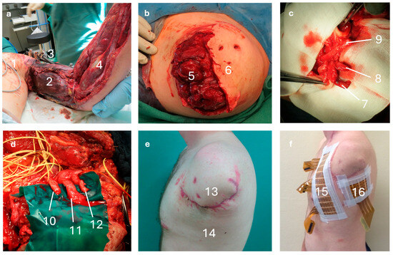
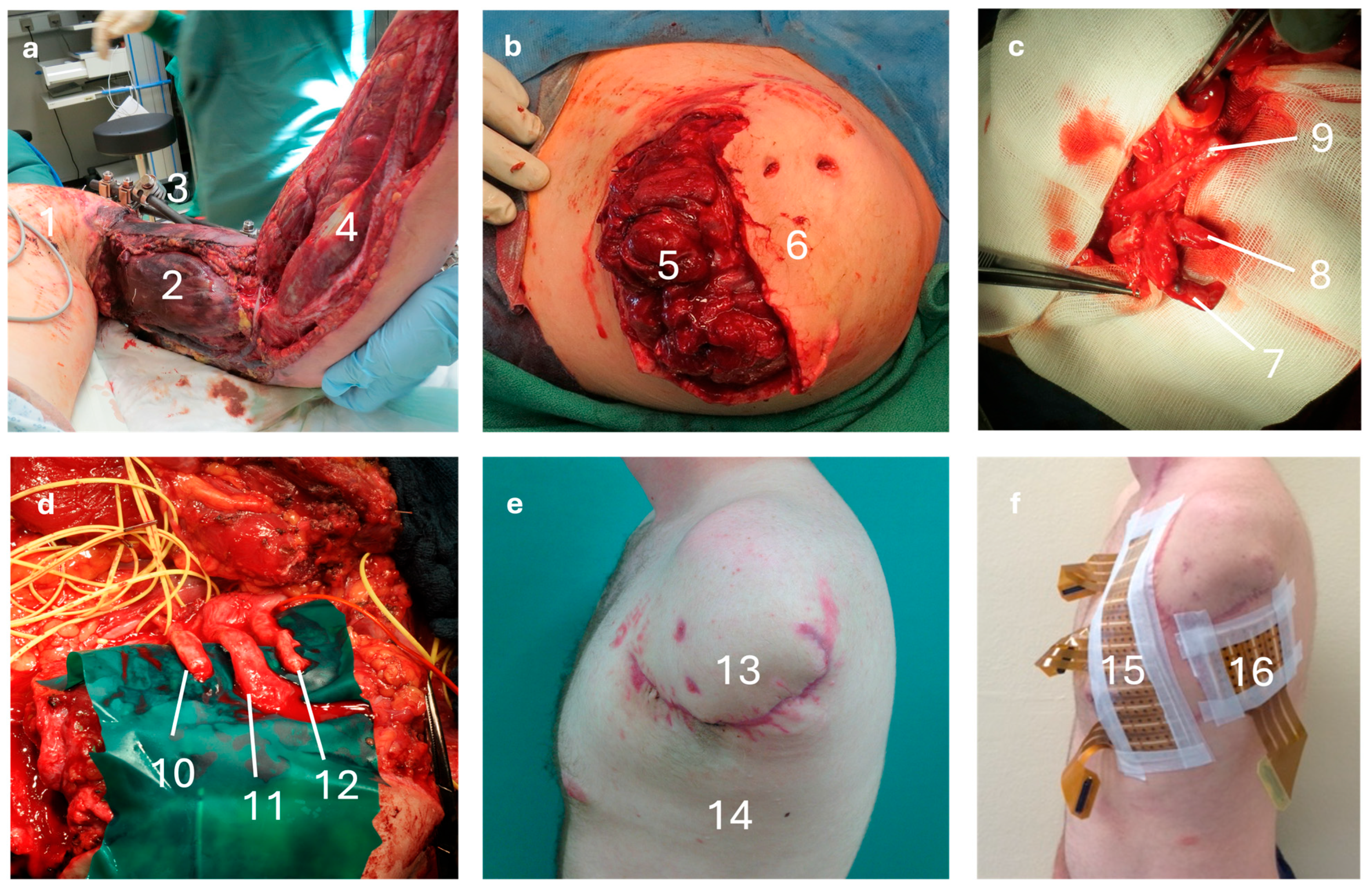
In cases of both shoulder disarticulation and glenohumeral amputation, multiple surgical approaches may be required due to the extensive anatomical region involved. As trauma mechanisms are often complex and highly variable, individualized adaptations to the operative strategy are frequently necessary. In both patients, treatment followed a two-stage (and in some situations multi-stage) protocol. During the first stage, post-traumatic care and definitive amputation were performed, and the operative field was prepared to enable a tailored reconstructive strategy (see Figure 2). A subsequent second-stage procedure was then carried out to perform TMR under optimized tissue conditions and to establish a stable soft-tissue environment.
Figure 2.
Intraoperative photography from patient with shoulder disarticulating amputation: (a) Initial presentation with extensive soft tissue defect and necrotic muscles after failed replantation (1), necrotic muscles of upper extremity (2), external fixation (3), and remaining viable tissue (4). (b) First stage after glenohumeral amputation and soft tissue coverage with thoracic wall muscles (5) and skin-flap margins (6). (c) Proximal identification of donor nerves using intraoperative neurostimulation: radial (7), ulnar (8), and median nerve (9). (d) Second-stage TMR with denervated recipient muscles and aligned donor nerves: musculocutaneous (10), median (11), and ulnar (12). (e) Stable postoperative stump contour (13) with well-healed thoracic incisions (14). (f) Early surface EMG demonstrating activation of reinnervated muscle groups (15, 16).
For patients with shoulder disarticulation, an extended superior approach was employed. A generous incision along the lateral clavicle and acromial stump provided access to the proximal brachial plexus. Wide elevation of the skin flaps and release of residual deltoid fascia improved visualization and facilitated mobilization of the plexus elements. Residual peripheral nerves—including the musculocutaneous, median, ulnar, radial, and axillary components—were identified proximally and isolated with the assistance of intraoperative neurostimulation. Recipient muscles were selected based on the preserved functional segments of the trapezius, the clavicular portion of the pectoralis major, and the remaining thoracic wall musculature. These muscles were denervated as proximally as possible to minimize competitive reinnervation. TMR was subsequently performed by transferring donor nerves into adjacent target muscles in a tension-free manner, ensuring reproducible anatomical alignment and sufficient soft-tissue coverage.
For patients with a glenohumeral amputation level, a standardized subclavicular approach was used. A broad incision extending along the deltopectoral groove exposed the underlying pectoralis major. Improved access to the neurovascular bundle was achieved by detaching the pectoralis minor from the coracoid. Donor and recipient structures were identified and prepared under intraoperative neurostimulation. As in shoulder-disarticulation cases, recipient muscles were denervated proximally, and residual peripheral nerves were coapted to nearby target muscles without tension. The nerve matrix employed for each of the four TH and two GH/SD patients included in this study is summarized in Table 1 and follows standardized anatomical principles. Intraoperative photographic documentation of the amputation at the glenohumeral level was limited due to ethical and consent-related constraints. To ensure reproducibility and anatomical clarity, the surgical approaches are described in detail in the text and complemented by established anatomical and surgical roadmaps as cited.
After surgery, the rehabilitation protocol lasted for two years. On average, it takes about three months for reinnervation to occur. During this initial postoperative phase, the focus therefore lies on scar care, lymphatic drainage, and postoperative pain management. Between 3 and 5 months, the creation of the socket began, along with the start of remote rehabilitation using a prosthesis mounted on a base to avoid pulling on the scars.
In clinical practice at both centers, individualized prosthetic fitting played a central role in the rehabilitation process. Prosthetic parameters—including length, size, and weight (with material selection based on patient habitus and functional demands)—were initially determined during the first fitting session and routinely adjusted throughout rehabilitation. Functional alignment with respect to upper-body posture and prevention of shoulder or back pain was monitored. Follow-up assessments were conducted by experienced orthopedic technicians who evaluated posture, compensatory movement patterns, and prosthetic comfort. Modifications to prosthetic alignment, weight distribution, or suspension systems were performed when necessary to optimize function and reduce compensatory strain.
Starting from the 15th month, the prostheses were fully assembled, and the situational training in occupational therapy could begin. The patients were grouped by treatment center: Göttingen (n = 3, 2 TH, 1 SD) and Nantes (n = 3, 2 TH, 1 SD). The Göttingen center featured a mature TMR program with prior experience in nerve transfer and osseointegration. The Nantes center had recently initiated its TMR program, with two surgeries performed under supervision from Göttingen surgeons. All the patients were pain-free at the time of testing and used comparable myoelectric prostheses.
The patients were evaluated for their functional performance in both groups using the Box and Block test (BBT). Due to differences in clinical routines and follow-up protocols between centers, the Clothespin Relocation Test (CRT) was performed only in Göttingen, while the WHOQOL-BREF and Visual Analogue Scale (VAS) for assessment of pain were collected exclusively in Nantes.
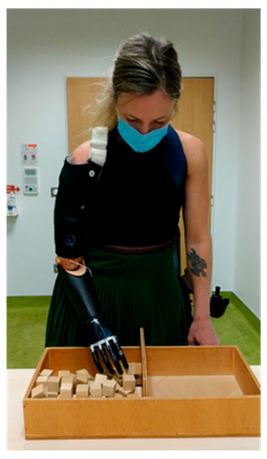
The BBT was performed following the standardized procedure described by Mathiowetz et al. [10]. The patients were seated or, in the case of the German patients, standing in front of a partitioned wooden box and instructed to transfer 2.5 cm square wooden blocks from one compartment to the other, one block at a time, using their prosthetic hand (see Figure 3). The number of blocks transferred within 60 s was recorded as a measurement outcome.
Figure 3.
Patient performing Box and Block test.
The CRT is a standardized tool for the assessment of hand dexterity and was later on adapted and reintroduced by Kuiken et al. [11,12]. In the adapted version the patients were instructed to relocate five spring-loaded clothespins from a horizontal bar to a vertical one and then return them using their prosthetic hand. The total time (in minutes) required to complete the relocation cycle was recorded.
The French group of patients was tested for the WHOQOL-BREF, an abbreviated version of the WHOQOL-100 questionnaire [13], which is an efficient tool to assess the patient’s quality of life in the context of multiple life domains. The WHOQOL-BREF is a validated 26-item abbreviated version of the WHOQOL-100, assessing four domains: physical health, psychological wellbeing, social relationships, and environment. The patients responded using a 5-point Likert scale. Domain scores were calculated and transformed to a 0–100 scale: higher scores indicate better quality of life. Lastly, investigators used the VAS to quantify patient-reported pain intensity. The patients marked their perceived pain level on a 10 cm horizontal line, anchored with “no pain” at 0 cm on one side and “worst imaginable pain” at 10 cm on the other. The distance (in millimeters) from the “no pain” anchor to the patient’s mark was recorded.
Both centers used a set of diverse prosthetic systems (Ottobock 12k50/12k100/12k110, Dynamic arm plus, MyoHand Variplus Speed, Ottobock, Duderstadt, Germany). Based on the post-surgical anatomy, different electrode set-ups could be applied (5/6/6 electrodes for German patients, 5/6/3 electrodes for French patients) for control read-out. An advanced signal processing device (MyoBock, Ottobock, Duderstadt, Germany) allowed for diverse intuitive motor control.
Furthermore, we performed a comparison of the patient’s skin-to-skin duration in minutes as an estimation of the burden that the surgery imposes on the patients and investigated the patients’ need for pain killers postoperatively.
Experiments were conducted under approval of the local ethics commitee (Ethics application: No. 1/1/17 at University Medical Center Göttingen and listed in the German clinical trials register (DRKS00037592). GenAI (ChatGPT 5.0) was used for text editing and in a preliminary digital review process. For a transparent reporting of the nonrandomized publications evaluations a TREND checklist can be found in the Supplementary Materials.
3. Results
Within the first two years of rehabilitation ( T0 = surgery), we observed improvements in both functional performance and patient-reported quality of life.
3.1. Box and Block Test (BBT)
All six patients across both centers increased in BBT scores.
3.1.1. Göttingen Group: The Three Patients Treated in Germany Showed Improvements
- Case 1: from 5.3 (±0.58) to 12.3 (±2.08) points (10.8 and 14.1 months post-op).
- Case 2: from 5.5 (±0.7) to 11.6 (±0.58) points (6.7 and 17.9 months post-op).
- Case 3: from 9.3 (±1.15) to 12.0 (±1.73) points (10.0 and 10.2 months post-op).
3.1.2. French Group: Patients in Nantes Demonstrated Two Phases of Improvement
- Early Phase (within 1 year)
- Case 1: 6.0 (±2.83) → 13.0 points (3 and 12 months post-op).
- Case 2: 7.0 (±0) → 10.0 points (3 and 12 months post-op).
- Case 3: 7.5(±6.3) → 16.0 points (3 and 12 months post-op).
- Extended Follow-up (up to 24 months)
- Case 1: 11.0 → 18.0 points (3 and 24 months post-op).
- Case 2: 14.0 → 17.0 points (3 and 24 months post-op).
- Case 3: 10.5 → 19.0 points (3 and 24 months post-op).
3.2. Clothespin Relocation Test (CRT)
This test was performed only in the German cohort to assess prosthesis dexterity. All three patients showed a decrease in the time needed to fulfill the tasks.
- Case 1: from 2:35 min to 0:45 min (10.8 and 14.1 months post-op).
- Case 2: from 5:33 min to 1:42 min (7.6 and 17.9 months post-op).
- Case 3: from 0:48 min to 0:34 min (10 and 10.2 months post-op).
These improvements support the functional gains observed in the BBT, indicating better prosthetic control.
3.3. Skin-on-Skin Time (According to Anesthesiologic Protocols)
3.3.1. German Group
- Case 1: 245 min.
- Case 2: 274 min.
- Case 3: 310 min.
3.3.2. French Group
- Case 1: 260 min.
- Case 2: 280 min.
- Case 3: 492 min.
German case 3 was a combined TMR surgery, also including an osseointegration procedure, and therefore must be interpreted as an overestimation of the actual surgery time for TMR only.
3.4. WHOQOL-BREF and Visual Analog Scale (VAS) for Pain Assessment
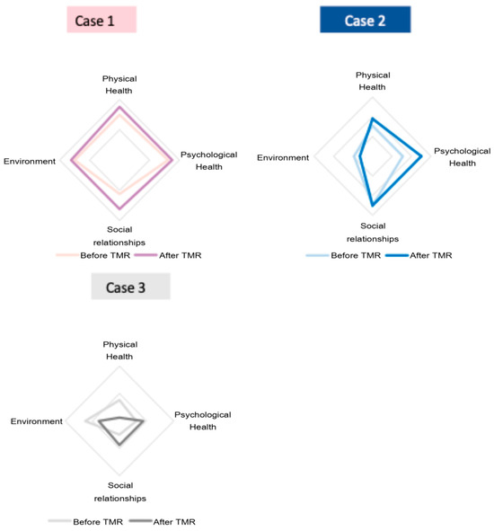
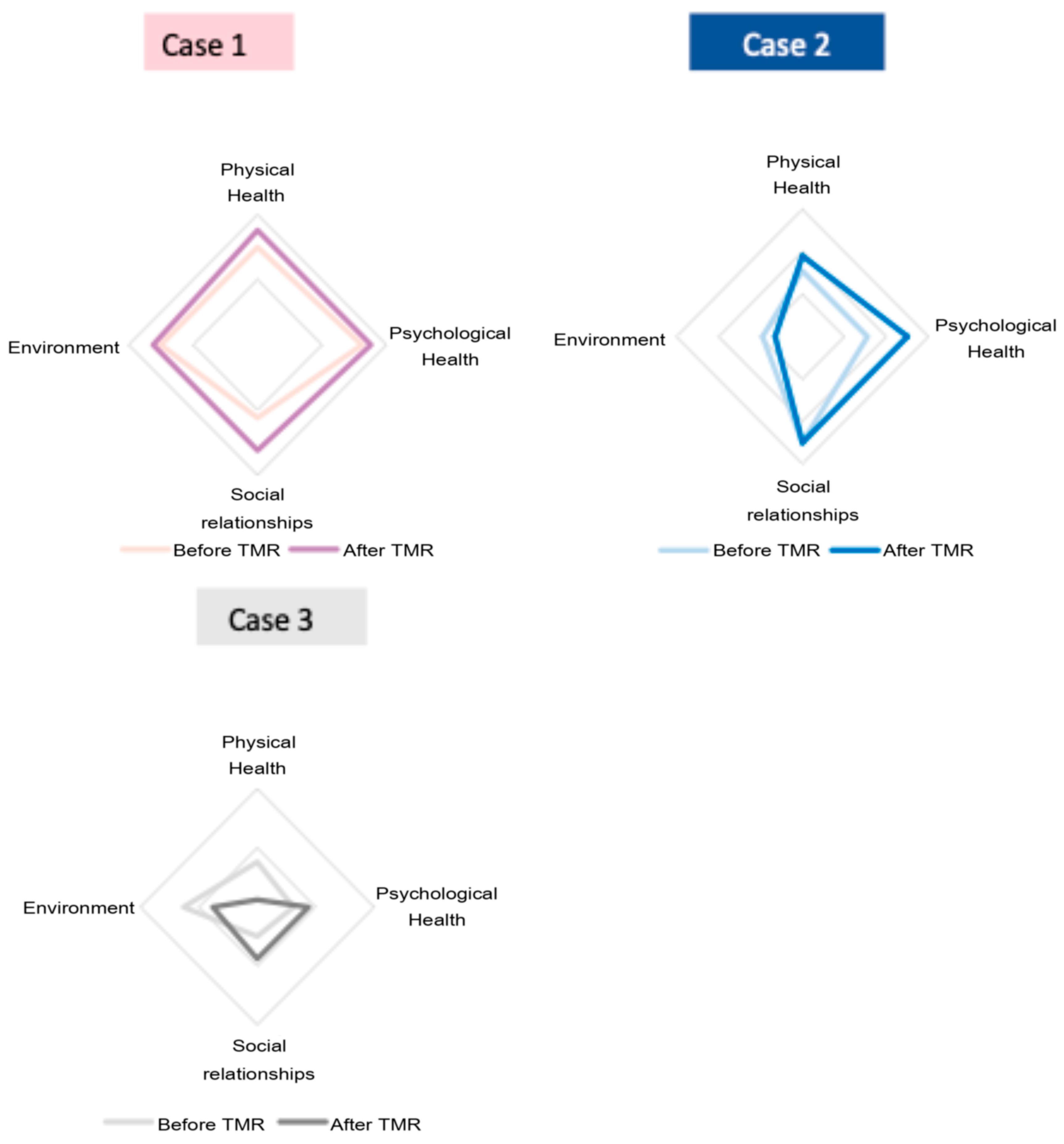
The evaluation of the change in the patient’s quality of life using the WHOQOL-BREF showed a more nuanced picture (Figure 4). All the patients reported an increase in social relationships and psychological health, and two of three patients reported an increase in physical health. One patient, however, described a decrease in physical health, and two patients described a decrease in their interaction with the environment.
Figure 4.
WHOQOL-BREF results for each of the 3 cases treated in Nantes, France.
Importantly, all six patients discontinued the use of pain medication at 2 years following TMR. Furthermore, the French patients reported a decrease from 65–82 mm to 0–31 mm across the patients’ Visual Analog Scale (VAS) pain scores.
4. Discussion
This dual-center case series suggests that TMR is a reproducible procedure yielding functional and psychosocial benefits, even in settings where surgical programs are newly implemented. The functional outcomes, particularly in the BBT and CRT, point to a consistent improvement in prosthetic control, likely driven by enhanced EMG signal acquisition due to optimized nerve-muscle transfers.
To place our results in context, Kuiken et al. [1] described the Box and Block test 5 months after surgery. Presented was the number of blocks moved after TMR in patients with shoulder-level amputation using custom made myoelectric prosthesis, with results of 14 ± 4 blocks moved (n = 3). However, a baseline result before training is not given. In TMR subjects using a pattern recognition control mode in a 6-week home trial, Hargrove et al. observed a statistically significant increase in BBT performance (p = 0.03), with subjects increasing from an average of 10 to 12 blocks moved.
In another work presenting a comparison of pattern recognition vs. direct control, Hargrove et al. [14] found end-trial block counts of 13.4 ± 2.6 (pattern recognition) vs. 15.6 ± 2.7 (direct control), though that study did not show significant pre/post improvement.
Compared to these benchmarks, our cohort’s average gain of 5.23 and 6.17 and absolute average values of 11.96 and 13 for the German and the French group, respectively, are slightly lower than the values reported. The differences may reflect variation in baseline status, rehabilitation protocols, device sophistication, or training intensity. The relative consistency in direction of improvement across studies helps reinforce the plausibility of functional gains through TMR and prosthetic training, but direct quantitative comparisons remain limited by reporting heterogeneity.
The timeline of reinnervation was primarily determined by the anatomical distance between the donor nerve and the target muscle following neuroma excision. As these distances typically ranged only a few centimeters, reinnervation was expected within the first postoperative months based on the established axonal regeneration rate of approximately 1 mm/day. Reinnervation was monitored clinically and functionally during routine follow-up, including observation of voluntary contraction, surface EMG detection of transcutaneous muscle activation, and functional activation during occupational therapy using tabletop prosthetic training devices.
Exact timepoints could not be consistently documented for all the patients due to heterogeneous follow-up schedules and variable attendance between the participating centers, which is a major limitation of our study; nevertheless, all the patients demonstrated detectable muscle activation during the early rehabilitation phase, enabling the initiation of targeted functional training.
Furthermore, the recent literature has highlighted that upper-limb prosthesis use can influence whole-body biomechanics [15,16]. While these data support the plausibility of gait or whole-body biomechanical effects in upper-limb prosthesis users, the current evidence is characterized by small sample sizes, mixed populations, and a lack of standardized gait-analysis protocols specifically tailored to this group. In particular, spatiotemporal gait parameters have not consistently demonstrated significant deviations in adult upper-limb prosthesis users [17]. In our study, we therefore focused on patient-reported outcomes, upper-limb functional assessments, and surgical parameters, which directly reflect the aims of knowledge-transfer and targeted muscle reinnervation. Nonetheless, we anticipate integrating gait or kinematic analyses into future investigations that target long-term compensatory mechanisms and whole-body function.
Pain relief was another major outcome: all the patients stopped using pain medication, and VAS scores dropped substantially. This supports the idea that secondary TMR offers not only functional restoration but also significant neuromodulatory effects.
In the cases presented, the collaboration between the experienced and new centers proved successful, indicating that the procedure may not require a steep learning curve when guided by standardized protocols. However, surgical time differences may reflect not only surgeon expertise but also institutional routines, team dynamics, and access to specialized tools.
The study has several limitations. The small sample size and heterogeneous patient profiles (e.g., amputation level, evaluation tools) preclude strong statistical conclusions. Differences in prosthetic hardware and electrode configurations across centers may have influenced control performance and functional outcomes and should be considered when interpreting cross-centre comparisons. Furthermore, the heterogeneity in outcome measures beyond the Box and Block test serves as a common ground. With CRT capturing functional performance and WHOQOL reflecting subjective well-being, this creates two different perspectives on the post-surgical rehabilitation phase and limits direct comparison. Future studies could benefit from greater harmonization, for example, through consensus-driven core outcome sets that integrate both functional and patient-reported outcomes, thereby improving comparability across studies. This project served as a test run for international collaboration in this field, and although surgical assimilation and good clinical outcomes could be demonstrated in a limited set of patients, future projects will also aim to create a platform of exchange beyond surgical expertise to harmonize interdisciplinary approaches in post-surgical rehabilitation. More broadly, while CRT and WHOQOL assess different but complementary domains—objective functional capacity versus subjective quality of life—their heterogeneity underscores the need for harmonization, whether through validated crosswalks between functional and QoL measures or the adoption of mixed-methods frameworks.
Moreover, neither patients nor therapists were blinded, which could introduce assessment bias. Selection bias may have occurred, as patients with high motivation and compliance may have been more likely to complete the full protocol.
Despite these limitations, our results underscore the potential of TMR to improve prosthetic control and quality of life. Future multicenter studies with standardized protocols are needed to validate these findings and explore broader applicability.
5. Conclusions
This dual-center case series highlights the clinical and collaborative potential of TMR as a reproducible and transferable surgical technique for improving prosthetic control and quality of life in patients with amputation of the upper limb. The results suggest that with guidance and standardized protocols, new centers can successfully implement TMR with similar BBT gains across centers in this small cohort; a pronounced learning curve was not apparent in early cases under guided protocols. Importantly, all the patients experienced reduced reliance on pain medication and improved dexterity over time. These findings support the broader dissemination of TMR techniques and emphasize the importance of structured, multidisciplinary rehabilitation in maximizing patient benefit. Future multicenter studies are warranted to validate these early results in larger, more diverse populations.
Supplementary Materials
The following supporting information can be downloaded at: https://www.mdpi.com/article/10.3390/prosthesis8020015/s1, TREND Checklist for Non-Randomized Study.
Author Contributions
Conceptualization, G.F. and E.d.K.-H.; methodology, D.W., E.A., and G.F.; software; validation, E.d.K.-H., J.P., A.S.B., S.B., C.B., and E.A.; data curation, J.F.G.; writing—original draft preparation, J.F.G., G.F., and E.d.K.-H.; writing—review and editing and visualization, W.L., G.B., and M.D.C.; supervision, G.F. All authors have read and agreed to the published version of the manuscript.
Funding
This research received no external funding.
Institutional Review Board Statement
The study was conducted in accordance with the Declaration of Helsinki and approved by the Ethics Committee of the Medical University of Göttingen (protocol code: No. 1/1/17, date of approval: 18 July 2017). Experiments were conducted under approval of the German ethics votum (Ethics application: No. 1/1/17) at University Medical Center Göttingen and listed in the German clinical trials register (DRKS00037592).
Informed Consent Statement
Informed consent was obtained from all subjects involved in the study. Written informed consent has been obtained from the patients to publish this paper.
Data Availability Statement
Data presented in this study can be provided upon request to the responding author, Gunther Felmerer.
Acknowledgments
During the preparation of this manuscript/study, the authors used ChatGPT o4 for the purposes of text editing and in a preliminary digital review process. The authors have reviewed and edited the output and take full responsibility for the content of this publication.
Conflicts of Interest
The study was conducted in close cooperation with the company Ottobock (Germany, Duderstadt) however the authors declare no conflict of interest. The funders had no role in the design of the study; in the collection, analyses, or interpretation of data; in the writing of the manuscript; or in the decision to publish the results.
Abbreviations
The following abbreviations are used in this manuscript:
| BBT | Box and Blocks Test |
| CRT | Clothespin relocation test |
| EMG | Electromyografy |
| QoL | Quality of Life |
| SD | Shoulder Disarticulation |
| TH | Transhumeral |
| TMR | Targeted Muscle Reinnervation |
| UL | Upper limb |
| VAS | Visual Analog Scale |
| WHOQOL | World health organization Quality of Life |
References
- Kuiken, T.A.; Miller, L.A.; Lipschutz, R.D.; Lock, B.A.; Stubblefield, K.; Marasco, P.D.; Zhou, P.; Dumanian, G.A. Targeted reinnervation for enhanced prosthetic control in a bilateral shoulder disarticulation amputee. JAMA 2004, 28, 245–253. [Google Scholar]
- Dumanian, G.A.; Potter, B.K.; Mioton, L.M.; Ko, J.H.; Cheesborough, J.E.; Souza, J.M.; Ertl, W.J.; Tintle, S.M.; Nanos, G.P.; Valerio, I.L.; et al. Targeted muscle reinnervation treats neuroma and phantom pain in major limb amputees: A randomized clinical trial. Ann. Surg. 2019, 270, 238–246. [Google Scholar] [CrossRef] [Scilit] [PubMed]
- Goodyear, E.G.; O’Brien, A.L.; West, J.M.; Huayllani, M.T.; Huffman, A.C.; Souza, J.M.; Schulz, S.A.; Moore, A.M. Targeted muscle reinnervation at the time of major limb amputation reduces symptomatic neuroma recurrence. Plast. Reconstr. Surg. 2024, 153, 154–163. [Google Scholar] [CrossRef] [Scilit] [PubMed]
- Valerio, I.L.; Dumanian, G.A.; Jordan, S.W.; Mioton, L.M.; Bowen, J.B.; West, J.M.; Porter, K.; Ko, J.H.; Souza, J.M.; Potter, B.K. Preemptive treatment of phantom and residual limb pain with targeted muscle reinnervation at the time of major limb amputation. J. Am. Coll. Surg. 2019, 228, 217–226. [Google Scholar] [CrossRef] [Scilit] [PubMed]
- Souza, J.M.; Cheesborough, J.E.; Ko, J.H.; Cho, M.S.; Kuiken, T.A.; Dumanian, G.A. Targeted muscle reinnervation: A novel approach to postamputation neuroma pain. Clin. Orthop. Relat. Res. 2014, 472, 2984–2990. [Google Scholar] [CrossRef] [Scilit] [PubMed]
- Chang, B.L.; Mondshine, J.; Attinger, C.E.; Kleiber, G.M. Targeted muscle reinnervation improves pain, ambulation, and prosthesis control in highly comorbid amputees. Plast. Reconstr. Surg. 2021, 148, 376–386. [Google Scholar] [CrossRef] [Scilit] [PubMed]
- Zimbulis, A.J.; An, V.V.; Symes, M.; Duraku, L.S.; Gaston, R.G.; Eberlin, K.R.; Sivakumar, B. Targeted muscle reinnervation versus standard nerve treatment for neuroma pain: A systematic review and meta-analysis. Hand 2024, 20, 1197–1206. [Google Scholar] [CrossRef] [Scilit] [PubMed]
- Gart, M.S.; Souza, J.M.; Dumanian, G.A. Targeted Muscle Reinnervation in the Upper Extremity Amputee: A Technical Roadmap. J. Hand Surg. Am 2015, 40, 1877–1888. [Google Scholar] [CrossRef] [Scilit] [PubMed]
- Aszmann, O.; Bergmeister, K.D.; Salminger, S. Targeted Muscle Reinnervation for Prosthetic Control: Standardized Nerve Transfer Matrices and Perioperative Protocol. Hand Clinic. 2021, 37, 415–424. [Google Scholar]
- Mathiowetz, V.; Volland, G.; Kashman, N.; Weber, K. Adult norms for the box and block test of manual dexterity. Am. J. Occup. Ther. 1985, 39, 386–391. [Google Scholar] [CrossRef] [Scilit] [PubMed]
- Kuiken, T.; Miller, L.; Lipschutz, R.; Stubblefield, K.; Dumanian, G. Prosthetic command signals following targeted hyper-reinnervation nerve transfer surgery. In Proceedings of the 2005 IEEE, Engineering in Medicine and Biology 27th Annual Conference, Shanghai, China, 1–4 September 2005; pp. 7652–7655. [Google Scholar]
- Kyberd, P.; Hussaini, A.; Maillet, G. Characterisation of the Clothespin Relocation Test as a functional assessment tool. J. Rehabil. Assist. Technol. Eng. 2018, 18, 5. [Google Scholar] [CrossRef] [Scilit] [PubMed]
- Harper, A.; Power, M. Development of the World Health Organization WHOQOL-BREF quality of life assessment. The WHOQOL Group. Psychol. Med. 1998, 28, 551–558. [Google Scholar]
- Hargrove, L.J.; Miller, L.A.; Turner, K.; Kuiken, T.A. Myoelectric Pattern Recognition Outperforms Direct Control for Transhumeral Amputees with Targeted Muscle Reinnervation: A Randomized Clinical Trial. Sci. Rep. 2017, 7, 13840. [Google Scholar] [CrossRef] [Scilit] [PubMed]
- Armstrong, K.; Brinkmann, J.T.; Stine, R.; Gard, S.A.; Major, M.J. Do Upper Limb Loss and Prosthesis Use Affect Lower Limb Gait Dynamics? J. Prosthet. Orthot. 2019, 33, 249–254. [Google Scholar] [CrossRef] [Scilit] [PubMed]
- Bloomer, C.; Wang, S.; Kontson, K. Kinematic analysis of motor learning in upper limb body-powered bypass prosthesis training. PLoS ONE 2020, 15, e0226563. [Google Scholar] [CrossRef] [Scilit] [PubMed]
- Dunn, J.A.; Gomez, N.G.; Wong, B.; Sinclair, S.K.; Henninger, H.B.; Foreman, K.B.; Bachus, K.N. Transhumeral prosthesis use affects upper body kinematics and kinetics. Gait Posture 2024, 112, 59–66. [Google Scholar] [CrossRef] [Scilit] [PubMed]

Disclaimer/Publisher’s Note: The statements, opinions and data contained in all publications are solely those of the individual author(s) and contributor(s) and not of MDPI and/or the editor(s). MDPI and/or the editor(s) disclaim responsibility for any injury to people or property resulting from any ideas, methods, instructions or products referred to in the content. |
© 2026 by the authors. Licensee MDPI, Basel, Switzerland. This article is an open access article distributed under the terms and conditions of the Creative Commons Attribution (CC BY) license.