


Abstract
Background: The restorative material used to seal the screw access channel in screw-retained implant-supported crowns may influence the biomechanical behavior and mechanical performance of the restoration. Objective: To evaluate the influence of different screw access channel restorative materials on stress distribution and deformation in porcelain-fused-to-metal (PFM) and monolithic zirconia implant-supported crowns using three-dimensional finite element analysis (FEA). Materials and Methods: Three-dimensional finite element models of screw-retained PFM and monolithic zirconia implant-supported crowns were developed. Four restorative materials—direct composite resin, resin matrix ceramic (RMC), lithium disilicate (LDS), and zirconia inlays—were used to restore the screw access channel. Each model was subjected to 200 N vertical and oblique occlusal loads. Equivalent von Mises (EVM) stress and total deformation were calculated and compared among the different restorative materials and crown types. Results: In PFM crowns, lithium disilicate and zirconia inlays produced lower stress transfer under vertical loading than direct composite resin and resin matrix ceramic. Under oblique loading, the differences in stress distribution among restorative materials were less pronounced. Zirconia inlays exhibited the highest internal stress concentrations but the lowest deformation, whereas direct composite resin demonstrated the greatest deformation. In monolithic zirconia crowns, deformation values were comparable among zirconia, lithium disilicate, and resin matrix ceramic inlays, indicating that the high stiffness of the zirconia crown largely governed the biomechanical response. Conclusions: Both the crown material and the restorative material used to seal the screw access channel affected the biomechanical performance of implant-supported restorations. Lithium disilicate and zirconia demonstrated more favorable mechanical behavior than direct composite resin, which exhibited greater deformation. These findings suggest that indirect restorative materials may provide biomechanical advantages over direct composite resin, particularly in restorations subjected to higher occlusal loads.
1. Introduction
Implant-supported crowns (ISCs) offer a viable and popular option for replacing missing teeth without removing tooth structure, unlike conventional fixed partial dentures. They are also generally preferred as a treatment option for missing teeth over removable partial dentures [1,2].
From a clinical perspective, the mechanical resistance of an implant-supported restoration is considered a crucial factor influencing the long-term success of dental implants [1,3]. There are two options to retain the crown to the implant abutment: either by cement or screw. Cement-retained implant prostheses have certain advantages over screw-retained implant prostheses, such as reduced fabrication complexity, costs, better passivity of fit, development of proper occlusal contacts, and superior esthetics [4,5]. The screw-retained implant crown design includes an access opening to tighten the abutment screw; after that, the access is closed with a composite restoration. The screw-retained implant provides different advantages like removal for implant maintenance, repairs, or hygiene without distraction, and no residual cement retained under it leads to peri-implantitis compared with the cemented type, providing an ideal solution for a limited interocclusal space [6]. The screw-retained implant crown has some disadvantages, especially in the esthetic area, and the screw access hole may interfere with the occlusal table integrity, potentially affecting the integrity and design of the occlusal surface [7,8]. A screw access hole in the crown-retained implant developed a weak point in the restoration design structure [9]. The main problem is that the composite in the screw access channel is a weak point that creates physical, mechanical, and biological problems. The common complications compared with ceramic are aesthetic issues due to shade mismatch, color change, and surface roughness over time then material worries (leakage, wear), and finally, the reparability aspect [10,11]. The materials used for crown construction also play a role in the fracture resistance value [12]. The most common mechanical complication associated with the composite restoration of the screw channel is polymerization shrinkage stress, combined with no bonding between the composite and ceramic, resulting in contact failure and microleakage [13]. The rough surface and microleakage of the composite influence plug accumulation [13,14]. Although different types of composite restorations are used to obturate the screw access opening, these defects persist [11,14]. The presence of an SAH (Sscrew Aacsses Hole) decreased fracture resistance in glass ceramic and composite resin crowns; however, it did not significantly affect the crowns. Filling the access opening with composite does not fully restore the crown’s original strength. The deep and narrow channel may hinder the complete curing of the composite restoration by light curing. Reviewing the research predicted that all studies used different composites to obturate the access opening to overcome disadvantages such as strength, microleakage, discoloration, and other disadvantages. This study was conducted to evaluate the influence of using different restoration materials with a higher modulus of elasticity as indirect inlay restorations, compared with direct composite resin restoration, to obturate the screw-retained implant access opening. A finite element stress analysis was used to investigate the stress distribution and the deformation of the crown screw access hole.
The null hypothesis is that there are no observed differences in EVM stress distributions and total deformations between the direct and indirect restorative materials used to fill the screw access channel.
2. Materials and Methods
2.1. Geometry Modeling
Two three-dimensional finite element (FE) models were developed for stress analysis. The first model consisted of a screw-retained monolithic zirconia crown supported by a dental implant, whereas the second model comprised a screw-retained porcelain-fused-to-metal (PFM) crown supported by the same implant configuration. Each model was created in two separate parts that were subsequently assembled into a complete three-dimensional model for finite element analysis.
2.2. Preparation of Part A: Mandibular Bone Model
A three-dimensional model of the mandibular segment, including cortical and cancellous bone, was reconstructed from cone-beam computed tomography (CBCT) data. The CBCT scan consisted of 185 slices acquired with a voxel size of 68 µm, a field of view of 11.8 cm, and a slice thickness of 0.3 mm. The image dataset was exported in Digital Imaging and Communications in Medicine (DICOM) format and imported into 3D Slicer (version 5.12.0) for image segmentation. Automated global thresholding was used to isolate the mandibular region corresponding to the right first molar area. The segmented model was exported as a stereolithography (STL) file and refined using Geomagic Design X (version 2025.1.0). Final surface optimization was performed in Autodesk Meshmixer (version 3.5) before the model was imported into SolidWorks 2024 for assembly and further processing.
2.3. Preparation of Part B: Implant-Supported Crown Model
The mandibular segment of a dental simulator mannequin, following removal of the right first molar, was digitized using an intraoral scanner (Omnicam, Sirona Dental Systems GmbH (Dentsply Sirona), Bensheim, Germany) and reproduced as a full-arch resin model by additive manufacturing. A titanium implant fixture (4.0 mm diameter × 10.0 mm length; Osstem Implant, Seoul, Republic of Korea) was placed in the right mandibular first molar region of the printed model. After the placement of the implant and scan body, the model was scanned using an intraoral scanner, and the digital data were exported to Exocad software (DentalCAD 3.2 Elefsina) for prosthetic design.
A screw-retained monolithic zirconia crown was designed with a cylindrical occlusal access channel measuring 2.0 mm in diameter and extending from the occlusal surface to the gutta-percha covering the abutment screw. The completed restoration was exported as an STL file and imported into SolidWorks for final refinement.
The PFM crown model was generated from the zirconia crown by dividing the full-contour restoration into two layers: a 1.0 mm-thick veneering porcelain layer and a metal coping occupying the remaining thickness. The final prosthetic assembly consisted of the implant fixture, abutment, abutment screw, gutta-percha plug, cement layer (a glass ionomer cement was used for all tested materials). The internal surfaces were prepared according to the specific requirements of each restorative material. Internal Surface Treatment Zirconia: The internal surface was air-abraded with 30–50-μm Al2O3 particles at low pressure, followed by thorough cleaning and drying. HF acid and silane treatment were not used, as zirconia does not contain a silica glass phase. Lithium disilicate: The internal surface was treated with hydrofluoric acid (HF) at the concentration and duration recommended by the manufacturer, followed by thorough rinsing and drying. Silane application was then performed to enhance surface interaction before cementation. Resin-matrix/resin nano-ceramic: The internal surface was cleaned and air-abraded with fine Al2O3 particles at controlled low pressure, followed by cleaning, drying, and the application of the manufacturer-recommended primer when indicated. After the respective surface treatments, all specimens were cemented using glass ionomer cement under standardized conditions, seating of the restoration under standardized pressure, and removal of excess cement. The cement was allowed to set according to the manufacturer’s instructions. The STL file was further optimized in Autodesk Meshmixer to improve compatibility with finite element analysis software. The same preprocessing workflow used for Part A was applied to Part B.
2.4. Assembly of the Finite Element Models
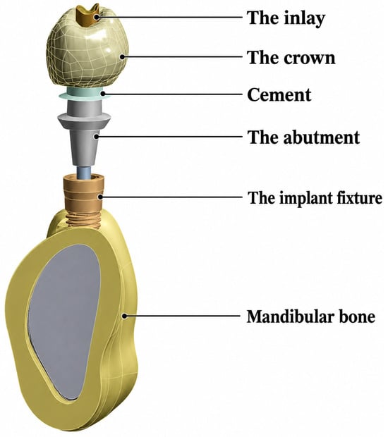
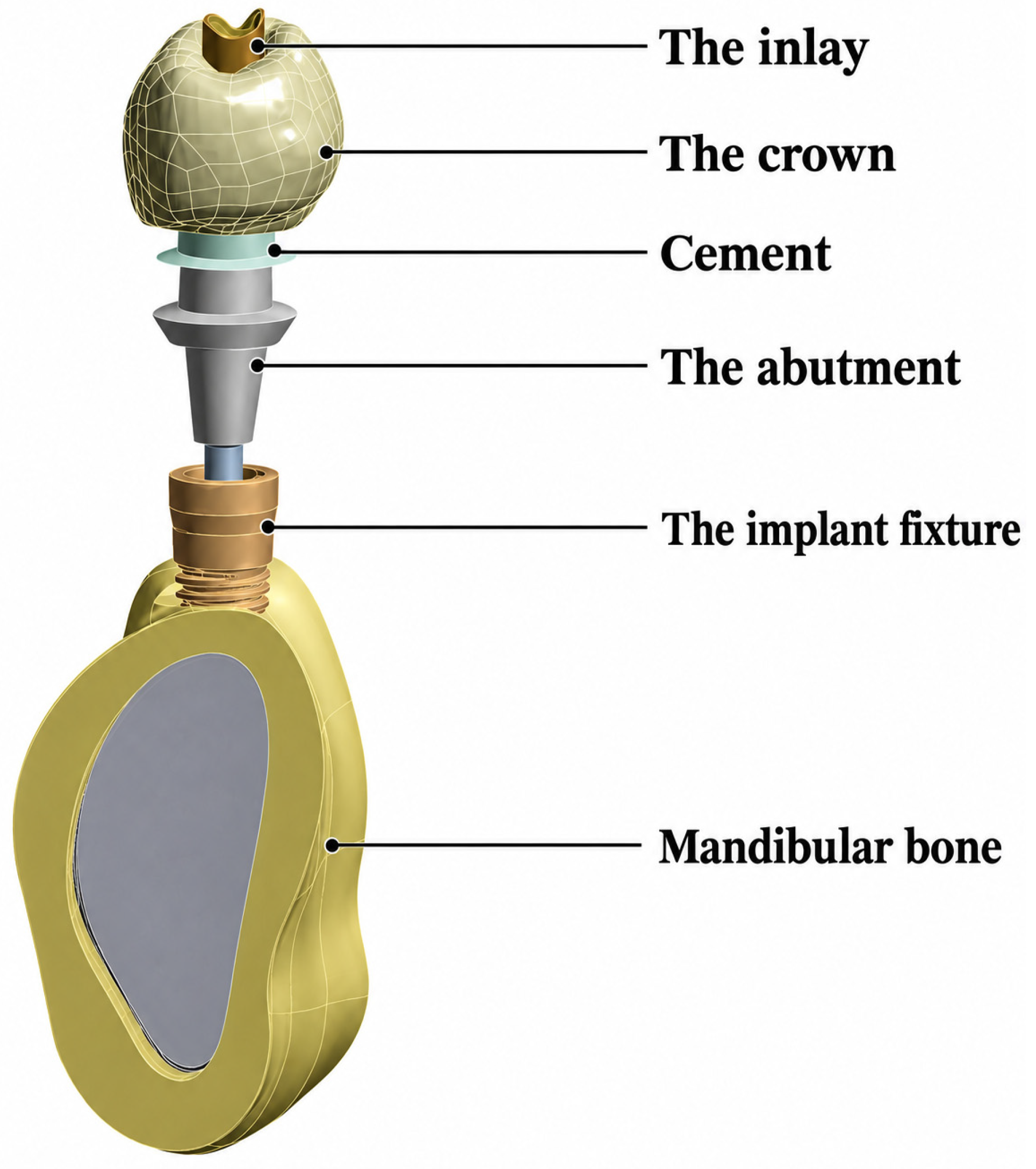
The mandibular bone model (Part A) and the prosthetic assembly (Part B) were assembled in SolidWorks to generate the complete finite element models, consisting of the supporting bone, implant fixture, abutment, cement layer, and definitive restoration (Figure 1). The assembled models were exported as STL files and further refined in Autodesk Meshmixer (version 3.0) to improve mesh quality before finite element analysis. All interfaces between model components were assumed to have perfectly bonded contact, except for the interface between the access-channel restorative material and the crown, which was defined as a frictional contact with a coefficient of friction of 0.15.
Figure 1.
Model parts.
Finite Element Model Design: two principal crown designs were evaluated:
Model A: Screw-retained monolithic zirconia crown with an occlusal access channel.
Model B: Screw-retained porcelain-fused-to-metal (PFM) crown created by dividing the full-contour crown into a 1.0-mm porcelain veneer supported by a metal coping using the design tools in ANSYS Workbench.
For each crown design, eight finite element models were generated according to the restorative material used to fill the access channel and the loading direction (vertical or oblique), resulting in a total of 16 models.
2.5. Material Properties, Boundary Conditions, and Loading
All material properties, including the elastic modulus and Poisson’s ratio, were assigned to their respective model components in ANSYS Workbench (version 2022 R1) according to values reported in the literature (Table 1).
Table 1.
Mechanical properties data inserted into Ansys software (version 2022 R1).
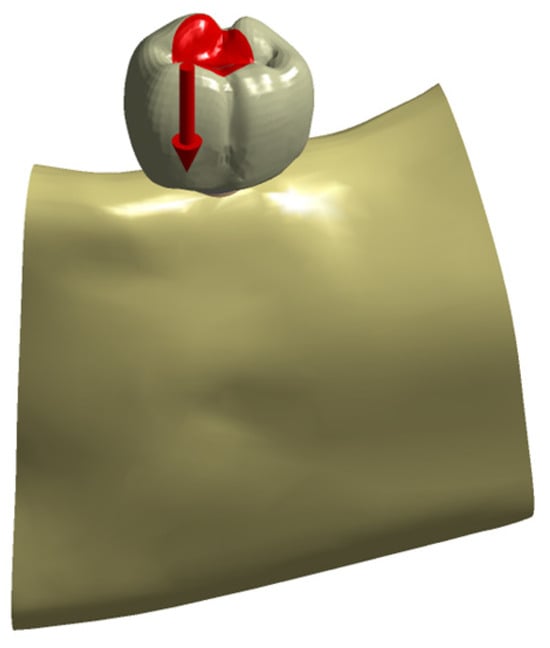
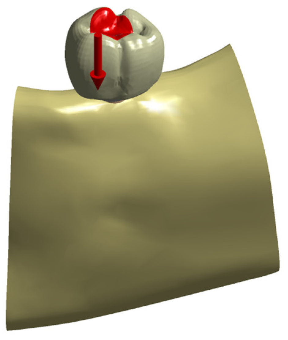
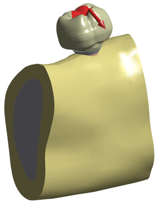
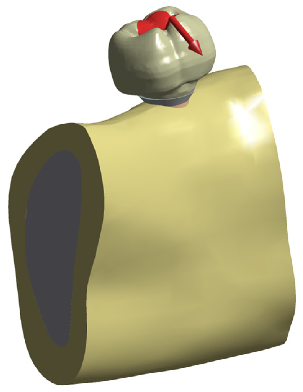
The outer surface of the cortical bone was constrained in all directions (X, Y, and Z axes) to simulate rigid fixation. A static load of 200 N was applied to the occlusal surface under two loading conditions: vertically along the implant axis obliquely at 45 degrees, as illustrated in Figure 2 and Figure 3. This allows the effects of restorative material and loading direction to be compared.
Figure 2.
The direction of the vertical force as shown by the red arrow along the axis of the implant.
Figure 3.
The direction of the oblique force at 45 degrees to the long axis of the implant.
All prepared assemblies were meshed for finite element simulations. Ten-node, three-dimensional tetrahedral elements were employed to accommodate the irregular meshes and complex geometries of the dental structures. Standard mesh convergence tests were performed for all models at each testing stage. The mesh was considered satisfactory when further reduction in element size did not result in significant changes in the maximum stress values, indicating that the original geometry was adequately represented.
Following the definition of the material properties, contact conditions, boundary conditions, and loading, an appropriate finite element mesh was generated for each model (Figure 1).
2.6. Outcome Measures
Finite element analysis was performed to evaluate two primary outcomes: the von Mises stress distribution within the restoration and surrounding components and the total deformations of the inlay materials and the crowns.
3. Results
3.1. Equivalent von Mises Stress (EVM)
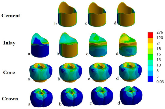
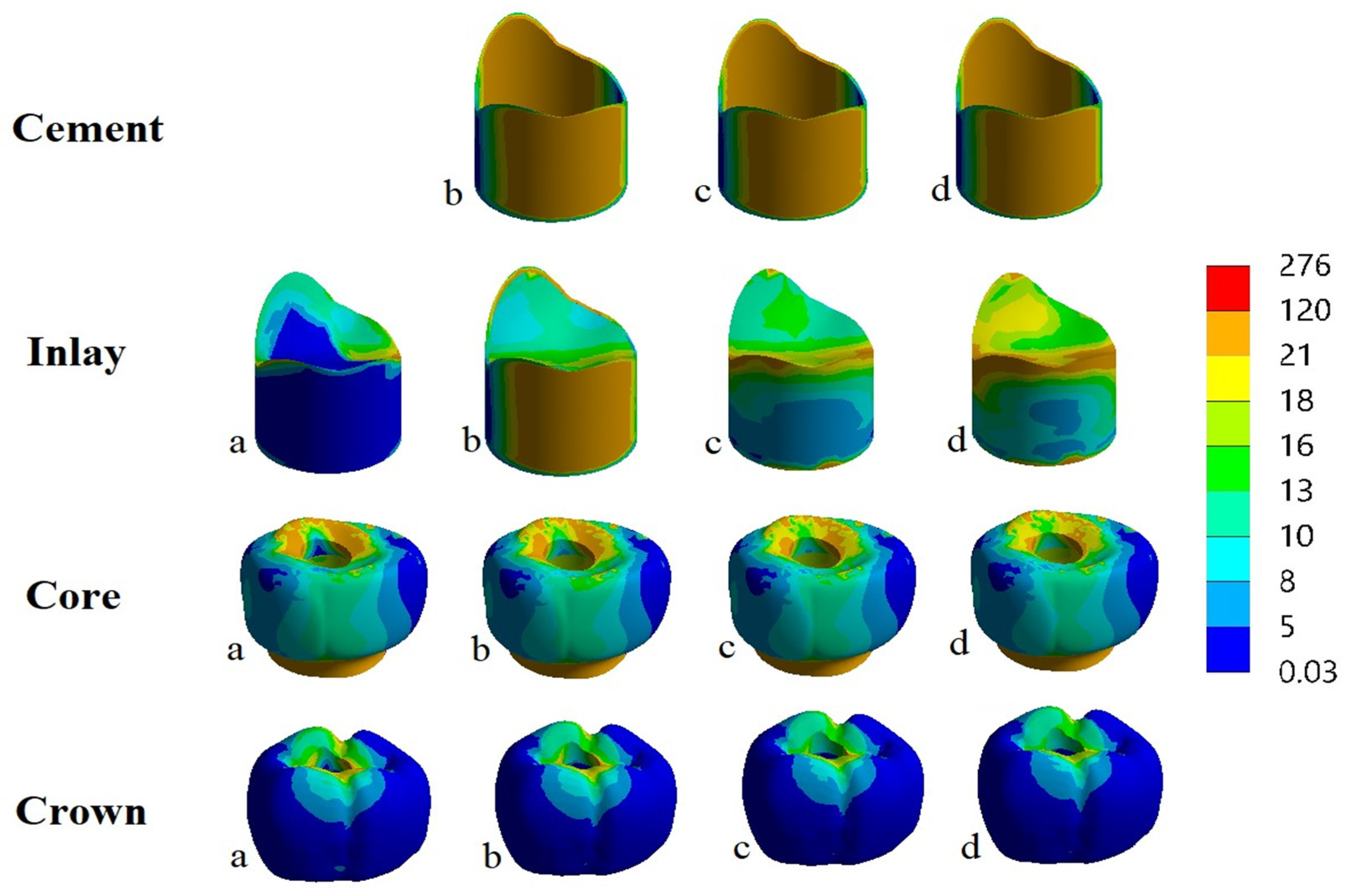
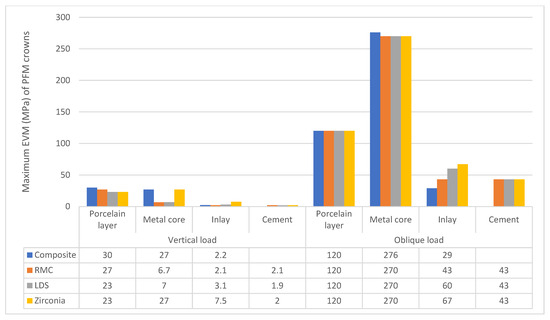
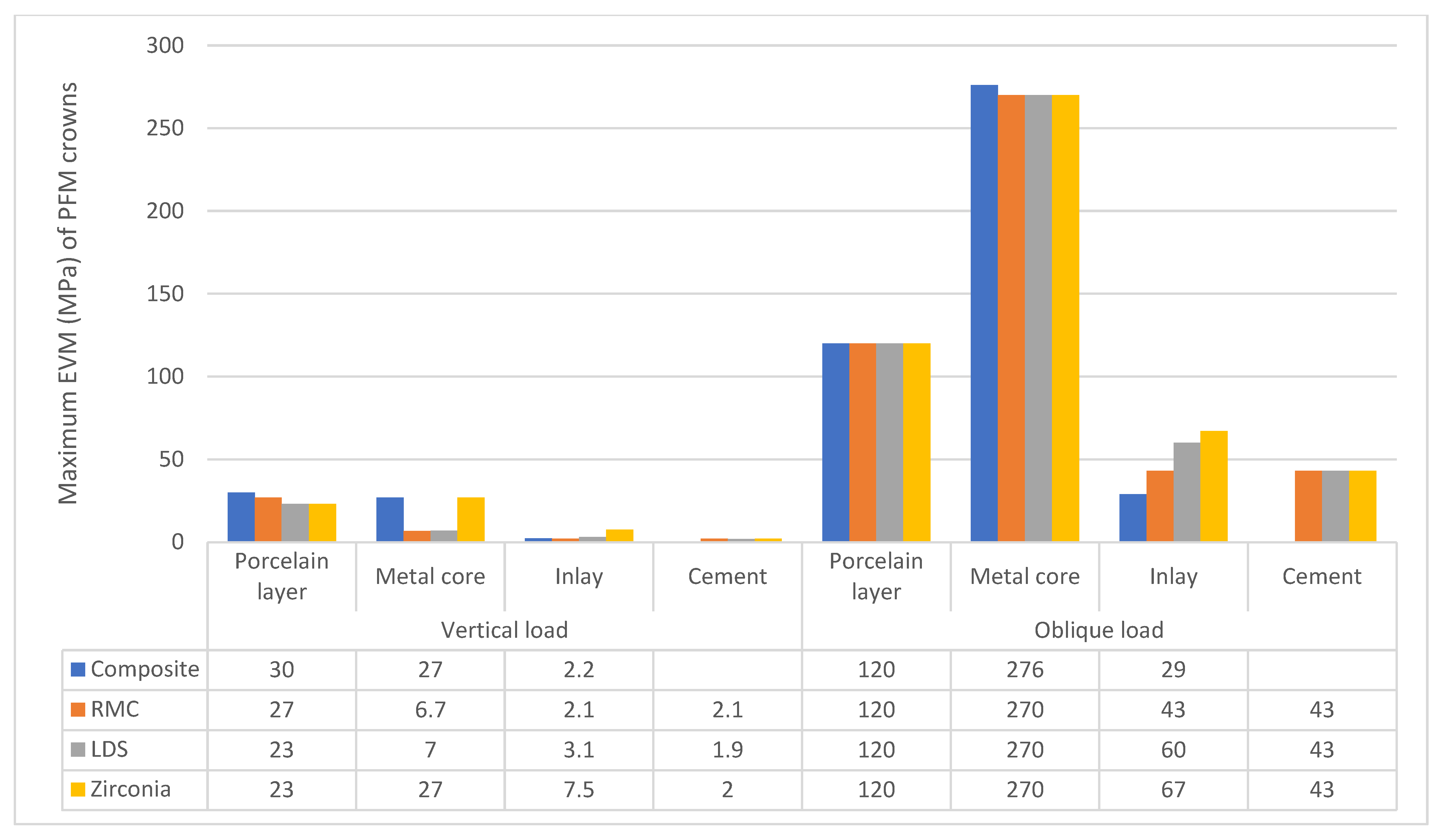
The stress distributions are presented in a stress contour plot, and the maximum stress is reported as a bar chart. We standardized the legends within each group to make visual comparisons easier.
Under vertical load, the highest stress was transmitted to the porcelain layer when filled with direct composite materials. As for the inlay itself, the zirconia group had the highest maximum EVM.
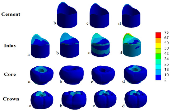
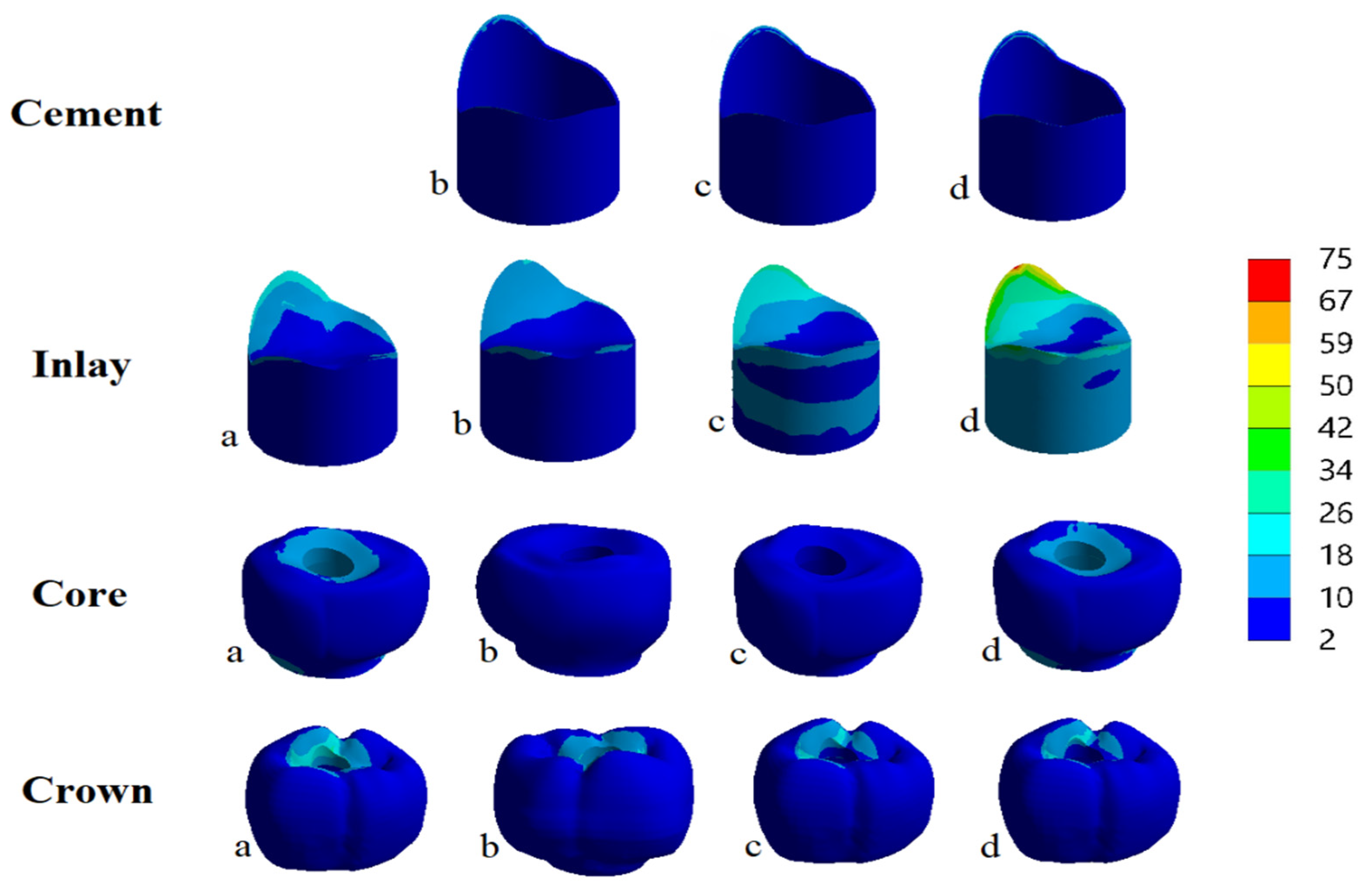
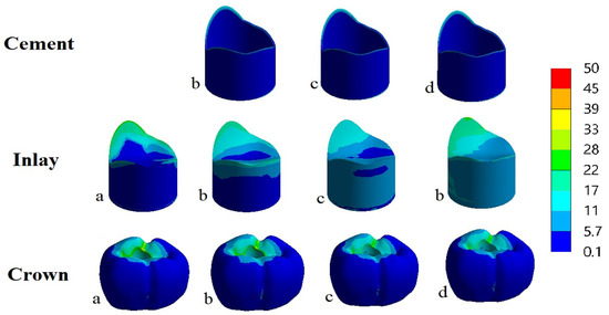
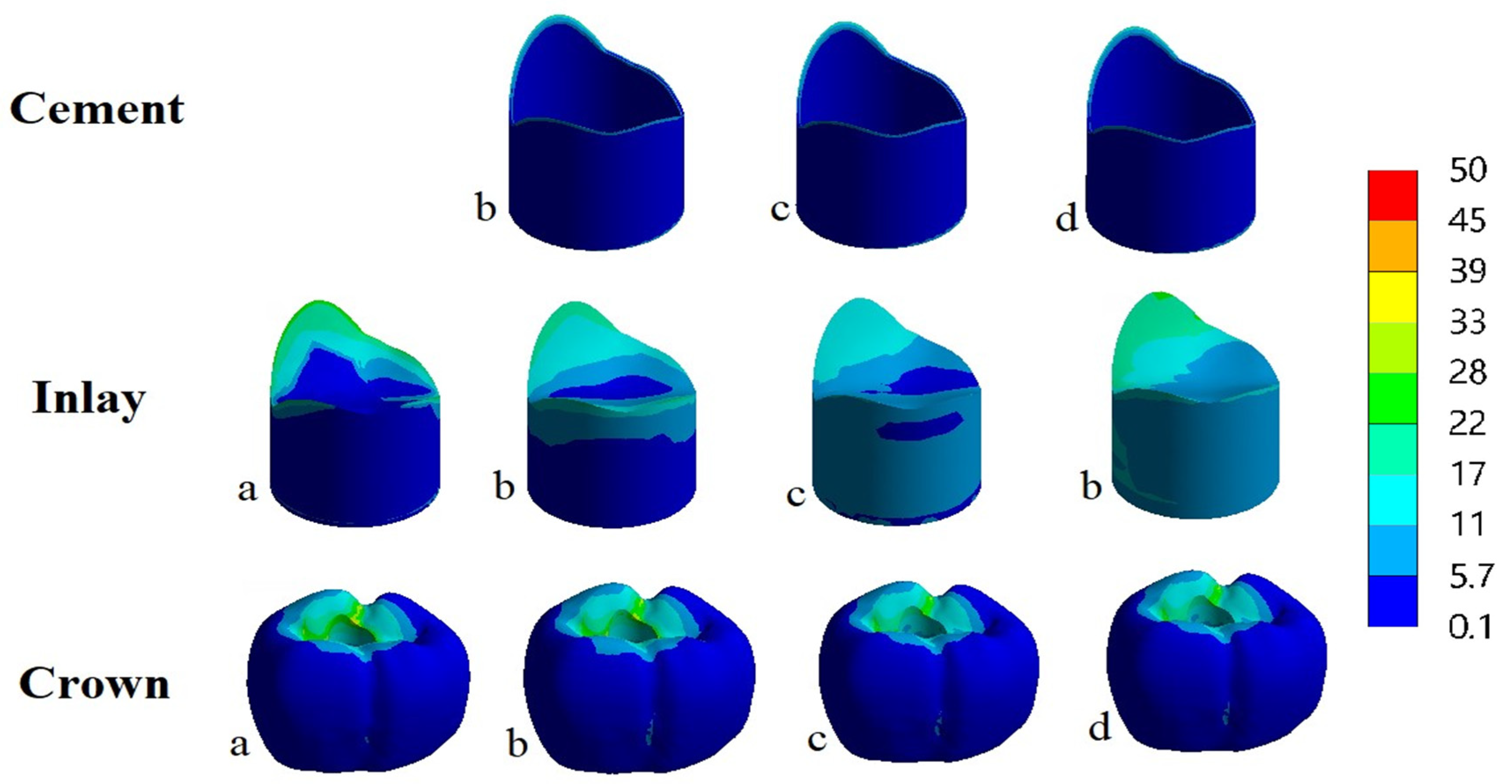
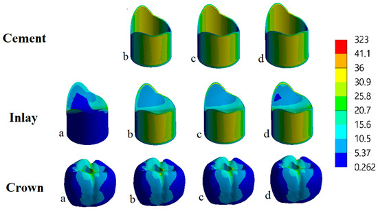
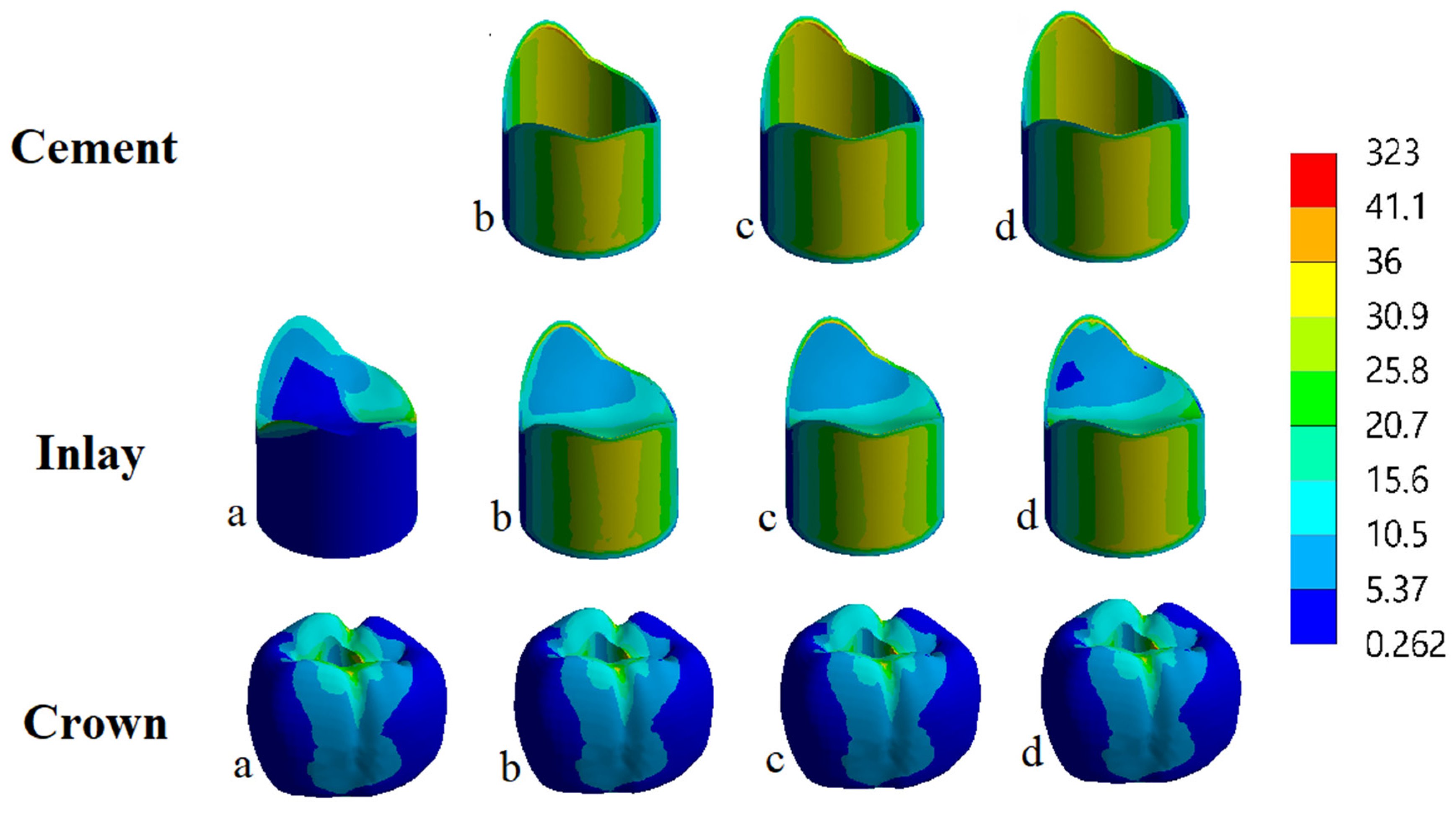
The EVM stress distributions for the PFM and zirconia crowns under vertical and oblique loading are presented in Figure 4, Figure 5, Figure 6, Figure 7, Figure 8 and Figure 9.
Figure 4.
EVM distributions (MPa) of PFM crown under vertical load on the (a) composite, (b) resin matrix ceramic, (c) lithium disilicate, and (d) zirconia.
Figure 5.
EVM distributions (MPa) of PFM crown under oblique load on the (a) composite, (b) resin matrix ceramics, (c) lithium disilicate and (d) zirconia.
Figure 6.
Maximum EVM stress distributions (MPa) of PFM crown under vertical and oblique load.
Figure 7.
von Mises stress distributions (MPa) of the zirconia crown under vertical load on the (a) composite, (b) resin matrix ceramic, (c) lithium disilicate, and (d) zirconia.
Figure 8.
EVM distributions (MPa) of the zirconia crown under oblique load on the (a) composite, (b) resin matrix ceramic, (c) lithium disilicate, and (d) zirconia.
Figure 9.
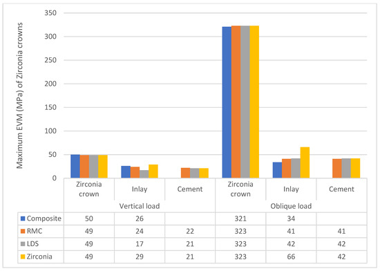
Maximum EVM stress distributions (MPa) of the zirconia crown under vertical and oblique load.
For the PFM crowns under vertical loading, the maximum EVM stress values in the porcelain layer were 30 MPa, 27 MPa, 23 MPa, and 23 MPa for the composite, resin matrix ceramic (RMC), lithium disilicate (LDS), and zirconia inlay groups, respectively. The corresponding maximum EVM stress values in the metal core were 27 MPa, 6.7 MPa, 7 MPa, and 27 MPa, respectively. For the inlay materials, the maximum EVM stress values were 2.2 MPa, 2.1 MPa, 3.1 MPa, and 7.5 MPa for the composite, RMC, LDS, and zirconia groups, respectively.
While the main difference between the groups under oblique load found in inlays is the same as under vertical load, the zirconia inlay had the highest EVM.
Under oblique loading, the maximum EVM stress values for the porcelain layer were 120 MPa for the composite group and 120 MPa for the RMC, LDS, and zirconia groups. The maximum EVM stress values recorded in the metal core were 276 MPa, 270 MPa, 270 MPa, and 270 MPa for the composite, RMC, LDS, and zirconia groups, respectively. The corresponding maximum EVM stress values in the inlay were 29 MPa, 43 MPa, 60 MPa, and 67 MPa, respectively. Overall, the zirconia inlay exhibited the highest EVM stress among the tested inlay materials under oblique loading (Figure 6).
Regarding the zirconia crown, the highest difference between groups was found in the inlay layer, where the zirconia inlay presented the highest (EVM) stress under both vertical and oblique load.
For the zirconia crowns, the maximum EVM stress values in the crown under vertical loading were 50 MPa, 49 MPa, 49 MPa, and 49 MPa for the composite, RMC, LDS, and zirconia inlay groups, respectively. The corresponding maximum EVM stress values in the inlay were 26 MPa, 24 MPa, 17 MPa, and 29 MPa, respectively. Under oblique loading, the maximum EVM stress values in the zirconia crown were 321 MPa, 323 MPa, 323 MPa, and 323 MPa for the composite, RMC, LDS, and zirconia groups, respectively. The corresponding maximum EVM stress values in the inlay were 34 MPa, 41 MPa, 42 MPa, and 66 MPa, respectively (Figure 9).
The maximum EVM stress within the zirconia crown was therefore similar among the four inlay materials under both loading conditions. In contrast, the zirconia inlay showed the highest EVM stress among the tested inlay materials under oblique loading.
3.2. Total Deformation
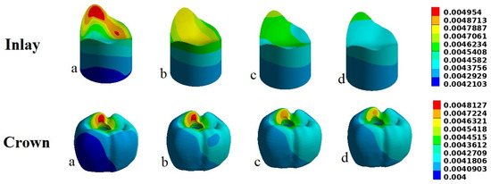
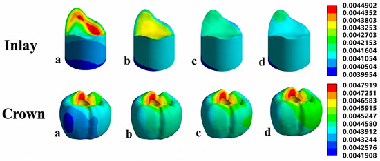
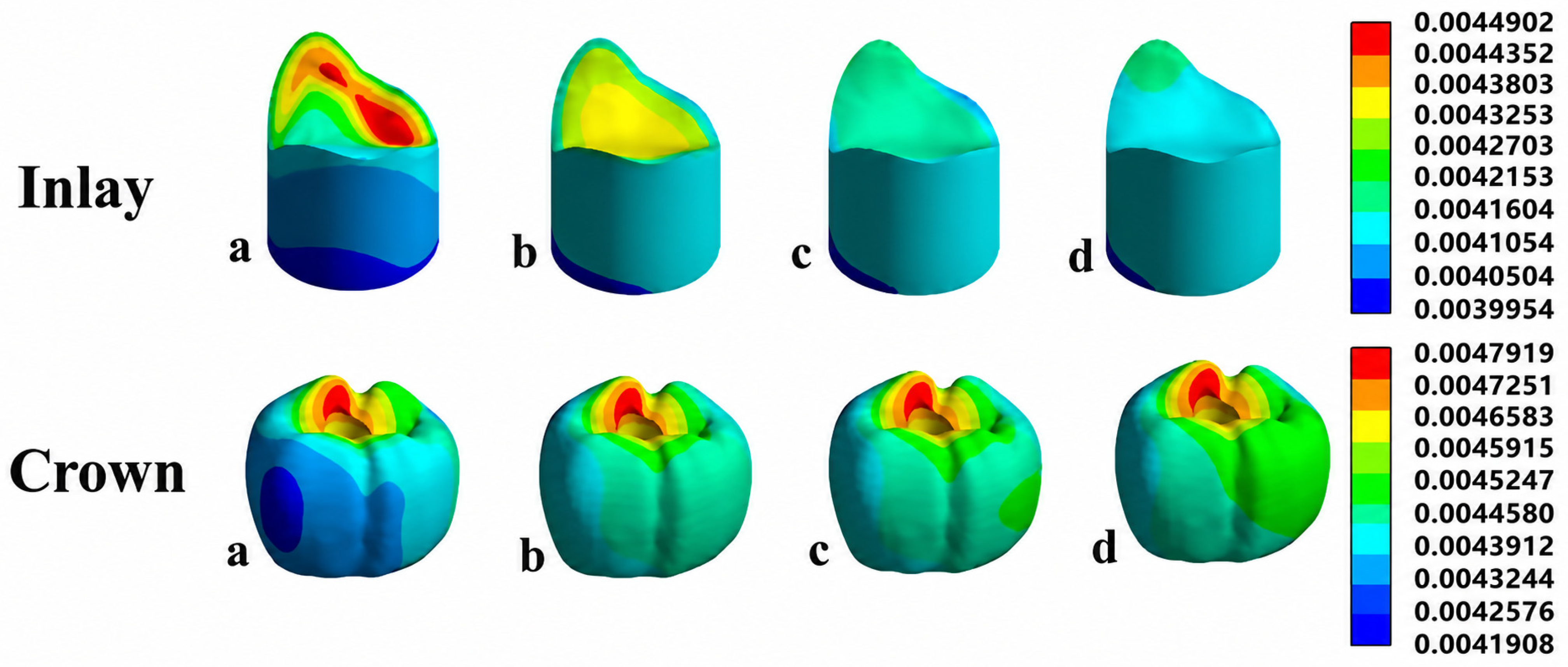
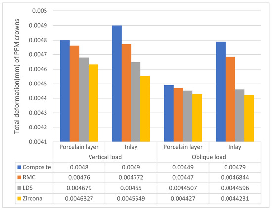
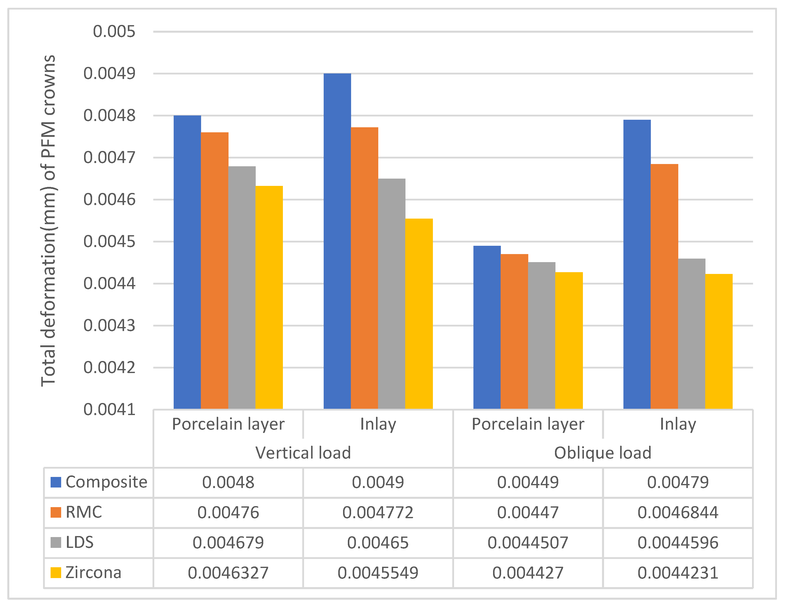
The total deformation values for the PFM crowns are presented in Figure 10, Figure 11 and Figure 12. Under vertical loading, the maximum deformation values of the porcelain layer were 0.00480 mm, 0.00476 mm, 0.004679 mm, and 0.004633 mm for the composite, RMC, LDS, and zirconia groups, respectively. The corresponding deformation values of the inlay were 0.00490 mm, 0.004772 mm, 0.004650 mm, and 0.004555 mm, respectively.
Figure 10.
Total deformation (mm) of PFM crown under vertical load on the (a) composite, (b) resin matrix ceramic, (c) lithium disilicate, and (d) zirconia.
Figure 11.
Total deformation (mm) of PFM crown under oblique load on the (a) composite, (b) resin matrix ceramic, (c) lithium disilicate, and (d) zirconia.
Figure 12.
Total deformation (mm) of PFM crown under oblique and vertical loads.
Under oblique loading, the maximum deformation values of the porcelain layer were 0.004491 mm, 0.004470 mm, 0.004451 mm, and 0.004427 mm for the composite, RMC, LDS, and zirconia groups, respectively. The corresponding deformation values of the inlay were 0.004790 mm, 0.004684 mm, 0.004460 mm, and 0.004423 mm, respectively. Thus, the composite group exhibited the highest deformation, whereas the zirconia group showed the lowest deformation under both loading conditions.
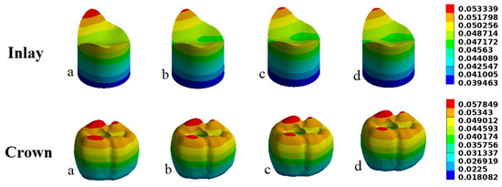
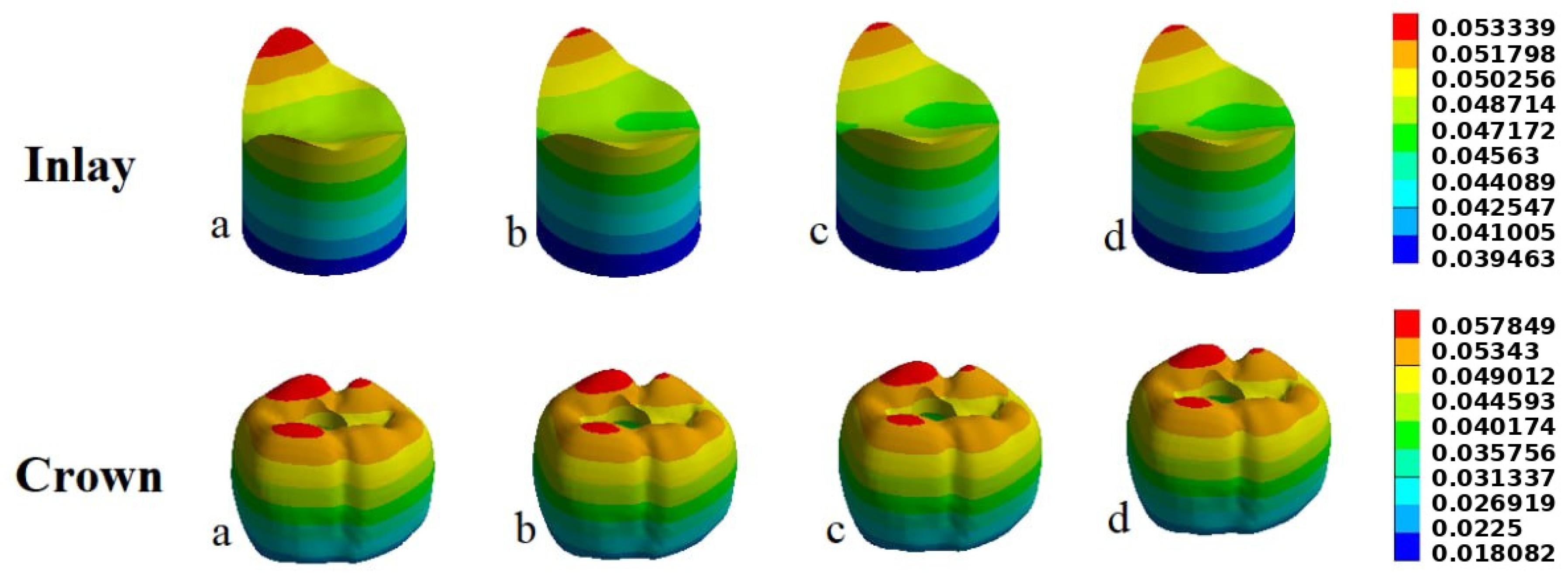
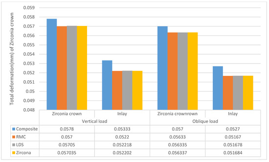
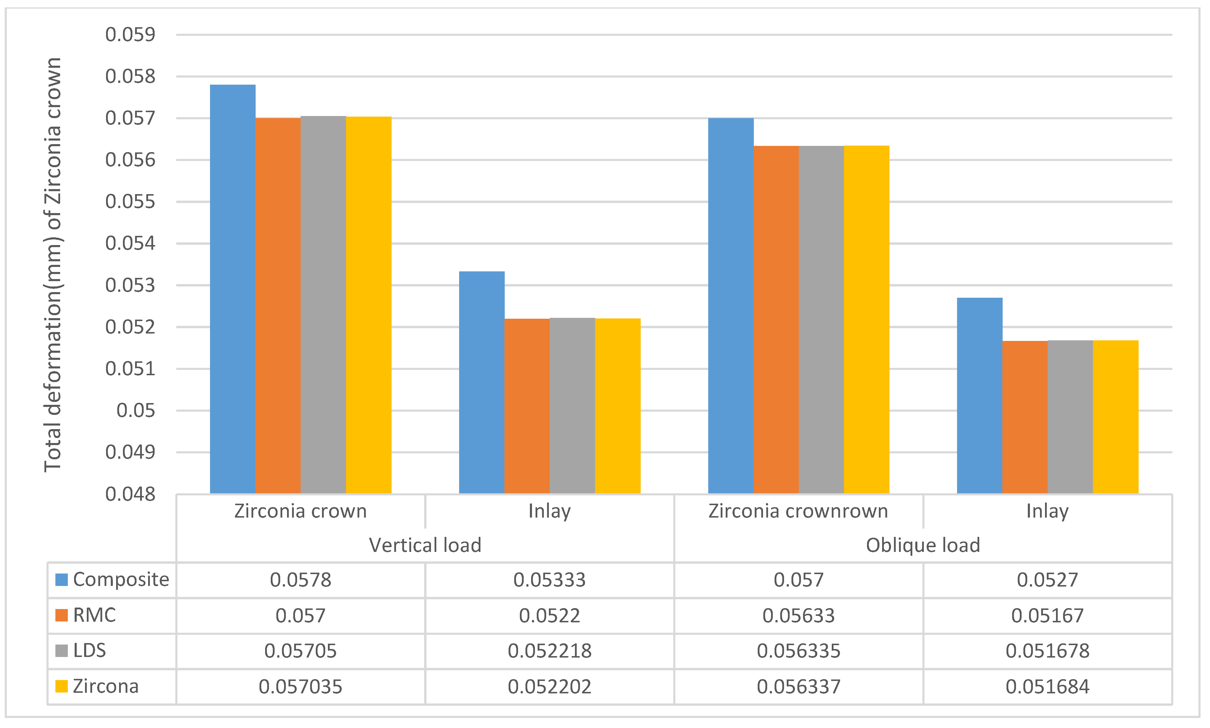
For the zirconia crowns, the total deformations are presented in Figure 13, Figure 14 and Figure 15. The maximum deformation values of the crown under vertical loading were 0.05780 mm, 0.05700 mm, 0.05705 mm, and 0.05704 mm for the composite, RMC, LDS, and zirconia groups, respectively. The corresponding deformation values of the inlay were 0.05333 mm, 0.05220 mm, 0.052218 mm, and 0.052202 mm, respectively.
Figure 13.
Total deformation (mm) of the zirconia crown under vertical load on the (a) composite, (b) resin matrix ceramic, (c) lithium disilicate, and (d) zirconia.
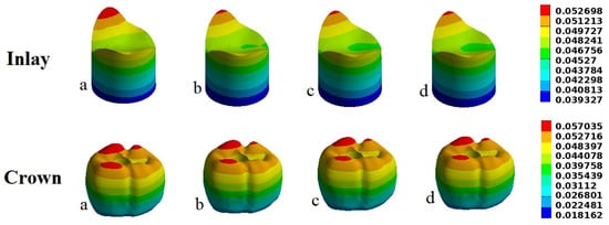
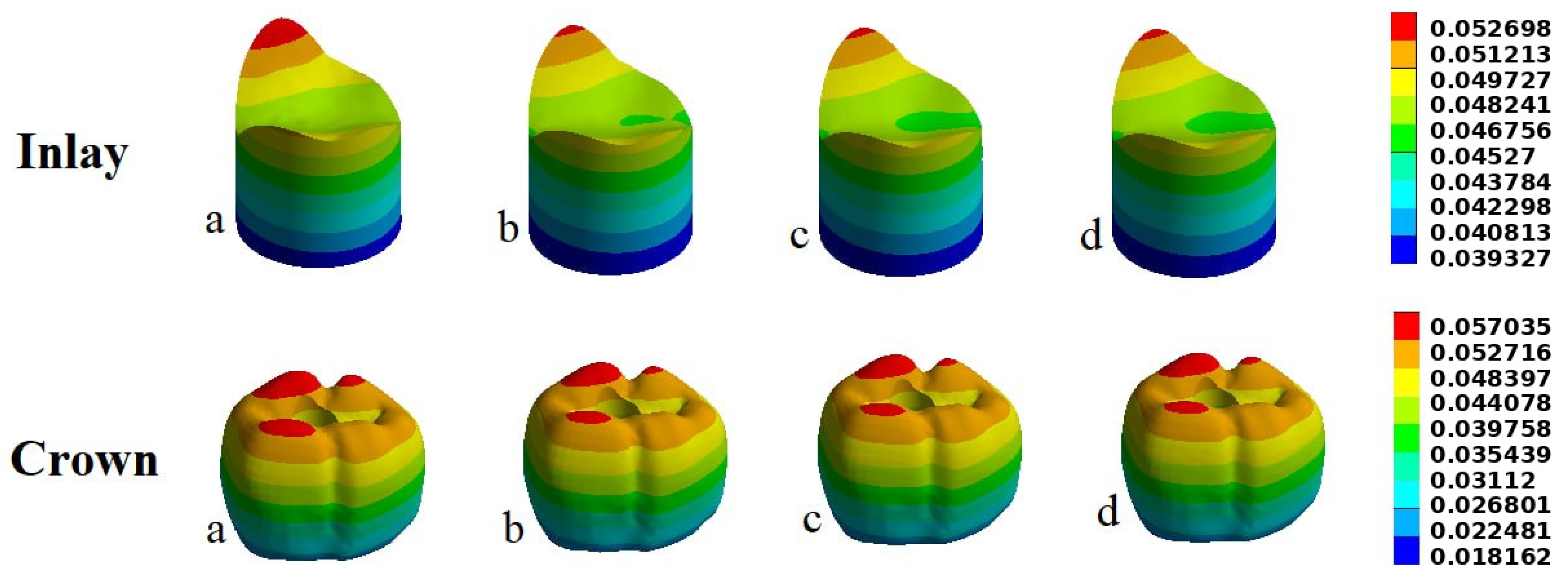
Figure 14.
Total deformation (mm) of the zirconia crown under oblique load on the (a) composite, (b) resin matrix ceramic, (c) lithium disilicate, and (d) zirconia.
Figure 15.
Total deformation (mm) of the zirconia crown under vertical and oblique loads.
Under oblique loading, the maximum deformation values of the zirconia crown were 0.05700 mm, 0.05633 mm, 0.056335 mm, and 0.056337 mm for the composite, RMC, LDS, and zirconia groups, respectively. The corresponding deformation values of the inlay were 0.05270 mm, 0.05167 mm, 0.051678 mm, and 0.051684 mm, respectively.
Overall, the composite group demonstrated the highest deformation values, while the differences among the RMC, LDS, and zirconia groups were relatively small, particularly for the zirconia crown. Under both loading conditions, the deformation of the zirconia crown and its inlay was within a narrow range across the four restorative materials.
4. Discussion
Overall, screw-retained dental prostheses have the advantage of more predictable retrievability [20,21,22]. When the screw-retained implant crown is fabricated, the screw access hole may interfere with the occlusal table integrity, potentially affecting the integrity and design of the occlusal surface. However, using composite restoration in the screw hole may be abraded, subsided, or even dislocated under long-term chewing, which limits the use of screw-retained implants in clinical application. The present study compared the stress distribution and the deformation of the crown screw access hole filled by direct or indirect restoration materials using finite element analysis.
The findings demonstrated differences in EVM stress distribution and total deformation among the restorative materials and therefore did not support the proposed null hypothesis.
Based on the chosen analysis criterion, von Mises stress was selected because it is widely used in finite element analysis (FEA) to assess the safety and reliability of mechanical components and [23] structures. This measure captures the combined effects of tensile, compressive, and shear stresses, offering a comprehensive view of the stress state within a material. It is particularly valuable for evaluating materials that undergo plastic deformation, as it provides a more accurate prediction of yielding and failure compared to other stress measures, especially in the context of metallic dental implants [24,25,26].
The present study demonstrated that the stress distribution varied according to the elastic modulus and other mechanical properties of the restorative materials, but the crown distributed most of the stress, and lower levels of stress occurred in the cement layer and the inlay material. This result was consistent with the previous studies [27,28].
In PFM crowns, the brittle nature of the porcelain layer mandates the use of a material that has minimal stress transformation to fill the access hole [23,29]. In the present study, the highest stress was transmitted to the porcelain layer when filled with the direct composite material, followed by the RMC inlay. While the LDS and the zirconia inlay transmitted the least EVM stress under the vertical occlusal load. However, under the oblique occlusal load, the differences in stress concentrations between the groups were minimal.
Clinical evaluations confirmed that metal frameworks provide the fracture resistance necessary for long-term stability; therefore, stress distribution on the metal core is less crucial than the porcelain layer because of the ductile nature of the metal, which makes the failure less probable [29,30].
For the inlay materials, the zirconia group exhibited the highest EVM stress concentration under both vertical and oblique loads, followed by LDS, RMC, and the composite group. These results can be explained by the fact that zirconia has the highest compressive and flexural strength by a significant margin. Lithium disilicate ranks second, offering a balanced mix of strength and aesthetics. Hybrid ceramics (polymer-infiltrated) have the lowest compressive strength but provide high flexibility and shock absorption [31,32].
The maximum EVM stress on the zirconia crowns was roughly equivalent regardless of the type of inlay used, whereas the zirconia inlay had the highest EVM stress under both vertical and occlusal loads. These results were in agreement with the results of Kintopp [27], who studied the effect of different restorative materials on partial posterior restorations using FEA. They concluded that zirconia has the highest stress concentration, followed by polymer-infiltrated ceramic network and composite resin.
There is no article in the literature to compare the present study to, but the nearest study was by Ma et al. [33], who studied the load-bearing properties of lithium disilicate and zirconia occlusal using FEA, and they concluded that achieving long-term stability in ceramic structures requires clinicians to effectively balance the physical properties of the restorative materials with those of the supporting structure.
The present study demonstrated distinct differences in total deformation among the groups. These findings can be explained by the inherent mechanical properties of each material and corroborated by previous studies [34,35].
Among the tested materials with PFM crowns, zirconia showed the least deformation. This outcome is consistent with its very high elastic modulus and its well-documented fracture toughness [35,36]. Such rigidity makes zirconia highly resistant to occlusal forces. Lithium disilicate displayed moderate deformation, reflecting its balance between strength and elasticity. It is less rigid than zirconia and therefore better able to absorb occlusal forces [37]. While resin matrix ceramics showed greater deformation than the purely ceramic materials, this flexibility may compromise marginal integrity over time [38]. Composite inlays exhibited the highest deformation, which raises concerns about microleakage, marginal breakdown, and reduced load-bearing capacity [22].
This study revealed clear differences in how these inlays respond to occlusal loading inside the zirconia crown compared to PFM crown. There are no differences between the zirconia, lithium disilicate, and resin matrix ceramic deformation.
These findings can be attributed to the mechanical dominance of the zirconia crown, which possesses an exceptionally high elastic modulus and fracture strength. Once the inlays are cemented within the zirconia framework, the crown functions as a rigid shell that governs the overall stress distribution. This rigidity effectively masks the intrinsic differences among the inlay materials, resulting in comparable deformation responses across zirconia, lithium disilicate, and resin matrix ceramics [39].
From a clinical standpoint, zirconia remains the most appropriate choice for restorations that demand high strength, although its inherent rigidity may increase the risk of stress concentration within the tooth structure. Lithium disilicate, by contrast, offers a favorable balance between esthetics and mechanical performance, making it suitable for both anterior and posterior applications. Resin matrix ceramics provide enhanced stress absorption, yet their greater flexibility may compromise marginal adaptation over time. Meanwhile, composite inlays are valued for their ease of handling and cost-effectiveness, though their lower durability under heavy occlusal forces limits their long-term reliability in load-bearing regions.
This study has a few notable limitations. First, material properties were simplified by assuming isotropy and homogeneity, which oversimplifies the naturally complex, anisotropic, and heterogeneous characteristics of dental tissues and materials. Second, the loading conditions were simplified to just two force types applied to occlusal contacts. In reality, clinical occlusal forces are highly dynamic and shaped by variables like diet and parafunctional habits. To build on these findings, future research should investigate how diverse material properties, variable occlusal forces, and alternative restoration designs impact the long-term clinical success of dental restorations.
5. Conclusions
Within the limitations of this three-dimensional finite element analysis, both the crown material and the restorative material used to seal the screw access channel influenced stress distribution and deformation. Lithium disilicate and zirconia demonstrated more favorable biomechanical behavior than direct composite resin, particularly in PFM crowns under vertical loading. In monolithic zirconia crowns, the high stiffness of the crown minimized differences among restorative materials. Further in vitro and clinical studies are required to validate these findings.
Author Contributions
Conceptualization, K.M. and S.R.; Methodology, L.A.S., R.K. and R.M.T.; Software, R.I., L.A.S. and R.M.; Validation, K.M.; Formal analysis, K.M., L.A.S. and S.R.; Investigation, L.A.S., R.K. and S.R.; Resources, R.I., H.M. and R.K.; Data curation, R.I. and R.M.; Writing—original draft, S.N.D.; Supervision, A.A.Z., S.N.D. and R.M.T.; Project administration, A.A.Z., H.M., R.M., S.N.D. and R.M.T.; Funding acquisition, H.M. All authors have read and agreed to the published version of the manuscript.
Funding
This research received no external funding.
Institutional Review Board Statement
Not applicable.
Informed Consent Statement
Not applicable.
Data Availability Statement
The original contributions presented in this study are included in the article. Further inquiries can be directed to the corresponding author.
Conflicts of Interest
The authors declare no conflict of interest.
References
- Goodacre, C.J.; Bernal, G.; Rungcharassaeng, K.; Kan, J.Y. Clinical complications in fixed prosthodontics. J. Prosthet. Dent. 2003, 90, 31–41. [Google Scholar] [CrossRef] [Scilit] [PubMed]
- Zitzmann, N.U.; Marinello, C.P. Treatment plan for restoring the edentulous maxilla with implant-supported restorations: Removable overdenture versus fixed partial denture design. J. Prosthet. Dent. 1999, 82, 188–196. [Google Scholar] [CrossRef] [Scilit] [PubMed]
- Bidra, A.S.; Rungruanganunt, P.; Gauthier, M. Clinical outcomes of full arch fixed implant-supported zirconia prostheses: A systematic review. Eur. J. Oral Implantol. 2017, 10, 35–45. [Google Scholar] [PubMed]
- Sailer, I.; Mühlemann, S.; Zwahlen, M.; Hämmerle, C.H.; Schneider, D. Cemented and screw-retained implant reconstructions: A systematic review of the survival and complication rates. Clin. Oral Implant. Res. 2012, 23, 163–201. [Google Scholar] [CrossRef] [Scilit] [PubMed]
- Michalakis, K.X.; Hirayama, H.; Garefis, P.D. Cement-retained versus screw-retained implant restorations: A critical review. Int. J. Oral Maxillofac. Implant. 2003, 18, 719–728. [Google Scholar]
- Wittneben, J.G.; Joda, T.; Weber, H.P.; Brägger, U. Screw retained vs. cement retained implant-supported fixed dental prosthesis. Periodontol. 2000 2017, 73, 141–151. [Google Scholar] [CrossRef] [Scilit] [PubMed]
- Manawar, A.; Dhanasekar, B.; Aparna, I.; Naim, H. Factors influencing success of cement versus screw-retained implant restorations: A clinical review. J. Osseointegration 2012, 4, 43–47. [Google Scholar] [CrossRef] [Scilit]
- Shadid, R.; Sadaqa, N. A comparison between screw-and cement-retained implant prostheses. A literature review. J. Oral Implantol. 2012, 38, 298–307. [Google Scholar] [CrossRef] [Scilit] [PubMed]
- Derafshi, R.; Farzin, M.; Taghva, M.; Heidary, H.; Atashkar, B. The Effects of New Design of Access Hole on Porcelain Fracture Resistance of Implant-Supported Crowns. J. Dent. 2015, 16, 61–67. [Google Scholar]
- Sarrafzadeh, A.; Nakhostin, A.; Jadidi, S.; Safari, M. Color Assessment of Screw-Retained Implant Prostheses Access Hole Fillings: An In-Vitro Study. Front. Dent. 2024, 21, 46. [Google Scholar] [CrossRef] [Scilit] [PubMed]
- AlQarawi, F.K.; AlShammasi, B.M.; AlBasry, Z.A.; AlAwami, H.Z.; AlEraky, D.M.; AlJindan, R.Y.; Alrumaih, H.S.; Alshahrani, F.A. Microleakage and Bacterial Adhesion with Three Restorative Materials Used to Seal Screw-access Channels of Implant Abutments: An: In vitro: Study. Saudi J. Med. Med. Sci. 2021, 9, 241–247. [Google Scholar] [CrossRef] [Scilit] [PubMed]
- Liwwattanakorn, T.; Kamonkhantikul, K.; Homsiang, W.; Arksornnukit, M. Effect of Screw Access Hole on Fracture Resistance Among 3 Different Implant-Supported Crown Materials After Cyclic Loading: An In Vitro Study; Research Square Platform LLC: Durham, NC, USA, 2024. [Google Scholar]
- Mendes Tribst, J.P.; de Oliveira Dal Piva, A.M.; Inês Gonçalves, N.; Souto Borges, A.L.; Antonio Bottino, M.; Johannes Kleverlaan, C. Polymerization Shrinkage and Push-out Bond Strength of Different Composite Resins for Sealing the Screw-access Hole on Implant-supported Crowns. J. Adhes. Dent. 2020, 22, 523–530. [Google Scholar] [CrossRef] [Scilit] [PubMed]
- Shim, J.S.; Li, C.-Y.; Won, J.-E.; Ryu, J.J. Evaluation of microleakage and push-out bond strength of various composite resins for sealing the screw-access channel in implant-supported restorations. J. Prosthet. Dent. 2022, 128, 764.e1–764.e7. [Google Scholar] [CrossRef] [Scilit] [PubMed]
- Sevimay, M.; Turhan, F.; Kiliçarslan, M.; Eskitascioglu, G. Three-dimensional finite element analysis of the effect of different bone quality on stress distribution in an implant-supported crown. J. Prosthet. Dent. 2005, 93, 227–234. [Google Scholar] [CrossRef] [Scilit] [PubMed]
- Lemos, C.A.A.; Verri, F.R.; Santiago Júnior, J.F.; Almeida, D.A.d.F.; Batista, V.E.d.S.; Noritomi, P.Y. Retention system and splinting on morse taper implants in the posterior maxilla by 3D finite element analysis. Braz. Dent. J. 2018, 29, 30–35. [Google Scholar] [CrossRef] [Scilit] [PubMed][Green Version]
- Acharya, P.H.; Patel, V.V.; Duseja, S.S.; Chauhan, V.R. Comparative evaluation of peri-implant stress distribution in implant protected occlusion and cuspally loaded occlusion on a 3 unit implant supported fixed partial denture: A 3D finite element analysis study. J. Adv. Prosthodont. 2021, 13, 79. [Google Scholar] [CrossRef] [Scilit] [PubMed]
- Tamrakar, S.K.; Mishra, S.K.; Chowdhary, R.; Rao, S. Comparative analysis of stress distribution around CFR-PEEK implants and titanium implants with different prosthetic crowns: A finite element analysis. Dent. Med. Probl. 2021, 58, 359–367. [Google Scholar] [CrossRef] [Scilit] [PubMed]
- Sannino, G.; Gloria, F.; Ottria, L.; Barlattani, A. Influence of finish line in the distribution of stress trough an all ceramic implant-supported crown.: A 3D Finite Element Analysis. Oral Implantol. 2009, 2, 14. [Google Scholar]
- Fiorillo, L.; D’Amico, C.; Ronsivalle, V.; Cicciù, M.; Cervino, G. Single dental implant restoration: Cemented or screw-retained? A systematic review of multi-factor randomized clinical trials. Prosthesis 2024, 6, 871–886. [Google Scholar] [CrossRef] [Scilit]
- Found, A.A.; Shah, S.; Fouda, M.A.; Santos, M.J.M.; Butler, S. Survey of screw-retained versus cement-retained implant restorations used in both education and private dental practices. J. Dent. Educ. 2024, 88, 1029–1039. [Google Scholar] [CrossRef] [Scilit] [PubMed]
- Li, F.; Wu, M.; Zhang, X.; Bi, M.; Shen, M.; Zhou, Z. The effect of varying the position of implant screw access opening on the stress distribution of sealing composite resin: A 3D-FEA study. BMC Oral Health 2026, 26, 271. [Google Scholar] [CrossRef] [Scilit] [PubMed]
- Al-Moaleem, M.M.; Shah, F.K.; Khan, N.S.; Porwal, A. The effect of thermocycling on the bonding of different restorative materials to access opening through porcelain fused to metal restorations. J. Adv. Prosthodont. 2011, 3, 186–189. [Google Scholar] [CrossRef] [Scilit] [PubMed]
- Ibrahim, R.O.; Al-zahawi, A.R.; Sabri, L.A. Mechanical and thermal stress evaluation of PEEK prefabricated post with different head design in endodontically treated tooth: 3D-finite element analysis. Dent. Mater. J. 2021, 40, 508–518. [Google Scholar] [CrossRef] [Scilit] [PubMed]
- Ausiello, P.; Dal Piva, A.M.D.O.; Di Lauro, A.E.; Garcia-Godoy, F.; Testarelli, L.; Tribst, J.P.M. Mechanical behavior of Alkasite posterior restorations in comparison to polymeric materials: A 3D-FEA study. Polymers 2022, 14, 1502. [Google Scholar] [CrossRef] [Scilit] [PubMed]
- Zhu, S.-P.; Ye, W.-L.; Correia, J.A.; Jesus, A.M.; Wang, Q. Stress gradient effect in metal fatigue: Review and solutions. Theor. Appl. Fract. Mech. 2022, 121, 103513. [Google Scholar] [CrossRef] [Scilit]
- de Almeida Kintopp, C.C.; Diógenes, A.N.; Lopes, R.T.; Weber, K.R.; Rezende, C.E.E.; da Rosa Kaizer, M.; Gonzaga, C.C. Effect of different preparations and restorative materials on partial posterior restorations: A 3D FEA study using μCT data. J. Prosthet. Dent. 2025, 133, 1056.e1–1056.e7. [Google Scholar] [CrossRef] [Scilit] [PubMed]
- Tribst, J.P.M.; Dal Piva, A.M.D.O.; Madruga, C.F.L.; Valera, M.C.; Borges, A.L.S.; Bresciani, E.; de Melo, R.M. Endocrown restorations: Influence of dental remnant and restorative material on stress distribution. Dent. Mater. 2018, 34, 1466–1473. [Google Scholar] [CrossRef] [Scilit] [PubMed]
- DuVall, N.B.; DeReis, S.P.; Vandewalle, K.S. Fracture strength of various titanium-based, CAD-CAM and PFM implant crowns. J. Esthet. Restor. Dent. 2021, 33, 522–530. [Google Scholar] [CrossRef] [Scilit] [PubMed]
- Pjetursson, B.E.; Sailer, I.; Merino-Higuera, E.; Spies, B.C.; Burkhardt, F.; Karasan, D. Systematic review evaluating the influence of the prosthetic material and prosthetic design on the clinical outcomes of implant-supported multi-unit fixed dental prosthesis in the posterior area. Clin. Oral Implant. Res. 2023, 34, 86–103. [Google Scholar] [CrossRef] [Scilit] [PubMed]
- ElSayed, H.M.; Yousief, S.A.; Alanazi, S.; Alyami, M.H.; Emam, A.N.M.; Alshammari, M.N.; Bukhary, F.; Robaian, A.; Alharthi, R.; Khuthija Khanam, H.M.; et al. Stress Distribution of Hybrid Ceramic Implant-supported Superstructure Versus Zirconia and Lithium Disilicate Superstructures. Open Dent. J. 2026, 20, e18742106441241. [Google Scholar] [CrossRef] [Scilit]
- Succaria, F.; Morgano, S.M. Prescribing a dental ceramic material: Zirconia vs lithium-disilicate. Saudi Dent. J. 2011, 23, 165. [Google Scholar] [CrossRef] [Scilit] [PubMed]
- Ma, L.; Guess, P.C.; Zhang, Y. Load-bearing properties of minimal-invasive monolithic lithium disilicate and zirconia occlusal onlays: Finite element and theoretical analyses. Dent. Mater. 2013, 29, 742–751. [Google Scholar] [CrossRef] [Scilit] [PubMed]
- Chen, S.; Chen, R.; Chen, Y.; Zheng, Z.; Lin, J. The impact of preparation dimensions and material choice on stress distribution in MOD inlays: A 3D finite element study. BMC Oral Health 2026, 26, 259. [Google Scholar] [CrossRef] [Scilit] [PubMed]
- Manziuc, M.; Kui, A.; Chisnoiu, A.; Labuneț, A.; Negucioiu, M.; Ispas, A.; Buduru, S. Zirconia-reinforced lithium silicate ceramic in digital dentistry: A comprehensive literature review of our current understanding. Medicina 2023, 59, 2135. [Google Scholar] [CrossRef] [Scilit] [PubMed]
- Zhang, Y.; Lawn, B.R. Novel zirconia materials in dentistry. J. Dent. Res. 2018, 97, 140–147. [Google Scholar] [CrossRef] [Scilit] [PubMed]
- Denry, I.; Kelly, J. Emerging ceramic-based materials for dentistry. J. Dent. Res. 2014, 93, 1235–1242. [Google Scholar] [CrossRef] [Scilit] [PubMed]
- Ruse, N.; Sadoun, M. Resin-composite blocks for dental CAD/CAM applications. J. Dent. Res. 2014, 93, 1232–1234. [Google Scholar] [CrossRef] [Scilit] [PubMed]
- Zhang, Z.; Zheng, K.; Li, E.; Li, W.; Li, Q.; Swain, M.V. Mechanical benefits of conservative restoration for dental fissure caries. J. Mech. Behav. Biomed. Mater. 2016, 53, 11–20. [Google Scholar] [CrossRef] [Scilit] [PubMed]

Disclaimer/Publisher’s Note: The statements, opinions and data contained in all publications are solely those of the individual author(s) and contributor(s) and not of MDPI and/or the editor(s). MDPI and/or the editor(s) disclaim responsibility for any injury to people or property resulting from any ideas, methods, instructions or products referred to in the content. |
© 2026 by the authors. Licensee MDPI, Basel, Switzerland. This article is an open access article distributed under the terms and conditions of the Creative Commons Attribution (CC BY) license.