


Abstract
Background/Objectives: Marginal and internal adaptation are important determinants of the clinical performance of CAD/CAM zirconia fixed restorations. This systematic review and meta-analysis evaluated the influence of workflow type (digital versus analogue impression workflows) on the clinical marginal and internal fit of tooth-supported CAD/CAM zirconia fixed restorations. Methods: The review was conducted in accordance with the Preferred Reporting Items for Systematic Reviews and Meta-Analyses (PRISMA) 2020 statement and was prospectively registered in PROSPERO. PubMed/MEDLINE, Embase, Web of Science, Cochrane CENTRAL, IEEE Xplore, ClinicalTrials.gov, and Google Scholar were searched for clinical studies published from January 2014 through June 2026. Ten clinical studies met the eligibility criteria. Because most comparative studies used within-subject designs, the paired nature of the data was incorporated into the quantitative synthesis. Random-effects meta-analyses were performed using restricted maximum likelihood estimation and Hartung–Knapp adjusted confidence intervals, with sensitivity analyses across plausible within-pair correlations. Results: Four studies provided directly comparable data for quantitative synthesis of marginal fit and overall internal fit. Digital workflows showed a trend toward smaller marginal and internal discrepancies than analogue workflows (MD = −21.89 μm; 95% CI, −57.59 to 13.81 μm; p = 0.146; I2 = 88.3%), but the difference was not statistically significant. Overall internal fit similarly favoured digital workflows (MD = −21.65 μm; 95% CI, −57.66 to 14.36 μm; p = 0.152; I2 = 68.7%) without a statistically significant difference. Exploratory location-specific analyses of internal fit also showed no statistically significant differences. Sensitivity analyses across different assumed within-pair correlations did not materially alter the conclusions. Conclusions: Digital impression workflows showed a trend toward smaller marginal and internal discrepancies than analogue workflows; however, the pooled differences were not statistically significant. The considerable heterogeneity and limited number of quantitatively comparable studies warrant cautious interpretation. Further clinical studies using standardized fit definitions, measurement protocols, and appropriate reporting of paired outcomes are needed.
1. Introduction
Marginal and internal adaptation of a fixed dental prosthesis at its margins and internal surfaces can influence its biological and mechanical behaviour over time [1]. These two aspects of fit represent different features of restoration adaptation. Marginal fit describes the relationship between the cervical restoration margin and the preparation finish line. Excessive discrepancy at this interface may contribute to cement degradation, microleakage, plaque retention, recurrent caries, and periodontal complications [2,3]. Internal fit, in contrast, represents the space separating the prepared tooth surface from the intaglio surface of the restoration. The magnitude and distribution of this space may affect restoration seating, retention, fracture resistance, and cement thickness [2,3]. Therefore, in assessing the clinical performance of a workflow, both marginal fit and internal fit should be considered.
In an analogue workflow, the intraoral situation is transferred using an elastomeric impression material, such as vinyl polysiloxane [1] or polyether [4], followed by the production of a stone cast [5] and subsequent laboratory procedures [6]. Because information is transferred through several physical steps, inaccuracies introduced during impression making, cast production, pattern fabrication, or laboratory processing may accumulate [7]. Conventional impressions may also increase treatment and laboratory time and can be less comfortable for patients while requiring appropriate infection-control procedures [8,9].
Digital restorative workflows use intraoral scanning (IOS) together with CAD/CAM technology to acquire and process clinical information electronically [5,10]. Direct digital acquisition can reduce reliance on physical impressions and casts and permits the rapid transfer of clinical data to the laboratory [11,12]. The resulting three-dimensional dataset can also be inspected immediately, allowing the clinician to evaluate the scanned preparation and occlusal information before proceeding with fabrication [7,13]. Nevertheless, intraoral scanning is not equally predictable in every clinical situation. Accuracy may be affected when margins are deeply subgingival, soft tissues are mobile, or extensive multi-unit or edentulous areas must be captured [14]. Consequently, digital acquisition should be evaluated against conventional methods under clinical rather than exclusively laboratory conditions.
Yttria-stabilized zirconia is widely used for fixed prosthodontic restorations because it combines favourable mechanical behaviour with biocompatibility and increasingly improved optical properties [7,8]. Earlier 3Y-TZP materials were characterized primarily by high strength and fracture toughness, whereas newer 4Y and 5Y formulations were developed to provide greater translucency [15,16]. The increase in translucency is accompanied by changes in mechanical properties and processing requirements [16]. Irrespective of zirconia generation, restoration fit reflects multiple stages of the CAD/CAM pathway rather than impression accuracy alone. Errors may arise during clinical data acquisition, cast production or digitization, CAD processing and cement-space definition, milling, and dimensional changes associated with sintering [1,7].
Several systematic reviews have evaluated the influence of conventional versus digital impression workflows on the marginal and internal fit of fixed restorations [17,18,19,20,21]. However, some of these reviews included both in vitro and clinical studies [17,19,20] and incorporated heterogeneous combinations of relevant system variables. These variables included restoration materials, such as zirconia, lithium disilicate, and Co-Cr alloys [19,20]; restoration manufacturing methods [17,18,19]; and impression workflows involving conventional, digital, or combined conventional-digital techniques [19]. Furthermore, marginal and internal fit outcomes were not consistently available across all studies included in previous reviews [21]. Consequently, uncertainty remains regarding the influence of impression workflow on the clinical marginal and internal fit of CAD/CAM zirconia fixed restorations. Several clinical investigations comparing contemporary digital and analogue workflows have subsequently provided additional evidence regarding marginal and/or internal adaptation [3,9,10,12]. An updated synthesis restricted to clinical studies is therefore warranted to clarify the available clinical evidence while accounting for differences in study design, measurement methodology, and the paired nature of many clinical comparisons.
Accordingly, the present systematic review and meta-analysis focused on clinical studies of tooth-supported CAD/CAM zirconia fixed restorations and incorporated methodological considerations relevant to the quantitative synthesis of within-subject comparative data. The search was restricted to studies published from 1 January 2014 onward to capture the contemporary period of clinical implementation of direct intraoral scanning technologies [22]. The purpose of this systematic review and meta-analysis was to determine the influence of impression workflow type (digital versus analogue) on the clinical marginal and internal fit of tooth-supported CAD/CAM zirconia fixed restorations. The null hypothesis was that no significant difference would be observed in marginal or internal fit between the two workflows.
2. Materials and Methods
2.1. Protocol Registration
The review followed the PRISMA 2020 reporting framework [23]. Before study selection began, the protocol was registered with PROSPERO (CRD42024550353), and the eligibility criteria, methodological procedures, and outcomes of interest were defined prospectively. Supplementary Material S1 contains the complete PRISMA 2020 checklist.
2.2. Focused Question (PICO)
The focused research question was developed according to the Population, Intervention, Comparison, and Outcome (PICO) framework:
- Population (P): patients receiving tooth-supported CAD/CAM zirconia fixed restorations.
- Intervention (I): digital workflow using direct intraoral optical scanners (IOSs).
- Comparison (C): analogue conventional workflow using elastomeric impressions.
- Outcome (O): marginal fit and internal fit of zirconia restorations.
2.3. Eligibility Criteria
For study eligibility and data extraction, marginal and internal fit were predefined according to Holmes et al. [2] as separate outcome domains. Marginal fit was defined as the marginal gap, measured as the perpendicular distance between the cervical finish line of the prepared tooth and the cervical margin of the restoration. Internal fit was defined as the distance between the intaglio surface of the restoration and the corresponding prepared tooth surface, including the axial and occlusal/lingual walls.
Studies were eligible if they met the following criteria: (1) involved human participants in a clinical setting; (2) evaluated tooth-supported CAD/CAM zirconia fixed restorations; (3) directly compared a workflow incorporating direct intraoral scanning (IOS) with an analogue elastomeric-impression workflow; (4) reported marginal fit, internal fit, or both; (5) were published in English; and (6) were published between 1 January 2014 and 30 June 2026. The 2014 start date was selected to capture the contemporary period of clinical implementation of direct intraoral scanning technologies [22].
Studies were excluded if they were in vitro or laboratory studies, animal studies, case reports, case series, single-arm studies, studies without a comparator group, reviews, conference abstracts, finite element analyses, or studies that did not otherwise meet the predefined population, intervention, comparator, outcome, or zirconia-restoration eligibility criteria.
2.4. Information Sources
A comprehensive electronic literature search was conducted in PubMed/MEDLINE, Embase, Web of Science, Cochrane CENTRAL, IEEE Xplore, ClinicalTrials.gov, and Google Scholar to identify eligible clinical studies published from 1 January 2014 to 30 June 2026. The electronic search was supplemented by manual screening of reference lists of the included studies. Database-specific controlled vocabulary, where applicable, and free-text terms were combined using Boolean operators to capture concepts related to fixed dental restorations, CAD/CAM or computer-aided workflows, digital and analogue impression techniques, and restoration accuracy or adaptation. Zirconia-specific eligibility was subsequently confirmed during study screening. Where supported by the respective database, search filters were applied to restrict the results to the predefined publication period (1 January 2014–30 June 2026), English-language publications, and clinical or clinical-trial evidence. All information sources were last searched on 30 June 2026.
The detailed database-specific strategies are presented in Table 1.
Table 1.
Database-specific electronic search strategies used to identify eligible studies.
2.5. Study Selection
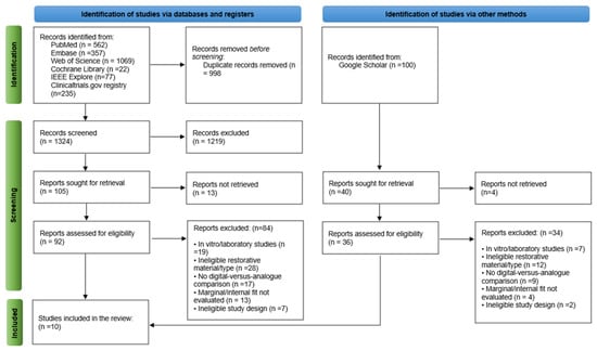
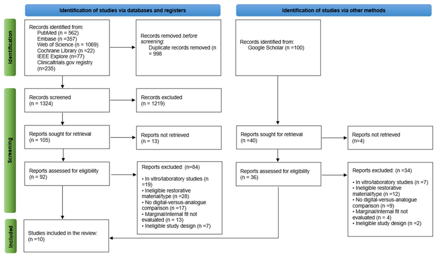
Search results were transferred to Rayyan QCRI, where duplicate records were removed. R.M. and K.M.F. independently evaluated titles and abstracts and subsequently assessed the full texts of potentially eligible reports. Any disagreement was first discussed between the two reviewers; unresolved decisions were referred to P.E. Screening agreement was high (Cohen’s κ = 0.89; 95% CI, 0.82–0.96). Figure 1 summarizes the selection process according to PRISMA 2020.
Figure 1.
PRISMA 2020 flow diagram of the study selection process.
2.6. Data Extraction
F.S.A. and Z.A. independently collected the study data using a previously piloted extraction form, after which P.E. verified the extracted information. Recorded items included study design and location; participant and restoration numbers; restoration type and zirconia material; intraoral scanner, CAD software, milling system, and cement-space parameters; and the material and technique used for the analogue impression. For marginal and internal fit, available means, standard deviations, measurement sites, and assessment methods were recorded. The two fit outcomes were treated separately throughout the review.
2.7. Risk of Bias Assessment
Risk of bias was independently assessed by two reviewers (P.E. and R.M.) using validated assessment tools appropriate to the methodological design of each included study. Studies incorporating randomized allocation of the intervention or treatment sequence [1,4,9,10,12] were evaluated using the Cochrane Risk of Bias 2 (RoB 2) tool, whereas the remaining non-randomized comparative studies were evaluated using the Risk of Bias in Non-randomized Studies of Interventions (ROBINS-I) tool [24].
For randomized studies, the RoB 2 [25] assessment considered bias arising from the randomization process, deviations from intended interventions, missing outcome data, measurement of the outcome, and selection of the reported result. For randomized within-subject, crossover, or split-mouth studies, the paired nature of the comparison and the method used to allocate the intervention or treatment sequence were additionally considered.
For non-randomized comparative studies, ROBINS-I assessment considered bias due to confounding, selection of participants, classification of interventions, deviations from intended interventions, missing data, measurement of outcomes, and selection of the reported result [24]. Disagreements between reviewers were resolved through discussion and consensus.
2.8. Statistical Analysis
Quantitative synthesis was performed when at least two studies reported clinically and methodologically comparable continuous outcomes. Mean differences (MDs) in marginal and internal fit were expressed in micrometers (μm) and calculated as the digital-workflow value minus the analogue-workflow value.
Because most eligible comparative studies employed within-subject designs, in which the same tooth or preparation was assessed using both digital and conventional (analogue) workflows, the paired nature of the observations was accounted for in the meta-analysis. For studies reporting paired data, the variance of the mean difference was estimated from the reported standard deviations of the digital and conventional measurements using an assumed within-pair correlation coefficient. Because the included studies did not report either the standard deviation of the paired differences or the within-pair correlation coefficient, a correlation of r = 0.50 was prespecified for the primary analysis. To assess the robustness of the pooled estimates to this assumption, sensitivity analyses were conducted using alternative correlation coefficients of r = 0.25 and r = 0.75, representing lower and higher degrees of within-subject correlation, respectively. This assumption was necessary because the precision of an effect estimate from a paired design depends on the correlation between measurements obtained from the same participant; when this correlation is not reported, it must be estimated to appropriately account for the paired nature of the data.
Random-effects meta-analyses were performed using restricted maximum likelihood (REML) estimation. Because only a small number of studies contributed to each quantitative synthesis, Hartung–Knapp-adjusted confidence intervals were used for pooled estimates. Statistical heterogeneity was quantified using the I2 statistic and between-study variance (τ2). A 95% prediction interval was calculated to estimate the range of effects that might be expected in a future clinically comparable study.
Marginal fit and overall internal fit were synthesized separately. When sufficiently comparable regional data were available, exploratory analyses were performed for axial, axio-occlusal/crest, and occlusal internal-fit measurements. Studies were excluded from a specific quantitative synthesis when the outcome definition, data structure, or statistical reporting did not permit derivation of a valid and clinically comparable effect estimate. Statistical analyses were performed using Python (version 3.13.5) with NumPy (version 2.3.5) and SciPy (version 1.17.0). Statistical significance was set at p < 0.05.
2.9. Reporting Bias Assessment
Reporting bias was not formally assessed because fewer than 10 studies contributed to each meta-analysis. Funnel-plot assessment and statistical tests for funnel-plot asymmetry were therefore not performed because these methods have limited power and may yield misleading interpretations when only a small number of studies are available.
2.10. Certainty of Evidence
The certainty of evidence for the primary outcomes of marginal fit and overall internal fit was evaluated using the Grading of Recommendations Assessment, Development and Evaluation (GRADE) framework [26]. Certainty was considered according to risk of bias, inconsistency, indirectness, imprecision, and potential publication bias. Because the quantitative evidence included both randomized and non-randomized comparative clinical studies, the certainty judgment considered the design and methodological limitations of the individual studies together with the magnitude of heterogeneity and precision of the pooled estimates.
3. Results
3.1. Study Selection
The systematic search identified 2422 records from the electronic databases and additional sources. After duplicate removal, the remaining records were screened by title and abstract according to the predefined eligibility criteria. A total of 128 reports were retrieved for full-text assessment. Following full-text evaluation, 10 clinical studies met all eligibility criteria and were included in the qualitative synthesis [1,3,4,6,7,8,9,10,12,27]. Of these, four studies provided directly comparable paired data suitable for the primary meta-analysis of marginal fit [1,3,4,10], and the same four studies provided sufficiently comparable data for quantitative synthesis of overall internal fit [1,3,4,10]. Additional studies contributed to the qualitative synthesis but were not quantitatively pooled when their outcome definition, data structure, or available statistical information did not permit calculation of a valid comparable effect estimate [6,7,8,9,12,27]. The study-selection process is presented in Figure 1.
3.2. Study Characteristics
Ten clinical studies published between 2015 and 2026 met the eligibility criteria [1,3,4,6,7,8,9,10,12,27]. The studies predominantly evaluated tooth-supported single zirconia crowns, whereas Ahrberg et al. [4] and Morsy et al. [10] also evaluated three-unit fixed dental prostheses. Digital workflows incorporated contemporary intraoral scanners, including Lava C.O.S., iTero, TRIOS, CEREC Omnicam, and CS3500, whereas analogue workflows used polyvinyl siloxane or polyether impressions followed by stone-cast fabrication and laboratory digitization.
The evidence included both randomized and non-randomized comparative clinical designs. Five studies incorporated randomized allocation of the impression technique, intervention, or treatment sequence [1,4,9,10,12], whereas five studies were non-randomized comparative investigations [3,6,7,8,27]. All but one study [12] used within-subject, crossover, or split-mouth comparisons, permitting digital and analogue workflows to be evaluated within the same participant or on matched clinical preparations.
Marginal and/or internal adaptation was predominantly assessed using silicone replica techniques followed by microscopic measurement at predefined locations [1,3,4,7,8,9,10,12,27]. Berrendero et al. [6] instead evaluated marginal adaptation clinically using a probe of defined tip diameter.
The detailed study characteristics are presented in Table 2.
Table 2.
Detailed characteristics of the included studies.
3.3. Risk of Bias Assessment
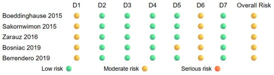
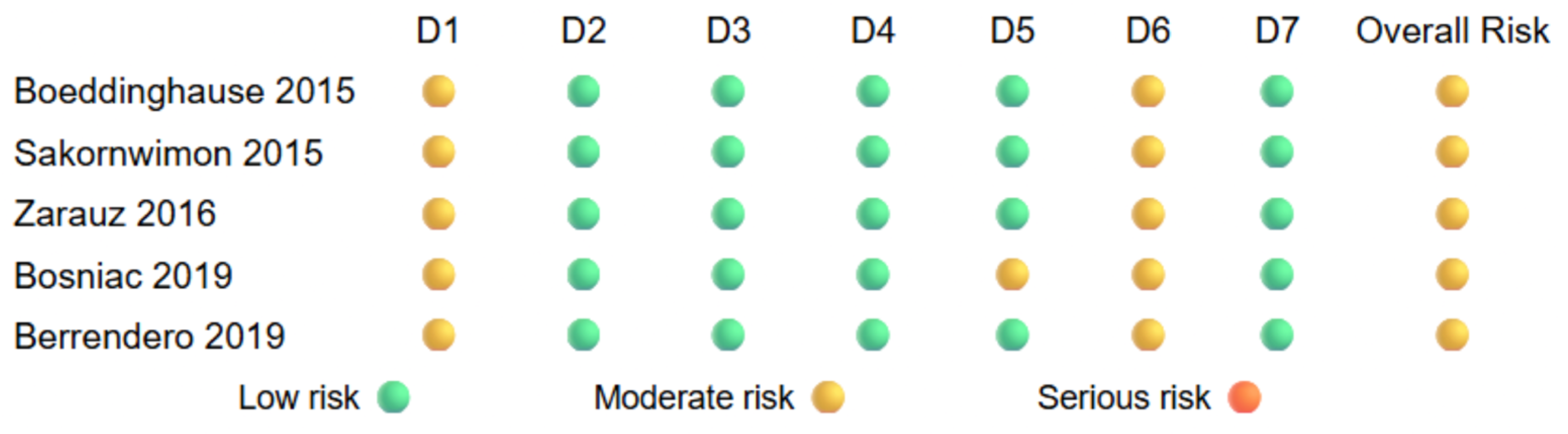
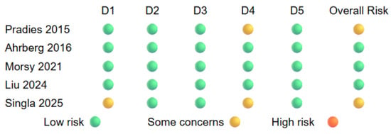
Risk-of-bias assessments are presented in Figure 2 and Figure 3. The five non-randomized comparative studies [3,6,7,8,27] were evaluated using ROBINS-I (Figure 2). The principal methodological concerns were related to potential confounding and outcome measurement, particularly where assessor blinding, operator-related factors, or measurement procedures were incompletely reported. Other domains generally presented fewer concerns, although individual judgments varied according to study methodology.
Figure 2.
Risk-of-bias assessment of the five non-randomized comparative clinical studies [3,6,7,8,27] using the Risk of Bias in Non-randomized Studies of Interventions (ROBINS-I) tool. D1, bias due to confounding; D2, bias due to selection of participants; D3, bias due to classification of interventions; D4, bias due to deviations from intended interventions; D5, bias due to missing data; D6, bias due to measurement of outcomes; D7, bias due to selection of the reported result.
Figure 3.
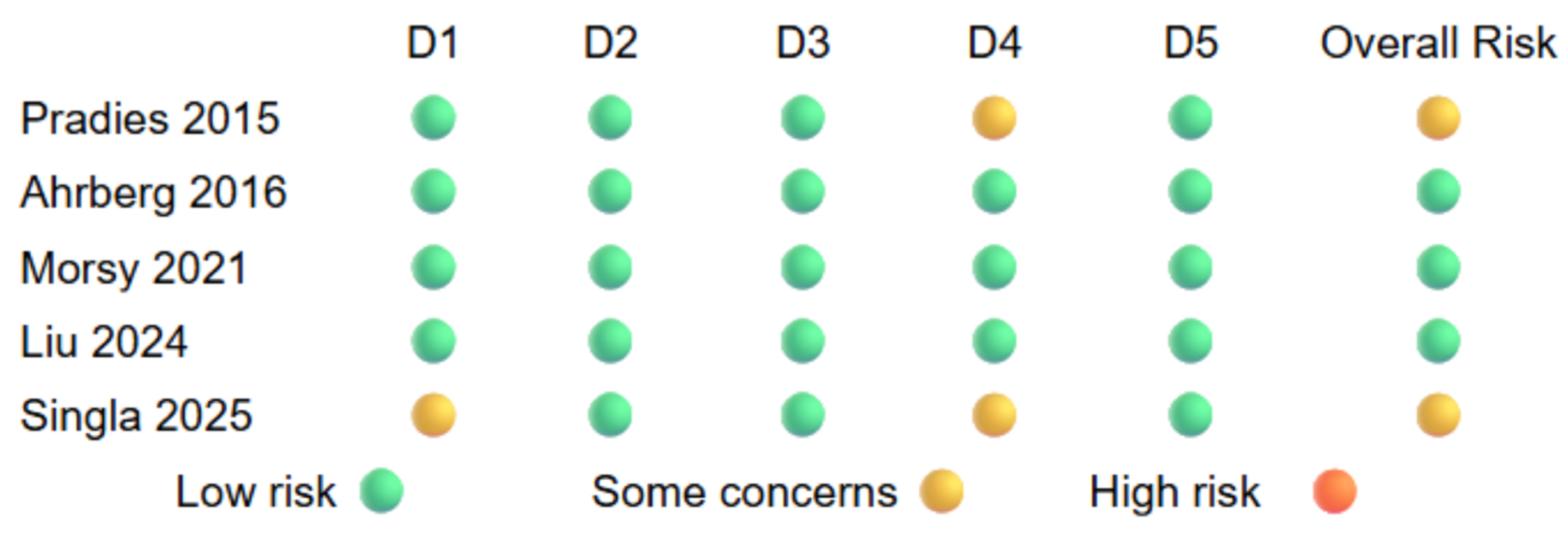
Risk-of-bias assessment of the five clinical studies incorporating randomized allocation [1,4,9,10,12] using the Cochrane Risk of Bias 2 (RoB 2) tool. D1, bias arising from the randomization process; D2, bias due to deviations from intended interventions; D3, bias due to missing outcome data; D4, bias in measurement of the outcome; D5, bias in selection of the reported result.
The five studies incorporating randomized allocation [1,4,9,10,12] were evaluated using RoB 2 (Figure 3). Potential concerns primarily involved deviations from intended interventions and outcome measurement because complete blinding of the clinical operator to digital versus analogue impression procedures was inherently difficult. Nevertheless, several studies incorporated randomized treatment or sequence allocation, blinded outcome assessment, or both.
Overall, the risk-of-bias assessment demonstrated methodological variability across the available clinical evidence, which was considered when interpreting the pooled findings.
3.4. Qualitative Synthesis
3.4.1. Marginal Fit
The qualitative findings demonstrated considerable variability in marginal adaptation across studies and workflows [1,3,6,7,8,9,10,12,27]. Most studies reporting continuous micrometric measurements showed smaller marginal discrepancies with direct digital impressions than with analogue impressions [1,3,4,7,9,10,12], whereas Sakornwimon et al. [8] reported no statistically significant difference between the two workflows. Berrendero et al. [6], using a clinical probe-based assessment rather than a directly comparable continuous micrometric outcome, reported better marginal adaptation for digitally derived crowns.
Among the studies providing continuous marginal-gap measurements, Pradíes et al. [1] reported mean values of 76.33 ± 65.32 μm for the digital workflow and 91.46 ± 72.17 μm for the analogue workflow. Zarauz et al. [3] reported corresponding values of 80.29 ± 26.24 μm and 135.51 ± 48.78 μm, and Ahrberg et al. [4] reported 61.08 ± 24.77 μm and 70.40 ± 28.87 μm, respectively. Morsy et al. [10] similarly reported smaller marginal discrepancies with the digital workflow (30.91 ± 15.15 μm) than with the analogue workflow (40.02 ± 19.50 μm).
Some studies reported marginal-fit outcomes but could not be incorporated into the primary quantitative synthesis because of multiple digital scanner arms sharing a common analogue comparator [7,27], site-specific marginal measurements without a directly comparable overall participant-level estimate [8], categorical clinical assessment [6], or outcome structures that were not directly comparable with the continuous paired marginal-gap outcome used for the primary synthesis [9,12]. These studies were retained in the qualitative synthesis to avoid inappropriate unit-of-analysis assumptions.
3.4.2. Internal Fit
Internal adaptation was reported less consistently than marginal adaptation, with substantial variation in the anatomical regions and measurement protocols used across studies. Measurement locations included the axial walls, axio-occlusal or crestal regions, and occlusal fossae [1,3,4,10,12]. Among studies reporting an overall internal-discrepancy value, Pradíes et al. [1] reported a lower mean discrepancy for the digital workflow than for the analogue workflow (149.86 ± 65.84 μm versus 165.77 ± 69.64 μm, respectively). Similarly, Zarauz et al. [3] reported lower internal discrepancy for the digital workflow (111.40 ± 54.04 μm) than with the analogue workflow (173.00 ± 92.65 μm). Morsy et al. [10] also reported lower mean internal discrepancy for the digital workflow (30.86 ± 13.57 μm) compared with the analogue workflow (41.86 ± 18.94 μm).
Location-specific findings were reported by Ahrberg et al. [4] who identified a statistically significant difference at the mid-occlusal region, with lower mean discrepancy for the digital workflow than for the analogue workflow (155.57 ± 49.85 μm vs. 171.51 ± 60.98 μm, respectively). Differences between workflows at the other evaluated measurement locations were comparatively smaller and did not demonstrate the same level of statistical significance [4].
Because internal adaptation was assessed at different anatomical landmarks and using non-uniform measurement protocols, the reported values were not considered directly interchangeable across all studies. Accordingly, study eligibility for quantitative synthesis was determined separately for overall internal-fit outcomes and location-specific measurements, with pooling undertaken only when the measurement region, outcome definition, and reported data were sufficiently comparable.
3.5. Quantitative Synthesis (Meta-Analysis)
3.5.1. Marginal Fit
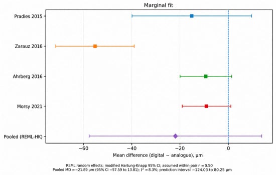
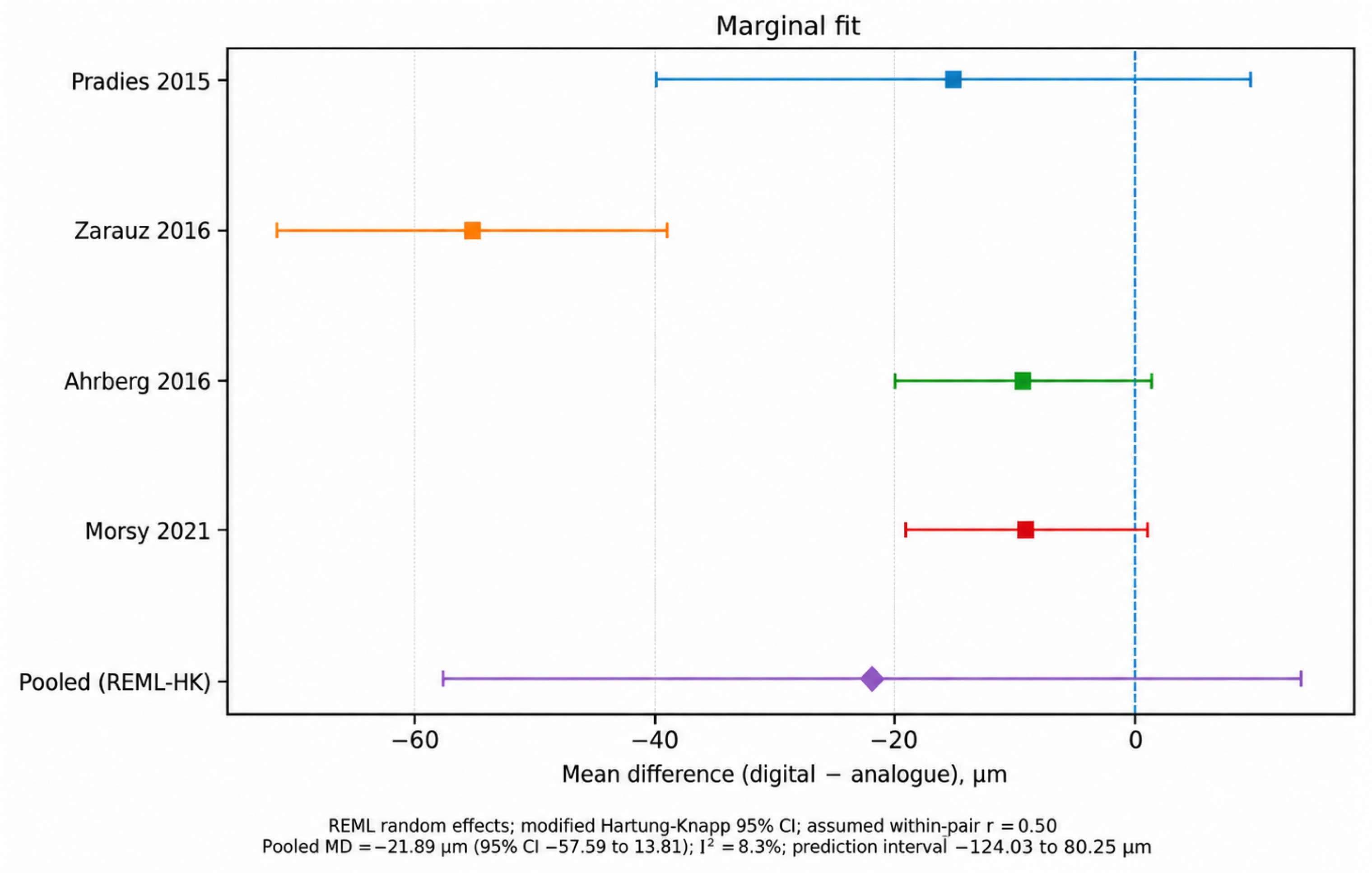
Four clinical studies [1,3,4,10] provided directly comparable marginal-gap means and standard deviations for paired digital and analogue restorations and were included in the primary quantitative synthesis. Mean differences were calculated as digital minus analogue values; therefore, negative values favoured the digital workflow.
Using an assumed within-pair correlation of r = 0.50, the REML random-effects meta-analysis yielded a pooled MD of −21.89 μm, favouring the digital workflow; however, the difference was not statistically significant (95% CI, −57.59 to 13.81 μm; p = 0.146) (Figure 4). Considerable heterogeneity was observed (I2 = 88.3%; τ2 = 437.72). The 95% prediction interval ranged from −124.03 to 80.25 μm, indicating substantial variability in the magnitude and potentially the direction of the workflow effect across comparable clinical settings (Table 3).
Figure 4.
Forest plot of the paired random-effects meta-analysis comparing marginal fit of CAD/CAM zirconia restorations fabricated using digital and analogue workflows [1,3,4,10]. Mean differences are expressed as digital minus analogue values in μm; negative values favour the digital workflow. The primary analysis assumed a within-pair correlation of r = 0.50 and used REML estimation with Hartung–Knapp adjusted confidence intervals.
Table 3.
Study-level data included in the paired meta-analysis of marginal fit.
Accordingly, although the pooled point estimate favoured digital workflows, the available clinical evidence did not demonstrate a statistically reliable overall difference in marginal fit between digital and analogue workflows.
3.5.2. Correlation-Sensitivity Analysis for Marginal Fit
Because the primary studies did not report within-pair correlations, sensitivity analyses were performed using assumed correlation coefficients of r = 0.25, 0.50, and 0.75. The pooled estimate remained similar across all assumptions. At r = 0.25, the pooled MD was −21.85 μm (95% CI, −57.90 to 14.21 μm; p = 0.149; I2 = 83.9%); at r = 0.50, the pooled MD was −21.89 μm (95% CI, −57.59 to 13.81 μm; p = 0.146; I2 = 88.3%); and at r = 0.75, the pooled MD was −21.91 μm (95% CI, −57.17 to 13.36 μm; p = 0.142; I2 = 92.7%) (Table 4). Thus, the direction and statistical interpretation of the pooled marginal-fit estimate were not materially dependent on the assumed within-pair correlation.
Table 4.
Sensitivity analysis of marginal fit according to the assumed within-pair correlation coefficient.
3.5.3. Overall Internal Fit
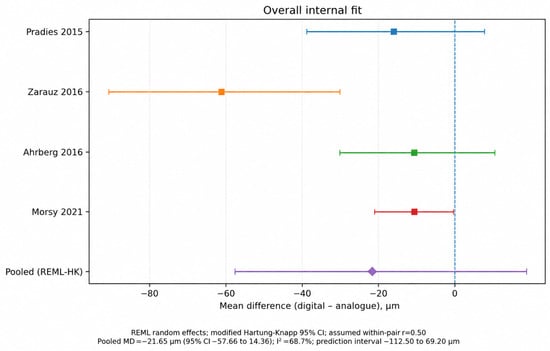
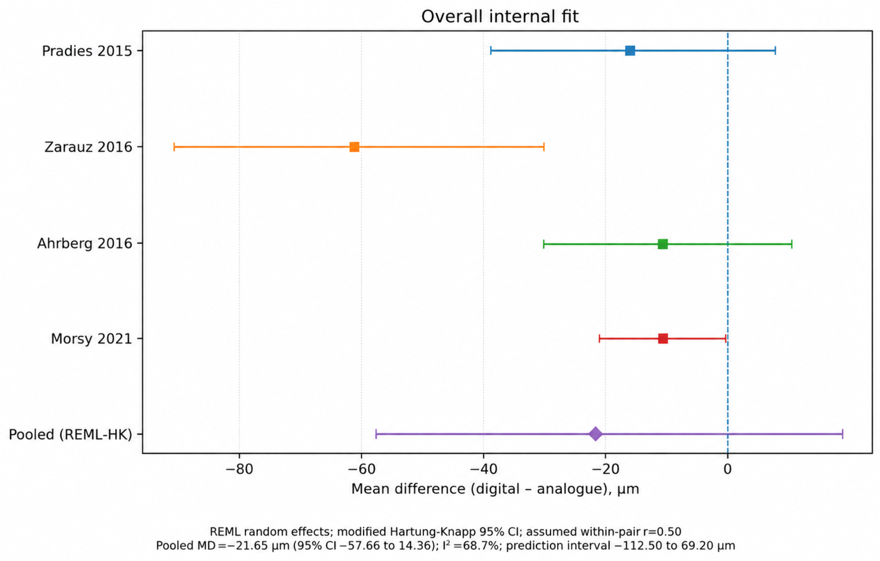
Four paired clinical studies [1,3,4,10] reported sufficiently comparable overall internal-fit data for quantitative synthesis. Using an assumed within-pair correlation of r = 0.50, the random-effects meta-analysis favoured digital workflows, with a pooled MD of −21.65 μm; however, the difference was not statistically significant (95% CI, −57.66 to 14.36 μm; p = 0.152) (Figure 5). Heterogeneity was substantial (I2 = 68.7%; τ2 = 317.88), and the 95% prediction interval ranged from −112.50 to 69.20 μm (Table 5). Thus, the available clinical evidence did not establish a consistent overall internal-fit advantage for either workflow.
Figure 5.
Forest plot of the paired random-effects meta-analysis comparing overall internal fit of CAD/CAM zirconia restorations fabricated using digital and analogue workflows [1,3,4,10]. Negative mean differences favour the digital workflow. The primary analysis assumed a within-pair correlation of r = 0.50.
Table 5.
Study-level data included in the paired meta-analysis of overall internal fit.
3.5.4. Correlation-Sensitivity Analysis for Overall Internal Fit
Sensitivity analyses using alternative within-pair correlations yielded similar conclusions. At r = 0.25, the pooled MD was −20.08 μm (95% CI, −54.00 to 13.83 μm; p = 0.156; I2 = 56.4%); at r = 0.50, the pooled MD was −21.65 μm (95% CI, −57.66 to 14.36 μm; p = 0.152; I2 = 68.7%); and at r = 0.75, the pooled MD was −22.84 μm (95% CI, −60.22 to 14.55 μm; p = 0.147; I2 = 80.9%) (Table 6). Thus, the absence of a statistically significant difference in overall internal fit was consistent across plausible assumptions regarding the within-pair correlation.
Table 6.
Correlation-sensitivity analysis for overall internal fit.
3.5.5. Location-Specific Internal Fit
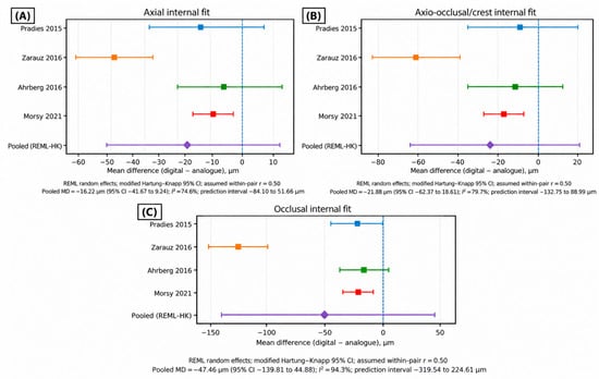
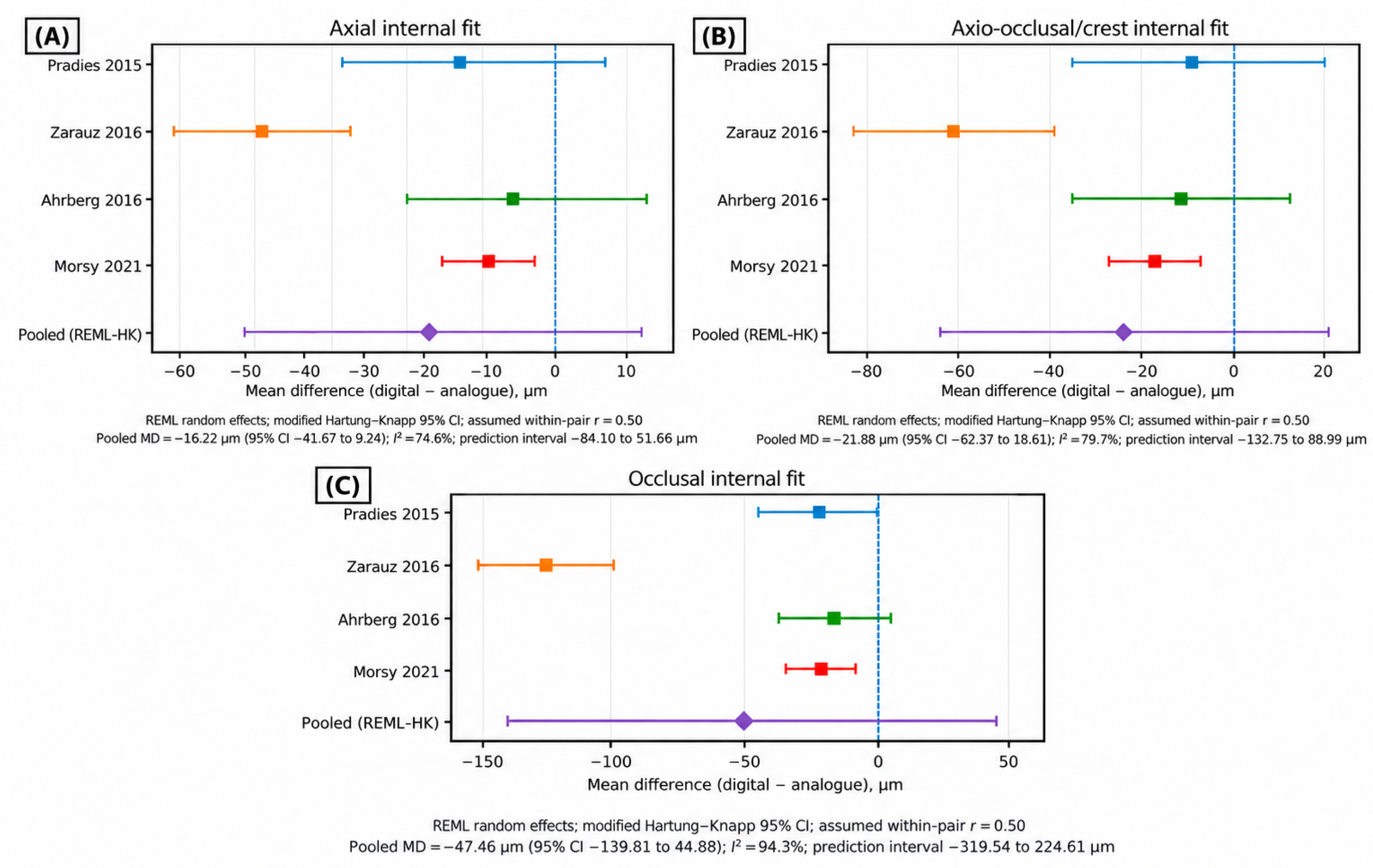
Exploratory location-specific meta-analyses were performed using data from studies reporting sufficiently comparable anatomical measurement regions [1,3,4,10]. For the axial region, the pooled MD was −16.22 μm (95% CI, −41.67 to 9.24 μm; p = 0.136; I2 = 74.6%); for the axio-occlusal/crest region, the pooled MD was −21.88 μm (95% CI, −62.37 to 18.61 μm; p = 0.184; I2 = 79.7%); and for the occlusal region, the pooled MD was −47.46 μm (95% CI, −139.81 to 44.88 μm; p = 0.200; I2 = 94.3%) (Figure 6). Although all pooled point estimates favoured digital workflows, none reached statistical significance. The particularly high heterogeneity in the occlusal analysis indicates considerable variation in measurement landmarks and clinical protocols and warrants cautious interpretation.
Figure 6.
Forest plot of the axial internal fits (A), axio-occlusal/crest internal fits (B), and occlusal internal fits (C) [1,3,4,10].
3.6. Certainty of Evidence
The certainty of evidence was rated as very low for both marginal fit and overall internal fit. For marginal fit, certainty was downgraded because of methodological limitations across the contributing studies, considerable inconsistency (I2 = 88.3%), and imprecision, as both the pooled confidence interval and prediction interval crossed the line of no effect (Table 7). For overall internal fit, certainty was similarly downgraded because of methodological limitations, substantial inconsistency (I2 = 68.7%) (Table 7), and imprecision, with the pooled confidence interval and prediction interval including both a potential benefit of digital workflows and little or no difference. Consequently, the pooled estimates should be interpreted cautiously and should not be considered definitive evidence of superiority of either workflow.
Table 7.
GRADE summary of findings.
4. Discussion
The present systematic review and meta-analysis synthesized clinical evidence on the marginal and internal fit of CAD/CAM zirconia fixed restorations produced using digital and analogue workflows. The principal finding is that digital workflows demonstrated a consistent trend toward smaller marginal and internal discrepancies. However, after accounting for the paired nature of the evidence and substantial between-study heterogeneity, neither the pooled marginal nor overall internal fit differences reached statistical significance. Consequently, the available clinical evidence supports the comparability, rather than the definitive superiority, of digital workflows relative to conventional analogue techniques for the adaptation of zirconia restorations. Given the very low certainty of evidence for both primary outcomes, these differences should be interpreted cautiously and should not be considered evidence of clinical superiority of the digital workflow.
Interpretation of marginal adaptation should consider both the statistical findings and their clinical relevance. The pooled mean difference of approximately 22 μm favouring digital workflows must be contextualized within commonly cited clinically acceptable thresholds, which typically range from 100 to 120 μm for fixed dental restorations [10]. Although a universally accepted threshold for marginal discrepancy has not been established, a mean difference of 22 μm represents a modest magnitude that is unlikely to independently influence biological outcomes, such as cement dissolution, microleakage, or secondary caries, provided both workflows remain within acceptable limits [28]. Furthermore, the wide 95% prediction interval, which crossed the line of no effect and spanned both clinically advantageous and disadvantageous ranges, indicates that the true effect in any given future clinical setting remains highly uncertain. Therefore, while the direction of effect suggests a possible average advantage for intraoral scanning (IOS), this finding should not be interpreted as evidence of definitive clinical superiority.
Internal adaptation findings mirrored those of marginal fit, with a similar pooled advantage of approximately 22 μm for digital workflows. Internal adaptation is primarily relevant to mechanical aspects of restoration performance, including retention, resistance, and cement film thickness [2]. The exploratory location-specific analyses revealed that while all regional point estimates favoured digital workflows, the occlusal region exhibited the largest difference but also the most extreme heterogeneity [1,4]. This variation can be attributed to fundamental differences in how internal fit is measured across anatomical landmarks. Measurements at occlusal fossae or axio-occlusal transitions are highly sensitive to the specific geometry of the preparation, the CAD design algorithms, and the assigned cement space settings, which varied across the included studies [4,7,10,12,27]. Consequently, treating all internal-fit measurements as interchangeable is methodologically inappropriate, and the high heterogeneity precludes a confident conclusion regarding regional internal adaptation differences.
The substantial statistical heterogeneity observed across both primary outcomes is likely attributable to considerable clinical and methodological variability. Clinically, the included studies evaluated different intraoral scanner systems, ranging from older wavefront sampling technologies [1] to contemporary confocal scanners [10,12]. Additionally, the studies used different analogue impression materials, which possess inherent differences in dimensional stability and elastomeric recovery [4,6]. Furthermore, variations in CAD software algorithms, cement-space parameters, milling strategies, and zirconia sintering protocols introduce cumulative discrepancies that are distinct to digital and analogue workflows [4,7,10,12,27]. Methodologically, the reliance on different measurement techniques (e.g., silicone replica technique under varying microscopic magnifications versus clinical probing) [1,3,4,10,12] and the lack of standardized anatomical measurement landmarks fundamentally limit direct comparability and likely contributed to the high heterogeneity. Such methodological variability reduces direct comparability and likely contributed to the particularly high heterogeneity observed in the location-specific analyses. Standardized definitions and measurement landmarks are therefore needed in future clinical investigations. Accordingly, the pooled estimates represent only the subset of studies with sufficiently comparable quantitative data and should be interpreted alongside the broader qualitative findings from all 10 clinical studies included.
An inherent methodological challenge in synthesizing this evidence is the predominance of within-subject, crossover, or split-mouth designs [1,3,4,7,8,9,10,27]. While advantageous for controlling inter-individual variability in preparation geometry and biological response, these designs require accounting for within-pair correlation. Thus, the present analysis incorporated the paired nature of the data. Because the original studies did not report within-pair correlations or standard deviations of paired differences, a correlation of r = 0.50 was used for the primary analyses, and sensitivity analyses were performed using r = 0.25 and r = 0.75. The similarity of the pooled estimates across these assumptions indicates that the overall interpretation was not materially dependent on the selected correlation coefficient. Future within-subject clinical studies should therefore report the standard deviation of paired differences and/or the within-pair correlation directly, which would permit more precise quantitative synthesis without reliance on assumed correlation coefficients.
The risk-of-bias profile further tempers the certainty of the evidence, which was rated as very low. While half of the included studies randomized the intervention sequence, complete blinding of clinical operators to digital versus analogue workflows is inherently impossible, introducing potential performance bias. Additionally, non-randomized studies exhibited confounding related to operator experience and sequence effects. Despite these methodological limitations, the direction of the pooled estimates was consistently toward digital workflows, suggesting that methodological bias alone does not explain the observed trend.
The present findings should also be considered in relation to previous systematic reviews [17,18,19,20,21]. Hasanzade et al. [20] evaluated both in vivo and in vitro evidence comparing digital and conventional impression workflows. Al-Odinee et al. [21] focused on clinical studies of CAD/CAM zirconia restorations and reported statistically significant differences favouring digital scanning for marginal and internal fit. In the present review, the direction of the pooled effects similarly favoured the digital workflow; however, the pooled differences did not reach statistical significance. The present analysis restricted quantitative synthesis to studies providing sufficiently comparable outcome definitions and data structures and accounted for the paired structure of within-subject comparisons. REML random-effects models with Hartung–Knapp-adjusted confidence intervals, prediction intervals, and sensitivity analyses across plausible within-pair correlations were additionally used to characterize uncertainty. These methodological considerations may partly account for the wider confidence intervals observed in the present analysis. The difference between the present findings and those reported in previous reviews [17,18,19,20,21] concerns the statistical certainty surrounding the estimated workflow effect. From a clinical standpoint, the available evidence does not demonstrate a statistically significant difference between digital and analogue impression workflows when they are compared solely on marginal or internal adaptation. Instead, the available evidence suggests that both workflows can produce clinically acceptable marginal discrepancies, and workflow selection should be based on a broader clinical rationale. Digital workflows may offer practical advantages, including enhanced patient comfort, reduced chairside time, streamlined laboratory communication, and improved contamination control [8,12]. Conversely, analogue workflows retain validity, particularly in clinical scenarios involving deep subgingival margins where soft-tissue control and retraction are challenging for optical scanning, or in extensive edentulous spans where IOS accuracy may be compromised [10,17]. Consequently, the clinical relevance of a given marginal discrepancy should be interpreted in the context of restoration design, margin location, cementation procedures, periodontal conditions, and duration of clinical service rather than according to a single cutoff.
This review has several strengths, including a focused PICO question, a comprehensive search strategy, prospective registration, and the exclusion of single-arm in vitro studies. Furthermore, the quantitative synthesis appropriately accounted for the paired nature of the data, used Hartung–Knapp-adjusted confidence intervals suitable for small meta-analyses, and incorporated prediction intervals to convey the expected range of future clinical effects.
Four limitations of the study are acknowledged. First, although 10 clinical studies met the eligibility criteria, only four provided sufficiently comparable paired data for each primary meta-analysis [1,3,4,10]. Within-pair correlations were not reported and therefore had to be assumed, and internal-fit measurement locations were not fully standardized. Second, results from some of the eligible studies could not be quantitatively pooled because of differences in study design, outcome reporting, data structure, site-specific reporting, categorical outcomes, or insufficient statistical information [6,7,8,9,12,27]. Third, the small number of studies included in each meta-analysis precluded the reliable formal assessment of publication bias. Fourth, restriction of the search to studies published in English may have introduced language-selection bias. Future clinical trials should prioritize methodological standardization and transparent reporting. Researchers should adopt standardized definitions for marginal and internal adaptation, utilize consistent anatomical measurement landmarks, and report within-pair correlations or the standard deviations of paired differences to facilitate accurate future meta-analyses. Furthermore, investigations should move beyond surrogate fit measurements to evaluate long-term clinical outcomes, including restoration survival, biological complications, and the impact of different cementation protocols. In addition, cement-space settings, restoration-seating and cementation protocols, and fit-measurement procedures should be consistently reported.
5. Conclusions
Considering the limitations of the available clinical evidence, CAD/CAM zirconia restorations produced through digital workflows showed smaller marginal and internal discrepancies than restorations produced through analogue workflows. However, neither the pooled marginal-fit difference nor the overall internal-fit difference reached statistical significance. Considerable heterogeneity, wide prediction intervals, and the very low certainty of evidence indicate that the magnitude of any workflow-related difference remains uncertain and may vary across clinical settings. Therefore, the current evidence does not establish definitive superiority of either workflow based solely on marginal or internal fit. Future clinical studies should use standardized definitions and measurement protocols and report within-pair correlations or standard deviations of paired differences. These improvements should enhance future quantitative analyses.
Supplementary Materials
The following supporting information can be downloaded at: https://www.mdpi.com/article/10.3390/prosthesis8100104/s1. Supplementary S1. The PRISMA 2020 checklist.
Author Contributions
Conceptualization, P.E.; methodology, P.E. and R.M.; validation, P.E. and R.M.; formal analysis, P.E. and R.M.; investigation, K.M.F., F.S.A., and Z.A.; resources, P.E.; data curation, P.E. and R.M.; writing—original draft preparation, R.M., K.M.F., F.S.A., and Z.A.; writing—review and editing, P.E.; visualization, R.M.; supervision, P.E.; project administration, P.E. All authors have read and agreed to the published version of the manuscript.
Funding
This research received no external funding.
Institutional Review Board Statement
Not applicable.
Informed Consent Statement
Not applicable.
Data Availability Statement
The data used in this systematic review and meta-analysis were extracted from previously published studies. The data underlying the quantitative analyses are available from the corresponding author upon reasonable request.
Conflicts of Interest
The authors declare no conflicts of interest.
References
- Pradíes, G.; Zarauz, C.; Valverde, A.; Ferreiroa, A.; Martínez-Rus, F. Clinical evaluation comparing the fit of all-ceramic crowns obtained from silicone and digital intraoral impressions based on wavefront sampling technology. J. Dent. 2015, 43, 201–208. [Google Scholar] [CrossRef] [Scilit] [PubMed]
- Holmes, J.R.; Bayne, S.C.; Holland, G.A.; Sulik, W.D. Considerations in measurement of marginal fit. J. Prosthet. Dent. 1989, 62, 405–408. [Google Scholar] [CrossRef] [Scilit] [PubMed]
- Zarauz, C.; Valverde, A.; Martinez-Rus, F.; Hassan, B.; Pradies, G. Clinical evaluation comparing the fit of all-ceramic crowns obtained from silicone and digital intraoral impressions. Clin. Oral Investig. 2016, 20, 799–806. [Google Scholar] [CrossRef] [Scilit] [PubMed]
- Ahrberg, D.; Lauer, H.C.; Ahrberg, M.; Weigl, P. Evaluation of fit and efficiency of CAD/CAM fabricated all-ceramic restorations based on direct and indirect digitalization: A double-blinded, randomized clinical trial. Clin. Oral Investig. 2017, 20, 291–300. [Google Scholar] [CrossRef] [Scilit] [PubMed]
- Ellakany, P.; Al-Harbi, F.; El Tantawi, M.; Mohsen, C. Evaluation of the accuracy of digital and 3D-printed casts compared with conventional stone casts. J. Prosthet. Dent. 2022, 127, 438–444. [Google Scholar] [CrossRef] [Scilit] [PubMed]
- Berrendero, S.; Salido, M.P.; Ferreiroa, A.; Valverde, A.; Pradíes, G. Comparative study of all-ceramic crowns obtained from conventional and digital impressions: Clinical findings. Clin. Oral Investig. 2019, 23, 1745–1751. [Google Scholar] [CrossRef] [Scilit] [PubMed]
- Bosniac, P.; Rehmann, P.; Wöstmann, B. Comparison of an indirect impression scanning system and two direct intraoral scanning systems in vivo. Clin. Oral Investig. 2019, 23, 2421–2427. [Google Scholar] [CrossRef] [Scilit] [PubMed]
- Sakornwimon, N.; Leevailoj, C. Clinical marginal fit of zirconia crowns and patients’ preferences for impression techniques using intraoral digital scanner versus polyvinyl siloxane material. J. Prosthet. Dent. 2016, 118, 386–391. [Google Scholar] [CrossRef] [Scilit] [PubMed]
- Singla, N.; Datta, P.; Rani, G.; Tripathi, S.; Chaudhary, N.; Pal, S. Comparative evaluation of marginal fit, prosthodontic and patient centered outcomes of milled zirconia crown using conventional and digital impression techniques—A split mouth, randomized clinical trial. J. Dent. Spec. 2025, 13, 192–197. [Google Scholar] [CrossRef] [Scilit]
- Morsy, N.; El Kateb, M.; Azer, A.; Fathalla, S. Fit of monolithic multilayer zirconia fixed partial dentures fabricated by conventional versus digital impression: A clinical and laboratory investigations. Clin. Oral Investig. 2021, 25, 5363–5373. [Google Scholar] [CrossRef] [Scilit] [PubMed]
- Ellakany, P.; Tantawi MEl Mahrous, A.A.; Al-Harbi, F. Evaluation of the Accuracy of Digital Impressions Obtained from Intraoral and Extraoral Dental Scanners with Different CAD/CAM Scanning Technologies: An In Vitro Study. J. Prosthodont. 2022, 31, 314–319. [Google Scholar] [CrossRef] [Scilit] [PubMed]
- Liu, X.; Feng, K.; Dong, L.; Lixia, L.; Ni, L.; Zheng, D. Accuracy and Efficiency of Digitally Fabricated All-Ceramic Crowns from Conventional Impressions and Intraoral Scans: A Single-Blind Clinical Randomized Controlled Trial. Int. J. Prosthodont. 2024, 37, 8–15. [Google Scholar] [CrossRef] [Scilit] [PubMed]
- Ellakany, P.; Aly, N.M.; Al-Harbi, F. Accuracy of 3D Printed and Digital Casts Produced from Intraoral and Extraoral Scanners with Different Scanning Technologies: In Vitro Study. J. Prosthodont. 2022, 31, 521–528. [Google Scholar] [CrossRef] [Scilit] [PubMed]
- Mangano, F.G.; Admakin, O.; Margiani, B.; Solop, I.; Latuta, N. An experimental strategy for capturing the margins of prepared single teeth with an intraoral scanner: A prospective clinical study on 30 patients. Int. J. Environ. Res. Public Health 2020, 17, 392. [Google Scholar] [CrossRef] [Scilit] [PubMed]
- Refaie, A.; Fouda, A.; Bourauel, C.; Singer, L. Marginal gap and internal fit of 3D printed versus milled monolithic zirconia crowns. BMC Oral Health 2023, 23, 448. [Google Scholar] [CrossRef] [Scilit] [PubMed]
- Güney, B.; Yılmaz Biçer, E.; Doğan, D.G.; Bankoğlu Güngör, M. Effects of Yttria Content and Margin Design on the Fracture Resistance of Monolithic Zirconia Crowns. J. Funct. Biomater. 2026, 17, 219. [Google Scholar] [CrossRef] [Scilit] [PubMed]
- Sinha, A.; Srivastava, G.; Panda, S.K.; Mohapatra, A.; Tabarak, N.; Choudhury, G.K. Comparative evaluation of marginal and internal fit of multi-unit fixed dental prostheses: A systematic review and meta-Analysis of conventional versus digital impressions. Dent. Res. J. 2026, 23, 17. [Google Scholar] [CrossRef] [Scilit] [PubMed]
- Ahmed, W.M.; Shariati, B.; Gazzaz, A.Z.; Sayed, M.E.; Carvalho, R.M. Fit of tooth-supported zirconia single crowns—A systematic review of the literature. Clin. Exp. Dent. Res. 2020, 6, 700–716. [Google Scholar] [CrossRef] [Scilit] [PubMed]
- Sarafidou, K.; Chatziparaskeva, M.; Chatzikamagiannis, D.; Mpotskaris, V.; Tortopidis, D.; Bakopoulou, A.; Kokoti, M. Evaluation of marginal/internal fit of fixed dental prostheses after digital, conventional, and combination impression techniques: A systematic review. Eur. J. Oral Sci. 2022, 130, e12902. [Google Scholar] [CrossRef] [Scilit] [PubMed]
- Hasanzade, M.; Aminikhah, M.; Afrashtehfar, K.I.; Alikhasi, M. Marginal and internal adaptation of single crowns and fixed dental prostheses by using digital and conventional workflows: A systematic review and meta-analysis. J. Prosthet. Dent. 2021, 126, 360–368. [Google Scholar] [CrossRef] [Scilit] [PubMed]
- Al-Odinee, N.M.; Al-Haddad, A.; Alshammari, A.F.; Madfa, A.A. Marginal and internal fit of zirconia CAD-CAM fabricated with conventional impression and digital scanning techniques: A systematic review and meta-analysis. J. Prosthet. Dent. 2026, 136, 681–690. [Google Scholar] [CrossRef] [Scilit] [PubMed]
- Ting-shu, S.; Jian, S. Intraoral Digital Impression Technique: A Review. J. Prosthodont. 2015, 24, 313–321. [Google Scholar] [CrossRef] [Scilit] [PubMed]
- Page, M.J.; McKenzie, J.E.; Bossuyt, P.M.; Boutron, I.; Hoffmann, T.C.; Mulrow, C.D.; Shamseer, L.; Tetzlaff, J.M.; Akl, E.A.; Brennan, S.E.; et al. The PRISMA 2020 statement: An updated guideline for reporting systematic reviews. BMJ 2021, 372, n71. [Google Scholar] [CrossRef] [Scilit] [PubMed]
- Sterne, J.A.; Hernán, M.A.; Reeves, B.C.; Savović, J.; Berkman, N.D.; Viswanathan, M.; Henry, D.; Altman, D.G.; Ansari, M.T.; Boutron, I.; et al. ROBINS-I: A tool for assessing risk of bias in non-randomised studies of interventions. BMJ 2016, 355, i4919. [Google Scholar] [CrossRef] [Scilit] [PubMed]
- Sterne, J.A.C.; Savović, J.; Page, M.J.; Elbers, R.G.; Blencowe, N.S.; Boutron, I.; Cates, C.J.; Cheng, H.Y.; Corbett, M.S.; Eldridge, S.M.; et al. RoB 2: A revised tool for assessing risk of bias in randomised trials. BMJ 2019, 366, l4898. [Google Scholar] [CrossRef] [Scilit] [PubMed]
- Guyatt, G.H.; Oxman, A.D.; Vist, G.E.; Kunz, R.; Falck-Ytter, Y.; Alonso-Coello, P.; Schünemann, H.J.; GRADE Working Group. GRADE: An emerging consensus on rating quality of evidence strength of recommendations. BMJ 2008, 336, 924–926. [Google Scholar] [CrossRef] [Scilit] [PubMed]
- Boeddinghaus, M.; Breloer, E.S.; Rehmann, P.; Wöstmann, B. Accuracy of single-tooth restorations based on intraoral digital and conventional impressions in patients. Clin. Oral Investig. 2015, 19, 2027–2034. [Google Scholar] [CrossRef] [Scilit] [PubMed]
- Wu, L.; Sun, Z.; Zhao, J.; Zheng, Y. Retrospective clinical study of monolithic zirconia crowns fabricated with a straightforward completely digital workflow. J. Prosthet. Dent. 2022, 128, 913–918. [Google Scholar] [CrossRef] [Scilit] [PubMed]

Disclaimer/Publisher’s Note: The statements, opinions and data contained in all publications are solely those of the individual author(s) and contributor(s) and not of MDPI and/or the editor(s). MDPI and/or the editor(s) disclaim responsibility for any injury to people or property resulting from any ideas, methods, instructions or products referred to in the content. |
© 2026 by the authors. Licensee MDPI, Basel, Switzerland. This article is an open access article distributed under the terms and conditions of the Creative Commons Attribution (CC BY) license.