


Abstract
Background/Objectives: This in vitro study evaluated the wear resistance and fracture load of newly introduced ceramic-reinforced hybrid 3D-printed permanent resin tooth materials and compared their mechanical performance with prefabricated polymethylmethacrylate denture teeth and a conventional 3D-printed resin tooth material. Methods: Mandibular first molars were digitally scanned in an anatomic form for fracture load testing and a flattened occlusal form for wear depth evaluation. Four 3D-printed materials were fabricated: one conventional 3D-printed resin (NextDent C&B MFH) and three ceramic-reinforced hybrid resins (VarseoSmile, Crowntec, and Flexcera Smile). Half the specimens underwent thermal cycling (10,000 cycles). Wear testing was performed in a chewing simulator using a ceramic antagonist under 49 N for 120,000 cycles. Wear depth (µm) was quantified with 3D deviation analysis using Geomagic software. Fracture load was measured using a universal testing machine. Data were analyzed using two-way ANOVA and Tukey post hoc comparisons (α = 0.05). Results: Ceramic-reinforced hybrid materials exhibited significantly lower wear depth and higher fracture load values than prefabricated polymethylmethacrylate and conventional 3D-printed resin tooth materials before and after thermal cycling (p < 0.001). Conventional 3D-printed resin tooth materials consistently showed the greatest wear depth and lowest fracture load. Thermal cycling significantly reduced mechanical performance in all groups. Conclusions: Under the present in vitro conditions, ceramic-reinforced hybrid 3D-printed resin tooth materials demonstrated superior wear behavior and fracture resistance to prefabricated and conventional 3D-printed resin tooth materials.
1. Introduction
Prefabricated acrylic denture teeth remain essential in complete denture prostheses because their mechanical durability is critical in influencing occlusal stability, masticatory efficiency, and long-term prosthesis performance [1,2]. Wear of denture teeth leads to a progressive loss of vertical dimension, altered occlusal relationships, reduced chewing efficiency, and diminished patient satisfaction, ultimately shortening the functional lifespan of the prosthesis [3,4]. Previous studies [5,6,7] have consistently shown that excessive occlusal wear contributes to a loss of cusp morphology, occlusal instability, and the need for earlier prosthesis replacement, underscoring the importance of developing denture tooth materials with improved wear resistance. In addition to wear, fracture resistance plays a critical role in denture longevity, as insufficient structural strength increases the risk of tooth fracture under functional loading, leading to compromised occlusion, repair needs, and the reduced service life of the prosthesis [2,8]. Therefore, evaluating both wear resistance and fracture strength is essential for determining the clinical suitability of emerging denture tooth materials [2,9].
The reinforcement of polymer-based dental materials has been widely explored through the incorporation of various micro- and nanofillers, including silica, zirconia, alumina, glass, and other inorganic particles designed to enhance mechanical strength, wear resistance, and structural stability [10,11]. These fillers modify the resin matrix by improving hardness and increasing resistance to crack propagation [12]. When fabricating artificial teeth for complete dentures, ceramic-based fillers are considered clinically suitable and biocompatible for long-term intraoral use [13]. Ceramic fillers provide the necessary esthetic translucency, color stability, and mechanical reinforcement without compromising the biological safety or processing requirements of denture tooth materials [14].
Digital technologies have transformed the fabrication of complete dentures, with three-dimensional (3D) printing emerging as a significant advancement in modern prosthodontics [15,16]. Additive manufacturing enables streamlined workflows, reduced clinical visits, and reproducible prostheses, offering an alternative to conventional and milled denture fabrication methods [17]. Recent reviews highlight that the vat photopolymerization resins used for printed denture bases and teeth can achieve clinical performance comparable to traditional dentures, with favorable adaptation, patient satisfaction, and reduced treatment time [18]. Moreover, 3D-printed complete dentures have demonstrated comparable or superior retention, comfort, and patient-reported outcomes relative to conventional dentures, supporting their growing clinical acceptance [19]. Current evidence also indicates that printed denture materials, when processed under validated protocols, can match or exceed the mechanical properties of conventional options [20,21]. However, long-term durability and material behavior remain areas requiring further investigation. These developments underscore the expanding role of 3D printing in removable prosthodontics and the need for continued evaluation of emerging resin materials used for denture teeth [20].
Advances in additive manufacturing have led to the introduction of 3D-printed denture tooth resins with increasingly improved mechanical behavior. Recent investigations show that high-impact and resin-modified printable materials can achieve mechanical properties comparable to conventional denture teeth, especially when optimized post-processing enhances polymerization efficiency and structural cohesion [22]. Consequently, recent developments in printable denture tooth materials have increasingly incorporated ceramic micro- or nanofillers to enhance mechanical performance, positioning ceramic-reinforced hybrid (CRH) resins as a logical progression in the advancement of durable 3D-printed prosthodontic materials [14,23]. Although thermomechanical aging can reduce the strength of polymer-based materials, several newly developed permanent CRH resins maintain favorable wear resistance, fracture strength, and surface stability under simulated intraoral conditions [20]. Comparative studies further indicate that permanent printable resins demonstrate superior polymerization efficiency and lower monomer elution to temporary printed materials, contributing to improved reliability and biocompatibility [24]. Collectively, these findings support the growing viability of permanent 3D-printed denture teeth as durable alternatives to traditional prefabricated options while emphasizing the need for continued evaluation of emerging printable resin systems as the technology evolves.
Material composition plays a critical role in the mechanical performance of denture teeth. Careful selection is essential to minimize abrasive wear, and this requires consideration of both the manufacturing process and the intrinsic properties of the resin [25]. With the development of 3D-printed technology and materials, denture teeth were fabricated from tooth resins designated for provisional resin restoration according to the different systems used [9,26,27]. Recently, studies have compared the mechanical performance of 3D-printed resins to prefabricated PMMA teeth as well as to milled teeth and found that 3D-printed teeth showed low mechanical performance, which affects the longevity of denture performance [9,25]. Materials reinforced with fillers consistently demonstrate superior wear resistance, making them more suitable for maintaining long-term occlusal stability and functional durability [28]. Recent developments in permanent 3D-printed denture tooth materials have demonstrated notable improvements in wear resistance and mechanical performance. Several newly introduced permanent resins have shown wear behavior comparable to, and in some cases exceeding, that of prefabricated acrylic teeth [2]. Fracture strength was similar between prefabricated acrylic teeth and permanent 3D-printed resins before aging; however, aging procedures resulted in reduced fracture strength in the printed materials [2]. Based on this, different studies recommended new materials, new technologies, and fluid resin modifications using nanofillers [10,11,27,28]. All these suggestions were investigated and resulted in a new resin being used for permanent restoration. These resins include nanoceramic-filled resins and improvements in printing technologies [21,28,29]. From a clinical perspective, understanding the wear resistance and fracture load behavior of emerging CRH permanent 3D-printed denture tooth materials is essential for selecting durable materials that maintain occlusal stability and reduce the risk of tooth wear or fracture during clinical use [21,25]. As 3D-printed resin tooth materials become more widely incorporated into daily prosthodontic practice, clinicians require evidence-based guidance to determine whether these new printable formulations can reliably replace prefabricated PMMA denture teeth [8,27,29]. By providing comparative mechanical data under simulated intraoral aging conditions, this study offers clinically relevant information that may support material selection and improve long-term denture tooth performance.
A comprehensive comparison of different CRH permanent 3D-printed denture tooth systems is still lacking despite a few studies examining individual materials. Most available investigations have evaluated only a single printable resin type against prefabricated denture teeth, providing limited insight into how newer permanent printable formulations perform across essential mechanical criteria. Although recent permanent printable polymers show improved mechanical performance, their wear resistance remains largely unexamined, and no study has directly compared these newly launched materials while evaluating their mechanical behavior in terms of wear and fracture load. This lack of information highlights a clear knowledge gap, indicating that more comprehensive investigation is required to determine how emerging CRH permanent 3D-printed systems perform in terms of wear behavior and fracture load properties, which are essential for occlusal stability and long-term denture longevity.
Accordingly, the present study aimed to investigate three newly developed CRH 3D-printed resin tooth materials—VarseoSmile (VS), CrownTec (CT), and Flexcera Smile (FS)—representing the latest generation of reinforced printable materials intended for permanent prosthodontic use. These resins incorporate ceramic fillers within hybrid polymer matrices to enhance wear resistance and structural stability. Their mechanical performance was compared with a conventional 3D-printed resin (NextDent, ND) and prefabricated PMMA denture teeth to provide a comprehensive evaluation under controlled in vitro conditions. The null hypothesis stated that no significant differences would exist in wear resistance or fracture load among CRH materials, prefabricated PMMA denture teeth, and conventional 3D-printed resin, both before and after thermal aging.
2. Materials and Methods
2.1. 3D-Printed Materials
Four 3D-printed resin tooth materials were investigated in this study: one conventional 3D-printed resin (NextDent, ND) and three CRH 3D-printed resins (VarseoSmile, VS; Crowntec, CT; and Flexcera Smile, FS). These materials represent current commercially available systems for permanent denture tooth fabrication and differ in resin composition, filler content, and manufacturer-specific printing and post-curing protocols.
2.2. Study Design
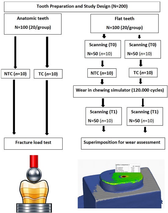
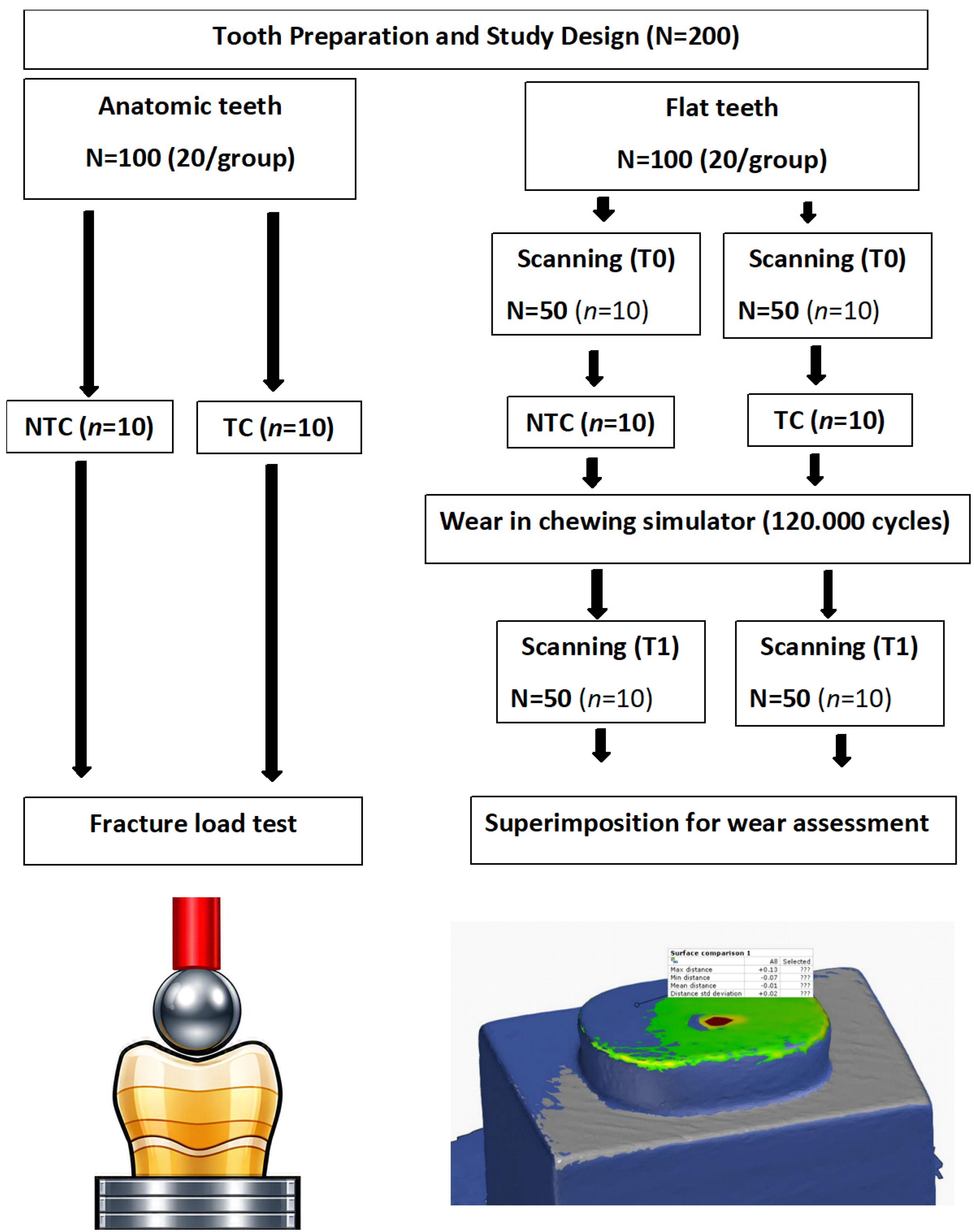
A total of 200 denture teeth were required for this study: 40 prefabricated (PF) PMMA acrylic teeth and 160 3D-printed teeth, allocated according to the tested property and specimen design (anatomic and flat configurations). The study specimens were divided into two main categories according to the outcome evaluated: anatomic denture teeth for fracture load testing and flattened denture teeth for wear analysis (Figure 1). For anatomic teeth, half the teeth (N = 50, 10/resin tooth material) were directly subjected to fracture load testing, whereas the other half underwent thermal cycling before testing. The flattened teeth were initially scanned to establish the baseline surface morphology (T0). Half the specimens (N = 50, 10/resin tooth material) were subjected directly to the standardized chewing simulation protocol, while the other half underwent thermal cycling before receiving the same chewing simulation procedure. After completing their respective aging and wear protocols, all flattened teeth were scanned again (T1) to quantify the resulting wear. Digital scans obtained at two time points (T0 and T1) were superimposed and analyzed to quantify wear-related changes [30].
Figure 1.
Study design illustrating specimen grouping and allocation, including the aging procedures applied for each test. TC, thermal cycling; NTC, no thermal cycling.
2.3. Sample Size Calculation
Sample size for this study was determined a priori using the mean differences and standard deviations reported in a previous investigation on the wear behavior and fracture resistance of 3D-printed denture teeth [9]. These values were entered into GPower (version 3.1, Universität Düsseldorf, Düsseldorf, Germany) with a two-tailed test, an alpha level of 0.05, and 80% power, yielding a requirement of n = 10 specimens per group at a 95% confidence level. While the prior study employed a two-group comparison, the present experiment adopted a 5-material × 2-aging-condition factorial design, resulting in ten experimental groups. The requirement of 10 specimens per group was therefore applied to each cell of the factorial structure, providing adequate power to detect main effects, whereas interaction effects were not specifically powered. The chosen sample size was based on prior mean differences and standard deviations for wear and fracture load outcomes, with effect-size assumptions specified using Cohen’s f.
2.4. Tooth Resins Used, Specimen Grouping, and Tooth Printing
Forty PF mandibular molar anatomic teeth were selected (Major Dent-V, MAJOR Prodotti Dentari S.P.A., Moncalieri, Italy), fabricated from high cross-linked polymethyl methacrylate (PMMA). Twenty teeth remained without any modifications for fracture load testing. The remaining 20 teeth were flattened using a customized putty jig, confirming standardization of the tooth flattening process. One anatomic and one flattened tooth were scanned, creating a Standard Tessellation Language (STL) file. The STL file was imported into the printing system for slicing and the designing of layer thickness, printing orientations, and support structures and positions. The 3D-printed resins and printers used in the present study, in addition to the printing parameters, post-curing cleaning protocol, and post-curing conditions, are detailed and summarized in Table 1. After complete polymerization, all support structures were removed and finished and then stored in distilled water until the aging and testing procedures.
The classification of VarseoSmile, CrownTec, and Flexcera Smile as the ceramic-reinforced hybrid (CRH) materials is based on the best manufacturer-supported information currently available. Although companies differ in the level of compositional detail they disclose, all three products are identified in technical datasheets, safety datasheets, or regulatory documentation [31,32,33,34], with additional evidence from published studies detailing filler type and concentration [13,14,35]. These sources characterize the materials as hybrid photopolymers containing ceramic or nanoceramic fillers. For some materials, manufacturers restrict access to exact filler percentages or detailed chemical composition; therefore, only verified descriptors are reported in Table 1. This classification reflects the most transparent and evidence-based grouping possible and provides a consistent framework for interpreting the mechanical behavior of these materials while acknowledging that compositional information is not equally available across all products.
Table 1.
Materials used and specific printing parameters.
2.5. Aging Procedures
As shown in Figure 1, the teeth allocated for thermal aging were placed in a thermal cycling machine (SD Mechatronik Thermocycler, Feldkirchen-Westerham, Germany) and subjected to 10,000 thermal cycles (5 °C to 55 °C with a 30 s dwell time) corresponding to approximately one year of simulated clinical service under the present in vitro conditions [36]. For specimens aged for the wear assessment, a chewing simulator (CS-4.2, SD Mechatronik GMBH, Feldkirchen, Germany) was used, as detailed in a previous study [26]. Specimens were subjected to a 49 N load in a chewing simulator programmed to deliver a 2 mm vertical descent and a 0.5 mm horizontal antagonist displacement at 1.53 Hz, using a Ø6 mm steatite (standardized ceramic ball) antagonist in accordance with International Organization for Standardization Technical Specification (ISO/TS) 14569-2 [37]. The antagonist moved horizontally back and forth over a 2 mm path at a frequency of 1.6 Hz. Each specimen underwent 120,000 cycles in distilled water [9,38], which is equivalent to approximately six months of simulated clinical service under the present in vitro conditions. Under these chewing simulation conditions, specimens were subjected to a cyclic 49 N vertical load combined with horizontal antagonist movement, with the latter exerting the dominant influence on wear generation [37].
2.6. Specimen Testing
Wear resistance was evaluated using a two-stage scanning protocol [30] designed to isolate the effect of thermal cycling on surface loss according to the detailed study design (Figure 1). At baseline, all flattened specimens were scanned using a desktop scanner (E4 scanner; 3Shape A/S, Copenhagen, Denmark) to obtain reference STL files (T0). Half the specimens were then subjected directly to the chewing simulation phase under standardized load and antagonist conditions, whereas the other half underwent thermal cycling followed by the same chewing simulation protocol. After completion of the respective procedures, a second scan was performed (T1) to quantify the resulting wear. This approach was used to identify the specific thermal effect on denture tooth wear.
After aging, the STL files from the time points (T0, T1) were imported into Geomagic Control X (3D Systems, Inc., Rock Hill, SC, USA) for wear evaluation. The “Reference Scan” and “Test Scan” were designated as reference and measured data, respectively. The flat, unworn surface of the reference scan was segmented and selected as the comparison area. Initial alignment and best-fit alignment procedures were performed, and multiple cross-sectional views were generated to verify proper superimposition [9]. A 3D comparison was performed in Geomagic Control X using a color-map tolerance of 50 µm and a range of 100 µm, where blue shading indicated areas of material loss. The software generated a quantitative report, and the negative average value was recorded as the wear depth for each specimen. For each specimen, the comparison area was standardized. Wear depth was defined as the negative average deviation, expressed in µm, representing the mean linear surface loss over the worn area [9,39]. This parameter was selected as the statistical outcome, providing a stable and representative measure of overall wear.
For fracture load testing, a universal testing machine (Instron Model 8871, Instron Corp., Norwood, MA, USA) was used. Non-aged and aged anatomic teeth were mounted on the Instron testing machine with the occlusal surface oriented upward. Each specimen was securely positioned so that the applied load contacted the central groove and engaged the cusp slopes (Figure 1). A stainless-steel ball indenter (7-mm radius) was used to deliver force at a crosshead speed of 1 mm/min until failure occurred, and the maximum load at fracture (N) was recorded [9]. To minimize localized contact damage and ensure uniform load distribution, a 1.5 mm rubber sheet was placed between the occlusal surface and the indenter during testing [26,40].
2.7. Statistical Analysis
Data normality was verified using the Shapiro–Wilk test, and nonsignificant outcomes confirmed a normal distribution for all variables. Accordingly, parametric analyses were performed. The effects of material type, thermal cycling, and their interaction were assessed using two-way ANOVA, followed by Tukey’s post hoc test for pairwise comparisons. Within-material comparisons between the NTC and TC groups were derived as formal simple-effects analyses from the ANOVA model, with a multiplicity-control procedure applied. Effect sizes were reported as partial η2, and group means were presented with 95% confidence intervals to indicate estimate precision. All statistical procedures were conducted in SPSS Statistics version 29 (IBM Corp., Armonk, NY, USA), with the significance level set at p < 0.05.
3. Results
3.1. Wear Resistance Results
The two-way ANOVA (Table 2) showed a significant main effect of material type on wear resistance (F = 698.258, df = 4, p < 0.001, η2 = 0.811), indicating substantial differences in wear depth among the five resin materials. Thermal cycling also demonstrated a significant main effect (F = 385.684, df = 1, p < 0.001, η2 = 0.969), confirming that aging increased wear across all groups. The material × thermal cycling interaction was significant (F = 4.094, df = 4, p = 0.004, η2 = 0.154), demonstrating that the influence of thermal cycling on wear depth varied depending on the resin type. Group means are reported with 95% confidence intervals to indicate estimate precision.
Table 2.
Two-way ANOVA for wear depth (µm) analysis.
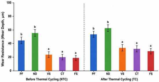
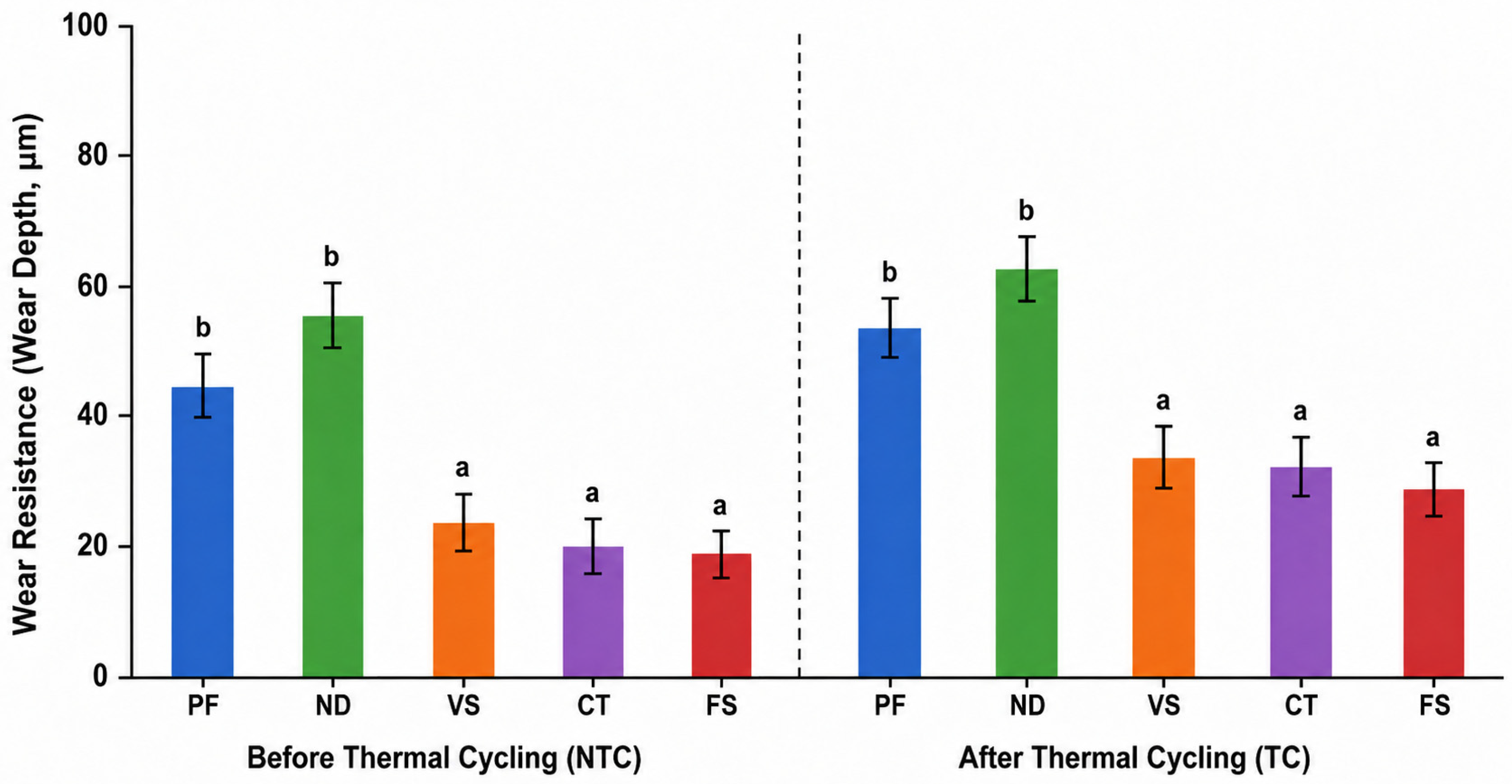
Wear-resistance outcomes (wear depth) for all materials are summarized in Table 3 and Figure 2. Before thermal cycling, clear differences were observed among the tested resin tooth materials. The CRH resin tooth materials (VS, CT, and FS) exhibited the lowest wear depth, indicating superior wear resistance, with no significant differences among them. PF showed intermediate wear resistance, whereas ND demonstrated the highest wear depth, reflecting the lowest baseline performance. After thermal cycling, the same performance pattern persisted; however, the magnitude of wear increase differed among materials, consistent with significant interaction. All materials exhibited higher wear depth after aging, but the CRH resin tooth materials (VS, CT, FS) maintained significantly better wear resistance than PF and ND. ND continued to show the greatest wear after thermal cycling, confirming its inferior stability under thermal stress.
Table 3.
Wear resistance (wear depth, µm) of printed and prefabricated resin tooth material.
Figure 2.
Wear depth of prefabricated PMMA teeth and printed resin tooth material before and after thermal cycling. Different lowercase letters indicate statistically significant pairwise differences, whereas the same letters indicate nonsignificant differences (p > 0.05).
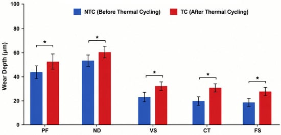
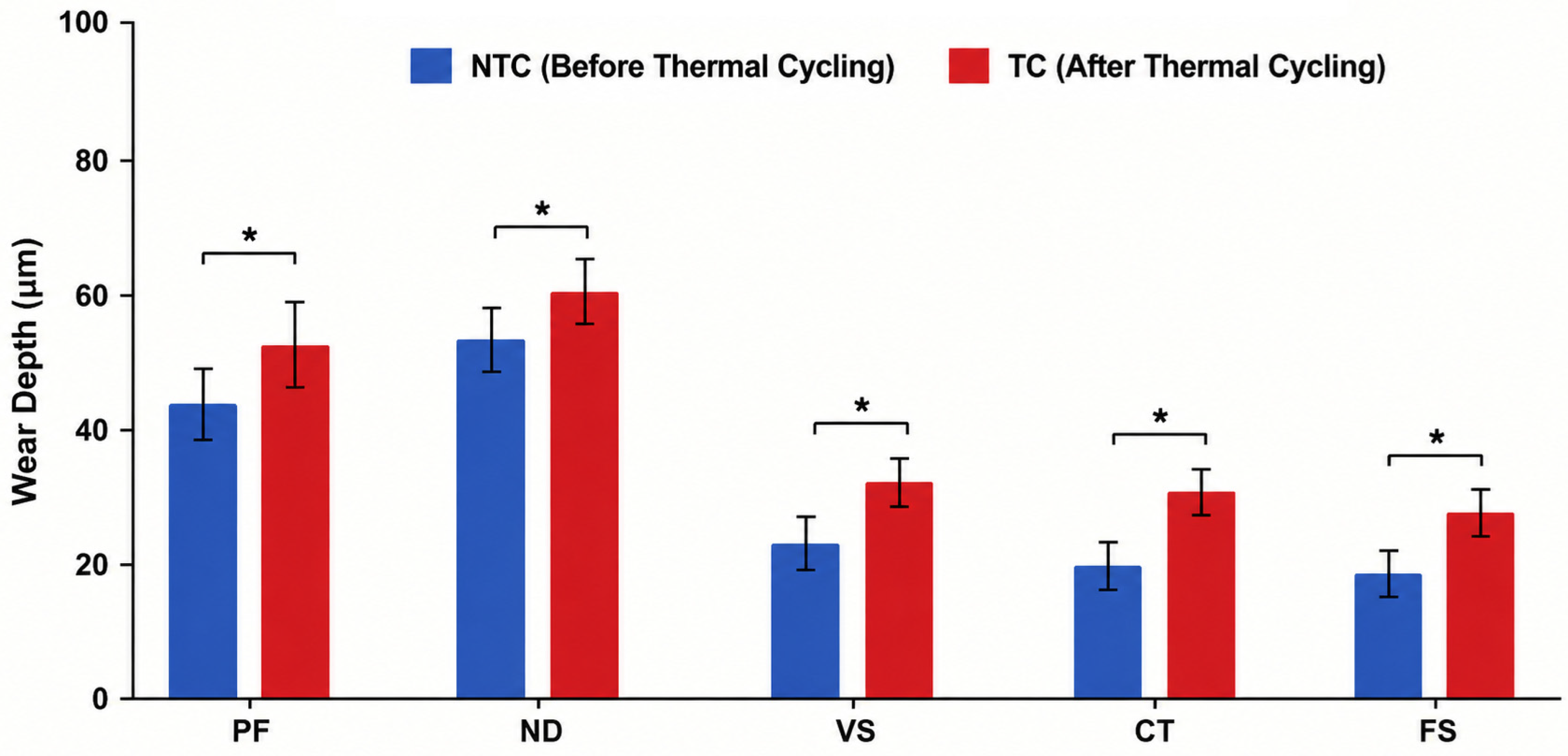
Simple-effects comparisons (Figure 3) demonstrated that thermal cycling significantly increased wear depth within each material group (PF: p = 0.004; ND: p = 0.009; VS: p= 0.016; CT: p = 0.006; FS: p = 0.032). Despite this decline, the CRH resin tooth materials remained the best-performing group overall.
Figure 3.
Thermal cycling effect on wear resistance (wear depth, µm) per materials. * p-value < 0.05 indicates statistical significance.
3.2. Fracture Load Results
The two-way ANOVA (Table 4) demonstrated a significant main effect of thermal cycling on fracture load (F = 1319.455, df = 1, p < 0.001, η2 = 0.936), indicating that aging reduced fracture resistance across all materials. A significant main effect of resin type was also observed (F = 1692.446, df = 4, p < 0.001, η2 = 0.987), confirming substantial differences in fracture load performance among the five tested materials. In addition, the thermal cycling × resin interaction was significant (F = 30.664, df = 4, p < 0.001, η2 = 0.577), showing that the extent to which thermal cycling affected fracture load varied according to material composition. Group means are reported with 95% confidence intervals to indicate the estimated precision.
Table 4.
Two-way ANOVA for fracture load (N) analysis.
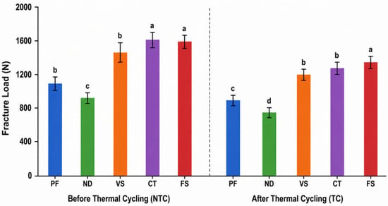
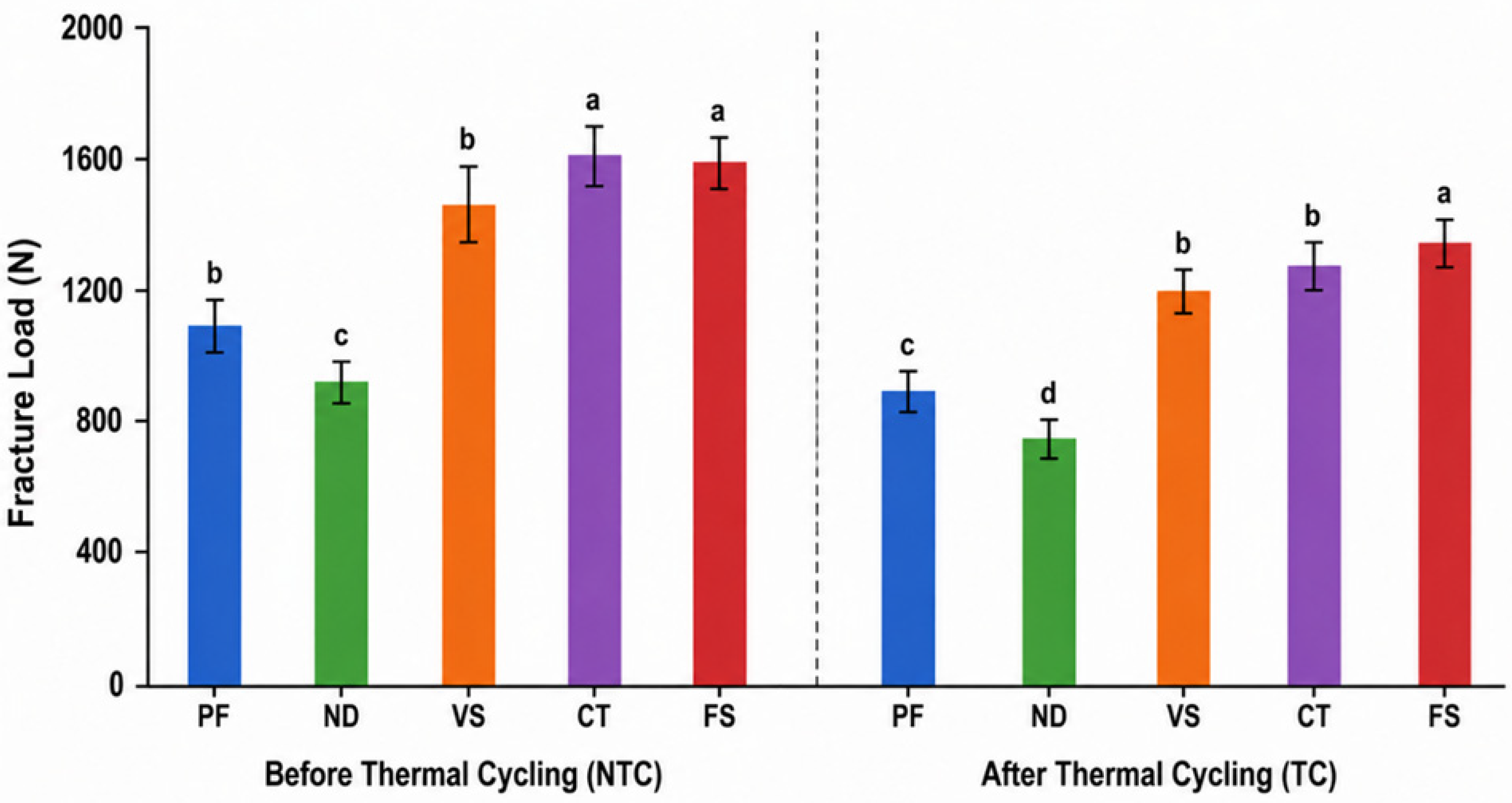
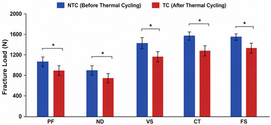
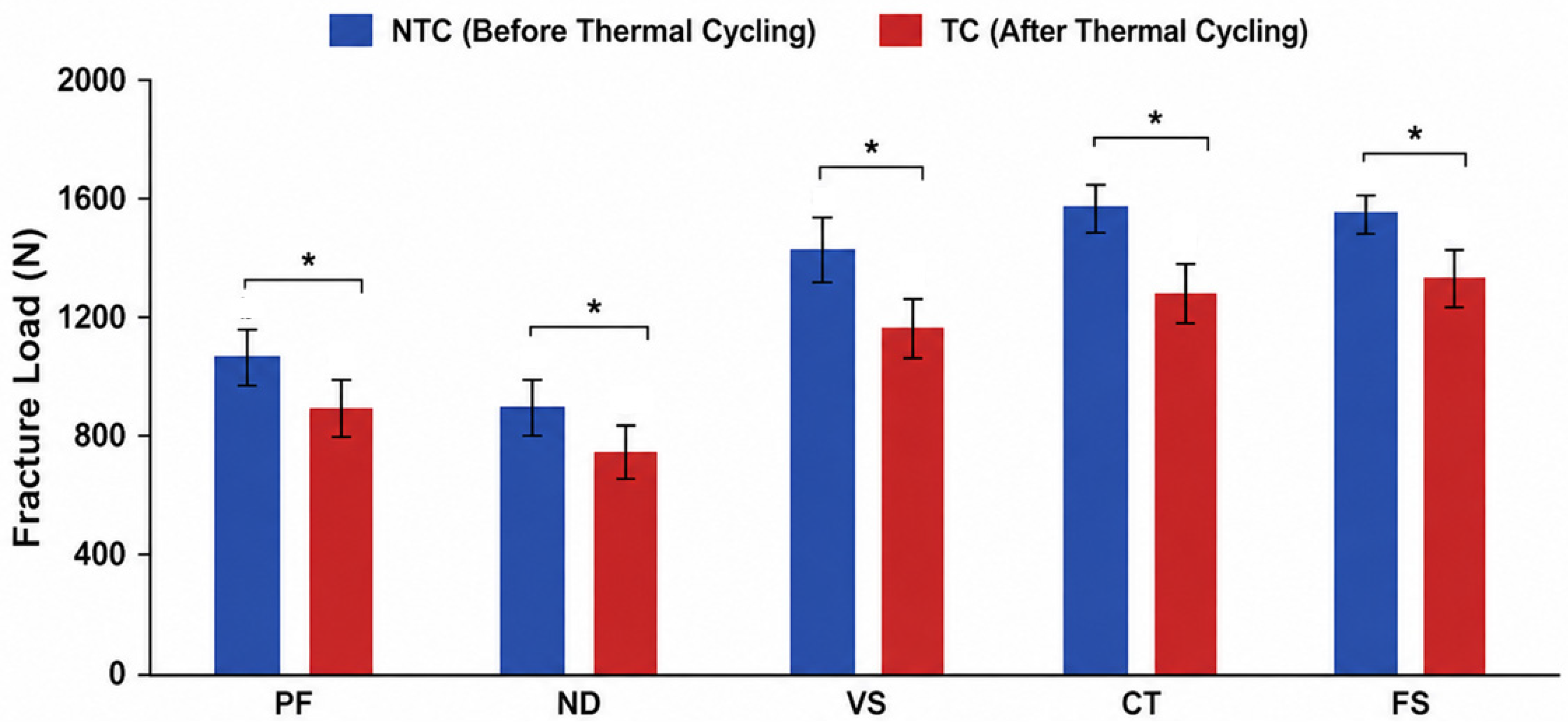
Fracture load outcomes for all materials are presented in Table 5 and Figure 4. Before thermal cycling, the CRH resin tooth materials (VS, CT, FS) exhibited the highest fracture load values, with CT showing the greatest resistance, followed by FS and VS. PF demonstrated moderate strength, whereas ND showed the lowest fracture load, indicating inferior structural performance prior to aging. Following thermal cycling, fracture load values decreased across all materials, but the magnitude of reduction varied among groups, consistent with the significant interaction. The CRH resin tooth materials (VS, CT, FS) maintained the highest fracture load values after aging and remained statistically comparable to one another (p > 0.05). PF exhibited a marked reduction, while ND continued to show the lowest fracture load compared with all other materials (p < 0.001).
Table 5.
Fracture load (N) of prefabricated PMMA teeth and printed resin tooth material.
Figure 4.
Fracture load (N) of prefabricated PMMA teeth and printed resin tooth material before and after thermal cycling. Different lowercase letters indicate statistically significant pairwise differences, whereas the same letters indicate nonsignificant differences (p > 0.05).
Simple-effects comparisons (Figure 5) demonstrated that thermal cycling produced a significant decline in fracture load within each material group (PF: p < 0.001; ND: p = 0.001; VS: p = 0.009; CT: p = 0.017; FS: p = 0.003). Despite this reduction, the CRH resin tooth materials remained the best-performing group overall.
Figure 5.
Thermal cycling effect on fracture load (N) per materials. * p-value < 0.05 indicates statistical significance.
4. Discussion
This study aimed to investigate and compare the mechanical performances of permanent CRH 3D-printed resin teeth before and after thermal cycling in comparison with prefabricated PMMA teeth and ND 3D-printed resins. Based on the study findings, there are clear differences in wear resistance and fracture load before and after thermal cycling. The permanent CRH resin teeth (VS, CT, FS) exhibited the lowest wear depth and the highest fracture load values, significantly outperforming the prefabricated and conventional 3D-printed resin under all conditions. Thermal cycling significantly increased wear and reduced fracture load for all tested resin tooth materials; however, the CRH resin tooth materials remained the least affected and preserved the highest overall mechanical performance. These findings reject the null hypothesis and show that material type and thermal aging significantly affect wear resistance and fracture load.
The present study was undertaken to evaluate the mechanical performance of newly introduced CRH 3D-printed resin tooth materials, as their strength and durability have not yet been adequately investigated. Most existing research on 3D-printed denture teeth has focused on provisional resins, which historically represented the only printable options available [27,41]. Consequently, the literature largely documents the mechanical limitations of these temporary materials, including reduced wear resistance, lower fracture strength, and susceptibility to thermal degradation [9,41]. With the recent development of permanent, reinforced 3D-printed denture tooth resins incorporating improved filler technology and enhanced cross-linking [8], there is a clear need to determine whether these modern materials can match or exceed the performance of prefabricated denture teeth. Addressing this gap, the current study provides one of the first comparative assessments of these CRH 3D-printed resin tooth materials [20].
In this study, a steatite antagonist was selected to simulate enamel-like occlusal contact under controlled laboratory conditions, as steatite provides a standardized and reproducible surface with mechanical properties comparable to natural enamel. Steatite produces a moderate, physiologically relevant abrasive pattern that allows meaningful comparison between denture tooth materials [42]. Its uniform composition, hardness, and surface finish minimize variability and ensure consistent contact with mechanics across specimens, thereby improving the reliability of wear measurements [28,42].
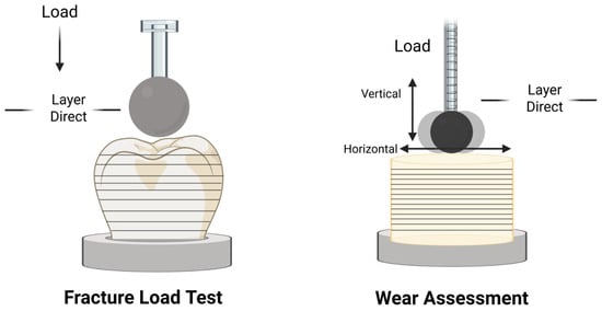
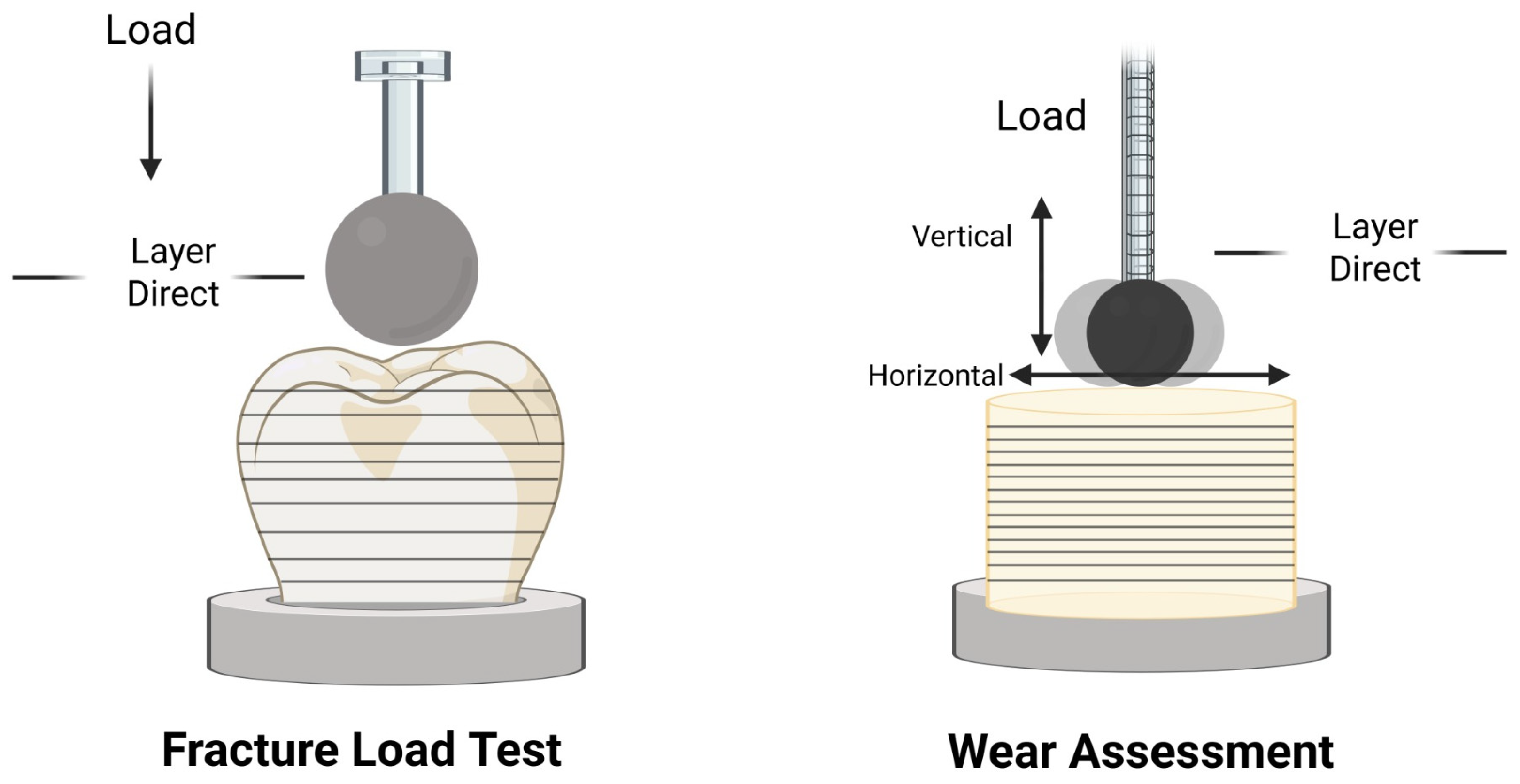
CRH 3D-printed resin tooth materials demonstrated markedly superior wear resistance, exhibiting the lowest wear depth both before and after thermal cycling. This reflects the improved material construction of new permanent printable resins, which incorporate higher cross-linking density and ceramic fillers [28]. Based on the literature, the densely cross-linked polymer network reduces chain mobility and limits surface softening under mechanical load. While the ceramic fillers stabilize the surface against micro-cutting and micro-fatigue mechanisms [10,11,43], these features produce harder, more dimensionally stable material capable of maintaining structural integrity under repeated occlusal contact [43]. In this study, all teeth were printed at a 0-degree orientation, positioning the layer planes parallel to the build platform and aligning the applied wear load parallel to these layers. With respect to cyclic wear loading, the protocol combined vertical and horizontal movements (Figure 6), with the horizontal component being predominant; therefore, the wear load acted mainly parallel to the printed layer structure. The statistical outcome was expressed as wear depth (negative average deviation). This configuration minimizes interlayer shear and promotes more uniform distribution of the applied forces across the printed structure [2]. Moreover, the presence of ceramic fillers within and between layers enhances abrasive resistance, as the fillers act as hard micro-reinforcements that limit polymer chain mobility and reduce surface degradation under repeated cycles [27].
Figure 6.
Schematic illustration of the layer orientation in relation to the direction of the applied load during the fracture load and wear resistance tests.
The enhanced wear behavior of the CRH 3D-printed resin tooth materials used in the present study is in agreement with a previous study that compared different 3D-printed resin teeth and reported the superior performance of these materials, comparable to CAD-CAM milled denture teeth [8]. However, this is in disagreement with a previous study [38] that evaluated provisional 3D-printed resins and reported superior wear resistance to prefabricated and milled teeth under simulated chewing. Rungsitsathien et al. [38] further noted that applying a 50 µm surface glaze did not significantly improve the wear resistance of 3D-printed resin tooth material. These discrepancies may be attributed to differences in experimental methodology, measurement approaches, and antagonist selection, particularly as Rungsitsathien et al. employed a self-antagonist design [38].
The 3D-printed resin (ND) exhibited the highest wear depth under all testing conditions, indicating comparatively low performance in the present in vitro evaluation. The increased wear depth is attributed to the material composition and absences of reinforcing fillers like CRH resin tooth material. This inferior performance is consistent with earlier investigations of provisional printable resins, which repeatedly reported accelerated wear, reduced hardness, and greater susceptibility to hydrothermal degradation due to their lower cross-linking density and absence of reinforcing fillers [43]. Additionally, the photopolymer resins were originally formulated for short-term use, and their softer polymer matrices undergo rapid surface breakdown when subjected to functional loading and thermal fluctuations [44].
Thermal cycling increased in wear depth across all materials, confirming that simulated intraoral aging compromises surface durability. The increase in wear after thermal aging may be attributed to water sorption, which acts as a plasticizer within the polymer matrix [40]. Additionally, the change temperatures accelerate the uptake of water, intensifying its softening effect on the resin network, as reported in the literature on PMMA and early 3D-printed resins [9]. This is consistent with Srinivasan et al., who reported that conventional PMMA and printable resins exhibit pronounced degradation after aging, resulting in decreased hardness and increased abrasion [15]. Despite these hydrothermal effects, CRH 3D-printed resin tooth materials showed the smallest increase in wear, reflecting their enhanced cross-linked polymer and ceramic filler reinforcement, which together limit hydrothermal softening and stabilize the surface under changing temperatures [17]. This superior performance after aging underscores the enhanced structural characteristics of CRH materials and reflects their favorable wear and fracture load behavior compared with prefabricated PMMA teeth and conventional 3D-printed resins under the present in vitro conditions.
The fracture load of 3D-printed denture teeth is strongly influenced by printing orientation, as the direction of the printed layers determines whether the applied load interacts directly with the printed layers or across the interlayer interfaces. This relationship reflects the inherent anisotropy of additively manufactured polymers, where mechanical performance varies according to how the load is positioned relative to the layer architecture created during printing [27]. The denture teeth in this study were printed at a 0-degree printing orientation, meaning the layers were printed horizontally. During fracture testing, the applied load was directed vertically onto the occlusal surface, positioning the force perpendicular to the printed layer arrangement (Figure 6). This configuration closely simulates clinical loading conditions and allows evaluation of how the layer structure responds to compressive stress [2].
CRH 3D-printed denture tooth resins (VS, CT, FS) demonstrated the highest fracture load values when compared with both prefabricated PMMA teeth and ND printed materials. This increase in fracture load might be attributed to the material composition and ceramic filler content [8]. The cross-linked polymer network in CRH resin tooth materials increases the rigidity of the polymer matrix by restricting molecular chain mobility, which enhances material stiffness and reduces susceptibility to surface softening under occlusal loading. This dense network improves structural cohesion and allows the material to better withstand compressive and shear stresses compared with conventional photopolymer resins [45]. Moreover, the presence of ceramic and hybrid fillers improves resistance to fatigue-related damage by acting as hard microstructural barriers that limit crack initiation and propagation under applied load [46]. The findings of the present study are consistent with those reported by Alqahtani et al. [45], who demonstrated that the indirect tensile fracture values of 3D-printed denture teeth were higher than those of prefabricated PMMA teeth. The findings of the present study differ from those reported by Al-Fodeh et al. [2], who reported a comparable fracture strength between prefabricated PMMA teeth and 3D-printed resin tooth material before aging. This variation may be attributed to differences in the resin formulations as well as the specific fracture-testing methodology employed in each study.
The 3D-printed resin (ND) consistently exhibited the lowest fracture load values, confirming its limited structural integrity and reinforcing its role as a provisional rather than definitive restorative material. This inferior performance is largely attributable to its less cross-linked polymer network and absence of filler reinforcement, which reduces resistance to crack initiation and propagation under occlusal loading [15,40]. These factors emphasized that conventional photopolymer resins were initially formulated for short-term interim use, and their softer matrices undergo rapid mechanical fatigue compared with prefabricated PMMA teeth. Previous investigations of conventional printable resins have reported similar findings, documenting markedly low fracture strength [15,40]. By contrast, Gad et al. [9] compared the fracture resistance of conventional 3D-printed resins and reported the high fracture resistance of 3D-printed resin compared with prefabricated PMMA before thermal aging. The variation might be attributed to the aging protocol and testing procedures and conditions.
The fracture load of all resins decreased significantly after thermal aging. This decrease is mainly attributed to water sorption during thermal cycling, as discussed earlier. This increased water sorption resulted in plasticization of the polymer matrix and the development of microcracks that weaken the internal structure under repeated thermal stress [2,9,47]. Previous studies evaluating conventional PMMA teeth and 3D-printed resin teeth have similarly reported substantial decreases in fracture load following thermal cycling [2,9]. The present findings therefore align closely with the previous literature, confirming that thermal cycling is a critical aging factor that adversely affects the fracture behavior of denture tooth materials [47].
Despite the negative effect of thermal aging on all materials, CRH resin tooth materials showed the highest fracture load compared with prefabricated PMMA teeth and conventional ND 3D-printed teeth. This improvement is largely attributed to the presence of ceramic fillers, which decrease water sorption and limit hydrothermal softening of the polymer matrix, as reported in previous investigations [48]. By reducing moisture uptake, these fillers minimize plasticization and microcrack formation during thermal cycling, allowing the material to retain higher stiffness and fracture load values [49]. Consequently, the reinforced polymer network maintains its integrity under fluctuating temperature conditions, explaining the enhanced performance of fracture load after thermal cycling. On the other hand, the 3D-printed resin (ND) showed the greatest proportional reduction in fracture resistance after thermal cycling, indicating its limited structural stability under hydrothermal conditions. This decline is primarily related to its lower cross-linking density and the absence of ceramic filler reinforcement.
The present findings have important clinical implications for selecting denture tooth materials in definitive prosthodontic treatment. The ceramic-reinforced hybrid (CRH) resin tooth materials (VS, CT, FS) demonstrated consistently superior mechanical performance, combining low wear depth with high fracture load values both before and after thermal cycling. Their stability under simulated intraoral conditions indicates that these printable materials can maintain performance under the tested in vitro loading conditions. Although the ceramic-reinforced hybrid materials exhibited superior wear resistance and fracture load performance under the present experimental conditions, these findings should not be extrapolated to long-term clinical behavior. The thermal cycling protocol, limited chewing simulation, and static fracture testing used in this study provide controlled laboratory comparisons but do not replicate the multifactorial nature of intraoral aging, including cyclic fatigue, enzymatic degradation, pH fluctuations, and patient-specific occlusal dynamics. Therefore, the present results should be interpreted as in vitro mechanical performance rather than evidence of long-term clinical reliability. Confirmation of clinical durability requires fatigue-based investigations and well-designed clinical studies that evaluate performance under real functional loading over time.
At the same time, the current literature shows that the mechanical behavior of 3D-printed dental materials is influenced not only by intrinsic resin composition but also by printing and post-processing parameters, including printer type, light wavelength, exposure time, layer thickness, build orientation, and curing intensity. Accordingly, the improved performance observed for CRH materials in this study may reflect the combined contribution of ceramic filler content, the enhanced cross-linking, and the material-specific manufacturing workflow rather than filler composition alone. These mechanisms, although supported by previous studies, were not directly investigated here and should be interpreted as literature-based hypotheses rather than experimentally verified causal explanations. Overall, these results indicate that CRH resin tooth materials had superior wear resistance and fracture strength to prefabricated PMMA teeth and conventional 3D-printed resins under the present in vitro conditions. However, these findings should not be extrapolated to claims of long-term clinical durability or suitability.
The present in vitro study protocols provide controlled and comparable mechanical data; however, they cannot fully replicate the complexity of long-term intraoral function. The chewing simulation applied standardized vertical loading under controlled conditions, which does not reproduce the wide variability in patient-specific occlusal patterns, lateral excursions, and parafunctional habits or the combined chemical and mechanical degradation occurring clinically. Likewise, the fracture load experiment assessed static loading to catastrophic failure, offering insight into ultimate strength but not into fatigue resistance or crack propagation under repeated cyclic loading. These limitations are inherent to in vitro methodologies and must be considered when interpreting the clinical relevance of the findings; therefore, the results should be viewed as an evaluation of comparative mechanical performance under controlled laboratory conditions rather than direct predictors of long-term intraoral behavior. Additional limitations relate to the experimental design. The loading protocols represent simplified chewing conditions and do not incorporate parafunctional movements or long-term cyclic fatigue. Another important limitation is that material composition could not be isolated from printer-specific parameters and post-curing protocols, meaning the observed differences may reflect the combined effects of resin formulation and manufacturing workflow. Future studies should standardize printing conditions to determine the independent contribution of each factor.
Furthermore, only a limited number of materials were tested, restricting the generalizability of the findings. The fracture load test employed a steatite antagonist due to its enamel-like mechanical behavior, providing a standardized and clinically relevant opposing surface. However, this choice limits applicability to situations involving natural opposing teeth. Clinical scenarios often involve antagonists made of ceramics, composites, or metal alloys, each with distinct hardness, surface roughness, and contact mechanics. As a result, the present findings may not fully represent the range of restorative materials encountered clinically. Future research should incorporate long-term fatigue loading, chemical aging protocols, multi-antagonist wear simulations, and detailed microstructural characterization. In addition, clinical trials are required to validate these findings under real functional conditions, including patient-specific occlusal variability, hygiene behaviors, and prosthesis maintenance patterns.
5. Conclusions
Within the limitations of this study, it could be concluded that
- Ceramic-reinforced hybrid materials demonstrated significantly higher wear resistance than conventional 3D-printed materials and prefabricated PMMA resin tooth materials under the experimental conditions.
- Wear depth remained low for ceramic-reinforced hybrid materials both before and after thermal cycling, indicating favorable wear behavior in this laboratory setting.
- Fracture load values were highest in ceramic-reinforced hybrid materials, exceeding those of prefabricated PMMA and conventional 3D-printed resin tooth materials prior to and following thermal cycling.
- Thermal cycling significantly reduced wear resistance and fracture load across all tested materials; however, ceramic-reinforced hybrid materials were least affected and maintained the most favorable mechanical performance within the limits of the applied in vitro protocols.
- The findings indicate that ceramic-reinforced hybrid materials performed better under the specific in vitro conditions used in this study. Confirmation of long-term clinical performance requires fatigue testing and clinical investigations.
Author Contributions
Conceptualization, M.M.G. and A.A.M.; methodology, M.M.G.; software, E.A.N.; validation, A.R., H.k.A. and S.A.; formal analysis, A.A.B. and A.F.E.; investigation, A.A.M., E.A.N. and A.E.N.; resources, A.A.M. and A.E.N.; data curation, M.M.G. and A.F.E.; writing—original draft preparation, M.M.G., A.A.B., H.k.A., A.R., S.A. and A.F.E.; writing—review and editing, E.A.N., M.M.G., A.A.M., A.R., H.k.A., A.R., A.A.B., S.A., A.E.N. and A.F.E.; visualization, A.F.E.; supervision, M.M.G.; project administration, S.A.; funding acquisition, A.R. and A.E.N. All authors have read and agreed to the published version of the manuscript.
Funding
This research received no external funding.
Institutional Review Board Statement
Not applicable.
Informed Consent Statement
Not applicable.
Data Availability Statement
Data is contained within the article.
Conflicts of Interest
The authors declare no conflicts of interest.
References
- Zafar, M.S. Prosthodontic Applications of Polymethyl Methacrylate (PMMA): An Update. Polymers 2020, 12, 2299. [Google Scholar] [CrossRef] [Scilit]
- Al-Fodeh, R.S.; Al-Dwairi, Z.N.; Almasri, M.; Baba, N. Z Mechanical properties of 3D-printed resin denture teeth: An in vitro study. J. Prosthodont 2024, 35, 1273–1280. [Google Scholar] [CrossRef] [Scilit]
- Kamonwanon, P.; Yodmongkol, S.; Chantarachindawong, R.; Thaweeboon, S.; Thaweeboon, B.; Srikhirin, T. Wear resistance of a modified polymethyl methacrylate artificial tooth compared to five commercially available artificial tooth materials. J. Prosthet. Dent. 2015, 114, 286–292. [Google Scholar] [CrossRef] [Scilit]
- Ghazal, M.; Yang, B.; Ludwig, K.; Kern, M. Two-body wear of resin and ceramic denture teeth in comparison to human enamel. Dent. Mater. 2008, 24, 502–507. [Google Scholar] [CrossRef] [Scilit]
- Ramanna, P.K.; Basavaraj, G.G.; Mishra, D.; Nagabhairava, R.; Rao, A.; Jacob, S.S.; Munshi, N. Assessment of Wear Resistance of Three Commercially Available Different Denture Teeth: A Comparative Study. J. Contemp. Dent. Pract. 2025, 26, 791–794. [Google Scholar] [CrossRef] [Scilit]
- Abdulhameed, N.; Volschow, B.; Abedi, T.; Bifaretti, N.; Slipak, I.; Manibo, A.; Hussein, H. Clinical wear of different types of denture teeth after one year in service: A clinical study. J. Prosthet. Dent. 2025, 134, 1164–1170. [Google Scholar] [CrossRef] [Scilit]
- Wang, Z.; Su, Y.; Wang, J.; Liu, Y.; Xing, W. Occlusal parameters and wear of artificial teeth in complete dentures with lingualized versus bilateral balanced occlusion: A randomized clinical trial. BMC Oral Health 2024, 24, 1405. [Google Scholar] [CrossRef] [Scilit]
- Abdelfattah, M.Y.; Al Humayyani, N.; Alwthinani, F.K.; Alzahrani, A.H.; Alotaibi, A.O.; Yousef, M.; Sayed Ahmed, A.; Ali, A. In vitro evaluation of the mechanical and optical properties of 3D printed vs CAD/CAM milled denture teeth materials. Saudi Dent. J. 2024, 36, 1227–1232. [Google Scholar] [CrossRef] [Scilit]
- Gad, M.M.; Alghamdi, R.; Al-Ghamdi, R.; Al-Jefri, A.; Akhtar, S.; Khan, S.Q.; Alalawi, H.; Al-Qarni, F.D. Wear and Fracture Resistance of 3D-Printed Denture Teeth: An In Vitro Comparative Study. J. Prosthodont. 2023, 32, 170–177. [Google Scholar] [CrossRef] [Scilit]
- Aati, S.; Akram, Z.; Ngo, H.; Fawzy, A.S. Development of 3D printed resin reinforced with modified ZrO2 nanoparticles for long-term provisional dental restorations. Dent. Mater. 2021, 37, e360–e374. [Google Scholar] [CrossRef] [Scilit]
- Morad, M.S.; El-Ebiary, M.A.; Kenawy, E.R.; Elshenawy, E.A. Modification of 3D printing resin with surface treated mesoporous silica nanoparticles: Structural and mechanical characterization. BMC Oral Health 2026, 26, 719. [Google Scholar] [CrossRef] [Scilit]
- Mok, Z.H.; Proctor, G.; Thanou, M. Emerging nanomaterials for dental treatments. Emerg. Top. Life Sci. 2020, 4, 613–625. [Google Scholar] [CrossRef] [Scilit]
- Mandurino, M.; Cortili, S.; Coccoluto, L.; Greco, K.; Cantatore, G.; Gherlone, E.F.; Vichi, A.; Paolone, G. Mechanical Properties of 3D Printed vs. Subtractively Manufactured Composite Resins for Permanent Restorations: A Systematic Review. Materials 2025, 18, 985. [Google Scholar] [CrossRef] [Scilit]
- Mudhaffer, S.; Haider, J.; Silikas, N.; Satterthwaite, J. Edge strength of definitive 3D-printed restorative resin materials. Dent. Mater. 2025, 41, 745–754. [Google Scholar] [CrossRef] [Scilit]
- Srinivasan, M.; Kamnoedboon, P.; McKenna, G.; Angst, L.; Schimmel, M.; Özcan, M.; Müller, F. CAD-CAM removable complete dentures: A systematic review and meta-analysis of trueness of fit, biocompatibility, mechanical properties, surface characteristics, color stability, time-cost analysis, clinical and patient-reported outcomes. J. Dent. 2021, 113, 103777. [Google Scholar] [CrossRef] [Scilit]
- Goodacre, B.J.; Goodacre, C.J. Additive Manufacturing for Complete Denture Fabrication: A Narrative Review. J. Prosthodont. 2022, 31, 47–51. [Google Scholar] [CrossRef] [Scilit]
- Revilla-León, M.; Özcan, M. Additive Manufacturing Technologies Used for Processing Polymers: Current Status and Potential Application in Prosthetic Dentistry. J. Prosthodont. 2019, 28, 146–158. [Google Scholar] [CrossRef] [Scilit]
- Schubert, O.; Edelhoff, D.; Güth, J.F.; Schweiger, J. Advances in 3D Printing for Removable Prosthetics-Insights and Perspectives. J. Esthet. Restor. Dent. 2026, 38, 520–529. [Google Scholar] [CrossRef] [Scilit]
- Abdelnabi, M.H.; Swelem, A.A. 3D-Printed Complete Dentures: A Review of Clinical and Patient-Based Outcomes. Cureus 2024, 16, e69698. [Google Scholar] [CrossRef] [Scilit]
- Sabanik, P.; Liu, T.C.; Tabatabaeian, M.; Rocha, M.G.; Surathu, N.; Lawson, N.C. A Review of Current Systems, Materials, and Protocols for 3D-Printed Splints, Crowns, and Dentures. J. Esthet. Restor. Dent. 2026, 38, 635–651. [Google Scholar] [CrossRef] [Scilit]
- Flottes, Y.; Smail, Y.; Palomino-Durand, C.; Attal, J.P.; Ceinos, R.; François, P.; Dursun, E. Properties of 3D printed resins for definitive dental restorations: A systematic review. J. Prosthet. Dent. 2026, 135, e6–e27. [Google Scholar] [CrossRef] [Scilit]
- Alhotan, A.; Silikas, N.; Ibrahim, Y.M.; Al-Johani, H.; Abdul-Monem, M.M. Mechanical performance of high-impact 3D-printed denture base resins subjected to thermomechanical aging. Dent. Mater. 2026, in press. [Google Scholar] [CrossRef] [Scilit]
- Awad, M.M.; Fawzy, A.S. Do the Physicomechanical Properties of 3D-Printed Resin-Based Composite Materials Support Their Use for Definitive Restorations? A Narrative Review. Aust. Dent. J. 2025, 70, S67–S81. [Google Scholar] [CrossRef] [Scilit]
- Intarak, N.; Prommanee, S.; Somkana, S.; Srijunbarl, A.; Kamnoedboon, P.; Khan, A.S.; Porntaveetus, T.; Srinivasan, M. Comparative analysis of monomer elution, polymerization efficiency, mechanical properties and biocompatibility of 3D-printed provisional and permanent dental resins. J. Dent. 2026, 175, 106833. [Google Scholar] [CrossRef] [Scilit]
- Veerapeindee, P.; Rungsiyakull, P.; Jia-Mahasap, W. Wear resistance of 3D printed, milled, and prefabricated methacrylate-based resin materials: An in vitro study. J. Prosthet. Dent. 2025, 133, 904.e1–904.e8. [Google Scholar] [CrossRef] [Scilit]
- Cha, H.S.; Park, J.M.; Kim, T.H.; Lee, J.H. Wear resistance of 3D-printed denture tooth resin opposing zirconia and metal antagonists. J. Prosthet. Dent. 2020, 124, 387–394. [Google Scholar] [CrossRef] [Scilit]
- Tanthasri, T.; Jia-Mahasap, W.; Rungsiyakull, P. Wear resistance of 3D printed denture teeth: Influence of printing technology and build orientation. J. Prosthet. Dent. 2026, 135, 391.e1–391.e9. [Google Scholar] [CrossRef] [Scilit]
- Gentle, M.; Alsahafi, T.; Bashiri, R.; Abreu-Serrano, A.; Sulaiman, T.A. Wear resistance of additive-manufactured denture teeth following mastication simulation. J. Prosthet. Dent. 2026, 135, 390.e1–390.e7. [Google Scholar] [CrossRef] [Scilit]
- Pot, G.J.; Van Overschelde, P.A.; Keulemans, F.; Kleverlaan, C.J.; Tribst, J.P.M. Mechanical Properties of Additive-Manufactured Composite-Based Resins for Permanent Indirect Restorations: A Scoping Review. Materials 2024, 17, 3951. [Google Scholar] [CrossRef] [Scilit]
- Zeidan, A.A.E.; Helal, M.A. Evaluation of the effect of thermocycling on the trueness and precision of digitally fabricated complete denture bases. BMC Oral Health 2024, 24, 983. [Google Scholar] [CrossRef] [Scilit]
- NextDent. CB MFH (Micro Filled Hybrid). NextDent: Soesterberg, The Netherlands. Available online: https://nextdent.com/products/cb-mfh-micro-filled-hybrid/ (accessed on 21 September 2025).
- BEGO Bremer GmbH. VarseoSmile Crown Plus: Safety Data Sheet; BEGO Bremer GmbH: Bremen, Germany, 2025; Available online: https://usa.bego.com/wp-content/uploads/2022/08/VarseoSmile-Crown-plus_791918_USA_US_V-1.0.0_SDB.pdf (accessed on 22 September 2025).
- Saremco Dental AG. Crowntec Safety Data Sheet; Saremco Dental AG: Rebstein, Switzerland, 2025; Available online: https://docs.saremco.ch/ifu/saremco_print_crowntec/US_Version/D600248_GA_saremco-print-CROWNTEC_EN_USA_edited-06-2023_DRUCK_frei.pdf (accessed on 22 September 2025).
- Desktop Health. Flexcera Smile Ultra+ Instructions for Use; Desktop Health: Boston, MA, USA, 2025; Available online: https://knowledge.desktophealth.com/Materials-DH/Instructions-for-Use/Flexcera-Smile-Ultra-Plus/Instructions%20for%20Use%20-%20Flexcera%20Smile%20Ultra%20plus%20US%20rev.11.pdf (accessed on 22 September 2025).
- Duarte, S., Jr.; Phark, J.H. Advances in Dental Restorations: A Comprehensive Review of Machinable and 3D-Printed Ce-ramic-Reinforced Composites. J. Esthet. Restor. Dent. 2025, 37, 257–276. [Google Scholar] [CrossRef] [Scilit]
- Gale, M.S.; Darvell, B.W. Thermal cycling procedures for laboratory testing of dental restorations. J. Dent. 1999, 27, 89–99. [Google Scholar] [CrossRef] [Scilit]
- ISO/TS 14569-2; Dental Materials—Guidance on Testing of Wear—Part 2: Wear by Two- and/or Three-Body Contact. International Organization for Standardization (ISO): Geneva, Switzerland, 2001.
- Rungsitsathien, S.; Peampring, C. Wear of 3D-printed resin denture teeth under self-antagonist conditions after chewing simulation. J. Adv. Prosthodont. 2026, 18, 25–35. [Google Scholar] [CrossRef] [Scilit]
- Witecy, C.; Ganss, C.; Wöstmann, B.; Schlenz, M.B.; Schlenz, M.A. Monitoring of Erosive Tooth Wear with Intraoral Scanners In vitro. Caries Res. 2021, 55, 215–224. [Google Scholar] [CrossRef] [Scilit]
- Gad, M.M.; Alalawi, H.; Akhtar, S.; Al-Ghamdi, R.; Alghamdi, R.; Al-Jefri, A.; Al-Qarni, F.D. Strength and Wear Behavior of Three-Dimensional Printed and Prefabricated Denture Teeth: An In Vitro Comparative Analysis. Eur. J. Dent. 2023, 17, 1248–1256. [Google Scholar] [CrossRef] [Scilit]
- Elsharkawy, Y.M.M.; Mohamed, H.T.; Saeedi, T.M.A.S.; Farahat, A.M.S. Comparative in vitro study on wear resistance and colour stability of 3D-printed, milled, and conventional PMMA denture teeth. BMC Oral Health 2025, 25, 656. [Google Scholar] [CrossRef] [Scilit]
- Preis, V.; Hahnel, S.; Behr, M.; Rosentritt, M. Contact wear of artificial denture teeth. J. Prosthodont. Res. 2018, 62, 252–257. [Google Scholar] [CrossRef] [Scilit]
- Lalvay, E.J.A.; de Carvalho, F.G.; da Silva, A.R.; Morganti, F.A.C.; Collares, F.M.; Münchow, E.A. 3D-printed resins intended for long-term restoration vs. conventionally used materials: A systematic review and network meta-analysis of in vitro studies. J. Dent. 2026, 167, 106531. [Google Scholar] [CrossRef] [Scilit]
- Srinivasan, M.; Kalberer, N.; Kamnoedboon, P.; Mekki, M.; Durual, S.; Özcan, M.; Müller, F. CAD-CAM complete denture resins: An evaluation of biocompatibility, mechanical properties, and surface characteristics. J. Dent. 2021, 114, 103785. [Google Scholar] [CrossRef] [Scilit]
- Alqahtani, N.M.; Chaturvedi, S.; Tomar, S.S.; Kumari, L.; Gill, S.; Nayan, K.; Shariff, M.; Bhagat, T.V.; Addas, M.K.; Chaturvedi, M. Fracture toughness of 3D printed denture teeth. Technol. Health Care 2023, 31, 247–258. [Google Scholar] [CrossRef] [Scilit]
- Díez-Pascual, A.M. PMMA-Based Nanocomposites for Odontology Applications: A State-of-the-Art. Int. J. Mol. Sci. 2022, 23, 10288. [Google Scholar] [CrossRef] [Scilit]
- Muhammad, N.; Sarfraz, Z.; Zafar, M.S.; Liaqat, S.; Rahim, A.; Ahmad, P.; Alsubaie, A.; Almalki, A.S.A.; Khandaker, M.U. Characterization of various acrylate based artificial teeth for denture fabrication. J. Mater. Sci. Mater. Med. 2022, 33, 17. [Google Scholar] [CrossRef] [Scilit]
- Ferreira, I.; Calazans Neto, J.V.; Cândido Dos Reis, A. Artificial teeth obtained by additive manufacturing: Wear resistance aspects. A systematic review of in vitro studies. Heliyon 2024, 10, e23279. [Google Scholar] [CrossRef] [Scilit]
- Britto, V.T.; Cantelli, V.; Collares, F.M.; Bertol, C.D.; Della Bona, Á. Biomechanical properties of a 3D printing polymer for provisional restorations and artificial teeth. Dent. Mater. 2022, 38, 1956–1962. [Google Scholar] [CrossRef] [Scilit]

Disclaimer/Publisher’s Note: The statements, opinions and data contained in all publications are solely those of the individual author(s) and contributor(s) and not of MDPI and/or the editor(s). MDPI and/or the editor(s) disclaim responsibility for any injury to people or property resulting from any ideas, methods, instructions or products referred to in the content. |
© 2026 by the authors. Licensee MDPI, Basel, Switzerland. This article is an open access article distributed under the terms and conditions of the Creative Commons Attribution (CC BY) license.