


Abstract
This study investigated the impact of moderate teat-end hyperkeratosis on milk somatic cell count (SCC) and bacterial count (BC) in Holstein cows. Comparing teat-end scores (TS) 2 and 3, we found that TS3 quarters exhibited significantly higher SCC (p = 0.017) despite having shorter days in milk than TS2 quarters. While BC in foremilk (BC1) remained stable, post-milking samples (BC2) decreased during sequential sampling; however, a significant Group × Time interaction (p = 0.020) revealed a less pronounced bacterial reduction in TS3. These findings tentatively suggest that moderate hyperkeratosis (TS3) may be associated with subclinical mammary irritation and altered bacterial clearance dynamics, independent of the absolute bacterial load. Managing teat-end morphology may help optimize milk quality, even when bacterial contamination is minimal.
1. Introduction
Bovine mastitis is a major disease that causes substantial economic losses for dairy farmers, making prevention critically important. The condition of the teat end is a key determinant of udder health, serving as the primary physical barrier against intramammary infection [1]. Teat-end hyperkeratosis is commonly evaluated using the National Mastitis Council teat-end scoring system, based on the classification proposed by Mein et al. [2]. Scores are then categorized into four groups, from normal (teat-end score [TS]1) to severe hyperkeratosis (TS4) [3]. Increasing severity of teat-end hyperkeratosis has been associated with compromised integrity of the teat canal, increased susceptibility to pathogen penetration, and higher risk of mastitis [4]. Severe hyperkeratosis in particular may impair teat defense mechanisms during the post-milking period, when the teat canal is relaxed and more vulnerable to bacterial invasion [5]. Further, clinical studies have reported that severe teat-end hyperkeratosis is associated with a higher isolation rate of mastitis-causing pathogens from milk, increased milk somatic cell count (SCC) as a well-established indicator of mammary inflammation, and alterations in intramammary antimicrobial factors [6].
Although severe hyperkeratosis clearly affects milk quality and udder health, the implications of mild to moderate TS remain unclear. A systematic review indicated that moderate hyperkeratosis does not consistently correlate with indicators of milk quality, such as SCC [4]. Bacterial contamination is a primary determinant of mastitis; but the host inflammatory response (reflected by SCC) is affected not only by bacterial load, but also by structural and physiological characteristics of the teat [7]. Discrepancies between bacterial exposure and cellular immune response observed under field conditions suggest that teat-end morphology itself may modulate the local inflammatory environment [8].
Despite such insights, the understanding of the implications of intermediate TSs (particularly TS2 and TS3) remains limited. Obtaining a better grasp of how moderate teat-end hyperkeratosis affects milk bacterial load and SCC is essential for improving udder health monitoring and developing effective strategies to prevent mastitis. Accordingly, the present study compared milk bacterial load and SCC between cows with TS2 and TS3 to determine whether morphological differences at the teat-end are reflected in milk-based health indicators.
2. Materials and Methods
This study was conducted using data obtained from a previously published field trial evaluating the efficacy of an external teat sealant (ETS) in lactating dairy cows [9]. Whereas the original study investigated the preventive effects of the ETS against mastitis, the present study represents a secondary analysis with a distinct focus on the association between teat-end condition and milk-based health indicators. To completely eliminate any potential confounding variables or carry-over effects from the original sealant treatment, only quarters assigned to the non-treated negative control group in the original trial were utilized in the present analysis. Consequently, the cows and quarters evaluated in this study received no active treatment or intervention throughout the original study period, ensuring that the presented results reflect purely the physiological and morphological relationship between teat-end hyperkeratosis and milk parameters without any treatment-induced bias. The study was carried out on lactating Holstein cows from a commercial dairy herd in Kagoshima Prefecture, Japan. The herd comprised approximately 700 Holstein cows housed in a free-stall barn and fed a total mixed ration, with an average daily milk yield of approximately 40 kg per cow. Cows were milked three times daily (at 0500 h, 1200 h, and 1900 h) using an 8-cow, double-parallel milking parlor. Only clinically healthy cows with no signs of clinical mastitis at the time of sampling were included in the study.
Individual quarters were enrolled if they met all the following criteria: (1) TS of 2 or 3 based on the classification by Mein et al. [2]; (2) milk SCC < 200,000 cells/mL, measured using an ADAM™ SCC automated somatic cell counter (NanoEntek Co., Seoul, Republic of Korea) according to the instructions from the manufacturer; (3) live bacterial count <1000 colony-forming units (CFU)/mL, assessed using CompactDry™ TC (Shimadzu Diagnostics Corp., Tokyo, Japan); (4) absence of Staphylococcus aureus, as confirmed by milk culture using CompactDry™ X-SA (Shimadzu Diagnostics Corp.). Based on these screening criteria, six cows (17 quarters) were enrolled in the study. Days in milk (DIM) and parity of the enrolled cows were recorded.
Sampling was conducted during the morning (0500 h) and evening (1900 h) milking sessions, yielding a total of 4 sampling points over 2 days. Quarter milk samples were aseptically collected twice during each session in accordance with National Mastitis Council guidelines [10]. The first sample (Sample 1) was obtained from the initial foremilk prior to pre-dipping to evaluate bacterial contamination around the teat duct. After sufficient fore-stripping to check for abnormalities and stimulate milk let-down, the second sample (Sample 2) was collected immediately before attachment of the milking unit. This sequential sampling was designed to differentiate between external contamination and the internal mammary environment.
CFU values for Sample 1 and Sample 2 were determined using the spread-plate method and designated as bacterial count (BC)1 and BC2, respectively. Within 12 h of collection, 10 µL of each milk sample was plated onto AccuRate™ sheep blood agar (Shimadzu Diagnostics Corp.). Plates were incubated at 37 °C for 24 h, after which the CFU per milliliter of milk was determined. Although the lower limit of detection (LOD) for BC was 100 CFU/mL, samples with no colony formation were assigned a value of 50 CFU/mL (half the LOD) to minimize statistical bias, in accordance with the method of Hornung and Reed [11]. The SCC of Sample 2 was determined immediately after milk collection using an ADAM™ SCC automated somatic cell counter, as described above.
All statistical analyses were performed using open-source R software (ver. 4.4.2; R Foundation for Statistical Computing, Vienna, Austria). For statistical analysis, BC and SCC were transformed into common logarithms (log10) to normalize the distribution and stabilize variance. To evaluate longitudinal changes between the two groups, a linear mixed-effects model (LMM) was applied using the lme4 package. Fixed effects included group (TS2 vs. TS3), time (a 4-level factor), DIM, and the Group × Time interaction. To account for within-subject variability resulting from repeated measurements, and to address the hierarchical structure of quarters nested with cows, cow ID and quarter ID were included as random effects, and a random-intercept structure was adopted, as shown below:
Model selection was conducted using the Akaike information criterion (AIC). Several candidate models, including those with and without the group × time interaction, were compared, and the model with the lowest AIC value was selected as the best-fitting model. The assumption of the liner mixed models was thoroughly evaluated using residual diagnostics. The normality of the residuals was verified by the Shapiro–Wilk test and Q-Q plots, and the homogeneity of variance was evaluated by visuality inspecting residual plots. Furthermore, p-values for the fixed effects were estimated using Satterthwaite’s approximation for degrees of freedom via the lmerTest package in R. Statistical significance was set at p < 0.05. Descriptive statistics are presented as the mean ± standard error, unless otherwise specified.
The research protocol for this study was approved by the Okayama University of Science Experimental Animal Committee (approval no.: Jitsu 2024-072; 30 October 2024).
3. Results
3.1. Animal Characteristics and Data Overview
A total of six cows (17 quarters) met the inclusion criteria and were included in the analysis. Of the 17 quarters, 11 were classified as TS2 and 6 as TS3. Across the four sampling sessions, 44 samples from TS2 quarters and 24 samples from TS3 quarters were analyzed. No quarters showed a change in TS during the study period. Mean parity was 1.3 ± 0.5 (mean ± standard deviation [SD]) in the TS2 group and 1.0 ± 0.0 (mean ± SD) in the TS3 group, with no significant difference between the two groups (Mann–Whitney U test). By contrast, DIM was significantly higher in the TS2 group (317.8 ± 93.7 days), compared with the TS3 group (155.3 ± 41.0 days; Mann–Whitney U test, p < 0.001). The estimated marginal means (EMMeans), their 95% confidence intervals (95% CI), and the specific sample sizes (n) for each group across all sampling time points are detailed in Table 1.
Table 1.
Estimated marginal means (EMMeans) and 95% confidence intervals (CIs) for bacterial counts (log10 BC1 and BC2) and somatic cell counts (log10 SCC) across sampling time points.
3.2. Bacterial Counts (log10 BC1 and log10 BC2)
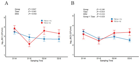
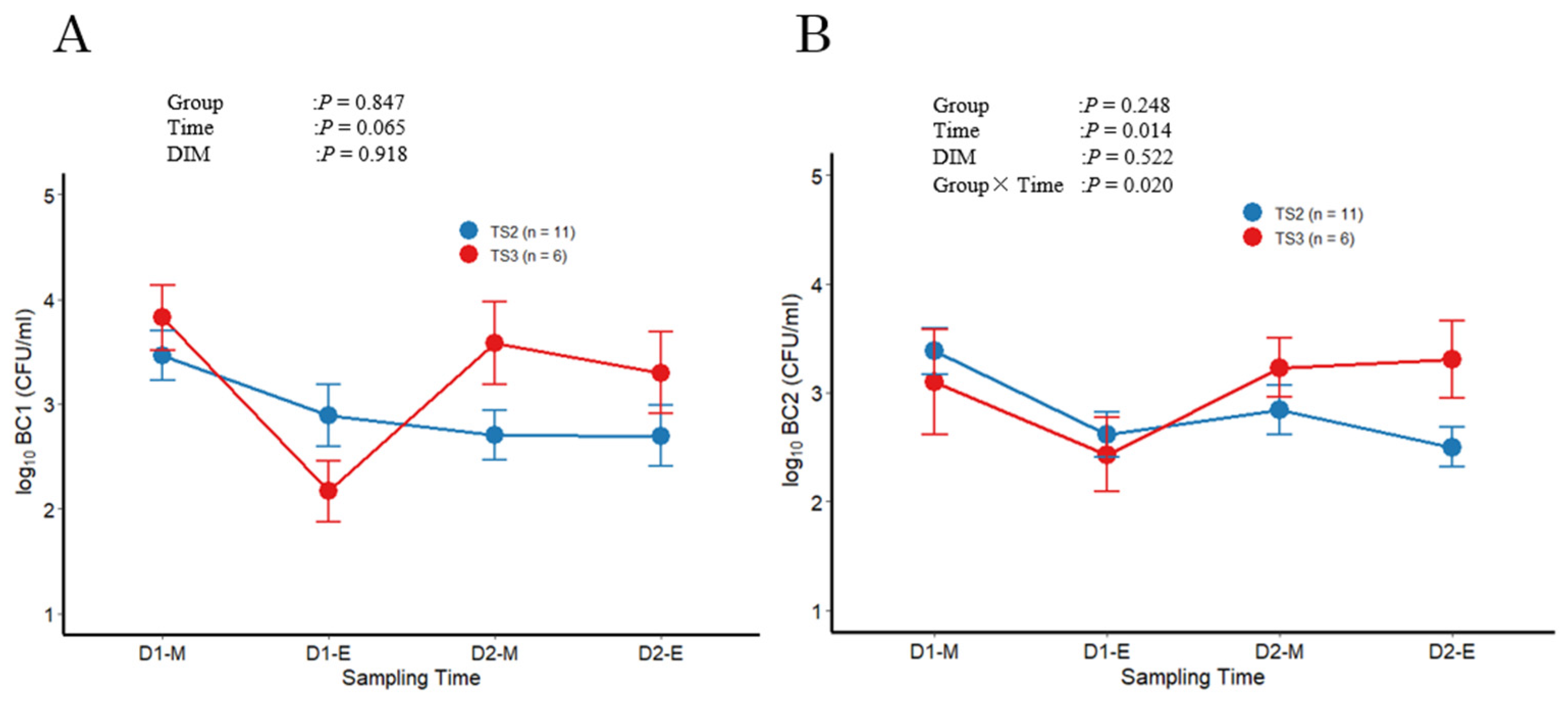
Regarding the bacterial counts in log10BC1, model selection based on the AIC favored a simple model without the Group × Time interaction. Under this best-fit model, no significant main effects were observed for either group (p = 0.847) or time (p = 0.065), indicating the baseline bacterial loads on the teat end prior to milking remained statistically stable across the groups through the sequential sampling process (Figure 1A).
Figure 1.
Change in log-transformed bacterial count (BC) in foremilk (A) and pre-milking samples (B) across sampling times in mammary quarters classified as teat-end score (TS) 2 (n = 11) or TS3 (n = 6). Total BC was determined at four sampling points (D1-M: Day 1 morning, D1-E: Day 1 evening, D2-M: Day 2 morning, D2-E: Day 2 evening). Error bars represent mean ± standard error (SE).
Analysis of log10BC2 revealed a significant main effect of time (p = 0.014) and, more importantly, a significant interaction between group and time (p = 0.020) (Figure 1B). This significant interaction indicates that the pattern of bacterial decline over time differed between the two groups. Specifically, the reduction in bacterial load during sequential sampling was less pronounced in the TS3 group compared to the TS2 group.
3.3. Somatic Cell Counts (log10 SCC)
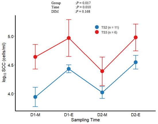
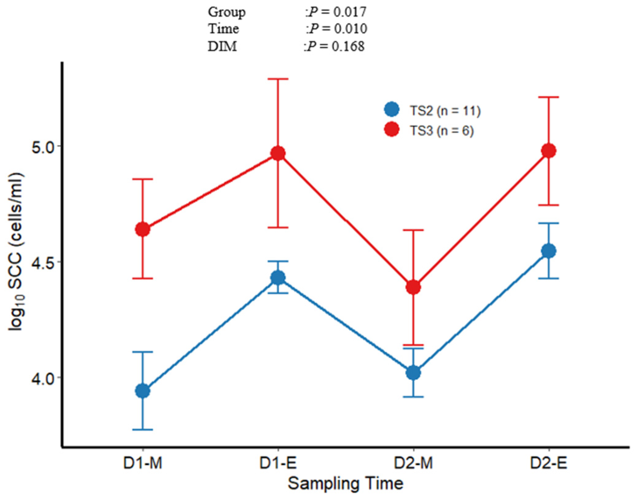
Regarding SCC, the AIC-based model comparison supported the simpler model lacking the interaction term. The final analysis was therefore conducted using this model. The TS3 group exhibited significantly higher log10SCC values compared with the TS2 group (p = 0.017) (Figure 2). In addition, log10SCC increased significantly over time (p = 0.010), whereas the effect of DIM was not significant (p = 0.168).
Figure 2.
Change in log-transformed somatic cell count (log10SCC) across sampling times in mammary quarters classified as TS2 (n = 11) or TS3 (n = 6). SCC was determined at four sampling points (D1-M: Day 1 morning, D1-E: Day 1 evening, D2-M: Day 2 morning, D2-E: Day 2 evening). Error bars represent mean ± SE.
4. Discussion
In this study, cows in the TS3 group exhibited significantly higher SCC than those in the TS2 group, even though the BCs in milk were comparable. Although SCC generally increases during late lactation [12], the TS2 group showed a significantly longer period of days in milk than the TS3 group. However, the SCC was higher in TS3 cows. These results indicate that moderate progression of teat-end hyperkeratosis potentially influences the inflammatory response in cows independent of bacterial load. Consequently, these data provide new insights into the functional relevance of an intermediate TS.
Previous studies have reported that severe hyperkeratosis can impair the physical barrier function of the teat canal, increasing the risk of intramammary infection [5,8]. However, the impact of mild-to-moderate hyperkeratosis on milk bacterial contamination has been inconsistent across studies. For instance, Breen et al. [13] reported a significant association between hyperkeratosis and infection risk, whereas Bhutto et al. [14] found no consistent link between mild to moderate hyperkeratosis and risk of clinical mastitis or elevated SCC. Routine mastitis surveillance programs often assume that an increase in SCC reflects bacterial challenge [15], whereas our results suggest that teat-end morphology can elevate SCC potentially independent of the absolute bacterial load. This phenomenon could be attributed to non-infectious factors, such as the influence of stress hormones [16], physical loads during milking (e.g., milking process control vacuum pressure), and specific morphological characteristics of the teat [17], which may collectively stimulate an inflammatory response. Furthermore, the significant Group × Time interaction observed for BC2 (specifically, the more pronounced reduction in bacterial load in the TS2 group compared to the TS3 group) suggests that moderate morphological changes (i.e., TS3) might also influence the efficiency of differences in bacterial trends over time, as directly indicated by the significant interaction term. Consequently, TS3 may potentially contribute to elevated SCC through a combination of mechanical irritation and variations in how bacterial numbers decline over time. Although we cannot draw definitive conclusions as these non-infectious parameters were not directly measured, our findings suggest that teat-end morphology can elevate SCC potentially independent of bacterial load, or otherwise influence the temporal reduction in bacterial numbers. This aligns with recent evidence indicating that physical and environmental factors can contribute to inflammatory responses in the mammary gland, even in the absence of significant bacterial colonization [18].
Regarding the diurnal variation observed in our dataset (Figure 2), the significantly higher SCC levels in the evening compared to the morning may be largely attributed to a dilution or concentration effect. Shorter milking intervals during the day typically result in lower milk yields, which consequently concentrate the somatic cells present in the milk. Furthermore, the marked variance (high SD) observed within the TS3 group underscores the biological heterogeneity inherent to moderate hyperkeratosis. Quarters classified as TS3 may experience varying degrees of microstructural barrier disruption, localized mechanical stress, or hyperkeratotic tissue fragmentation during milking. This structural diversity likely leads to highly individualized subclinical immune responses and fluctuating cell shedding among distinct animals, thereby driving up the standard deviation despite the small sample size.
Several limitations must be acknowledged when interpreting these findings. First, as a pilot investigation, the sample size was small, consisting of six cows and 17 quarters (with only six quarters in the TS3 group). Second, the study relies on an opportunistic sampling strategy rather than a longitudinal monitoring design, which inherently limits our duration of observation to a short sampling period. Third, a marked imbalance in days in milk (DIM) between groups represents a potential confounder. Although the linear mixed model statistically controlled for DIM as a covariate, this baseline discrepancy may still limit our ability to fully isolate the independent impact of teat-end morphology. Lastly, our strict inclusion criteria (SCC < 200,000 cells/mL and bacterial count <1000 CFU/mL) introduced a deliberate selection effect that restricted data variation. While this stringent filtering was essential to isolate the non-infectious physiological relationships by eliminating active intramammary infection (IMI), it precludes the evaluation of bacterial challenge models or the extrapolation of our findings to quarters experiencing active mastitis. Additionally, because multiple outcomes (BC1, BC2, and SCC) were evaluated independently without statistical correction for multiple testing, the potential for Type I error inflationary risk should be considered. Therefore, given these constraints in experimental design, sampling strategy, and observation period, these preliminary trends must be interpreted with caution to avoid any biological over interpretation.
Taken together, these findings suggest that management practices aimed at reducing teat-end hyperkeratosis may help lower SCC and improve milk quality, even when bacterial contamination is minimal. Future large-scale longitudinal studies with a wider spectrum of SCC and differential SCC data [19] are warranted to validate these relationships across diverse health statuses and determine whether persistent TS3 increases subsequent IMI risk. Additionally, further research should investigate the underlying mechanisms, including changes in keratin structure, mechanical forces during milking, and inflammatory mediators at the teat end.
In conclusion, within the cohort evaluated in this pilot study, cows with TS3 exhibited persistently higher or increasing SCC compared with TS2 cows, despite the absence of the elevated bacterial counts. While inflammatory mediators and microstructural changes were not directly quantified, these findings tentatively suggest that moderate hyperkeratosis may be associated with subclinical mammary irritation or altered clearance dynamics. Therefore, teat-end morphology should be considered a potential factor in udder health monitoring, even when bacterial levels appear controlled.
Author Contributions
Y.S. conceived the study, participated in the design and coordination of the study. Y.S. and T.K. collected samples on the farm, prepared samples, analyzed them in the laboratory, analyzed and interpreted data, and wrote the manuscript. K.H. and A.G. participated in the data analyses and drafting of the manuscript. Y.I. and N.Y. supervised the entire study. All of the authors provided critical feedback, assisted with research, and prepared the final manuscript. All authors have read and agreed to the published version of the manuscript.
Funding
This research was funded by Nitto Denko Corporation (Miyagi, Japan) under Grant Agreement No. 2409YS-1R.
Institutional Review Board Statement
The study was conducted in accordance with the Declaration of Helsinki and approved by the Experimental Animal Committee of Okayama University of Science (Approval No.: Jitsu 2024-072; 30 October 2024).
Informed Consent Statement
The farmer was informed about the purpose and the methods of the study. Oral consent was obtained from the farmer before commencement of the study.
Data Availability Statement
The data presented in this study are available on request from the corresponding author due to the privacy of research participants.
Acknowledgments
The authors would like to thank the dairy farmer and farm personnel for providing access to the cows and for their valuable cooperation throughout the study. We also extend our sincere appreciation to Takeshi Sakuma, Shin Kamijo, and Toshiharu Izaki for their technical assistance and support.
Conflicts of Interest
This research was funded by Nitto Denko Corp. (Miyagi, Japan) under grant agreement no. 2409YS-1R. The sponsor had no control over the interpretation, writing, or publication of this work. The corresponding author had full access to all the data in the study and had final responsibility for the decision to submit for publication.
Abbreviations
The following abbreviations are used in this manuscript:
| BC1 | bacterial counts in first foremilk |
| BC2 | bacterial counts in milk just before the milking unit was attached |
| SCC | somatic cell counts |
| TS | teat-end score |
References
- Paulrud, C.O. Basic concepts of the bovine teat canal. Vet. Res. Commun. 2005, 29, 215–245. [Google Scholar] [CrossRef] [Scilit] [PubMed]
- Mein, G.A.; Neijenhuis, F.; Morgan, W.F.; Reinemann, D.J.; Hillerton, J.E.; Baines, J.R.; Ohnstad, I.; Rasmussen, M.D.; Timms, L.; Britt, J.S.; et al. Evaluation of bovine teat condition in commercial dairy herds: 1. Noninfectious factors. In Proceedings of the 2nd International Symposium on Mastitis and Milk Quality, Vancouver, BC, Canada, 13–15 September 2001; National Mastitis Council Inc.: Vancouver, BC Canada, 2001; pp. 347–351. Available online: https://www.researchgate.net/publication/237767923_Evaluation_of_Bovine_Teat_Condition_in_Commercial_Dairy_Herds_1_Non-Infectious_Factors (accessed on 21 February 2026).
- National Mastitis Council (NMC). Guidelines for Evaluating Teat Skin Condition. 2007. Available online: https://www.nmconline.org/wp-content/uploads/2016/09/Guidelines-for-Evaluating.pdf (accessed on 13 December 2025).
- Pantoja, J.C.F.; Correia, L.B.N.; Rossi, R.S.; Latosinski, G.S. Association between teat-end hyperkeratosis and mastitis in dairy cows: A systematic review. J. Dairy Sci. 2020, 103, 1843–1855. [Google Scholar] [CrossRef] [Scilit] [PubMed]
- Paduch, J.H.; Mohr, E.; Krömker, V. The association between teat end hyperkeratosis and teat canal microbial load in lactating dairy cattle. Vet. Microbiol. 2012, 17, 353–359. [Google Scholar] [CrossRef] [Scilit] [PubMed]
- Hisaeda, K.; Ohta, N.; Yamamoto, N.; Goto, A.; Inoue, Y.; Shinozuka, Y.; Suzuki, N.; Isobe, N. Relationship of teat canal score to milk somatic cell count, pathogen infection, and antimicrobial components in Holstein dairy cows. J. Vet. Med. Sci. 2025, 87, 497–502. [Google Scholar] [CrossRef] [Scilit] [PubMed]
- Rainard, P.; Foucras, G.; Boichard, D.; Rupp, R. Invited review: Low milk somatic cell count and susceptibility to mastitis. J. Dairy Sci. 2018, 101, 6703–6714. [Google Scholar] [CrossRef] [Scilit] [PubMed]
- Guarín, J.F.; Paixão, M.G.; Ruegg, P.L. Association of anatomical characteristics of teats with quarter-level somatic cell count. J. Dairy Sci. 2017, 100, 643–652. [Google Scholar] [CrossRef] [Scilit] [PubMed]
- Shinozuka, Y.; Kanda, T.; Hisaeda, K.; Goto, A.; Inoue, Y.; Yamamoto, N. Effect of External Teat Sealant on the Prevention of Intramammary Infection for Milking Cows: A Randomized Cross-Over Design Study. Microorganisms 2025, 13, 819. [Google Scholar] [CrossRef] [Scilit] [PubMed]
- Hogan, J.S.; Gonzalez, R.N.; Harmon, R.J.; Nickerson, S.C.; Oliver, S.P.; Pankey, J.W.; Smith, K.L. Laboratory Handbook on Bovine Mastitis, 3rd ed.; National Mastitis Council Inc.: Madison, WI, USA, 1999. [Google Scholar]
- Hornung, R.W.; Reed, L.D. Estimation of Average Concentration in the Presence of Nondetectable Values. Appl. Occup. Environ. Hyg. 1990, 5, 46–51. [Google Scholar] [CrossRef] [Scilit]
- Stocco, G.; Cipolat-Gotet, C.; Stefanon, B.; Zecconi, A.; Francescutti, M.; Mountricha, M.; Summer, A. Herd and animal factors affect the variability of total and differential somatic cell count in bovine milk. J. Anim. Sci. 2023, 101, 406. [Google Scholar] [CrossRef] [Scilit] [PubMed]
- Breen, J.E.; Bradley, A.J.; Green, M.J. Quarter and cow risk factors associated with a somatic cell count greater than 199,000 cells per milliliter in United Kingdom dairy cows. J. Dairy Sci. 2009, 92, 3106–3115. [Google Scholar] [CrossRef] [Scilit] [PubMed]
- Bhutto, A.L.; Murray, R.D.; Woldehiwet, Z. Udder shape and teat-end lesions as potential risk factors for high somatic cell counts and intra-mammary infections in dairy cows. Vet. J. 2010, 183, 63–67. [Google Scholar] [CrossRef] [Scilit] [PubMed]
- Alhussien, M.N.; Dang, A.K. Milk somatic cells, factors influencing their release, future prospects, and practical utility in dairy animals: An overview. Vet. World 2018, 11, 562–577. [Google Scholar] [CrossRef] [Scilit] [PubMed]
- Wegner, T.N.; Schuh, J.D.; Nelson, F.E.; Stott, G.H. Effect of stress on blood leucocyte and milk somatic cell counts in dairy cows. J. Dairy Sci. 1976, 59, 949–956. [Google Scholar] [CrossRef] [Scilit] [PubMed]
- Uí, C.A.; Silva, B.P.; Ryan, E.G.; McAloon, C.I.; Browne, M.; Upton, J. Associations between vacuum-, milk-flow-, and teat-based milking parameters and somatic cell count. J. Dairy Sci. 2026, 109, 4123–4138. [Google Scholar] [CrossRef] [Scilit] [PubMed]
- Stanek, P.; Żółkiewski, P.; Januś, E.A. Review on Mastitis in Dairy Cows Research: Current Status and Future Perspectives. Agriculture 2024, 14, 1292. [Google Scholar] [CrossRef] [Scilit]
- Fonseca, M.; Kurban, D.; Roy, J.P.; Santschi, D.E.; Molgat, E.; Yang, D.A.; Dufour, S. Usefulness of differential somatic cell count for udder health monitoring: Identifying referential values for differential somatic cell count in healthy quarters and quarters with subclinical mastitis. J. Dairy Sci. 2025, 108, 3917–3928. [Google Scholar] [CrossRef] [Scilit] [PubMed]

Disclaimer/Publisher’s Note: The statements, opinions and data contained in all publications are solely those of the individual author(s) and contributor(s) and not of MDPI and/or the editor(s). MDPI and/or the editor(s) disclaim responsibility for any injury to people or property resulting from any ideas, methods, instructions or products referred to in the content. |
© 2026 by the authors. Licensee MDPI, Basel, Switzerland. This article is an open access article distributed under the terms and conditions of the Creative Commons Attribution (CC BY) license.