


Abstract
Direct comparisons of MnO2 polymorphs in synthetic benzylic oxidation remain limited, particularly regarding the combined effects of crystallinity, morphology, surface area, and surface-associated water. In this study, α-, β-, and γ-MnO2 were prepared by hydrothermal methods and benchmarked against nano-MnO2 for the oxidation of diphenylmethane to benzophenone. XRD, TGA, FTIR, BET, and SEM analyses confirmed phase-defined crystalline polymorphs, distinct wire- or rod-like morphologies, and marked differences in surface area and water retention. Nano-MnO2 exhibited a porous, poorly crystalline nanoscale structure with the highest surface area and delivered the greatest oxidation efficiency under aerobic, atmospheric, and anaerobic conditions. Among the crystalline phases, α-MnO2 showed the highest activity despite its lower BET surface area than β-MnO2, indicating that surface-associated water is more influential than surface area alone. The loss of activity after drying at 120 °C, prolonged storage, or reuse further supports the critical role of labile surface-bound water. Overall, this work establishes a structure–morphology–water–reactivity relationship for MnO2-mediated arylmethylene oxidation and identifies water-rich nano-MnO2 as the most effective material for converting benzylic substrates to ketones.
1. Introduction
The fabrication of highly versatile manganese(IV) oxide (MnO2) continues to attract significant research interest due to its broad applicability in fields such as supercapacitors, biosensors, electromagnetics, photonics, fuel cells, ion-sieves, and catalysis [1,2,3,4,5,6,7,8]. Owing to its polymorphic nature, MnO2 occurs in several crystallographic forms—including α-, β-, and γ-MnO2—all constructed from MnO6 octahedra arranged in distinct connectivity patterns [9,10]. These polymorphs exhibit unique structural flexibility, giving rise to distinctive physical and chemical properties [5]. Researchers worldwide have extensively explored the structural diversity and surface morphologies of MnO2, leveraging these characteristics for a wide range of industrial applications [11,12].
Recently, we reported a nano-MnO2 material that efficiently oxidizes arylmethylene groups to ketones [13]. This material showed higher catalytic activity than commercial MnO2 and Attenburrow MnO2 [14,15]. Thermogravimetric analysis suggested that its enhanced performance was associated with retained surface-bound water. To examine whether this feature is unique to the nanoscale material or broadly relevant across MnO2 structures, we synthesized representative crystalline polymorphs and compared their reactivity with nano-MnO2 under identical oxidation conditions.
Preparation of phase-defined MnO2 polymorphs was therefore essential. Several routes have been reported for MnO2 synthesis, including sol-based, co-precipitation, ion-exchange, microwave-assisted, hydrothermal, and chemical reduction methods [16,17,18,19,20,21,22,23,24,25]. Among these, hydrothermal synthesis is widely used because it enables control over crystallinity, phase, and morphology [26,27]. Accordingly, α-, β-, and γ-MnO2 were prepared hydrothermally to ensure a consistent basis for comparing structure, water retention, surface area, and catalytic performance [16,19,26].
This study establishes a direct relationship between MnO2 polymorphism, surface-associated water, surface area, and catalytic oxidation efficiency within a single comparative framework. Although the synthesis, morphology, and broad catalytic applications of α-, β-, and γ-MnO2 are well documented [27,28], fewer studies have examined how adsorbed and structural water influence benzylic oxidation across phase-defined MnO2 materials. By benchmarking hydrothermally prepared polymorphs against nano-MnO2 under aerobic, atmospheric, and anaerobic conditions, this work clarifies the origin of the enhanced activity of nano-MnO2 and highlights the importance of hydrated surface environments. Recent studies similarly show that surface hydroxyl coverage, adsorbed oxygen, and hydrated MnO2 surfaces can strongly modulate oxidative catalysis [28,29].
Prior studies have compared MnO2 polymorphs in environmental and gas-phase oxidation reactions, including toluene, formaldehyde, carbon monoxide, nitric oxide, and volatile organic compound oxidation [30,31,32]. These reports demonstrate that phase structure, tunnel dimensions, oxygen vacancies, surface oxygen species, and redox properties strongly influence catalytic performance [33]. However, direct comparisons of α-, β-, and γ-MnO2 for synthetic benzylic oxidation, particularly arylmethylene-to-ketone conversion and benchmarking against nano-MnO2, remain limited. Although MnO2 is well established as a selective oxidant for benzylic and allylic alcohols [34], its phase-dependent behavior in direct arylmethylene oxidation is less explored. The present study addresses this gap by correlating oxidation efficiency with phase, morphology, BET surface area, and water retention.
2. Materials and Methods
2.1. X-Ray Powder Diffraction
Powder X-ray diffraction (XRD) patterns were collected on a Bruker D8 Advance diffractometer purchased from Bruker using Cu Kα radiation (λ = 1.5406 Å) at 40 kV and 40 mA.
2.2. Scanning Electron Microscopy
The size and morphology of the products were determined by scanning electron microscopy (SEM) images taken with a FEI Quanta 600 field emission scanning electron microscope purchased from Gatan equipped with an Evex EDS X-ray analysis system obtained from Evex Analytical Instruments at an accelerating voltage of 20 kV.
2.3. Surface Area
Surface areas were measured by the Brunauer–Emmett–Teller (BET) method using N2 adsorption on a Quantachrome instrument procured from Microtrac and calculated by the multipoint method.
2.4. Fourier Transform Infrared Spectroscopy
Fourier transform infrared (FTIR) spectra were recorded from KBr pellets over 400–4000 cm−1 using a PerkinElmer System 2000 instrument purchased from Thermo-Fisher.
2.5. Thermal Properties
Thermogravimetric analysis (TGA) was performed using a Mettler Toledo TGA/DSC 1 STAR® system sourced from Mettler Toledo. Samples were heated to 800 °C at a ramp rate of 10 °C/min.
2.6. General Method of Oxidation Under Conventional Conditions
In a typical oxidation, MnO2 (25 wt%) was added to a stirred solution of diphenylmethane in THF, and the mixture was heated at 80 °C for 6–24 h until TLC indicated complete consumption of the starting material. After cooling, the mixture was filtered and washed with ethyl acetate (3 × 10 mL). The combined organic layers were washed with brine, dried over MgSO4, filtered, and concentrated under reduced pressure. Purification by column chromatography afforded the corresponding ketone.
3. Results and Discussion
3.1. Synthesis of Polymorphic Forms of MnO2
The crystalline MnO2 polymorphs required for this study were prepared using literature hydrothermal procedures [16,26] as specified in Table 1. α-MnO2 was obtained from manganese sulfate and ammonium persulfate in the presence of ammonium sulfate, whereas β-MnO2 was prepared from manganese sulfate and ammonium persulfate without ammonium sulfate. γ-MnO2 required modified conditions using potassium chlorate as an oxidant and a higher reaction temperature. α- and β-MnO2 were formed at 120 °C over 12 h, while γ-MnO2 required 160 °C for 18 h. These differences in reagent composition, temperature, and reaction time illustrate the sensitivity of MnO2 phase formation to hydrothermal conditions and provide phase-defined materials for subsequent structure–property comparisons.
Table 1.
Experimental summary for the synthesis of α-, β-, and γ-MnO2.
3.2. Synthesis of Nano-MnO2
Wang and co-workers previously reported nano-MnO2 for supercapacitor applications [17,35]. We subsequently found that this material functions as an effective oxidant for arylmethylene oxidations [13]. The material was later applied to the synthesis of drug-relevant scaffolds with antibacterial activity [36,37]. The nano-MnO2 used here was prepared from KMnO4 and D-glucose under ultrasonic conditions, as described below.
To a stirred solution of 1.5 g KMnO4 and 0.05 g PEG-600 dispersed in 100 mL deionized water, a mixture of 0.5 g D-glucose, 5 mL absolute ethanol, and 50 mL deionized water was added. The addition of PEG regulates the crystallization of the MnO2 formed [28]. The resulting glucose solution was introduced into the KMnO4 mixture and subjected to ultrasonic irradiation (100 W, 40 kHz) for 10 min, during which the reaction temperature remained near 60 °C. The precipitated product was collected, washed with deionized water and ethanol, and dried at 70 °C, affording the desired MnO2 material in >99% conversion.
We repeated the same synthesis at pH 2 and pH 6, but the resulting MnO2 samples exhibited poor performance in the oxidation reaction and were not further evaluated. Based on literature precedent, PEG-600 was retained as the structure-directing agent due to its ability to promote the formation of nanotubular and nanowire morphologies during MnO2 assembly.
3.3. X-Ray Powder Diffraction Analysis
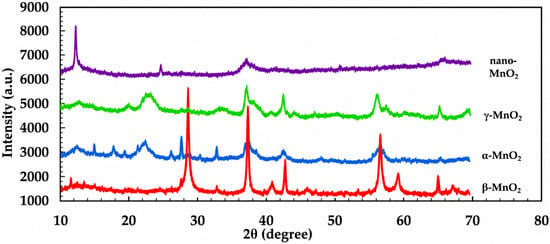
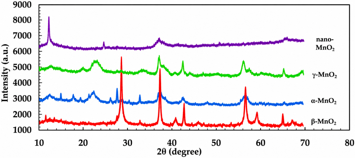
Once the various MnO2 polymorphs were synthesized, the structures were confirmed with X-ray diffraction studies. The X-ray diffraction (XRD) patterns of α-, β-, and γ-MnO2, along with nano-MnO2, are shown in Figure 1. The XRD diffraction peaks for β-MnO2 (red) correspond to the standard diffraction peaks indexed to the pure tetragonal phase of β-MnO2 [space group: P42/mnm (136)] with lattice constants a = 4.398 Å and c = 2.873 Å (PDF #01-071-0071), which are in good agreement with reported data for pure β-MnO2 [26]. The XRD pattern for α-MnO2 (blue) can be indexed to the pure tetragonal phase of α-MnO2 [space group: I4/m (87)] with lattice constants a = 9.7847 Å and c = 2.863 Å (JCPDS 44-0141), and the pattern for γ-MnO2 (green) can be indexed to the pure orthorhombic phase of γ-MnO2 with lattice constants a = 6.36 Å, b = 10.15 Å, and c = 4.09 Å (JCPDS 14-0644).
Figure 1.
Powder X-ray diffraction (XRD) patterns of the prepared α-, β-, and γ-MnO2 samples.
The XRD data confirm the successful preparation of α-, β-, and γ-MnO2, each displaying the characteristic reflections of its expected crystalline phase. β-MnO2 exhibits the sharpest and most intense diffraction peaks, indicating the highest crystallinity among the crystalline samples. In contrast, nano-MnO2 shows weak, broad diffraction features consistent with a poorly crystalline or amorphous structure, a feature commonly observed for nanoscale MnO2 materials prepared under rapid redox conditions [38].
Unlike the highly ordered long-range structures of the crystalline polymorphs, amorphous nano-MnO2 lacks periodic atomic arrangements and therefore exhibits greater structural disorder [39]. This disorder creates interstitial cavities and loosely packed atomic domains capable of accommodating substantial amounts of surface-bound and trapped water, a feature that we hypothesize contributes to its enhanced oxidation performance [40].
In contrast, the slower nucleation and growth processes associated with the hydrothermal synthesis of α-, β-, and γ-MnO2 yield more crystalline materials with fewer sites for water retention [41,42]. No impurity peaks were detected in any of the samples, confirming the absence of secondary phases.
The XRD results therefore show that crystallinity alone does not predict catalytic behavior: β-MnO2 is the most crystalline phase, whereas α-MnO2 is more active in oxidation. This contrast points to additional controlling factors, particularly surface-associated water and accessible reactive sites.
3.4. Thermogravimetric Analysis (TGA)
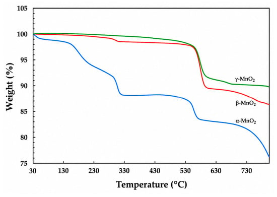
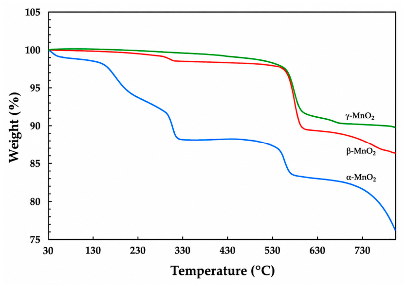
After phase assignment by XRD, thermogravimetric analysis was used to compare water retention in the MnO2 materials. The ability of each polymorph to retain water is closely related to its crystal framework: α-MnO2, with its 2 × 2 tunnel structure, can accommodate more H2O than β-MnO2 with 1 × 1 tunnels or γ-MnO2 with a 1 × 2 tunnel framework. Previous work also showed that nano-MnO2 contains substantial surface water, which contributes to its higher catalytic activity [13]. In the present samples, mass loss between 90 and 300 °C is assigned mainly to physiosorbed and chemisorbed water, whereas loss at 550–600 °C corresponds to structural water (Figure 2). α-MnO2 retained more surface-associated water than β- and γ-MnO2, especially in the 90–150 °C region associated with labile water. This trend parallels catalytic performance, where α-MnO2 gave higher oxidation yields than β- and γ-MnO2 when dried at 90 °C. This was in line with our previous observation of nano-MnO2 exhibiting higher mass loss of 14.2% over a temperature range of 100–150 °C while the Attenburrow and commercial MnO2 registered mass loss of 9.5% and 3.6%, respectively [13]. From this observation, we deduced that with greater porosity and surface area, nano-MnO2 retains more adsorbed water and exhibits greater reactivity followed by α-MnO2.
Figure 2.
TGA results for different crystalline forms of MnO2 samples.
3.5. Brunauer–Emmett–Teller (BET) Analysis
BET analysis was used to determine whether differences in catalytic activity correlate with accessible surface area. Nitrogen adsorption–desorption measurements were analyzed by the multipoint BET method, and the results are summarized in Table 2.
Table 2.
BET surface area of the prepared MnO2 samples.
The BET surface areas decreased in the order nano-MnO2 > β-MnO2 > α-MnO2 > γ-MnO2. Nano-MnO2 showed the largest surface area (169.20 m2/g), approximately five times higher than β-MnO2 and nine- to fifteen-fold higher than α- and γ-MnO2. This high value is consistent with reports that nanocrystalline or poorly crystalline MnO2 phases often possess small particle sizes, defect-rich domains, interparticle voids, and mesoporosity [43,44]. The crystalline polymorphs showed lower accessible surface areas, as expected for more ordered materials in which crystal growth and aggregation can reduce pore accessibility [43,45]. β-MnO2 had the highest surface area among the crystalline phases, likely reflecting smaller crystallites or more open secondary aggregates under the present synthesis conditions [45,46]. However, α-MnO2 was more active in oxidation despite its lower BET surface area, indicating that surface area is not the sole determinant of reactivity. Instead, morphology, crystallite size, pore connectivity, hydration, oxygen vacancies, lattice oxygen mobility, and Mn oxidation-state distribution must also be considered [43,44,45,46].
3.6. Fourier Transform Infrared (FTIR) Spectroscopy
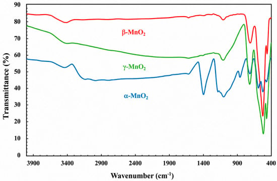
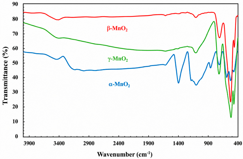
FTIR spectroscopy was used to further evaluate the composition and water-associated features of the α-, β-, and γ-MnO2 samples (Figure 3). Spectra were collected in transmission mode after 250 scans at a resolution of 4 cm−1. Bands near 700 and 475 cm−1 correspond to O–Mn–O stretching vibrations, confirming formation of MnO2 and agreeing with the XRD assignments. Broad absorptions between 4000 and 3000 cm−1 are assigned to O–H stretching from interlayer or structural water, while the band near 1616 cm−1 corresponds to bending vibrations of physically adsorbed water. The absorption at 1116 cm−1 is attributed to –OH bending associated with Mn centers, and bands at 517 and 487 cm−1 further support Mn–O stretching modes in octahedral MnO2 layers. The simultaneous presence of lattice Mn–O and water-associated bands indicates that both structural and adsorbed water are present in these samples. α-MnO2 also exhibits a distinct band at 1380 cm−1, consistent with reported data for this polymorph [39]. Weaker bands in the same region for β- and γ-MnO2 support the conclusion that water-associated surface chemistry contributes to the observed differences in oxidation reactivity.
Figure 3.
FTIR spectra of the as-prepared α-, β-, and γ-MnO2 samples.
3.7. Scanning Electron Microscopy (SEM) Analysis
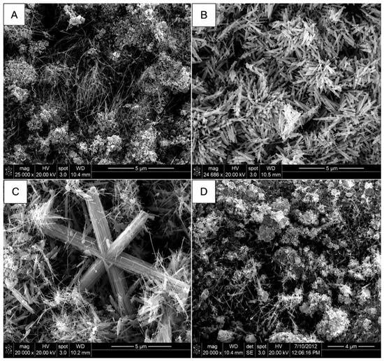
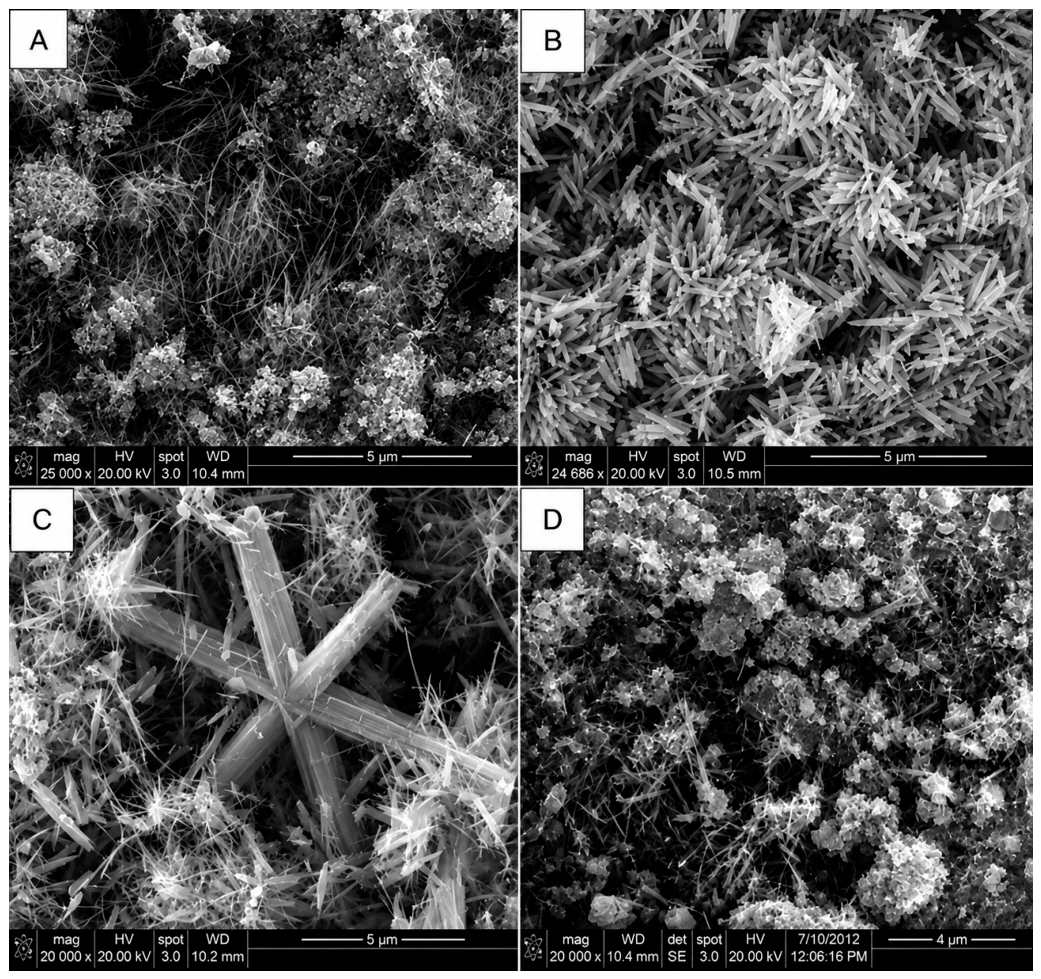
The particle size and morphology of the different crystallographic forms of α-, β-, and γ-MnO2, together with nano-MnO2, were examined by SEM, as shown in Figure 4. The images reveal pronounced morphology-dependent differences among the MnO2 materials, consistent with their distinct crystal structures and synthetic conditions and with prior reports showing that hydrothermal variables strongly control MnO2 phase, aspect ratio, aggregation, and secondary particle architecture [47,48,49,50]. As shown in Figure 4A, α-MnO2 is composed predominantly of elongated, wire-like crystallites with diameters of approximately 10–50 nm and lengths extending to several micrometers. This morphology closely resembles previously reported hydrothermal α-MnO2 nanowires, which are commonly described as one-dimensional structures with tens-of-nanometers diameters and micrometer-scale lengths [47,48]. These high-aspect-ratio nanowires appear as intertwined bundles, generating interparticle voids and open channels that may facilitate adsorption of water molecules and access of organic substrates to redox-active surface sites. The relatively open morphology of α-MnO2 is therefore consistent with literature emphasizing that one-dimensional MnO2 architectures can enhance catalytic performance through improved surface exposure, pore connectivity, and mass transport [49].
Figure 4.
SEM images of (A) α-MnO2, (B) β-MnO2, (C) γ-MnO2, and (D) nano-MnO2.
In contrast, the SEM image of β-MnO2 in Figure 4B shows well-defined nanorods with more rigid and angular morphologies, including square or rectangular cross-sectional features. These rods have diameters of approximately 40–150 nm and lengths of 2–5 µm. Similar β-MnO2 rod- or stick-like morphologies have been reported for hydrothermally prepared β-MnO2, where the rutile-type 1 × 1 tunnel structure promotes more compact, highly crystalline one-dimensional growth relative to more open tunnel phases [28,38]. The larger rod diameter and more compact morphology suggest a higher degree of crystallite growth and structural ordering, which agrees with the sharp diffraction features observed in the XRD analysis. Although β-MnO2 displayed a higher BET surface area than α-MnO2, its more crystalline and compact rod-like architecture likely provides fewer hydrated defects and less accessible surface-bound water, which may explain why its oxidation activity was lower than that of α-MnO2. This interpretation agrees with comparative studies showing that MnO2 catalytic behavior depends not only on external surface area, but also on crystal phase, defect density, exposed facets, oxygen mobility, and surface chemistry [51].
The γ-MnO2 sample shown in Figure 4C exhibits a mixed morphology consisting mainly of nanorods, with occasional branched or six-armed star-like structures. The rod diameters are approximately 30–90 nm and the lengths extend to several micrometers. Compared with α- and β-MnO2, the γ-polymorph appears more heterogeneous, with localized aggregation and less uniform particle distribution. Such morphological heterogeneity is consistent with reports that γ-MnO2 often contains intergrowths or structural disorder and that its observed SEM morphology is highly sensitive to precursor chemistry, oxidant strength, temperature, and growth time [28,38,52]. This morphology is consistent with its lower BET surface area and poorer catalytic performance. The reduced porosity and lower abundance of accessible hydrated surface sites likely limit substrate interaction with Mn redox centers, contributing to the diminished oxidation efficiency observed for γ-MnO2.
Nano-MnO2, shown in Figure 4D, displays a markedly different morphology from the hydrothermally prepared crystalline phases. Rather than forming discrete long rods or wires, nano-MnO2 consists of small, approximately spherical or granular particles assembled into a highly porous, loosely packed network. This morphology is consistent with reports that nanostructured MnO2 materials can form porous particle networks with limited large-scale aggregation [38]. This texture produces extensive interparticle voids and a large accessible surface, in agreement with the substantially higher BET surface area measured for this material [38]. Comparable poorly crystalline or nanostructured MnO2 materials have been reported to show high surface area, abundant pore volume, defect-rich domains, and enhanced electrolyte or substrate accessibility, all of which can contribute to improved catalytic or electrochemical activity [48,49,50,51,52,53]. The relatively uniform dispersion of nanosized particles further increases the number of exposed Mn–O surface sites available for substrate contact.
Overall, the SEM analysis supports the conclusion that morphology alone does not fully dictate catalytic performance. β-MnO2 possesses a larger BET surface area than α-MnO2, yet α-MnO2 exhibits higher oxidation activity, indicating that surface hydration and the accessibility of water-associated active sites are critical contributors. This distinction is important because related MnO2 studies frequently correlate catalytic or electrochemical activity with nanowire/nanorod morphology and surface area, but increasingly emphasize that phase structure, oxygen vacancies, structural disorder, and bound water can be equally decisive [47,52]. Nano-MnO2 combines favorable morphological attributes—small particle size, high porosity, extensive surface exposure, and abundant water-retaining sites—which collectively rationalize its superior performance in the oxidation of diphenylmethane to benzophenone. Thus, when considered together with XRD, TGA, FTIR, and BET data, the SEM observations support a structure–morphology–water–reactivity relationship across the MnO2 materials examined here and are broadly consistent with literature describing morphology-controlled MnO2 catalysis [38,51].
3.8. Catalytic Oxidation Activity
The prepared α-, β-, γ-, and nano-MnO2 samples were evaluated as oxidants for the conversion of diphenylmethane to benzophenone to correlate activity with phase, morphology, water content, and surface area. Samples dried at two different temperatures were compared under aerobic, atmospheric, and anaerobic conditions. Yields are reported as isolated yields from triplicate reactions performed for 6–24 h using 25 wt% catalyst unless otherwise stated.
Two separate samples of each MnO2 form were dried at different temperatures (90 °C and 120 °C), and their catalytic activities were compared. Each reaction was performed in triplicate under aerobic (O2), atmospheric (air), and anaerobic (N2) conditions, and the results are summarized in Table 3.
Table 3.
Oxidation of diphenylmethane to benzophenone using α-, β-, γ-, and nano-MnO2.
Under aerobic conditions, all catalysts produced benzophenone in modest to high yields, consistent with the participation of adsorbed molecular oxygen on the MnO2 surface. Under anaerobic conditions, nano-MnO2 clearly outperformed the crystalline polymorphs, consistent with its combination of high surface area and greater surface-water content. Among the crystalline phases, α-MnO2 gave the highest yields, even though β-MnO2 had the larger BET surface area. γ-MnO2, which combines low surface area with low water retention, showed the poorest performance. These trends indicate that retained surface-associated water is a stronger predictor of activity than BET surface area or crystallinity alone.
Drying temperature had a pronounced effect on reactivity. Samples dried at 90 °C retained more labile surface-bound water and therefore showed higher activity, whereas drying at 120 °C reduced the activity of all MnO2 materials. This behavior supports the role of adsorbed water in the oxidation process and is consistent with reports that heat treatment can alter MnO2 hydration, surface hydroxyl density, porosity, and catalytic performance [51,52].
Catalyst-loading studies from 25 to 500 wt% (Table 4) further show that the crystalline polymorphs require higher loadings to reach useful conversion, whereas nano-MnO2 remains highly effective at low loading. For the crystalline samples, activity generally follows the trend α-MnO2 > β-MnO2 > γ-MnO2, paralleling the relative amount of retained surface water rather than the BET surface-area trend. These data reinforce the conclusion that efficient oxidation requires both accessible surface sites and a hydrated surface environment.
Table 4.
Effect of catalyst loading on the oxidation of diphenylmethane to benzophenone.
Yields are reported as isolated yields from reactions performed under the stated catalyst loadings. Storage and regeneration experiments further support the importance of surface-bound water. In previous work, nano-MnO2 stored at 90 °C for 4–10 months showed a gradual decline in catalytic activity [13]. Similar, but faster, deactivation was observed for α-, β-, and γ-MnO2, whose yields dropped from approximately 25% to negligible levels within one month. The loss of activity is attributed to depletion or reorganization of surface-bound water. Nano-MnO2 recovered up to 75% of its activity after sequential washing with 1 M HCl, 1 M NaOH, and water, followed by drying at 90 °C, whereas the crystalline polymorphs showed little recovery. Adding 0.5–1.0% water directly to the reaction mixture did not improve reactivity and instead lowered yields, indicating that externally added bulk water cannot replace the organized surface-bound water present in the active catalyst. Literature reports similarly show that prolonged heating or thermal treatment can remove surface water and alter accessible surface area, hydration state, and catalytic behavior in MnO2 materials [43,53].
Reusability followed the same trend. Nano-MnO2 could be recycled for approximately three to four runs, with yields of about 30–70% depending on cycle and conditions, whereas the crystalline polymorphs showed substantially lower recyclability. γ-MnO2 was particularly poor, giving trace to no conversion on the second run even at 50 wt% catalyst loading. These results suggest that the hydrated, defect-rich surface of nano-MnO2 is more readily maintained or partially regenerated than the surfaces of the more crystalline polymorphs.
4. Conclusions
α-, β-, and γ-MnO2 were prepared by controlled hydrothermal methods and benchmarked against nano-MnO2 for the oxidation of diphenylmethane to benzophenone. XRD, TGA, FTIR, BET, and SEM analyses confirmed pronounced differences in crystallinity, morphology, accessible surface area, and water retention. The crystalline polymorphs displayed phase-defined diffraction patterns and wire- or rod-like morphologies, whereas nano-MnO2 showed broad diffraction features, a porous nanoscale architecture, and the highest BET surface area.
Catalytic evaluation showed that MnO2-mediated arylmethylene oxidation is governed by the combined effects of surface accessibility and retained surface-associated water rather than by crystallinity or BET surface area alone. Nano-MnO2 was the most efficient material under aerobic, atmospheric, and anaerobic conditions, while α-MnO2 was the most active crystalline polymorph despite having a lower surface area than β-MnO2. The decline in activity after drying at 120 °C, prolonged storage, or reuse further confirms the importance of labile surface-bound water for maintaining oxidative performance.
Overall, this work defines a structure–morphology–water–reactivity relationship for MnO2-mediated benzylic oxidation. Water-rich nano-MnO2 is the most effective oxidant for arylmethylene-to-ketone conversion, and α-MnO2 is the best crystalline analog when surface hydration is preserved. These findings emphasize the need to control synthesis, drying, storage, and regeneration conditions to maintain the hydrated surface environment required for efficient oxidation. Future studies that tune hydration, porosity, defect density, and substrate scope should support the development of more sustainable MnO2-based oxidants with high activity and reduced reagent waste.
Author Contributions
Conceptualization, B.N., S.K.L. and R.A.B.; methodology, B.N. and S.K.L.; validation and formal analysis, S.K.L.; investigation, S.K.L. and R.A.B.; resources, S.K.L. and R.A.B.; data curation, S.K.L.; writing—original draft preparation, B.N. and S.K.L.; writing—review and editing, B.N., R.A.B. and K.D.A.; visualization, K.D.A.; supervision, R.A.B. and K.D.A.; project administration, R.A.B.; funding acquisition, R.A.B. All authors have read and agreed to the published version of the manuscript.
Funding
The authors are grateful to the Oklahoma State University College of Arts and Sciences for support of the Oklahoma Statewide NMR Facility, including funds for a 400 MHz NMR spectrometer. This facility was established with support from the NSF (BIR-9512269), the Oklahoma State Regents for Higher Education, the W. M. Keck Foundation, and Conoco, Inc. The authors also thank the OSU College of Arts and Sciences for support in upgrading departmental FTIR instrumentation.
Institutional Review Board Statement
Not applicable.
Informed Consent Statement
Not applicable.
Data Availability Statement
The data supporting the findings of this study are available within the article.
Conflicts of Interest
Author Sathish Kumar Lageshetty was employed by Midwest Medical Isotopes. Author Baskar Nammalwar was employed by Vividion Therapeutics. Author Kevin D. Ausman was employed by Spark Therapeutics. The funding sponsors had no role in the design of the study; in the collection, analyses, or interpretation of data; in the writing of the manuscript; or in the decision to publish the results. The remaining authors declare no conflicts of interest.
References
- Sui, N.; Duan, Y.; Jiao, X.; Chen, D. Large-Scale Preparation and Catalytic Properties of One-Dimensional α/β-MnO2 Nanostructures. J. Phys. Chem. C 2009, 113, 8560–8565. [Google Scholar] [CrossRef] [Scilit]
- Thackeray, M.M. Manganese oxides for lithium batteries. Prog. Solid State Chem. 1997, 25, 1–71. [Google Scholar] [CrossRef] [Scilit]
- Truong, T.T.; Liu, Y.; Ren, Y.; Trahey, L.; Sun, Y. Morphological and crystalline evolution of Nanostructured MnO2 and its application in lithium-air batteries. ACS Nano 2012, 6, 8067–8077. [Google Scholar] [CrossRef] [Scilit] [PubMed]
- An, J.; Peng, X.; Xu, S.; Xu, Z.; Wang, J. Fabrication of coral like carbon black/MnO2 nano composites from commercial carbon black and their application in supercapacitors. RSC Adv. 2015, 5, 97080–97088. [Google Scholar] [CrossRef] [Scilit]
- Chen, Y.D.; Wu, Q.M.; Huang, B.; Tang, L.R. Ultrasound-assisted one pot synthesis of dispersable Nano-MnO2 in the presence of nanocellulose. Adv. Mater. Res. 2013, 634–638, 2318–2323. [Google Scholar] [CrossRef] [Scilit]
- Boppana, V.B.R.; Jiao, F. Nanostructured MnO2: An efficient and robust water oxidation catalyst. Chem. Commun. 2011, 47, 8973–8975. [Google Scholar] [CrossRef] [Scilit] [PubMed]
- Simon, P.; Gogotsi, Y. Materials for electrochemical capacitors. Nat. Mater. 2008, 7, 845–854. [Google Scholar] [CrossRef] [Scilit] [PubMed]
- Feng, R.X.; Dong, H.; Wang, Y.D.; Ai, X.P.; Cao, Y.L.; Yang, H.X. A simple and high efficient direct borohydride fuel cell with MnO2-catalyzed cathode. Electrochem. Commun. 2005, 7, 449–452. [Google Scholar] [CrossRef] [Scilit]
- Ananth, M.V.; Pethkar, S.; Dakshinamurthi, K. Distortion of MnO6 octahedra and electrochemical activity of Nstutite-based MnO2 polymorphs for alkaline electrolytes—An FTIR study. J. Power Sources 1998, 75, 278–282. [Google Scholar] [CrossRef] [Scilit]
- Devaraj, S.; Munichandraiah, N. Effect of crystallographic structure of MnO2 on its electrochemical capacitance properties. J. Phys. Chem. C 2008, 112, 4406–4417. [Google Scholar] [CrossRef] [Scilit]
- Jiang, X.; Li, C.; Long, Z.; Hou, X. Selectively Enhanced Molecular Emission Spectra of Benzene, Toluene and Xylene with nano-MnO2 in Atmospheric Ambient Temperature Dielectric Barrier Discharge. Anal. Methods 2015, 7, 400–404. [Google Scholar] [CrossRef] [Scilit]
- Mallat, T.; Baiker, A. Oxidation of Alcohols with Molecular Oxygen on Solid Catalysts. Chem. Rev. 2004, 104, 3037–3058. [Google Scholar] [CrossRef] [Scilit] [PubMed]
- Nammalwar, B.; Fortenberry, C.; Bunce, R.A.; Lageshetty, S.K.; Ausman, K.D. Efficient oxidation of arylmethylene compounds using nano-MnO2. Tetrahedron Lett. 2013, 54, 2010–2013. [Google Scholar] [CrossRef] [Scilit]
- Taylor, R.J.K.; Reid, M.; Foot, J.; Raw, S.A. Tandem Oxidation Processes Using Manganese Dioxide: Discovery, Applications, and Current Studies. Acc. Chem. Res. 2005, 38, 851–869. [Google Scholar] [CrossRef] [Scilit] [PubMed]
- Attenburrow, J.; Cameron, A.F.B.; Chapman, J.H.; Evans, R.M.; Hems, B.A.; Jansen, A.B.A.; Walker, T. A synthesis of vitamin a from cyclohexanone. J. Chem. Soc. (Resumed) 1952, 1094–1111. [Google Scholar] [CrossRef] [Scilit]
- Khan, Y.; Durrani, S.K.; Mehmood, M.; Khan, M.R. Mild hydrothermal synthesis of γ-MnO2 nanostructures and their phase transformation to α-MnO2 nanowires. J. Mater. Res. 2011, 26, 2268–2275. [Google Scholar] [CrossRef] [Scilit]
- Wang, H.-Q.; Yang, G.-f.; Li, Q.-Y.; Zhong, X.-X.; Wang, F.-P.; Li, Z.-S.; Li, Y.-H. Porous nano-MnO2: Large scale synthesis via a facile quick-redox procedure and application in a supercapacitor. New J. Chem. 2011, 35, 469–475. [Google Scholar] [CrossRef] [Scilit]
- Luo, Y. Preparation of MnO2 nanoparticles by directly mixing potassium permanganate and polyelectrolyte aqueous solutions. Mater. Lett. 2007, 61, 1893–1895. [Google Scholar] [CrossRef] [Scilit]
- Dong, Y.; Li, K.; Jiang, P.; Wang, G.; Miao, H.; Zhang, J.; Zhang, C. Simple hydrothermal preparation of α-, β-, and γ-MnO2 and phase sensitivity in catalytic ozonation. RSC Adv. 2014, 4, 39167–39173. [Google Scholar] [CrossRef] [Scilit]
- Finney, J.L. The structure of water: A historical perspective. J. Chem. Phys. 2024, 160, 060901. [Google Scholar] [CrossRef] [Scilit] [PubMed]
- Wang, X.; Li, Y. Selected-Control Hydrothermal Synthesis of α- and β-MnO2 Single Crystal Nanowires. J. Am. Chem. Soc. 2002, 124, 2880–2881. [Google Scholar] [CrossRef] [Scilit] [PubMed]
- Rao, C.N.R.; Vivekchand, S.R.C.; Biswas, K.; Govindaraj, A. Synthesis of inorganic nanomaterials. Dalton Trans. 2007, 3728–3749. [Google Scholar] [CrossRef] [Scilit] [PubMed]
- Pang, S.C.; Anderson, M.A.; Chapman, T.W. Novel Electrode Materials for Thin-Film Ultracapacitors: Comparison of Electrochemical Properties of Sol-Gel-Derived and Electrodeposited Manganese Dioxide. J. Electrochem. Soc. 2000, 147, 444–450. [Google Scholar] [CrossRef] [Scilit]
- Ding, Y.S.; Shen, X.F.; Gomez, S.; Luo, H.; Aindow, M.; Suib, S.L. Hydrothermal Growth of Manganese Dioxide into Three-Dimensional Hierarchical Nanoarchitectures. Adv. Funct. Mater. 2006, 16, 549–555. [Google Scholar] [CrossRef] [Scilit]
- Wu, M.; Snook, G.A.; Chen, G.Z.; Fray, D.J. Redox deposition of manganese oxide on graphite for supercapacitors. Electrochem. Commun. 2004, 6, 499–504. [Google Scholar] [CrossRef] [Scilit]
- Hong, S.; Jin, S.; Deng, Y.; Garcia-Mendez, R.; Kim, K.; Utomo, N.; Archer, L. Efficient Scalable Hydrothermal Synthesis of MnO2 with Controlled Polymorphs and Morphologies for Enhanced Battery Cathodes. ACS Energy Lett. 2023, 8, 1744–1751. [Google Scholar] [CrossRef] [Scilit]
- Baral, A.; Satish, L.; Zhang, G.; Ju, S.; Ghosh, M.K. A Review of Recent Progress on Nano MnO2: Synthesis, Surface Modification and Applications. J. Inorg. Organomet. Polym. Mater. 2021, 31, 899–922. [Google Scholar] [CrossRef] [Scilit]
- Tharat, B.; Untarabut, P.; Junkaew, A.; Suthirakun, S. First-Principles Investigation of Hydroxyl Species Formation on β-MnO2 (110) for Catalytic Oxidation Applications. ACS Omega 2025, 10, 12097–12108. [Google Scholar] [CrossRef] [Scilit] [PubMed]
- Zhang, R.; Yang, L.; Yang, J.; Niu, Q.; Zhu, B. Effects of Drying Operation on the Mn2+ Removal Activity of MnOx: Performance and Mechanism. Water 2025, 17, 261. [Google Scholar] [CrossRef] [Scilit]
- Huang, N.; Qu, Z.; Dong, C.; Qin, Y.; Duan, X. Superior performance of alpha@beta-MnO2 for the toluene oxidation: Active interface and oxygen vacancy. Appl. Catal. A Gen. 2018, 560, 195–205. [Google Scholar] [CrossRef] [Scilit]
- Pang, G.; Wang, D.; Zhang, Y.; Ma, C.; Hao, Z. Catalytic activities and mechanism of formaldehyde oxidation over gold supported on MnO2 microsphere catalysts at room temperature. Front. Environ. Sci. Eng. 2016, 10, 447–457. [Google Scholar] [CrossRef] [Scilit]
- Lu, T.; Su, F.; Zhao, Q.; Li, J.; Zhang, C.; Zhang, R.; Liu, P. Catalytic oxidation of volatile organic compounds over manganese-based oxide catalysts: Performance, deactivation and future opportunities. Sep. Purif. Technol. 2022, 296, 121436. [Google Scholar] [CrossRef] [Scilit]
- Li, S.; Tu, L.; Li, X.; Chen, P.; Zhang, P.; Zhang, Y.; Ding, S. Investigation into the effect of MnO2 structure toward thermal/microwave-assisted catalytic oxidation of benzene. Appl. Surf. Sci. 2025, 707, 163649. [Google Scholar] [CrossRef] [Scilit]
- Aoyama, T.; Sonoda, N.; Yamauchi, M.; Toriyama, K.; Anzai, M.; Ando, A.; Shioiri, T. Chemical Manganese Dioxide (CMD), an Efficient Activated Manganese Dioxide. Application to Oxidation of Benzylic and Allylic Alcohols. Synlett 1998, 35–36. [Google Scholar] [CrossRef] [Scilit]
- Wang, X.; Yang, X.; Yi, X.; Min, X.; Jia, Y. Rapid synthesis of manganese dioxide nanoparticles for enhanced compatibility and theranostic applications. RSC Adv. 2025, 15, 3060–3066. [Google Scholar] [CrossRef] [Scilit] [PubMed]
- Nammalwar, B.; Bourne, C.R.; Bunce, R.A.; Wakeham, N.; Bourne, P.C.; Ramnarayan, K.; Mylvaganam, S.; Berlin, K.D.; Barrow, E.W.; Barrow, W.W. Inhibition of bacterial dihydrofolate reductase by 6-alkyl-2,4-diaminopyrimidines. ChemMedChem 2012, 7, 1474–1482. [Google Scholar] [CrossRef] [Scilit] [PubMed]
- Nammalwar, B.; Bourne, C.R.; Wakeham, N.; Bourne, P.C.; Barrow, E.W.; Muddala, N.P.; Bunce, R.A.; Berlin, K.D.; Barrow, W.W. Modified 2,4-diaminopyrimidine-based dihydrofolate reductase inhibitors as potential drug scaffolds against Bacillus anthracis. Bioorg. Med. Chem. 2015, 23, 203–211. [Google Scholar] [CrossRef] [Scilit] [PubMed]
- Xie, C.; Xu, Z.; Zheng, Y.; Wang, S.; Dai, M.; Xiao, C. Research Progress on the preparation of Manganese dioxide nanomaterials and their electrochemical applicatons. Nanomaterials 2024, 14, 1283. [Google Scholar] [CrossRef] [Scilit] [PubMed]
- Chang, J.-K.; Tsai, W.-T. Material Characterization and Electrochemical Performance of Hydrous Manganese Oxide Electrodes for Use in Electrochemical Pseudocapacitors. J. Electrochem. Soc. 2003, 150, A1333–A1338. [Google Scholar] [CrossRef] [Scilit]
- Boyom-Tatchemo, F.W.; Devred, F.; Ndiffo-Yemeli, G.; Laminsi, S.; Gaigneaux, E.M. Plasma-induced redox reactions synthesis of nanosized α-, γ- and δ-MnO2 catalysts for dye degradation. Appl. Catal. B Environ. 2020, 260, 118159. [Google Scholar] [CrossRef] [Scilit]
- Birgisson, S.; Saha, D.; Iversen, B.B.F. Understanding crystallization pathways leading to manganese oxide polymorph formation. Cryst. Growth Des. 2018, 18, 827–838. [Google Scholar] [CrossRef] [Scilit] [PubMed]
- Ghosh, S.K. Diversity in the Family of Manganese Oxides at the Nanoscale: From Fundamentals to Applications. ACS Omega 2020, 5, 25493–25504. [Google Scholar] [CrossRef] [Scilit] [PubMed]
- Yang, P.; Wang, J.; Wang, H.; Wang, S.; Yang, C.; He, Y. Physicochemical properties of different crystal forms of manganese dioxide prepared by a liquid phase method and their quantitative evaluation in capacitor and battery materials. Nanoscale Adv. 2023, 5, 3396–3413. [Google Scholar] [CrossRef] [Scilit] [PubMed]
- Taranu, B.-O.; Novaconi, S.D.; Ivanovici, M.; Goncalves, J.N.; Rus, F.S. α-MnO2 Nanowire Structure Obtained at Low Temperature with Aspects in Environmental Remediation and Sustainable Energy Applications. Appl. Sci. 2022, 12, 6821. [Google Scholar] [CrossRef] [Scilit]
- Wu, J.; Huang, H.; Yu, L.; Hu, J. Controllable Hydrothermal Synthesis of MnO2 Nanostructures. Adv. Mater. Phys. Chem. 2013, 3, 201–205. [Google Scholar] [CrossRef]
- Song, H.; Xu, L.; Chen, M.; Cui, Y.; Wu, C.; Qiu, J.; Xu, L.; Cheng, G.; Hu, X. Recent progresses in the synthesis of MnO2 nanowire and its application in environmental catalysis. RSC Adv. 2021, 11, 35494–35513. [Google Scholar] [CrossRef] [Scilit] [PubMed]
- Rajamanickam, N.; Ganesan, P.; Rajashabala, S.; Ramachandran, K. Structural and optical properties of α-MnO2 nanowires and β-Mn2 nanorods. AIP Conf. Proc. 2014, 1591, 267–269. [Google Scholar] [CrossRef] [Scilit]
- Shivakumar, S.R.; Ko, J.M.; Kim, D.Y.; Kim, B.C.; Wallace, G.G. Performance evaluation of CNT/polypyrrole/MnO2 composite electrodes for electrochemical capacitors. Electrochim. Acta 2007, 52, 7377–7385. [Google Scholar] [CrossRef] [Scilit]
- Diantoro, M.; Istiqomah, I.; Fath, Y.A.; Naiskhudin, N.; Alias, Y.; Meevasana, W. Potential of MnO2 based composite and numerous morphological for enhancing supercapacitors performance. Int. J. Appl. Ceram. Technol. 2023, 20, 2077–2098. [Google Scholar] [CrossRef] [Scilit]
- Dong, Y.; Jin, B.; Liu, S.; Gao, J.; Wang, K.; Su, F. Abundant Oxygen Vacancies Induced by the Mechanochemical Process Boost the Low-Temperature Catalytic Performance of MnO2 in NH3-SCR. Catalysts 2022, 12, 1291. [Google Scholar] [CrossRef] [Scilit]
- Wu, H.; Zhang, R.; Kang, B.; Guo, X.; Di, Z.; Wang, K.; Jia, J.; Wei, Y.; Wang, Z. Morphological impact of 1-dimensional to 3-dimensional manganese dioxides on catalytic ozone decomposition correlated with crystal facets and lattice oxygen mobilities. Environ. Sci. Nano 2025, 12, 418–432. [Google Scholar] [CrossRef] [Scilit]
- Liang, L.; Jia, M.; Zhao, M.; Deng, Y.; Tang, J.; He, X.; Liu, Y.; Yan, K.; Yu, X.; Yang, H.; et al. Progress of Nanomaterials based on Manganese dioxide in the field of tumor diagnosis and therapy. Int. J. Nanomed. 2024, 19, 8883–8900. [Google Scholar] [CrossRef] [Scilit] [PubMed]
- Ma, J.; Cheng, Q.; Pavlinek, V.; Saha, P.; Li, C. Morphology-controllable synthesis of MnO2 hollow nanospheres and their supercapacitive performance. New J. Chem. 2013, 37, 722–728. [Google Scholar] [CrossRef] [Scilit]

Disclaimer/Publisher’s Note: The statements, opinions and data contained in all publications are solely those of the individual author(s) and contributor(s) and not of MDPI and/or the editor(s). MDPI and/or the editor(s) disclaim responsibility for any injury to people or property resulting from any ideas, methods, instructions or products referred to in the content. |
© 2026 by the authors. Licensee MDPI, Basel, Switzerland. This article is an open access article distributed under the terms and conditions of the Creative Commons Attribution (CC BY) license.