


Abstract
Theranostic nanoparticles, which integrate diagnostic imaging and therapeutic delivery within a single nanoplatform, represent a transformative paradigm in oncological nanomedicine. Despite substantial preclinical progress, the field faces persistent gaps in rational nanoparticle design informed by tumor biology, preclinical model fidelity, and clinical translation. This review critically synthesizes theranostic nanoparticle research across three underexplored domains. First, we examine tumor microenvironment features—reactive oxygen species dynamics, glutathione gradients, hypoxia, and proteasomal dysregulation—as mechanistic drivers of nanoparticle responsiveness. Second, we evaluate redox-responsive and proteasome-targeted nanoplatforms that exploit these cues for stimuli-triggered drug release and simultaneous imaging readout. Third, we address the unmet need for three-dimensional organoid and microfluidic tumor models as predictive preclinical testing environments, given the well-documented limitations of conventional two-dimensional cultures. Cancer subtype-specific applications are discussed for breast cancer, HPV-associated malignancies, colorectal cancer, and prostate cancer. Clinical translation barriers—including pharmacokinetic constraints, protein corona formation, immune clearance, anti-PEG antibodies, complement activation-related pseudoallergy, and FDA/EMA regulatory pathways—are addressed from a clinical oncology perspective. The review concludes with a research roadmap integrating proteomics-guided nanoparticle engineering, patient-derived organoid biobanks, and artificial intelligence-assisted design as priority areas for next-generation oncological theranostics.
1. Introduction
Cancer constitutes one of the most formidable challenges in contemporary medicine. According to the most recent GLOBOCAN estimates, approximately 20.6 million new cancer cases (19.5 million excluding nonmelanoma skin cancer) were diagnosed worldwide in 2024, with the annual global burden projected to rise to approximately 34 to 35 million new cases by 2050 [1]. Despite remarkable advances in surgery, conventional chemotherapy, targeted molecular agents, and immunotherapy, the five-year survival rates for many advanced and metastatic malignancies remain disappointing, underscoring the urgent need for more sophisticated, tumor-selective, and clinically translatable therapeutic approaches [1].
The limitations inherent to conventional cancer therapies are well established. Systemic chemotherapy agents distribute indiscriminately throughout the body, causing collateral toxicity to healthy tissues and frequently failing to sustain therapeutic concentrations at tumor sites [2]. Targeted therapies, while representing a substantial advance over cytotoxic chemotherapy, are constrained by the heterogeneity of tumor cell populations, the emergence of acquired resistance mechanisms, and the limited number of druggable oncogenic targets [3]. Immunotherapy, although transformative for certain malignancies, benefits only a subset of patients and is associated with immune-related adverse events that can be life-threatening [4]. These clinical realities have driven sustained interest in nanotechnology-based platforms capable of overcoming the delivery barriers, improving drug bioavailability at tumor sites, and enabling simultaneous diagnostic readout [5].
Nanomedicine offers a fundamentally distinct approach to cancer treatment. By engineering drug carriers at the nanoscale, it becomes possible to exploit tumor-specific biological characteristics for preferential drug accumulation [6]. The concept of theranostics, derived from the fusion of therapeutics and diagnostics, represents the logical extension of nanomedicine toward fully integrated, real-time guided cancer management [6]. A theranostic nanoparticle integrates at least one diagnostic modality, such as magnetic resonance imaging, positron emission tomography, fluorescence imaging, or photoacoustic imaging, with one or more therapeutic payloads within a single, spatiotemporally controlled nanoplatform [7]. This dual functionality enables clinicians and researchers to simultaneously monitor drug biodistribution, assess tumor response, and adapt treatment parameters in real time, capabilities that are simply impossible with conventional pharmaceutical formulations.
The enhanced permeability and retention (EPR) effect, first described by Matsumura and Maeda in 1986, constitutes the foundational rationale for passive nanoparticle targeting in solid tumors [8]. Tumor vasculature is characterized by poorly organized, highly fenestrated endothelial junctions and defective lymphatic drainage, collectively enabling the preferential extravasation and intratumoral retention of macromolecules and nanoparticles with hydrodynamic diameters in the range of 10 to 500 nanometres [9]. While the EPR effect has served as the conceptual cornerstone of nano-oncology for nearly four decades, its translational utility is increasingly recognized as tumor-type-dependent, heterogeneous across patients, and subject to significant interspecies variability between murine xenograft models and human tumors [3]. This understanding has catalyzed a paradigm shift toward active targeting strategies and stimuli-responsive nanoplatform designs that operate in concert with the specific physicochemical and biological characteristics of the tumor microenvironment.
The tumor microenvironment represents far more than a passive backdrop for cancer cells. It is a dynamic, biochemically complex ecosystem characterized by elevated concentrations of reactive oxygen species (ROS), overexpression of intracellular glutathione (GSH), hypoxic gradients, acidic extracellular pH, and pervasive dysregulation of the ubiquitin–proteasome system [10]. Each of these tumor microenvironment characteristics represents both a driver of tumor progression and an exploitable trigger for stimuli-responsive nanoparticle systems that selectively release their payloads in response to the specific biochemical conditions encountered within the tumor [11]. This mechanistic convergence between cancer biology and nanoparticle engineering is central to the design of next-generation theranostic platforms.
Of particular mechanistic relevance to this review is the proteasomal axis within the tumor microenvironment. The 26S proteasome is the principal cellular machinery for regulated protein degradation, and its activity is substantially upregulated in many cancer types to sustain the elevated proteotoxic demands of rapidly proliferating malignant cells [12]. Proteasome inhibitors such as bortezomib represent established clinical agents in haematological malignancies, and their potential in solid tumors is constrained primarily by pharmacokinetic limitations and systemic toxicity that could be addressed through nanoparticle encapsulation [13]. The integration of proteasome-targeted therapy within a theranostic nanoplatform remains a critically underexplored frontier, one that this review specifically addresses.
Another fundamental gap in the field concerns the preclinical evaluation of theranostic nanoparticles. The overwhelming majority of published studies employ conventional two-dimensional cell monolayer cultures for initial screening, systems that fail to recapitulate the three-dimensional architecture, oxygen gradients, extracellular matrix composition, and cell-to-cell signaling dynamics of actual tumors [14]. The emergence of patient-derived tumor organoids and tumor-on-chip microfluidic devices offers a transformative opportunity to evaluate nanoparticle behavior in physiologically relevant three-dimensional contexts before advancing to animal studies and clinical trials [15]. This review advocates for systematic integration of such advanced preclinical models into the theranostic nanoparticle development pipeline.
This review is positioned at a specific intersection that the existing literature has not yet addressed as an integrated whole. Reviews of redox-responsive nanomedicine have tended to specialize narrowly: recent glutathione-focused reviews provide detailed chemical linker taxonomies (disulfide, diselenide, transition-metal, and prodrug strategies) with comparatively limited attention to translational barriers and tumor heterogeneity [16], while broader redox-responsive nanocarrier reviews survey ROS and GSH triggers together largely as a drug-delivery engineering problem rather than in connection with a specific therapeutic target. Parallel reviews of nanoparticle-enabled targeted protein degradation have surveyed proteasome- and lysosome-directed strategies, including PROTAC and molecular glue delivery, largely independent of the tumor redox microenvironment that governs when and where such payloads are released [17]. General theranostic nanoparticle reviews, meanwhile, typically catalog nanoplatform chemistries and imaging modalities without a unifying therapeutic mechanism or a critical stance on the preclinical models used to validate them. This review differs from each of these precedents in combining four elements within a single mechanistic and translational framework: the coupling of redox-responsive (ROS- and GSH-triggered) drug release specifically with proteasome-targeted therapy, using bortezomib as a mechanistic exemplar; an explicit critique of two-dimensional preclinical validation paired with an evidence-based case for patient-derived organoid and microfluidic testing platforms; application to cancer subtypes, including HPV-associated malignancies, that are underrepresented in the redox-responsive nanomedicine literature; and a forward-looking, phased research roadmap intended to guide the field toward clinically translatable designs rather than incremental proof-of-concept demonstrations.
The present review is structured as follows: Section 2 provides a detailed mechanistic account of the tumor microenvironment features most relevant to theranostic nanoparticle design. Section 3 systematically classifies theranostic nanoparticle platforms by composition and physicochemical characteristics. Section 4 focuses on the novel intersection of redox-responsive nanoplatforms with proteasome-targeted therapy. Section 5 critically evaluates organoid and microfluidic models as advanced preclinical testing environments. Section 6 examines cancer subtype-specific applications. Section 7 addresses clinical translation barriers and regulatory pathways. Section 8 delineates future research priorities.
2. The Tumor Microenvironment: Redox Signaling, Metabolic Reprogramming, and Proteasomal Dysregulation
2.1. Hallmarks of the Tumor Microenvironment and Their Nanoparticle Design Implications
Tumors are not merely masses of malignant cells but complex ecosystems that actively co-evolve with surrounding stromal components, including cancer-associated fibroblasts, tumor-associated macrophages, endothelial cells, immune cells, and the extracellular matrix [10]. The defining hallmarks of cancer include sustained proliferative signaling, evasion of growth suppressors, resistance to cell death, replicative immortality, induction of angiogenesis, activation of invasion and metastasis, reprogramming of energy metabolism, and evasion of immune destruction [10]. Each of these hallmarks generates physicochemical features within the tumor microenvironment that can be exploited for stimuli-responsive nanoparticle activation.
From the perspective of nanoparticle design, the most tractable tumor microenvironment features are those that differ sufficiently and consistently from normal tissue to enable selective nanoparticle activation at the tumor site while preserving systemic safety [18]. These features include elevated intracellular glutathione concentrations, aberrant reactive oxygen species accumulation, hypoxia-driven extracellular acidification, overexpression of specific enzymes such as matrix metalloproteinases and cathepsins, and dysregulation of the proteasomal degradation machinery [11]. The following subsections address each of these features in mechanistic detail.
2.2. Reactive Oxygen Species Dynamics and Glutathione Gradients
Reactive oxygen species, encompassing hydrogen peroxide, superoxide anion, hydroxyl radical, and singlet oxygen, arise in tumors from both mitochondrial oxidative phosphorylation and a range of enzymatic sources, most notably the NADPH oxidase (NOX) family, which is frequently overexpressed or dysregulated across multiple solid tumor types and can constitute a dominant, mitochondria-independent source of intracellular and extracellular ROS [19]. Collectively, these mitochondrial and enzymatic sources are substantially elevated in most solid tumors as a consequence of oncogene activation, mitochondrial dysfunction, NOX pathway dysregulation, and the hypoxic-reoxygenation cycles characteristic of poorly vascularized tumor regions [19,20]. The intratumoral concentration of hydrogen peroxide, the most stable and diffusible ROS species, has been estimated at 50 to 100 micromolar in solid tumors, compared to approximately 1 to 100 nanomolar in normal, non-malignant tissue, creating a chemical gradient spanning roughly three to five orders of magnitude that can be exploited for ROS-responsive drug release [20,21]. Paradoxically, cancer cells simultaneously maintain elevated concentrations of the antioxidant glutathione to protect against ROS-mediated oxidative damage and to sustain reductive conditions for proliferative signaling [22]. Intracellular glutathione concentrations in tumor cells typically range from 1 to 10 millimolar, approximately fourfold higher than normal cells, and up to 1000-fold higher than extracellular concentrations [22]. This pronounced concentration differential between the intracellular and extracellular compartments renders glutathione-sensitive chemical linkers particularly attractive for designing nanoparticles that remain stable in circulation but undergo rapid disassembly upon internalization into the reducing intracellular environment [11]. The disulfide bond is the most widely exploited GSH-responsive chemical moiety, as it undergoes rapid thiol-disulfide exchange in the presence of millimolar intracellular glutathione concentrations, triggering drug release with high spatiotemporal fidelity [11].
The coexistence of elevated ROS and high intracellular GSH in tumor cells, which might appear contradictory, reflects a sophisticated redox homeostatic adaptation that cancer cells employ to sustain proliferative signaling while avoiding lethal oxidative stress [22]. This redox equilibrium presents a unique opportunity for dual-responsive nanoparticle design: platforms bearing both ROS-cleavable extracellular linkers, such as thioketal or arylboronic ester moieties, and GSH-responsive intracellular release elements can achieve sequential, spatially resolved payload delivery that maximizes the therapeutic index while minimizing premature systemic drug release [11].
2.3. Hypoxia and Extracellular Acidification
Hypoxia, defined as tissue oxygen tensions below 10 mmHg, is a near-universal feature of solid tumors arising from the inadequacy of the abnormal tumor vasculature to supply oxygen at the rate demanded by rapidly proliferating cancer cells [16]. Chronic hypoxia drives the stabilization of hypoxia-inducible factor 1-alpha (HIF-1α), a master transcriptional regulator that upregulates glycolytic enzymes, angiogenic factors including vascular endothelial growth factor, and drug efflux transporters that collectively promote treatment resistance [10]. From a nanoparticle design perspective, hypoxia-responsive platforms incorporating nitroimidazole moieties or azobenzene linkers, which undergo selective bioreduction under hypoxic conditions, enable tumor zone-specific drug release that spares normally oxygenated tissue [18].
Extracellular acidification is an inevitable metabolic consequence of the Warburg effect, wherein cancer cells preferentially utilize aerobic glycolysis rather than oxidative phosphorylation for energy generation, producing lactic acid and driving the extracellular pH of solid tumors to values typically between 6.5 and 7.0, compared to the physiological pH of 7.4 in normal tissue [18]. This acidic extracellular microenvironment has motivated the design of pH-responsive nanoparticles incorporating acid-labile bonds such as hydrazone, acetal, or Schiff base linkages [18]; however, the acid-catalyzed hydrolysis kinetics of these bonds are strongly pH-dependent, and clinically meaningful cleavage rates are reached primarily within the more acidic endosomal and lysosomal compartments (pH approximately 4.5–5.5) rather than in the milder acidic extracellular tumor space (pH 6.5–7.0) [23]. Representative hydrazone-linked polymer–drug conjugates, for instance, release approximately 50 percent of their payload within 4 to 6 h at pH 5.0–6.0 but require more than 22 h to reach the same release at pH 7.4, a kinetic gap too wide for substantial drug release to occur extracellularly during typical tumor residence times [23]. Consequently, most hydrazone-, acetal-, and Schiff base-based designs are more accurately understood as intracellular, endosomal/lysosomal-triggered release systems that act after nanoparticle internalization, while the mild extracellular acidity instead serves complementary roles such as promoting surface charge reversal or aggregation-mediated retention that enhance cellular uptake.
2.4. Proteasomal Dysregulation as a Therapeutic and Theranostic Vulnerability
The ubiquitin–proteasome system is the principal intracellular machinery for regulated protein degradation, responsible for the turnover of approximately 80 percent of cellular proteins via the ubiquitin-mediated targeting of substrates to the 26S proteasome, a 2.5-megadalton proteolytic complex [12]. In cancer cells, ubiquitin–proteasome system activity is substantially upregulated to meet the elevated demand for clearance of misfolded, damaged, and regulatory proteins that arise from the accelerated metabolic rate and genomic instability of malignant cells [12]. This proteasomal upregulation is particularly pronounced in haematological malignancies, where proteasome inhibitors such as bortezomib and carfilzomib have achieved regulatory approval and demonstrated clinical efficacy, but is also detectable across a broad spectrum of solid tumors including breast, colorectal, and lung carcinomas [13].
The proteasome represents a compelling theranostic target for several interrelated reasons. First, its elevated activity in tumor cells provides a biologically specific trigger for proteasome-responsive nanoparticle systems designed to release payloads in proportion to local proteasomal activity. Second, inhibition of the proteasome in cancer cells provokes the accumulation of cytotoxic misfolded proteins, disrupts the degradation of pro-apoptotic regulatory proteins such as p53 and IκBα, and ultimately triggers the unfolded protein response and apoptotic cascades selectively in proteotoxically stressed cancer cells [12]. Third, proteasome inhibitors can be encapsulated within theranostic nanoparticles to circumvent the pharmacokinetic limitations, including rapid plasma clearance, poor solid-tumor penetration, and peripheral neuropathy, that restrict the clinical utility of free proteasome inhibitors in solid tumor indications [13,24]. The combination of proteasome inhibition with real-time imaging within a single nanoplatform thus represents a mechanistically motivated and clinically justified theranostic strategy that has received insufficient systematic investigation in the literature.
3. Classification of Theranostic Nanoparticle Platforms
Theranostic nanoparticles span a broad spectrum of compositions, architectures, and physicochemical properties, each conferring distinct advantages and limitations that determine their suitability for specific clinical scenarios [2]. The following classification organizes these platforms according to their primary compositional categories: organic nanocarriers, inorganic nanocarriers, and hybrid nanoplatforms. Table 1 provides a comparative summary of the major platform types.
Table 1.
Comparative overview of major theranostic nanoparticle platforms in oncology.
The physicochemical parameters summarized in Table 1 are not merely descriptive: particle size, polydispersity, and surface charge collectively govern the formation of the protein corona that develops within seconds of nanoparticle exposure to blood, and it is this corona, rather than the nanoparticle’s engineered surface, that circulating cells and the mononuclear phagocyte system actually encounter in vivo [39]. Surface charge is a primary determinant of corona composition and thickness: highly charged nanoparticles preferentially adsorb a dense opsonin layer and are cleared rapidly, whereas near-neutral or moderately anionic formulations adsorb a thinner, more stable corona and achieve longer circulation half-lives. fundamentally altering the protein species recruited to the surface. Because complement activation and infusion-reaction risk are themselves charge- and corona-dependent, these parameters carry direct translational consequences beyond simple pharmacokinetics [39].
3.1. Organic Nanocarriers
3.1.1. Liposomes
Liposomes are spherical, self-assembling vesicular structures composed of one or more phospholipid bilayers enclosing an aqueous core, with typical diameters ranging from 80 to 200 nanometres [25]. Their biocompatibility, capacity to encapsulate both hydrophilic agents in the aqueous core and hydrophobic agents within the lipid bilayer, and amenability to surface functionalization with polyethylene glycol and targeting ligands have established liposomes as the most clinically advanced nanoparticle platform in oncology [25]. Doxil, a polyethylene glycol-coated liposomal formulation of doxorubicin, represents the first nanoparticle-based cancer therapy to receive regulatory approval by the United States Food and Drug Administration in 1995, demonstrating substantially improved pharmacokinetics and reduced cardiotoxicity compared to the free drug [25]. This cardioprotective effect arises mechanistically from two complementary factors. First, free doxorubicin is reduced to a semiquinone radical intermediate that generates reactive oxygen species and damages myocardial mitochondrial membranes, and because the myocardium has comparatively limited free-radical scavenging capacity and a high affinity for doxorubicin relative to other tissues, it is disproportionately vulnerable to this oxidative injury [39]. Second, the continuous, tight-junctioned capillary endothelium of healthy cardiac tissue—in contrast to the leaky, fenestrated vasculature of tumors—largely excludes the ~80–100 nm PEGylated liposome from crossing into the myocardial interstitium, while the PEG corona simultaneously slows drug release and reduces reticuloendothelial clearance, together limiting the peak plasma concentration of free, bioavailable doxorubicin that reaches cardiac tissue [39].
In the theranostic context, liposomes have been equipped with diagnostic functionalities through the encapsulation of MRI contrast agents such as gadolinium chelates or manganese oxides within the aqueous core, or through surface conjugation of radiolabeled moieties for positron emission tomography tracking [6]. The simultaneous loading of chemotherapeutic agents and imaging probes within a single liposomal vehicle enables real-time monitoring of drug delivery kinetics without the need for separate diagnostic procedures. Surface modification of liposomes with stimuli-responsive lipid components that undergo phase transitions in response to elevated temperature, ROS, or acidic pH has further enabled spatiotemporally controlled drug release at tumor sites [18].
3.1.2. Polymeric Nanoparticles
Polymeric nanoparticles, particularly those formulated from biodegradable polymers such as poly(lactic-co-glycolic acid), poly(lactic acid), and poly(caprolactone), offer a highly versatile platform for theranostic applications owing to their tunable size, surface chemistry, and degradation kinetics [28]. The encapsulation efficiency of PLGA-based nanoparticles for both hydrophobic and hydrophilic payloads is well established, and their degradation via hydrolysis into naturally occurring metabolic intermediates ensures favorable biocompatibility profiles [28]. Crucially, the synthesis of PLGA nanoparticles is readily scalable using established emulsification and nanoprecipitation techniques, a manufacturing advantage of considerable relevance to eventual clinical translation.
Theranostic PLGA nanoparticles have been constructed by co-encapsulating chemotherapeutic drugs with fluorescent probes, quantum dots, or MRI contrast agents, or by incorporating near-infrared dye-labeled polymer matrices that enable tumor visualization simultaneously with controlled drug delivery [28]. Surface functionalization with polyethylene glycol extends plasma circulation half-life by reducing recognition and clearance by the mononuclear phagocyte system, while conjugation of targeting ligands including antibodies, aptamers, peptides, and small molecules enables receptor-mediated active targeting to cancer cells expressing specific surface markers [26]. The physical and chemical versatility of polymeric nanoparticles makes them particularly well-suited to the incorporation of stimuli-responsive design elements, including pH-sensitive acetal linkers, ROS-cleavable thioketal moieties, and enzyme-triggered degradation sequences, which enable tumor-selective payload release [18,36].
3.2. Inorganic Nanocarriers
3.2.1. Superparamagnetic Iron Oxide Nanoparticles
Superparamagnetic iron oxide nanoparticles (SPIONs), composed of magnetite or maghemite cores with diameters typically between 5 and 50 nanometres, are among the most extensively studied inorganic nanocarriers for biomedical applications [30]. Their superparamagnetic behavior enables highly efficient transverse relaxation enhancement of water protons, generating strong T2-weighted negative contrast in magnetic resonance imaging and enabling sensitive tumor detection at doses well below those required by conventional gadolinium-based contrast agents [30]. For theranostic applications, the surface of SPIONs can be modified with polymer coatings, lipid shells, or silica matrices that serve as reservoirs for therapeutic payloads including chemotherapeutic drugs, nucleic acids, and photosensitizers [30]. The ability to apply external magnetic fields to direct SPION accumulation toward tumor sites represents an additional tumor-targeting strategy distinct from the EPR effect, termed magnetic targeting, and the alternating magnetic field-induced heat generation of SPIONs enables magnetic hyperthermia therapy, wherein sustained tumor heating to cytotoxic temperatures between 42 and 46 degrees Celsius can be combined with real-time MRI thermometry for image-guided thermal ablation [30]. The clinical translation of magnetic targeting, however, is constrained by a fundamental physical limitation: the gradient strength of an externally applied magnetic field falls off steeply with distance from the magnet surface, restricting effective particle capture to superficial or easily accessible lesions. In early clinical trials, magnetic drug targeting was effective only for tumors within approximately 5 mm of the body surface, and theoretical calculations indicate that intravenously administered particles smaller than 500 nm—the size range required for adequate circulation time—can be magnetically captured only within roughly 18 mm of the skin surface using typical rare-earth external magnets, even though larger micron-scale particles could in principle be targeted to depths of several centimetres [40]. This creates an intrinsic design conflict for systemically administered SPIONs: the sub-200-nanometre size needed to avoid rapid reticuloendothelial clearance and achieve an adequate circulation time is largely incompatible with the particle size and field strength required for magnetic capture of deep-seated tumors, meaning that magnetic targeting in its current form remains most clinically viable for superficial, catheter-accessible, or locally injected tumor sites rather than as a general systemic targeting strategy [40].
3.2.2. Gold Nanoparticles
Gold nanoparticles occupy a unique position in cancer theranostics owing to their exceptional physicochemical properties, including size and shape-dependent localized surface plasmon resonance, high X-ray attenuation coefficient, straightforward surface functionalization via gold–thiol chemistry, and remarkable biocompatibility [32]. The localized surface plasmon resonance of gold nanorods and nanostars, which is tunable to the near-infrared optical window of 650 to 950 nm where tissue absorption is minimized, enables deep-tissue photoacoustic imaging and photothermal therapy with high spatial specificity [41]. Upon irradiation with near-infrared light, gold nanoparticles absorb photons and convert them into heat with extraordinary efficiency, enabling hyperthermic ablation of cancer cells within the zone of nanoparticle accumulation while leaving surrounding tissue unaffected [41].
In the theranostic context, gold nanoparticles function simultaneously as computed tomography contrast agents through their high X-ray attenuation properties and as photoacoustic imaging probes through their strong optical absorption, providing multimodal diagnostic capability without requiring additional contrast agent administration [32]. Surface functionalization of gold nanoparticles with polyethylene glycol stabilizes their colloidal dispersity and extends circulation time, while conjugation of cancer-specific targeting ligands including anti-EGFR, anti-HER2, and anti-PSMA antibodies enables receptor-mediated active targeting [32].
3.2.3. Quantum Dots and Emerging Inorganic Platforms
Quantum dots are semiconductor nanocrystals that exhibit size-dependent fluorescent emission with an extraordinarily narrow spectral bandwidth, high quantum yield, and exceptional photostability compared to organic fluorophores [34]. The tunability of quantum dot emission across the visible and near-infrared spectrum enables multiplexed imaging of multiple biological targets simultaneously, a capability uniquely suited to theranostic applications where tracking of nanoparticle biodistribution, tumor marker expression, and treatment response are required concurrently [34]. However, concerns regarding the cytotoxicity of cadmium-containing quantum dots have driven the development of cadmium-free alternatives based on indium phosphide, silicon, and carbon quantum dots that offer comparable optical properties with substantially improved biocompatibility profiles [34]. Beyond quantum dots, mesoporous silica nanoparticles, upconversion nanoparticles, and copper sulfide platforms represent additional emerging inorganic theranostic agent classes under active investigation [18].
3.3. Hybrid Nanoplatforms
Hybrid nanoplatforms combine organic and inorganic components within a single nanoparticle architecture to achieve synergistic integration of their complementary properties, achieving a level of multifunctionality that exceeds what either component alone can provide [26]. Lipid-coated inorganic nanoparticles, wherein a SPION or gold nanoparticle core is enveloped within a lipid bilayer or polymer matrix, combine the imaging contrast properties of the inorganic core with the biocompatibility, drug-loading capacity, and surface functionalization versatility of the organic shell [36]. The design of hybrid nanoplatforms is increasingly guided by the principle of functional modularity, wherein each component of the nanoparticle is specifically engineered for one defined function: the core provides imaging contrast, the polymer matrix provides controlled drug release, and the surface carries targeting ligands and anti-fouling coatings [26].
This functional modularity comes at a manufacturing cost that is rarely acknowledged alongside its biological advantages. Each additional layer in a lipid-coated inorganic architecture—core synthesis, surface ligand exchange, lipid or polymer coating, and terminal functionalization—constitutes a discrete unit operation with its own yield loss and quality-control checkpoint, and these losses compound multiplicatively rather than additively across the full synthesis sequence. Batch-to-batch reproducibility is particularly difficult to maintain for multilayer hybrid architectures under Good Manufacturing Practice conditions, where small variations in mixing, temperature, or raw material lot can propagate through each coating step and manifest as core–shell delamination, incomplete lipid coverage, or aggregation that would not necessarily appear at laboratory bench scale [42]. Consequently, the attrition rate from a bench-scale proof-of-concept to a GMP-compliant clinical batch is substantially higher for multicomponent hybrid nanoplatforms than for single-component systems—a translational bottleneck discussed in greater detail in Section 7.3.
4. Redox-Responsive and Proteasome-Targeted Theranostic Nanoplatforms
The following section addresses what we argue represents the most significant unexplored intersection in current theranostic nanoparticle research: the deliberate combination of redox-responsive release mechanisms with proteasome inhibitor payloads within a single theranostic nanoplatform (Figure 1). Table 2 summarizes key published studies in redox-responsive and proteasome-targeted theranostic nanoparticle systems.
Figure 1.
Tumor Microenvironment-Responsive Activation of a Theranostic Nanoparticle. Sequential steps: (a) systemic circulation and EPR-mediated tumor accumulation, (b) extracellular ROS-triggered shell disassembly via oxidation of an arylboronic ester linker to a phenol, (c) intracellular GSH-triggered drug release via reductive cleavage of a disulfide linker, (d) simultaneous imaging signal generation. Representative chemical structures of the ROS- and GSH-responsive linkers are shown in the inset. Created in BioRender. Begüm Kurt (2026). Available at https://BioRender.com (accessed on 17 July 2026).
Table 2.
Selected studies of redox-responsive and proteasome-targeted theranostic nanoparticle platforms.
4.1. ROS-Responsive Theranostic Systems
ROS-responsive nanoparticles are designed to remain kinetically stable in the relatively low-ROS environment of systemic circulation but to undergo rapid structural degradation upon encountering the elevated hydrogen peroxide concentrations characteristic of the tumor microenvironment [11]. The principal chemical strategies employed for ROS-responsive drug release include arylboronic ester moieties that are converted to phenol derivatives upon reaction with hydrogen peroxide, thioketal linkages that undergo ROS-mediated chain scission to release the conjugated drug, and bilirubin-based linkers that oxidize to biliverdin upon ROS exposure [11]. The incorporation of imaging functionality into ROS-responsive nanoparticles has been achieved through multiple strategies, including fluorescence activation events, the uncaging of a pro-fluorescent substrate, or the unmasking of a pre-encapsulated MRI contrast agent, providing an optical or MRI signal that reports the occurrence of drug release at the tumor site [18].
Chemodynamic Therapy: Fenton-Based ROS Amplification
A related but mechanistically distinct strategy for exploiting the tumor redox environment is chemodynamic therapy (CDT), in which the therapeutic effect arises not from triggered cargo release but from the in situ catalytic generation of cytotoxic hydroxyl radicals (OH) within the tumor itself [43]. CDT relies on the Fenton reaction, in which ferrous iron (Fe2+) catalytically decomposes hydrogen peroxide into hydroxyl radicals and hydroxide ion (Fe2+ + H2O2 → Fe3+ + OH + OH−), and on Fenton-like reactions employing other redox-active transition metals such as copper, manganese, and cobalt that catalyze analogous H2O2 decomposition [44,45]. Because the classical Fenton reaction proceeds most efficiently under strongly acidic conditions (pH 3–4) and requires a continuous supply of reduced metal ion and hydrogen peroxide, its direct application within the comparatively mild acidic tumor extracellular environment (pH 6.5–7.0) and at typical intratumoral H2O2 concentrations is substantially less efficient than under optimal laboratory conditions, motivating the nanoparticle-based catalytic amplification strategies described below [45].
Nanozymes—nanomaterials engineered to reproduce the catalytic activity of natural enzymes—have become the principal vehicle for concentrating and accelerating Fenton and Fenton-like chemistry at the tumor site. Iron oxide, manganese oxide, copper-based, and metal–organic framework nanozymes exhibiting peroxidase-mimicking activity co-localize catalytic metal centers with the nanoparticle surface, increasing local H2O2 turnover and hydroxyl radical flux far beyond what is achievable with freely diffusing metal ions while simultaneously enabling the imaging-relevant functionalities discussed elsewhere in this review [44].
The efficacy of CDT is nonetheless fundamentally constrained by the limited and heterogeneous endogenous H2O2 concentration available within tumor tissue. Glucose oxidase (GOx) has emerged as the principal enzymatic strategy for overcoming this constraint: GOx catalyzes the oxidation of intratumoral glucose to gluconic acid and hydrogen peroxide, thereby supplying additional Fenton reaction substrate directly at the reaction site while simultaneously depleting the glucose supply that fuels tumor glycolytic metabolism, a dual mechanism often termed starvation-enhanced chemodynamic therapy. The gluconic acid byproduct further locally acidifies the tumor microenvironment, shifting local conditions closer to the acidic optimum of Fenton catalysis and creating a self-reinforcing catalytic cascade [44,45].
This GOx–Fenton cascade strategy, however, introduces its own translational challenges, including the systemic toxicity risk associated with circulating free GOx, competing consumption of the same molecular oxygen pool relied upon by hypoxia-responsive and photodynamic strategies described elsewhere in this review, and a continued dependence on reliable nanoparticle accumulation at the tumor site to achieve therapeutically meaningful local concentrations of both catalyst and substrate.
4.2. GSH-Responsive Theranostic Systems
The intracellular glutathione gradient between tumor cells and the extracellular space provides a highly specific trigger for nanoparticle disassembly and drug release, as disulfide bond cleavage by glutathione occurs only within the reducing intracellular milieu and is kinetically negligible in the oxidizing extracellular compartment [22]. Disulfide-crosslinked polymeric nanoparticles represent the most extensively studied class of GSH-responsive theranostic platforms, wherein the disulfide bonds serve both as the structural crosslinks maintaining nanoparticle integrity during circulation and as the release triggers that are selectively cleaved upon cellular internalization [11]. In theranostic applications, GSH-responsive nanoparticles have been designed to incorporate fluorescent or MRI-active imaging probes within the same disulfide-crosslinked matrix such that the GSH-mediated structural disassembly simultaneously releases the therapeutic payload and activates the imaging probe through dequenching or conformational rearrangement [11].
The GSH-responsive release paradigm described above implicitly assumes a spatially uniform, adequately reducing intracellular environment throughout the tumor mass, an assumption that is frequently violated in large solid tumors. The elevated glutathione concentrations characteristic of viable, metabolically active tumor cells [22] depend on continuous enzymatic regeneration of reduced glutathione by glutathione reductase and the thioredoxin system, machinery that requires functional cellular metabolism and is progressively compromised as tumor regions transition from the well-perfused periphery through a hypoxic intermediate zone to a necrotic core [46]. Within necrotic tissue, cell death eliminates the enzymatic infrastructure that maintains the reduced glutathione pool, and the local reduction potential can no longer be assumed to support efficient thiol–disulfide exchange. Experimental work in multicellular tumor spheroids has directly demonstrated this limitation: densely packed spheroid cores restrict glutathione diffusion into disulfide-crosslinked micelle interiors, measurably slowing nanoparticle disassembly and drug release relative to monolayer culture [46]. For theranostic nanoparticles designed to penetrate large, necrosis-containing solid tumors, this raises the practical concern that disulfide-based release may be substantially attenuated or absent precisely in the deep tumor regions hardest to reach by other means—a limitation that reinforces the need for the three-dimensional, necrosis-inclusive preclinical models discussed in Section 5.
4.3. Dual ROS/GSH-Responsive Theranostic Platforms
Dual-responsive nanoplatforms designed to respond independently to both extracellular ROS and intracellular GSH offer a stepwise drug release mechanism with superior spatiotemporal precision compared to single-stimulus systems [11]. In typical dual-responsive architectures, the outer layer of the nanoparticle is engineered to respond to extracellular hydrogen peroxide, causing partial structural rearrangement and facilitating nanoparticle cellular uptake, while the inner drug-loaded matrix contains disulfide or diselenide crosslinks that undergo GSH-mediated cleavage only after endosomal escape into the cytoplasm [11]. Diselenide-linked polymers represent a particularly elegant dual-responsive design, as the Se-Se bond is cleavable by both ROS through oxidation to seleninic acid and by GSH through thiol–selenide exchange, enabling drug release in response to either stimulus and providing two independent tumor-selective activation pathways [11].
4.4. Proteasome-Targeted Theranostic Nanoparticles: A Critical Gap and Emerging Opportunity
Targeted protein degradation encompasses several mechanistically distinct strategies that should not be treated as a single category. Direct proteasome inhibitors such as bortezomib act by broadly blocking the catalytic activity of the 26S proteasome itself. Proteolysis-targeting chimeras (PROTACs), by contrast, hijack the same ubiquitin–proteasome system to achieve selective degradation of a single target protein, and critically depend on preserved proteasome function to do so (Section 4.5). Molecular glues represent a third, structurally distinct approach: unlike bifunctional PROTACs, they are typically monovalent small molecules that induce or stabilize a direct protein–protein interaction between an E3 ubiquitin ligase and a target protein through molecular surface complementarity, without a linking scaffold. This review focuses primarily on the first category, using bortezomib as a mechanistic exemplar, because it is here that redox-responsive nanoparticle design offers a specific, mechanistically grounded advantage rather than a purely pharmacokinetic one.
This advantage arises because proteasome inhibition and cellular redox stress are mechanistically coupled rather than independent. Bortezomib-induced proteasome inhibition causes rapid accumulation of unfolded and misfolded proteins within the endoplasmic reticulum, triggering the unfolded protein response; a substantial fraction of unfolded protein response signaling in turn drives overproduction of reactive oxygen species, a process considered the principal mechanism of bortezomib-induced apoptosis in multiple myeloma and other malignancies [47]. Redox-responsive nanoparticle release is therefore not merely a delivery convenience for bortezomib but a mechanistically self-reinforcing pairing: the tumor’s elevated baseline ROS/GSH gradient triggers initial drug release, and the resulting proteasome inhibition further elevates intracellular ROS via the unfolded protein response, compounding oxidative stress specifically within the treated cancer cell. This mechanistic logic does not extend to PROTACs or molecular glues, whose degradative function depends on intact proteasome activity; co-delivering such agents alongside a direct proteasome inhibitor within the same nanoplatform would be mechanistically counterproductive, since proteasome inhibition would block the degradation machinery the PROTAC or molecular glue requires. Redox-responsive design therefore serves a different, purely pharmacokinetic role for PROTAC-based nano-delivery systems (Section 4.5), where it functions to achieve tumor-selective release rather than to reinforce the mechanism of action itself.
Bortezomib was the first proteasome inhibitor to receive regulatory approval, achieving Food and Drug Administration authorization in 2003 for relapsed refractory multiple myeloma and subsequently for mantle cell lymphoma [12]. Its mechanism of action involves selective, reversible inhibition of the chymotrypsin-like catalytic activity of the 26S proteasome ß5 subunit through covalent boronic acid–peptide bond formation, disrupting the regulated degradation of pro-apoptotic proteins and triggering endoplasmic reticulum stress, NF-kß pathway suppression, and mitochondrial apoptosis selectively in proteotoxically stressed cancer cells [12,13]. Despite its clinical success in haematological malignancies, the application of bortezomib to solid tumors has been constrained by its dose-limiting peripheral neuropathy, rapid plasma clearance, and poor penetration into the dense extracellular matrix of solid tumor masses [13].
A defining structural feature of bortezomib is also a central formulation liability: the dipeptidyl boronic acid pharmacophore that mediates covalent inhibition of the proteasome ß5 subunit is a promiscuous Lewis acid that spontaneously and reversibly complexes with cis-diol-containing species encountered in the bloodstream and in standard culture media, including glucose, sialic acid residues, and catecholamine derivatives [48]. This non-specific diol exchange competes with the drug’s engagement of its intended target and can result in premature displacement of bortezomib from a delivery vehicle, accelerated systemic clearance, and a reduced tumor-available fraction prior to nanoparticle-triggered release. Encapsulation strategies based solely on physical entrapment or electrostatic association do not resolve this vulnerability, since the free drug remains free to re-equilibrate with circulating diols.
A chemically rational solution exploits this same boronic acid reactivity as a deliberate, high-affinity anchoring strategy rather than treating it purely as a liability. Catechol- and dopamine-functionalized polymer carriers form a dynamic covalent boronate ester with the boronic acid group of bortezomib, favoring carrier-bound drug over incidental complexation with serum monosaccharides and thereby retaining the drug stably within the nanocarrier during circulation [48]. Because the resulting boronate ester bond is intrinsically pH-labile, the same linkage doubles as the release trigger: the mildly acidic tumor extracellular environment and endolysosomal compartment shift the esterification equilibrium toward hydrolysis, liberating active bortezomib preferentially at the tumor site (Figure 2). This catechol–boronate ester design principle underlies the estrone-functionalized copolymer system described above [43] and has been demonstrated more broadly in catechol-containing polyelectrolyte complex nanoparticles engineered specifically for stable bortezomib loading and sustained release [48].
Figure 2.
Boronic Acid–Diol Exchange as Both a Formulation Liability and a Targeted-Release Strategy for Bortezomib. Bortezomib’s boronic acid pharmacophore reacts reversibly with cis-diol-containing species. (left) In the absence of a diol-selective carrier, bortezomib forms weak, low-affinity complexes with serum diols such as glucose and sialic acid, permitting free exchange and premature drug release before tumor accumulation. (right) Catechol- or dopamine-functionalized polymer carriers instead form a high-affinity, dynamic covalent boronate ester with the same boronic acid group, protecting bortezomib during systemic circulation; the pH-labile ester bond is subsequently hydrolyzed in the acidic tumor microenvironment, triggering site-selective drug release.
Nanoparticle encapsulation addresses these pharmacokinetic limitations through several complementary mechanisms. First, encapsulation within a polymer matrix physically shields bortezomib from the plasma proteases and boronic acid-reactive proteins that accelerate its clearance in free form, extending the plasma half-life and tumor exposure duration [13,24]. Second, the EPR-mediated passive accumulation of nanoparticles within the tumor interstitium increases local bortezomib concentrations relative to systemic administration, potentially enabling tumor-effective doses at total drug loads below the threshold for peripheral neuropathy [13]. Third, surface functionalization of bortezomib-loaded nanoparticles with tumor-targeting ligands enables active accumulation preferentially within cancer cells rather than peripheral nerve tissues, directly addressing the toxicity mechanism that limits dose escalation in free drug regimens [24].
Chen and colleagues demonstrated a particularly innovative approach wherein bortezomib was loaded within estrone-functionalized, pH and ROS dual-responsive copolymeric nanoparticles that preferentially target estrogen receptor-expressing gallbladder cancer cells via receptor-mediated endocytosis [43]. Upon acidification in the endolysosomal compartment and ROS exposure within the cancer cell, the nanoparticle released bortezomib in a controlled fashion, triggering reversible proteasome inhibition and accumulation of polyubiquitinated proteins that overwhelmed the cancer cell’s proteostatic capacity [43]. The concomitant inclusion of chlorin e6 as a photosensitizer within the nanoparticle enabled photodynamic activation, creating a synergistic combination of proteasome inhibition and ROS-mediated cytotoxicity that demonstrated superior antitumor efficacy compared to free bortezomib or photodynamic therapy alone in xenograft models [28].
The incorporation of imaging functionality into proteasome-targeted nanoparticles remains an almost completely unexplored frontier. The vast majority of published bortezomib nanoparticle studies focus exclusively on therapeutic outcomes without any diagnostic imaging component [13,24]. We submit that a theranostic bortezomib nanoparticle integrating MRI contrast or fluorescent imaging capability alongside pH and GSH-responsive drug release would offer the clinically significant capability of monitoring real-time bortezomib biodistribution and tumor accumulation, thereby enabling individualized dosing optimization and treatment response assessment in solid tumor patients.
4.5. Nano-PROTACs: An Emerging Complement to Direct Proteasome Inhibition
Bortezomib and the other direct proteasome inhibitors discussed above act by broadly blocking the catalytic activity of the 26S proteasome itself. A mechanistically distinct and rapidly growing strategy, proteolysis-targeting chimeras (PROTACs), instead hijacks the same ubiquitin–proteasome system with much greater target selectivity: a PROTAC is a heterobifunctional small molecule that simultaneously binds a disease-relevant protein of interest and an E3 ubiquitin ligase, forcing their proximity so that the target protein is polyubiquitinated and degraded by the endogenous proteasome, without requiring any direct proteasome inhibition. This selectivity allows PROTACs to degrade individual oncoproteins that are otherwise considered undruggable by conventional small-molecule inhibitors. However, clinical translation of PROTACs has been substantially hindered by their high molecular weight, poor aqueous solubility and membrane permeability, and susceptibility to the “hook effect,” in which excess PROTAC concentration paradoxically reduces ternary complex formation and degradation efficiency.
Nanoparticle-based delivery, termed nano-PROTACs, has emerged as a principal strategy for overcoming these pharmacological limitations, and its development has proceeded along lines directly analogous to the redox- and pH-responsive design principles discussed throughout this review. Encapsulating or conjugating PROTACs within lipid, polymeric, or inorganic nanocarriers improves solubility and circulation stability while enabling tumor-selective, stimuli-responsive release that mitigates the hook effect by limiting systemic PROTAC exposure [49,50]. Stimuli-responsive nano-PROTAC designs incorporating ROS- or pH-cleavable linkers analogous to those described in Section 4.1, Section 4.2 and Section 4.3 have been proposed to achieve spatiotemporally controlled protein degradation specifically within the tumor microenvironment [51], suggesting a natural convergence between the redox-responsive nanoplatform strategies that are the focus of this review and the broader field of nanoparticle-assisted targeted protein degradation.
5. Three-Dimensional Organoid and Microfluidic Models for Preclinical Evaluation of Theranostic Nanoparticles
5.1. Fundamental Limitations of Conventional Two-Dimensional Models
The overwhelming majority of theranostic nanoparticle studies published to date employ conventional two-dimensional cell monolayer cultures grown on tissue culture plastic as their primary in vitro evaluation platform [14,52] (Figure 3). While two-dimensional cultures offer experimental simplicity and high throughput, they fail in critical respects to represent the physiological reality of solid tumors. In a two-dimensional monolayer, all cells are uniformly exposed to drug concentrations, oxygen tensions, and pH values that differ fundamentally from the spatial gradients existing within actual three-dimensional tumor masses [52]. Furthermore, two-dimensional cultures lack the extracellular matrix architecture, cell-to-cell junction signaling, and paracrine communication networks that profoundly influence nanoparticle uptake, intracellular trafficking, and cytotoxic response in actual tumors [52]. The absence of a three-dimensional diffusion barrier means that nanoparticle penetration depth cannot be assessed in monolayer culture, leading to systematic underestimation of the penetration challenge that theranostic nanoparticles will face in clinical settings [53].
Figure 3.
Comparative Preclinical Testing Models for Theranostic Nanoparticle Evaluation. (A): Conventional 2D monolayer culture (limited fidelity). (B): Patient-derived 3D tumor organoid with oxygen and NP penetration gradients. (C): Microfluidic tumor-on-chip with real-time NP tracking. Created in BioRender. Begüm Kurt (2026). Available at https://BioRender.com (accessed on 17 July 2026).
Critically, this uniform-exposure limitation is not a generic drug-delivery artifact but a direct threat to the validity of redox-responsive nanoparticle evaluation specifically: because two-dimensional cultures lack the oxygen and nutrient gradients that generate the spatial ROS and GSH heterogeneity present in vivo (Section 4.2), they cannot distinguish a genuinely tumor-selective redox-triggered release mechanism from one that would release its payload indiscriminately in any mildly oxidative environment, nor can they assess whether a proteasome-targeted payload such as bortezomib reaches a cytotoxically relevant concentration beyond the outermost, best-oxygenated cell layer.
5.2. Tumor Organoids as Predictive Theranostic Nanoparticle Testbeds
Tumor organoids, three-dimensional self-organizing cellular assemblies derived from patient tumor tissue or cancer cell lines embedded within extracellular matrix scaffolds such as Matrigel or collagen, represent a transformative advance in preclinical cancer modeling [14]. Organoids recapitulate the three-dimensional architecture, cellular heterogeneity, and tumor-specific molecular phenotype of the originating tumor with considerably greater fidelity than either two-dimensional monolayers or established cell line xenografts in immunocompromised mice [14]. In the context of theranostic nanoparticle evaluation, organoids offer the critical capabilities of studying nanoparticle penetration depth as a function of particle size and surface chemistry, assessing the spatial distribution of drug release within a structure that mimics the architecture of a clinical tumor, and monitoring the correlation between imaging signal and therapeutic response in three dimensions [52].
Patient-derived organoid biobanks, comprising organoids derived from biopsies of diverse tumor types and genetic backgrounds, enable drug response prediction with a patient specificity that is simply impossible with established cancer cell lines or xenograft models [52]. For theranostic nanoparticle development, organoid biobanks offer the additional capability of identifying tumor molecular subtypes that are most likely to respond to specific theranostic nanoparticle designs, enabling molecularly stratified preclinical screening that more faithfully mirrors the patient selection strategies that will be required in future clinical trials. It is important to distinguish organoids from the related but simpler tumor spheroid model discussed here: unlike patient-derived organoids, which self-organize from stem or progenitor cells within an extracellular matrix scaffold and retain the histological and genetic heterogeneity of the originating tumor, multicellular tumor spheroids are typically formed by aggregating established cancer cell lines under non-adherent conditions and generally lack this stem-cell-driven architectural complexity [54]. For example, Popescu and colleagues employed tumor spheroids, rather than patient-derived organoids, to evaluate nanoparticle-mediated doxorubicin delivery, demonstrating that nanoparticle formulation significantly altered the spatial pattern of drug-induced clonogenic inactivation within the spheroid relative to free drug [53].
Patient-derived organoids are therefore particularly well suited to evaluating the two central mechanistic claims of this review: that redox-responsive release can be spatially selective across a heterogeneous, necrosis-containing tumor architecture rather than uniform as in monolayer culture (Section 4.2), and that proteasome-targeted payloads such as bortezomib penetrate to a depth and reach an intracellular concentration sufficient for proteasome inhibition, rather than being effective only in the well-oxygenated periphery accessible to two-dimensional culture.
5.3. Tumor-on-Chip Microfluidic Platforms
Organ-on-chip technology applies the principles of microfluidics engineering to create miniaturized physiological systems that recreate the dynamic flow conditions, tissue–tissue interfaces, and mechanical forces of living organs within a transparent polymeric device compatible with high-resolution microscopy [15]. Tumor-on-chip platforms specifically recreate the tumor vascular environment by perfusing a vascular channel adjacent to a tumor organoid chamber, enabling the study of nanoparticle behavior from initial vascular perfusion through extravasation, interstitial transport, and cellular uptake in a continuous, real-time observable system [15]. For theranostic nanoparticles specifically, tumor-on-chip platforms enable direct correlation of the imaging signal generated by the nanoparticle with its spatial distribution and therapeutic effect, providing a mechanistic validation of the theranostic concept that is simply not achievable with conventional animal imaging studies [15].
This capability is particularly relevant for the theranostic nanoparticles discussed throughout this review, since it enables real-time, quantitative measurement of nanoparticle extravasation and penetration depth under physiological flow while directly pairing the resulting imaging readout with the redox-triggered release or proteasome-inhibition endpoint it is intended to report, a combined spatial and functional validation step that is not possible in static organoid or spheroid culture alone.
5.4. Integration of Proteomics-Guided Nanoparticle Design with Organoid Platforms
A particularly underexplored opportunity at the intersection of tumor organoid technology and theranostic nanoparticle design is the use of comprehensive redox proteomics data generated from patient-derived organoids to guide nanoparticle surface chemistry optimization. The oxidative modification status of the organoid proteome, which reflects the actual ROS and GSH environment of the patient’s tumor, can be systematically mapped using data-independent acquisition mass spectrometry-based redox proteomics approaches that enable unbiased quantification of cysteine oxidation events across thousands of proteins simultaneously [20,22]. By correlating organoid-derived redox proteomics data with the drug release kinetics of candidate theranostic nanoparticles in the same organoid system, it becomes possible to perform patient-specific matching between nanoparticle design parameters and the actual redox conditions present in the patient’s tumor [20].
6. Cancer Subtype-Specific Theranostic Nanoparticle Applications
While the design principles for theranostic nanoparticles are broadly applicable across malignancy types, optimal nanoparticle design must be adapted to the specific molecular profile, vascular architecture, immune microenvironment, and clinical management context of each cancer subtype. Table 3 provides a comparative summary of cancer subtype-specific applications.
Table 3.
Cancer subtype-specific theranostic nanoparticle applications: selected preclinical and early clinical findings.
6.1. Breast Cancer
Breast cancer is the most commonly diagnosed cancer among women worldwide, with an estimated 2.43 million new cases in 2024, and its molecular heterogeneity across the HER2-positive, hormone receptor-positive, and triple-negative subtypes necessitates subtype-specific treatment strategies [1]. Theranostic nanoparticles have been extensively developed for breast cancer, with the HER2-positive subtype representing the most tractable target owing to the high and relatively homogeneous surface expression of HER2 across tumor cells within individual patients, enabling efficient receptor-mediated active targeting by anti-HER2 antibody-conjugated nanoparticles [32]. Triple-negative breast cancer, which lacks targetable hormone receptors and HER2 amplification and is characterized by a particularly aggressive clinical course, represents a therapeutic context in which the non-specific but tumor-selective accumulation of nanoparticles via the EPR effect and redox-responsive drug release within the highly oxidative triple-negative breast cancer tumor microenvironment is especially compelling [13].
6.2. Human Papillomavirus-Associated Malignancies
Human papillomavirus is responsible for virtually all cases of cervical cancer and a substantial proportion of oropharyngeal, anal, vulvar, vaginal, penile, and bladder cancers, collectively representing approximately 690,000 cancer cases annually worldwide [60]. The distinct oncogenic biology of HPV-associated tumors, driven primarily by the E6 and E7 viral oncoproteins that inactivate p53 and pRb, respectively, creates specific therapeutic vulnerabilities that differ from their HPV-negative counterparts. Critically, E6 and E7 are intracellular oncoproteins and do not represent accessible surface targets amenable to conventional antibody-mediated nanoparticle targeting. Rather, their therapeutic relevance in the context of theranostic nanoparticles lies in their exploitation of the ubiquitin–proteasome system as an intracellular oncogenic vulnerability: HPV E6 protein mediates proteasomal degradation of p53 through interaction with the cellular E3 ubiquitin ligase E6AP, making the ubiquitin–proteasome pathway a molecularly specific therapeutic axis in HPV-driven malignancies that is directly amenable to proteasome-targeted nanoparticle strategies [12,60].
The therapeutic rationale for proteasome inhibitor-loaded theranostic nanoparticles in HPV-associated cancers is thus particularly well-grounded: by inhibiting the proteasome, such nanoparticles would simultaneously restore p53 activity through prevention of E6-mediated proteasomal degradation, induce proteotoxic stress through accumulation of polyubiquitinated substrates, and sensitize HPV-positive cancer cells to concurrent chemotherapy or radiotherapy [12]. The current landscape of nanoparticle research specifically targeting HPV-associated malignancies is substantially underdeveloped relative to the disease burden and the clear mechanistic rationale for targeted nanoparticle approaches [60]. The theranostic nanoparticle research community has, in effect, overlooked the 690,000 annual cases of HPV-associated cancer as a compelling clinical target for molecularly guided nanomedicine, a gap that this review specifically advocates for addressing.
6.3. Colorectal Cancer
Colorectal cancer represents the third most commonly diagnosed cancer and the second leading cause of cancer-related death worldwide, with an estimated 1.93 million new cases and 935,000 deaths in 2020 [57]. Gold nanorod-based theranostic platforms have demonstrated particular promise for colorectal cancer imaging and treatment, combining the high X-ray attenuation of gold for CT-guided tumor detection with near-infrared-mediated photothermal ablation of primary and metastatic lesions [32]. The hepatic tropism of colorectal cancer metastases is clinically significant, as the liver is an organ for which SPION-based MRI theranostics are particularly well-suited owing to the high hepatic uptake of intravenously administered SPIONs by Kupffer cells and the strong T2 signal contrast achievable in liver parenchyma [30]. Surface functionalization of gold nanorods with anti-EGFR antibodies, which targets the EGFR receptor overexpressed in the majority of colorectal adenocarcinomas, enables receptor-mediated active accumulation that substantially improves the tumor-to-normal tissue contrast ratio [32,41].
6.4. Prostate Cancer
Prostate cancer is the second most frequently diagnosed malignancy in men worldwide and exhibits a uniquely broad clinical spectrum ranging from indolent, low-grade disease managed by active surveillance to aggressive, castration-resistant metastatic disease associated with poor prognosis [61]. The unique biology of prostate cancer, particularly the dependency of early-stage disease on androgen receptor signaling and the near-universal expression of prostate-specific membrane antigen (PSMA) on prostate cancer cells, provides highly selective targeting opportunities for theranostic nanoparticle engineering [4]. PSMA-targeted theranostic nanoparticles represent an area of active development, directly motivated by the clinical success of small-molecule PSMA-targeted radioligand therapy with lutetium-177-PSMA-617, which demonstrated significant radiographic progression-free survival benefit in metastatic castration-resistant prostate cancer [4].
7. Clinical Translation: Challenges, Regulatory Considerations, and the Path Forward
Despite significant progress in preclinical nanomedicine research, transitioning theoretical theranostic concepts into approved clinical interventions remains a major hurdle. The translation of nanocarriers from bench to bedside requires overcoming complex biological barriers, ensuring reproducible large scale manufacturing, and meeting stringent regulatory standards. Analyzing the formulation strategies and therapeutic mechanisms of successfully commercialized oncological nanomedicines provides crucial insights for the future development of theranostic nanoplatforms. A comprehensive overview of clinically approved cancer nanomedicines alongside their primary regulatory milestones is summarized in Table 4.
Table 4.
Clinically approved cancer nanomedicines: lessons for theranostic nanoparticle clinical translation.
7.1. Overview of Clinically Approved Cancer Nanomedicines
Despite the enormous volume of preclinical theranostic nanoparticle research published over the past two decades, the number of nanoparticle-based cancer therapies with full regulatory approval remains remarkably small, and no formally dual-function theranostic nanoparticle that has received regulatory approval for simultaneous imaging and therapy in cancer has yet entered clinical use [58]. The approved nanoparticle formulations that do exist, including Doxil, Abraxane, Onivyde, Vyxeos, and Marqibo, are all therapeutic-only formulations without integrated imaging capability, and their development trajectories provide instructive lessons for the aspiring theranostic nanoparticle field [25,58].
The success of these approved formulations can be attributed to several common features: they improved the therapeutic index of already-approved drugs by modifying their pharmacokinetic profile rather than introducing a novel drug or novel mechanism; they demonstrated reproducible manufacturing processes that could be scaled to clinical requirements; and they showed clear clinical benefit in rigorously controlled randomized trials with predefined primary endpoints [58]. Future theranostic nanoparticle clinical development programs would benefit from emulating this development philosophy, focusing first on demonstrating that the addition of imaging functionality enhances the clinical utility of an otherwise established therapeutic nanoparticle [58].
7.2. Pharmacokinetics, Biodistribution, and Immune Clearance
The in vivo fate of a theranostic nanoparticle is determined by an intricate interplay of physicochemical parameters, including size, shape, surface charge, surface chemistry, and the composition and density of the protein corona that rapidly forms around the nanoparticle surface upon contact with plasma proteins [26,62]. This protein corona substantially alters the nanoparticle’s biological identity, modifying cellular uptake patterns and circulation half-life, and targeting ligand accessibility in ways that are difficult to predict from in vitro characterization alone [62,63]. This ligand-masking effect represents a particularly consequential and frequently underappreciated failure mode for actively targeted theranostic nanoparticles. Because the protein corona typically reaches a thickness of several nanometres, targeting ligands conjugated directly to the nanoparticle surface can become sterically buried beneath adsorbed serum proteins before the particle ever reaches its intended target, so receptor engagement measured in protein-free in vitro assays substantially overstates in vivo targeting efficiency. In a direct demonstration of this effect, nanoparticles with targeting ligands conjugated directly to the particle surface showed a 55 percent reduction in target cell binding when tested in serum compared to protein-free buffer, whereas conjugating the same ligands onto the outer, equilibrated layer of an already-formed protein corona substantially preserved targeting function [57]. Susceptibility to masking also depends on ligand size and conformation: small targeting molecules such as folate and transferrin tend to be more readily buried by the corona than bulkier antibody-based ligands, which can project further beyond the corona layer and partially retain receptor accessibility [62]. These findings indicate that active targeting strategies validated only in protein-free or low-serum in vitro conditions should be interpreted cautiously, and that corona-aware design strategies—such as ligand conjugation onto a pre-equilibrated corona or the use of anti-fouling spacers that project ligands beyond the corona thickness—may be necessary to preserve targeting function in vivo [64]. Nanoparticles with hydrodynamic diameters below approximately 8 nanometres are cleared by renal filtration, while those above approximately 200 nanometres are rapidly sequestered by the mononuclear phagocyte system, leaving a relatively narrow therapeutic size window of approximately 10 to 150 nanometres for systemic tumor-targeted delivery [26]. Polyethylene glycol surface modification has been the standard strategy for prolonging nanoparticle circulation half-life, but its efficacy is limited by the generation of anti-PEG antibodies upon repeated administration, which dramatically accelerates PEGylated nanoparticle clearance and can trigger complement activation-related pseudoallergy (CARPA) [27,36,65]. Anti-PEG immunogenicity is now recognized as a clinically relevant barrier: pre-existing anti-PEG antibodies have been detected in a significant proportion of treatment-naive patients, raising important questions about patient stratification for PEGylated nanoparticle therapies [38]. Alternative anti-fouling strategies under active investigation include zwitterionic polymer coatings, polysarcosine, and cell membrane camouflage approaches wherein the nanoparticle surface is enveloped with red blood cell or platelet membranes to enable immune evasion [4,66].
A further translational consideration specific to the redox-responsive and chemodynamic strategies discussed throughout this review is the intrinsic toxicity of the redox-active materials themselves, independent of any therapeutic payload. Transition-metal-based nanozymes and Fenton-like catalysts (iron, copper, manganese, and related species) generate cytotoxic hydroxyl radicals by design, and while nanoparticle-confined catalysis is intended to restrict this reactivity to the tumor site, incomplete tumor retention and eventual biodegradation can release free metal ions into systemic circulation; copper in particular has been associated with dose-limiting hepatic and neurological toxicity at elevated systemic exposure, constraining the therapeutic window of copper-based Fenton-like nanocatalysts [67]. Glucose oxidase-based cascade systems introduce an analogous concern, since free or prematurely released GOx acting outside the tumor could deplete systemic glucose and generate off-target oxidative stress in healthy tissue. These considerations mean that the biosafety evaluation of redox-active nanoplatforms must extend beyond standard nanotoxicology endpoints to include dose-dependent characterization of the catalytic byproducts specific to each redox-active material class.
7.3. Manufacturing, Scale-Up, and Quality Control
The transition from laboratory-scale nanoparticle synthesis to clinical-grade manufacturing at kilogram scales represents one of the most formidable practical barriers to theranostic nanoparticle translation [58]. The physicochemical properties of nanoparticles that determine their biological behavior are highly sensitive to subtle variations in raw material quality, processing conditions, temperature, and pH that can be rigorously controlled in small research batches but are substantially more challenging to maintain at industrial manufacturing scales [58]. Batch-to-batch variability in nanoparticle size distribution, encapsulation efficiency, surface functionalization density, and particle morphology—collectively addressed under the chemistry, manufacturing, and controls (CMC) framework of regulatory submissions—is recognized by both the FDA and EMA as a primary source of translational failure for nanomedicines [68,69]. Good manufacturing practice-compliant production of clinical-grade nanoparticles requires not only sophisticated process control and analytical characterization but also the development of validated, regulatory-acceptable quality control assays for each critical quality attribute of the nanoparticle product [3,58,68]. The ICH Q8-Q10 guidelines on pharmaceutical development, quality risk management, and pharmaceutical quality systems provide the applicable regulatory framework, though their application to nanosized drug products requires adaptation to account for the size-dependent, surface-sensitive nature of nanoparticle critical quality attributes [69].
Actively targeted nanoparticles introduce an additional layer of CMC complexity beyond the size, encapsulation efficiency, and morphology attributes discussed above, since the targeting ligand itself must be characterized and controlled as a distinct critical quality attribute. Conventional bioconjugation chemistries, such as EDC/NHS coupling to surface-exposed amines, attach ligands at random orientations and stoichiometries, and only a fraction of the nominal ligand loading is typically accessible for receptor engagement; single-molecule quantification studies have found that as few as 20 to 40 percent of randomly conjugated antibodies remain functionally accessible on the nanoparticle surface, a figure that can be improved to approximately 70 percent through site-specific, orientation-controlled conjugation strategies such as engineered cysteine residues, click chemistry, or Fc-directed capture domains [70,71]. Ligand density itself is not simply a matter of maximization: an intermediate ligand density is frequently optimal for cell binding, since excessive ligand loading can promote nanoparticle aggregation, increase immunogenic potential, and paradoxically reduce targeting efficiency through steric crowding among adjacent ligands, while insufficient density fails to achieve the multivalent avidity needed for effective receptor engagement [70]. These considerations compound the batch-to-batch reproducibility challenge described above, since ligand density and functional orientation must each be independently controlled and verified at every manufacturing scale, typically requiring orthogonal analytical methods—such as enzyme-linked immunosorbent assays, surface plasmon resonance, or super-resolution single-molecule imaging—that extend well beyond the standard physicochemical release testing (size, PDI, zeta potential) applied to non-targeted nanoparticle formulations [70]. The absence of standardized, cross-laboratory characterization protocols for targeting ligand functionality, an issue partially addressed by recently proposed reporting guidelines such as MIRIBEL (Minimum Information Reporting in Bio-Nano Experimental Literature), remains an underappreciated contributor to the limited clinical translation of actively targeted nanomedicines relative to their passively targeted counterparts [70].
7.4. Regulatory Pathways and Clinical Trial Design
From a regulatory perspective, theranostic nanoparticles present unique classification challenges, as they simultaneously function as drug delivery systems, as medical devices for imaging, and potentially as diagnostic agents, placing them at the intersection of regulatory categories governed by different divisions within regulatory agencies such as the Food and Drug Administration and the European Medicines Agency [58,68,69]. Under the FDA framework, theranostic nanoparticles may qualify as combination products under 21 CFR Part 3, requiring designation through the Office of Combination Products, which determines the lead regulatory center—typically the Center for Drug Evaluation and Research for drug-device combinations. The EMA’s reflection paper on liposomal products and the Committee for Medicinal Products for Human Use nanomedicine guidelines similarly underscore the need for demonstration of physicochemical comparability between clinical and pre-clinical batches, impurity profiling, and long-term stability data under accelerated conditions [68]. Early regulatory engagement through pre-IND or Scientific Advice meetings is therefore essential for theranostic nanoparticle developers, enabling alignment on the non-clinical package, imaging endpoint qualification, and biomarker strategy before Phase I initiation [69]. Clinical trial design for theranostic nanoparticles requires careful consideration of co-primary endpoints that capture the dual clinical value of the platform, with therapeutic efficacy endpoints complemented by imaging endpoints that quantify the correlation between nanoparticle accumulation, drug delivery confirmation, and tumor response [6,72]. To consolidate the redox-responsive strategies discussed across this review and support their comparative evaluation, Table 5 summarizes the representative payload, imaging modality, principal translational limitation, and current development stage for each approach (Table 5).
Table 5.
Comparative Summary of Redox-Responsive Theranostic Strategies Discussed in This Review.
8. Future Perspectives and Proposed Research Priorities
The future of oncological nanomedicine relies on transitioning from empirical formulation approaches toward molecularly guided and clinically driven platform designs. Overcoming the persistent biological barriers and manufacturing complexities of theranostic systems requires a structured roadmap that spans fundamental mechanistic discovery through to final regulatory integration. A phased strategic timeline detailing the key research milestones, technological readiness levels, and priority domains across near term, medium term, and long term horizons is illustrated in Figure 4.
Figure 4.
Research Roadmap for Next-Generation Theranostic Nanoparticle Development in Oncology. Priority milestones integrating redox proteomics, organoid platforms, HPV-targeted nanoparticles, and AI-assisted design across three temporal horizons. Created in BioRender. Begüm Kurt (2026). Available at https://BioRender.com (accessed on 17 July 2026).
8.1. Proteomics-Guided Nanoparticle Engineering
The integration of quantitative redox proteomics data into the nanoparticle design process represents a fundamentally unexplored but mechanistically compelling research priority for the field. By systematically characterizing the oxidative modification status of the tumor proteome across a library of patient-derived organoids representing multiple cancer types and molecular subtypes, it becomes possible to empirically determine the range of ROS and GSH concentrations that theranostic nanoparticle stimuli-responsive elements must be designed to respond to in specific patient populations [20,22]. This proteome-level understanding of the redox tumor microenvironment would transform nanoparticle design from a generic approach based on average published tumor microenvironment parameters to a precision medicine exercise in which nanoparticle responsive elements are calibrated to the actual redox biochemistry of the individual patient’s tumor. Redox proteomics datasets generated from patient-derived organoids before and during theranostic nanoparticle treatment can identify protein oxidation signatures that predict treatment response, providing mechanistic biomarkers for patient stratification that are grounded in the biology of both the nanoparticle drug release mechanism and the cancer cell’s redox signaling networks [20].
8.2. Artificial Intelligence-Assisted Nanoparticle Design
The multidimensional parameter space governing nanoparticle biological behavior cannot be efficiently navigated by conventional empirical trial-and-error experimentation [3]. Artificial intelligence approaches, particularly deep learning neural networks and Gaussian process-based Bayesian optimization algorithms, offer the capacity to learn complex, nonlinear structure–activity relationships from existing nanoparticle datasets and to predict optimal nanoparticle formulation parameters for specific tumor biological contexts without exhaustive experimental screening [3,4]. The development of curated, standardized databases of theranostic nanoparticle characterization data, including physicochemical parameters, protein corona composition, biodistribution profiles, imaging performance metrics, and therapeutic outcomes across diverse cancer models, would provide the training datasets necessary for meaningful AI-guided nanoparticle design.
8.3. HPV-Associated Cancers as a Priority Theranostic Target
As articulated in Section 6.2, the 690,000 annual cases of HPV-associated cancer represent a critically underserved population for theranostic nanoparticle development [60]. The mechanistic rationale for proteasome-targeted theranostic nanoparticles in HPV-associated malignancies is particularly compelling, given the direct role of the ubiquitin–proteasome system in E6-mediated p53 degradation and the consequent therapeutic logic of proteasome inhibition in this disease context [12,60]. A dedicated research program developing HPV-targeted, proteasome inhibitor-loaded, redox-responsive theranostic nanoparticles is warranted, integrating mechanistic expertise in HPV oncogenesis, proteasomal biology, redox biochemistry, and clinical oncology.
8.4. Standardization of Three-Dimensional Preclinical Evaluation
The adoption of tumor organoids and tumor-on-chip platforms as standard preclinical evaluation tools for theranostic nanoparticles requires the development of harmonized experimental protocols, standardized reporting parameters, and reference organoid models that enable meaningful inter-laboratory comparison of results [14,15]. Currently, the heterogeneity of organoid culture methods, extracellular matrix compositions, passage numbers, and characterization standards across different research groups makes it impossible to directly compare the nanoparticle evaluation results obtained in different laboratories, substantially impeding the field’s ability to build cumulative mechanistic knowledge [52]. The establishment of international consortia to define and disseminate standardized three-dimensional nanoparticle evaluation protocols represents a high-priority infrastructure investment for the theranostic nanoparticle field.
9. Discussion
This review has argued, through systematic synthesis of the current literature, that theranostic nanoparticle research in oncology stands at a critical juncture characterized by substantial preclinical momentum, a small but encouraging body of clinically approved nanoparticle therapeutics, and a series of identifiable, addressable gaps between the sophistication of laboratory nanoparticle engineering and the biological realities of human cancer. The three research gaps identified and prioritized in this review, the integration of redox-responsive drug release with proteasome-targeted therapy within a theranostic nanoplatform, the systematic adoption of three-dimensional organoid and microfluidic models for preclinical evaluation, and the development of theranostic nanoparticles tailored to HPV-associated malignancies, are distinguished by their strong mechanistic rationale, alignment with current research momentum, and direct clinical relevance.
The argument for redox-responsive proteasome-targeted theranostic nanoparticles rests on multiple mutually reinforcing pillars. First, the elevated ROS and GSH concentrations of the tumor microenvironment are well-established, quantified, and mechanistically linked to cancer cell biology in ways that provide rational chemical design parameters for responsive linker chemistry. Second, the proteasome is a validated therapeutic target in haematological malignancies with an established clinical proof of concept, and its upregulation in solid tumors creates a mechanistic basis for extending proteasome inhibition to this broader clinical context. Third, the pharmacokinetic limitations that have constrained free proteasome inhibitors in solid tumor applications are precisely the limitations that nanoparticle encapsulation is best positioned to overcome. Fourth, the imaging component of the proposed theranostic platform addresses the specific clinical need for real-time confirmation of drug delivery in solid tumor patients treated with nanoparticle-based therapies.
The case for three-dimensional organoid and tumor-on-chip evaluation as standard preclinical tools is supported by the extensive documentation of two-dimensional model failures to predict in vivo nanoparticle behavior and the growing body of evidence that organoid-derived drug response predictions correlate with clinical outcomes more accurately than conventional cell line data. The specific value of organoids for theranostic nanoparticle evaluation, namely their ability to simultaneously assess imaging signal distribution, drug penetration depth, and therapeutic response within a three-dimensional architecture that approximates clinical tumor structure, is a capability that is unique to three-dimensional models and irreplaceable by any two-dimensional approach regardless of its molecular sophistication.
The identification of HPV-associated malignancies as a priority target for theranostic nanoparticle development rests on the convergence of a substantial unmet clinical need, a compelling mechanistic rationale specifically linking the proteasomal axis to HPV oncogenesis, and the relative neglect of this disease area by the theranostic nanoparticle research community. The mechanistic alignment between proteasome inhibition and HPV E6-mediated p53 degradation is particularly noteworthy, as it suggests that proteasome-targeted theranostic nanoparticles might restore p53 function in HPV-positive cancer cells, a therapeutic effect that is impossible with conventional cytotoxic agents and that could represent a genuinely novel mechanism of action in this patient population [76].
From a broader perspective, this review argues that the field of theranostic nanoparticle oncology would benefit from a more systematic and mechanistically rigorous approach to the identification and exploitation of cancer-specific biological features as nanoparticle design parameters. The three research priorities identified in this review represent deliberate departures from an incremental approach, grounded in mechanistic tumor biology and directly motivated by specific, unmet clinical needs.
10. Conclusions
Theranostic nanoparticles represent a scientifically compelling and clinically promising approach to personalized cancer management, offering the unique capability of combining tumor-selective drug delivery with real-time imaging feedback within a single, pharmacologically unified platform. The present review has provided a comprehensive synthesis of the field while specifically identifying and advocating for three research priorities that remain largely unaddressed in the existing literature.
First, the deliberate integration of redox-responsive drug release mechanisms with proteasome inhibitor therapeutic payloads within a theranostic nanoplatform offers a mechanistically grounded and clinically motivated innovation that exploits both the elevated ROS and GSH of the tumor microenvironment and the well-validated proteasome vulnerability of cancer cells. The potential of this approach in triple-negative breast cancer, HPV-associated malignancies, and other proteotoxically stressed solid tumors justifies urgent experimental investigation.
Second, the adoption of patient-derived tumor organoids and tumor-on-chip microfluidic platforms as standard preclinical evaluation models for theranostic nanoparticles is an overdue and necessary step toward closing the translational gap between laboratory results and clinical outcomes. Proteomics-guided nanoparticle design, employing organoid-derived redox proteomics datasets to calibrate stimuli-responsive nanoparticle chemistry to individual patient tumor biochemistry, represents a genuinely novel approach to precision nanomedicine that the field is now technically equipped to pursue.
Third, HPV-associated malignancies constitute a substantially underserved clinical target for theranostic nanoparticle development, with a mechanistic rationale for proteasome-targeted approaches that is uniquely compelling in this disease context and a global disease burden of 690,000 annual cases that justifies dedicated research investment.
Progress toward these priorities will require coordinated investment in standardized three-dimensional preclinical platforms, international data-sharing for artificial intelligence-assisted nanoparticle design, and early regulatory engagement for theranostic nanoparticle clinical program design. Achieving these milestones will demand convergent expertise in redox proteomics, cancer genetics, three-dimensional tumor biology, and clinical oncology—a multidisciplinary integration that the field is now positioned to pursue.
Author Contributions
Conceptualization, M.O.C.; methodology, B.K.; validation, M.O.C., I.K.K.; formal analysis, B.K., I.K.K.; investigation, B.Y., M.O.; data curation, M.O.C.; writing—original draft preparation, B.K., B.Y.; writing—review and editing, B.Y., M.O.C. and M.O.; software, M.O.C.; visualization, B.Y. and M.O.; supervision, B.Y.; project administration, B.Y.; funding acquisition, B.Y. All authors have read and agreed to the published version of the manuscript.
Funding
This research received no external funding.
Institutional Review Board Statement
Not applicable. This review did not involve any new studies with human or animal subjects.
Informed Consent Statement
Not applicable. This review did not involve any new human subject research.
Data Availability Statement
No new data were created or analyzed in this study. Data sharing is not applicable to this article.
Acknowledgments
During the preparation of Figure 1, Figure 2, Figure 3 and Figure 4, the authors used a generative AI tool (OpenAI’s ChatGPT image-generation model DALL-E, BioRender-AI)) for initial visual conceptualization and drafting. All figures were subsequently reviewed, manually edited (BioRender), and verified for scientific accuracy by the authors, who take full responsibility for the final content and accuracy of all published figures.
Conflicts of Interest
The authors declare no conflicts of interest.
Abbreviations
| ROS | Reactive Oxygen Species |
| GSH | Glutathione |
| EPR | Enhanced Permeability and Retention |
| TME | Tumor Microenvironment |
| HIF-1α | Hypoxia-Inducible Factor 1-alpha |
| NF-κB | Nuclear Factor kappa-light-chain-enhancer of activated B cells |
| NP(s) | Nanoparticle(s) |
| PEG | Polyethylene Glycol |
| PLGA | Poly(lactic-co-glycolic acid) |
| SPION(s) | Superparamagnetic Iron Oxide Nanoparticle(s) |
| PDI | Polydispersity Index |
| RES | Reticuloendothelial System |
| MPS | Mononuclear Phagocyte System |
| CDT | Chemodynamic Therapy |
| GOx | Glucose Oxidase |
| BTZ | Bortezomib |
| UPS | Ubiquitin–Proteasome System |
| PROTAC | Proteolysis-Targeting Chimera |
| CMC | Chemistry, Manufacturing, and Controls |
| GMP | Good Manufacturing Practice |
| CQA | Critical Quality Attribute |
| ELISA | Enzyme-Linked Immunosorbent Assay |
| SPR | Surface Plasmon Resonance |
| DLS | Dynamic Light Scattering |
| MRI | Magnetic Resonance Imaging |
| PET | Positron Emission Tomography |
| CT | Computed Tomography |
| NIR | Near-Infrared |
| SERS | Surface-Enhanced Raman Scattering |
| PDT | Photodynamic Therapy |
| HER2 | Human Epidermal Growth Factor Receptor 2 |
| EGFR | Epidermal Growth Factor Receptor |
| PSMA | Prostate-Specific Membrane Antigen |
| CEA | Carcinoembryonic Antigen |
| TBR | Tumor-to-Background Ratio |
| HPV | Human Papillomavirus |
| TNBC | Triple-Negative Breast Cancer |
| NSCLC | Non-Small Cell Lung Cancer |
| MDS | Myelodysplastic Syndrome |
| AML | Acute Myeloid Leukemia |
| ALL | Acute Lymphoblastic Leukemia |
| MM | Multiple Myeloma |
| FDA | Food and Drug Administration |
| EMA | European Medicines Agency |
| ICH | International Council for Harmonisation |
| CHMP | Committee for Medicinal Products for Human Use |
| CFR | Code of Federal Regulations |
| IND | Investigational New Drug |
| CARPA | Complement Activation-Related Pseudoallergy |
| AI | Artificial Intelligence |
| PDMS | Polydimethylsiloxane |
| TRL | Technology Readiness Level |
References
- Sung, H.; Filho, A.M.; Laversanne, M.; Ferlay, J.; Siegel, R.L.; Soerjomataram, I.; Jemal, A.; Bray, F. Global cancer statistics 2024: GLOBOCAN estimates of incidence and mortality worldwide for 34 cancers in 186 countries. CA A Cancer J. Clin. 2026, 76, e70090. [Google Scholar] [CrossRef] [Scilit] [PubMed]
- Siddique, S.; Chow, J.C.L. Recent advances in functionalized nanoparticles in cancer theranostics. Nanomaterials 2022, 12, 2826. [Google Scholar] [CrossRef] [Scilit] [PubMed]
- Shi, J.; Kantoff, P.W.; Wooster, R.; Farokhzad, O.C. Cancer nanomedicine: Progress, challenges and opportunities. Nat. Rev. Cancer 2017, 17, 20–37. [Google Scholar] [CrossRef] [Scilit] [PubMed]
- Irvine, D.J.; Dane, E.L. Enhancing cancer immunotherapy with nanomedicine. Nat. Rev. Immunol. 2020, 20, 321–334. [Google Scholar] [CrossRef] [Scilit] [PubMed]
- Peer, D.; Karp, J.M.; Hong, S.; Farokhzad, O.C.; Margalit, R.; Langer, R. Nanocarriers as an emerging platform for cancer therapy. Nat. Nanotechnol. 2007, 2, 751–760. [Google Scholar] [CrossRef] [Scilit] [PubMed]
- Xue, Y.; Gao, Y.; Meng, F.; Luo, L. Recent progress of nanotechnology-based theranostic systems in cancer treatments. Cancer Biol. Med. 2021, 18, 639–654. [Google Scholar] [CrossRef] [Scilit] [PubMed]
- Muthu, M.S.; Leong, D.T.; Mei, L.; Feng, S.-S. Nanotheranostics—Application and further development of nanomedicine strategies for advanced theranostics. Theranostics 2014, 4, 660–677. [Google Scholar] [CrossRef] [Scilit] [PubMed]
- Matsumura, Y.; Maeda, H. A new concept for macromolecular therapeutics in cancer chemotherapy: Mechanism of tumoritropic accumulation of proteins and the antitumor agent smancs. Cancer Res. 1986, 46, 6387–6392. [Google Scholar] [PubMed]
- Maeda, H. The enhanced permeability and retention (EPR) effect in tumor vasculature: The key role of tumor-selective macromolecular drug targeting. Adv. Enzym. Regul. 2001, 41, 189–207. [Google Scholar] [CrossRef] [Scilit] [PubMed]
- Hanahan, D.; Weinberg, R.A. Hallmarks of cancer: The next generation. Cell 2011, 144, 646–674. [Google Scholar] [CrossRef] [Scilit] [PubMed]
- Yang, Y.; Sun, W. Recent advances in redox-responsive nanoparticles for combined cancer therapy. Nanoscale Adv. 2022, 4, 3504–3516. [Google Scholar] [CrossRef] [Scilit] [PubMed]
- Adams, J. The proteasome: A suitable antineoplastic target. Nat. Rev. Cancer 2004, 4, 349–360. [Google Scholar] [CrossRef] [Scilit] [PubMed]
- Li, X.; Zheng, N.; Zhao, T.; Zhu, R.; Wei, Y.; Sun, J.; He, Z.; Zheng, C. Progress on the application of bortezomib and bortezomib-based nanoformulations. Biomolecules 2022, 12, 51. [Google Scholar] [CrossRef] [Scilit] [PubMed]
- Clevers, H. Modeling development and disease with organoids. Cell 2016, 165, 1586–1597. [Google Scholar] [CrossRef] [Scilit] [PubMed]
- Park, S.E.; Georgescu, A.; Huh, D. Organoids-on-a-chip. Science 2019, 364, 960–965. [Google Scholar] [CrossRef] [Scilit] [PubMed]
- El Sadda, R.R. Glutathione responsive nanomedicine leverages tumor redox imbalance for targeted cancer theranostics. Discov. Oncol. 2026, 17, 45. [Google Scholar] [CrossRef] [Scilit] [PubMed]
- Hou, M.; Zhao, R.; Wang, S.; Jiang, B.; Yan, X. Targeted protein degradation technologies: Emerging mechanisms and nano-based innovations. Nano Res. 2025, 18, 94907983. [Google Scholar] [CrossRef] [Scilit]
- Zhou, W.; Jia, Y.; Liu, Y.; Chen, Y.; Zhao, P. Tumor microenvironment-based stimuli-responsive nanoparticles for controlled release of drugs in cancer therapy. Pharmaceutics 2022, 14, 2346. [Google Scholar] [CrossRef] [Scilit] [PubMed]
- Meitzler, J.L.; Antony, S.; Wu, Y.; Juhasz, A.; Liu, H.; Jiang, G.; Lu, J.; Roy, K.; Doroshow, J.H. NADPH oxidases: A perspective on reactive oxygen species production in tumor biology. Antioxid. Redox Signal. 2014, 20, 2873–2889. [Google Scholar] [CrossRef] [Scilit] [PubMed]
- Perillo, B.; Di Donato, M.; Pezone, A.; Di Zazzo, E.; Giovannelli, P.; Galasso, G.; Castoria, G.; Migliaccio, A. ROS in cancer therapy: The bright side of the moon. Exp. Mol. Med. 2020, 52, 192–203. [Google Scholar] [CrossRef] [Scilit] [PubMed]
- Stemberger, M.B.; Ju, J.A.; Thompson, K.N.; Mathias, T.J.; Jerrett, A.E.; Chang, K.T.; Ory, E.C.; Annis, D.A.; Mull, M.L.; Gilchrist, D.E.; et al. Hydrogen peroxide induces α-tubulin detyrosination and acetylation and impacts breast cancer metastatic phenotypes. Cells 2023, 12, 1266. [Google Scholar] [CrossRef] [Scilit] [PubMed]
- Schieber, M.; Chandel, N.S. ROS function in redox signaling and oxidative stress. Curr. Biol. 2014, 24, R453–R462. [Google Scholar] [CrossRef] [Scilit] [PubMed]
- Parshad, B.; Arora, S.; Singh, B.; Pan, Y.; Tang, J.; Hu, Z.; Patra, H.K. Towards precision medicine using biochemically triggered cleavable conjugation. Commun. Chem. 2025, 8, 100. [Google Scholar] [CrossRef] [Scilit] [PubMed]
- Korani, M.; Korani, S.; Zendehdel, E.; Jaafari, M.R.; Sathyapalan, T.; Sahebkar, A. Utilization of lipid-based nanoparticles to improve the therapeutic benefits of bortezomib. Anti-Cancer Agents Med. Chem. 2020, 20, 643–650. [Google Scholar] [CrossRef] [Scilit] [PubMed]
- Barenholz, Y. Doxil—The first FDA-approved nano-drug: Lessons learned. J. Control. Release 2012, 160, 117–134. [Google Scholar] [CrossRef] [Scilit] [PubMed]
- Albanese, A.; Tang, P.S.; Chan, W.C.W. The effect of nanoparticle size, shape, and surface chemistry on biological systems. Annu. Rev. Biomed. Eng. 2012, 14, 1–16. [Google Scholar] [CrossRef] [Scilit] [PubMed]
- Kozma, G.T.; Shimizu, T.; Ishida, T.; Szebeni, J. Anti-PEG antibodies: Properties, formation, testing and role in adverse immune reactions to PEGylated nano-biopharmaceuticals. Adv. Drug Deliv. Rev. 2020, 154–155, 163–175. [Google Scholar] [CrossRef] [Scilit] [PubMed]
- Danhier, F.; Ansorena, E.; Silva, J.M.; Coco, R.; Le Breton, A.; Préat, V. PLGA-based nanoparticles: An overview of biomedical applications. J. Control. Release 2012, 161, 505–522. [Google Scholar] [CrossRef] [Scilit] [PubMed]
- Mostafa, M.M.; Amin, M.M.; Zakaria, M.Y.; Hussein, M.A.; Shamaa, M.M.; Abd El-Halim, S.M. Chitosan surface-modified PLGA nanoparticles loaded with cranberry powder extract as a potential oral delivery platform for targeting colon cancer cells. Pharmaceutics 2023, 15, 606. [Google Scholar] [CrossRef] [Scilit] [PubMed]
- Thomas, R.; Park, I.-K.; Jeong, Y.Y. Magnetic iron oxide nanoparticles for multimodal imaging and therapy of cancer. Int. J. Mol. Sci. 2013, 14, 15910–15930. [Google Scholar] [CrossRef] [Scilit] [PubMed]
- Lee, J.Y.; Na, Y.R.; Na, C.M.; Im, P.W.; Park, H.W.; Kim, M.K.; Kim, Y.; You, J.H.; Kang, D.S.; Moon, H.E.; et al. 7-nm Mn0.5Zn0.5Fe2O4 superparamagnetic iron oxide nanoparticle (SPION): A high-performance theranostic for MRI and hyperthermia applications. Theranostics 2025, 15, 2883–2902. [Google Scholar] [CrossRef] [Scilit] [PubMed]
- Kesharwani, P.; Ma, R.; Sang, L.; Fatima, M.; Sheikh, A.; Abourehab, M.A.S.; Gupta, N.; Chen, Z.-S.; Zhou, Y. Gold nanoparticles and gold nanorods in the landscape of cancer therapy. Mol. Cancer 2023, 22, 98. [Google Scholar] [CrossRef] [Scilit] [PubMed]
- Mittal, S.; Kumar, C.; Mallia, M.B.; Sarma, H.D. Re-engineered theranostic gold nanoparticles for targeting tumor hypoxia. Mater. Adv. 2024, 5, 513–520. [Google Scholar] [CrossRef] [Scilit]
- Bhattacharya, R.; Mukherjee, P. Biological properties of ‘naked’ metal nanoparticles. Adv. Drug Deliv. Rev. 2008, 60, 1289–1306. [Google Scholar] [CrossRef] [Scilit] [PubMed]
- Abrishami, A.; Bahrami, A.R.; Nekooei, S.; Saljooghi, A.S.; Matin, M.M. Hybridized quantum dot, silica, and gold nanoparticles for targeted chemo-radiotherapy in colorectal cancer theranostics. Commun. Biol. 2024, 7, 393. [Google Scholar] [PubMed]
- Ulbrich, K.; Holá, K.; Šubr, V.; Bakandritsos, A.; Tuček, J.; Zbořil, R. Targeted drug delivery with polymers and magnetic nanoparticles: Covalent and noncovalent approaches, release control, and clinical studies. Chem. Rev. 2016, 116, 5338–5431. [Google Scholar] [CrossRef] [Scilit] [PubMed]
- Cerqueira, R.; Domingues, C.; Veiga, F.; Jarak, I.; Figueiras, A. Development and characterization of curcumin-loaded TPGS/F127/P123 polymeric micelles as a potential therapy for colorectal cancer. Int. J. Mol. Sci. 2024, 25, 7577. [Google Scholar] [CrossRef] [Scilit] [PubMed]
- Su, H.; Wang, F.; Ran, W.; Zhang, W.; Dai, W.; Wang, H.; Anderson, C.F.; Wang, Z.; Zheng, C.; Zhang, P.; et al. The role of critical micellization concentration in efficacy and toxicity of supramolecular polymers. Proc. Natl. Acad. Sci. USA 2020, 117, 4518–4526. [Google Scholar] [CrossRef] [Scilit] [PubMed]
- Li, X.R.; Cheng, X.H.; Zhang, G.N.; Wang, X.X.; Huang, J.M. Cardiac safety analysis of first-line chemotherapy drug pegylated liposomal doxorubicin in ovarian cancer. J. Ovarian Res. 2022, 15, 96. [Google Scholar] [CrossRef] [Scilit] [PubMed]
- Liu, J.F.; Jang, B.; Issadore, D.; Tsourkas, A. Use of magnetic fields and nanoparticles to trigger drug release and improve tumor targeting. WIREs Nanomed. Nanobiotechnol. 2019, 11, e1571. [Google Scholar] [CrossRef] [Scilit] [PubMed]
- Vines, J.B.; Yoon, J.-H.; Ryu, N.-E.; Lim, D.-J.; Park, H. Gold nanoparticles for photothermal cancer therapy. Front. Chem. 2019, 7, 167. [Google Scholar] [CrossRef] [Scilit] [PubMed]
- Liu, X.; Meng, H. Consideration for the scale-up manufacture of nanotherapeutics—A critical step for technology transfer. View 2021, 2, 20200190. [Google Scholar] [CrossRef] [Scilit]
- Chen, M.; Juengpanich, S.; Li, S.; Topatana, W.; Lu, Z.; Zheng, Q.; Cao, J.; Hu, J.; Chan, E.; Hou, L.; et al. Bortezomib-encapsulated dual responsive copolymeric nanoparticles for gallbladder cancer targeted therapy. Adv. Sci. 2022, 9, e2103895. [Google Scholar] [CrossRef] [Scilit] [PubMed]
- Cao, W.; Jin, M.; Yang, K.; Chen, B.; Xiong, M.; Li, X.; Cao, G. Fenton/Fenton-like metal-based nanomaterials combine with oxidase for synergistic tumor therapy. J. Nanobiotechnol. 2021, 19, 325. [Google Scholar] [CrossRef] [Scilit] [PubMed]
- Gao, H.; Cao, Z.; Liu, H.; Chen, L.; Bai, Y.; Wu, Q.; Yu, X.; Wei, W.; Wang, M. Multifunctional nanomedicines-enabled chemodynamic-synergized multimodal tumor therapy via Fenton and Fenton-like reactions. Theranostics 2023, 13, 1974–2014. [Google Scholar] [CrossRef] [Scilit] [PubMed]
- Lazzari, G.; Couvreur, P.; Mura, S. Multicellular tumor spheroids: A relevant 3D model for the in vitro preclinical investigation of polymer nanomedicines. Polym. Chem. 2017, 8, 4947–4969. [Google Scholar] [CrossRef] [Scilit]
- Ri, M. Endoplasmic-reticulum stress pathway-associated mechanisms of action of proteasome inhibitors in multiple myeloma. Int. J. Hematol. 2016, 104, 273–280. [Google Scholar] [CrossRef] [Scilit] [PubMed]
- Vehlow, D.; Wong, J.P.H.; Urban, B.; Weißpflog, J.; Gebert, A.; Schumacher, M.; Gelinsky, M.; Stamm, M.; Müller, M. Catechol containing polyelectrolyte complex nanoparticles as local drug delivery system for bortezomib at bone substitute materials. Pharmaceutics 2020, 12, 799. [Google Scholar] [CrossRef] [Scilit] [PubMed]
- Wu, X.; Shu, Y.; Zheng, Y.; Zhang, P.; Cong, H.; Zou, Y.; Cai, H.; Zha, Z. Recent advances in nanomedicine: Cutting-edge research on nano-PROTAC delivery systems for cancer therapy. Pharmaceutics 2025, 17, 1037. [Google Scholar] [CrossRef] [Scilit] [PubMed]
- Zhang, Y.; Wang, K.; Yang, Y.; Gong, T.; Zhang, Z.; Liu, R.; Fu, Y. Nanoparticle-mediated targeted protein degradation (NanoTPD): Principles, rational design and mechanisms of degradation. Nano Today 2026, 66, 102926. [Google Scholar] [CrossRef] [Scilit]
- Weng, Z.; Yu, J.; Liao, Y.; Zhang, Y.; Chen, C.; Chen, W. Emerging strategies in smart nano-PROTAC for stimuli-responsive protein degradation and precision cancer therapy. Nano Biomed. Eng. 2025, 17, 315–332. [Google Scholar] [CrossRef] [Scilit]
- Londoño-Berrio, M.; Castro, C.; Cañas, A.; Ortiz, I.; Osorio, M. Advances in tumor organoids for the evaluation of drugs: A bibliographic review. Pharmaceutics 2022, 14, 2709. [Google Scholar] [CrossRef] [Scilit] [PubMed]
- Popescu, R.C.; Kopatz, V.; Andronescu, E.; Savu, D.I.; Doerr, W. Nanoparticle-mediated drug delivery of doxorubicin induces a differentiated clonogenic inactivation in 3D tumor spheroids in vitro. Int. J. Mol. Sci. 2023, 24, 2198. [Google Scholar] [CrossRef] [Scilit] [PubMed]
- Gunti, S.; Hoke, A.T.K.; Vu, K.P.; London, N.R. Organoid and spheroid tumor models: Techniques and applications. Cancers 2021, 13, 874. [Google Scholar] [CrossRef] [Scilit] [PubMed]
- Lee, Y.-H.; Chang, D.-S. Fabrication, characterization, and biological evaluation of anti-HER2 indocyanine green-doxorubicin-encapsulated PEG-b-PLGA copolymeric nanoparticles for targeted photochemotherapy of breast cancer cells. Sci. Rep. 2017, 7, 46688. [Google Scholar] [CrossRef] [Scilit] [PubMed]
- Kim, Y.M.; Jung, H.; Choi, E.K.; Lee, J.-Y.; Kang, O.-J.; Kang, S.W.; Lee, S.-W.; Kim, H.; Kim, H. Evaluation of the anti-cancer efficacy of lipid nanoparticles containing siRNA against HPV16 E6/E7 combined with cisplatin in a xenograft model of cervical cancer. PLoS ONE 2024, 19, e0298815. [Google Scholar] [CrossRef] [Scilit] [PubMed]
- Siegel, R.L.; Wagle, N.S.; Cercek, A.; Smith, R.A.; Jemal, A. Colorectal cancer statistics, 2023. CA A Cancer J. Clin. 2023, 73, 233–254. [Google Scholar] [CrossRef] [Scilit] [PubMed]
- Tran, S.; DeGiovanni, P.-J.; Piel, B.; Rai, P. Cancer nanomedicine: A review of recent success in drug delivery. Clin. Transl. Med. 2017, 6, 44. [Google Scholar] [CrossRef] [Scilit] [PubMed]
- Seifert, R.; Alberts, I.L.; Afshar-Oromieh, A.; Rahbar, K. Prostate Cancer Theranostics: PSMA Targeted Therapy. PET Clin. 2021, 16, 391–396. [Google Scholar] [CrossRef] [Scilit]
- de Martel, C.; Ferlay, J.; Franceschi, S.; Vignat, J.; Bray, F.; Forman, D.; Plummer, M. Global burden of cancers attributable to infections in 2008: A review and synthetic analysis. Lancet Oncol. 2012, 13, 607–615. [Google Scholar] [CrossRef] [Scilit] [PubMed]
- Siegel, R.L.; Miller, K.D.; Wagle, N.S.; Jemal, A. Cancer statistics, 2023. CA A Cancer J. Clin. 2023, 73, 17–48. [Google Scholar] [CrossRef] [Scilit] [PubMed]
- Caracciolo, G.; Farokhzad, O.C.; Mahmoudi, M. Biological identity of nanoparticles in vivo: Clinical implications of the protein corona. Trends Biotechnol. 2017, 35, 257–264. [Google Scholar] [CrossRef] [Scilit] [PubMed]
- Nguyen, V.H.; Lee, B.-J. Protein corona: A new approach for nanomedicine design. Int. J. Nanomed. 2017, 12, 3137–3151. [Google Scholar] [CrossRef] [Scilit] [PubMed]
- Stordy, B.; Zhang, Y.; Sepahi, Z.; Khatami, M.H.; Kim, P.M.; Chan, W.C.W. Conjugating ligands to an equilibrated nanoparticle protein corona enables cell targeting in serum. Chem. Mater. 2022, 34, 6868–6882. [Google Scholar] [CrossRef] [Scilit]
- Szebeni, J.; Simberg, D.; González-Fernández, Á.; Barenholz, Y.; Dobrovolskaia, M.A. Roadmap and strategy for overcoming infusion reactions to nanomedicines. Nat. Nanotechnol. 2018, 13, 1100–1108. [Google Scholar] [CrossRef] [Scilit] [PubMed]
- Schöttler, S.; Becker, G.; Winzen, S.; Steinbach, T.; Mohr, K.; Landfester, K.; Mailänder, V.; Wurm, F.R. Protein adsorption is required for stealth effect of poly(ethylene glycol)- and poly(phosphoester)-coated nanocarriers. Nat. Nanotechnol. 2016, 11, 372–377. [Google Scholar] [CrossRef] [Scilit] [PubMed]
- Cheng, B.; Li, D.; Li, C.; Zhuang, Z.; Wang, P.; Liu, G. The application of biomedicine in chemodynamic therapy: From material design to improved strategies. Bioengineering 2023, 10, 925. [Google Scholar] [CrossRef] [Scilit] [PubMed]
- European Medicines Agency. Reflection Paper on the Data Requirements for Intravenous Liposomal Products Developed with Reference to an Innovator Liposomal Product. EMA/CHMP/806058/2009/Rev. 02. 2020. Available online: https://www.ema.europa.eu (accessed on 17 July 2026).
- Food Drug Administration. Drug Products Including Biological Products that Contain Nanomaterials: Guidance for Industry; U.S. Department of Health and Human Services: Washington, DC, USA, 2022.
- Woythe, L.; Tito, N.B.; Albertazzi, L. A quantitative view on multivalent nanomedicine targeting. Adv. Drug Deliv. Rev. 2021, 169, 1–21. [Google Scholar] [CrossRef] [Scilit] [PubMed]
- Woythe, L.; Madhikar, P.; Feiner-Gracia, N.; Storm, C.; Albertazzi, L. A single-molecule view at nanoparticle targeting selectivity: Correlating ligand functionality and cell receptor density. ACS Nano 2022, 16, 3785–3796. [Google Scholar] [CrossRef] [Scilit] [PubMed]
- Cheng, Z.; Al Zaki, A.; Hui, J.Z.; Muzykantov, V.R.; Tsourkas, A. Multifunctional nanoparticles: Cost versus benefit of adding targeting and imaging capabilities. Science 2012, 338, 903–910. [Google Scholar] [CrossRef] [Scilit] [PubMed]
- Zhou, J.; Li, K.; Zang, X.; Xie, Y.; Song, J.; Chen, X. ROS-responsive galactosylated-nanoparticles with doxorubicin entrapment for triple negative breast cancer therapy. Int. J. Nanomed. 2023, 18, 1381–1397. [Google Scholar] [CrossRef] [Scilit] [PubMed]
- Shao, R.; Qiao, X.; Cao, L.; Man, J.; Guo, L.; Li, L.; Liu, W.; Li, L.; Wang, B.; Guo, L.; et al. Multimodal imaging and photothermal/chemodynamic therapy of cervical cancer using GSH-responsive MoS2@MnO2 theranostic nanoparticles. Discov. Nano 2023, 18, 122. [Google Scholar] [CrossRef] [Scilit] [PubMed]
- Chen, H.; Li, F.; Yao, Y.; Wang, Z.; Zhang, Z.; Tan, N. Redox dual-responsive and O2-evolving theranostic nanosystem for highly selective chemotherapy against hypoxic tumors. Theranostics 2019, 9, 90–103. [Google Scholar] [CrossRef] [Scilit] [PubMed]
- Mesri, E.A.; Feitelson, M.A.; Munger, K. Human viral oncogenesis: A cancer hallmarks analysis. Cell Host Microbe 2014, 15, 266–282. [Google Scholar] [CrossRef] [Scilit] [PubMed]
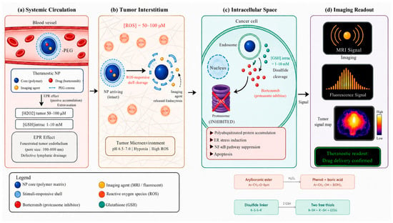
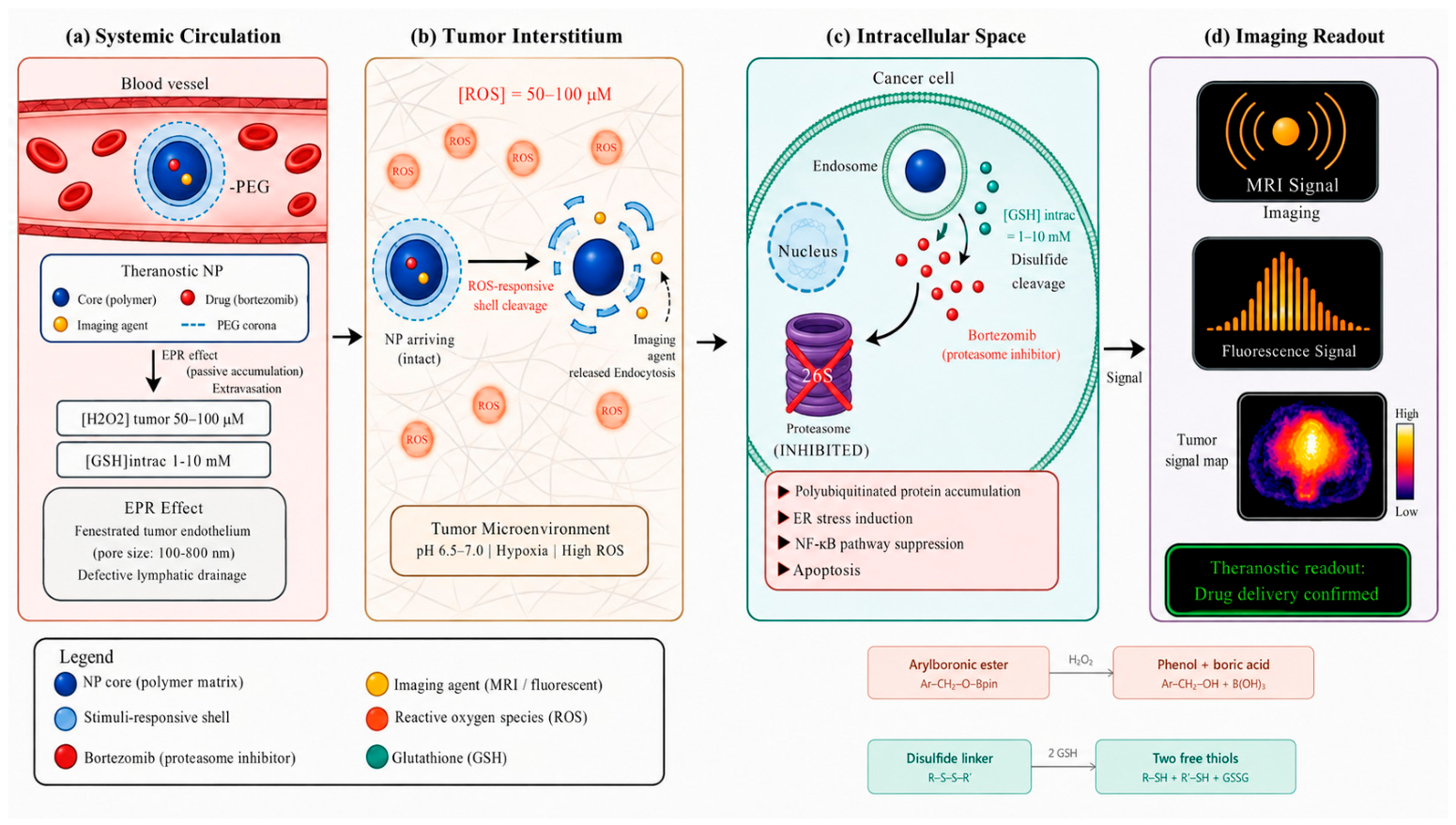
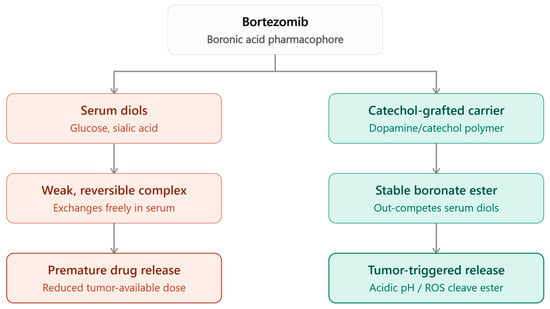
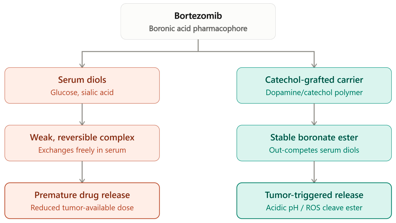
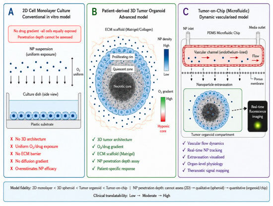
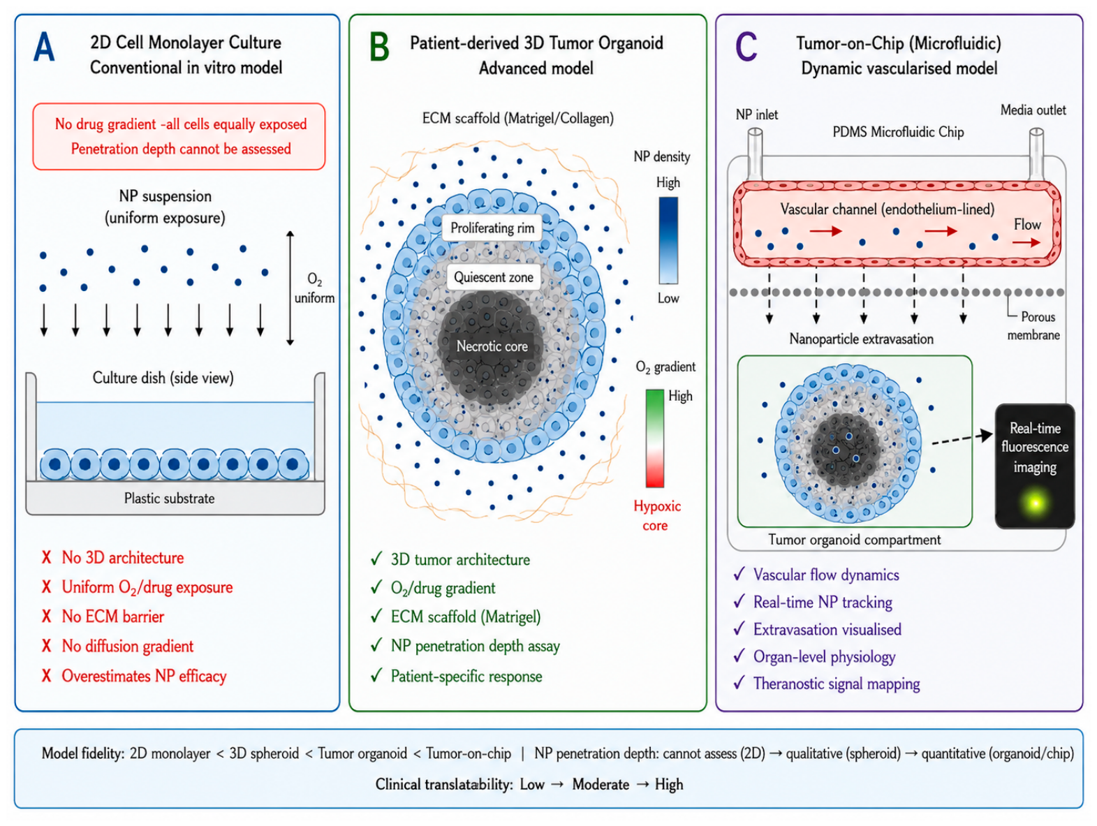
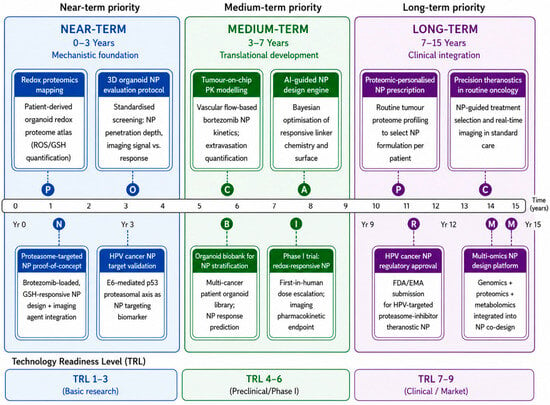
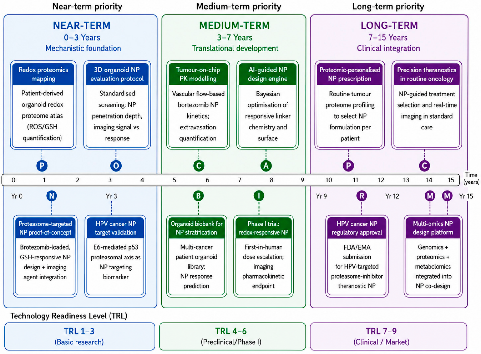

Disclaimer/Publisher’s Note: The statements, opinions and data contained in all publications are solely those of the individual author(s) and contributor(s) and not of MDPI and/or the editor(s). MDPI and/or the editor(s) disclaim responsibility for any injury to people or property resulting from any ideas, methods, instructions or products referred to in the content. |
© 2026 by the authors. Licensee MDPI, Basel, Switzerland. This article is an open access article distributed under the terms and conditions of the Creative Commons Attribution (CC BY) license.