


Abstract
Background: Kartagener syndrome (KS) is a rare subset of primary ciliary dyskinesia characterized by the triad of situs viscerum inversus (SVI), chronic sinusitis, and bronchiectasis. Laparoscopic cholecystectomy (LC) in patients with SVI is technically demanding because of mirror-image anatomy, while evidence supporting the use of indocyanine green (ICG) fluorescence in this setting is scarce. Case Presentation: We report the case of a 25-year-old woman with KS and SVI totalis who underwent elective LC for symptomatic cholelithiasis. The procedure was performed using a mirror American approach with four trocars and near-infrared ICG fluorescence cholangiography. ICG enabled real-time visualization of biliary anatomy and facilitated intraoperative orientation. The procedure was completed laparoscopically without intraoperative or postoperative complications, and the postoperative course was uneventful. Methods: A non-systematic narrative review of the literature was conducted to identify reported cases of LC in patients with SVI, including cases associated with KS. Studies published between 1991 and 2025 were retrieved from PubMed, Web of Science, Scopus, and Embase. Data were descriptively summarized, focusing on surgical technique, trocar placement, and reported use of ICG fluorescence. Results: A total of 143 articles were included. Most cases involved isolated SVI, while KS was reported only in a minority of patients. The mirror American technique and four-trocar configuration were the most frequently adopted approaches. Only three cases, including the present report, described the use of ICG fluorescence during LC in patients with SVI or KS. Conclusions: LC in patients with SVI is feasible but technically demanding. ICG fluorescence may assist intraoperative biliary orientation in complex anatomical settings; however, current evidence is extremely limited and should be considered hypothesis-generating only.
1. Introduction
Kartagener syndrome (KS) is a rare autosomal recessive disorder characterized by clinical and genetic heterogeneity [1]. Although Siewert first described KS in 1904, Kartagener first clinically characterized the syndrome in 1933 [2]. KS is a well-defined subset of primary ciliary dyskinesia (PCD), a genetically heterogeneous disorder caused by defects in the structure and function of motile cilia [3,4]. Approximately 50% of patients with PCD present with situs inversus; however, only those who also exhibit chronic sinusitis and bronchiectasis fulfill the diagnostic criteria for KS, for which its estimated prevalence is approximately 1 in 40,000 individuals in the general population [5]. The classic triad consists of chronic sinusitis, situs viscerum inversus (SVI), and bronchiectasis [2].
In the literature, no direct association between KS and cholelithiasis has been reported [2,3,6]. Currently, there is no evidence of an increased incidence of cholelithiasis in patients with KS compared to the general population [2].
Laparoscopic cholecystectomy (LC) is the surgical gold standard for symptomatic cholelithiasis [7]. One of the most serious complications of LC is iatrogenic bile duct injury (BDI) [7,8]. To reduce the risk of BDI, in 1995, Strasberg et al. suggested the identification of the Critical View of Safety (CVS) [9]. Given its critical role in surgical safety, the European Association of Endoscopic Surgery (EAES) recommends it as the best approach to cholecystectomy [10]. The CVS requires identification of Calot’s triangle and the hepatoduodenal ligament, mobilization of the gallbladder fundus, and identification, isolation, and division of the cystic duct and cystic artery [7,9].
In recent years, several technologies have been introduced to reduce the risk of BDI, including indocyanine green fluorescence (ICG) and artificial intelligence (AI) [7]. In particular, ICG facilitates identification of the CVS and, therefore, reduces the risk of BDI in cases of complex LC [6,11]. In patients with SVI, LC can be technically challenging, particularly for right-handed surgeons, due to the mirrored anatomy [2]. The first case of LC in a patient with SVI was reported in 1991, and since then, approximately one hundred cases have been described in the literature [12].
However, in the literature, only a very limited number of cases report the use of ICG fluorescence during LC in patients with SVI, and no standardized surgical technique or ICG dosage has been established. In patients with SVI, the mirrored biliary anatomy represents a potential risk factor for misidentification of biliary structures and bile duct injury during LC. In this setting, ICG fluorescence cholangiography may provide real-time visualization of the biliary tree and facilitate achievement of the CVS, particularly in complex or anatomically unfamiliar scenarios.
Therefore, the primary aim of this study is to report our experience with ICG-guided LC in a patient with KS and SVI. The secondary aim is to contextualize this experience within the existing literature on LC in patients with SVI, comparing operative approaches, trocar placement, and the reported use of ICG fluorescence.
2. Case Presentation
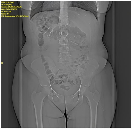
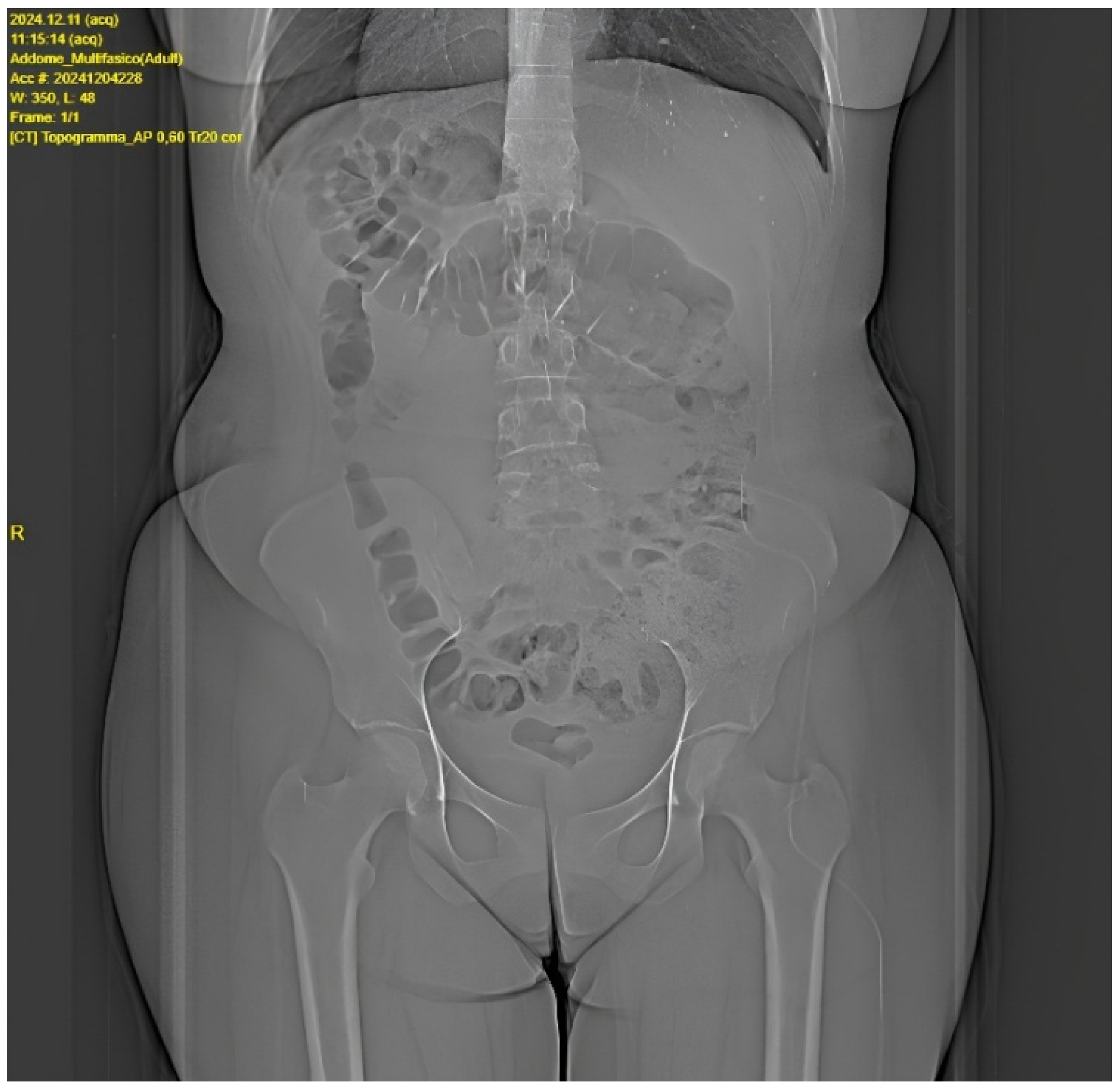
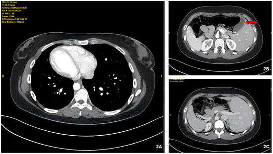
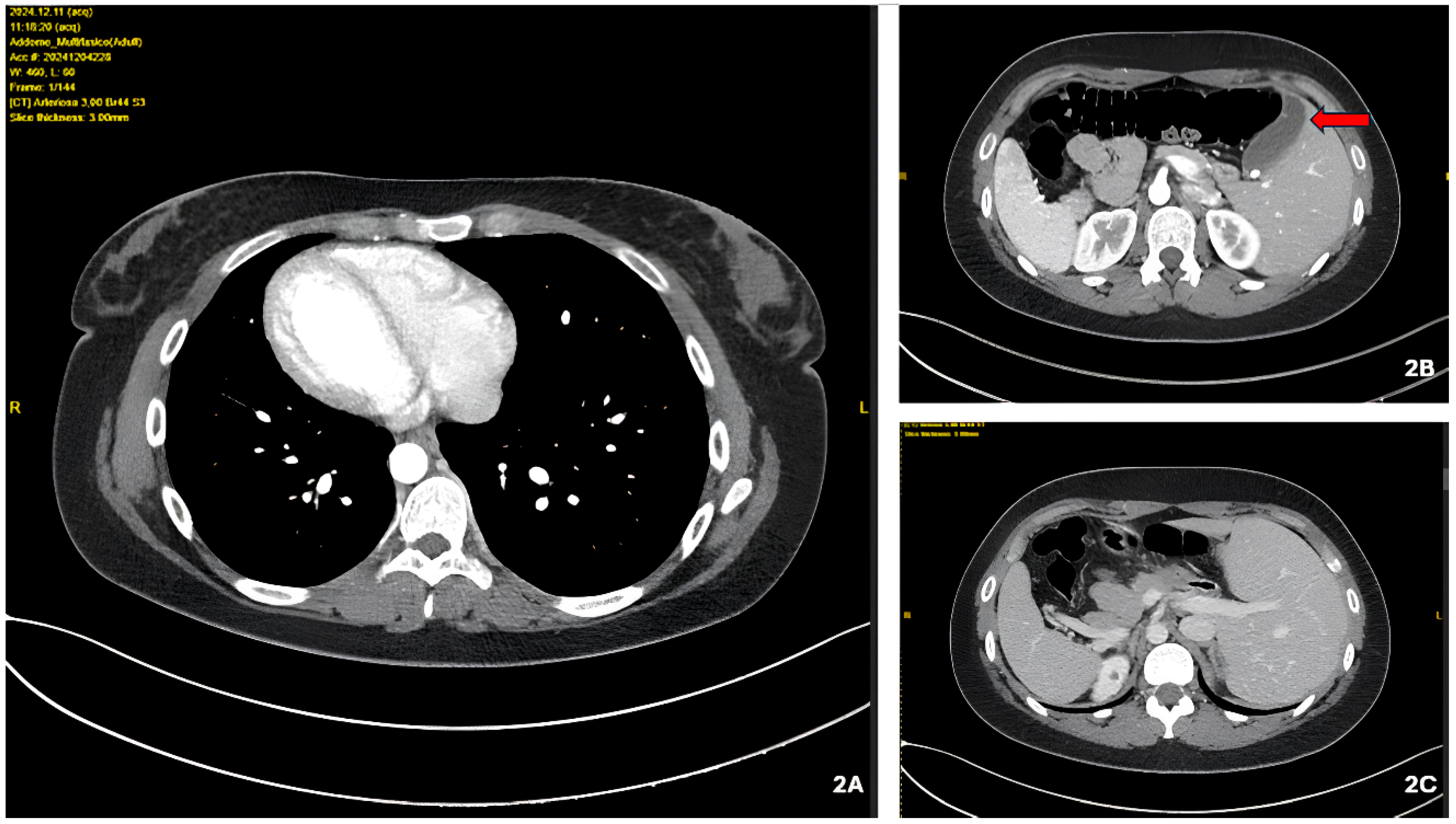
A 25-year-old Italian woman (Body Mass Index = 21.2) presented to our hospital with a history of recurrent biliary colic, with epigastric, left hypochondrium, and left subscapular pain, sometimes associated with nausea or vomiting. The patient exhibited the classic triad of KS: bronchiectasis, chronic sinusitis, and SVI totalis. KS had been previously diagnosed based on clinical and radiological findings by the pulmonology/ENT team based on the clinical phenotype and imaging (bronchiectasis and chronic sinusitis) in the setting of SVI totalis; previous medical documentation confirming the diagnosis was available in the patient’s records. Chest computed tomography demonstrated limited and stable bronchiectasis without signs of acute inflammatory activity. At baseline, the patient reported no dyspnea at rest and no recent lower respiratory tract infections. Preoperative anesthesiological evaluation revealed no contraindications to general anesthesia. Perioperatively, standard respiratory precautions were adopted (lung-protective ventilation and active airway clearance as needed), and no respiratory complications occurred. She reported no other comorbidities and had never undergone surgery. At preoperative surgical physical examination, vital signs were normal, and the patient was afebrile; the abdomen was soft and non-tender on both superficial and deep palpation, but a positive Murphy’s sign in the left hypochondrium was noted. The last biliary colic occurred three months before the preoperative visit. Laboratory tests were within normal limits. The patient was referred to our unit with a preoperative total-body computed tomography (CT) scan already performed, which clearly documented SVI totalis and cholelithiasis. Given the availability of detailed cross-sectional imaging and the need for accurate anatomical mapping in the context of mirror-image anatomy, an additional preoperative abdominal ultrasound was not considered necessary. The preoperative x-ray showed dextrocardia and the presence of the gastric fundus in the right abdominal region (Figure 1). Preoperative total body computed tomography (CT) showed SVI totalis and a 7 mm endocholecystic calculus (Figure 2). Given the history of symptomatic chronic cholelithiasis, elective LC under general anesthesia was scheduled. Standard perioperative measures were applied, including antibiotic prophylaxis according to local protocol, venous thromboembolism prevention (mechanical ± pharmacological prophylaxis based on individual risk assessment), and a multimodal postoperative analgesia regimen.
Figure 1.
Preoperative radiograph. Note the rise of the left hemidiaphragm due to the presence of the liver in the left hypochondrium.
Figure 2.
Preoperative computed axial tomography (CT) (2A) Note the dextrocardia; (2B) the red arrow indicates the gallbladder in left abdominal position; (2C) position of abdominal organs and vessels in mirrored anatomical position. The overall morphological picture, thoracic and abdominal, reflects SVI totalis.
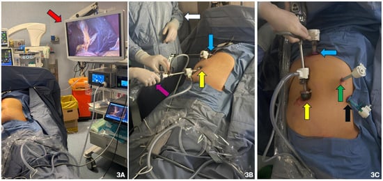
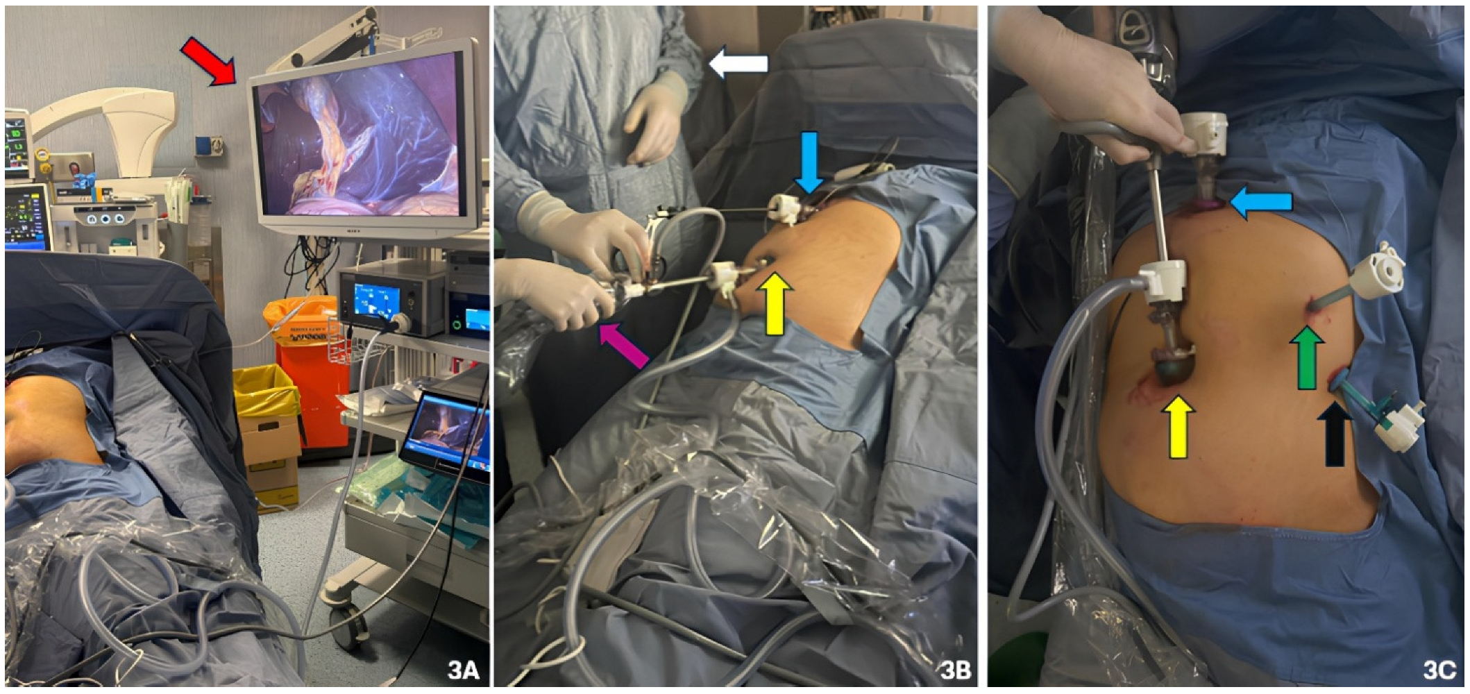
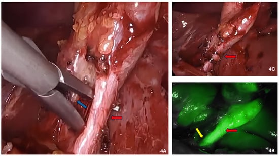
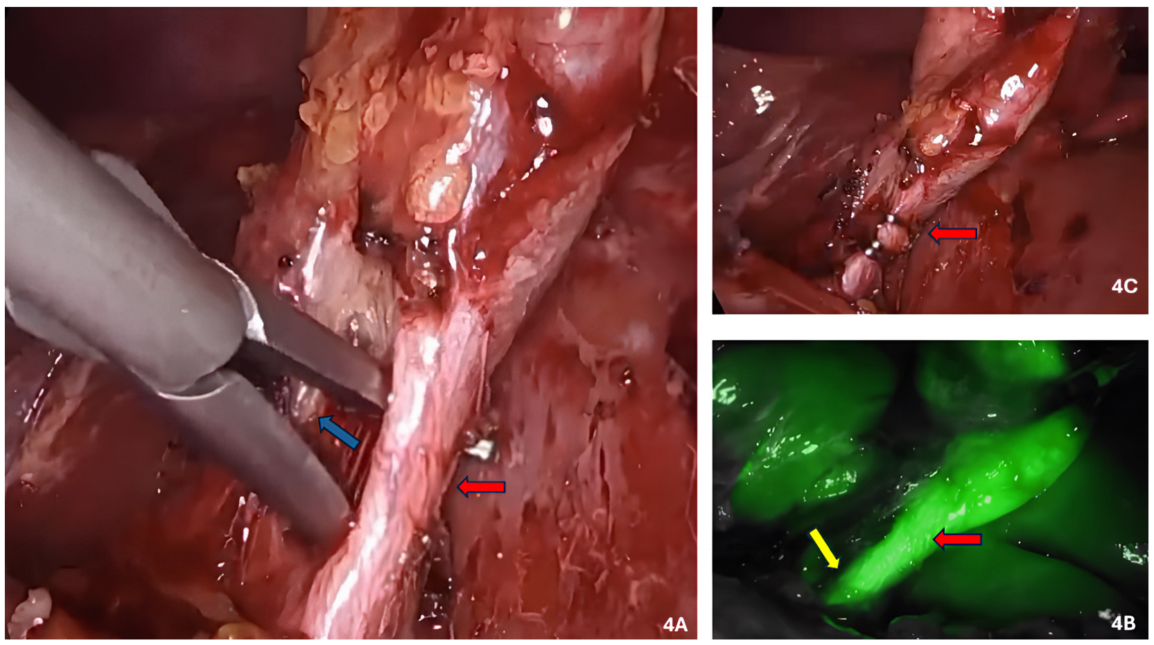
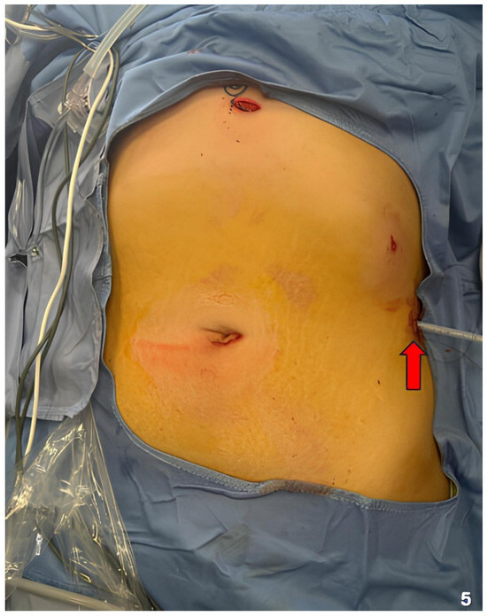
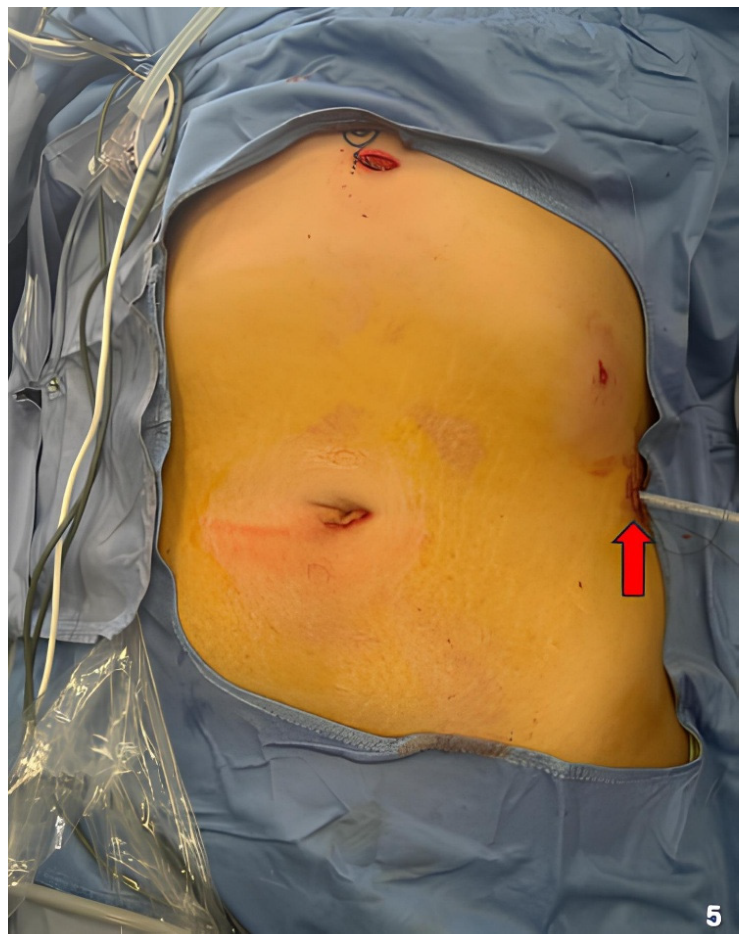
Before the surgical procedure, the patient signed informed consent. We diluted 25 mg of ICG in 10 mL of sterile normal saline. Then, the patient received a bolus of 0.2 mg/kg intravenously one hour before surgery. Fluorescence was displayed as an overlay on white-light imaging using the Stryker 1688 AIM 4K platform with a 30° laparoscope. Before ICG administration, contraindications were assessed, including a history of hypersensitivity reactions and liver function tests; no ICG-related adverse events occurred. To perform the LC, we adopted the “Mirror American” (MA) surgical technique. Therefore, we positioned the monitor on the left side of the patient and the first and second operators on the right side of the patient. The patient was placed in the reverse Trendelenburg position and lateralized on her right side. Under general anesthesia, LC began with a subumbilical minilaparotomy with an open technique. A 12 mm optical trocar was then inserted at this site. Exploration of the entire abdominal cavity confirmed SVI. The gallbladder appeared distended with inflammatory adhesions. We placed three more trocars: one 12 mm trocar subxiphoid, one 5 mm mid-clavicular left, and another 5 mm on the left anterior axillary subcostal line (Figure 3). Dissection and clip application were performed with the surgeon’s left hand through xiphoid access, while with their right hand, they retracted the Hartmann’s pouch through the trocar on the left mid-clavicular line. The second operator retracted the fundus with their right hand through the trocar positioned on the left anterior axillary line and moved the laparoscopic optic with their left hand through the periumbilical optical trocar. After initial adhesiolysis, we opened the anterior and posterior peritoneal layers. Subsequently, we isolated, clipped, and sectioned the elements of Calot’s triangle (cystic artery and cystic duct) under ICG fluorescence guidance. ICG fluorescence clearly highlighted the biliary anatomy and allowed reliable identification of the cystic duct and the common bile duct (Figure 4). Then, we performed a retrograde cholecystectomy and placed the gallbladder in a bag. A subhepatic drain was placed because of the initial inflammatory adhesions and the need to monitor for early postoperative bleeding or bile leakage in an anatomically unfamiliar setting; no bile leak was observed, and the drain remained serous and was removed on postoperative day 2. Finally, we extracted the gallbladder, removed the trocars under direct vision, and closed the abdominal parietal accesses of the trocars in layers (Figure 5). The primary operating surgeon was left-handed. During most of the operating times of the LC, their left hand held and used the monopolar hook, while their right hand used the Johann forceps. In contrast, the second operator was right-handed. The procedure lasted 75 min. The postoperative course was uneventful. In the postoperative period, we did not observe any complications, and the patient did not report abdominal pain, nausea, or vomiting. Six hours after surgery, the patient was fully mobilized. The drain was always free of serum or blood material, and we removed it on the second postoperative day (II POD) at the time of discharge.
Figure 3.
Position of laparoscopic operators and instruments. (3A) The red arrow indicates the position of the monitor to the left of the patient; (3B) the white arrow indicates the left-handed first surgeon to the right of the patient; the purple ferrule indicates the second surgeon to the right of the patient and to the right of the first operator; the yellow arrow indicates the 10 mm umbilical optical trocar; the blue arrow indicates the 10 mm subxiphoid trocar; (3C) the yellow arrow indicates the 10 mm umbilical optical trocar; the blue arrow indicates the 10 mm subxiphoid trocar; the green arrow indicates the 5 mm trocar on the left midclavicular line; the black arrow indicates the 5 mm trocar on the anterior axillary line.
Figure 4.
Intraoperative images during LC. (4A) Isolation of the cystic duct (red arrow) and posterior cystic artery (blue arrow); (4B) ICG identifies the cystic duct (red arrow) and the main biliary duct (yellow arrow); (4C) clipping and sectioning of the cystic duct (red arrow).
Figure 5.
At the end of the procedure, after removal of the trocars, a drainage tube was placed on the left side (red arrow).
Postoperative follow-up was performed 7 days after discharge and subsequently after another 30 days. No late biliary symptoms or related complications were reported beyond 30 days. The patient had normal vital parameters; the abdomen was soft, non-distended, and non-tender on superficial and deep palpation. After an abdominal ultrasound showed no intra-abdominal fluid collections, we removed the skin sutures made of absorbable multifilament thread. Histopathologic analysis showed chronic cholecystitis.
This case has been reported in line with the Case Report (CARE) Guidelines to ensure the accuracy and completeness of the report [13,14].
3. Discussion
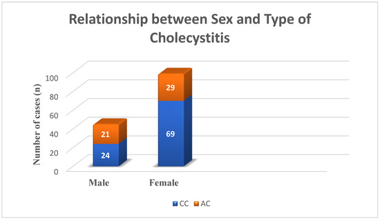
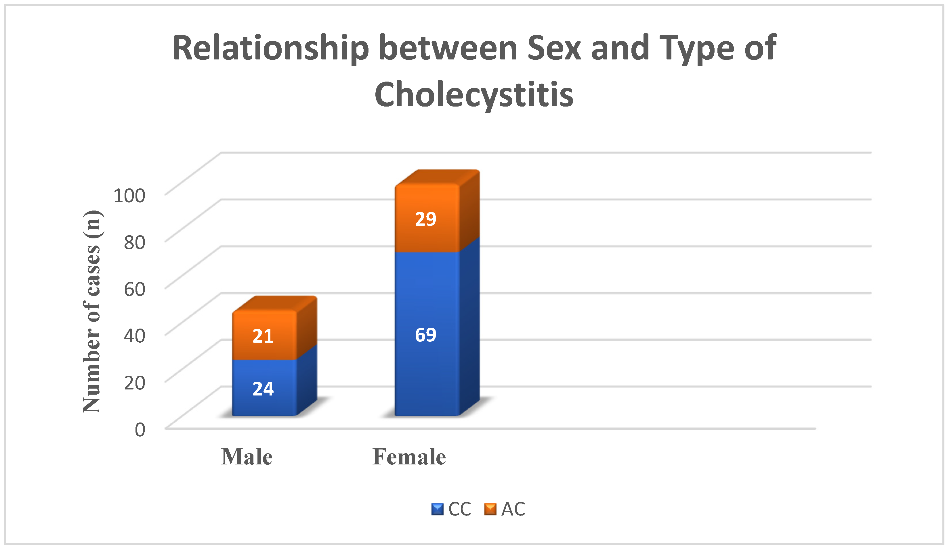
KS is a rare autosomal recessive genetic disorder, with an estimated prevalence ranging from approximately 1 in 30,000 to 1 in 40,000 individuals in the general population, characterized by SVI, bronchiectasis, and sinusitis [15,16,17]. SVI exists in partialis and totalis forms [18]. In this manuscript, the term SVI is used as a comprehensive term including both totalis and partialis forms. SVI partialis is characterized by total anatomical mirror inversion of either the thoracic organs or only the abdominal organs [18]. SVI totalis presents anatomical mirror inversion of both the thoracic and abdominal organs [18]. In these patients, symptomatic cholelithiasis may present with a different clinical manifestation compared with typical cholelithiasis [19]. The mirrored anatomy may result in atypical pain distribution mainly in the left hypochondrium but, in some cases, also in the epigastrium (30%) and in the right hypochondrium (10%) [2]. Less frequently, the authors report pain in the left subscapular area, as occurred in our case [2,20]. Therefore, the preoperative assessment of symptomatic cholelithiasis in these patients should always consider their congenital anatomical alteration and the different clinical phenomenology presentations [2,21]. Among the preoperative comorbidities, bronchiectasis may influence perioperative anesthetic management, even if, in the literature, no particular adverse events related to it are reported [22]. This is related to structural and functional abnormalities of motile cilia or non-functioning conditions [16]. Epidemiologically, LC in these patients is more common in females, and the average age is 46 [1,2]. From our review, in these patients, chronic cholecystitis (CC) is more frequent than acute cholecystitis (AC) in females, while it is almost superimposable in incidence in males (Figure 6). Almost all patients undergoing LC had SVI.
Figure 6.
Relationship between sex and type of cholecystitis. Percentages were calculated using the total number of included cases (n = 143). CC: Chronic cholecystitis; AC: acute cholecystitis.
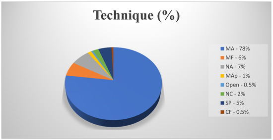
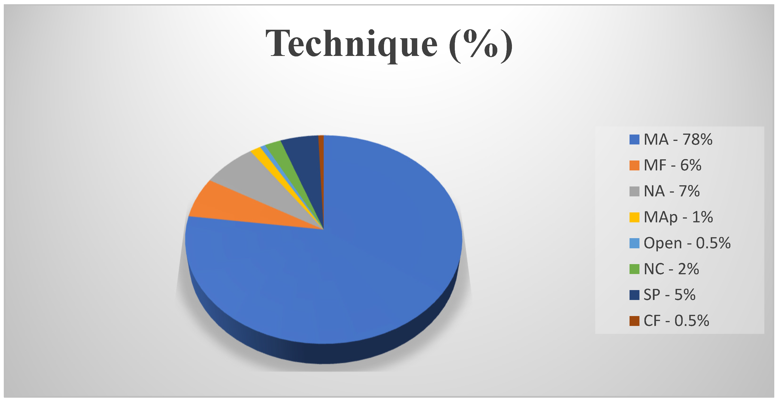
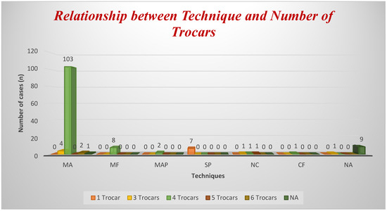
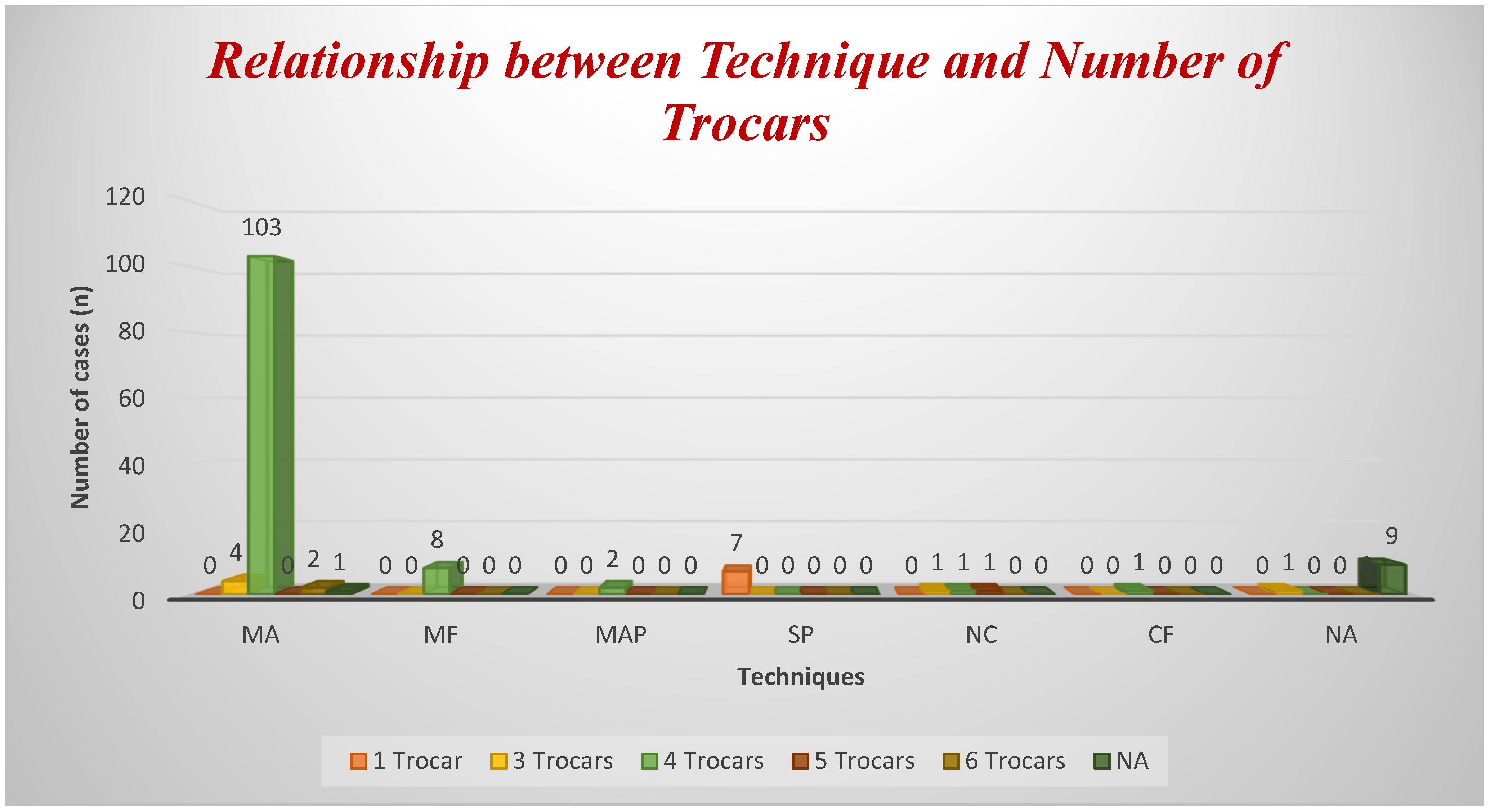
LC is also the gold standard in patients with SVI who have symptomatic cholelithiasis [2,20,21]. In this type of LC, the main difficulties concern the different arrangements of the operators and devices in the operating room and the presence of a mirrored anatomy [16,23,24]. Other important topics of discussion concern the position of the surgeons (mirror French technique [MF] vs. mirror American [MA] technique), the number of trocars to be used, and any postoperative complications. Currently, there is no standardization of LC in patients with KS [2]. However, from our review of the literature, common points emerge between the different authors. First, patients with KS do not seem to have more comorbidities than the general population [2]. Furthermore, bronchiectasis does not seem to negatively influence the anesthetic course, and in general, few authors address this issue [2]. In our case, the patient’s bronchiectasis did not negatively influence the pre- and post-operative anesthetic course. Over the years, three techniques have been described to perform LC in patients with SVI: MF, MA, and mixed approach (MAp) [2,11,25]. In only one case, a rendezvous was performed in a patient with SVI [26]. From our review, the technique most used and recommended by the authors is the MA (Figure 7). In a smaller number of cases, the authors preferred MF, while MAp was described in only three cases (Figure 7). We found only one case of conversion from LC to open cholecystectomy [27]. In 115 cases, the authors performed LC with four trocars (Figure 8). The use of single-port surgery occurred in seven cases (Figure 8).
Figure 7.
Percentages of surgical technique. Percentages were calculated using the total number of included cases (n = 143). MA: Mirror American; MF: mirror French; MAp: mixed approach; SP: single port; NC: not classifiable; CF: classical French; NA: not available; Open: open cholecystectomy.
Figure 8.
Relationship between technique and number of trocars. Percentages were calculated using the total number of included cases (n = 143). MA: Mirror American; MF: mirror French; MAp: mixed approach; SP: single port; NC: not classifiable; CF: classical French; NA: not available.
In the literature, the first case of a patient with KS undergoing LC dates back to 1991 [12]. In this case, the authors employed MA and four trocars; the first operator was right-handed [12]. The authors did not describe any difficulties during LC, nor in the postoperative period [12]. Takei et al. also performed LC with ease but without describing the surgical technique or providing specific technical notes [28]. Furthermore, in this case, the authors did not report the position of the operators, nor the number of trocars used [28]. Lipschutz et al. were the first to perform LC and endoscopic retrograde cholangiopancreatography with sphincterotomy [29]. However, even in this case, the authors do not describe the details of the surgical technique or the position of the operators [29]. Since 1991, the most widely used surgical technique has been MA (Figure 7). Left-handed surgeons benefit most from it because they can use their left hand to clip and dissect the anatomical elements and detach the gallbladder from the liver through the subxiphoid trocar [15,23]. In addition, the position of the first assistant on the right of the patient allows them to both maneuver the laparoscopic scope and use the fourth trocar on the left anterior axillary line [3,4,15,24]. Furthermore, in most cases of MA, the number of trocars used is four (Figure 8). Most authors state the need for high coordination between the operating surgeons during the LC [2]. In most cases, the first operator uses the right hand to retract the infundibulum and the left hand to dissect into the epigastric port [2,4,28,30,31]. In other cases, given the difficulty of performing the dissection with the left hand through the subxiphoid trocar, a second operator retracted the cystic infundibulum [2,4,15,16,23,24,25]. In fewer cases, the authors adopt the MF position, with the first operator between the patient’s spread legs, the second operator on the left of the patient, and the monitor on the left of the patient (Figure 8) [32,33,34,35]. Although MF is considered safe and effective, the greatest difficulty is the overlapping of laparoscopic instruments, especially for left-handed surgeons [23,25,33,34]. To avoid this problem, the MAp is safe and effective in cases of LC performed by a right-handed surgeon [2]. In this case, the first operator is on the right of the patient, while the second operator is between the patient’s legs (MF position); the arrangement of the trocars reflects the MA [2]. The authors report having obtained a greater view of the operating field and reduced interference between laparoscopic instruments [2]. In fact, this mixed position is a sort of MA translating from the right to the left: The first operator maintains the position on the right of the patient, while the second operator, instead of being on the right of the patient as in the MA, translates centrally between the patient’s legs, as in the MF [2]. In this position, the second operator uses the fourth trocar to traction the anterior peritoneal layer [2]. Overall, the number and type of postoperative complications were minimal. Indeed, Meza-Martinez et al. reported bilateral pulmonary edema, atelectasis, tachypnea, and desaturation [15]; Aburayya et al. described the occlusion of the common hepatic duct (CHD) [16]. In other cases, LC was concomitant with other surgeries. In fact, Taskin et al. performed it together with gastric banding using six trocars [36]; Djohan et al. associated laparoscopic appendectomy (LA), using four trocars in MA [37]; similarly, Borgaonkar et al. performed LA [38]. Finally, in other cases, LC has been associated with endoscopic retrograde cholangiopancreatography (ERCP) (Table 1) [29,39,40,41].
In the context of SVI, the role of ICG becomes particularly relevant, as the unfamiliar orientation of the biliary anatomy may increase the risk of misinterpretation during dissection. In standard LC, the reported clinical benefit of ICG fluorescence remains equivocal. While some studies have suggested potential advantages such as shorter operative times or lower conversion rates, other data do not confirm these findings; notably, Ambe et al. reported no significant differences between LC performed with and without ICG [11,42]. This discordance may reflect differences in study design, sample size, and statistical power; case complexity; surgeon experience; and heterogeneity in outcome definitions (e.g., operative time measured from skin incision vs. dissection time and variable thresholds for conversion). Therefore, any extrapolation of potential benefits to the SVI setting should be made cautiously, and the rationale for ICG in SVI should primarily be framed as improved intraoperative anatomical orientation rather than as an established reduction in operative time or conversion. In the literature, we found only two previous reports describing ICG fluorescence during LC in patients with SVI, and only one of these explicitly involved confirmed KS; including our case, three reports describe ICG use in SVI with/without KS. Tirelli et al [14]. described adopting a position with the first right-handed operator on the right of the patient (a 29-year-old woman with chronic cholecystitis) and the second operator between the patient’s legs, using four trocars [14,42]. Therefore, the position described is the MAp. The second operator maneuvered the laparoscopic optic with the left hand and tractioned the gallbladder infundibulum with the right hand [42]. We selected 0.2 mg/kg administered 60 min before skin incision to obtain biliary fluorescence with low background liver signals at the time of Calot’s triangle dissection. This regimen is consistent with the timing and dosing reported by Tirelli et al. [14], while other reports used shorter intervals; overall, published protocols remain heterogeneous, and no standardized regimen exists [14,42]. Finally, they did not report postoperative complications; discharge occurred on the first postoperative day. The difference compared to our approach is therefore in the position of the operators and in the function of the right hand of the second operator, who does not traction the gallbladder fundus. This difference is probably due to the right-handedness of the first operator of Tirelli compared to our case, while ours is left-handed. The other case concerns a 32-year-old man with SVI and a history of cholelithiasis [6]. The first operator and the camera operator positioned themselves on the right side of the patient, while the second operator positioned themselves on the left side [6]. The surgeons used four trocars, arranged in MA [6]. The right hand of the second operator cranially tractioned the gallbladder fundus, while the left hand of the first right-handed operator retracted the gallbladder infundibulum laterally [6]. With their right hand, the first operator performed the maneuvers of dissection, clipping, sectioning, and clotting during the LC [6]. The procedure lasted 45 min and the patient was discharged the day after surgery [6]. ICG was administered approximately half an hour before the start of LC [6]. Including our case, it can therefore be inferred that the three LCs using ICG were always performed using four trocars, with the MA position in two cases and MAp in the third. The first operator was always positioned to the right of the patient: In two cases, the operator was right-handed, while in our case, the operator was left-handed. The second operator always had a fundamental role in all three LCs and always had different positions. In fact, in our case, the second operator was positioned to the right of the patient and to the right of the first operator: With their left hand, they operated the laparoscopic optic, while with their right hand, they cranially tracted the gallbladder fundus. In the case reported by Tirelli et al. [14], the surgeon was positioned between the patient’s legs, operating the optic with their left hand and laterally retracting the infundibulum [43]. Finally, in the case of Rungsakulkij et al., the surgeon was positioned to the left of the patient and tracted the gallbladder fundus laterally, while another operator operated to the right of the patient and operated the laparoscopic optic [6]. Given the small number (3) of cases in the literature, including ours, we can state that there is no technique superior to the others for the execution of this type of LC. However, consistent with previous reports, it is always necessary for the surgical intervention to be performed by expert surgeons because coordination between the operators is crucial for the final outcome. Likewise, we can state that the presence of an expert left-handed first operator facilitates the execution of this type of LC, as they can use the instruments more ergonomically with their left hand, and consequently, the overlapping of the laparoscopic instruments does not occur. Therefore, the right-handed surgeon requires a more experienced assistant surgeon (second operator) with whom they already have excellent coordination, whether pulling the fundus or the gallbladder infundibulum. Finally, the presence of a third operator operating the laparoscopic optic could help the second operator by relieving them of the need to use their left hand to operate on the optic. For the same reason, we can state that intraoperative timing is operator-dependent. Finally, we agree with the other two authors that ICG has been of great help in identifying the biliary tract in an anatomical context that is certainly not familiar given its mirrored appearance. Notably, most of the reported cases involved isolated SVI without a confirmed diagnosis of KS (Table 1). This finding suggests that KS represents only a minority of reported cases undergoing LC and that most available surgical evidence primarily reflects anatomical challenges related to SVI rather than KS-specific clinical features. For this reason, the use of ICG in LC patients with SVI appears to be a useful adjunct for intraoperative anatomical orientation.
Although the available evidence is limited to three published reports, including the present case, ICG fluorescence appears to be a useful adjunct for intraoperative biliary visualization in this anatomical setting. However, these observations are based on a very small number of cases and cannot be generalized; further reports are needed to better define its potential role.
4. Narrative Review Methodology
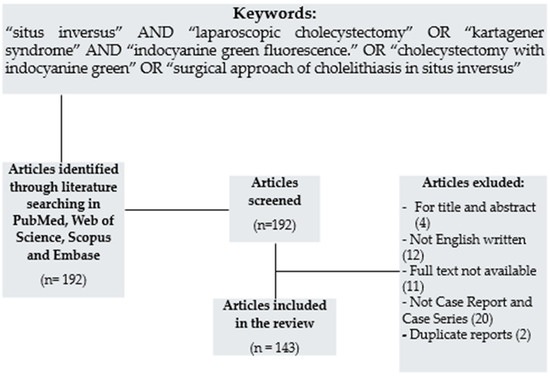
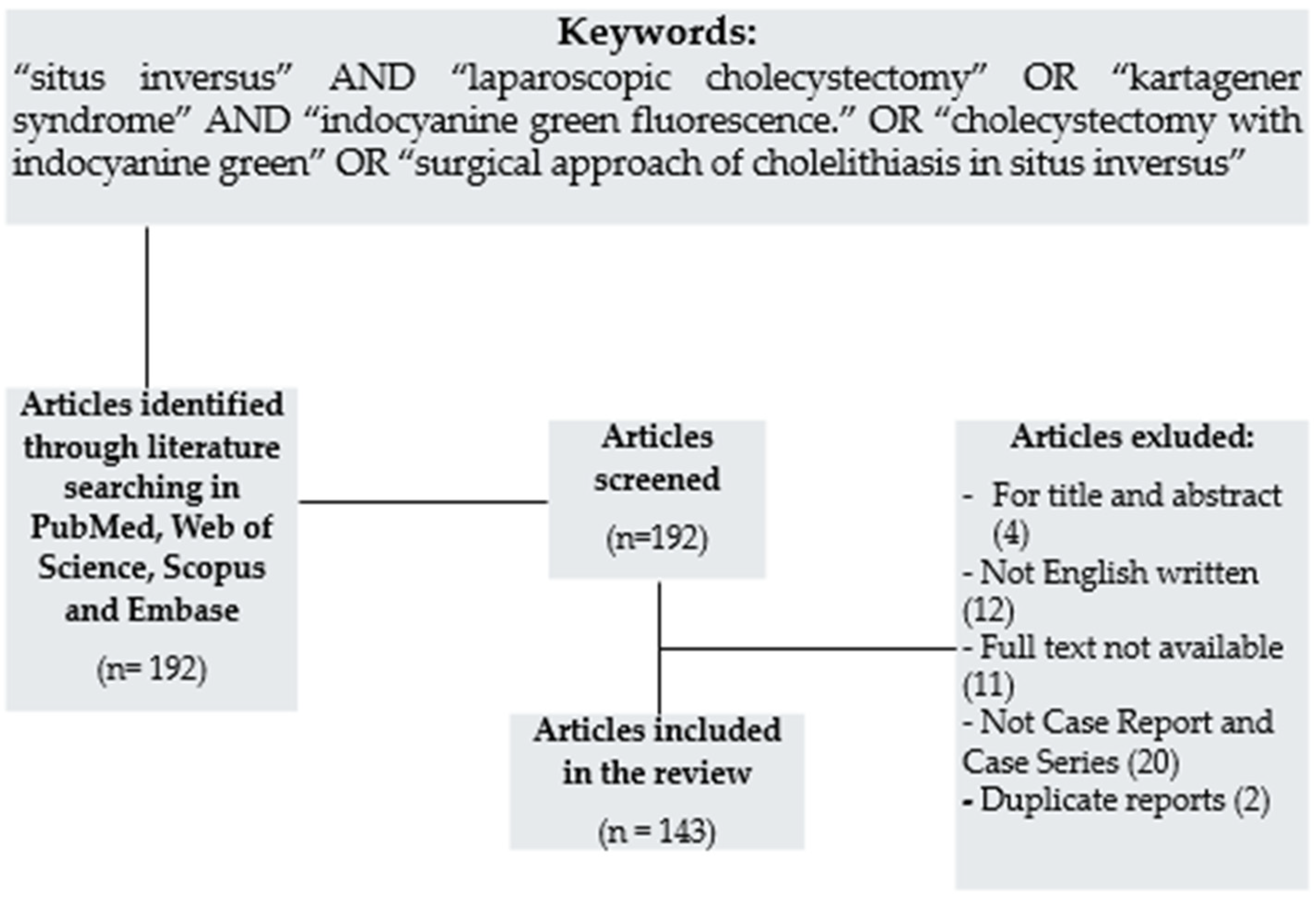
We performed a narrative, non-systematic review aimed to provide a descriptive overview of surgical techniques reported for LC in patients with SVI, including cases associated with KS. This review was not designed as a systematic review or meta-analysis, and no formal protocol registration was performed. Given the rarity of the condition, the predominance of case reports/small case series, and the marked heterogeneity in reporting and surgical techniques, a systematic review with meta-analysis was not feasible. We searched PubMed, Web of Science, Scopus, and Embase for studies on LC published from 1 January 1991 to 30 June 2025 using the following keywords: “situs inversus” AND “laparoscopic cholecystectomy” OR “Kartagener syndrome” AND “indocyanine green fluorescence” OR “cholecystectomy with indocyanine green” OR “surgical approach of cholelithiasis in situs inversus” (Figure 9). The search strategy was intentionally broad to maximize sensitivity. The last search was performed on 30 June 2025. Filters were limited to the English language and the availability of the abstract and full text. When articles reported more than one patient, each patient was extracted and recorded as an individual case. Missing data were coded as “NR” and were not imputed. Given the descriptive aim of this work and the predominance of case reports, no formal quality assessment or PRISMA-based systematic reporting was undertaken (Figure 9). We included only articles written in English with available abstracts and full texts, including case reports and case series, whether monocentric or multicentric, or prospective or retrospective. Patients could be of any age with a clear diagnosis of SVI (partialis or totalis). When explicitly reported, the presence of KS was recorded as a comorbidity (SVI with bronchiectasis and chronic sinusitis). To ensure data accuracy, all extracted cases were independently reviewed by two authors. When an article reported more than one patient, each patient was extracted and recorded as an individual case (one row per case) in Table 1. Variables extracted included year, sex, age, presentation (acute/chronic), situs inversus type, reported KS diagnosis, operative setup/technique, trocar number, adjunct intraoperative imaging (e.g., cholangiography/ERCP/ICG), and postoperative outcomes when available. Missing data were coded as ‘NR’ and were not imputed. Duplicate reports and overlapping case descriptions were identified and removed after cross-checking author names, publication year, patient demographics, and surgical details. Discrepancies were resolved by consensus. We focused on articles describing the surgical technique, the number of trocars used, and postoperative complications. If these variables were not available, the articles were still included when other relevant data were reported, such as age, sex, and preoperative acute or chronic cholelithiasis. If some of this data was not available, we reported this in Table 1 as not reported (NR). We excluded duplicates (including overlapping case descriptions), incomplete reports, and non-peer-reviewed preprint articles. No formal quality appraisal was performed because the objective of this non-systematic narrative review was descriptive (mapping of technical variables), and the evidence largely consisted of anecdotal case reports/case series. Given the descriptive purpose of this non-systematic narrative review and the heterogeneity and incomplete reporting of several variables across individual case reports, no formal descriptive statistical analysis was performed.
Figure 9.
Literature search and selection process for the narrative review.
Therefore, 143 articles were included in the final analysis, and the results of the review are presented descriptively, without quantitative synthesis. Table 1 reports the individual cases extracted from the literature and represents the dataset used for the descriptive analysis.
Table 1.
The 143 articles divided by category. SVI: Situs viscerum inversus; F: female; M: male; CC: chronic cholecystitis; AC: acute cholecystitis; T: totalis; P: partialis; MA: mirror American; MF: mirror French; Map: mixed approach; NA: not available; NC: not classifiable; CF: classical French; NO: absent; ICG: indocyanine Green; KS: Kartagener syndrome.
5. Conclusions
LC in patients with SVI is technically more demanding than standard procedures due to the mirrored biliary and vascular anatomy and the need for ergonomic adaptation by the surgical team. In patients with associated KS, additional perioperative considerations may arise due to underlying respiratory involvement. In this anatomically complex setting, ICG cholangiography allows real-time visualization of the biliary tree and may facilitate intraoperative anatomical orientation during dissection. Although the available evidence is limited to a very small number of reported cases, including ours, no procedure-related complications were reported in association with ICG use. Given the lack of comparative data, outcome events, and adequate statistical power, the present findings should be considered hypothesis-generating only. According to the Oxford Centre for Evidence-Based Medicine, the level of evidence of the available data corresponds to Level 4, and no conclusions regarding clinical effectiveness, risk reduction, or reproducibility can be drawn.
Author Contributions
Conceptualization, A.F.; methodology, G.S., V.V., and L.R.; software, A.P. and V.D.; validation, M.A. and Y.B.A.; formal analysis, F.C. and A.C. (Alessio Cece); investigation, A.F. and G.S.; resources, A.F. and V.V.; data curation, A.F., L.R., V.D., Y.B.A., and F.C.; writing—original draft preparation, A.F.; writing—review and editing, A.A. and F.K.; visualization, G.S., V.V., A.P., A.C. (Armando Calogero); supervision, G.S. and F.K.; project administration, A.F. and F.K. All authors have read and agreed to the published version of this manuscript.
Funding
This research received no external funding.
Institutional Review Board Statement
Ethical review and approval were waived because this study reports a retrospective description of routine clinical care in a single patient, with fully anonymized data and no experimental intervention. According to institutional policy and national regulations governing clinical research, ethics committee approval is not required for the publication of individual case reports. Written informed consent for publication was obtained from the patient.
Informed Consent Statement
Informed consent was obtained from all subjects involved in this study. Written informed consent has been obtained from the patient(s) to publish this paper.
Data Availability Statement
The data presented in this study are available upon request from the corresponding author (the data are not publicly available due to privacy restrictions).
Conflicts of Interest
The authors declare no conflicts of interest and that they have no known competing financial interests or personal relationships that could have appeared to influence the work reported in this paper.
Abbreviations
The following abbreviations are used in this manuscript:
| KS | Kartagener syndrome |
| LC | Laparoscopic cholecystectomy |
| SVI | Situs viscerum inversus |
| ICG | Indocyanine green |
| MA | Mirror American technique |
| MF | Mirror French technique |
| MAp | Mixed approach technique |
| CF | Classical French technique |
| SP | Single port |
| AC | Acute cholecystitis |
| CC | Chronic cholecystitis |
| NA | Not available |
| NC | Not classifiable |
References
- Chaouch, M.A.; Jerraya, H.; Dougaz, M.W.; Nouira, R.; Dziri, C. A Systematic Review of Laparoscopic Cholecystectomy in Situs Inversus. J. Investig. Surg. 2021, 34, 324–333. [Google Scholar] [CrossRef] [Scilit]
- Palomba, G.; Dinuzzi, V.P.; Manigrasso, M.; Milone, M.; De Palma, G.D.; Aprea, G. Laparoscopic Cholecystectomy with a Mixed Approach in a Patient with Kartagener Syndrome: Technical Report and Review of Literature. Indian J. Surg. 2021, 83, 411–417. [Google Scholar] [CrossRef] [Scilit]
- He, T.; Zou, J.; Song, H.; Yi, B.; Sun, K.; Yang, J.; Lei, T.; Xu, L.; Li, G. Laparoscopic Cholecystectomy in a Patient with Situs Inversus Totalis Presenting with Cholelithiasis: A Case Report. Front. Surg. 2022, 9, 874494. [Google Scholar] [CrossRef] [Scilit] [PubMed]
- Enciu, O.; Toma, E.A.; Tulin, A.; Georgescu, D.E.; Miron, A. Look beyond the Mirror: Laparoscopic Cholecystectomy in Situs Inversus Totalis—A Systematic Review and Meta-Analysis (and Report of New Technique). Diagnostics 2022, 12, 1265. [Google Scholar] [CrossRef] [Scilit]
- Wang, L.; Zhao, X.; Liang, H.; Zhang, L.; Li, C.; Li, D.; Meng, X.; Meng, F.; Gao, M. Novel Compound Heterozygous Mutations of DNAH5 Identified in a Pediatric Patient with Kartagener Syndrome: Case Report and Literature Review. BMC Pulm. Med. 2021, 21, 263. [Google Scholar] [CrossRef] [Scilit]
- Rungsakulkij, N.; Tangtawee, P. Fluorescence Cholangiography during Laparoscopic Cholecystectomy in a Patient with Situs Inversus Totalis: A Case Report and Literature Review. BMC Surg. 2017, 17, 43. [Google Scholar] [CrossRef] [Scilit]
- Fernicola, A.; Palomba, G.; Capuano, M.; De Palma, G.D.; Aprea, G. Artificial Intelligence Applied to Laparoscopic Cholecystectomy: What Is the next Step? A Narrative Review. Updates Surg. 2024, 76, 1655–1667. [Google Scholar] [CrossRef] [Scilit]
- Waage, A.; Nilsson, M. Iatrogenic Bile Duct Injury: A Population-Based Study of 152 776 Cholecystectomies in the Swedish Inpatient Registry. Arch. Surg. 2006, 141, 1207–1213. [Google Scholar] [CrossRef] [Scilit]
- Strasberg, S.M.; Hertl, M.; Soper, N.J. An Analysis of the Problem of Biliary Injury during Laparoscopic Cholecystectomy. J. Am. Coll. Surg. 1995, 180, 101–125. [Google Scholar]
- Eikermann, M.; Siegel, R.; Broeders, I.; Dziri, C.; Fingerhut, A.; Gutt, C.; Jaschinski, T.; Nassar, A.; Paganini, A.M.; Pieper, D.; et al. Prevention and Treatment of Bile Duct Injuries during Laparoscopic Cholecystectomy: The Clinical Practice Guidelines of the European Association for Endoscopic Surgery (EAES). Surg. Endosc. 2012, 26, 3003–3039. [Google Scholar] [CrossRef] [Scilit]
- Gangemi, A.; Danilkowicz, R.; Elli, F.E.; Bianco, F.; Masrur, M.; Giulianotti, P.C. Could ICG-Aided Robotic Cholecystectomy Reduce the Rate of Open Conversion Reported with Laparoscopic Approach? A Head to Head Comparison of the Largest Single Institution Studies. J. Robot. Surg. 2017, 11, 77–82. [Google Scholar] [CrossRef] [Scilit]
- Campos, L.; Sipes, E. Laparoscopic Cholecystectomy in a 39-Year-Old Female with Situs Inversus. J. Laparoendosc. Surg. 1991, 1, 123–126. [Google Scholar] [CrossRef] [Scilit]
- Gagnier, J.J.; Kienle, G.; Altman, D.G.; Moher, D.; Sox, H.; Riley, D.; Allaire, A.; Aronson, J.; Carpenter, J.; Gagnier, J.; et al. The CARE Guidelines: Consensus-Based Clinical Case Report Guideline Development. J. Diet. Suppl. 2013, 10, 381–390. [Google Scholar] [CrossRef] [Scilit] [PubMed]
- Tirelli, F.; Grieco, M.; Biondi, A.; Belia, F.; Persiani, R. Laparoscopic Cholecystectomy with Indocyanine Green Fluorescence in Patient with Situs Inversus Totalis. Mini-Invasive Surg. 2021, 5, 15. [Google Scholar] [CrossRef] [Scilit]
- Meza-Martinez, D.A.; Palomino-Payan, J.A.; Andrade-Ordoñez, S.J.; Fematt-Rodriguez, B.J.; Muñuzuri-Camacho, M.A.; Gonzalez-Quezada, A. Overcoming Anatomical Challenges: Laparoscopic Cholecystectomy in Situs Inversus Totalis. Cureus 2023, 15, e46371. [Google Scholar] [CrossRef] [Scilit] [PubMed]
- Aburayya, B.I.; Obeidat, L.R.; Kitana, F.I.; Al Khatib, O.; Romman, S.; Hamed, O.H. Complete Common Bile Duct Injury after Laparoscopic Cholecystectomy in Situs Inversus Totalis: A Case Report, Review of the Literature and Illustrative Case Video. Int. J. Surg. Case Rep. 2024, 115, 109265. [Google Scholar] [CrossRef] [Scilit]
- Choumad, S.; Qajia, H.; Abourak, C.; Ebedda, A.; Nassar, I.; Imrani, K. Kartagener syndrome rarely revealed by chronic diarrhea in an adult: A case report. Radiol. Case Rep. 2025, 21, 676–679. [Google Scholar] [CrossRef] [Scilit] [PubMed] [PubMed Central]
- Idu, M.; Jakimowicz, J.; Iuppa, A.; Cuschieri, A. Hepatobiliary Anatomy in Patients with Transposition of the Gallbladder: Implications for Safe Laparoscopic Cholecystectomy. Br. J. Surg. 1996, 83, 1442–1443. [Google Scholar] [CrossRef] [Scilit]
- Kumar, S.; Fusai, G. Laparoscopic Cholecystectomy in Situs Inversus Totalis with Left-Sided Gall Bladder. Ann. R. Coll. Surg. Engl. 2007, 89, W16–W18. [Google Scholar] [CrossRef] [Scilit]
- Alam, A.; Santra, A. Laparoscopic Cholecystectomy in a Case of Situs Inversus Totalis: A Review of Technical Challenges and Adaptations. Ann. Hepato-Biliary-Pancreat. Surg. 2017, 21, 84–87. [Google Scholar] [CrossRef] [Scilit]
- Mohamed, H.K.; Albendary, M.; Wuheb, A.A.; Ali, O.; Mohammed, M.J.; Osman, M.; Elshikhawoda, M.S.M.; Mohamedahmed, A.Y. A Systematic Review and Meta-Analysis of Bag Extraction Versus Direct Extraction for Retrieval of Gallbladder After Laparoscopic Cholecystectomy. Cureus 2023, 15, e35493. [Google Scholar] [CrossRef] [Scilit] [PubMed]
- Cheng, L.; Dong, Y.; Liu, S. Anesthetic Management of Patients with Kartagener Syndrome: A Systematic Review of 99 Cases. J. Cardiothorac. Vasc. Anesth. 2023, 37, 1021–1025. [Google Scholar] [CrossRef] [Scilit] [PubMed]
- Chiu, B.-Y.; Chuang, S.-H.; Chuang, S.-C.; Kuo, K.-K. Laparoscopic Common Bile Duct Exploration to Treat Choledocholithiasis in Situs Inversus Patients: A Technical Review. World J. Clin. Cases 2023, 11, 1939–1950. [Google Scholar] [CrossRef] [Scilit] [PubMed]
- Suleimanov, V.; Al Asker, H.; Al Hawaj, K.; Alhashim, I.W.; Al Rebh, F.N.; Al Askar, H.; Alhashim, I.W. Laparoscopic Cholecystectomy in a Morbidly Obese Patient with Situs Inversus Totalis: A Case Report. Cureus 2022, 14, e32304. [Google Scholar] [CrossRef] [Scilit]
- Nguyen, C.; Schutter, D. Adaptability in the Midst of Anatomical Challenges: A Case of Situs Inversus Totalis in Laparoscopic Cholecystectomy. Cureus 2024, 16, e58770. [Google Scholar] [CrossRef] [Scilit]
- Brañes, A.; Pérez, G. Laparoendoscopic Rendezvous Procedure in a Patient with Situs Inversus Totalis: A Case Report and Review of the Literature. Clin. Case Rep. 2020, 8, 2595–2599. [Google Scholar] [CrossRef] [Scilit]
- Di Bella, A.; Bruscino, A.; Alemanno, G.; Bergamini, C.; Giordano, A.; Iacopini, V.; Bisogni, D.; Prosperi, P. Urgent Cholecystectomy in Patient with Left-Sided Gallbladder Case Report and Review of the Literature. Ann. Ital. Chir. 2020, 9, S2239253X2003234X. [Google Scholar]
- Takei, H.T.; Maxwell, J.G.; Clancy, T.V.; Tinsley, E.A. Laparoscopic Cholecystectomy in Situs Inversus Totalis. J. Laparoendosc. Surg. 1992, 2, 171–176. [Google Scholar] [CrossRef] [Scilit]
- Lipschutz, J.H.; Canal, D.F.; Hawes, R.H.; Ruffolo, T.A.; Besold, M.A.; Lehman, G.A. Laparoscopic Cholecystectomy and ERCP with Sphincterotomy in an Elderly Patient with Situs Inversus. Am. J. Gastroenterol. 1992, 87, 218–220. [Google Scholar]
- Herrera Ortiz, A.F.; Lacouture, J.C.; Sandoval Medina, D.; Gómez Meléndez, L.J.; Uscategui, R. Acute Cholecystitis in a Patient with Situs Inversus Totalis: An Unexpected Finding. Cureus 2021, 13, e15799. [Google Scholar] [CrossRef] [Scilit]
- Yadav, A.; Ray, S.; Nundy, S. Ectopic Gall Bladder: A Case Report. SAGE Open Med. Case Rep. 2021, 9, 2050313X211036777. [Google Scholar] [CrossRef] [Scilit] [PubMed]
- Bedioui, H.; Chebbi, F.; Ayadi, S.; Makni, A.; Fteriche, F.; Ksantini, R.; Jouini, M.; Kacem, M.; Ben Safta, Z. Laparoscopic Cholecystectomy in a Patient with Situs Inversus. Ann. Chir. 2006, 131, 398–400. [Google Scholar] [CrossRef] [Scilit] [PubMed]
- Oms, L.M.; Badia, J.M. Laparoscopic Cholecystectomy in Situs Inversus Totalis: The Importance of Being Left-Handed. Surg. Endosc. 2003, 17, 1859–1861. [Google Scholar] [CrossRef] [Scilit] [PubMed]
- Fanshawe, A.E.E.; Qurashi, K. Laparoscopic Cholecystectomy for Gallstone Pancreatitis in a Patient with Situs Inversus Totalis. J. Surg. Case Rep. 2017, 2017, rjx003. [Google Scholar] [CrossRef] [Scilit]
- Evoli, L.P.; Miglionico, L.; Graziosi, L.; Cavazzoni, E.; Bugiantella, W.; Dei Santi, V.; Donini, A. Laparoscopic Cholecystectomy for a Symptomatic Cholelithiasis in a Patient Presenting Situs Viscerum Inversus Totalis. A Case Report. Ann. Ital. Chir. 2012, 83, 63–66. [Google Scholar]
- Taskin, M.; Zengin, K.; Ozben, V. Concomitant Laparoscopic Adjustable Gastric Banding and Laparoscopic Cholecystectomy in a Super-Obese Patient with Situs Inversus Totalis Who Previously Underwent Intragastric Balloon Placement. Obes. Surg. 2009, 19, 1724–1726. [Google Scholar] [CrossRef] [Scilit]
- Djohan, R.S.; Rodriguez, H.E.; Wiesman, I.M.; Unti, J.A.; Podbielski, F.J. Laparoscopic Cholecystectomy and Appendectomy in Situs Inversus Totalis. JSLS J. Soc. Laparoendosc. Surg. 2000, 4, 251–254. [Google Scholar]
- Borgaonkar, V.; Deshpande, S.; Kulkarni, V. Response to Comment on “Laparoscopic Cholecystectomy and Appendicectomy in Situs Inversus Totalis”. J. Minim. Access Surg. 2012, 8, 165. [Google Scholar] [CrossRef] [Scilit]
- Abhilash; Reddy, A.; Paramasivam, S.; Alexander, N.; Ravisankar, V.; Thillai, M. Management of a Patient with Situs Inversus Totalis with Acute Cholecystitis and Common Bile Duct Stones: A Case Report. Int. J. Surg. Case Rep. 2014, 5, 821–823. [Google Scholar] [CrossRef] [Scilit]
- El Hajj, I.I.; Sherman, S.; Ceppa, E.P.; Lehman, G.A. ERCP and Laparoscopic Cholecystectomy in a Patient with Situs Inversus Totalis. Dig. Liver Dis. 2017, 49, 1374. [Google Scholar] [CrossRef] [Scilit]
- Hu, Y.; Zeng, H.; Pan, X.L.; Lv, N.H.; Liu, Z.J.; Hu, Y. Therapeutic Endoscopic Retrograde Cholangiopancreatography in a Patient with Situs Inversus Viscerum. World J. Gastroenterol. 2015, 21, 5744. [Google Scholar] [CrossRef] [Scilit]
- Ambe, P.C.; Plambeck, J.; Fernandez-Jesberg, V.; Zarras, K. The Role of Indocyanine Green Fluoroscopy for Intraoperative Bile Duct Visualization during Laparoscopic Cholecystectomy: An Observational Cohort Study in 70 Patients. Patient Saf. Surg. 2019, 13, 2. [Google Scholar] [CrossRef] [Scilit] [PubMed]
- Kamitani, S.; Tsutamoto, Y.; Hanasawa, K.; Tani, T. Laparoscopic cholecystectomy in situs inversus totalis with “inferior” cystic artery: A case report. World J. Gastroenterol. 2005, 11, 5232–5234. [Google Scholar] [PubMed] [PubMed Central]
- Goh, P.; Tekant, Y.; Shang, N.S.; Ngoi, S.S. Laparoscopic cholecystectomy in a patient with empyema of the gallbladder and situs inversus. Endoscopy 1992, 24, 799–800. [Google Scholar] [CrossRef] [Scilit] [PubMed]
- Drover, J.W.; Nguyen, K.T.; Pace, R.F. Laparoscopic cholecystectomy in a patient with situs inversus viscerum: A case report. Can. J. Surg. 1992, 35, 65–66. [Google Scholar] [PubMed]
- Huang, S.M.; Chau, G.Y.; Lui, W.Y. Laparoscopic cholecystectomy for cholelithiasis in a patient with situs inversus totalis. Endoscopy 1992, 24, 802–803. [Google Scholar] [CrossRef] [Scilit] [PubMed]
- Schiffino, L.; Mouro, J.; Levard, H.; Dubois, F. Cholecystectomy via laparoscopy in situs inversus totalis. A case report and review of the literature. Minerva Chir. 1993, 48, 1019–1023. (In Italian) [Google Scholar] [PubMed]
- McDermott, J.P.; Caushaj, P.F. ERCP and laparoscopic cholecystectomy for cholangitis in a 66-year-old male with situs inversus. Surg. Endosc. 1994, 8, 1227–1229. [Google Scholar] [CrossRef] [Scilit] [PubMed]
- Malatani, T.S. Laparoscopic cholecystectomy in situs inversus totalis: A case report and a review of the literature. Ann. Saudi Med. 1996, 16, 458–459. [Google Scholar] [CrossRef] [Scilit] [PubMed]
- Crosher, R.F.; Harnarayan, P.; Bremner, D.N. Laparoscopic cholecystectomy in situs inversus totalis. J. R. Coll. Surg. Edinb. 1996, 41, 183–184. [Google Scholar] [PubMed]
- D’Agata, A.; Boncompagni, G. Videolaparocolecistectomia in situ viscerum inversus totalis [Video laparoscopic cholecystectomy in situ viscerum inversus totalis]. Minerva Chir. 1997, 52, 271–275. (In Italian) [Google Scholar] [PubMed]
- Habib, Z.; Shanafey, S.; Arvidsson, S. Laparoscopic cholecystectomy in situs viscerum inversus totalis. Ann. Saudi Med. 1998, 18, 247–248. [Google Scholar] [CrossRef] [Scilit] [PubMed]
- Demetriades, H.; Pramateftakis, M.G.; Kanellos, I.; Angelopoulos, S.; Mantzoros, I.; Betsis, D. Retained gallbladder remnant after laparoscopic cholecystectomy. J. Laparoendosc. Adv. Surg. Tech. 2008, 18, 276–279. [Google Scholar] [CrossRef] [Scilit] [PubMed]
- Komborozos, V.; Papoudos, M.; Yannopoulos, P. Laparoscopic cholecystectomy, in a patient with situs inversus totalis. Ann. Gastroenterol. 2000, 13, 323–324. [Google Scholar]
- Wong, J.; Tang, C.N.; Chau, C.H.; Luk, Y.W.; Li, M.K. Laparoscopic cholecystectomy and exploration of common bile duct in a patient with situs inversus. Surg. Endosc. 2001, 15, 218. [Google Scholar] [CrossRef] [Scilit] [PubMed]
- Donthi, R.; Thomas, D.J.; Sanders, D.; Schmidt, S.P. Report of laparoscopic cholecystectomy in two patients with left-sided gallbladders. JSLS J. Soc. Laparoendosc. Surg. 2001, 5, 53–56. [Google Scholar] [PubMed] [PubMed Central]
- Nursal, T.Z.; Baykal, A.; Iret, D.; Aran, O. Laparoscopic cholecystectomy in a patient with situs inversus totalis. J. Laparoendosc. Adv. Surg. Tech. 2001, 11, 239–241. [Google Scholar] [CrossRef] [Scilit] [PubMed]
- Yaghan, R.J.; Gharaibeh, K.I.; Hammori, S. Feasibility of laparoscopic cholecystectomy in situs inversus. J. Laparoendosc. Adv. Surg. Tech. 2001, 11, 233–237. [Google Scholar] [CrossRef] [Scilit] [PubMed]
- Al-Jumaily, M.; Hoche, F. Laparoscopic cholecystectomy in situs inversus totalis: Is it safe? J. Laparoendosc. Adv. Surg. Tech. 2001, 11, 229–231. [Google Scholar] [CrossRef] [Scilit] [PubMed]
- Franklin, M.E., Jr.; Almeida, J.A.; Pérez, E.R.; Michaelson, R.L.; Majarrez, A. Cholecystectomy and appendectomy by laparoscopy in a patient with situs inversus totalis. A case report and review of the literature. Asoc. Mex. Cir. Endosc. 2001, 2, 150–153. [Google Scholar]
- Trongé, A.; Monestés, J.; Trongé, C.; Genna, A. Situs inversus abdominal: A propósito de un caso [Abdominal situs inversus: Report of case]. Acta Gastroenterol. Latinoam. 2002, 32, 43–45. (In Spanish) [Google Scholar] [PubMed]
- Polychronidis, A.; Karayiannakis, A.; Botaitis, S.; Perente, S.; Simopoulos, C. Laparoscopic cholecystectomy in a patient with situs inversus totalis and previous abdominal surgery. Surg. Endosc. 2002, 16, 1110. [Google Scholar] [CrossRef] [Scilit] [PubMed]
- Kang, S.B.; Han, H.S. Laparoscopic exploration of the common bile duct in a patient with situs inversus totalis. J. Laparoendosc. Adv. Surg. Tech. 2004, 14, 103–106. [Google Scholar] [CrossRef] [Scilit] [PubMed]
- Docimo, G.; Manzi, F.; Maione, L.; Canero, A.; Veneto, F.; Lo Schiavo, F.; Sparavigna, L.; Amoroso, V.; De Rosa, M.; Docimo, L. Case report: Laparoscopic cholecystectomy in situs viscerum inversus. Hepato-Gastroenterology 2004, 51, 958–960. [Google Scholar] [PubMed]
- McKay, D.; Blake, G. Laparoscopic cholecystectomy in situs inversus totalis: A case report. BMC Surg. 2005, 5, 5. [Google Scholar] [CrossRef] [Scilit] [PubMed] [PubMed Central]
- Puglisi, F.; Troilo, V.L.; De Fazio, M.; Capuano, P.; Lograno, G.; Catalano, G.; Martines, G.; Memeo, V. Cholecystectomy in situs viscerum inversus totalis. Does laparoscopy increase the pitalls? Chir. Ital. 2006, 58, 179–183. [Google Scholar] [PubMed]
- Aydin, U.; Unalp, O.; Yazici, P.; Gurcu, B.; Sozbilen, M.; Coker, A. Laparoscopic cholecystectomy in a patient with situs inversus totalis. World J. Gastroenterol. 2006, 12, 7717–7719. [Google Scholar] [CrossRef] [Scilit] [PubMed] [PubMed Central]
- Kirshtein, B.; Lantsberg, S.; Mizrahi, S.; Lantsberg, L. Laparoscopic cholecystectomy for acute cholecystitis in patient with situs viscerum inversus. Surg. Laparosc. Endosc. Percutaneous Tech. 2006, 16, 169–171. [Google Scholar] [CrossRef] [Scilit] [PubMed]
- Machado, N.O.; Chopra, P. Laparoscopic cholecystectomy in a patient with situs inversus totalis: Feasibility and technical difficulties. JSLS J. Soc. Laparoendosc. Surg. 2006, 10, 386–391. [Google Scholar] [PubMed] [PubMed Central]
- Shah, A.Y.; Patel, B.C.; Panchal, B.A. Laparoscopic cholecystectomy in-patient with situs inversus. J. Minim. Access Surg. 2006, 2, 27–28. [Google Scholar] [CrossRef] [Scilit] [PubMed] [PubMed Central]
- Fernandes, M.N.; Neiva, I.N.; de Assis Camacho, F.; Meguins, L.C.; Fernandes, M.N.; Meguins, E.M. Three-port laparoscopic cholecystectomy in a brazilian Amazon woman with situs inversus totalis: Surgical approach. Case Rep. Gastroenterol. 2008, 2, 170–174. [Google Scholar] [CrossRef] [Scilit] [PubMed] [PubMed Central]
- Hamdi, J.; Abu Hamdan, O. Laparoscopic cholecystectomy in situs inversus totalis. Saudi J. Gastroenterol. 2008, 14, 31–32. [Google Scholar] [CrossRef] [Scilit] [PubMed] [PubMed Central]
- Pavlidis, T.E.; Psarras, K.; Triantafyllou, A.; Marakis, G.N.; Sakantamis, A.K. Laparoscopic cholecystectomy for severe acute cholecystitis in a patient with situs inversus totalis and posterior cystic artery. Diagn. Ther. Endosc. 2008, 2008, 465272. [Google Scholar] [CrossRef] [Scilit] [PubMed] [PubMed Central]
- García-Núñez, L.M.; Soto-Ortega, L.E.; Payró-Hernández, L.E.; Cabello-Pasini, R. Colecistectomía laparoscópica en situs inversus: La desventaja de ser diestro [Laparoscopic cholecystectomy in situs inversus: The disadvantage of being right-handed]. Rev. Gastroenterol. Mex. 2008, 73, 149–152. (In Spanish) [Google Scholar] [PubMed]
- Rosen, H.; Petrosyan, M.; Mason, R.J. Cholecystitis in Situs Inversus Totalis. Radiol. Case Rep. 2015, 3, 226. [Google Scholar] [CrossRef] [Scilit] [PubMed] [PubMed Central]
- Eisenberg, D. Cholecystectomy in situs inversus totalis: A laparoscopic approach. Int. Med. Case Rep. J. 2009, 2, 27–29. [Google Scholar] [CrossRef] [Scilit] [PubMed] [PubMed Central]
- Ghosh, M.; Ghosh, S. Kartagener’s syndrome. A case report. Indian J. Child Health 1963, 12, 322–323. [Google Scholar] [PubMed]
- Velimezis, G.; Vassos, N.; Kapogiannatos, G.; Koronakis, D.; Salpiggidis, C.; Perrakis, E.; Perrakis, A. Left-sided Gallbladder in the Era of Laparoscopic Cholecystectomy: A Single-center Experience. Am. Surg. 2015, 81, 1249–1252. [Google Scholar] [PubMed]
- Pataki, I.; Soultan, T.G.; Chanis, W. Cholecystitis miatt végzett laparoscopos cholecystectomia situs inversus totalisban [Laparoscopic cholecystectomy in totalis situs inversus for cholecystitis]. Magy. Seb. 2010, 63, 23–25. (In Hungarian) [Google Scholar] [CrossRef] [Scilit] [PubMed]
- Hall, T.C.; Barandiaran, J.; Perry, E.P. Laparoscopic cholecystectomy in situs inversus totalis: Is it safe? Ann. R. Coll. Surg. Engl. 2010, 92, W30–W32. [Google Scholar] [CrossRef] [Scilit] [PubMed] [PubMed Central]
- González Valverde, F.M.; Gómez Ramos, M.J.; Méndez Martínez, M.; Pérez Montesinos, J.M.; Tamayo Rodríguez, M.E.; Ruiz Marín, M.; Marín-Blázquez, A.A. Colecistectomía laparoscópica en una paciente con situs inversus totalis [Laparoscopic cholecystectomy in a patient with situs inversus totalis]. Acta Gastroenterol. Latinoam. 2010, 40, 264–267. (In Spanish) [Google Scholar] [PubMed]
- Sandu, C.; Toma, M. Colecistectomia laparoscopică la o bolnavă cu situs inversus total [Laparoscopic cholecystectomy in a patient with situs inversus totalis]. Chirurgia 2010, 105, 705–707. (In Romanian) [Google Scholar] [PubMed]
- Patle, N.M.; Tantia, O.; Sasmal, P.K.; Khanna, S.; Sen, B. Laparoscopic cholecystectomy in situs inversus-our experience of 6 cases. Indian J. Surg. 2010, 72, 391–394. [Google Scholar] [CrossRef] [Scilit] [PubMed] [PubMed Central]
- Jindal, V.; Misra, M.C.; Bansal, V.K.; Choudhury, N.; Garg, S.K.; Khan, R.N.; Krishna, A.; Panwar, R.; Rewari, V. Technical challenges in laparoscopic cholecystectomy in situs inversus. J. Laparoendosc. Adv. Surg. Tech. 2010, 20, 241–243. [Google Scholar] [CrossRef] [Scilit] [PubMed]
- Arif, S.H.; Mohammed, A.A. Agenesis of the gall bladder, an unexpected finding during laparoscopy; case report. Ann. Med. Surg. 2020, 55, 66–68. [Google Scholar] [CrossRef] [Scilit] [PubMed] [PubMed Central]
- Han, H.J.; Choi, S.B.; Kim, C.Y.; Kim, W.B.; Song, T.J.; Choi, S.Y. Single-incision multiport laparoscopic cholecystectomy for a patient with situs inversus totalis: Report of a case. Surg. Today 2011, 41, 877–880. [Google Scholar] [CrossRef] [Scilit] [PubMed]
- Uludag, M.; Yetkin, G.; Kartal, A. Single-incision laparoscopic cholecystectomy in situs inversus totalis. JSLS J. Soc. Laparoendosc. Surg. 2011, 15, 239–243. [Google Scholar] [CrossRef] [Scilit] [PubMed] [PubMed Central]
- Borgaonkar, V.D.; Deshpande, S.S.; Kulkarni, V.V. Laparoscopic cholecystectomy and appendicectomy in situs inversus totalis: A case report and review of literature. J. Minim. Access Surg. 2011, 7, 242–245. [Google Scholar] [CrossRef] [Scilit] [PubMed] [PubMed Central]
- Weber-Sánchez, A.; Bravo-Torreblanca, C.; Garteiz-Martínez, D.; Carbó-Romano, R.; Vega-Rivera, F.; Hernández, R. Colecistectomía con exploración de vías biliares laparoscópica en un paciente de 60 años con situs inversus; informe de un caso [Case report: Laparoscopic cholecystectomy and common bile duct exploration in a 60 year-old patient with situs inversus]. Rev. Gastroenterol. Mex. 2011, 76, 255–259. (In Spanish) [Google Scholar] [PubMed]
- Seo, K.W.; Yoon, K.Y. Laparoscopy-assisted distal gastrectomy for early gastric cancer and laparoscopic cholecystectomy for gallstone with situs inversus totalis: A case report. J. Korean Surg. Soc. 2011, 81, S34–S38. [Google Scholar] [CrossRef] [Scilit] [PubMed] [PubMed Central]
- Iusco, D.R.; Sacco, S.; Ismail, I.; Bonomi, S.; Virzì, S. Three-trocar laparoscopic cholecystectomy in patient with situs viscerum inversus totalis: Case report and review of the literature. Il G. Chir. 2012, 33, 10–13. [Google Scholar] [PubMed]
- Elbeshry, T.M.; Ghnnam, W.M. Retrograde (fundus first) Laparoscopic Cholecystectomy in Situs Inversus Totalis. Sultan Qaboos Univ. Med. J. 2012, 12, 113–115. [Google Scholar] [CrossRef] [Scilit] [PubMed] [PubMed Central]
- Lochman, P.; Hoffmann, P.; Kočí, J. Elective laparoscopic cholecystectomy in a 75-year-old woman with situs viscerum inversus totalis. Wideochir. Inne Tech. Maloinwazyjne 2012, 7, 216–219. [Google Scholar] [CrossRef] [Scilit] [PubMed] [PubMed Central]
- Demiryilmaz, I.; Yilmaz, I.; Albayrak, Y.; Peker, K.; Sahin, A.; Sekban, N. Laparoscopic cholecystectomy in patients with situs inversus totalis: Literature review of two patients. Iran. Red Crescent Med. J. 2012, 14, 826–828. [Google Scholar] [CrossRef] [Scilit] [PubMed] [PubMed Central]
- de Campos Martins, M.V.; Pantaleão Falcão, J.L.; Skinovsky, J.; de Faria, G.M. Single-port cholecystectomy in a patient with situs inversus totalis presenting with cholelithiasis: A case report. J. Med. Case Rep. 2012, 6, 96. [Google Scholar] [CrossRef] [Scilit] [PubMed] [PubMed Central]
- Pahwa, H.S.; Kumar, A.; Srivastava, R. Laparoscopic cholecystectomy in situs inversus: Points of technique. BMJ Case Rep. 2012, 2012, bcr2012006170. [Google Scholar] [CrossRef] [Scilit] [PubMed] [PubMed Central]
- Bozkurt, S.; Coskun, H.; Atak, T.; Kadioglu, H. Single Incision Laparoscopic Cholecystectomy in situs Inversus Totalis. J. Surg. Tech. Case Rep. 2012, 4, 129–131. [Google Scholar] [CrossRef] [Scilit] [PubMed] [PubMed Central]
- Pezzolla, A.; Lattarulo, S.; Disabato, M.; Barile, G.; Primiceri, G.; Paradies, D. Colecistectomia laparoscopica per colecistite acuta in gravidanza: Il caso di una portatrice di situs viscerum inversus [Laparoscopic cholecystectomy for acute cholecystitis in pregnancy: A case report in woman with situs viscerum inversus]. Recenti Prog. Med. 2012, 103, 578–580. (In Italian) [Google Scholar] [CrossRef] [Scilit] [PubMed]
- Salama, I.A.; Abdullah, M.H.; Houseni, M. Laparoscopic cholecystectomy in situs inversus totalis: Feasibility and review of literature. Int. J. Surg. Case Rep. 2013, 4, 711–715. [Google Scholar] [CrossRef] [Scilit] [PubMed] [PubMed Central]
- Singh, K.; Dhir, A. Laparoscopic cholecystectomy in situs inversus totalis: A case report. Surg. Technol. Int. 2002, 10, 107–108. [Google Scholar] [PubMed]
- Arya, S.V.; Das, A.; Singh, S.; Kalwaniya, D.S.; Sharma, A.; Thukral, B.B. Technical difficulties and its remedies in laparoscopic cholecystectomy in situs inversus totalis: A rare case report. Int. J. Surg. Case Rep. 2013, 4, 727–730. [Google Scholar] [CrossRef] [Scilit] [PubMed] [PubMed Central]
- Ali, M.S.; Attash, S.M. Laparoscopic cholecystectomy in a patient with situs inversus totalis: Case report with review of literature. BMJ Case Rep. 2013, 2013, bcr2013201231. [Google Scholar] [CrossRef] [Scilit] [PubMed] [PubMed Central]
- Khiangte, E.; Newme, I.; Patowary, K.; Phukan, P. Single-port laparoscopic cholecystectomy in situs inversus totalis using the E.K. glove port. J. Minim. Access Surg. 2013, 9, 180–182. [Google Scholar] [CrossRef] [Scilit] [PubMed] [PubMed Central]
- Stojcev, Z.; Duszewski, M.; Bobowicz, M.; Galla, W.; Maliszewski, D. Laparoscopic cholecystectomy in a patient with total situs inversus—Case report. Pol. J. Surg. 2013, 85, 141–144. [Google Scholar] [CrossRef] [Scilit] [PubMed]
- Zhang, S.N.; Li, L.; Ran, J.H.; Liu, J.; Liang, Y.; Gao, Y.; Yang, M.J. Laparoscopic cholecystectomy in situs inversus totalis: A case report. Chin. Med. Sci. J. 2013, 28, 245–247. [Google Scholar] [CrossRef] [Scilit] [PubMed]
- Raghuveer, M.N.; Diwakar, S.R. Extraocular sebaceous carcinoma on the chest wall—A case report. J. Clin. Diagn. Res. 2014, 8, ND05-7. [Google Scholar] [CrossRef] [Scilit] [PubMed] [PubMed Central]
- Reddy, P.K.; Subramanian, R.V.; Yuvaraja, S. Laparoscopic cholecystectomy for left-sided gallbladder (sinistroposition). JSLS J. Soc. Laparoendosc. Surg. 2005, 9, 356–357. [Google Scholar] [PubMed] [PubMed Central]
- Raghuveer, M.N.; Mahesh Shetty, S.; Sunil Kumar, B.B. Laparoscopic cholecystectomy in situs inversus totalis. J. Clin. Diagn. Res. 2014, 8, ND03–ND05. [Google Scholar] [CrossRef] [Scilit] [PubMed] [PubMed Central]
- Fang, J.F.; Zheng, Z.H.; Wei, B.; Chen, T.F.; Lei, P.R.; Huang, J.L.; Huang, L.J.; Wei, H.B. Laparoscopic resection for rectal cancer and cholecystectomy for patient with situs inversus totalis. J. Minim. Access Surg. 2015, 11, 210–212. [Google Scholar] [CrossRef] [Scilit] [PubMed] [PubMed Central]
- Deguchi, Y.; Mitamura, K.; Omotaka, S.; Eguchi, J.; Sakuma, D.; Sato, M.; Nomura, N.; Ito, T.; Grimes, K.L.; Inoue, H. Single-incision cholecystectomy in a patient with situs inversus totalis presenting with cholelithiasis: A case report. Asian J. Endosc. Surg. 2015, 8, 347–349. [Google Scholar] [CrossRef] [Scilit] [PubMed]
- Phothong, N.; Akaraviputh, T.; Chinswangwatanakul, V.; Trakarnsanga, A. Simplified technique of laparoscopic cholecystectomy in a patient with situs inversus: A case report and review of techniques. BMC Surg. 2015, 15, 23. [Google Scholar] [CrossRef] [Scilit] [PubMed] [PubMed Central]
- Butt, M.Q.; Chatha, S.S.; Ghumman, A.Q.; Rasheed, A.; Farooq, M.; Ahmed, J. Laparoscopic cholecystectomy for left sided gallbladder in situs inversus totalis. J. Coll. Physicians Surg. Pak. 2015, 25, S22–S23. [Google Scholar] [PubMed]
- Ahmed, E.M. Laparoscopic cholecystectomy in a patient with situs inversus totalis: A case report on how to obtain a critical view of safety? Egypt. J. Surg. 2015, 34, 293–298. [Google Scholar]
- Ahmed, Z.; Khan, S.A.; Chhabra, S.; Yadav, R.; Kumar, N.; Vij, V.; Saxena, D.; Talera, D.; Kankaria, J.; Gupta, S.; et al. Our experience with surgery in situs inversus: Open peptic perforation repair and laparoscopic cholecystectomy in 1 patient and 3 patients respectively. Int. J. Surg. Case Rep. 2016, 29, 34–38. [Google Scholar] [CrossRef] [Scilit] [PubMed] [PubMed Central]
- Mora-Guzmán, I.; de Nova, J.L.M.; Costaleite, K.V.; Martín-Pérez, E. Acute Cholecystitis in a Situs Inversus Totalis Patient. Cir. Esp. 2016, 94, 181. [Google Scholar] [CrossRef] [Scilit]
- Thomas, D.; Kenneth, W. Case Report of Situs Inversus Totalis and Laparoscopic Cholecystectomy. Int. Surg. 2016, 101, 347–351. [Google Scholar] [CrossRef] [Scilit]
- Ren, J.J.; Li, S.D.; Geng, Y.J.; Xiao, R. Modified laparoscopic cholecystectomy technique for treatment of situs inversus totalis: A case report. J. Int. Med. Res. 2017, 45, 1261–1267. [Google Scholar] [CrossRef] [Scilit] [PubMed] [PubMed Central]
- Kafadar, M.T.; Yalcin, M.; Daduk, Y.; Soyalp, M.; Gok, M.A. Laparoscopic Cholecystectomy for Acute Cholecystitis in Situs Inversus Totalis: An Extraordinary Approach. Eurasian J. Emerg. Med. 2018, 17, 82–85. [Google Scholar] [CrossRef] [Scilit]
- Haddad, A.; Sebai, A.; Atri, S.; Daghfous, A.; Safta, Z.B. Acute Cholecystitis in Two Patients Carrying a Situs Inversus: A Diagnostic and Therapeutic Challenge. J. Clin. Case Rep. 2017, 7, 7. [Google Scholar] [CrossRef]
- Centeno, D.M.; França, T.G.; Centeno, A.A.; neves Noronha, M.A.; Mainardi, C.R.; Sousa, D.O.; Silva, L.C. Laparoscopic Approachin Patient with Situs Inversus Totalis and Colelithiasis: A case report. Int. Arch. Med. 2017, 10, 253. [Google Scholar] [CrossRef] [Scilit]
- Ahmad, R.; Shafique, M.S.; Ahmad, S.H.; Mehmood, S.; Khan, J.S. Inverting the laparoscopic cholecystectomy: A case of acute biliary pancreatitis in situs inversus. Int. Surg. J. 2017, 4, 3515–3518. [Google Scholar] [CrossRef] [Scilit]
- Takalkar, Y.P.; Koranne, M.S.; Vashist, K.S.; Khedekar, P.G.; Garale, M.N.; Rege, S.A.; Dalvi, A.N. Laparoscopic cholecystectomy with choledochoduodenostomy in a patient with situs inversus totalis. J. Minimal Access Surg. 2018, 14, 241–243. [Google Scholar] [CrossRef] [Scilit] [PubMed]
- Jhobta, R.S.; Gupta, A.; Negi, B.; Negi, K. Single-incision laparoscopic cholecystectomy using conventional instruments in situs inversus totalis. BMJ Case Rep. 2018, 11, e226404. [Google Scholar] [CrossRef] [Scilit] [PubMed] [PubMed Central]
- Hu, L.; Chai, Y.; Yang, X.; Wu, Z.; Sun, H.; Wang, Z. Duodenoscope combined with laparoscopy in treatment of biliary stones for a patient with situs inversus totalis: A case report. Medicine 2019, 98, e14272. [Google Scholar] [CrossRef] [Scilit] [PubMed] [PubMed Central]
- Boufettal, R.; Erguibi, D.; Hajri, A.; Idrissi, A.; Jai, S.R.; Chehab, F. Cholécystectomie laparoscopique sur situs inversus [Laparoscopic cholecystectomy in a patient with situs inversus]. Pan Afr. Med. J. 2018, 31, 183. (In French) [Google Scholar] [CrossRef] [Scilit] [PubMed] [PubMed Central]
- Petit, J.A.; Rajkomar, K. Laparoscopic Cholecystectomy in Situs Inversus: Case Study and Literature Review. Internet J. Surg. 2018, 35, 1. [Google Scholar]
- AlKhlaiwy, O.; AlMuhsin, A.M.; Zakarneh, E.; Taha, M.Y. Laparoscopic cholecystectomy in situs inversus totalis: Case report with review of techniques. Int. J. Surg. Case Rep. 2019, 59, 208–212. [Google Scholar] [CrossRef] [Scilit] [PubMed] [PubMed Central]
- Mohammed, A.A.; Arif, S.H. Laparoscopic cholecystectomy for left-sided gall bladder in situs inversus totalis patient, a technically demanding procedure. Ann. Med. Surg. 2019, 47, 1–4. [Google Scholar] [CrossRef] [Scilit] [PubMed] [PubMed Central]
- Malik, F.S.; Butt, U.I.; Khan, W.H.; Bilal, S.M.; Umar, M.; Umer, S. Laparoscopic Cholecystectomy in Situs Inversus Totalis. J. Coll. Physicians Surg. Pak. 2019, 29, 1000–1002. [Google Scholar] [CrossRef] [Scilit] [PubMed]
- Du, T.; Hawasli, A.; Summe, K.; Meguid, A.A.; Lai, C.; Sadoun, M. Laparoscopic Cholecystectomy in a Patient with Situs Inversus Totalis: Port Placement and Dissection Techniques. Am. J. Case Rep. 2020, 21, e924896. [Google Scholar] [CrossRef] [Scilit] [PubMed] [PubMed Central]
- Ponce Leon, F.; Fiorencio, M.H.; Leal, C.P.; Santos, A.R. Laparoscopic cholecystectomy in a patient with situs inversus totalis after videolaparoscopic sleeve-Case report. Int. J. Surg. Case Rep. 2020, 71, 202–204. [Google Scholar] [CrossRef] [Scilit] [PubMed] [PubMed Central]
- Chatzifotiou, D.; Schnell, M.; Lupascu, B.; Gundlach, M. A Rare Case of a Left-sided Gallbladder Accompanied with an Aplastic Cystic Duct in a Patient with Acute Cholecystitis. Am. J. Case Rep. 2020, 21, e920821. [Google Scholar] [CrossRef] [Scilit] [PubMed] [PubMed Central]
- Moyon, C.M.A.; Rojas, C.L.; Moyon, C.F.X.; Aguayo, W.G.; Molina, G.A.; Ochoa, C.; Neira, A.; Vinueza, M.E. Acute cholecystitis and residual choledocholithiasis in a situs inversus patient, successful laparoscopic approach and ERCP a case report from Ecuador. Ann. Med. Surg. 2020, 54, 101–105. [Google Scholar] [CrossRef] [Scilit] [PubMed] [PubMed Central]
- Raut, S.; Limbu, Y.; Pudasaini, P.; Gongal, S.; Maharjan, D.K. Laparoscopic Cholecystectomy in Situs Inversus Totalis with Stage 5 Chronic Kidney Disease: A Case Report. JNMA J. Nepal. Med. Assoc. 2023, 61, 893–896. [Google Scholar] [CrossRef] [Scilit] [PubMed] [PubMed Central]
- Song, J.; Fincher, R.; McCool, M.; McCague, A.; Wisniewski, P. Laparoscopic Cholecystectomy in a Patient with Situs Inversus: A Case Report and Literature Review. Cureus 2023, 15, e50598. [Google Scholar] [CrossRef] [Scilit] [PubMed] [PubMed Central]
- Girão de Caires, F.; Girão de Caires, A.; Rietsch Flores, P.; Alves, D.G.; Camarneiro, R. The Management and Challenges of Laparoscopic Cholecystectomy in Situs Inversus Abdominalis. Cureus 2023, 15, e50012. [Google Scholar] [CrossRef] [Scilit] [PubMed] [PubMed Central]
- Abu-Oddos, N.; Abu-Jeyyab, M.; Al Mse’adeen, M.; Rawshdeh, A.; Al-Jafari, M.; Abu-Oddos, S.I.; Shahin, M.; Rawashdeh, B. Laparoscopic Cholecystectomy in a Patient with Situs Inversus Totalis and a Double Superior Vena Cava. Am. J. Case Rep. 2023, 24, e938774. [Google Scholar] [CrossRef] [Scilit] [PubMed] [PubMed Central]
- Bhargava, V.; Vaddavalli, V.V.; Abuji, K.; Palle, P.; Ramavath, K. Two Surgeons’ Technique for Laparoscopic Cholecystectomy in Situs Inversus for a Right-Handed Surgeon: Technical and Ergonomic Considerations. Cureus 2023, 15, e38161. [Google Scholar] [CrossRef] [Scilit] [PubMed] [PubMed Central]
- Badipati, R.; Maity, S.; Maddasani, M.; Ali, S.M.G.; Khatoon, F.N.; Kasinikota, L.D.; Gunturu, K.; Prameela, G. A rare case report of Mirizzi syndrome type III treatment algorithm in situs inversus totalis, large ventricular septal defect and transposition of great arteries in a young diabetic patient. Ann. Hepato-Biliary-Pancreat. Surg. 2023, 27, 322–327. [Google Scholar] [CrossRef] [Scilit] [PubMed] [PubMed Central]
- Cunningham, B.; Blades, D.; McArdle, G. Laparoscopic cholecystectomy in situs inversus totalis-surgical technique and procedure safety using anatomical checkpoints. J. Surg. Case Rep. 2024, 2024, rjae450, Erratum in J. Surg. Case Rep. 2024, 2024, rjae530. https://doi.org/10.1093/jscr/rjae530. [Google Scholar] [CrossRef] [Scilit] [PubMed] [PubMed Central]
- Nassr, B.; Nassr, H.; Allouzi, A.; Abdalla, A.; Shaheen, T.; Alkhatabi, A.; Alkhatabi, O. Reversing the Norm: Successful Cholecystectomy in a Patient with Situs Inversus. Cureus 2024, 16, e59957. [Google Scholar] [CrossRef] [Scilit] [PubMed] [PubMed Central]
- Azadbakht, M.; Azadbakht, S.; Daniali, S. Biliary stone in patient with situs inversus totalis. Int. J. Surg. Case Rep. 2024, 122, 110073. [Google Scholar] [CrossRef] [Scilit] [PubMed] [PubMed Central]
- Althunayan, S.A.; AlRubaysh, N.S.; Alshaban, J.A.; Ali, S.O. Acute Cholecystitis in a Patient with Situs Inversus. Cureus 2024, 16, e60172. [Google Scholar] [CrossRef] [Scilit] [PubMed] [PubMed Central]
- Abdullah, H.S.; Alomar, T.H.; Alamri, R.S.; Alalawi, A.A. Gallstones in a Looking-Glass: A Case Report on the Successful Laparoscopic Management of Cholelithiasis in Situs Inversus Totalis. Cureus 2024, 16, e67734. [Google Scholar] [CrossRef] [Scilit] [PubMed] [PubMed Central]
- Lakhey, P.; Shrestha, M.; Sharma, R.; Lakhey, P.J. Laparoscopic Cholecystectomy in Situs Inversus Totalis: A Case Report. Clin. Case Rep. 2025, 13, e70222. [Google Scholar] [CrossRef] [Scilit] [PubMed] [PubMed Central]
- Vázquez-Guerra, X.C.; Rosales-Isais, J.A.; Hernandez-Garza, V.I.; Valdivia-Balderas, J.M.; Alvarez-Lozada, L.A.; Quiroga-Garza, A. Intraoperative Diagnosis of Situs Inversus Totalis in Laparoscopic Cholecystectomy: Taking into Consideration Patient-Centered Care in a Limited-Resource Case. Cureus 2025, 17, e78501. [Google Scholar] [CrossRef] [Scilit] [PubMed] [PubMed Central]
Disclaimer/Publisher’s Note: The statements, opinions and data contained in all publications are solely those of the individual author(s) and contributor(s) and not of MDPI and/or the editor(s). MDPI and/or the editor(s) disclaim responsibility for any injury to people or property resulting from any ideas, methods, instructions or products referred to in the content. |
© 2026 by the authors. Licensee MDPI, Basel, Switzerland. This article is an open access article distributed under the terms and conditions of the Creative Commons Attribution (CC BY) license.