


Abstract
Background/Objectives: Helicobacter pylori infection induces a systemic humoral immune response that reflects both bacterial virulence and host immune regulation. While anti-H. pylori IgG is widely used as a marker of infection, its ability to predict the topographic distribution and biological activity of gastritis is limited. The objective of this study was to evaluate whether the relative predominance of systemic IgG versus IgA antibodies, IgG subclasses, and antigen-specific IgG reactivity could better reflect the features and topography of gastric inflammation. Methods: A total of 123 patients with dyspeptic symptoms, confirmed H. pylori infection, and histologically verified gastritis were included. Anti-H. pylori IgG and IgA levels were measured by ELISA, IgG1 and IgG2 subclasses by subclass-specific assays, and antigen-specific IgG reactivity (CagA, VacA, UreB66, 30 kDa, and UreA 26 kDa) by Western blot. Histopathological parameters of the antral and corpus mucosa were graded according to the updated Sydney system. Receiver operating characteristic (ROC) analysis and univariate and multivariate logistic regression were applied to identify predictors of gastritis topography. Results: Anti-H. pylori IgG levels correlated with the grade and activity of inflammation in the antrum, whereas IgA correlated with inflammatory parameters in the corpus. IgG1 and IgG2 showed limited associations with antral inflammatory activity. IgA showed the best diagnostic performance for pangastritis/corpus-predominant gastritis, while IgG2 best identified antrum-predominant gastritis. The combined serological profile defined as IgG > IgA together with 30 kDa antigen positivity was independently associated with antrum-predominant gastritis in multivariate analysis (OR 2.516; 95% CI 1.004–6.308). Conclusions: The systemic balance between IgG and IgA responses reflects the topographic distribution of H. pylori-associated gastritis. IgG predominance combined with 30 kDa antigen seropositivity represents an independent serological predictor of antrum-predominant gastritis and may improve non-invasive stratification of gastric inflammation.
Keywords:
Helicobacter pylori; IgG; IgA; IgG1; IgG2; 30 kDa antigen; gastritis topography; serological biopsy 1. Introduction
Helicobacter pylori infection is most commonly acquired in early childhood and, if untreated, persists lifelong. Although the majority of infected individuals remain asymptomatic, H. pylori is the principal etiological factor underlying chronic gastritis, peptic ulcer disease and distal gastric adenocarcinoma. Clinical outcomes are not determined solely by the presence of the bacterium but by the severity and topographic distribution of gastric inflammation, shaped by bacterial virulence factors and the host immune response [1].
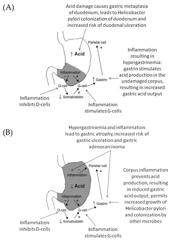
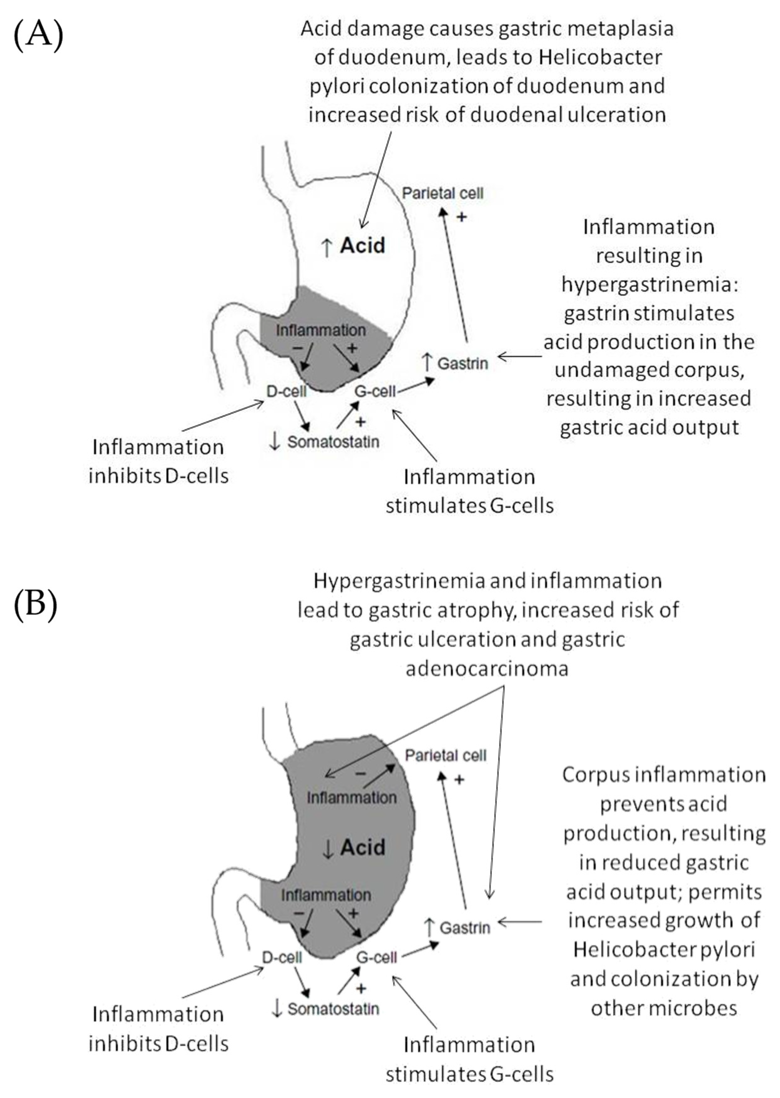
Distinct phenotypes of chronic gastritis are closely associated with specific clinical trajectories. Antrum-predominant gastritis, typically linked to increased gastric acid secretion, represents the characteristic background for duodenal ulcer disease. In contrast, pangastritis and corpus-predominant gastritis, which are accompanied by normal or decreased acid output, predispose to gastric ulceration, atrophic gastritis and gastric cancer. These patterns parallel the regional distribution of H. pylori colonization and are sufficiently robust to permit the prediction of ulcer risk on the basis of the gastritis phenotype [2] (Figure 1).
Figure 1.
Pathophysiological mechanisms and acid secretion patterns in (A) antrum-predominant gastritis and (B) pangastritis. In antrum-predominant gastritis, inflammation inhibits D-cells, resulting in decreased somatostatin secretion, increased gastrin release from G-cells, and stimulation of parietal cells leading to increased gastric acid output and elevated risk of duodenal ulceration. In pangastritis, corpus inflammation suppresses parietal cell function, resulting in reduced acid secretion, hypergastrinemia, progression to gastric atrophy, and increased risk of gastric ulceration and adenocarcinoma.
Beyond disease-specific mechanisms, H. pylori remains a major global public health concern due to its causal relationship with gastric cancer. Population-based eradication studies demonstrate that suppression of H. pylori can significantly reduce the gastric cancer incidence. A landmark community eradication program in the Matsu Islands (Taiwan) reported a 53% reduction in gastric cancer over more than a decade of follow-up [3]. However, emerging randomized population-based data indicate that the benefit of mass screening may be more modest than previously anticipated [4].
The infection elicits a pronounced innate and adaptive immune response, marked by local mucosal inflammation and robust systemic IgG and IgA antibody production. Despite this strong immune activation, H. pylori typically persists through multiple immune-evasion strategies, including modified lipopolysaccharide structure, molecular mimicry via Lewis antigens, and modulation of dendritic cell function with downstream effects on T-cell differentiation [5]. The interplay between pro- and anti-inflammatory mechanisms, combined with systemic and mucosal immunity, is thought to be central to the determination of disease phenotype [1,5].
In recent years, increasing interest has focused on non-invasive biomarkers for detecting gastric precancerous lesions and stratifying the cancer risk. Serum pepsinogens, gastrin-17, and H. pylori serology remain the most widely evaluated markers; however, their diagnostic accuracy is moderate, population-dependent, and insufficient to replace histology [6]. Likewise, available non-invasive diagnostic tests—serology, stool antigen testing, and the urea breath test—differ markedly in their capacity to identify active infection, with serology lacking the ability to discriminate current from past infection [7]. Advanced molecular and proteomic approaches have been proposed, but none can reliably capture both active infection and the inflammatory phenotype of the gastric mucosa [7,8,9].
In high-incidence regions, combined serological screening using anti-H. pylori IgG and serum pepsinogens (the ABC method) has been shown to effectively stratify gastric cancer risk and may represent a cost-effective alternative to annual endoscopic screening, as demonstrated in long-term Markov analyses from Japan [10]. Longitudinal Korean data similarly confirm that the ABC method predicts development of gastric neoplasia [11]. These limitations underscore the need for novel non-invasive tools that better reflect the topographic distribution and intensity of gastric inflammation.
Previous studies indicate that systemic anti-H. pylori IgG and IgA responses differ between benign and malignant gastroduodenal diseases, reflecting distinct host–pathogen interaction patterns. Population-based investigations demonstrate that both IgG and IgA levels increase with the severity of gastric atrophy, intestinal metaplasia and dysplasia [12]. Prospective data further suggest that not only seropositivity but also the magnitude of the response to specific virulence factors carries clinical relevance [13].
Our earlier work showed that IgG antibodies targeting specific H. pylori antigens—including CagA, VacA, the 30 kDa antigen and UreA (26 kDa)—correlate with both the degree and the anatomical distribution of gastric inflammation, as well as with clinical outcomes [14,15,16]. Further pediatric and adult studies confirm that IgG and IgA titers rise in parallel with inflammatory activity and bacterial virulence [17].
We have also demonstrated that patients with gastric cancer exhibit significantly lower systemic IgG levels and a higher frequency of IgA predominance compared with individuals with superficial or atrophic gastritis, suggesting a qualitatively distinct humoral immune profile in malignant disease [18,19].
Given these findings, it remains unclear whether the systemic balance between IgG and IgA, combined with antigen-specific seroreactivity, can serve as a reliable non-invasive marker of gastritis topography. Although the present study is based on the same patient cohort as several of our previously published reports, the analyses, outcome measures and research questions addressed here are distinct and have not been reported previously. Therefore, the aim of the present study was to evaluate whether the relative predominance of systemic IgG versus IgA antibodies, together with antigen-specific seroreactivity, reflects the histopathological features and topographic distribution of Helicobacter pylori-associated gastritis.
2. Results
2.1. Study Population Characteristics
A total of 123 patients with Helicobacter pylori-associated gastritis were included in the analysis. The median age of the study population was 63 years (range 21–87). The sample comprised 48 men (39.0%) and 75 women (61.0%).
Based on histopathological evaluation, 74 patients (60.2%) had antrum-predominant gastritis, whereas 4 patients (3.3%) had corpus-predominant gastritis, and 45 patients (36.6%) had pangastritis. Due to the small number of corpus-predominant cases (n = 4), corpus-predominant gastritis and pangastritis (4 + 45) were combined into a single corpus-involving group for comparative analyses (n = 49, 39, 8%), focusing on the presence versus absence of corpus inflammation rather than distinct etiologies. Accordingly, the binary outcome used throughout the analyses was intentionally defined as the presence versus absence of corpus involvement, rather than as distinct nosological entities. Conclusions regarding isolated corpus-predominant gastritis as a separate phenotype are, therefore, limited due to the small number of such cases (n = 4).
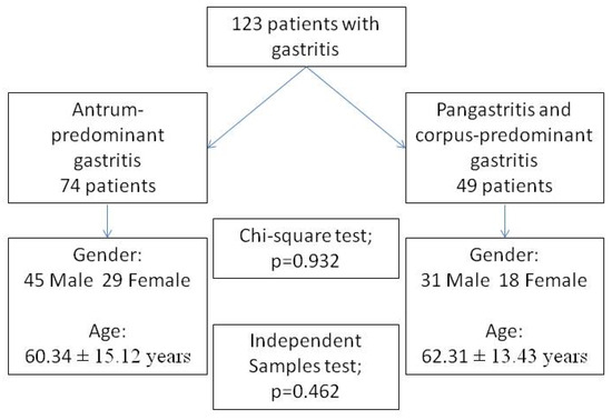
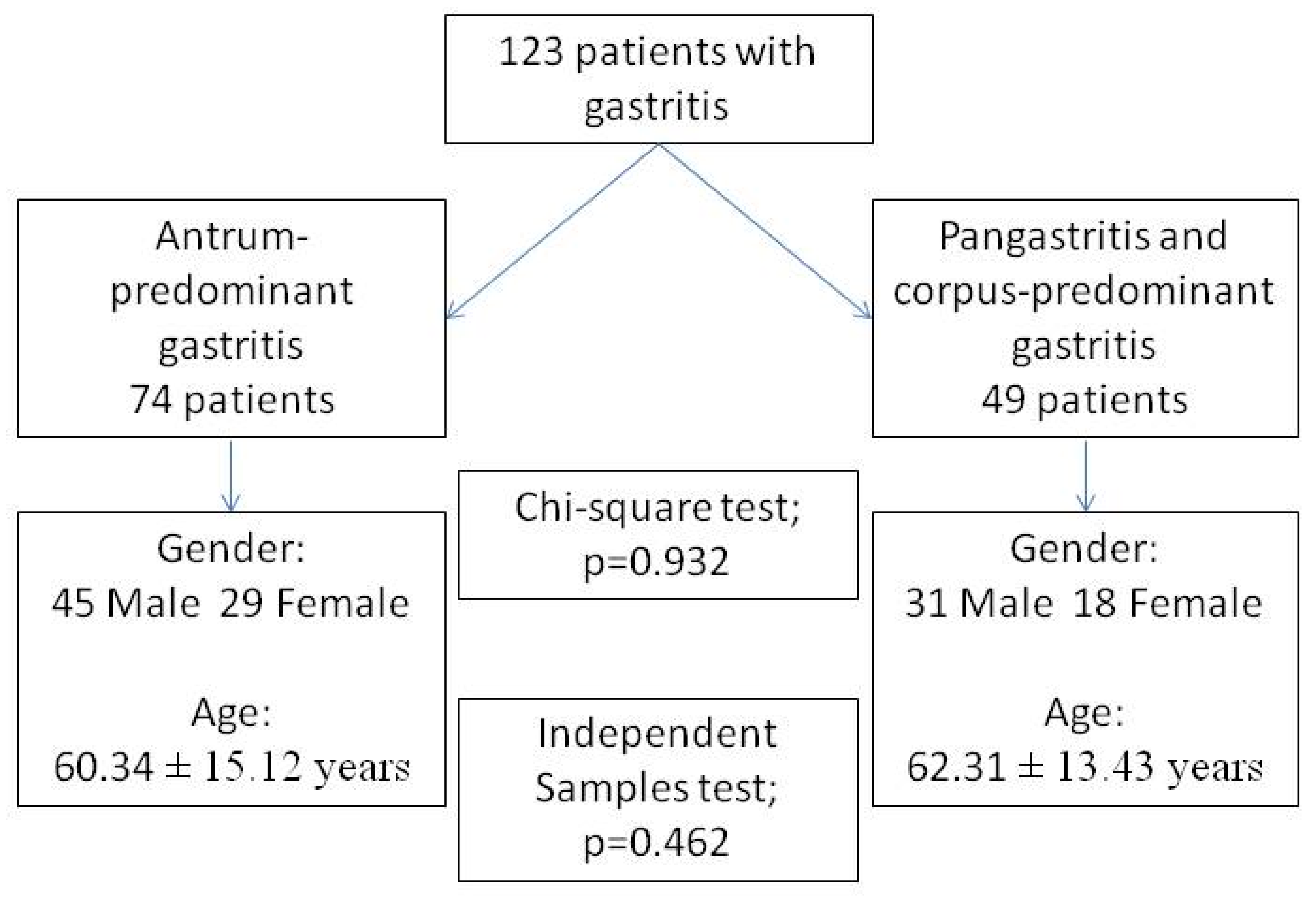
The distribution of patients by gastritis type, age and sex is illustrated in Figure 2.
Figure 2.
Distribution of patients according to gastritis topography, sex, and age. The study included 123 patients with Helicobacter pylori-associated gastritis, of whom 74 had antrum-predominant gastritis, and 49 had pangastritis or corpus-predominant gastritis. There were no significant differences between the groups in sex distribution (χ2 test, p = 0.932) or age (independent samples t-test, p = 0.462).
2.2. Correlations Between Immunoglobulins and Histopathological Activity
The correlations between serum anti-H. pylori IgG, IgA, IgG1 and IgG2 antibodies and histopathological parameters of the antral and corpus mucosa are summarized in Table 1.
Table 1.
Correlations between anti-Helicobacter pylori IgG, IgA, IgG1 and IgG2 antibodies and histopathological parameters of inflammation in the antral and corpus mucosa.
Serum IgG showed weak but statistically significant positive correlations with both the grade (r = 0.189, p = 0.037) and activity (r = 0.182, p = 0.044) of antral inflammation, whereas serum IgA correlated with inflammatory parameters (inflammation r = 0.199, p = 0.027, inflammatory activity r = 0.181, p = 0.045) in the corpus mucosa, indicating a topography-dependent relationship between serological responses and histological activity.
The IgG1 and IgG2 subclasses, as well as the IgG1/IgG2 ratio, showed weak but statistically significant positive correlations with antral inflammatory activity, while no significant associations were observed with gastric atrophy or intestinal metaplasia. Selected combined serological profiles further demonstrated divergent associations with inflammatory parameters between the antrum and corpus.
2.3. Immunoglobulin Predominance, Antigen-Specific Reactivity and Histopathology
Correlations between IgG/IgA and IgG1/IgG2 predominance in combination with antigen-specific IgG reactivity (CagA, VacA, UreB66, 30-kDa and UreA 26-kDa) and histopathological parameters are shown in Table 2.
Table 2.
Correlations between IgG/IgA and IgG1/IgG2 predominance combined with antigen-specific IgG reactivity (CagA, VacA, UreB66, 30 kDa and UreA 26 kDa) and histopathological parameters of antral and corpus mucosa.
Predominance of IgG over IgA was mainly associated with inflammatory activity in the antral mucosa, whereas IgA predominance was more frequently related to inflammatory parameters in the corpus. These associations were antigen-dependent, with different H. pylori antigens showing distinct correlation patterns. Overall, antigen-specific IgG profiles further strengthened the relationship between serological phenotype and gastritis topography.
2.4. ROC Analysis of Individual Immunoglobulins
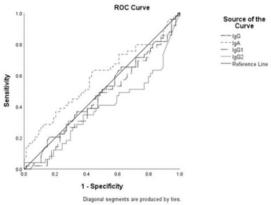
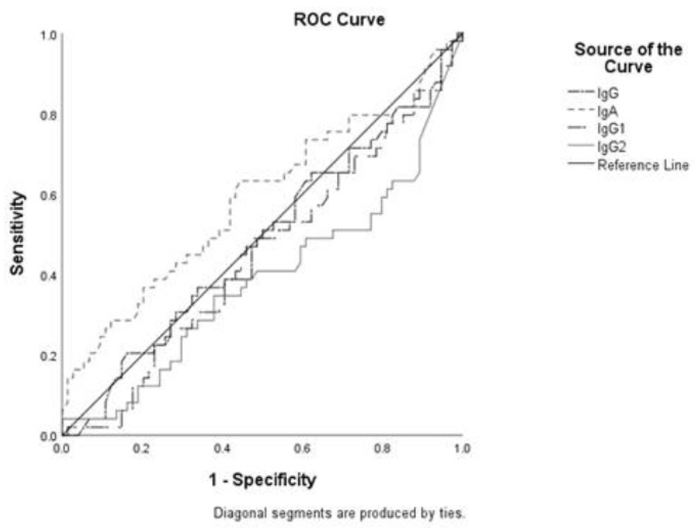
Receiver operating characteristic (ROC) curves for anti–H. pylori IgG, IgA, IgG1 and IgG2 in discriminating antrum-predominant gastritis from corpus-involving gastritis are presented in Table 3 and Figure 3.
Table 3.
Receiver operating characteristic (ROC) analysis of anti-Helicobacter pylori IgG, IgA, IgG1 and IgG2 for differentiating pangastritis/corpus-predominant gastritis and antrum-predominant gastritis.
Figure 3.
Receiver operating characteristic (ROC) curves of anti-Helicobacter pylori IgG, IgA, IgG1 and IgG2 for discrimination between antrum-predominant gastritis and pangastritis/corpus-predominant gastritis. The diagonal line represents the reference line. Area under the curve (AUC), sensitivity, specificity and optimal cut-off values are presented in Table 3.
- For corpus-involving gastritis, the highest discriminatory performance was observed for IgA (AUC = 0.587).
- For antrum-predominant gastritis, the highest AUC was obtained for IgG2 (AUC = 0.614).
For all ROC analyses, antrum-predominant gastritis was consistently defined as the positive class. When AUC values below 0.5 were observed, this was interpreted as indicating inverse discrimination (i.e., higher values associated with the opposite phenotype). For clarity, no separate reciprocal ROC models were constructed; instead, interpretation focused on the directionality of associations.
Overall, the diagnostic performance of individual immunoglobulin classes was modest.
2.5. Distribution of Serological Profiles According to Gastritis Topography
The distribution of IgG/IgA and IgG1/IgG2 predominance and their combinations with antigen-specific IgG reactivity in patients with different gastritis phenotypes are summarized in Table 4.
Table 4.
Anti-Helicobacter pylori IgG vs. IgA and IgG1 vs. IgG2 predominance and their combinations with antigen-specific IgG reactivity (CagA, VacA, UreB66, 30 kDa and UreA 26 kDa) in antrum-predominant gastritis and pangastritis/corpus-predominant gastritis.
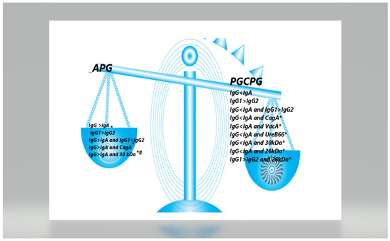
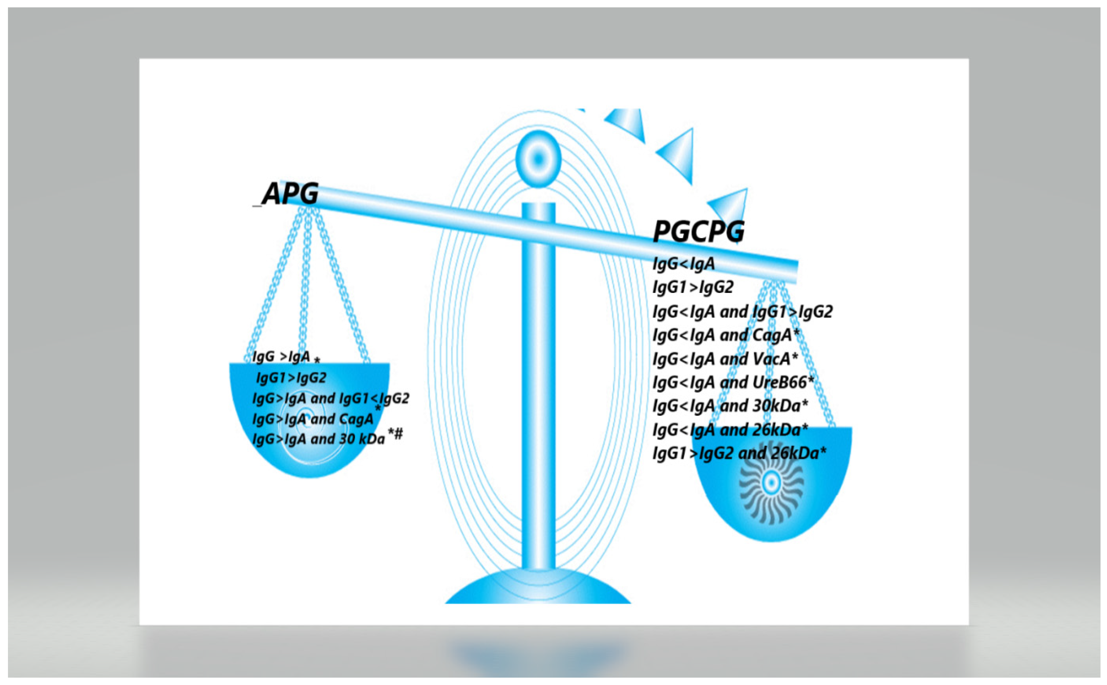
IgG > IgA predominance was significantly more common in antrum-predominant gastritis, whereas IgG < IgA predominance was significantly more frequent in corpus-involving gastritis. The combination of IgG > IgA + 30 kDa antigen positivity showed the strongest association with antrum-predominant gastritis, while IgG < IgA + UreA 26 kDa positivity characterized corpus-involving disease.
These distributions are illustrated in Figure 4.
Figure 4.
Distribution of significant serological predictors of antrum-predominant gastritis (APG) and pangastritis/corpus-predominant gastritis (PG/CPG). Asterisks indicate statistically significant associations in univariate analysis (* p < 0.05), while the hash symbol (#) denotes independent predictors identified by multivariate logistic regression.
2.6. Logistic Regression Analysis
The univariate and multivariate logistic regression models evaluating serological predictors of gastritis type are shown in Table 5.
Table 5.
Univariate and multivariate logistic regression analysis of serological predictors of gastritis type (antrum-predominant vs. pangastritis/corpus-predominant gastritis).
In the univariate analysis, several combinations of immunoglobulin ratios and antigen-specific reactivities were significantly associated with gastritis topography.
In the multivariate model, only the combination of IgG > IgA and 30 kDa antigen seropositivity remained an independent predictor of antrum-predominant gastritis (OR = 2.516, 95% CI 1.004–6.308, p = 0.049).
Helicobacter pylori Colonization and Histopathological Parameters
The relationships between H. pylori colonization density and histopathological parameters are presented in Table 6.
Table 6.
Correlation between the degree of Helicobacter pylori colonization in the antrum and corpus and histopathological parameters of gastric mucosa and gastritis type.
The degree of Helicobacter pylori colonization in the antrum correlated strongly with the grade (r = 0.466, p < 0.001) and activity (r = 0.692, p < 0.001) of antral inflammation, as well as with the inflammatory activity in the corpus (r = 0.345, p < 0.001), whereas no consistent direct correlations were observed between the bacterial density and individual serological parameters, indicating that antibody levels do not directly reflect the bacterial load.
- Antral colonization correlated strongly with both the grade (r = 0.466, p < 0.001) and activity (r = 0.692, p < 0.001) of antral inflammation and moderately with corpus inflammatory activity.
- Corpus colonization was strongly associated with both antral and corpus inflammatory activity, as well as with the grade of corpus inflammation.
These correlations further support the relationship between colonization patterns and gastritis topography.
3. Discussion
This study provides an integrated serological, microbiological and histopathological assessment of H. pylori-associated gastritis and demonstrates that the relative balance between systemic IgG and IgA, particularly when combined with specific antigen seroreactivities, reflects the topographic distribution of gastric inflammation. The principal finding is that IgG predominance accompanied by 30 kDa antigen seropositivity independently predicts antrum-predominant gastritis, whereas IgA predominance, especially with CagA, VacA and UreA 26 kDa reactivity, characterizes pangastritis and corpus-predominant inflammation. These results agree with our earlier observations of lower IgG levels and frequent IgA predominance in patients with gastric cancer, suggesting progressive shifts in systemic humoral responses along the gastric carcinogenesis continuum [18,19].
Beyond the overall IgG/IgA predominance, IgG subclass responses were also examined. IgG1 and IgG2 predominance showed no consistent associations with histological parameters, although IgG2 demonstrated limited discriminatory ability for antrum-predominant gastritis and IgG1/IgG2 predominance differed between antral and corpus-involving disease, indicating a modest relationship with gastritis topography rather than severity.
The identified serological profiles align with established mechanistic models of gastritis progression. Antrum-predominant gastritis is generally associated with preserved or enhanced acid secretion, creating an environment conducive to high bacterial density and strong local inflammation. This is reflected serologically by IgG predominance. In contrast, the transition toward pangastritis and corpus-predominant disease is marked by reduced acid output, broader mucosal involvement and a shift toward IgA-dominant responses. Histological detection of H. pylori is frequently false-negative in seropositive subjects with gastric adenocarcinoma and decreased secretory capacity due to pronounced hypochlorhydria, despite ongoing systemic antibody production [20].
Microbiological findings support this model. Antral colonization correlated strongly with the grade and activity of antral inflammation, while corpus colonization demonstrated an even stronger association with corresponding corpus inflammation. The inverse relationship between antral colonization and intestinal metaplasia supports the concept that advanced mucosal remodeling creates progressively unfavorable conditions for bacterial persistence [21,22]. Long-term cohort studies further show that H. pylori is strongly associated with active inflammation, moderately with atrophy, and minimally with intestinal metaplasia, and that eradication reverses inflammation and atrophy but not established metaplasia [23].
Two distinct immunological phenotypes were identified: (1) antrum-predominant gastritis, characterized by IgG predominance and 30-kDa antigen positivity, and (2) pangastritis/corpus-predominant disease, characterized by IgA predominance in combination with CagA, VacA, UreB66 and UreA 26 kDa reactivity. Multivariate analyses confirmed IgG > IgA plus 30 kDa positivity as the only independent predictor of antrum-predominant gastritis.
From an immunopathological perspective, IgG predominance suggests a systemic Th1-biased response consistent with active inflammation and higher antigenic exposure in the antrum. CagA seropositivity and a predominance of systemic IgG responses have been repeatedly associated with higher histological inflammatory activity across different age groups [17]. Conversely, IgA predominance likely reflects enhanced mucosal immune activation associated with long-standing infection and prolonged antigenic stimulation, although the cross-sectional design of the study precludes direct assessment of the disease duration. These interpretations are consistent with experimental evidence demonstrating that divergent humoral profiles correspond to distinct oxidative stress and antioxidant patterns [24].
The current concept of serological gastric biopsy relies primarily on pepsinogen measurements and anti-H. pylori IgG, but the diagnostic performance remains moderate and varies across populations [6]. Although useful for cancer risk stratification, the ABC method does not reliably capture inflammatory phenotypes or regional distribution of disease [25]. Serological testing has several well-recognized limitations that must be acknowledged. Serological assays are limited by moderate sensitivity and specificity, antibody persistence [7], false negatives [26], dependence on preserved secretory function [27], temporal discrepancies between histology and serology [12], and quantitative variability in antibody titers [28]. Although ROC analysis was used to explore the potential diagnostic performance of individual immunoglobulin classes, the observed discriminatory ability was modest overall. For clarity and clinical interpretability, a consistent outcome direction was applied in the analysis, focusing on the biological meaning of the associations rather than on reciprocal model construction. This approach avoids potential confusion arising from reciprocal ROC models and allows clearer clinical interpretation of the discriminatory direction.
Combined IgG/IgA predominance testing may compensate for some of these limitations, as a subset of IgG-negative individuals are IgA-positive [26]. False-negative IgG results have also been documented in advanced mucosal atrophy [26,28].
Unlike pepsinogen-based methods, which primarily reflect corpus atrophy, the proposed IgG/IgA predominance-weighted antigen-specific profiling appears better suited to capturing active inflammatory phenotypes and their anatomical distribution. These findings suggest that immune profiling may complement existing functional biomarkers by providing insight not only into mucosal loss but also into ongoing immunological activity.
Several limitations merit consideration. The cross-sectional design precludes temporal inference. The study was not designed to directly compare conventional serological biopsy with the proposed model using ROC-based diagnostics, and potential improvements in diagnostic performance therefore remain hypothetical pending prospective validation. Although corpus-predominant gastritis and pangastritis represent distinct histopathological entities, they were pooled in the present analysis due to the limited number of isolated corpus-predominant cases. This approach emphasizes corpus involvement as a shared biological feature relevant to immune response patterns. While autoimmune gastritis may present with corpus-predominant atrophy, patients with autoimmune disorders were excluded, and no histological patterns suggestive of advanced autoimmune gastritis were observed. Nevertheless, residual heterogeneity within the corpus-involving group cannot be completely excluded and represents a limitation of the study.
In conclusion, the predominance between systemic IgG and IgA responses combined with antigen-specific seroreactivity reflects the topographic pattern of gastric inflammation. IgG predominance with 30 kDa positivity predicts antrum-predominant gastritis, whereas IgA-dominant profiles characterize corpus-involving disease. These findings support the potential role of advanced serological immune profiling as a non-invasive tool for the stratification of H. pylori-associated gastritis.
4. Materials and Methods
4.1. Study Design and Patients
This observational cross-sectional study represents a predefined secondary analysis of previously collected data and stored biological samples derived from a larger investigation entitled “Systemic humoral immune response to Helicobacter pylori in patients with dyspepsia, peptic ulcer disease and gastric cancer.” The present analysis was predefined and approved by the institutional Ethics Committee prior to data processing.
The original study was conducted at the Military Medical Academy during 2009 and included consecutive adult patients undergoing diagnostic upper gastrointestinal endoscopy for dyspeptic symptoms.
- Inclusion criteria
- 1.
- Age ≥ 18 years,
- 2.
- Availability of gastric biopsies from both the antrum and corpus,
- 3.
- Confirmed Helicobacter pylori infection based on histological detection (H&E and/or Giemsa staining).
- 4.
- Availability of complete serological data.
- Exclusion criteria
- 1.
- Previous H. pylori eradication therapy,
- 2.
- Use of antibiotics, proton pump inhibitors or bismuth compounds within four weeks prior to endoscopy,
- 3.
- History of gastric surgery,
- 4.
- Severe systemic disease, autoimmune disorders or immunodeficiency,
- 5.
- Incomplete clinical or laboratory data.
A total of 123 patients were included in the final analysis.
4.2. Endoscopic Evaluation and Biopsy Collection
All patients underwent standard diagnostic esophagogastroduodenoscopy after fasting for at least 6 h. Endoscopy was performed using a standard Olympus video endoscope (Olympus 2951 Ishikawa-machi, Hachioji-shi, Tokyo 192-8507, Japan). Premedication consisted of 1% lidocaine spray for pharyngeal anesthesia or intravenous analgosedation, depending on clinical indication.
Endoscopic reporting adhered to the criteria of the European Society of Gastrointestinal Endoscopy (ESGE).
Biopsies were obtained according to the Updated Sydney System:
- Two biopsies from the antrum (lesser and greater curvature),
- Two biopsies from the corpus (lesser and greater curvature), using standard Olympus biopsy forceps.
Biopsy specimens were fixed in buffered formalin, embedded in paraffin, sectioned at 2–3 μm thickness, and stained with hematoxylin and eosin for routine histopathological assessment. Giemsa staining was additionally performed for the detection of H. pylori.
4.3. Histopathological Evaluation
Histological assessment was performed by experienced gastrointestinal pathologists who were blinded to the serological results. For both the antrum and corpus, the following parameters were evaluated according to the Updated Sydney Classification:
- Chronic inflammation,
- Inflammatory activity,
- Glandular atrophy,
- Intestinal metaplasia,
- H. pylori density.
Each parameter was graded on a 0–3 scale (0 = none, 1 = mild, 2 = moderate, 3 = severe).
Based on histological findings, patients were categorized as having one of the following:
- Antrum-predominant gastritis,
- Corpus-predominant gastritis,
- Pangastritis.
4.4. Microbiological Diagnosis of Helicobacter pylori
The diagnosis of H. pylori infection was established by histological detection of the organism using hematoxylin and eosin and Giemsa staining. Only patients with histologically verified infection were included in the analysis. Giemsa staining was routinely performed in cases where chronic inflammation was present on hematoxylin and eosin staining, but where Helicobacter pylori organisms were not readily identified. The aim of using both stains was to maximize the diagnostic sensitivity rather than to compare the staining methods; therefore, a formal statistical correlation between H&E and Giemsa staining was not performed.
4.5. Serological Analysis
Venous blood samples were collected at the time of endoscopy or within 24 h thereafter as part of the original study protocol. Serum was separated by centrifugation and stored at −20 °C until analysis.
4.6. Determination of Total IgG and IgA
Total anti–Helicobacter pylori IgG and IgA antibodies were measured using commercial ELISA kits (EUROIMMUN Medizinische Labordiagnostika AG Seekamp 31 23560 Lübeck, Germany); catalog number EI 2080-9601, isotype-specific variants: EI 2080-9601 G for IgG and EI 2080-9601 A for IgA). Antigens were derived from the H. pylori Lior1 (Brussels) strain. According to the manufacturer’s documentation, the results are reported in standardized arbitrary units derived from optical density values using kit-specific calibration curves.
4.7. Determination of IgG Subclasses
IgG1 and IgG2 subclasses were quantified using monoclonal antibodies:
- Anti-human IgG1 clone 8c/6-39 (biotin-conjugated),
- Anti-human IgG2 clone HP-6014 (biotin-conjugated),
from (Inc. PO Box 14508 St. Louis, MO 63178, USA), in combination with EUROIMMUN plates coated with H. pylori antigens (Lior1strain) (EUROIMMUN Medizinische Labordiagnostika AG Seekamp 31 23560 Lübeck, Germany).
4.8. Antigen-Specific IgG Detection
Antigen-specific IgG responses to
- CagA,
- VacA,
- UreB (66 kDa),
- 30 kDa antigen,
- UreA (26 kDa),
were determined using a commercial Western blot assay (VIRAMED ViraBlot Helicobacter pylori IgG; VIRAMED Biotech AG, Planegg, Germany; catalog number CW 2080-5005 G), based on European clinical H. pylori isolates.
Optical density values were expressed in arbitrary units according to the calibration curves provided by the manufacturer. Seropositivity cut-off values were applied as recommended.
Both anti-H. pylori IgG and IgA antibodies were measured using the same commercial ELISA platform, based on an identical antigenic substrate and calibrated optical density units, allowing intra-assay comparability between immunoglobulin classes.
IgG/IgA predominance was defined on an individual level as a binary within-subject comparison, with IgG predominance assigned when the IgG exceeded IgA and IgA predominance when the IgA exceeded IgG. This variable was used exclusively as an operational indicator of the prevailing systemic humoral immune response and was not analyzed as a continuous quantitative ratio. The same binary approach was applied to IgG1/IgG2 predominance.
4.9. Statistical Analysis
Statistical analysis was performed using IBM SPSS version 26.0. The normality of continuous variables was assessed using the Kolmogorov–Smirnov test. Continuous variables were presented as the mean ± standard deviation or median (interquartile range), as appropriate, while the categorical variables were summarized as frequencies and percentages.
- Group comparisons were performed using the Independent Samples t-test or Mann–Whitney U test for continuous variables and the χ2 test or Fisher’s exact test for categorical variables.
- Relationships between variables were examined using Spearman’s rank correlation.
- Receiver operating characteristic (ROC) curve analysis was used to assess the diagnostic performance of IgG, IgA, IgG1 and IgG2 in discriminating antrum-predominant gastritis from corpus-predominant/pangastritis.
- The area under the curve (AUC), sensitivity, specificity and optimal cut-off values were calculated using the Youden index.
- Univariate and multivariate logistic regression analyses were performed to identify independent predictors of antrum-predominant gastritis. Variables with p < 0.05 in univariate analysis were included in the multivariate model. The results are expressed as odds ratios (ORs) with 95% confidence intervals (CIs). In logistic regression analyses, antrum-predominant gastritis was coded as the dependent outcome (1) and corpus-involving gastritis as 0. Age and sex were evaluated as potential covariates but were not retained in the final model, as they were not significantly associated with the gastritis type.
A p-value < 0.05 was considered statistically significant.
Given the exploratory and hypothesis-generating nature of this study and the biological interdependence of the evaluated markers, no formal correction for multiple testing was applied. This approach increases the risk of type I error, and the findings should therefore be interpreted with appropriate caution. Multivariate logistic regression was used to identify the most robust associations within this exploratory framework, but it does not replace formal multiplicity correction.
4.10. Ethics Approval
The original study protocol and informed consent procedure were approved by the Ethics Committee of the Military Medical Academy. A separate approval was obtained from the same Ethics Committee for the present secondary analysis of previously collected data and stored biological samples (Approval No. 178/2025, Date: 20 November 2025). Written informed consent was obtained from all participants at the time of enrollment and included permission for future analyses. All procedures were conducted in accordance with the Declaration of Helsinki.
Author Contributions
Conceptualization, N.M. and S.M.; methodology, N.M.; software, N.R.; validation, N.M., I.T. and E.R.; formal analysis, S.M.; investigation, N.M., I.T. and E.R.; resources, N.R.; data curation, S.M.; writing—original draft preparation, N.M. and S.M.; writing—review and editing, N.M., I.T., N.R., E.R. and S.M.; visualization, N.R.; supervision, N.M. and N.R.; project administration, N.M. All authors have read and agreed to the published version of the manuscript.
Funding
This research received no external funding.
Institutional Review Board Statement
The study was conducted in accordance with the Declaration of Helsinki, and approved by the Ethics Committee of Military Medical Academy, Belgrade (protocol code 178/2025 and date of approval 28 November 2025).
Informed Consent Statement
Informed consent was obtained from all subjects involved in the study.
Data Availability Statement
The data presented in this study are available on request from the corresponding author due to privacy and ethical reasons.
Conflicts of Interest
The authors declare no conflict of interest.
References
- White, J.R.; Winter, J.A.; Robinson, K. Differential inflammatory response to Helicobacter pylori infection: Etiology and clinical outcomes. J. Inflamm. Res. 2015, 8, 137–147. [Google Scholar] [CrossRef] [Scilit]
- Dixon, M.F. Patterns of inflammation linked to ulcer disease. Best Pr. Res. Clin. Gastroenterol. 2000, 14, 27–40. [Google Scholar] [CrossRef] [Scilit]
- Chiang, T.-H.; Chang, W.-J.; Chen, S.L.-S.; Yen, A.M.-F.; Fann, J.C.-Y.; Chiu, S.Y.-H.; Chen, Y.-R.; Chuang, S.-L.; Shieh, C.-F.; Liu, C.-Y.; et al. Mass eradication of Helicobacter pylori to reduce gastric cancer incidence: A long-term cohort study on Matsu Islands. Gut 2021, 70, 243–250. [Google Scholar] [CrossRef] [Scilit]
- Lee, Y.C.; Chiang, T.H.; Chiu, H.M.; Su, W.W.; Chou, K.C.; Chen, S.L.; Yen, A.M.; Fann, J.C.; Chiu, S.Y.; Chuang, S.L.; et al. Screening for Helicobacter pylori to Prevent Gastric Cancer: A Pragmatic Randomized Clinical Trial. JAMA 2024, 332, 1642–1651. [Google Scholar] [CrossRef] [Scilit]
- Sijmons, D.; Guy, A.J.; Walduck, A.K.; Ramsland, P.A. Helicobacter pylori and the role of lipopolysaccharide variation in innate immune evasion. Front. Immunol. 2022, 13, 868225. [Google Scholar] [CrossRef] [Scilit]
- Romanczyk, M.; Osmola, M.; Link, A.; Druet, A.; Hemont, C.; Martin, J.; Chapelle, N.; Matysiak-Budnik, T. Non-invasive markers for the detection of gastric precancerous conditions. Cancers 2024, 16, 2254. [Google Scholar] [CrossRef] [Scilit]
- Ghazanfar, H.; Javed, N.; Reina, R.; Thartori, O.; Ghazanfar, A.; Patel, H. Advances in diagnostic modalities for Helicobacter pylori infection. Life 2024, 14, 1170. [Google Scholar] [CrossRef] [Scilit] [PubMed]
- Costa, L.C.M.C.; Carvalho, M.G.; Pereira, A.L.C.G.; Teixeira Neto, R.G.; Andrade Figueiredo, L.C.; Barros-Pinheiro, M. Diagnostic methods for Helicobacter pylori. Med. Princ. Pr. 2024, 33, 173–184. [Google Scholar] [CrossRef] [Scilit] [PubMed]
- Sun, M.; Liu, E.; Yang, L.; Cao, H.; Han, M. A scoping review of worldwide guidelines for diagnosis and treatment of Helicobacter pylori infection. Syst. Rev. 2025, 14, 107. [Google Scholar] [CrossRef] [Scilit]
- Malfertheiner, P.; Megraud, F.; Rokkas, T.; Gisbert, J.P.; Liou, J.M.; Schulz, C.; Gasbarrini, A.; Hunt, R.H.; Leja, M.; O'Morain, C.; et al. Management of Helicobacter pylori infection: The Maastricht VI/Florence consensus report. Gut 2022, 71, 1724–1762. [Google Scholar] [CrossRef] [Scilit] [PubMed]
- Saito, S.; Azumi, M.; Muneoka, Y.; Nishino, K.; Ishikawa, T.; Sato, Y.; Terai, S.; Akazawa, K. Cost-effectiveness of combined serum anti-Helicobacter pylori IgG antibody and serum pepsinogen concentrations for screening for gastric cancer risk in Japan. Eur. J. Health Econ. 2018, 19, 545–555. [Google Scholar] [CrossRef] [Scilit] [PubMed]
- Kwak, H.W.; Choi, I.J.; Cho, S.J.; Lee, J.Y.; Kim, C.G.; Kook, M.C.; Ryu, K.W.; Kim, Y.-W. Characteristics of gastric cancer according to Helicobacter pylori infection status. J. Gastroenterol. Hepatol. 2014, 29, 1671–1677. [Google Scholar] [CrossRef] [Scilit]
- Li, S.; Lu, A.P.; Zhang, L.; Li, Y.D. Anti-Helicobacter pylori IgG and IgA antibody responses and clinical presentations in patients with precancerous lesions. World J. Gastroenterol. 2003, 9, 755–758. [Google Scholar] [CrossRef] [Scilit]
- Nishizawa, T.; Sakitani, K.; Suzuki, H.; Yamakawa, T.; Takahashi, Y.; Yamamichi, N.; Watanabe, H.; Seto, Y.; Koike, K.; Toyoshima, O. A combination of serum anti-Helicobacter pylori antibody titer and Kyoto classification score could provide a more accurate diagnosis of H pylori. United European Gastroenterol. J. 2019, 7, 343–348. [Google Scholar] [CrossRef] [Scilit]
- Manojlovic, N.; Tufegdžić, I.; Ristanovic, E.; Bokonjić, D. Serum IgG antibodies against Helicobacter pylori low molecular weight antigens 50 kDa, 30 kDa and Urease A 26 kDa, along with vacuolating cytotoxin A, are associated with the outcome of the infection. Vojnosanit. Pregl. 2020, 77, 405–412. [Google Scholar] [CrossRef] [Scilit]
- Manojlović, N.; Tufegdžić, I.; Ristanović, E.; Bokonjić, D. Simultaneous and alternative IgG seroreactivity against Helicobacter pylori antigens VacA, 30 kDa and 50 kDa is a better biomarker approach for the outcome of infection than VacA and 50 kDa alone. Vojnosanit. Pregl. 2020, 79, 71. [Google Scholar] [CrossRef] [Scilit]
- Manojlovic, N.; Tufegdzic, I.; Ristanovic, E.; Bokonjic, D. Seroreactivity against Helicobacter pylori VacA, 50 kDa and 30 kDa along with alarm features may improve the diagnostic approach to uninvestigated dyspepsia: A pilot study. Vojnosanit. Pregl. 2020, 79, 448–455. [Google Scholar] [CrossRef] [Scilit]
- Manojlovic, N.; Babic, D.; Filipovic-Ljeshovic, I.; Pilcevic, D. Anti-Helicobacter pylori IgG and IgA response in gastric cancer and chronic gastritis. Hepatogastroenterology 2008, 55, 807–813. [Google Scholar]
- Kishikawa, H.; Ojiro, K.; Nakamura, K.; Katayama, T.; Arahata, K.; Takarabe, S.; Miura, S.; Kanai, T.; Nishida, J. Previous Helicobacter pylori infection-induced atrophic gastritis: A distinct disease entity in an understudied population without a history of eradication. Helicobacter 2020, 25, e12669. [Google Scholar] [CrossRef] [Scilit]
- Zhang, C.; Yamada, N.; Wu, Y.L.; Wen, M.; Matsuhisa, T.; Matsukura, N. Helicobacter pylori infection, atrophy and intestinal metaplasia in gastric disease. World J. Gastroenterol. 2005, 11, 791–796. [Google Scholar] [CrossRef] [Scilit] [PubMed]
- Asfeldt, A.M.; Steigen, S.E.; Lochen, M.L.; Straume, B.; Johnsen, R.; Bernersen, B.; Florholmen, J.; Paulssen, E.J. The natural course of Helicobacter pylori infection on endoscopic findings in a population during 17 years of follow-up: The Sørreisa gastrointestinal disorder study. Eur. J. Epidemiol. 2009, 24, 649–658. [Google Scholar] [CrossRef] [Scilit]
- Smirnova, O.V.; Sinyakov, A.A.; Kasparov, E.V. Role of IgG, IgA and IgE antibodies against Helicobacter pylori in oxidative stress in chronic gastritis. Biomedicines 2022, 10, 2053. [Google Scholar] [CrossRef] [Scilit]
- Dinis-Ribeiro, M.; Libânio, D.; Uchima, H.; Spaander, M.C.W.; Bornschein, J.; Matysiak-Budnik, T.; Tziatzios, G.; Santos-Antunes, J.; Areia, M.; Chapelle, N.; et al. Management of epithelial precancerous conditions and early neoplasia of the stomach (MAPS III): European Society of Gastrointestinal Endoscopy (ESGE), European Helicobacter and Microbiota Study Group (EHMSG) and European Society of Pathology (ESP) Guideline update 2025. Endoscopy 2025, 57, 504–554. [Google Scholar] [CrossRef] [Scilit] [PubMed]
- Toyoshima, O.; Nishizawa, T.; Arita, M.; Kataoka, Y.; Sakitani, K.; Yoshida, S.; Yamashita, H.; Hata, K.; Watanabe, H.; Suzuki, H. Helicobacter pylori infection in subjects negative for high-titer serum antibody. World J. Gastroenterol. 2018, 24, 1419–1428. [Google Scholar] [CrossRef] [Scilit] [PubMed]
- Kim, J.H.; Lee, S.Y.; Lee, S.P.; Kim, J.H.; Sung, I.K.; Park, H.S.; Shim, C.S.; Han, H.S. The histologic detection of Helicobacter pylori in seropositive subjects is affected by pathology and secretory ability of the stomach. Helicobacter 2018, 23, e12480. [Google Scholar] [CrossRef] [Scilit]
- Kishikawa, H.; Kimura, K.; Takarabe, S.; Kaida, S.; Nishida, J. Helicobacter pylori antibody titer and gastric cancer screening. Dis. Markers 2015, 2015, 156719. [Google Scholar] [CrossRef] [Scilit] [PubMed]
- Shams, S.; Vesali Jamshid, Z.; Shahbazi, T.; Hasani, M.; Shams, E.; Ragolia, S. Comparison of serum IgG and IgA levels against Helicobacter pylori in patients with gastrointestinal symptoms. Int. Emerg. Med. 2018, 4, 105–108. [Google Scholar]
- Adachi, K.; Kishi, K.; Notsu, T.; Mishiro, T.; Sota, K.; Ishimura, N.; Ishihara, S. Serum anti-Helicobacter pylori IgG antibody titer in seronegative cases with different gastric atrophy status. Intern. Med. 2020, 59, 2817–2823. [Google Scholar] [CrossRef] [Scilit]

Disclaimer/Publisher’s Note: The statements, opinions and data contained in all publications are solely those of the individual author(s) and contributor(s) and not of MDPI and/or the editor(s). MDPI and/or the editor(s) disclaim responsibility for any injury to people or property resulting from any ideas, methods, instructions or products referred to in the content. |
© 2026 by the authors. Licensee MDPI, Basel, Switzerland. This article is an open access article distributed under the terms and conditions of the Creative Commons Attribution (CC BY) license.