


Abstract
Flexible implantable electrodes require biocompatibility, mechanical stability, and sufficient electrical conductivity for effective neural interfacing. This work examines ultrasonic treatment during poly(3,4-ethylenedioxythiophene):poly(styrenesulfonate) (PEDOT:PSS) impregnation of electrospun poly(D,L)-lactide (PLA) nonwoven scaffolds as a route to improve filler distribution and functional performance. Four sample types were studied: pristine PLA (untreated and sonicated) and PLA–PEDOT:PSS composites prepared with and without ultrasonication. Scanning electron microscopy shows that ultrasonic treatment suppresses the formation of continuous surface films and promotes homogeneous three-dimensional penetration of PEDOT:PSS throughout the fibrous network. As a result, electrical resistivity decreases by a factor of 7.3, from 294.4 to 40.2 Ω·m. Contact-angle measurements reveal markedly enhanced wettability, with sonicated composites exhibiting rapid water uptake (5–13 s), unlike non-sonicated controls. These findings demonstrate that ultrasound-assisted PEDOT:PSS impregnation yields conductive, highly wettable, and structurally stable scaffolds, highlighting their potential for flexible implantable neural electrodes.
1. Introduction
Flexible electronics hold great promise for the development of soft robotics, in vitro diagnostic systems, and wearable and implantable biomedical devices for continuous health monitoring [1]. Of particular importance is the critical task of creating implantable devices—neural prostheses—designed to interface with the nervous system to supplement, compensate for, or restore impaired or lost neurological functions [2]. The mechanical mismatch between conventional rigid electrodes and biological tissues is one of the critical problems of neuroengineering. This mismatch causes tissue damage, necrosis, and a release of bioactive substances that lead to tissue reorganization and glial scar formation. This, in turn, leads to degradation of electrode performance over time [2,3]. Significant current efforts are focused on the design of mechanically biomimetic polymers with complex molecular architectures capable of reproducing the stress–strain behavior of soft biological tissues, both in solvent-free states [4,5] and in gel form [6]. In parallel, increasing attention is devoted to the development of biomimetic conductive materials for applications in wearable and biointegrated electronics [6]. Beyond polymer-based and bioinspired systems, high-entropy materials are also being actively explored for advanced electrical, magnetic, and optical applications, further broadening the range of functional materials available for electronic technologies [7].
Among conventional polymers, polylactide (PLA) is one of the most promising biodegradable materials due to its biocompatibility, favorable mechanical properties, and non-toxic degradation products, making it a widely used candidate for tissue engineering and implantable medical devices [8]. However, the natural hydrophobicity of PLA (contact angle ~80°) limits its application in bioelectronics and requires functionalization. In recent years, there has been growing interest in conductive polymers such as poly(3,4-ethylenedioxythiophene):poly(styrenesulfonate) (PEDOT:PSS), which possesses excellent electrical properties, chemical stability, and biocompatibility [9]. Composite materials based on PEDOT:PSS demonstrate significant potential for application in organic electrochemical transistors and flexible biosensors [10]. The electrospinning method enables the fabrication of nonwoven materials with nanostructured morphology similar to the extracellular matrix, thereby promoting cell adhesion and enabling the development of tissue engineering scaffolds [11]. The combination of PLA and the conducting polymer PEDOT:PSS represents a promising platform for creating next-generation flexible and biocompatible electronics. For example, Chung et al. fabricated electrospun composite membranes based on PLA and poly(3-hydroxybutyrate-co-3-hydroxyvalerate) (PHBV), which were subsequently dip-coated with PEDOT:PSS to obtain conductive fibrous membranes [12]. Similarly Estrany et al. used chemical oxidative polymerization of EDOT monomers with PLA macromonomers to synthesize PEDOT-g-PLA graft copolymers with variable PEDOT content for Direct Ink Writing (DIW) 3D printing [13]. A key challenge in combining these materials lies in the interfacial adhesion between the components, since PEDOT:PSS is a highly hydrophilic and negatively charged dispersion, whereas PLA is hydrophobic and relatively inert.
Ultrasonic treatment represents a promising and well-established approach for modification of the fibers surface, enabling uniform particle distribution throughout the polymer matrix, as demonstrated in numerous reports [14,15,16,17]. The aim of this study is to investigate the effect of ultrasonic treatment on the structural and functional properties of the PLA-PEDOT:PSS composite and to evaluate its potential for application in implantable soft bioelectronics.
2. Materials and Methods
2.1. PLA Nanofibrous Scaffold Preparation
Poly-(D,L)-lactide with a molecular weight (Mw) of 110 kDa (PLA, Ingeo 4032d, NatureWorks, 650 Industrial Park Drive, P.O. Box 564, Blair, NE 68008, USA) was purchased from NatureWorks. 1,1,1,3,3,3-hexafluoro-2-propanol was purchased from Sigma-Aldrich (HFiP, 105228-2KG, Sigma-Aldrich, St. Louis, MO, USA). PEDOT:PSS (1:2.5 w/w) was purchased from Ossila (PEDOT:PSS Clevios™ PH 1000, 155090-83-8, Ossila Limited, Solpro Business Park, Windsor Street, Sheffield, UK).
Fibrous scaffolds composed of PLA were fabricated via an electrospinning system (NF-500 MECC Co., Ltd., Fukuda, Japan) (Figure 1). Polymer solutions of PLA (100 mg/mL) were prepared in HFIP and stirred for 24 h at 25 °C. After degassing, each solution was loaded into a 12 mL syringe fitted with a 22-gauge blunt-tip stainless steel needle. Electrospinning was performed onto an aluminum foil-covered collector under the following conditions: 25 °C, 1 mL/h feed rate, 275 mm tip-to-collector distance, and 35 kV applied voltage.
Figure 1.
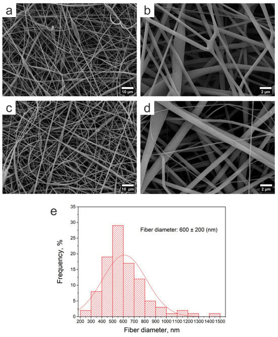
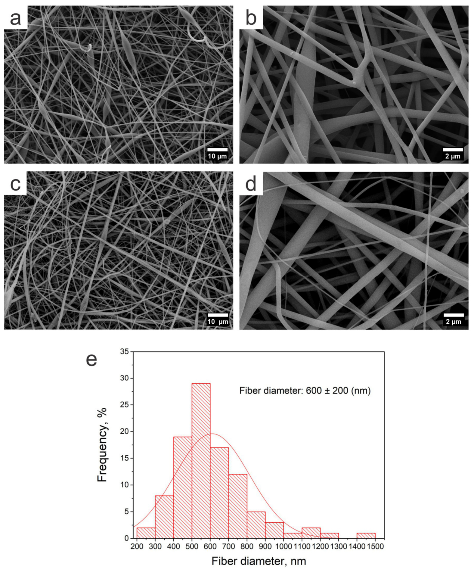
SEM images of the neat PLA nanofibrous scaffold without sonication (a,b) and after sonication (c,d), together with the corresponding fiber diameter distribution (e).
2.2. Samples Preparing and PEDOT:PSS Impregnation
The aqueous PEDOT:PSS dispersion (0.5 wt% solids) was prepared. To enhance the wettability of the hydrophobic material, 5 vol% ethanol was added to the dispersion. Rectangular samples (10 × 6 mm2) were cut from PLA nanofibrous scaffolds. Four types of samples were prepared: pristine PLA (uncoated): used as a control, pristine PLA-US (sonicated): prepared by placing it in a microcentrifuge (Eppendorf-type) tube containing the distillate water with subsequent sonication; PLA-PEDOT:PSS (non-sonicated): obtained by simple immersion, and PLA-PEDOT:PSS (sonicated): prepared using ultrasound-assisted impregnation. For the impregnation procedure, each PLA sample was placed in a microcentrifuge tube containing the PEDOT:PSS dispersion. The samples in the PLA-PEDOT:PSS (non-sonicated) group were simply immersed in the dispersion for 60 s. For the PLA-PEDOT:PSS (sonicated) and pristine PLA-US groups, the tubes containing the samples were placed in an ultrasonic bath (36 W, 40 kHz) for 60 s to facilitate PEDOT:PSS infiltration or just for sonication, respectively. After treatment, the excess dispersion/water was carefully removed, and the samples were dried at room temperature.
2.3. Morphological Analysis by Scanning Electron Microscope
The surface morphology of fabricated samples was examined by scanning electron microscopy (SEM; Carl Zeiss Crossbeam 550, Oberkochen, Germany) using an SE2 detector. Images were acquired at 10 kV accelerating voltage and 300 pA beam current. Samples were sputter-coated with a 10 nm Au/Pd (80:20) layer (Quorum Q150T S/E/ES Plus, Quorum Technologies Ltd., Laughton, East Sussex, UK) to improve conductivity.
2.4. Dynamic Mechanical Analysis
Mechanical properties were assessed using a dynamic mechanical analyzer, (DMA; TA Instruments RSA-G2, New Castle, DE, USA) in tensile mode. Rectangular specimens (≈10 × 3 mm2; thickness ≈ 120 μm) were tested at 25 °C at a constant elongation rate of 5 × 10⁻³ mm/min until fracture, without prior cyclic loading. Each group was measured in six samples. The evaluated parameters included the tensile modulus (determined from the linear region of the stress–strain curve), ultimate tensile strength (maximum stress before fracture), and elongation at break (strain at failure).
2.5. Specific Resistance Analysis
Rectangular samples (10 × 6 mm2) were prepared for electrical characterization. A central 6 × 6 mm2 square mask created 2 mm-wide contact areas on both sides. The samples were mounted in a Q150T sputtering system (Quorum Technologies Ltd., Laughton, East Sussex, UK) using a carbon tape and then coated with a 40 nm Au-Pd alloy in argon plasma. After sputtering, the mask was removed to reveal contact pads. Electrical resistance was measured with an RGK DM-12 digital multimeter (Uni-Trend Technology (China) Co., Ltd., Dongguan City, Guangdong Province, China) with probes placed on contact pads. The specific resistance ρ was calculated as:
where R is the electrical resistance of the sample, S is the cross-sectional area of the sample, and L is the sample length.
2.6. Contact Angle Measurements
Static and dynamic contact angles were assessed using a HO-IAD-CAM-01BC contact angle goniometer (Holmarc Opto-Mechatronics Pvt. Ltd., Kochi, Kerala, India) equipped with a camera for image and video acquisition. Distilled water served as the probe liquid to assess wettability and imbibition behavior. Samples were tested at room temperature under ambient laboratory conditions. For each group, measurements were conducted on three replicate specimens (n = 3). Static contact angles were recorded for neat PLA, where droplets remained stable over the observation period. For PEDOT:PSS-impregnated (sonicated and non-sonicated) samples exhibiting time-dependent spreading or absorption, video recording was performed, followed by frame-by-frame extraction at t = 0 s, 1 s intervals up to 5 s, and 2 s intervals thereafter until complete droplet imbibition or profile stabilization. Drop profiles were analyzed using ImageJ software (National Institutes of Health, Bethesda, MD, USA; version 1.53) with the Drop Analysis plugin implementing the low-bond axisymmetric drop shape analysis (LB-ADSA) method [16]. Reported values include initial contact angle (θ0), θ at 5 s (θ5s, where measurable), and time to complete absorption (tabs) for samples showing full imbibition.
Data are presented as mean ± standard deviation, with statistical comparisons via t-tests or ANOVA followed by Tukey’s post hoc test for multiple comparisons as appropriate. Differences were considered statistically significant at p < 0.05.
3. Results and Discussion
3.1. Morphological Analysis of Nonwoven Scaffolds by Scanning Electron Microscope
Analysis of the micrographs of the electrospun scaffolds recorded by scanning electron microscopy SEM (Figure 1 and Figure 2) revealed that the fibers are randomly oriented and tightly intertwined. The surface of the electrospun fibers appears relatively smooth, with no pronounced surface relief. The reference sample was the pristine PLA scaffold (Figure 1a,b), which exhibited an average fiber diameter of 600 ± 200 nm (Figure 1e). SEM of the sonicated pristine PLA scaffold (Figure 1c,d) revealed no significant morphological differences compared with the non-sonicated pristine PLA reference scaffold.
Figure 2.
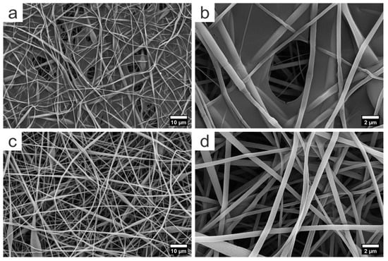
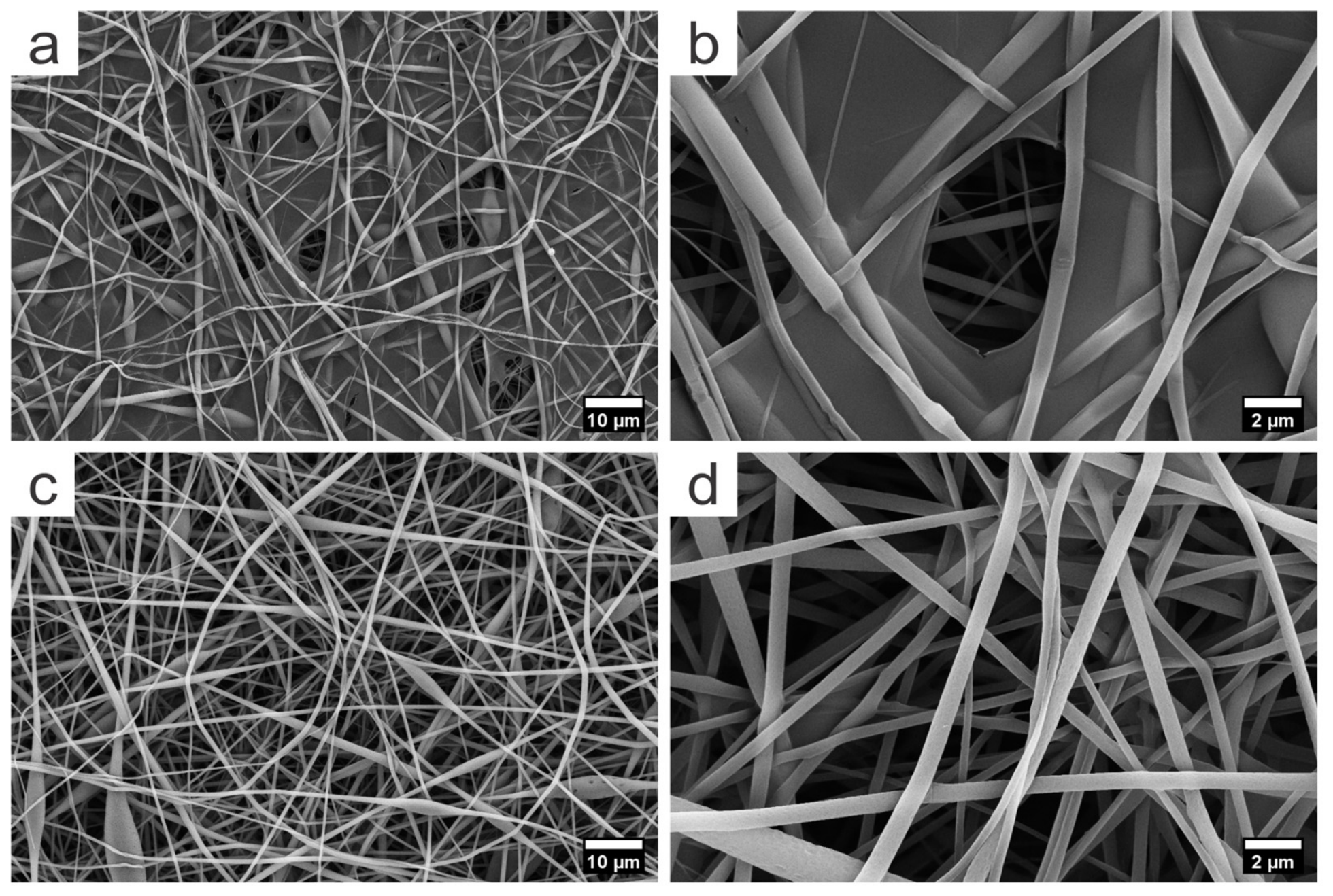
SEM micrographs of PLA–PEDOT:PSS composite scaffolds obtained without ultrasonic treatment (a,b), showing a continuous surface film, and after ultrasonic treatment (c,d), exhibiting the absence of a continuous film and textured fiber surfaces.
Two types of PLA–PEDOT:PSS fibrous scaffolds prepared by different impregnation procedures exhibited different surface morphologies. In the PLA-PEDOT:PSS sample, obtained without ultrasonic treatment, continuous film-like impregnation is observed on the scaffold surface (Figure 2a,b), indicating that PEDOT:PSS is mainly localized on the outer surface of the matrix, while the PLA fibers retain a smooth morphology comparable to that of the pristine scaffold. In contrast, for the ultrasonically treated sample (Figure 2c,d), no continuous film on the fiber surface is detected, which suggests improved penetration and distribution of PEDOT:PSS throughout the fibrous matrix. Moreover, the fiber surface becomes more textured, with small depressions and irregularities along the filaments, which may be associated with the action of ultrasound during the impregnation step.
It should be noted that previous studies have reported that ultrasonic treatment does not induce damage to the structure of nonwoven fibers [17,18].
The observed improvement in PEDOT:PSS distribution can be attributed to two main ultrasound-driven mechanisms. First, acoustic cavitation the—nucleation, growth, and collapse of microbubbles in the liquid medium—generates localized high-velocity microjets and pressure waves that overcome the hydrophobic barrier of the PLA fibers and drive the PEDOT:PSS dispersion into the interior of the fibrous matrix. Second, acoustic streaming enhances bulk microfluidic transport within the porous scaffold, promoting more homogeneous three-dimensional infiltration of the conductive phase compared to passive immersion.
3.2. Mechanical Properties
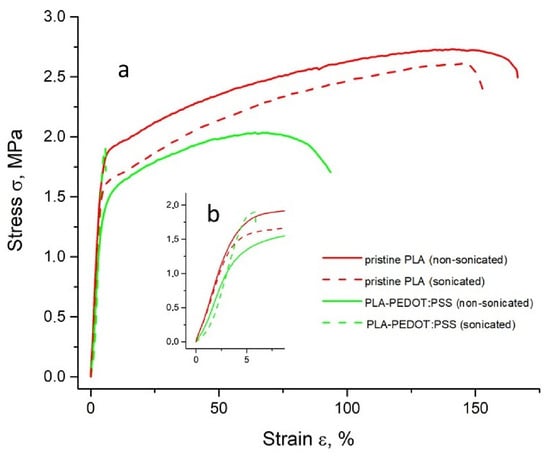
The results of mechanical analysis for sonicated and non-sonicated pristine PLA and PLA-PEDOT:PSS composites are shown in Table 1 and Figure 3.
Table 1.
Summary of mechanical properties of pristine PLA and PLA–PEDOT:PSS composites subjected to ultrasonic and non-ultrasonic processing. Data are presented as mean ± standard deviation.
Figure 3.
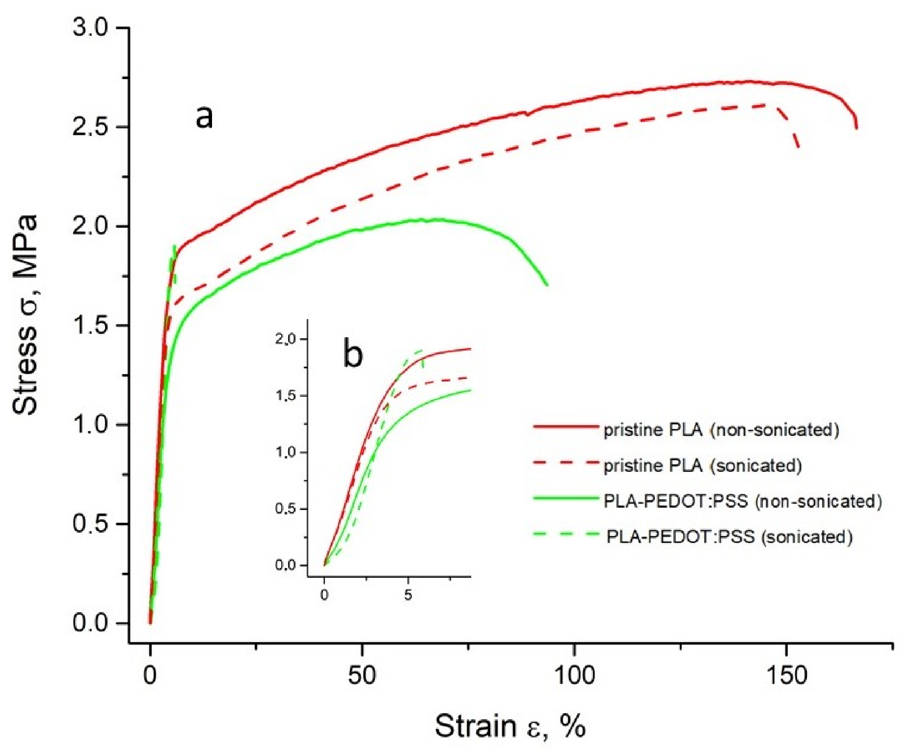
Stress–strain curves of the fabricated composites (a) and enlarged view of the initial linear region (b).
A pristine PLA scaffold subjected to ultrasonication in the absence of PEDOT:PSS was used as a sonication control. For pristine PLA (sonicated and non-sonicated) and for non-sonicated PLA–PEDOT:PSS, a clear deviation from linear elasticity in the stress–strain curves enabled determination of the yield strength. In contrast, sonicated PLA–PEDOT:PSS samples exhibited abrupt failure without a discernible yield point (Figure 3a,b); consequently, yield strength is not reported for this group (Table 1), consistent with its brittle mechanical response. No statistically significant differences in yield strength were observed among pristine PLA (sonicated and non-sonicated) and non-sonicated PLA–PEDOT:PSS (p = 0.08, one-way ANOVA), indicating that neither ultrasonication alone nor PEDOT:PSS impregnation without sonication affects yielding behavior.
The strain at elongation at break (ε) decreased from 153 ± 17% and 162 ± 14% for non-sonicated and sonicated pristine PLA, respectively, to 74 ± 9% for non-sonicated PLA–PEDOT:PSS, and further to 6 ± 2% for sonicated PLA–PEDOT:PSS (Table 1, Figure 3), demonstrating a pronounced transition toward brittle failure upon PEDOT:PSS impregnation, particularly under ultrasonication. No significant difference was observed between sonicated and non-sonicated pristine PLA, confirming that ultrasonication alone does not damage the fibrous structure.
PEDOT:PSS impregnation also reduced the ultimate tensile strength relative to pristine PLA (2.7 ± 0.3 MPa), yielding values of 2.0 ± 0.5 MPa and 2.0 ± 0.3 MPa for non-sonicated and sonicated PLA-PEDOT:PSS, respectively. This suggests that the conductive impregnation weakens the load-bearing fibrous network, while ultrasonication does not further affect this parameter. In contrast, Young’s modulus remained statistically unchanged across all samples (Table 1), indicating that the initial elastic stiffness is primarily governed by the PLA scaffold.
Despite the significant reduction in elongation at break of the sonicated PLA-PEDOT:PSS composite, a key mechanical finding of this study is the preservation of Young’s modulus across all sample groups (~34–47 MPa, Table 1), indicating that neither ultrasonication nor PEDOT:PSS impregnation alters the intrinsic elastic stiffness of the PLA fibrous scaffold. This is particularly relevant in the context of neural implantation, where mechanical mismatch between the electrode and the surrounding soft tissue—rather than absolute strain capacity—is considered the primary driver of chronic foreign body response, glial scarring, and long-term signal degradation [2,3,18]. The Young’s modulus of the present composites is several orders of magnitude lower than that of conventional silicon- or metal-based electrodes [19], placing them in a significantly more biomechanically compatible range for interfacing with neural tissue. While the elongation at break of the sonicated composite is reduced to 6 ± 2% due to the intrinsic brittleness of the PEDOT:PSS phase, this limitation is acknowledged and identified as a target for future optimization through post-treatment of the impregnated scaffolds with plasticizers such as ethylene glycol or DMSO, or through the addition of a compound with two or more hydroxyl groups, like glycerol or malic acid [20]. Another strategy to improve the mechanical robustness of PEDOT:PSS-based composites is the encapsulation of the brittle conducting phase within elastomeric layers, which promotes a more uniform distribution of strain and limits crack initiation and propagation under tension [9].
Overall, these results confirm that ultrasonication does not compromise the mechanical integrity of the PLA backbone, while PEDOT:PSS impregnation reduces ductility regardless of sonication—findings consistent with the SEM observations, which revealed no ultrasonication-induced structural damage to the fiber morphology (Figure 1) and demonstrated a uniform PEDOT:PSS coating across the nonwoven matrix (Figure 2c,d).
3.3. Electrical Properties
Ultrasonication during PEDOT:PSS impregnation had a pronounced effect on the electrical properties of the PLA-PEDOT:PSS composite. The electrical resistivity of the sonicated PLA-PEDOT:PSS samples was significantly lower (40.2 ± 20.0 Ω·m) than that of the non-sonicated PLA-PEDOT:PSS samples (294.4 ± 92.1 Ω·m) (Table 2), indicating a substantial improvement in electrical conductivity associated with the ultrasonication-assisted treatment. This reduction in resistivity suggests that ultrasonication promotes a more homogeneous distribution of the PEDOT:PSS phase within the fibrous network, facilitating the formation of more efficient conductive pathways throughout the mat.
Table 2.
Specific electrical resistivity (ρ) of non-sonicated and ultrasonically treated PLA–PEDOT:PSS samples, reported as mean ± standard deviation.
Although the two-probe method, which was used in this study, is in principle susceptible to contact resistance contributions [21], configurations with sputtered Au/Pd contact pads, defined by a shadow mask, minimize contact resistance relative to manually pressed probes. In addition, the high resistivity of both sample groups (40–294 Ω·m) places them firmly in the regime where sample resistance greatly exceeds any realistic contact resistance, rendering the relative contribution of the latter negligible. All groups were characterized under identical contact geometry, ensuring that the reported 7.3-fold reduction is a valid comparative result.
The pronounced reduction in electrical resistivity observed for the ultrasonically treated scaffolds is consistent with the morphological features revealed by SEM. In the non-sonicated PLA–PEDOT:PSS samples, the conductive polymer predominantly forms a continuous film on the outer surface of the fibrous mat, limiting the effective conductive cross-section and potentially leading to incomplete percolation across the scaffold thickness, which results in relatively high resistivity. In contrast, ultrasonically impregnated scaffolds exhibit no continuous surface film and show deeper penetration and a more homogeneous three-dimensional distribution of PEDOT:PSS throughout the fibrous network, accompanied by increased surface texturing of individual fibers. This interconnected conductive architecture facilitates more efficient charge transport, accounting for the significantly lower resistivity of the sonicated PLA–PEDOT:PSS scaffolds.
Although the electrical properties of the present composite fall below the thresholds typically cited for functional flexible neural electrodes [9], this is consistent with the proof-of-concept scope of the present study, which focuses on establishing the structure–property relationships of ultrasound-assisted impregnation rather than delivering a ready-to-implant electrode material. Further optimization of PEDOT:PSS loading and secondary solvent post-treatment (e.g., ethylene glycol, DMSO) represents a well-documented route toward electrode-grade conductivity [20,22,23,24,25] and constitutes a priority direction for future work.
In summary, ultrasound-assisted impregnation reduces the specific resistivity of the composite 7.3-fold, attributable to the formation of an interconnected three-dimensional PEDOT:PSS percolation network throughout the scaffold.
3.4. Contact Angle
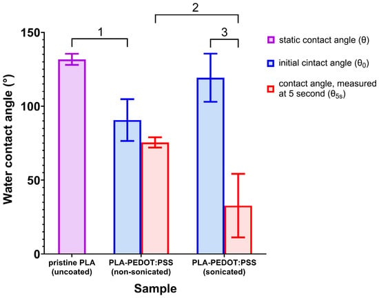
The results of contact angle measurements for pristine PLA and sonicated and non-sonicated PLA-PEDOT:PSS composites are shown in Figure 4. For the pristine PLA scaffolds, the water droplet preserved its shape throughout the observation period and the static contact angle remained essentially constant, indicating a hydrophobic, non-absorbing fibrous mat with limited liquid penetration into the interior. For the non-sonicated PLA-PEDOT:PSS samples, the initial contact angle θ0 was significantly lower than that of the pristine PLA, reflecting increased surface wettability due to the presence of the hydrophilic PEDOT:PSS phase. However, only a moderate decrease in θ(t) over time and the absence of complete droplet imbibition were observed within the experimental window. In contrast, the sonicated PLA-PEDOT:PSS exhibited θ0 values comparable to the non-sonicated group, but the time-dependent behavior differed drastically: the contact angle rapidly decreased and the droplet was fully absorbed within 5–13 s, evidencing high capillary uptake and good internal pore accessibility.
Figure 4.
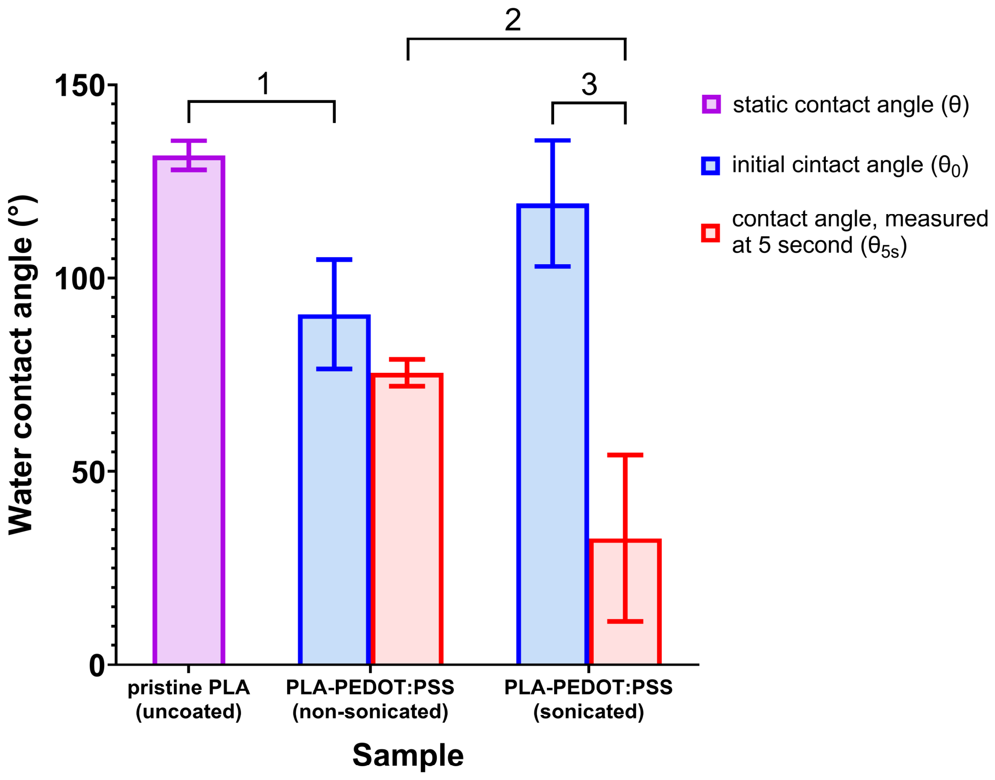
Contact angle measurements. Static contact angle (θ) of pristine PLA scaffolds. For non-sonicated and sonicated PLA-PEDOT:PSS composites, the initial contact angle (θ0) and the contact angle after 5 s (θ5s) were determined. Statistical significance: (1) p = 0.017, one-way ANOVA with Tukey post hoc test; (2) p = 0.027, independent-samples t-test; (3) p = 0.033, paired-samples t-test.
These wetting characteristics correlate well with the SEM observations of the PEDOT:PSS distribution in the fibrous matrices. In the non-sonicated PLA-PEDOT:PSS scaffolds, SEM revealed a continuous, film-like PEDOT:PSS impregnation on the outer surface while the underlying PLA fibers remained relatively smooth, which explains the reduced θ0 yet limited evolution of θ(t), as the liquid predominantly interacts with a surface film that only weakly communicates with the bulk porosity. By contrast, ultrasonically treated PLA-PEDOT:PSS scaffolds did not show such a continuous surface film; instead, PEDOT:PSS appeared more deeply penetrated and more homogeneously distributed throughout the fibrous network, and the fiber surfaces became rougher and more textured, with small depressions and irregularities attributed to the action of ultrasound. This three-dimensionally distributed conductive phase and increased micro-roughness enhance capillary effects and facilitate rapid liquid infiltration, consistent with the fast decay of θ(t) and complete droplet imbibition observed for the sonicated samples.
The contact angle data are also in line with the electrical characterization. The markedly lower electrical resistivity of the sonicated PLA-PEDOT:PSS scaffolds (40.2 ± 20.0 Ω·m) compared to the non-sonicated counterparts (294.4 ± 92.1 Ω·m) indicates that ultrasonication promotes the formation of an efficient, three-dimensional PEDOT:PSS percolation network within the mat. The same microstructural features responsible for continuous conductive pathways, namely deeper penetration of PEDOT:PSS and improved interconnection between coated fibers, also increase the effective internal surface area accessible to water and open capillary pathways within the scaffold. These effects collectively account for both the enhanced electrical conductivity and the more pronounced, time-dependent wetting behavior observed in the sonicated samples.
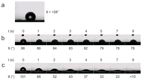
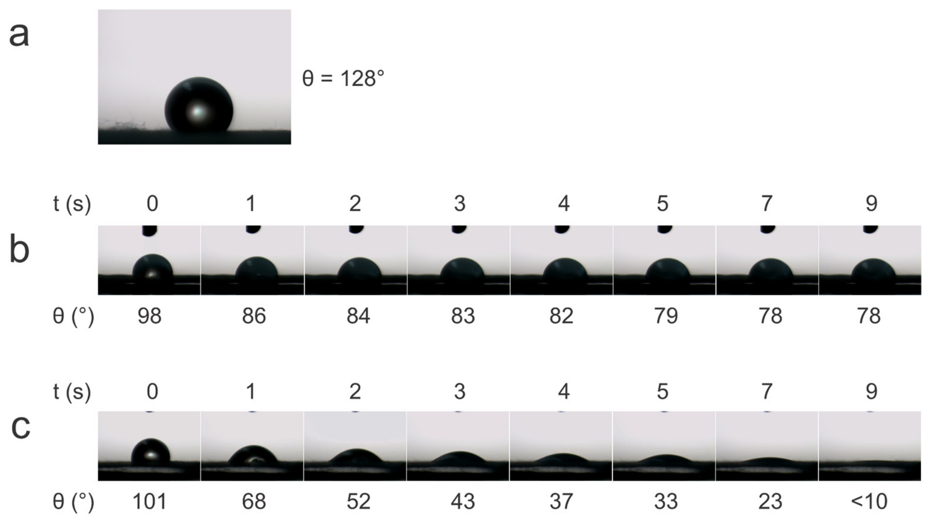
The time-resolved droplet images (Figure 5) further support and visualize the differences in wetting kinetics inferred from the averaged contact angle data. In agreement with the static measurements, the pristine PLA sample (Figure 5a) shows a single, essentially unchanged droplet shape with a high contact angle (θ ≈ 128°), confirming its hydrophobic [17], non-absorbing character over the entire observation period. For the non-sonicated PLA-PEDOT:PSS scaffold (Figure 5b), the frame sequence reveals only limited spreading: θ decreases from about 98° at t = 0 s to ≈78° at t = 9 s, and the droplet retains a well-defined cap-like shape, which is consistent with moderate wetting of a surface film and the absence of complete imbibition. In contrast, the sonicated PLA-PEDOT:PSS sample (Figure 5c) exhibits a rapid flattening and disappearance of the droplet, with θ decreasing from ≈101° at t = 0 s to below 10° by t = 9 s, directly illustrating the fast capillary uptake and full penetration of water into the ultrasonically treated fibrous matrix. It is known that more hydrophilic materials exhibit less cytotoxicity [19], which is important for implantable devices.
Figure 5.
Representative time evolution of water droplet profiles on different samples. Pristine PLA (a) showing static contact angle, non-sonicated PLA-PEDOT:PSS sample (b) showing limited spreading, sonicated PLA-PEDOT:PSS (c) showing rapid imbibition.
Beyond this indirect indication, several studies have directly demonstrated the biocompatibility of PEDOT:PSS-based composites in tissue engineering and neural interface applications. Conductive PEDOT:PSS-coated PLA/PHBV electrospun membranes have been shown to support fibroblast adhesion and proliferation without detectable cytotoxicity, highlighting their suitability for tissue engineering [12]. PEDOT-coated PLA fibrous scaffolds complied with cytotoxicity criteria and sustained the growth and neurite outgrowth of SH-SY5Y neural cells. In addition, fully polymeric PEDOT:PSS electrodes and PEDOT:PSS-functionalized scaffolds [26] have been successfully used as neural interfaces and electroconductive scaffolds in vivo or in advanced in vitro models, demonstrating acceptable tissue response and cytocompatibility [27,28]. Taken together, these studies indicate that the material components employed in the present study possess an established biocompatibility profile; however, a dedicated in vitro and in vivo evaluation of the specific ultrasonically treated PLA-PEDOT:PSS scaffolds will be required in future work.
Collectively, these wetting data confirm that ultrasonication promotes deep PEDOT:PSS infiltration, resulting in markedly enhanced hydrophilicity and capillary uptake relevant for cell adhesion in implantable applications.
4. Conclusions
Ultrasonic treatment during PEDOT:PSS impregnation fundamentally alters the distribution pattern of the conductive filler within the PLA fibrous matrix. While simple immersion in a PEDOT:PSS dispersion creates a surface film with limited penetration, ultrasonication promotes uniform 3D infiltration, as evidenced by the elimination of continuous surface impregnation and the development of textured fiber morphology in scanning electron microscopy images.
This structural modification yields substantial improvements in functional properties: specific resistivity decreased 7.3-fold (40.2 ± 20.0 Ω·m vs. 294.4 ± 92.1 Ω·m), and water contact angle kinetics demonstrated rapid imbibition (complete droplet absorption within 5–13 s) compared to limited spreading in non-sonicated samples. The simultaneous enhancement of electrical conductivity and capillary uptake indicates the formation of interconnected percolation networks throughout the scaffold thickness.
Ultrasonication does not compromise the intrinsic mechanical integrity of the PLA backbone, as confirmed by the unchanged Young’s modulus (≈47 MPa) and preserved strain at break in the pristine PLA controls (162 ± 14%). The observed reduction in elongation upon PEDOT:PSS loading (to 6 ± 2%) reflects the brittle nature of the conductive phase rather than ultrasound-induced damage. Nevertheless, improving the mechanical compliance of conductive scaffolds remains an important direction for future work. Possible strategies include blending PLA with flexible co-polymers such as poly(ethylene glycol) (PEG) [29] or polycaprolactone (PCL) [30], which have been shown to enhance the ductility of electrospun conductive scaffolds without compromising electrical performance.
Thus, ultrasound-assisted impregnation of electrospun PLA nonwoven scaffolds with PEDOT:PSS yields materials with low electrical resistivity and high hydrophilicity, enabling their consideration as promising platforms for future conductive structures integrated with the nervous system.
Author Contributions
Conceptualization, S.V.K., A.D.T. and D.A.I.; methodology, S.V.K. and A.D.T.; investigation, S.V.K. and A.D.T.; writing—original draft preparation, S.V.K., A.D.T. and V.Y.B. (Vadim Yu. Baula); writing—review and editing, D.A.I.; visualization, S.V.K. and A.D.T.; formal analysis, S.V.K., A.D.T. and V.Y.B. (Vadim Yu. Baula); resources, D.A.I., I.V.S., V.Y.B. (Vladimir Yu. Buzko) and R.P.Y.; data curation, S.V.K. and A.D.T.; supervision, D.A.I.; project administration, D.A.I.; funding acquisition, D.A.I. All authors have read and agreed to the published version of the manuscript.
Funding
This research was supported by the grant of the state program of the «Sirius» Federal Territory «Scientific and technological development of the «Sirius» Federal Territory» (Agreement № 18-03 date 10 September 2024).
Data Availability Statement
The original contributions presented in this study are included in the article. Further inquiries can be directed to the corresponding author.
Conflicts of Interest
The authors declare no conflicts of interest.
References
- Pang, C.; Lee, C.; Suh, K.-Y. Recent advances in flexible sensors for wearable and implantable devices. J. Appl. Polym. Sci. 2013, 130, 1429–1441. [Google Scholar] [CrossRef] [Scilit]
- Kravchenko, S.V.; Tyukalov, A.V.; Subcheva, E.N.; Parkhomenko, E.R.; Siaglova, E.D.; Anokhin, D.V.; Ivanov, D.A. PDMS–MWCNT nanocomposites: Mechanical, thermal and electrical properties for soft neuroprosthetic devices. Russ. Phys. J. 2025, 68, 1649–1656. [Google Scholar] [CrossRef] [Scilit]
- Feiner, R.; Dvir, T. Tissue–electronics interfaces: From implantable devices to engineered tissues. Nat. Rev. Mater. 2017, 3, 17076. [Google Scholar] [CrossRef] [Scilit]
- Clair, C.; Lallam, A.; Rosenthal, M.; Sztucki, M.; Vatankhah-Varnosfaderani, M.; Keith, A.N.; Cong, Y.; Liang, H.; Dobrynin, A.V.; Sheiko, S.S.; et al. Strained Bottlebrushes in Super-Soft Physical Networks. ACS Macro Lett. 2019, 8, 530–534. [Google Scholar] [CrossRef] [Scilit]
- Umarov, A.Z.; Collins, J.; Nikitina, E.A.; Moutsios, I.; Rosenthal, M.; Dobrynin, A.V.; Sheiko, S.S.; Ivanov, D.A. Enhancing the Biomimetic Mechanics of Bottlebrush Graft-Copolymers through Selective Solvent Annealing. Macromol. Rapid Commun. 2025, 46, 2400569. [Google Scholar] [CrossRef] [Scilit]
- Yin, J.; Wang, S.; Xiao, X.; Manshaii, F.; Scott, K.; Chen, J. Leveraging biomimetic materials for bioelectronics. Matter 2025, 8, 101961. [Google Scholar] [CrossRef] [Scilit]
- Gu, X.; Guo, X.-B.; Li, W.-H.; Jiang, Y.-P.; Liu, Q.-X.; Tang, X.-G. High-Entropy Materials for Application: Electricity, Magnetism, and Optics. ACS Appl. Mater. Interfaces 2024, 16, 53372–53392. [Google Scholar] [CrossRef] [Scilit]
- Hussain, M.; Khan, S.M.; Shafiq, M.; Abbas, N. A review on PLA-based biodegradable materials for biomedical applications. Giant 2024, 18, 100261. [Google Scholar] [CrossRef] [Scilit]
- Kayser, L.V.; Lipomi, D.J. Stretchable Conductive Polymers and Composites Based on PEDOT and PEDOT:PSS. Adv. Mater. 2019, 31, 1806133. [Google Scholar] [CrossRef] [Scilit]
- Tseng, H.-S.; Chen, Y.-L.; Zhang, P.-Y.; Hsiao, Y.-S. Additive Blending Effects on PEDOT:PSS Composite Films for Wearable Organic Electrochemical Transistors. ACS Appl. Mater. Interfaces 2024, 16, 13384–13398. [Google Scholar] [CrossRef] [Scilit] [PubMed]
- Prabhakaran, M.P.; Ghasemi-Mobarakeh, L.; Jin, G.; Ramakrishna, S. Electrospun conducting polymer nanofibers and electrical stimulation of nerve stem cells. J. Biosci. Bioeng. 2011, 112, 501–507. [Google Scholar] [CrossRef] [Scilit]
- Chang, H.C.; Sun, T.; Sultana, N.; Lim, M.M.; Khan, T.H.; Ismail, A.F. Conductive PEDOT:PSS coated polylactide (PLA) and poly(3-hydroxybutyrate-co-3-hydroxyvalerate) (PHBV) electrospun membranes: Fabrication and characterization. Mater. Sci. Eng. C 2016, 61, 396–410. [Google Scholar] [CrossRef] [Scilit]
- Dominguez-Alfaro, A.; Gabirondo, E.; Alegret, N.; De León-Almazán, C.M.; Hernandez, R.; Vallejo-Illarramendi, A.; Prato, M.; Mecerreyes, D. 3D Printable Conducting and Biocompatible PEDOT-graft-PLA Copolymers by Direct Ink Writing. Macromol. Rapid Commun. 2021, 42, 2100100. [Google Scholar] [CrossRef] [Scilit]
- Savelyeva, M.S.; Abalymov, A.A.; Lyubun, G.P.; Vidyasheva, I.V.; Yashchenok, A.M.; Douglas, T.E.L.; Gorin, D.A.; Parakhonskiy, B.V. Vaterite coatings on electrospun polymeric fibers for biomedical applications. J. Biomed. Mater. Res. A 2017, 105, 94–103. [Google Scholar] [CrossRef] [Scilit]
- Higuchi, J.; Klimek, K.; Wojnarowicz, J.; Opalińska, A.; Chodara, A.; Szałaj, U.; Dąbrowska, S.; Fudala, D.; Ginalska, G. Electrospun Membrane Surface Modification by Sonocoating with HA and ZnO:Ag Nanoparticles—Characterization and Evaluation of Osteoblasts and Bacterial Cell Behavior In Vitro. Cells 2022, 1, 1582. [Google Scholar] [CrossRef] [Scilit]
- Mulky, E.; Yazgan, G.; Maniura-Weber, K.; Luginbuehl, R.; Fortunato, G.; Bühlmann-Popa, A.-M. Fabrication of biopolymer-based staple electrospun fibres for nanocomposite applications by particle-assisted low temperature ultrasonication. Mater. Sci. Eng. C 2014, 45, 277–286. [Google Scholar] [CrossRef] [Scilit]
- Lionetto, F.; Dell’Anna, R.; Montagna, F.; Maffezzoli, A. Modeling of continuous ultrasonic impregnation and consolidation of thermoplastic matrix composites. Compos. Part Appl. Sci. Manuf. 2016, 82, 119–129. [Google Scholar] [CrossRef] [Scilit]
- Woods, G.A.; Rommelfanger, N.J.; Hong, G. Bioinspired Materials for In Vivo Bioelectronic Neural Interfaces. Matter 2020, 3, 1087–1113. [Google Scholar] [CrossRef] [Scilit] [PubMed]
- Zhao, H.; Liu, R.; Zhang, H.; Cao, P.; Liu, Z.; Li, Y. Research Progress on the Flexibility of an Implantable Neural Microelectrode. Micromachines 2022, 13, 386. [Google Scholar] [CrossRef] [Scilit]
- Fan, X.; Nie, W.; Tsai, H.; Wang, N.; Huang, H.; Cheng, Y.; Wen, R.; Ma, L.; Yan, F.; Xia, Y. PEDOT:PSS for Flexible and Stretchable Electronics: Modifications, Strategies, and Applications. Adv. Sci. 2019, 6, 1900813. [Google Scholar] [CrossRef] [Scilit] [PubMed]
- Dörling, B.; Jacobs, I.E.; Brunetti, I.; Pataki, N.J.; Yee, S.K.; Scheunemann, D.; Kang, K.; Zuo, G.; Reparaz, J.S.; Mori, T.; et al. Toward a Consensus Characterization Protocol for Organic Thermoelectrics. Adv. Mater. 2026, 38, e20430. [Google Scholar] [CrossRef] [Scilit]
- Khasim, S.; Pasha, A.; Lakshmi, M.; Chellasamy, P.; Kadarkarai, M.; Darwish, A.A.A.; Hamdalla, T.A.; Al-Ghamdi, S.A.; Alfadhli, S. Post treated PEDOT-PSS films with excellent conductivity and optical properties as multifunctional flexible electrodes for possible optoelectronic and energy storage applications. Opt. Mater. 2022, 125, 112109. [Google Scholar] [CrossRef] [Scilit]
- Park, H.; Lee, S.H.; Kim, F.S.; Choi, H.H.; Cheong, I.W.; Kim, J.H. Enhanced thermoelectric properties of PEDOT:PSS nanofilms by a chemical dedoping process. J. Mater. Chem. A 2014, 2, 6532–6539. [Google Scholar] [CrossRef] [Scilit]
- He, H.; Zhang, L.; Yue, S.; Yu, S.; Wei, J.; Ouyang, J. Enhancement in the Mechanical Stretchability of PEDOT:PSS Films by Compounds of Multiple Hydroxyl Groups for Their Application as Transparent Stretchable Conductors. Macromolecules 2021, 54, 1234–1242. [Google Scholar] [CrossRef] [Scilit]
- Lee, I.; Kim, G.W.; Yang, M.; Kim, T.-S. Simultaneously Enhancing the Cohesion and Electrical Conductivity of PEDOT:PSS Conductive Polymer Films using DMSO Additives. ACS Appl. Mater. Interfaces 2016, 8, 302–310. [Google Scholar] [CrossRef] [Scilit]
- Pires, L.S.; Melo, D.S.; Borges, J.P.; Henriques, C.R. PEDOT-Coated PLA Fibers Electrospun from Solutions Incorporating Fe(III)Tosylate in Different Solvents by Vapor-Phase Polymerization for Neural Regeneration. Polymers 2023, 15, 4. [Google Scholar] [CrossRef] [Scilit]
- Magaz, A.; Spencer, B.F.; Hardy, J.G.; Li, X.; Gough, J.E.; Blaker, J.J. Modulation of Neuronal Cell Affinity on PEDOT–PSS Nonwoven Silk Scaffolds for Neural Tissue Engineering. ACS Biomater. Sci. Eng. 2020, 6, 6906–6916. [Google Scholar] [CrossRef] [Scilit]
- Filho, G.; Júnior, C.; Spinelli, B.; Damasceno, I.; Fiuza, F.; Morya, E. All-Polymeric Electrode Based on PEDOT:PSS for In Vivo Neural Recording. Biosensors 2022, 12, 853. [Google Scholar] [CrossRef] [Scilit]
- Scaffaro, R.; Gulino, F.E.; Lopresti, F. Structure–property relationship and controlled drug release from multiphasic electrospun carvacrol-embedded polylactic acid/polyethylene glycol and polylactic acid/polyethylene oxide nanofiber mats. J. Ind. Text. 2020, 49, 943–966. [Google Scholar] [CrossRef] [Scilit]
- Munawar, M.A.; Schubert, D.W.; Nilsson, F. Mechanical tunability of oriented and random electrospun poly(ε-caprolactone) scaffolds via concentration, molecular weight, and environment. Sci. Rep. 2026, 16, 10507. [Google Scholar] [CrossRef] [Scilit] [PubMed]

Disclaimer/Publisher’s Note: The statements, opinions and data contained in all publications are solely those of the individual author(s) and contributor(s) and not of MDPI and/or the editor(s). MDPI and/or the editor(s) disclaim responsibility for any injury to people or property resulting from any ideas, methods, instructions or products referred to in the content. |
© 2026 by the authors. Licensee MDPI, Basel, Switzerland. This article is an open access article distributed under the terms and conditions of the Creative Commons Attribution (CC BY) license.