


Abstract
The article presents the research findings on a collection of ceramic crucibles excavated at the Koksharovka-1 hillfort, known as one of the most significant archaeological sites of the Medieval epoch (7th–13th centuries) in the Russian Far East. This study represents an investigation of melting crucibles as a distinct category of metalworking ceramics discovered in the research area. The study is based on an integrated approach combining traditional archaeological analysis with physicochemical methods, including scanning electron microscopy with energy-dispersive spectroscopy (SEM-EDS), X-ray fluorescence spectroscopy (XRF), Raman spectroscopy, electron probe microanalysis (EPMA), and computed microtomography (micro-CT). The research found that the morphological and technological properties of the crucibles were consistent with their function as containers for metal heat treatment. Traces of copper-based alloys as well as gold and silver processing were detected as a result of the crucible examination. For the first time, archaeological evidence of silver and gold metalworking in the Russian Far East during the Medieval epoch has been obtained.
1. Introduction
Crucibles, along with casting molds, are among the main ceramic inventories used since ancient times in the production of non-ferrous metal items. The discovery of crucibles at archaeological sites is of considerable interest as potential evidence of local metalworking [1,2,3,4,5]. Modern research on ceramic crucibles employing archaeometry methods focuses on the following features: morphological parameters, the composition of the ceramic paste, the presence of specialized coatings on the surfaces, and traces of functional usage, in particular, metallic traces. Collectively, these features allow for certain conclusions regarding the technological standards and skills involved in the manufacture of ceramic crucibles as specialized vessels for working with molten metal under high-temperature conditions, the specifics of the metalworking process, and the compositions of the metal alloys themselves [5,6,7,8,9,10,11,12].
Archaeometry studies of metalworking ceramics from the southern Russian Far East were initiated some years ago based on a collection of casting molds unearthed at one of the Medieval epoch sites [13]. The present article focuses on the ceramic crucibles discovered at the Koksharovka-1 hillfort. This is one of the currently known sites where ceramic crucibles with functional traces have been found [14,15]. Recently, a primary investigation of the crucibles unearthed at the Koksharovka-1 hillfort has revealed the significant potential of these materials as a source for studying past metalworking history. The most important and innovative result was the identification of evidence of gold and silver processing, together with the traces of copper-based alloys [15]. Previous studies of archaeological metal collections and technical ceramics indicated evidence of copper-based metallurgy and metalworking in the southern Russian Far East during the Medieval epoch, 7th–13th centuries [13,14,16,17,18]. However, no definite traces of local gold and silver processing had been detected until recently.
The current research complements and updates the primary study of the crucibles from the Koksharovka-1 hillfort with new materials and findings. The main research objectives are as follows: 1—determination of the morphological and technological features related to the manufacture of the crucibles and 2—detection and interpretation of probable traces of metals and alloys processed in crucibles. The study is intended to provide a general characterization of this unique assemblage of ceramic crucibles and contribute to the archaeometallurgy records of the region. The objectives were addressed through an interdisciplinary approach employing a set of physicochemical methods.
2. Archaeological Context
The Koksharovka-1 hillfort is located in the central Primor’ye region as a part of the southern Russian Far East, on the right bank of the Ussuri River, at a distance of about 150 km westward from the seacoast (see Figure 1).
Figure 1.
The research area, Primor’ye region, and site location of the Koksharovka-1 hillfort (red triangle).
The site represents two cultural and chronological horizons of the Medieval epoch. The lower horizon contains some remains of the Bohai State period, 698–926. The main upper horizon containing the remains of the post-Bohai period is dated in the interval of the late 10th–11th centuries according to the representative series of radiocarbon dates. This period, preceding the Jin Dynasty, or Jurchen Empire, period (1115–1234), is represented by a small number of known archaeological sites in the Primor’ye region. The Koksharovka-1 hillfort contributes to our knowledge of the economy, technologies, and culture of the local population during the post-Bohai period. Beyond its regional significance, the site offers a rare data point for medieval archaeometallurgy in the wider East Asian context, where well-documented crucible assemblages of comparable date remain scarce. Excavations conducted from 2008 to 2014 investigated a unique architectural complex belonging to the upper horizon of the 10th–11th centuries. This complex consists of seven wooden buildings and a courtyard with structures for economic and production purposes [19,20,21].
One of the structures was a rectangular surface construction with an area of 48 sq. m (see Figure S1). Its walls were oriented to the cardinal points. The foundation of the walls, with a width of 35–42 cm and a height of 7–42 cm, was built from rounded river pebbles and unworked stone. Bases for posts are located on the exterior side at the corners of the structure. Outside the structure, near its eastern wall, a concentration of fire-cracked stones with charcoal and pieces of burned daub was revealed, which could represent the remains of a furnace. Metal slags or ingots were not found [20] (pp. 45–46). Fragmented crucibles were discovered as two local concentrations inside the structure and as scattered finds near the southwestern and northeastern boundaries of the structure. This unearthed structure is tentatively interpreted as the remains of a casting workshop. Finds of crucibles were also made in other locations—specifically, several items were discovered in a test trench in the western part of the settlement. In general, it must be noted that there is a lack of archaeological evidence and materials such as metal slags, ingots, remains of melting constructions, and variability of specialized inventories, which could be essential for the reconstruction of technical and social patterns of the metalworking process at the settlement.
The total amount of the items identified previously as the crucibles is 32 items [15]. A subsequent reinspection of this assemblage raised doubts regarding the attribution of a subset of small, undiagnostic wall fragments (n = 10) lacking clear morphological features (rim, base, or characteristic wall curvature) and functional use-wear traces (slag deposits, glaze, or evidence of thermal impact). These fragments were excluded from further study, and the present research is based on the resulting subset of 22 objects with reliably established functional attribution as crucibles.
Other categories of archaeological ceramics unearthed at the settlement are ordinary pottery, pottery for special (ritual?) functions, and roof tiles. The ordinary pottery, exhibiting great diversity of shapes and sizes, was produced with the throwing-wheel technique and fired in reducing and oxidizing regimes. A small amount of fragmented stoneware with olive-greenish and brownish-yellow glazed walls is reported. The stoneware is interpreted as a product imported from Chinese ceramic kilns [19] (pp. 106–107, 132–163).
During the investigations, the Koksharovka-8 burial mound complex was excavated, located in immediate proximity to the hillfort area and associated with its architectural ensemble [20] (pp. 102–141). A representative series of metal decorative items was found here: 44 “leaflets” cut from gold foil, a bronze bracelet, and 26 silver ornaments (see Figures S2 and S3).
3. Materials and Methods
3.1. Materials
The assemblage of crucibles currently available for the intended study contains 22 items, including all unbroken and partially damaged ones discovered at the site, and series of small-sized fragments of walls, bottoms, and orifice parts. The examined crucibles were numbered as objects 1–22 (see Figure 2 and Figure 3; Table S1).
Figure 2.
Koksharovka-1 hillfort. Ceramic crucibles. Objects 1–7.
Figure 3.
Koksharovka-1 hillfort. Ceramic crucibles. Objects 8–22.
Macroscopic observations revealed features of the items’ morphology and size, color, textural characteristics of the surfaces and cores, and probable functional traces. Based on exterior surface appearances, crucibles were distinguished as those with a black glaze-like coating, crucibles with a yellow slip-like coating, and crucibles with uncoated surfaces. The functional traces present on most of the objects appeared as black- or dark gray-colored slightly crusted dross deposits; light-brownish plaque; dark spots on the interior surfaces; copper-red and greenish-turquoise spots on both surfaces; and bloating, corrugating, and cracking of the glaze and slip coatings. Notably, intensely thick slagged deposits were not present on the interiors.
Instrumental analyses of the crucibles were intended to obtain information on the textural and compositional features of ceramic fabrics and coatings and to reveal the traces of processed metals and alloys. Some additional materials were involved for the purpose of comparative study of ceramic fabrics and black glaze coatings.
Also, four fragments of ordinary pottery from the Koksharovka-1 hillfort were selected to examine the ceramic fabric composition. The task of comparing the characteristics of clay fabrics used for the making of ceramic crucibles and non-specialized utilitarian pottery from the same archaeological context is a common focus in studies of ancient technical ceramics [9]. The fragments were numbered as objects 23–26 (Table S1).
To compare black glaze coating composition, a fragment of archaeological black-glazed stoneware from the Primor’ye region was sampled as object 27 (see Figure S4c,d; Table S1). Black-glazed stoneware occurs at medieval sites dated to the Jin dynasty period of the early 12th–13th centuries. The sampled fragment comes from the well-known Shaiga hillfort [22]. It should be noted that the 22 crucibles examined in this study represent approximately 69% of the originally identified assemblage of 32 items (Section 2), the remaining 10 undiagnostic fragments having been excluded from analysis.
3.2. Methods
The research methodology focused on non-destructive instrumental techniques exclusively to avoid any damage to the unique archaeological objects, which are currently under special museum curation status. This was the reason that some established and effective instrumental techniques could not be applied in the presented study. One of these techniques is thin sectional petrography analysis, which provides useful information on ceramic fabrics compositions. This technique is adopted widely in the studies of archaeological ceramics, in particular, melting crucibles [7,9,23]. The set of techniques used in the current study included digital microscopy, portable X-ray fluorescence spectroscopy (pXRF), scanning Electron Microscopy with Energy Dispersive X-ray Spectroscopy (SEM-EDS), Electron probe microanalysis (EPMA), Raman spectroscopy, and computed microtomography (micro-CT).
3.2.1. Sampling Strategy
The application of specific methods is limited by the morphology and size of certain objects. Small-sized flat or slightly curved wall fragments are most appropriate for study by various methods. In contrast, the large sizes and curved contours of unbroken or partially damaged crucibles and of bottom parts significantly restrict the application of certain instrumental techniques. As noted, curved shapes of crucibles’ bottoms hinder or render impossible the analysis of the interior [5].
Not all examined crucibles were subjected to the full set of analytical techniques. Digital microscopy and pXRF were applied to the entire available assemblage of 22 objects. The selection of specific objects for the SEM-EDS, EPMA, Raman spectroscopy, and micro-CT was guided by a combination of criteria: (i) the presence of macroscopically or microscopically visible metallic residues, surface coatings, or droplets identified during preliminary visual and digital microscopic inspection, which prioritized objects for SEM-EDS, EPMA, and micro-CT examination; (ii) the morphological suitability of the specimen for a given technique, e.g., flat or slightly curved fragments were preferentially selected for SEM-EDS and EPMA, whereas access to interior surfaces for pXRF and EPMA was restricted for objects with well-preserved, closed reservoir shapes; and (iii) the state of preservation, ensuring that analysis could be performed without further damage to the object.
For the examination of the fabric of ordinary pottery, typical fragments of reducing-fired wheel-made wares of gray color, such as objects 23 and 24 (see Figure S4a,b), and typical fragments of oxidizing-fired wheel-made pottery, such as objects 25 and 26 (see Figure S4d), were selected. The measurements were taken on the exposed ceramic fabric areas appearing under the peeled slip layer.
Consequently, SEM-EDS, EPMA, Raman spectroscopy, and micro-CT were applied to purposefully selected subsets, as detailed in Table S1 and specified individually for each technique below.
3.2.2. Digital Microscopy
Digital microscopy is referenced in studies of archaeological ceramics, in particular, metalworking inventories, for the visual documentation of fabric, surface treatments, and residue patterns [11,24]. In this research study, digital microscopy was applied to both well-preserved and fragmented objects to obtain visual details of ceramic fabrics, glaze and slip coatings, and potential metal appearances. The analysis was conducted using a Levenhuk DTX 50 digital microscope (Levenhuk, Inc., Tampa, FL, USA) with a maximum optical zoom of 400×. The samples, which required no specific preparation regarding size or thickness, were examined under direct lighting to enhance the contrast of surface topography. The results are presented as a series of selected photo-images of the fractured cross-sections and surfaces.
3.2.3. Portable X-Ray Fluorescence Spectroscopy (pXRF)
The pXRF analysis as a non-destructive method is commonly applied in archaeometry for the rapid in situ characterization of artifacts, including technical ceramics, to identify the presence of major, minor, and trace elements indicative of raw materials or metalworking residues but without accurate quantitative estimation [5,8,11,25]. In this study, the pXRF technique was applied to both intact and fragmented crucibles to provide data on elemental compositions of surfaces with melting residue traces, surfaces free of visible residues, and surfaces with black glaze and yellow slip coatings. The measurements were performed using an Olympus Delta Professional DP 4000 analyzer (Waltham, MA, USA), which investigates surface areas with a spot size radius of 10–20 mm. The device’s applicability was significantly limited for examining the interior surfaces of crucibles with well-preserved, “closed” reservoirs due to physical access constraints. The analyzer was calibrated and operated in the “Alloy” measurement mode, which was optimized for the determination of metallic elements but has limitations in detecting light elements such as H, C, O, Na, K, Ca, Mg, N, and As. Therefore, we registered such basic elements of ceramic composition as Al, Si, Fe, Mg, Na, Ca, K, and some others and the element-metals as Cu, Pb, Sn, Ag, Au, and Zn, which was important in the context of research objectives. Three individual measurements were taken for each test spot. Taking into account semi-quantitative or moderately quantitative nature of the pXRF technique, in particular, applied to current study, the data are presented and discussed in qualitatively, through the categorizing detected elements as major, minor, or trace constituents.
3.2.4. Scanning Electron Microscopy with Energy Dispersive X-Ray Spectroscopy (SEM-EDS)
The SEM-EDS technique, which determines the elemental composition and the microstructure of an examined object, is widely applied to archaeological ceramic studies, in particular, to the investigation of metalworking ceramics [7,8,11,26,27]. In the present research, SEM-EDS was employed to examine the fabric, glaze, and potential metal evidence on the exterior and/or interior surfaces of ceramic crucibles. The analysis was conducted using a ZEISS EVO-40 scanning electron microscope (Oberkochen, Germany) equipped with an Oxford Instruments INCA-x sight EDS detector (Abingdon, UK) recognizing broad range of elements from the Be to U. The technique was applied to objects 12–20 and 23–27 following the sampling criteria described in Section 3.2.1. The objects were analyzed unpolished and without conductive coatings. The measurements were performed under low vacuum conditions (30–50 Pa) with an acceleration voltage of 20 kV, a working distance of 12.5–16.4 mm, and magnifications of 80×, 200×, and 500×. For each object, 1 to 3 test spots were selected, with 11 to 15 EDS spectra acquired per spot. Elemental concentrations of 0.5 wt% and higher were considered for reliable observation and subsequent interpretation. The original EDS measurements were processed under the standard procedure of element to oxide conversion. The data are presented and discussed as chemical compositions and their oxide components, such as Al2O3 (alumina), SiO2 (silica), Fe2O3 (iron), and the others, in weight % concentration.
3.2.5. Electron Probe Microanalysis (EPMA)
The EPMA as a non-destructive method for identifying elemental compositions in distinctive micro-areas of examined objects is applied to studies of archaeological materials, in particular, metalworking ceramics [7,11]. In the present research, EPMA was applied to objects 15, 16, and 22 following the criteria described in Section 3.2.1 to identify mineral inclusions in the fabric compound and to trace and identify metal particles inside the body and glaze coating. EPMA analysis was conducted on a JXA-8100 instrument (Tokyo, Japan) at the “Primorsky Center for Local Elemental and Isotope Analysis” of the Far East Geological Institute, Far Eastern Branch of the Russian Academy of Sciences, Vladivostok. The instrument was equipped with three wave spectrometers and an energy-dispersive spectrometer INCA x–sight (Oxford Instruments, England) assembled with a cathodoluminescence (CL) detector from Gatan and sensors MiniCL and CromaCL-2. Technical properties were the range of detected elements from B to U, voltage of 5–35 kV, light collection angle of 45°, crystal analyzers LiF, PET, TAP, LDE2, EDS resolution of 137 eV, and microscope magnification minimum of 40×. Instrumental analysis of the objects was conducted under a voltage of 20 kV and a probe current of 10 nA. Qualitative and semi-quantitative measurements were carried out based on the PhyRoZ procedure as a standard program of the EDS-analyzer Link ISIS applying a user-defined standard set. Carbon coating was applied to the examined surface before the analysis. The results of the investigation are presented as a series of selected BSE micrographs and panels of related EDS spectra.
3.2.6. Raman Spectroscopy
Raman spectroscopy as a technique providing data on the mineralogical and molecular composition of a substance is widely used in research on archaeological ceramic objects, although its application to the study of archaeological crucibles remains relatively limited, with specific examples including investigations of ceramic fabric composition [28]. In the present research, this method was applied to selected objects 8, 13, 15, 22, and 25 to identify mineralogical components of ceramic fabric and black glaze coating following the criteria described in Section 3.2.1 and to trace supposed evidence of metals on the crucibles’ surfaces.
The analysis was performed using an InVia Reflex Raman micro-spectrometer (Renishaw, Wotton-under-Edge, UK) coupled with a Leica DM2500 M microscope (Wetzlar, Germany) in incident light mode. A 532 nm diode-pumped laser with a power of 1.0 mW and an exposure time of 0.1 s was used for excitation, with 100 to 200 accumulations per spectrum to achieve an acceptable signal-to-noise ratio. A 20× objective lens (NA = 0.4, Leica) focused the laser to a spot of approximately 2 µm in diameter on the sample surface. Spectra were collected from three distinct locations per sample over a wavenumber range of 170 to 1800 cm−1 and processed using WiRE 4.4 software, employing smooth baseline subtraction and peak fit functions for identification.
3.2.7. Computed Microtomography (Micro-CT)
Micro-CT as a non-destructive and highly informative analytical technique is applied in archaeological ceramics studies to infer outer and inner morphology of an examined object to detect the areas of different X-ray density relating to the clay matrix, temper inclusions, and voids. The results of tomographic observations, represented as 2D and 3D images of zones and sites of intended interest, contribute to the reconstructions of ceramic fabrics and slips technologies, shaping methods, firing regimes, and functionality of studied objects [29]. In the present research, micro-CT was applied to selected objects to observe microstructural features of ceramic fabrics and glaze coatings and to visualize probable metal appearances. As prior studies show, micro-CT identification of metal particles within archaeological glass and ceramics is predicated on the significant difference in X-ray attenuation coefficients of certain materials [29,30].
Tomographic investigations were carried out using a Bruker SkyScan 1272 system (Kontich, Belgium) at the “Primorsky Center for Local Elemental and Isotope Analysis” of the Far East Geological Institute, Far Eastern Branch of the Russian Academy of Sciences, Vladivostok, with the following specifications: microfocus sealed X-ray source of 20–100 kV, 10 W, with less than 5 μm spot size at 4 W; X-ray detector 16 Mp (4904 × 3280 pixels); 0.35 μm pixel size at maximum magnification (nominal resolution); limitation of sample size to a diameter of 75 mm; and integrated filters of Al 0.25 mm, Al 0.5 mm, Al 1 mm, Al 0.5 mm + Cu 0.038 mm, and Cu 0.11 mm. The objects required no special preparation and were fixed in plastic containers by means of X-ray transparent materials to keep a vertical position during the scanning process. The parameters of X-ray acquisition were Cu 0.11 mm filter (X-ray source voltage of 100 kV and current of 100 μA), sample rotation from 0 to 180° with a 0.2° step, scaled image pixel size of 16.3 μm (object 22), 20 µm (object 20), 21 μm (object 15), and 21.7 µm (objects 11, 16, 17, and 18a), image format 1224 × 820 pixels and 1632 × 1092 pixels (object 22). To process the data, Bruker micro-CT (Kontich, Belgium) programs were used: NRecon 1.7.1.0 to reconstruct projection images and to create data sets of metal particles, DataViewer 1.5.3.4 to visualize cross-sections of the samples, and CTvox 3.3.0 r1403 to perform volume visualization and rendering.
4. Results
4.1. Crucibles as Ceramic Artefacts: Characteristics
4.1.1. Crucibles’ Morphology and Size
Two main shape types are distinguished. Type 1, represented by the majority of well-preserved objects, is a ceramic container with a narrowed conical bottom, a deep reservoir of round horizontal section, and an open orifice (see Figure 2 objects 2–7). The ratio of height to maximum orifice diameter is approximately 1.5–2. Where measurable, the height ranges from 4.7 to 7.7 cm. The bases are contoured by a parabolic or angled line and are not flattened. In cases where the upper part is well-preserved, the orifice features a pinched spout. The crucible wall thickness varies from 0.3 to 0.8 cm, typically being 0.6–0.7 cm. The capacities (volumes) of well-preserved crucibles were measured. They range from 20–25 mL (objects 2, 3, 4, 7) to 60–65 mL (object 5). Type 2 is represented by a single example of a low, flat-bottomed crucible with an oval horizontal section (see Figure 2 object 1). The reservoir height is 5.0 cm, while the maximum orifice width is 7.5 cm. The crucible’s volume is 35–40 mL. The wall and base thickness is 0.7–0.8 cm. The walls of both crucible types show slight contour asymmetry, indicating they were handmade rather than wheel-thrown.
4.1.2. Crucibles’ Fabrics
The description and characteristic of the crucibles’ fabrics are based on the observations on the color and texture as external features and some data on the structural and compositional patterns. The analytical procedures were restricted by the usage of non-destructive techniques. The determinations of chemical and mineralogical compositions are limited by the results obtained at the surfaced and cross-sectional areas without their damaging and the opportunity to apply the thin section petrography and XRD analyses as the techniques of high informative efficiency. It must be noted that the resulted evaluations of some properties of ceramic fabrics are preliminary rather that comprehensive ones.
Visual Observation and Digital Microscopy
Under macroscopic observation, the exterior surfaces of the crucibles, free of residue or dross deposits and coatings and fractured cross-sections cleaned of soil contamination, show a light color with whitish, creamy-whitish, or gray-whitish hues. No instance of reddish, orange, or brown-colored fabric was identified. For most of the objects, a distinctive textural feature visible in the cores (fractured cross-sections) under the naked eye and digital microscopy is the presence of mineral non-plastic inclusions. The inclusions appear mostly as semi-transparent whitish-gray and non-transparent white grains, ranging in size from 0.1–0.2 mm to 1.0–2.0 mm, often with angular sharp contours (see Figure 4a–e). The mineralogy of non-plastic inclusions is considered below.
Figure 4.
Results of digital microscopy. Crucibles’ cores and surfaces. Core (fractured cross-sections) images: (a): object 16; (b): object 4 (visible mineral inclusions); (c): object 19; (d): object 8 (visible mineral inclusions); (e): object 20 (visible mineral inclusions); (f): object 13; (g): object 11. Surface images: (h): object 11 (voids of probable burnt organic inclusions); (i): object 18a (fiber-like voids of probable organic inclusions); (j): object 2 (remains of yellow slip coating); (k): object 16 (black glaze coating); (l): object 13 (black glaze coating).
In contrast, some objects exhibit a fabric texture that appears more homogeneous, without protruding mineral inclusions (see Figure 4f,g). In some cases, probable traces of organic inclusions are supposed. At the surface of object 11, the traces appear as sub-angular voids, which can be interpreted, tentatively, as having been left by burnt particles of some kind of plant material, such as chopped grass or sawdust (see Figure 4h). At the surface of object 18a, the voids look like very narrow, elongated, and slightly curved channels, supposedly left by burnt thin fibers (see Figure 4i).
Micro-CT Results
Three-dimensional tomographic images of cut solid volumes of selected samples show of the fabrics saturated with mineral inclusions (see Figure 5a,b) and fabrics of relatively homogeneous texture (see Figure 6). A certain case is object 11, with the above-noted probable traces of burnt organic temper at the surface. Its tomographic cut solid volume shows the presence of mineral inclusions that are not as numerous and large-sized as those in objects 16 and 18a and some voids (pores) (see Figure 5e). The cut volume in the semi-transparent regime exhibits multiple internal pores, which seem to be morphologically comparable with the observed surface voids (see Figure 5f). This kind of porosity is different from the roundish-shaped, closed, and clearly outlined pores in object 17, which look like typical evidence of clay matrix vitrification (see Figure 5d).
Figure 5.
Results of micro-CT. Ceramic fabric. Three-dimensional tomographic images, showing the distribution of non-plastic inclusions and porosity in ceramic fabrics. (a–e): Cut solid volumes. (a): object 16; (b): object 18a; (c): object 13; (d): object 17; (e): object 11. (f): cut volume in semi-transparent regime, object 11. Green arrows: non-plastic inclusions. Yellow arrows: pores.
Figure 6.
Results of SEM-EDS. Object 13, interior surface. BSE image of SEM-EDS test spot area, showing the locally elevated TiO2/REE mineral inclusions. (a): The point of EDS spectrum N9 containing REE; (b): the point of EDS spectrum N 5 containing high TiO2; (c): the point of EDS spectrum N8 containing high TiO2. Referenced EDS spectra are given at the Figure S5.
pXRF and SEM-EDS Results
The elemental composition of the ceramic fabric was determined by pXRF and SEM-EDS on surfaces free of coatings and dross or plaque contamination. The pXRF measurements on the exteriors of objects 11, 18a, and 19 and on the interior of object 13 indicate Si and Al as major elements, together with Fe, Ti, and P as minor elements (Table 1). Notably, among the trace elements, Cu was detected on objects 11, 18a, and 19, and Pb on object 11.
Table 1.
Local elemental compositions of outer and interior surfaces measured by pXRF on the referenced objects; major elements (≥10%)—bold, minor elements (≥1.0%)—regular, trace elements (≤1.0%)—italic; n/a—non-attested.
SEM-EDS measurements were performed on the exteriors of objects 18a and 19, and on the interior of object 13 (Table 2). The description of the results is based on elemental data converted to oxide form. All samples show high alumina contents, up to 33–36%, together with low iron oxide contents. The considerable range of Al2O3 values observed within each object (e.g., 7.4–35.0% for object 18a) reflects the heterogeneity of individual SEM-EDS analytical spots, which variably intersect the clay matrix proper (higher Al2O3) or coarse quartz/mineral temper grains (lower Al2O3) rather than indicating compositional instability of the raw clay itself. Accordingly, the maximum values recorded for each object (33–36%) are considered the most representative of the alumina content of the clay matrix and are consistent with a refractory raw material. A characteristic trait of object 13 is the presence of REE elements La, Ce, and Nd detected together in a single EDS spectrum N9. In the BSE image of the test spot’s area, the spectrum corresponds to a bright light-colored inclusion less than 10 µm in size (Figure 6a). Another distinctive trait is the extremely wide range of TiO2 values, from 0.5 to 39.1%. In 9 of the 11 spectra taken at the test spot, the TiO2 content is less than the detection limit of 0.5% or ranges from 0.5 to 5.0%. Spectra N5 and N8 show a TiO2 content of 29.6% and 39.1%, respectively. In the BSE image of the test spot’s area, the referenced spectra are correlated with bright light-colored small-sized inclusions (Figure 6b,c). This wide range does not reflect a technologically meaningful variation in the bulk ceramic paste composition; rather, it results from the small analytical spot size of SEM-EDS relative to the size of discrete TiO2-bearing mineral grains so that the measured value depends on whether the probe area intersects such a grain.
Table 2.
Local oxide “compositions” (min.–max. oxide wt%) of the ceramic fabric measured by SEM-EDS on the referenced objects; the number of EDS spectra is given; metal wt% for La, Ce, and Nd; n/d: not detected; detection limit 0.5%.
EPMA Results
Non-plastic mineral inclusions visually observed in the ceramic fabric are identified through EPMA examination of the selected samples. The analysis performed on planar areas of the fractured cross-sections of objects 15 and 16 identified certain mineralogical components of the ceramic fabric. Quartz is represented as cracked and often sharp-angled grains 0.5–1.5 mm in size (see Figure 7a,b). Feldspar grains differ from the quartz inclusions in their smaller size, lighter color, and lower frequency (see Figure 7c). Titanium dioxide is represented in crystalline mineral form as grains up to 0.2 mm in size (see Figure 7a,d). Rare-earth elements (REE) are traced as bright white-colored inclusions (see Figure 7c).
Figure 7.
Results of EPMA. Ceramic fabric. BSE micrographs. (a): object 15; (b): object 16; (c): object 15; (d): object 16. Q—quartz, TiO2—titanium dioxide, Fsp—feldspar, REE—rare-earth elements. Numbers (bracketed) indicate relative EDS spectra (see Figure S6).
Raman Spectroscopy Results
Raman spectroscopy was applied to the interior of object 13, free of dross and plaque, and characterized by a very light color (see Figure S7a). Raman spectra were obtained at two test spots. At spot 1, the sequence of Raman peaks and bands at 147 cm−1, 241 cm−1, 443 cm−1, and 615 cm−1 is recognized (see Figure 8a). Prominent peaks at 147 cm−1 and 615 cm−1 are attributed to anatase and rutile, respectively, as crystalline forms of titanium dioxide, TiO2 [31]. The bands at 241 cm−1 and 443 cm−1 are characteristic of clay minerals of the kaolin group, and the peak at 462 cm−1 is interpreted as quartz, SiO2 [32]. In the spectrum of spot 2 (see Figure 8b), the band with a strong peak at 144 cm−1 corresponds to a signal of the anatase phase of TiO2. The low-intensity peaks at 194 cm−1, 397 cm−1, 517 cm−1, and 637 cm−1 are, in general, consistent with Raman signals of kaolinite clay material. These clays often contain the titanium dioxide in the anatase mineral form [32].
Figure 8.
Raman spectroscopy results. Ceramic fabric. Object 13, interior surface. (a): Raman spectrum of test spot 1; (b): Raman spectrum of test spot 2.
The results of EPMA and Raman spectroscopy are consistent with each other in the detection of titanium dioxide in rutile or anatase mineral form and consistent with the results of SEM-EDS examination of the object 13 interior surface indicating extremely high TiO2 at the points of non-plastic inclusions (see above). Furthermore, EPMA and SEM-EDS are consistent with each other in the detection of REE in the composition of the ceramic fabric. This confirms that the variability in TiO2 content noted above reflects the spatial resolution of point analyses rather than genuine compositional heterogeneity of the paste.
Summarizing the data on external features, in particular, color and chemical and mineralogical traits of ceramic fabric, the usage of raw clay material with refractory properties may be supposed preliminarily. These clays are characterized by a very light color before and after firing due to a low iron oxide content. Another distinctive trait is a high alumina oxide content, exceeding 30% in chemical composition [33,34]. Raman characteristics of selected object 13 are consistent with kaolinite clays, which are known as refractory materials [35]. Also, the presence of REE minerals detected in some objects by SEM-EDS and EPMA is a feature indicating refractory properties of the aluminosilicates [36,37]. For more precise recognition of refractory properties of crucibles’ clay material, additional analytical methods would be useful. Thin-section petrography and X-ray diffraction (XRD) could provide correct data on the mineralogy of clay matrix and inclusions and their thermal transformations [7,9].
In most cases, the crucible fabric can be interpreted as a combination of light clay, probably with refractory properties, and a mineral temper consisting of quartz grains and some amount of feldspar grains. The predominant size of quartz inclusions exceeds 0.2 mm, corresponding to a coarse temper. The prominent angularity of many grains can be interpreted as the result of intentional grinding or crushing of some kind of rock material for temper preparation [38,39]. Some objects exhibit fabrics prepared without any or with only a small amount of mineral temper.
In some cases, the addition of a vegetable temper may be supposed tentatively. However, this interpretation is based solely on the morphology of voids observed at the crucibles’ surfaces through digital microscopy and on their interiors through the micro-CT. No chemical or botanical confirmation of plant-derived material was undertaken.
The distinctive features of crucibles’ clay material are obvious in comparison with samples of local ordinary pottery. The SEM-EDS examination of the objects 23–26 as pottery fragments revealed a moderate alumina level, not exceeding 25%, and a significantly high iron level, exceeding 10% in its maximum values (Table 2). Light-orange hues of oxidizing-fired objects, in contrast with the whitish or creamy color of crucible fabric, indicates high iron content in the clay material (Figure S4c,d). Certainly, a moderate alumina level and high iron content correspond to ordinary low-firing clays but not to a raw material with prominent refractory properties [40].
4.1.3. The Coatings on Crucibles’ Surfaces
As noted in Section 3.1, the exterior surfaces of most of examined crucibles exhibit the evidence of some kinds of coatings distinguished visually by their color and textural features. This section considers the textural and compositional properties of exterior coatings revealed visually and instrumentally. The functional aspect of coatings is detailed in the consideration of crucibles as technical ceramics in the Section 5.
Black Glaze Coating
A glaze coating of black or dark-gray color, sometimes with a light greenish shade, is recognized on most of the examined objects. The glaze layer, 0.5 to 2.0 mm thick, differs clearly from the underlying body (see Figure 4a,d,e). The glaze surface shows striking evidence of deformation visible to the naked eye and under a digital microscope. Sometimes, the glaze looks glossy yet blistered, but more frequently, it is deeply cracked, shrunken, corrugated, and dull (see Figure 4k,l). Such external defects of the glaze as crazing, crackling, dulling, and blistering are explained as the result of thermal stresses from overheating, temperature rises, and sudden cooling [41,42].
The glaze layer, differing in its appearance from the ceramic fabric, is clearly visible in the tomographic 2D cross-section images (see Figure 9). Sometimes, the glaze exhibits a bloated, porous structure, indicating temperature impact (see Figure 9a,b). The uneven thickness of the glaze layer recognized in some slices is a result of thermal damage (see Figure 9c).
Figure 9.
Results of micro-CT. Glaze coating. Two-dimensional tomographic cross-section images, showing structural features of glaze layer on the crucibles’ exteriors. (a): object 20; (b): object 16; (c)—object 22. White arrows point to the glaze layer position.
The glaze elemental composition was determined by pXRF for a series of objects (Table 1). Si, Al, and Fe are detected as permanent major elements and Ti as a permanent minor element. Notably, most of the examined objects exhibit the minor and trace presence of certain metal elements in the glaze composition. Cu is registered in objects 1, 8, 10, and 12; Cu and Pb are represented in objects 5 and 7. Cu, Ag, Sn, and Pb are detected in object 4.
The results of SEM-EDS examination of the glaze local compositions are given in Table 3.
Table 3.
Local oxide “compositions” (min.–max. oxide wt%) of the glaze coating measured by SEM-EDS on the reference objects; the number of EDS spectra is given; metal wt% for Ag, Cl, Zr, La, Ce, and Nd; nd: not detected; detection limit 0.5%.
In all cases, SiO2 and Al2O3 are determined as major components. The high contents of Fe2O3 contrast sharply with the low levels of iron oxide in the ceramic fabric composition (Table 2). Notably, metals were detected as trace components in several cases: CuO in object 16 and Ag in objects 12 and 20. A distinctive trait of object 20 is the presence of the REE elements Ce, La, and Nd and the highest concentration of TiO2.
To determine the mineral composition, Raman spectroscopy was applied to object 16, which has a better-preserved glaze coating. The spectra taken at three testing spots (see Figure S7b) are presented (Figure 10a). Spectra 1 and 2 show protruding peaks around 310 cm−1 corresponding to iron oxide in the form of magnetite, Fe3O4. The weak shoulder- like appearances at around 300 cm−1 in spectrum 1 and at around 670 cm−1 in spectrum 3 may be interpreted as signatures of iron oxides as well [43]. The bands around 500 cm−1 in spectra 1, 2, and 3 and around 950 cm−1 in spectra 2 and 3 seem to be associated with a silica-rich glassy matrix, indicating the process of glaze formation [44]. The G band around 1600 cm−1 detects amorphous carbon black [45]. The amorphous carbon detected by Raman spectroscopy for the crucible sample may be explained as contamination from fuel soot during the melting process.
Figure 10.
Raman spectroscopy results. Black glaze. (a): object 16, exterior surface, Raman spectra of test spots 1, 2, and 3. (b): object 27, exterior surface, Raman spectra of test spots 1 and 2.
The examination of object 27, as a typical case of medieval black-glazed or brownish-black-glazed stoneware from the Primor’ye region, showed the following results. Under digital microscopy, the glaze looks like a compact layer of even thickness, differing clearly from the body (see Figure S4). The glaze, in its external appearance, is very similar to the glaze coatings on the above-mentioned crucible fragments (see Figure 4a,d,e). SEM-EDS detected SiO2 and Al2O3 as the major components. The contents of Na2O, MgO, K2O, CaO, TiO2, and Fe2O3 do not differ significantly from those registered for the crucibles’ glazes (Table 3). Two Raman spectra taken at the glaze surface (see Figure S7c) are presented (Figure 10b). The magnetite–maghemite complex of Fe3O4 + Fe2O3 is suggested by the bands at around 310–350 cm−1 and around 670 cm−1 [43]. The aluminosilicate glassy phase is detected at the 473 cm−1 and 961 cm−1 bands. On the whole, the black glaze coatings on the crucibles and the brownish-black glaze on the stoneware fragment show significant resemblance in their components, indicating an iron-containing aluminosilicate compound without lead as a potential flux.
Yellow Slip Coating
Another kind of exterior coating is a slip layer of yellow hues recognized on partially damaged objects 2, 3, and 6 (see Figure 2 objects 2, 3, 6). The slip layer, 0.2–0.3 mm thick, is poorly preserved and peeled off the body in many places. The slip texture shows evidence of bloating and damage under temperature impact (see Figure 4j). The pXRF examination of the yellow coating on objects 3 and 6 detected Si, Al, and Fe as major elements and Ti as a permanent minor element. It may be thought that the slip was prepared from ordinary low-firing clay with a high iron oxide content. Notably, in object 2, Cu and Sn are detected as minor and trace elements, respectively; in object 6, trace concentrations of Cu and Pb are registered; and in object 3, Cu is represented as a trace (Table 1).
4.2. Crucibles and Metals Processing: The Evidence
4.2.1. Visual Observation and Digital Microscopy
Under visual observation, spots and local areas of copper-red and greenish-turquoise colors are clearly distinguished on the exterior surfaces and less frequently on the interiors of objects 2, 3, 4, 6, and 9. Under digital microscopy, these appearances look like non-transparent “paint blots” (see Figure 11a,b). Bright red-colored areas on ceramic crucibles’ surfaces are considered indicators of copper melting [46]. Green-colored areas observed on archaeological ceramic crucibles are of the same origin as patina, resulting from the oxidizing processes of copper-tin alloys or bronzes (Figure 2 and Figure 4) [5,10,47,48].
Figure 11.
Results of digital microscopy. (a): object 2; exterior surface, a red-colored spot; (b): object 2, interior surface, a greenish-turquoise spot; (c): object 15, a metal droplet (arrowed); (d): object 22, metal droplets at the rim edge (circled in red); (e): object 22, metal droplets at the interior, below the rim area (circled in red).
Cases of metal droplets are registered on the interiors of objects 15 and 22. Invisible to the naked eye, they remained unnoticed during the primary field examination of artifact collections discovered at the Koksharovka-1 site. Digital microscopy revealed on object 15 a roundish-shaped and dull gray droplet about 1 mm in diameter located within the area of the artifact’s ink cipher (see Figure 11c). On object 22, a large and silver-shiny droplet 1.5 mm in size is located at the chipped edge of the broken orifice (see Figure 11d), and very small droplets are scattered nearby (see Figure 11e). Tentatively, based on their external features, the droplets were interpreted as silver metal.
4.2.2. The pXRF and SEM-EDS Results
The pXRF and SEM-EDS techniques applied to the interior surfaces of the crucibles detected evidence of the supposed metals and alloys via elemental compositional analysis.
The results of pXRF measurements show that in all objects, the elemental composition of the tested area combines Si and Al as basic major components of the ceramic material and copper, tin, lead, and silver present in major and minor values as potential evidence of metal and alloy processing (Table 1). Copper is the most frequent element, detected in 8 of the 12 examined interiors. In most cases, copper occurs with other elements such as tin (objects 2 and 3), tin and lead (objects 3, 9, 12), and silver and tin (object 4). Silver is detected in object 4 as a major element, and in objects 14, 16, 19, and 20 as a minor element. Gold, in contrast, exhibits the lowest frequency, detected as a trace element in objects 16 and 21.
According to the SEM-EDS measurements of a series of object interiors, the local compositions combine silica and alumina oxides as main components of the ceramic fabric and copper, tin, lead, silver, and gold as the metals (Table 4). A single object, 12, exhibits the co-occurrence of copper, tin, and lead. The combination of silver and lead is characteristic of object 19. Objects 15, 16, 17, and 20 show the pairing of gold and silver, together with some amount of copper, and in a single case (object 17) with tin.
Table 4.
Local oxide “compositions” (min.–max. oxide wt%) of the inner surfaces measured by SEM-EDS on the referenced objects; the test spot numbers and the number of EDS spectra are given; wt% for Ag, Au, Cl, and I; nd: not detected; detection limit 0.5%.
SEM BSE micrographs of objects 12, 15, 16, 17, and 20 show bright light-colored zones of metal concentrations: copper, lead, and tin compound (see Figure 12a); gold and silver combinations (see Figure 12b,d); gold, silver, and copper combinations (see Figure 12c,f,g); and silver and lead combination (see Figure 12e). The gold particles are detected as round-contoured bright spots of around 15 µm in size (see Figure 12f).
Figure 12.
Results of SEM-EDS. BSE micrographs. Metallic traces. (a): object 12, test spot 2; (b): object 15, test spot 2; (c): object 16, test spot 2; (d): object 17; (e): object 19; (f): object 20, test spot 1; (g): object 20, test spot 2.
The compositions of all objects are rich in iron. In object 12, the Fe2O3 concentration is up to 35.8%, and in object, it is 20 up to 43.0%. In objects 12 and 20, increased levels of iron oxide coincide with increased copper oxide levels. High iron levels in the crucibles’ interiors seem not to originate from the ceramic fabric material, which is poor in iron (Table 2). A noticeable feature is the presence of chlorine and/or sulfur as minor components in most of the objects. In contrast, in the compositions of the crucibles’ glazed exteriors, sulfur is not detected, and chlorine is present in single spectra for object 12 (Table 3).
Noteworthy is that in the gold-containing objects 15, 16, 17, and 20, iodine (I) is detected in various concentrations, up to 22.3% in object 16. The compositions of objects 12 and 19, which are free of gold, do not contain iodine (Table 4). This element is not detected in the compositions of the ceramic fabric and exterior surfaces.
The metal droplet on object 15 observed previously under digital microscopy is a special case of SEM-EDS examination (see Figure 11c). The BSE micrograph at ×80 magnification exhibits the droplet as a roundish bulge about 1000 µm in diameter, with an uneven surface looking flaked or corroded (see Figure 13). Testing spot 3 on object 15, corresponding to the bulge’s surface, indicates very low silica and alumina levels, the absence of alkalis, calcium, iron, and copper, together with an extremely high concentration of Ag, from 66.4 to 74.8%, and some amount of Au (Table 4).
Figure 13.
Results of SEM-EDS. Metal appearances. BSE micrograph. 80×. Object 15, metal (silver) droplet with a crusted and cracked surface.
4.2.3. The EPMA Results
The investigations were performed on objects 15 and 16 on the plane areas of the fractured cross-sections and on object 22 on the surface area along the broken orifice edge (see Figure S3). A set of data indicating various metals and metal compounds was obtained. The results are presented in the BSE micrographs (see Figure 14) and in the panel of the relative EDS spectra (see Figure S8).
Figure 14.
Results of EPMA. Metallic traces. BSE micrographs. (a): object 15; (b): object 16; (c): object 16; (d): object 15; (e): object 22, test spot 1; (f): object 22, test spot 2. Numbers (bracketed) indicate relative EDS spectra (see Figure S8).
Objects 15 and 16 exhibit evidence of metal inclusions corresponding to the body or fabric and to the glaze layer on the outer surface. Inside object 15, two lead particles of subangular shape and bright white color, less than 10 µm in size, are detected (see Figure 14a). Inside object 16, within a light-colored zone close to the border of the glaze layer, silver and gold are recognized (see Figure 14b). Object 16 exhibits the finest silver inclusions inside the glaze layer (see Figure 14c). Object 15 shows a case of silver and copper co-occurrence at the bordering zone between body and glaze (see Figure 14d). Two test spots on object 22 exhibit a wide range of metals. These include gold particles (see Figure 14e), silver particles (see Figure 14e,f), and zinc particles up to about 30 µm in size forming a chain (see Figure 14f). Small-sized compounds of copper, zinc, and lead and copper and zinc are registered (see Figure 14e).
In general, the EPMA analyses revealed metal inclusions inside the ceramic body, at the body–glaze border, and inside the glaze layer.
4.2.4. The Raman Spectroscopy Results
Objects 15 and 22 were selected for Raman measurements of visible metal droplets (see Figure S7d,e). Two Raman spectra taken at the metal droplet of object 15 interior exhibit apparent intensity at low wavenumbers, below 300 cm−1 (Figure 15a,b). Prominent peaks at around 244 cm−1 (Figure 15a) and 240–245 cm−1 (Figure 15b) seem to be correlated with Raman signatures of silver. Raman shifts of 239–248 cm−1 are mentioned as characteristics of silver oxide Ag2O [49]. A Raman shift at around 240 cm−1 is determined as a distinctive feature of the silver chloride compound, AgCl [50]. The peak at 122 cm−1 detected in both spectra is consistent with Ag lattice vibration modes at the Raman low wavenumbers interval of 90–147 cm−1 [49].
Figure 15.
Raman spectroscopy results. Identification of metallic traces (silver). Raman spectra. (a): object 15, interior surface, metal droplet, test spot 1. (b): object 15, interior surface, metal droplet, test spot 2. (c): object 22, interior surface, metal droplet. (d): red line—experimental powder AgCl, black line—object 8, interior surface, crusted dross area.
The Raman spectrum taken at the metal droplet of the object 22 shows significant intensity at low wavenumbers with a shifting peak at 123 cm−1 and weak bands at around 249 cm−1, 411 cm−1, and 607 cm−1 (Figure 15c). The band at 249 cm−1 is supposed to be the silver signature correlating with the Raman shift interval of 239–248 cm−1 of Ag2O in Table 9 in [49].
Object 8 was selected for examination at a dark crusted dross area on the interior surface near the rim part (see Figure S7f). In Figure 15, the obtained Raman spectrum (black line) is presented synchronically with Raman spectrum of the experimental powder substance of AgCl, silver chloride (red line). The latter exhibits prominent peaks at 123 cm−1 and 234 cm−1 consistent, respectively, to Ag lattice vibration modes interval and the AgCl Raman signature in Table 9 in [49]. The Raman spectrum taken at the crusted dross area on the crucible interior shows an intensive peak at 123 cm−1 and two symmetric peaks around 234 cm−1. With high probability, the traces of silver are identified for object 8.
4.2.5. The Micro-CT Results
Previous research has shown the possibilities of the tomographic technique in the identification of metal inclusions in archaeological glass and ceramics, in particular, in melting crucibles [29,51]. In the present study, the procedures of 2D and 3D imaging provided the visualization of metallic traces. Two groups of objects were examined subsequently.
Objects 15 and 22, as noted above, exhibit apparent metal droplets clearly visible under digital microscopy (see Figure 11c,d). The 2D shadow projections of the relevant object sections in frontal views show the droplets as well-defined black spots contrasting sharply in their X-ray density with the ceramic background. The 3D visualization of test spot areas in both objects revealed dense and solid volumetric formations of a bulbous-like configuration (see Figure 16a’,b’).
Figure 16.
Results of micro-CT. Metallic trace visualization. (a): Object 15 with surface metal droplet (blue arrow), (a’): 2D tomographic shadow projection (frontal view) and 3D reconstruction image of the droplet (black-square). (b): object 22 with surface metal droplet (blue arrowing), (b’): 2D tomographic shadow projection (frontal view) and 3D reconstruction image of the droplet (black-square, scale bar 100 µm).
Objects 16, 17, and 20, representing the second group, do not show visible surface metal droplets. The interiors of objects 16 and 20 are covered with light-brownish plaque and that of object 17—with dark crusted dross. The exterior surfaces exhibit glaze coating, well-preserved on object 16 and corrugated on objects 17 and 20 (see Figure 3 objects 16, 17, 20). The 2D tomographic shadow projections of the object sections in lateral (profile) views show black spots as points of intended interest (see Figure 17).
Figure 17.
Micro-CT results. Metallic trace visualization. (a): Object 16, (b): object 20, (c): object 17. Two-dimensional shadow projections of the objects’ sections (lateral/profile views) showing the spots/zones of high density (black-colored) and related 3D reconstruction images of probable metal formations (black-squares, scale bar 100 um for objects 16 and 20, and 1 mm for object 17).
Object 16 exhibits two distinct black spots inside the glaze layer on the exterior surface. Subsequent 3D visualization imaged dense drop-like volumetric formations corresponding to the certain spots (see Figure 17a). For object 20, fine-sized black spots are registered inside the body and at the interior. Visualized volumetric images of the spots look like smoothly outlined drops (see Figure 17b). Object 17 demonstrates a dense cluster of fine black spots and splashes at the interior level under a thin dross deposit. Three-dimensional imaging exhibits a scattering of spheroid-shaped and irregular-shaped formations varying in size (see Figure 17c). In general, based on the results of tomographic visualization of indubitable metal droplets on objects 15 and 22, the 2D and 3D images provided for objects 16, 17, and 20 are interpreted as evidence of metal formations. However, analytical identification of the metal compositions is not possible in the current study.
5. Discussion
In this section, we discuss the results obtained following the main research objectives stated in the Introduction. The results are focused on the characterization of crucibles as metalworking ceramics and the detection of probable traces of processed metals and alloys. The subject of the social and technical organization of the local production process at the Koksharovka-1 hillfort as the site where the crucible assemblage was found is beyond the scope of our study. As noted above (see Archaeological Context), current archaeological records are not sufficient for adequate interpretations in this field.
The investigated crucible assemblage is considered a certain chronological unity within the common framework of the late 10th–11th centuries according to carbon dating and the stratigraphy situation at the site, as noted above [21]. At present, no archaeological evidence of discrete chronological stages within this framework has been obtained. Therefore, we consider the studied crucibles as specialized inventories, which represent a conditionally simultaneous functional context.
Several methodological limitations of the present study should be explicitly acknowledged. First, the 22 objects examined in this study represent the full set of crucibles with reliably established morphological and functional attribution within the assemblage recovered at the site (see Section 3.1); however, within this verified assemblage, the sample sizes for individual techniques (particularly EPMA, applied to only three objects, and Raman spectroscopy, applied to three objects) are small and purposively, rather than randomly, selected (see Section 3.1), limiting the statistical generalizability of the results obtained by these specific methods. Second, pXRF data are semi-quantitative and were obtained in air-path mode, precluding detection of light elements (including oxygen) and requiring cautious interpretation of relative, rather than absolute, elemental proportions. Third, EPMA measurements were restricted to a limited number of accessible planar cross-sections and could not be extended to all objects due to morphological constraints. Finally, as noted above, the absence of thin-section petrography or XRD analysis limits the mineralogical resolution of the study, particularly regarding clay phase identification and high-temperature transformation products. These limitations do not undermine the overall validity of the results but should be taken into account when evaluating the strength of the technological and archaeological interpretations offered.
5.1. Crucibles as Technical Ceramics
Based on the research results presented in Section 4.1, this subsection concerns the studied crucibles as a certain category of metalworking ceramics. Presumably, the dominant type of crucible can be described as a container with a relatively deep conical-shaped reservoir. According to researchers of ancient technical ceramics, this form results from the evolution of crucibles as containers for metal smelting. A vessel with a deep reservoir implies heating from below and from the sides, with a more even and gradual distribution of the heat flow compared to a shallow, wide vessel. Crucibles with pointed or rounded bottoms, which reduce the risk of thermal shock, were convenient for placing into a layer of hot coals [6]. A feature of the crucible series (objects 2, 5, 6, 7, 11) is a prominent pouring lip/spout in the rim area. According to published data, crucibles with a deep reservoir, often featuring a functional pouring lip/spout, have been known since antiquity and are common in the medieval and post-medieval periods in various regions of the world. In general, ceramic melting crucibles were intended for multiple uses [4,5,7,10].
The studied collection does not display morphological uniformity. Alongside items with a deep reservoir and conical bottom, there are isolated examples of flat-bottomed, squat crucibles in the form of an oval bowl (see Figure 2 object 1). Archaeological contexts related to metalworking processes often provide evidence of the coexistence of ceramic crucibles of different morphological types and variations within specific chronological and cultural frameworks. This can be explained by the specifics of particular technological tasks, the working habits of different masters and workshops, changes in preferences among generations of foundry workers, and other reasons [1,4,6,52].
The fragmentary nature of the collection does not allow for the identified height parameters (ranging from 4.7 to 7.7 cm) and volume parameters (ranging from 20 to 65 mL) to be interpreted as stable dimensional standards. The hillfort may also have yielded crucibles of smaller or, conversely, larger sizes. Ceramic crucibles for smelting non-ferrous and precious metals from various medieval and historical archaeological contexts show considerable variation both in height, from 3.0 cm to 10 cm or more, and in volume, from small (less than 20 mL) to large (100–150 mL or more) [1,2,4,5,10,28].
The crucibles’ ceramic fabrics show obvious differences from the fabrics of ordinary pottery unearthed at the Koksharovka-1 site, which indicates, with probability, the usage of different raw clays for different categories of ceramics (see Section 4.1.2). For crucible-making, the intentional selection of raw material with refractory properties can be supposed. The most probable explanation, based on the obtained analytical data, is that a kaolinite-type clay rich in alumina and poor in iron was used, although this attribution remains inferential in the absence of direct mineralogical confirmation by thin-section petrography or XRD (see Section 4.1.2). In contrast, for ordinary pottery, low-firing iron-containing clay material was used. The Primor’ye region is rich in various clay deposits; in particular, sources of refractory materials are known at a distance of around 100 km westward from the Koksharovka-1 site [53] (pp. 119–124). However, no geological survey data are currently available to indicate the probable location of refractory raw clays for crucible making.
According to research data, refractory clays were used for the making of melting ceramic crucibles widely in Antiquity and the Middle Ages [6,10,52,54,55]. Refractory ceramic fabrics are especially appropriate for externally heated crucibles [3]. Sahlén emphasizes the importance of the correct approach to recognizing refractory clay materials intentionally selected for crucible making. The methodical focus should be placed on the mineralogical and geochemical characteristics of the clay fabric used for melting crucibles, casting molds, and ordinary pottery from the same archaeological context. Such investigations within broad chronological and cultural frameworks allow us to reveal and explain the changes and developments in the technology of technical ceramics, in particular, crucibles [9].
We consider the subject of refractory clays as raw materials for ceramic crucible making in the ancient metalworking of the Koksharovka-1 settlement and other local sites where ancient technical ceramics are found [13,14] to be a promising direction for further investigations.
A feature such as the presence of abundant mineral inclusions, in particular, large-sized quartz grains with traces of crushing in the fabric of most crucibles, seems to be non-accidental. The practice of introducing sandy temper, primarily quartz, into the molding clay of crucibles was known as early as the Bronze Age and was widespread in antiquity and the medieval period in various regions of the world. Researchers associate the addition of quartz with the technological task of increasing the thermal resistance of ceramic crucibles [2,6,8,38]. There are known examples of crucible fabrics combining white, or refractory, clay with quartz, sand, or grog temper [10,52,54].
In a few cases, the examined crucibles show morphological evidence preliminarily interpreted as traces of burnt organic temper, supposedly of vegetable origin (see Section 4.1.2). The organic tempering material added to clay paste leaves voids after firing, which contributes to producing a ceramic body with good thermal resistance properties. Together with quartz, sand, and grog, organic materials such as straw, chaff, and others are registered in the ceramic fabric of archaeological crucibles [12,39,56,57]. Confirming the presence and specific nature of organic temper at Koksharovka-1 through targeted archaeobotanical analysis remains a task for future research.
Special attention is paid to the surface treatment of the crucibles, in particular, evidence of exterior surface coatings. Clayish coatings similar to the coatings on objects 2, 3, and 6 were used in ancient metalworking to achieve more even temperature dynamics inside the crucible during the melting process, increase the thermal capacity of ceramic containers, decrease the risk of thermal shock, and protect against the penetration of fuel contaminants into the crucible walls. Archaeological evidence of this practice is known for the Bronze Age, Antiquity, and the Middle Ages [6,7,39,55].
The black glaze coatings on the representative series of examined crucibles are of significant interest. Can they be interpreted as the result of an intentional technological process or as an accidental effect of the melting process? Examples are known of the formation of a glaze layer due to physicochemical reactions of the ceramic material of crucibles under the influence of high temperatures and possible contact with metal substances. The formed glaze layer shows wide color variations from light shades to dark ones, with a predominance of yellow-green tones, uneven thickness, and a structural bond with the ceramic body [58,59].
The coatings under consideration on the crucibles from Koksharovka-1 are distinguished by features such as uniform black coloration and a clear structural differentiation from the ceramic body even in cases where the glaze appears deformed. Overall, there are grounds to interpret the black coatings on the external surfaces as the result of deliberate glazing of the crucibles. The glaze coatings applied to the crucible’s exterior surfaces could play a protective role during the melting process.
An additional argument for the artificial nature of the crucibles’ black glaze coatings is, as revealed above, their textural and compositional close similarity to the brownish-black glaze on stoneware from archaeological sites of the Jin (Jurchen Empire) period, 1115–1234, in the Primor’ye region. Both glazes seem to be related to the series of iron-based high-temperature non-leaded black- and dark brown-colored glazes for stoneware and porcelains produced in China beginning from the Tang period, 618–907, and becoming popular later [60,61,62]. Supposedly, the adoption of the iron-based high-temperature black glaze recipe for the surface coating of ceramic crucibles found at Koksharovka-1 site may be considered as evidence of extensive technological links and exchanges.
In general, ceramic crucibles from the Koksharovka-1 hillfort exhibit evidence of specialized technologies of fabric and surface treatment intended to design metalworking containers for high-temperature melting processes. Some variations in fabric temper materials and kinds of surface coatings may be caused by the existence of different workshops with their own technological skills and standards. Some considerations concerning the crucibles’ usage regime may be added. The studied objects exhibit evidence of significant thermal damage of exterior glaze and slip coatings (Section 4.1). This observation indicates with high probability repeated cycles of high-temperature heating and abrupt cooling of the crucibles’ conditions provoking glaze and slip deformations and damage.
5.2. Metals and Alloys Processed in the Crucibles
According to the results of visual observation, pXRF, SEM-EDS, and EPMA examinations, metallic traces are present on the interior and exterior surfaces and in the bodies of the crucibles (Section 4.2). In comparison with the exteriors and bodies, the interiors give the most striking evidence of metallic traces (Table 1 and Table 4). As researchers note, crucible interiors with residues, slags, and dross deposits are highly informative for detecting metallic traces, while the latter may be trapped inside the bodies and on exterior surfaces as well [5,11,63]. However, it is important to take into account certain factors influencing the interpretation of data obtained from crucible interiors.
One of the factors is the character of the crucibles’ fabric, in particular, its refractory properties. Fabrics made of usual, non-refractory clays undergo an intensive slag-formation process during the melting operation. Archaeological crucibles produced from such fabric have thick saturated slag or dross deposits on their interiors and are most informative for detecting metallic or alloy traces. On the contrary, crucibles made of refractory clays are characterized by poor slag or dross residues at their interiors, which are not so rich and representative in metallic remains [10]. Furthermore, it is important to take into account which part of the archaeological crucible is used for detecting metallic traces. Melting slag deposits are most intensive at bottom interiors and significantly less prominent at the walls and especially at the rim parts. Thus, crucible bottoms are preferable for observations of metallic traces [10]. Also, usually, slag or dross deposits at crucible interiors are the result of multiple uses of technical containers and represent not a one-time melting operation but several repeated ones with the same or different metals and their compositions [5,11]. Importantly, the physicochemical compounds formed as a result of direct contact of hot metal and technical ceramics such as crucibles and casting molds are always of a complicated mixed nature. Crucible interiors represent residual reaction products rather than direct samples of the originally processed metal or alloy. This allows us to provide mostly qualitative evaluations of metallic traces without exact quantitative indicators of the compositions of probable metals and alloys [11,63,64].
All referenced observations apply to the examined crucibles. As revealed, the containers are made of refractory clay material, and in most cases, their interiors do not exhibit intensive slagging. In the present study, most of the examined objects are fragments of crucible walls and rim parts, which are less informative for metal trace detection. The studied crucibles, as noted, show evidence of multiple usage, and the metallic traces at their interiors seem to be the result of not a single melting operation. According to the data of pXRF and SEM-EDS examinations of the interiors, in all cases, the elemental metals and components of crucible ceramic material are combined together in a mutual mixed chemical composition, reflecting complicated physical processes under temperature impact (Table 1 and Table 4). As a result, our considerations, based significantly on the data from crucible interiors, do not claim to be highly comprehensive. Main attention will be paid, in general, to the probable range of metals and alloys processed in the crucibles.
Consequently, the elemental associations discussed below are interpreted as probable qualitative evidence consistent with the processing of particular alloy types but not as direct measurements of the original alloy composition. This cautious interpretive limitation does not, however, weaken the scientific value of the obtained data for the subject of archaeometallurgy in the southern Russian Far East.
First, evidence of copper-based alloys may be distinguished. Visually, these are red-colored and green-colored local spots on the surfaces indicating surely copper- and copper-tin-containing remains (Section 4.2.1). On the interiors of some crucibles, combinations of copper, tin, and lead in major and minor values are detected by pXRF and SEM-EDS (Table 1 and Table 4). For objects 3, 9, and 12, the detected traces can be correlated with a Cu-Sn-Pb alloy. Traces consistent with a Cu-Sn alloy are supposed for objects 2, 4, 18, and 20. Remains of copper-based alloys are documented by analytical studies for ceramic crucibles of a wide chronological range—from the Bronze Age to the Medieval period [5,8,28,38,52].
An interesting case is the evidence of zinc prills and compounds of zinc and copper and zinc, copper, and lead detected in object 22 based on the EPMA results (see Figure 11). Archaeometry studies of archaeological crucibles interpret the Cu-Zn and Cu-Zn-Pb traces as evidence of brass and leaded brass alloys, respectively [1,5,28,52]. The production of brass, starting in the Roman period, underwent significant worldwide development during the Medieval period [65].
Silver processing is documented in several objects. The most striking evidence is the visible droplets at the interior of object 15 and at the rim part of object 22 (Figure 9c). The droplet on object 15 exhibits traces of silver chloride based on Raman spectroscopy results. Silver chloride is known as a product of metal corrosion processes, in particular, under environmental conditions. Corrosion damages silver object surfaces, producing cracking and crusting at the microtextural level and darkening and dulling the color [47]. As revealed, such microtextural features as crusting and cracking and the dull dark-colored appearance of the droplet on object 15 allow us to suppose a corrosion process. On the contrary, the droplet on object 22, according to Raman spectroscopy results and its shiny external appearance, seems to be uncorroded silver. Another case of silver chloride detection by Raman spectroscopy is an interior black dross area on object 8 (see Figures S7f and S8 object 8).
Silver prills are documented by the results of EPMA analysis in object 16 and in object 22 (Section 4.2.3). A case of a silver and lead compound is recognized in object 19 based on SEM-EDS analysis (Section 4.2.2). This composition is tentatively consistent with argentiferous lead ore smelting for subsequent cupellation to extract silver, a technology documented in ancient silver metallurgy [66,67], including crucible-based lead-silver smelting in China in the 7th–13th centuries [68,69]; however, given the residual and mixed nature of crucible interior compositions (see above), this remains a probable rather than a definitive technological interpretation.
In a series of crucibles (objects 15, 16, 17, 20, and 22), SEM-EDS and EPMA examinations detected the gold as separate globule-like particles or in the compounds with silver, or with silver and copper (see Section 4.2.2 and Section 4.2.3). In some studies, gold prills and globules at the interiors and exteriors and within the crucibles’ bodies are interpreted as evidence of the melting or refining of gold-bearing alloys [3,5]. Compound-like traces of gold and silver or gold, silver, and copper may indicate the processing of natural gold containing impurities. In particular, alluvial or placer-type gold often contains a significant content of silver [11].
At present, we do not suggest any unambiguous interpretation of the discovered gold and silver traces in the examined crucibles. It is impossible to say for sure whether these traces are the results of noble metals melting or re-melting technological operations. Do these traces reflect systematic or large-scale gold/silver working at the settlement, or, with certain probability, some episodic experiences? These questions may be left for further investigations.
A separate comment may be suggested regarding the presence of iodine (I) together with Au and Ag in objects 15, 16, 17, and 20. At present, no grounds exist for an unambiguous interpretation of this observation. It is interesting to note the reported case of a medieval archaeological crucible with iodine present in the composition of a silver corrosion crust [3]. However, the examined crucibles exhibit iodine only in the gold-containing samples, which precludes explaining its presence as a matter of chance. In this regard, modern practical research in the field of bioorganic chemistry is of interest, as it has demonstrated the possibility of using iodine from marine microorganisms for the effective purification of native gold [70]. Very tentatively, it may be supposed that the presence of iodine in the studied objects is somehow linked with the process of gold refining or cupellation. However, this preliminary opinion strongly needs further investigation to be confirmed or rejected.
Some comments may be added regarding other traces of metal-working processes inside the studied crucibles. According to SEM-EDS data, a distinctive feature is the presence of sulfur and/or chlorine at the interiors of most of the examined objects, in contrast to their almost complete absence at the exteriors (see Table 3 and Table 4).
Objects 15, 16, and 20 contain traces of Ag and Au, and object 19 contains Ag. The presence of sulfur and chlorine in the compositions of crucible slags containing Cu, Ag, and Au traces has been reported (Table 2 and Table 3) [3,5,10].
The occurrence of S and Cl as volatile agents and impurities in Cu, Ag, and Au ores is derived from metallogenic processes in magmatic rocks [71]. In the considered case, S and Cl are supposed to be indicators of certain geochemical types of silver and/or gold ore materials.
The examined crucibles show almost equally high levels of iron content at both surfaces. The high iron at the exteriors seems to be due mainly to the black glaze composition based on an iron-containing pigment, as revealed. However, the increased iron at the interiors seems to be of different origin. Researchers note cases of high iron, sometimes up to more than 40%, in slag residue compositions at crucible interiors. Increased iron may be explained as contamination from metal ore smelting processes [10,12]. Iron oxides are among the most common impurities in some copper, silver, and gold ores [72,73].
Supposedly, the obtained results indicate local non-ferrous metal-working at the Koksharovka-1 hillfort. In this connection, it is interesting to cite the pXRF analysis data on the chemical compositions of several bronze, silver, and gold objects from the assemblage of decorative metal unearthed in the neighboring burial mound Koksharovka-8 referenced in Section 2. One bronze object showed nearly 100% copper. The analysis of three silver objects showed major Ag up to 92.89% and minor Cu up to 7.12%. Two silver objects showed Ag as a major element, up to more than 90.0%, and Sn and Pb as minor elements. In the composition of the gold foil, the major elements are Au up to 80.70% and Ag up to 19.08% and Cu is a trace element. In the researchers’ opinion, the copper, silver, and gold objects from the burial mound were not of imported origin but were made from local raw materials [74]. It may be said that the analytical data on the compositions of metal objects from the mound do not contradict the probability of their origin from the alloys produced in the examined ceramic crucibles.
Currently available geological records confirm that in the eastern Primor’ye region, there are known developed copper and copper–zinc–lead ore deposits [75]. Gold, gold–silver, silver–gold, and gold–copper ore and gravel deposits are developed in the southern, central, western, and northern Primor’ye regions [76,77]. Obviously, the local raw material base was conducive to non-ferrous metalworking activities during the Medieval epoch. In this connection, it is appropriate to refer to the archaeometallurgy evidence from the cemeteries and residential sites of the Bohai Kingdom, 698–926, excavated in Jilin and Heilongjiang provinces of Northeast China, nearby the Primor’ye region. The investigations of metal artifacts together with the remains of mining and smelting technological processes indicate, with high probability, the usage of local copper ores for metalworking [78]. These results, presenting a temporal and regional context quite close to the materials considered in our study, support the assumption about the usage of local ores, in particular, copper resources, in metalworking at the Koksharovka-1 site.
The identified range of metals and alloys processed with ceramic crucibles discovered at the Koksharovka-1 hillfort aligns in some features with the known characteristics of non-ferrous metallurgy of the Medieval epoch in the Russian Far East. Based on previous examination of metal artifacts and metallic traces on technical ceramics from some archaeological sites, the dominating role of multi-component Cu–Sn, Cu–Pb, and Cu–Sn–Pb bronzes with highly variable tin and lead contents has been revealed for the Bohai State period, 698–926 [13,14,17] (pp. 53–72, 84, 85), [18]. Subsequently, these bronze alloy types were practiced in the Jin Dynasty, or Jurchen State, period, 1115–1234 [17] (pp. 73–85).
As for the case of Cu–Zn and Cu–Zn–Pb (brass and leaded brass) detection, published data indicate that brass alloys were not characteristic of Bohai metalworking and occur only as sporadic exceptions rather than as a deliberately produced material. In the southern Russian Far East, a more systematic approach to brass production emerged later, during the Jin Dynasty period, 1115–1234 [17] (pp. 74–76, 85). The evidence of brass alloys in crucibles from the post-Bohai horizon of the Koksharovka-1 hillfort allows us to suppose a new technological choice. That might be the result of short-lived trials, imported raw materials, or contact with regions where brass was already in use.
The detection of gold and silver traces in a representative series of crucibles is documented for the first time for any medieval settlement in the southern Russian Far East. No direct analogues of crucible-based noble metal processing are currently known in the region. This novelty underscores the unique status of Koksharovka-1 in the early medieval archaeometallurgy of the Russian Far East. In this context, it may be added that on the Korean Peninsula, neighboring Primor’ye, the processing of silver and gold had been actively developing since the early centuries CE. Medieval archaeological sites, 6th–13th centuries, have yielded not only finished artifacts but also evidence of their production [79] (pp. 37–55), [80]. Therefore, it is possible to suppose some technological links between metalworking at the Koksharovka-1 site in the Primor’ye region and the archaeometallurgy of the Korean Peninsula during the Medieval past.
6. Concluding Remarks
The points suggested below summarize the main results of the conducted study.
1. The archaeometry study of the ceramic crucible assemblage unearthed at the Koksharovka-1 hillfort in the Primor’ye region contributes to the history of archaeometallurgy in the southern Russian Far East within the chronological framework of the Medieval epoch, 7th–13th centuries, and, in particular, of the post-Bohai period, 10th–11th centuries.
2. The examined crucible collection presents a category of technical ceramics corresponding to the aims and conditions of metalworking processes. The set of morphological and technological features of the crucibles indicates, in some measure, a deliberate approach to the manufacture of technical containers intended for a specific function—high-temperature metal processing. The results of crucible fabric analyses lead to the assumption about the usage of refractory clays for crucibles making in contrast to daily non-specialized pottery made of ordinary low-firing clays. This hypothesis is promising for further investigations adding new methods and materials, in particular, the data on local geology.
A distinctive feature of crucible-making technology is the surface coating of an iron-based black glaze related to Chinese iron-based high-temperature glazes produced during the Tang–Jin periods. This observation can be interpreted, preliminarily, as evidence of complicated technological networks in East Asia and the Far East during the Medieval period, providing interactions between different manufacturing activities. The interpretation of the crucibles’ black glazes from the Koksharovka-1 hillfort in a broad temporal and regional context can be defined as a subject for further research.
3. Ceramic crucibles from Koksharovka-1 hillfort exhibit a broad range of traces of non-ferrous metalworking. The traces allow us to distinguish supposedly copper-based alloys Cu-Sn, Cu-Sn-Pb, Cu-Zn, and Cu-Zn-Pb. This conclusion, while preliminary, is consistent with previous research data on the compositions of bronze artifacts from medieval archaeological sites in the Primor’ye region. As noted, the alloys Cu-Sn and Cu-Sn-Pb are common for the bronzes of the Bohai period, 698–926, and the Jin period, 1115–1234; however, zinc-containing alloys are a characteristic trait of non-ferrous metalworking of the Jin period.
The detection of Cu-Zn and Cu-Zn-Pb as probable alloy traces may be interpreted as the evidence of some technological affinity of the metalworking patterns of the post-Bohai period represented by materials from Koksharovka-1 hillfort and those of the Jin period. This assumption is supported by the revealed technological similarity between the black glaze coating at the crucibles from Koksharovka-1 hillfort and the black glaze at stoneware from Jin period sites in Primor’ye region.
4. The conducted study highlights the Koksharovka-1 hillfort as the only medieval settlement in the Russian Far East where the traces of gold and silver processing in crucibles were discovered. The assemblage of decorative gold and silver objects unearthed in the burial mound Koksharovka-8 near the hillfort is unique for the research area as well. As a preliminary version, it may be suggested that the buried decorative objects were produced from metal materials processed at the hillfort.
The unique status of the Koksharovka-1 hillfort as a supposed local place of gold and silver metalworking in the Primor’ye region during the 10th–11th centuries is raising a research task to find probable technological connections in neighboring territories of East Asia and the Far East. In particular, the Korean Peninsula, where archaeological records of ancient noble metalworking are widely presented, is considered a promising region for the comparative studies.
5. The presented research experience confirms the efficiency of the archaeometry approach, including a set of physicochemical non-invasive methods for the investigation of such rare archaeological objects as melting crucibles. The authors find it promising to apply this research strategy to other assemblages of technical ceramics unearthed earlier at archaeological sites in the southern Russian Far East. That would provide a new informational level of studies in regional archaeometallurgy.
Supplementary Materials
The following supporting information can be downloaded at https://www.mdpi.com/article/10.3390/heritage9080296/s1.
Author Contributions
Conceptualization, I.S.Z.; methodology, I.S.Z., A.V.B., and I.Y.B.; validation, I.S.Z. and I.Y.B.; formal analysis, I.S.Z., A.V.B.; investigation, I.S.Z., A.V.B., A.A.K. (Aleksander A. Karpenko), A.A.K. (Alexander A. Karabtsov) and N.A.K.; writing—original draft preparation, I.S.Z.; writing—review and editing, I.S.Z., A.V.B., I.Y.B., A.A.K. (Alexander A. Karabtsov) and A.A.K. (Aleksander A. Karpenko); visualization, I.S.Z., A.A.K. (Alexander A. Karabtsov), A.A.K. (Aleksander A. Karpenko) and N.A.K.; supervision, I.S.Z.; project administration, I.S.Z.; funding acquisition, I.Y.B. All authors have read and agreed to the published version of the manuscript.
Funding
The study was financially supported within the State Assignment of the Ministry of Science and Higher Education of the Russian Federation, topic No. FZNS-2025-0018. The authors also utilized the resources of the Far East Geological Institute, Far Eastern Branch of the Russian Academy of Sciences (Vladivostok, Russia).
Data Availability Statement
No new data were created or analyzed in this study. Data sharing is not applicable to this article.
Acknowledgments
The authors are cordially grateful to Maria A. Ushkova (Far East Geological Institute, Vladivostok) for the conducting of micro-CT investigations of ceramic crucibles and Denis V. Fomin (A.V. Zhirmunsky National Scientific Center of Marine Biology, Vladivostok) for the technical supporting of the SEM-EDS examinations.
Conflicts of Interest
The authors declare no conflicts of interest.
References
- Persov, N.E.; Saracheva, T.G.; Soldatenkova, V.V. Crucibles from Excavations of the Jewelers’ Quarter in Tver. Istor. Zhurnal Nauchnye Issled. 2016, 6, 750–759. (In Russian) [Google Scholar] [CrossRef] [Scilit]
- Sahlén, D. Ceramic Evidence from Non-Ferrous Metallurgy in the Mälaren Valley during the Viking Age. In Prehistoric Pottery across the Baltic; Bergholm: Oxford, UK, 2016; pp. 75–80. [Google Scholar]
- Merkel, S.; Schlotfeldt, S.; Struckmeyer, K. Gold, Silver and Bronze Analyses of Three Fragments of Technical Ceramics from Elsfleth-Hogencamp, Germany. Metalla 2017, 27, 19–28. [Google Scholar] [CrossRef] [Scilit]
- Rudenko, K.A. Crucibles of the 6th–7th Centuries AD of the Imenkovo Culture. Theory Pract. Archaeol. Res. 2020, 31, 65–81. (In Russian) [Google Scholar] [CrossRef] [Scilit]
- Orfanou, V.; Birch, T.; Sindbæk, S.M.; Feveile, C.; Barfod, G.H.; Lesher, C.E. On Diverse Arts: Crucible Metallurgy and the Polymetallic Cycle at Scandinavia’s Earliest Viking Town, Ribe (8th–9th c. CE), Den-mark. Archaeol. Anthropol. Sci. 2021, 13, 81. [Google Scholar] [CrossRef] [Scilit]
- Bayley, J.; Rehren, T. Towards a Functional and Typological Classification of Crucibles. In Metals and Mines: Studies in Archaeometallurgy; La Niece, S., Hook, D., Craddock, P., Eds.; Archetype Books: London, UK, 2007; pp. 46–55. [Google Scholar]
- König, D.; Serneels, V. Roman Double-Layered Crucibles from Autun/France: A Petrological and Geo-chemical Approach. J. Archaeol. Sci. 2013, 40, 156–165. [Google Scholar] [CrossRef] [Scilit]
- Rehren, T.; Asderaki, E.; Skafida, E.; Karnava, A. Bronze Age Crucibles from the Kastro-Palaia Settle-ment, Volos, Greece—A Contradiction of Form and Function? Hist. Metall. 2013, 47, 111–124. [Google Scholar]
- Sahlén, D. Selected with care-the technology of crucibles in late prehistoric Scotland. A petrographic and chemical assessment. J. Archaeol. Sci. 2013, 40, 4207–4221. [Google Scholar] [CrossRef] [Scilit]
- Rademakers, F.W.; Rehren, T. Seeing the Forest for the Trees: Assessing Technological Variability in Ancient Metallurgical Crucible Assemblages. J. Archaeol. Sci. Rep. 2016, 7, 588–596. [Google Scholar] [CrossRef] [Scilit]
- Martinón-Torres, M.; Veronesi, U.; Guerrero Arenas, J.L. Goldsmithing Traditions and Innovations in Colonial Colombia: An Analytical Study of Crucibles from Santa Cruz de Mompox. Post-Mediev. Archaeol. 2018, 52, 147–169. [Google Scholar] [CrossRef] [Scilit]
- Davey, C.J.; Santarelli, B.; Rehren, T.H. Egyptian Middle Kingdom copper: Analysis of a crucible from Buhen in the Petrie Museum. J. Archaeol. Sci. Rep. 2021, 36, 102859. [Google Scholar] [CrossRef] [Scilit]
- Zhushchikhovskaya, I.S.; Buravlev, I.Y. Ancient Ceramic Casting Molds from the Southern Russian Far East: Identification of Alloy Traces via Application of Nondestructive SEM-EDS and pXRF Methods. Heritage 2021, 4, 2643–2667. [Google Scholar] [CrossRef] [Scilit]
- Gelman, E.I.; Kojima, Y. Production of Bronze by the Bohai Population in the Ilistaya River Valley. Humanit. Res. Russ. Far East 2013, 5, 49–57. (In Russian) [Google Scholar]
- Klyuev, N.A.; Gridasova, I.V. Evidence of Metallurgical Production and Metalworking at the Koksharovka-1 Walled Town in Primorye. In Multidisciplinary Research in Archaeology. Issue 3: Crafts and Trades; IIAE FEBRAS: Vladivostok, Russia, 2017; pp. 67–75. (In Russian) [Google Scholar]
- Zhushchikhovskaya, I.S.; Klyuev, N.A.; Balagurova, A.V. Ceramic Crucibles from the Koksharovka-1 Settlement: New Evidence of Metalworking in the Southern Russian Far East during the Medieval Period. Theory Pract. Archaeol. Res. 2025, 37, 185–211. (In Russian) [Google Scholar] [CrossRef] [Scilit]
- Kon’kova, L.V. Bronze Casting Production in the South of the USSR Far East. In The Turn of the 2nd–1st Millennia BC–The 13th Century AD; Nauka: Leningrad, USSR, 1989; 123p. (In Russian) [Google Scholar]
- Balagurova, A.V.; Nikitin, Y.G.; Buravleva, A.A.; Buravlev, I.Y. On the Technical and Technological Aspects of the Manufacture of Early Medieval Bronze Items. Proc. Inst. Hist. Archaeol. Ethnogr. FEB RAS 2025, 47, 198–214. [Google Scholar] [CrossRef]
- Klyuev, N.A.; Artemova, A.V.; Bessonova, E.A.; Bondarenko, O.V.; Gladchenkov, A.A.; Gelman, E.I.; Gridasova, I.V.; Dorofeeva, N.A.; Emelyanova, T.A.; Zalischak, V.B.; et al. The Walled Town of Koksharovka-1 in Primorye: Results of Excavations of the Russian-Korean Expedition in 2008–2011. Part 1; Institute of History, Archaeology and Ethnology FEB RAS: Vladivostok, Russia; National Research Institute of Cultural Heritage of the Republic of Korea: Daejeon, Republic of Korea, 2012; p. 320. (In Russian) [Google Scholar]
- Klyuev, N.A.; Chi, B.; Bessonova, E.A.; Gelman, E.I.; Gridasova, I.V.; Ivliev, A.L.; Kim, V.; Koptev, A.A.; Lee, M.; Lim, N.; et al. Archaeological Sites Koksharovka-1 and Koksharovka-8 in Primorye: Results of Research of the Russian-Korean Expedition in 2012–2014; Institute of History, Archaeology and Ethnology FEBRAS: Vladivostok, Russia; National Research Institute of Cultural Heritage of the Republic of Korea: Daejeon, Republic of Korea, 2015; p. 220. (In Russian) [Google Scholar]
- Klyuev, N.A.; Belova, I.V. Results of Studies of Post-Bokhai Sites in Priomorye (Koksharovka-1 Settlement, Koksharovka-8 Burial Complex. Archaeol. Eurasian Steppes 2024, 2, 307–317. (In Russian) [Google Scholar]
- Gelman, E.I. Glazed Ceramics and Porcelain from Medieval Sites of Primorye; Institute of History, Archaeology and Ethnology FEBRAS: Vladivostok, Russia, 1999; ISBN 5-7442-1177-2. (In Russian) [Google Scholar]
- Quinn, P.S. (Ed.) Interpretating Silent Artefacts: Petrographic Approach to Archaeological Ceramics; Archaeopress: Oxford, UK, 2009; ISBN 9781905739295. [Google Scholar]
- Wang, C.; Gao, Z.; Wang, Q.; Gao, J.; Wang, Q. The Diversity of Bronze Production Technologies during the Eastern Zhou Dynasty Revealed by Analysis of Slags from the Baidian and Xincun Sites in Central China. Herit. Sci. 2024, 12, 333. [Google Scholar] [CrossRef] [Scilit]
- Saage, R.; Russow, E. Urban Casting Tools as Evidence for Transfer of Technology across the Baltic Sea in 13th- to 17th-Century Estonia. Mediev. Archaeol. 2020, 64, 330–353. [Google Scholar] [CrossRef] [Scilit]
- Sahlen, D. Scanning Electron Microscopy and Ceramic Technology: Crucibles from Late Prehistoric Scotland. In SEM and Microanalysis in the Study of Historical Technology, Materials and Conservation, Conference Proceeding; Cartwright, C., Ed.; Archetype Publications: London, UK, 2012; pp. 190–192. [Google Scholar]
- Li, C.; Li, Y.; Wang, L.; Chen, K.; Liu, S. Primary Research on the Bronze Technology of Lower Xiajiadian Culture in Northeastern China. Herit. Sci. 2019, 7, 75. [Google Scholar] [CrossRef] [Scilit]
- Chazhengina, S.Y.; Summanen, I.M.; Svetov, S.A. Rare Crucible from Medieval Karelian Hillfort: Mineralogical Fingerprints of Functional Use. Minerals 2021, 11, 648. [Google Scholar] [CrossRef] [Scilit]
- Rigby, S.P. Use of Computerised X-ray Tomography in the Study of the Fabrication Methods and Conservation of Ceramics, Glass and Stone Building Materials. Heritage 2024, 7, 5687–5722. [Google Scholar] [CrossRef] [Scilit]
- Machado, A.S.; Oliveira, D.F.; Gama Filho, H.S.; Latini, R.; Bellido, A.V.B.; Assis, J.T.; Anjos, M.J.; Lopes, R.T. Archeological Ceramic Artifacts Characterization through Computed Microtomography and X-ray Fluorescence. X-Ray Spectrom. 2017, 46, 427–434. [Google Scholar] [CrossRef] [Scilit]
- Ocaňa, M.; Garcia-Ramos, J.V.; Serna, C.J. Low-Temperature Nucleation of Rutile Observed by Raman Spectroscopy during Crystallization of TiO2. J. Am. Ceram. Soc. 1992, 75, 2010–2012. [Google Scholar] [CrossRef] [Scilit]
- Ikehata, K.; Arakawa, Y.; Ishibashi, J. Raman Microspectroscopic Study of Reference Clay Minerals and Alteration Minerals in Volcanic Ejecta from the 7 March 2012 Phreatic Eruption on Ioto Island (Iwo-jima), Izu-Bonin Arc, Japan. Vib. Spectrosc. 2021, 114, 103247. [Google Scholar] [CrossRef] [Scilit]
- Djangang, C.N.; Elimbi, A.; Melo, U.C.; Lecomte, G.L.; Nkombou, C.; Soro, J.; Yvon, J.; Blanchart, P.; Njopwouo, D. Refractory Ceramics from Clays of Mayouom and Myan in Cameroon. Appl. Clay Sci. 2008, 39, 10–18. [Google Scholar] [CrossRef] [Scilit]
- Salihu, S.A.; Suleiman, I.Y. Comparative Analysis of Physical and Chemical Characteristics of Selected Clays Deposits Found in Kebbi State, Nigeria. Int. J. Phys. Sci. 2018, 13, 163–173. [Google Scholar]
- Chàvez, G.L.; Johns, W.D. Mineralogical and ceramic properties of refractory clays from central Missouri (USA). Appl. Clay Sci. 1995, 9, 407–424. [Google Scholar] [CrossRef] [Scilit]
- Hower, J.C.; Ruppert, L.E.; Eble, C.F. Lanthanide, Yttrium, and Zirconium Anomalies in the Fire Clay Coal Bed, Eastern Kentucky. Int. J. Coal Geol. 1999, 39, 141–153. [Google Scholar] [CrossRef] [Scilit]
- Liu, Q.; Wang, F.; Qiu, X.; An, D.; He, Z.; Zhang, Q.; Xie, Z. Effects of La and Ce on Microstructure and Properties of SiC/Al Composites. Ceram. Int. 2020, 46, 1232–1235. [Google Scholar] [CrossRef] [Scilit]
- Masioli, E.; Artioli, D.; Bianchetti, P.; Di Pilato, S.; Guida, G.; Salvatori, S.; Sidoti, G.; Vidale, M. Cop-per-Melting Crucibles from the Surface of Altyn-Depe, Turkmenistan (ca 2500–2000 BC). Paléorient 2006, 32, 157–174. [Google Scholar] [CrossRef] [Scilit]
- Ivanov, S.; Kovalev, I.; Rehren, T. Technical Ceramics from Late Period to Roman Kom Tuman (Mem-phis), Egypt, Reveal Collocation of Multiple High-Temperature Industries. Adv. Archeomater. 2025, 6, 100055. (In Russian) [Google Scholar] [CrossRef] [Scilit]
- Vasić, M.V.; Gencel, O.; Velasco, P.M. From Raw Clay to Ceramics: Evaluating the Suitability of Raw Materials through Compositional Analysis. Boletín Soc. Española Cerámica Vidr. 2025, 64, 100455. [Google Scholar] [CrossRef] [Scilit]
- Chen, T.; Gong, B.; Tang, C. Origin and Evolution of Cracks in the Glaze Surface of a Ceramic during the Cooling Process. Materials 2023, 16, 5508. [Google Scholar] [CrossRef] [Scilit] [PubMed]
- Youssef, E.; Mostafa, N.; Khoury, J.E.; Merhej, T.; Lteif, R. Glaze Surface Defects Causes and Prevention Controls. J. Ceram. Sci. Technol. 2022, 13, 1–10. [Google Scholar] [CrossRef]
- Hanesch, M. Raman Spectroscopy of Iron Oxides and (Oxy)hydroxides at Low Laser Power and Possible Applications in Environmental Magnetic Studies. Geophys. J. Int. 2009, 177, 941–948. [Google Scholar] [CrossRef] [Scilit]
- Colomban, P. Glasses, Glazes and Ceramics—Recognition of the Ancient Technology from the Raman Spectra. In Raman Spectroscopy in Archaeology and Art History; Edwards, H.G.M., Chalmers, J.M., Eds.; Royal Society of Chemistry: Cambridge, UK, 2005; Chapter 13; pp. 192–206. [Google Scholar]
- Buzgar, N.; Apopei, A.I.; Diaconu, V.; Buzatu, A. The Composition and Source of the Raw Material of Two Stone Axes of Late Bronze Age from Neamț County (Romania)—A Raman Study. Analele Științifice Univ. “Al. I. Cuza” Iași Ser. Geol. 2013, 59, 5–22. [Google Scholar]
- Pettersson, P.E. The Quality of the Craft. EXARC J. 2013, 2013, 2. [Google Scholar]
- Oudbashi, O.; Wanhill, R. Long-Term Embrittlement of Ancient Copper and Silver Alloys. Heritage 2021, 4, 2287–2319. [Google Scholar] [CrossRef] [Scilit]
- Boccaccini, F.; Riccucci, C.; Messina, E.; Pascucci, M.; Bosi, F.; Chelazzi, D.; Guaragnone, T.; Baglioni, P.; Ingo, G.M.; Di Carlo, G. Reproducing Bronze Archaeological Patinas through Intentional Burial: A Comparison between Short- and Long-Term Interactions with Soil. Heliyon 2023, 9, e19626. [Google Scholar] [CrossRef] [Scilit] [PubMed]
- Martina, I.; Wiesinger, R.; Jembrih-Simburger, D.; Schreiner, M. Micro-Raman Characterization of Silver Corrosion Products: Instrumental Set Up and Reference Database. E-Preserv. Sci. 2012, 9, 1–8. [Google Scholar]
- Huang, Q.; Qin, D.; Xia, Y. Seeing Is Believing: What Is on the Surface of Silver Nanocrystals Suspended in Their Original Reaction Solution. Chem. Sci. 2024, 15, 6321. [Google Scholar] [CrossRef] [Scilit] [PubMed]
- Chiu, I.H.; Osawa, T.; Ninomiya, K.; Takeda, S.I.; Takahashi, T.; Katsuragawa, M.; Watanabe, S.; Kubo, K.; Saito, T.; Mizumoto, K.; et al. Nondestructive 3D Elemental Imaging of Edo’s Archaeological Artifacts via Muonic X-ray Measurements. Npi. Herit. Sci. 2025, 13, 154. [Google Scholar] [CrossRef] [Scilit]
- Eniosova, N.; Rehren, T.H. Metal Melting crucibles from medieval Novgorod. In The Archaeology of Medieval Novgorod in Context: Studies in Centre/Periphery Relations; Oxbow Books: Oxford, UK, 2012; pp. 210–224. [Google Scholar]
- Galperina, M.K.; Slepnev, Y.S.; Erokhina, L.V. The Perspectives of the Development of Raw Materials Base for Ceramics Industry; Stroiizdat: Moscow, Russia, 1973. (In Russian) [Google Scholar]
- Martinón-Torres, M.; Freestone, I.C.; Hunt, A.; Rehren, T. Mass-Produced Mullite Crucibles in Medieval Europe: Manufacture and Material Properties. J. Am. Ceram. Soc. 2008, 91, 2071–2074. [Google Scholar] [CrossRef] [Scilit]
- Gardner, C.; Vekinis, G.; Müller, N.S.; Freestone, I. High Temperature Performance of Two-Layered Ceramics and the Implications for Roman Crucibles. Archaeometry 2020, 62, 935–951. [Google Scholar] [CrossRef] [Scilit]
- Evely, D.; Hein, A.; Nodarou, E. Crucibles from Palaikastro, East Crete: Insights into Metallurgical Technology in the Aegean Late Bronze Age. J. Archaeol. Sci. 2012, 39, 1821–1836. [Google Scholar] [CrossRef] [Scilit]
- Craddock, P.T. Refractories: Ceramics with a Purpose. Old Potter’s Alm. 2013, 18, 9–20. [Google Scholar] [CrossRef] [Scilit]
- Veronesi, U.; Rehren, T.H.; Martinón-Torres, M. The Philosophers and the Crucibles. New Data on the 17th–18th Century Remains from the Old Ashmolean Laboratory, Oxford. J. Archaeol. Sci. Rep. 2021, 35, 102684. [Google Scholar] [CrossRef] [Scilit]
- Klein, S.; Fischer Lechner, S.; Berthold, C.; Sessing, J.; Kirnbauer, T.; Zeiler, M.; Essling Wintzer, W. Lead Glazed Ceramic Fragments: Intentional Glazing or Metallurgical Accident? Metallogr. Micro-Struct. Anal. 2023, 12, 246–261. [Google Scholar] [CrossRef] [Scilit]
- Ma, C.; Luo, H.; Wang, F.; Zhu, J.; Wang, D.; Wang, T.; Chen, C.H. Provenance study of Tang-dynasty blackglazed porcelains unearthed at the Yingou site. Herit. Sci. 2026, 4, 48. [Google Scholar] [CrossRef] [Scilit]
- Wu, L.; Nie, Y.; Li, J.; Wu, J.; Shi, W.Y.; Jiang, Y. Chemical Compositions and Chromatic Mechanism of High-Temperature Iron-Series Glazed Wares from the Guangyuan Kiln in Sichuan Province, Southwest China During the Song Dynasty. Materials 2024, 17, 6221. [Google Scholar] [CrossRef] [Scilit] [PubMed]
- Wang, T.; He, Z.; Xia, S.; Sciau, P.; Shao, A.; Ren, Z.; Wang, F.; Zhu, J.; Wang, Y.; Li, Q.; et al. Micro-Raman Study of Chinese Iron-Based Brown Wares Produced During Song Dynasty (960–1279 CE): Probing Crystals in the Glazes. Heritage 2025, 8, 480. [Google Scholar] [CrossRef] [Scilit]
- Dungworth, D. A note on the analysis of crucibles and moulds. Hist. Metall. 2000, 34, 114–119. [Google Scholar]
- Kearns, T.; Martinon-Torres, M.; Rehren, T. Metal to Mould: Alloy Identification in Experimental Casting Moulds Using XRF. Hist. Metall. 2010, 44, 48–58. [Google Scholar]
- Brownsword, R. Medieval Metalwork: An Analytical Study of Copper-Alloy Objects. Hist. Metall. 2004, 38, 84–105. [Google Scholar]
- Conophagos, C. Smelting Practice at Ancient Laurion (Greece). In Early Pyrotechnology: The Evolution of the First Fire-Using Industries; Wertime, T.E., Wertime, S.F., Eds.; Smithsonian Institution Press: Washington, DC, USA, 1982; pp. 181–191. [Google Scholar]
- Craddock, P. Production of Silver across the Ancient World. ISIJ Int. 2014, 54, 1085–1092. [Google Scholar] [CrossRef] [Scilit]
- Liu, S.; Rehren, T.; Chen, J.; Xu, C.; Venunan, P.; Larreina-Garcia, D.; Martinon-Torres, M. Bullion Production in Imperial China and Its Significance for Sulphide Ore Smelting World-Wide. J. Archaeol. Sci. 2015, 55, 151–165. [Google Scholar] [CrossRef] [Scilit]
- Liu, S.; Rehren, T.; Qin, D.; Chen, J.; Zhou, W.; Martinón-Torres, M.; Huang, X.; Qian, W. Coal-Fuelled Crucible Lead-Silver Smelting in 12th–13th Century China: A Technological Innovation in the Age of Deforestation. J. Archaeol. Sci. 2019, 104, 75–84. [Google Scholar] [CrossRef] [Scilit]
- Khaing San, Y.; Sugai, Y.; Sasaki, K. Gold Dissolution from Ore with Iodide-Oxidising Bacteria. Sci. Rep. 2019, 9, 4178. [Google Scholar] [CrossRef] [Scilit] [PubMed]
- Grondahl, C.; Zajacz, Z. Sulfur and Chlorine Budgets Control the Ore Fertility of Arc Magmas. Nat. Commun. 2022, 13, 4218. [Google Scholar] [CrossRef] [Scilit] [PubMed]
- Cooke, S.R.B.; Aschenbrennen, S. The Occurrence of Metallic Iron in Ancient Copper. J. Field Archaeol. 1975, 2, 251–266. [Google Scholar] [CrossRef] [Scilit]
- Yurichev, A.N. Gold and Silver Accessory Minerals in Ultramafites of the Kyzyr-Burlyuksky Ultra-mafic Massif (Western Sayan). Ores Met. 2021, 4, 109–120. (In Russian) [Google Scholar] [CrossRef] [Scilit]
- Popov, V.K.; Klyuev, N.A.; Nozdrachev, E.A.; Sleptsov, I.Y. Chemical Composition of Gold, Silver and Copper Items in the Koksharovka-8 Burial Mound (Primorye, 10th Century) and Their Possible Sources. Bull. Irkutsk State Univ. Geoarchaeol. Ethnol. Anthropol. Ser. 2015, 12, 37–46. (In Russian) [Google Scholar]
- Khanchuk, A.I.; Arkhipov, G.I.; Ivanov, V.V. Copper Resources of the Russian Far East: Situation, Problems and Prospects of Development. Vestn. DVO RAN 2019, 2, 12–24. (In Russian) [Google Scholar] [CrossRef]
- Stepanov, V.A. Metallogeniya Zolota Primorya (Gold Metallogeny of Primorye). Vestnik Amurskogo Gosudarstvennogo Universiteta. Seriya: Estestvennye i Ekonomicheskie Nauki. 2012. (In Russian) Available online: https://cyberleninka.ru/article/n/metallogeniya-zolota-primorya (accessed on 9 May 2026).
- Popov, M.A.; Petukhov, V.I.; Garbuzov, S.P. Mineral Resource Base of Primorsky Krai (Review). FEFU Sch. Eng. Bull. 2016, 4, 129–141. (In Russian) [Google Scholar]
- Li, C.; Wu, S.; Li, Q.; Song, Y.; Jia, Y.; Li, Y. Research on copper production of the Bohai Kingdom in Northeast China. Herit. Sci. 2022, 10, 147. [Google Scholar] [CrossRef] [Scilit]
- Kim, C.M.; Lee, B.H. Artifacts and Workshops in Baekje; E-Maek Press: Buyeo, Republic of Korea, 2006; p. 167. [Google Scholar]
- Lee, H. Silla Metalwork: Discoveries from Gyeongju. J. Korean Art Archaeol. 2019, 13, 67–81. [Google Scholar] [CrossRef] [Scilit] [PubMed]
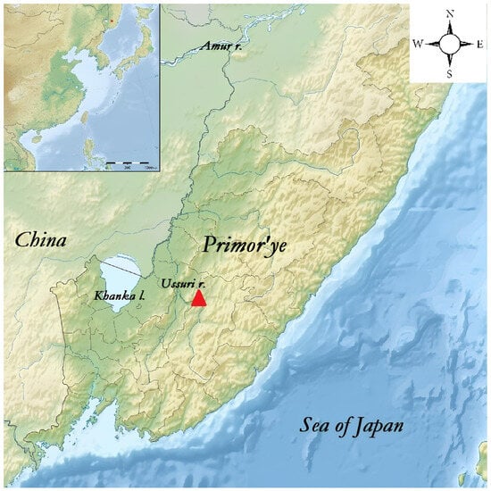
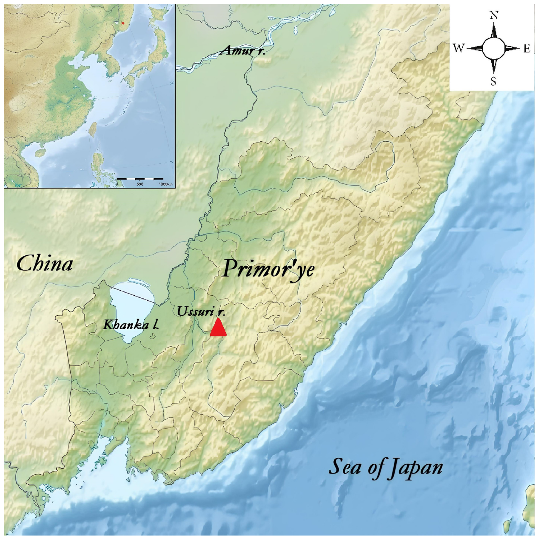
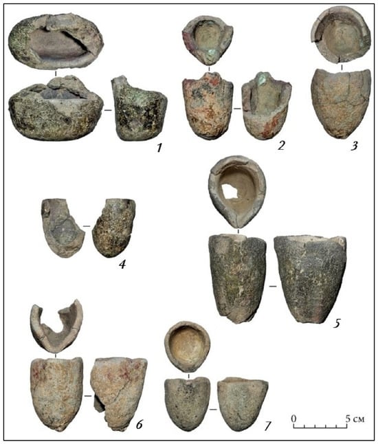
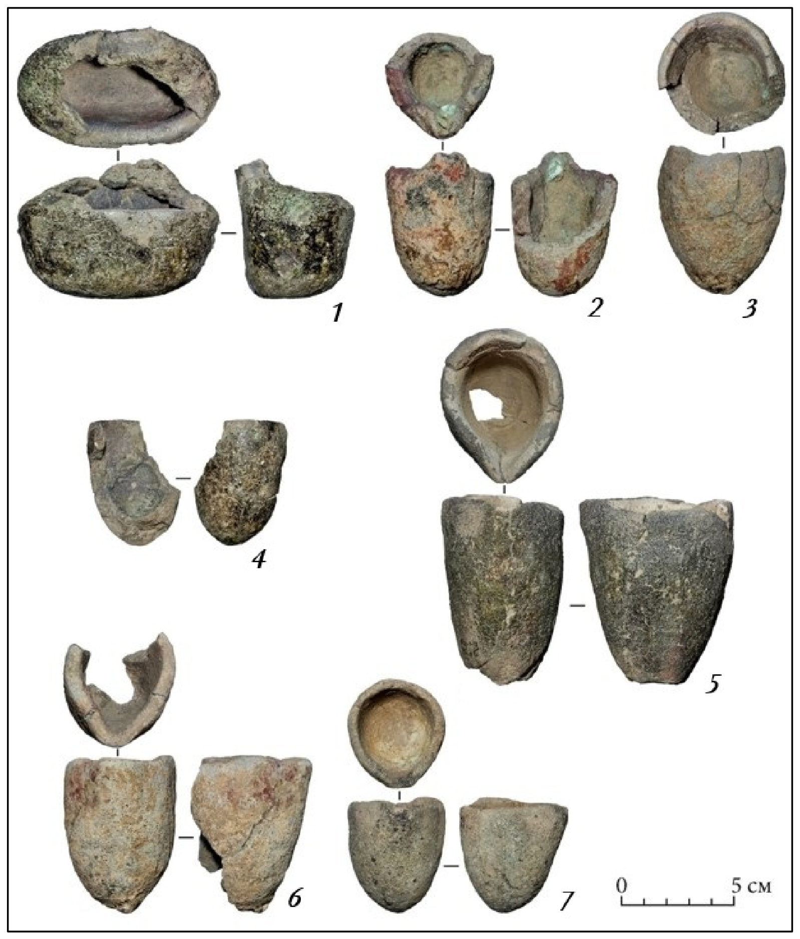
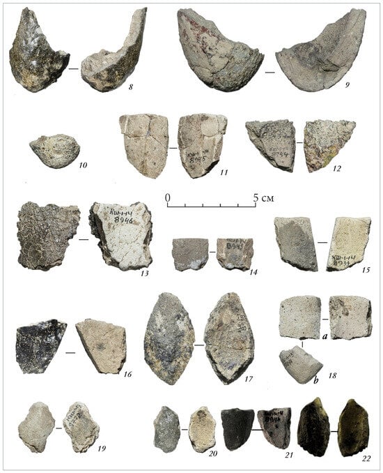
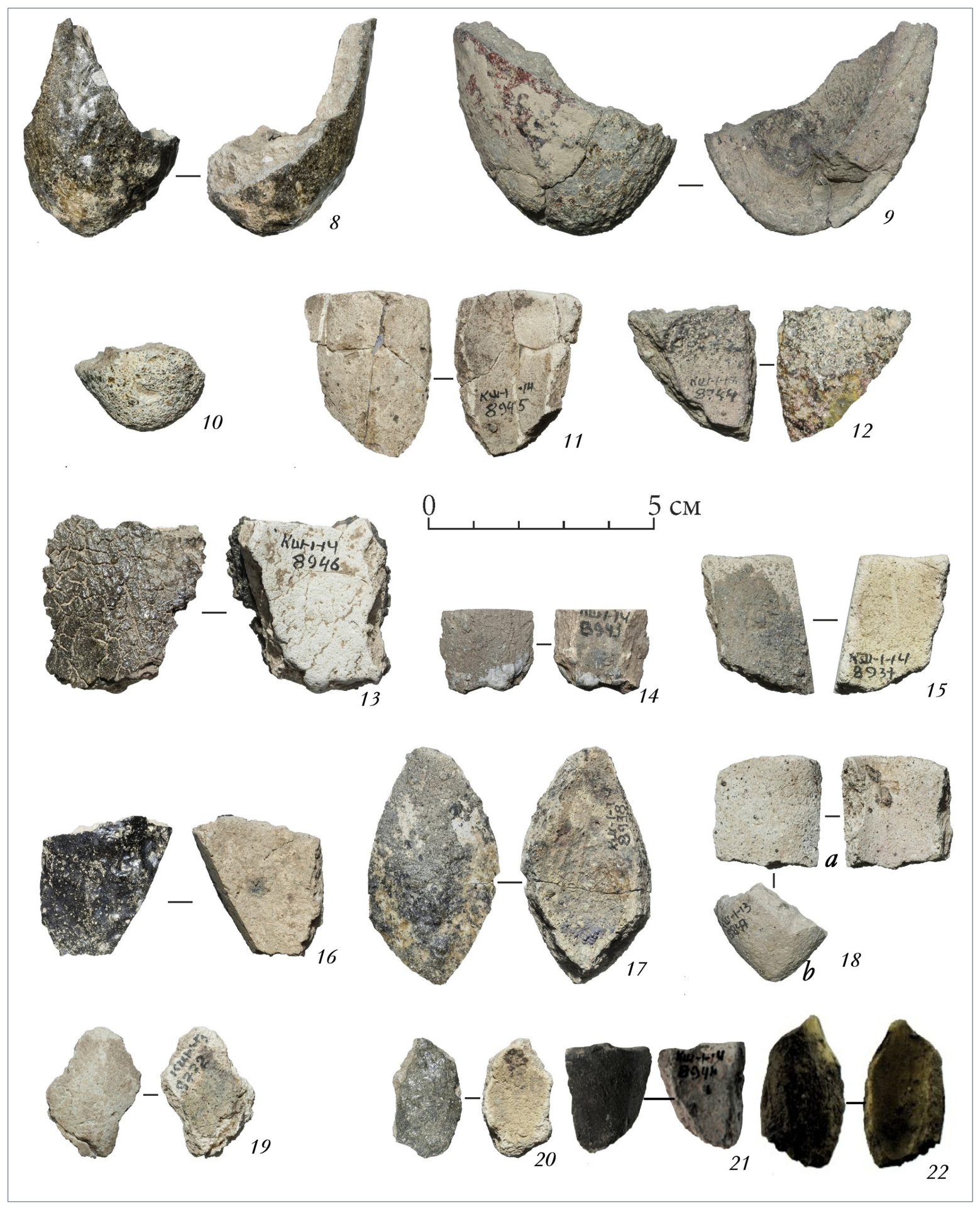
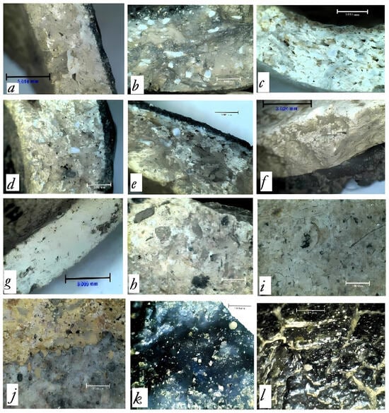
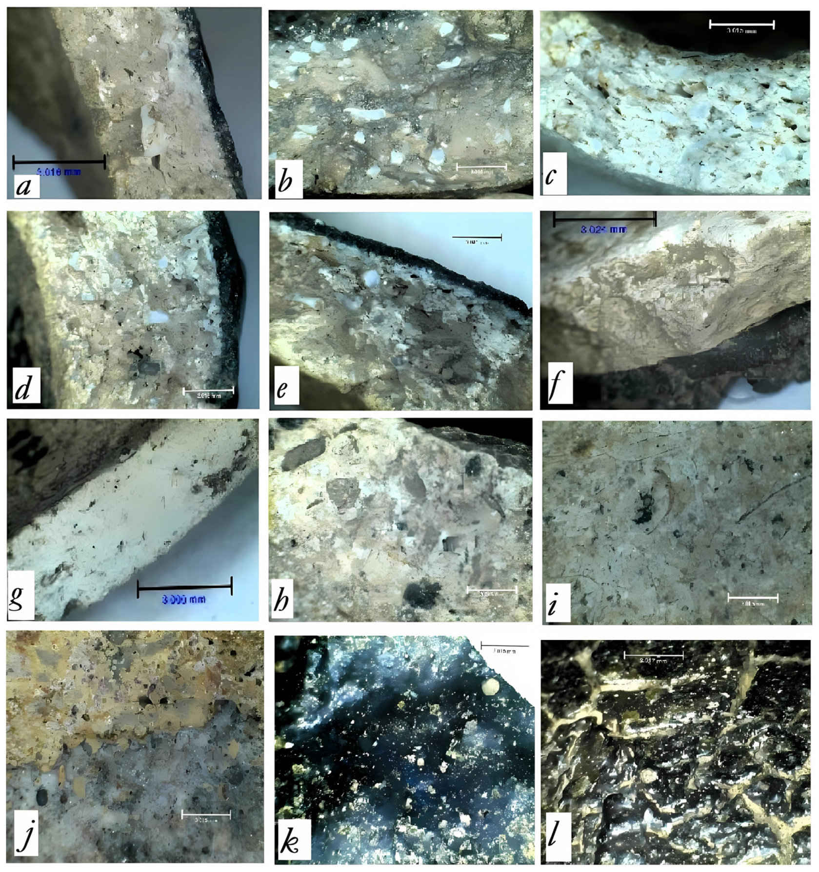
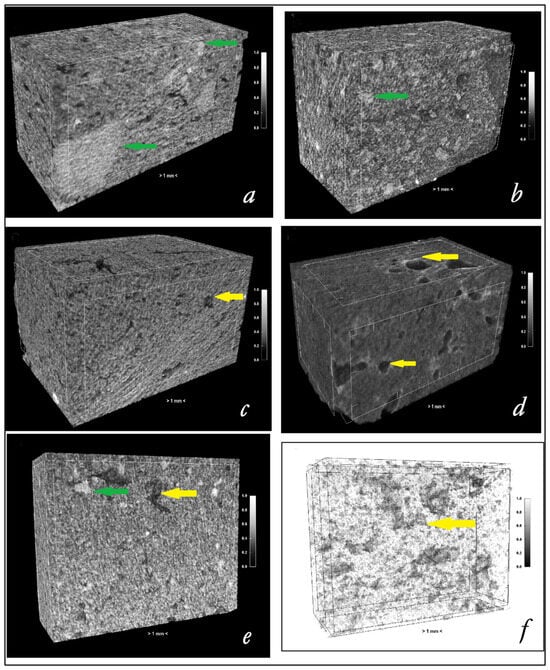
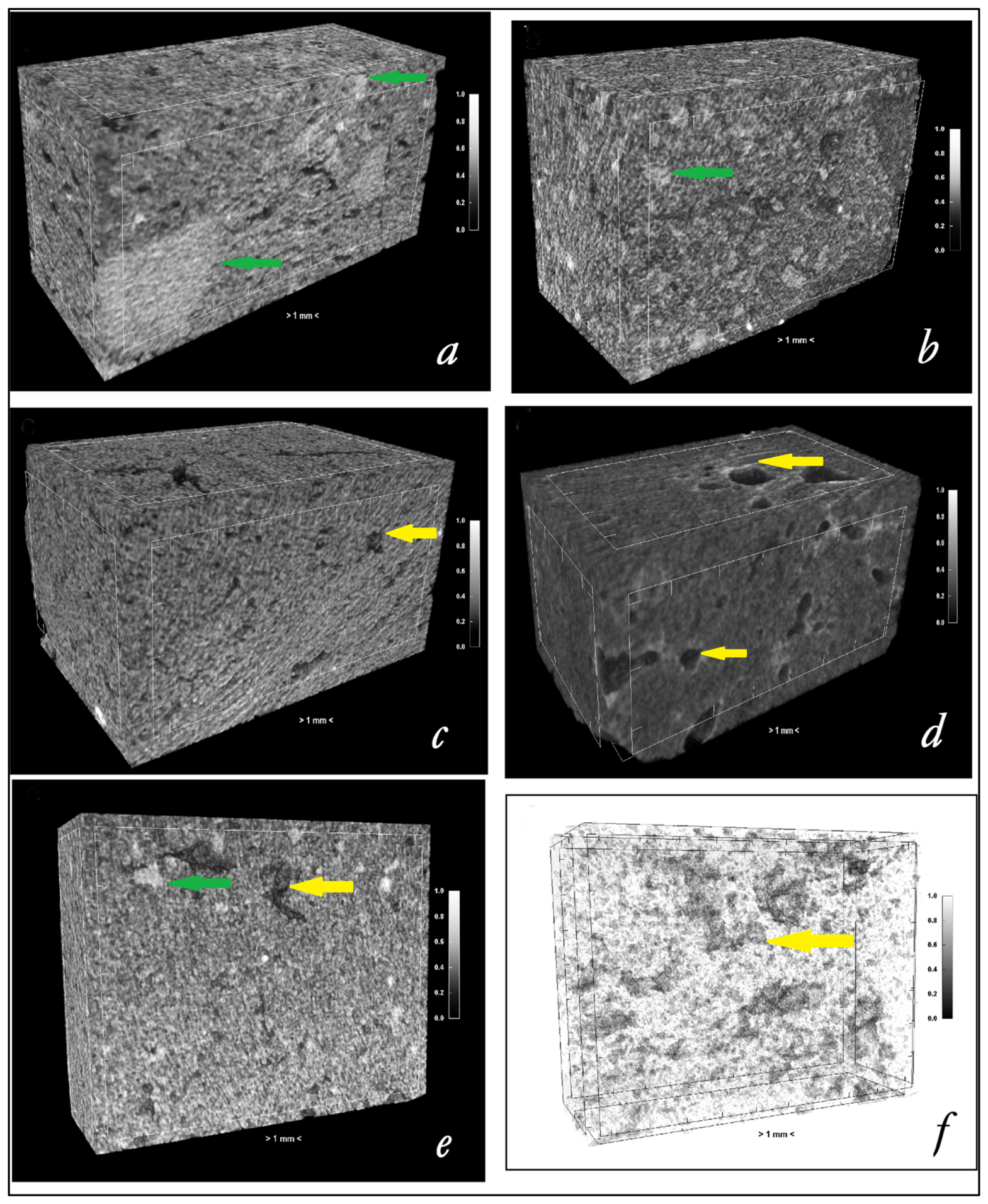
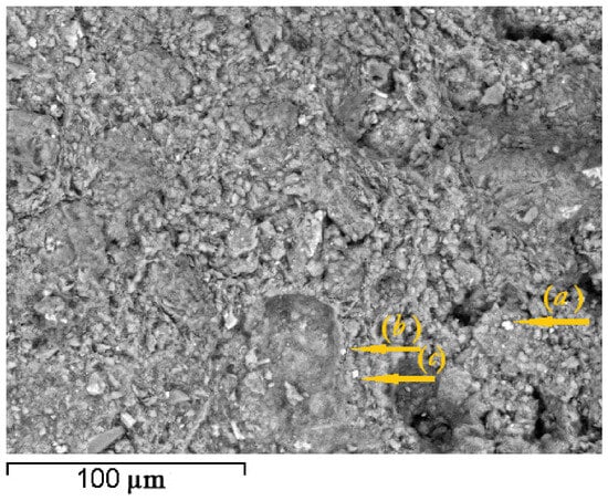
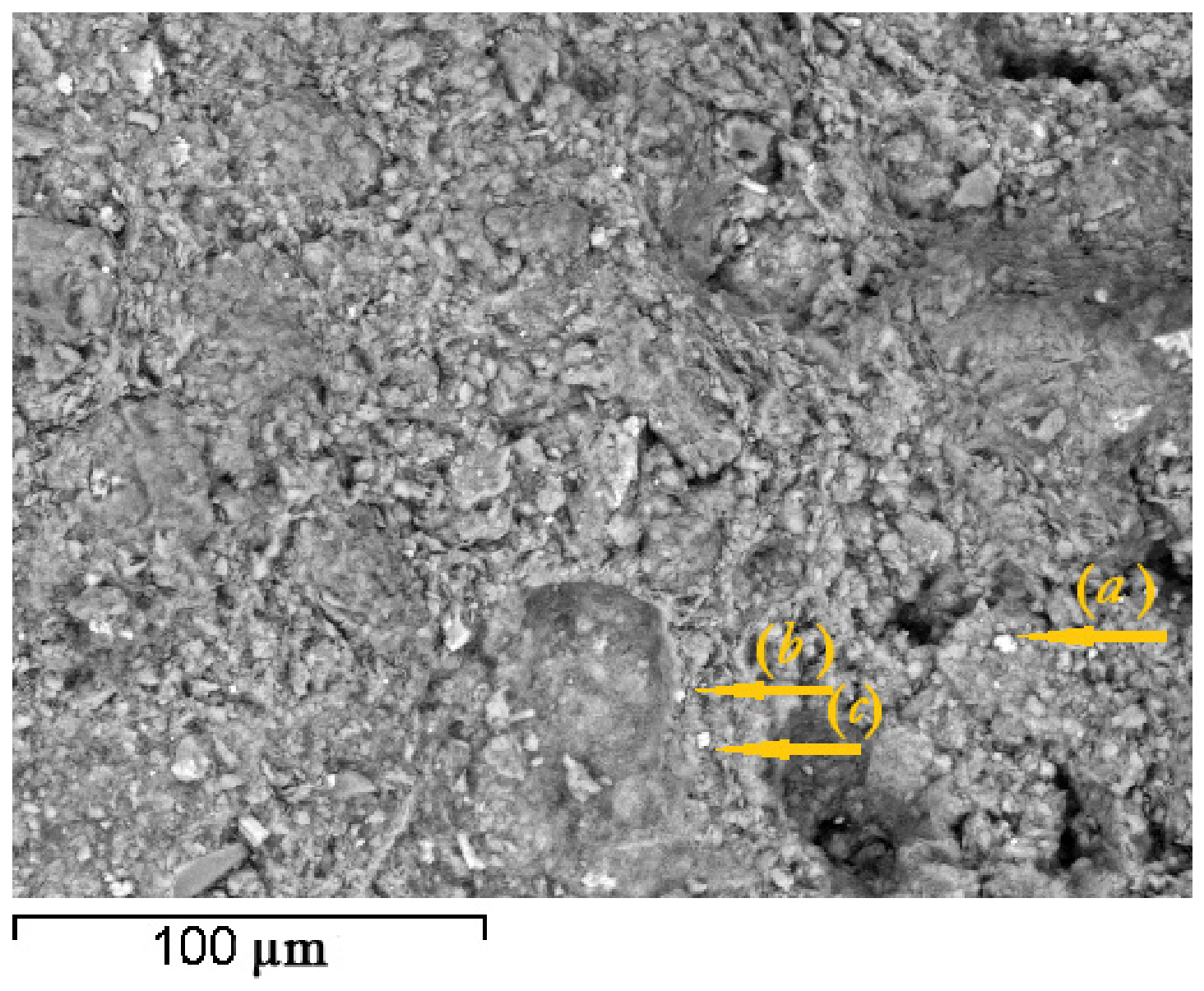
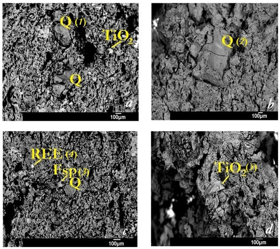
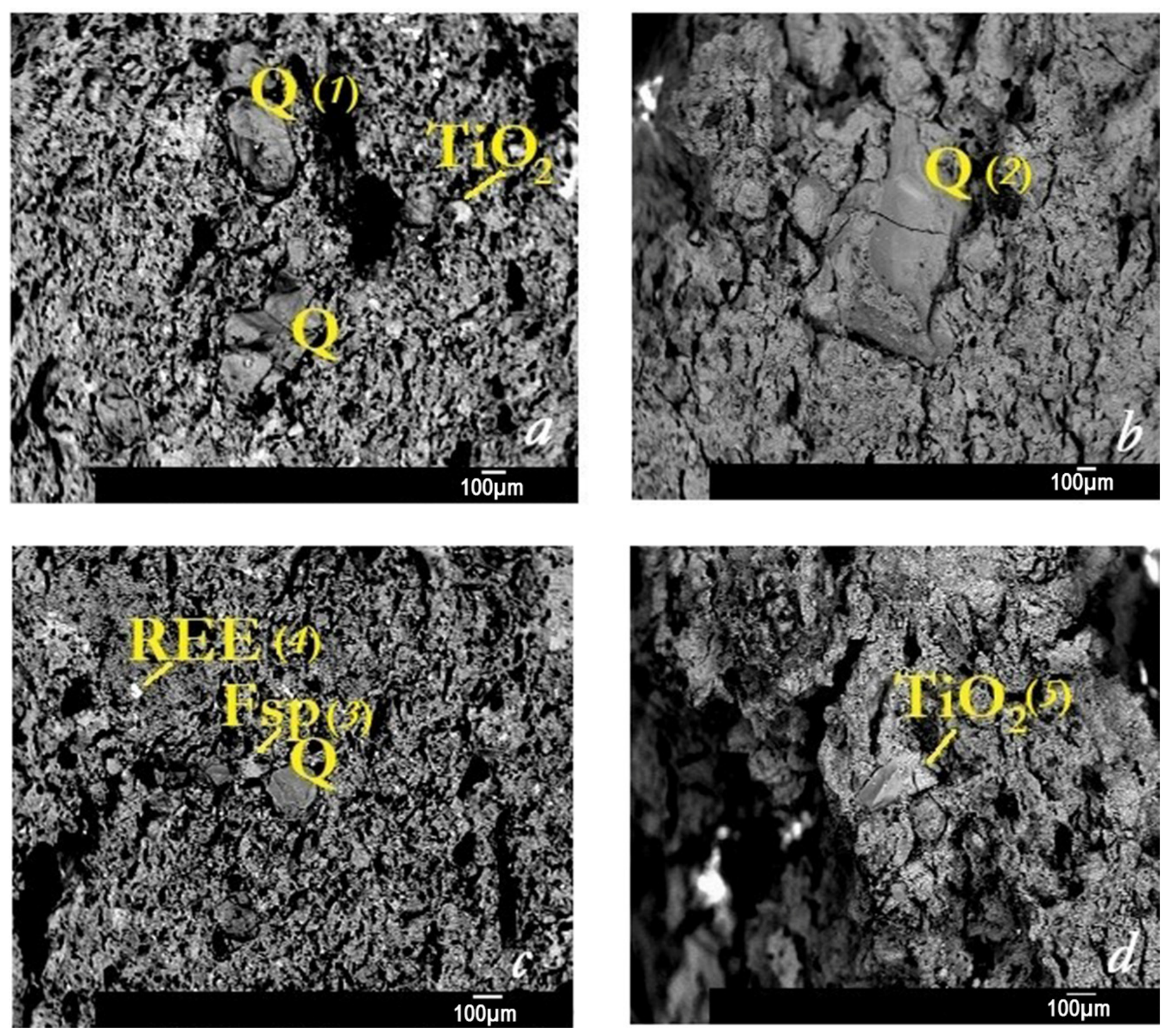
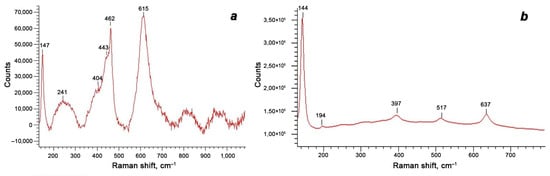
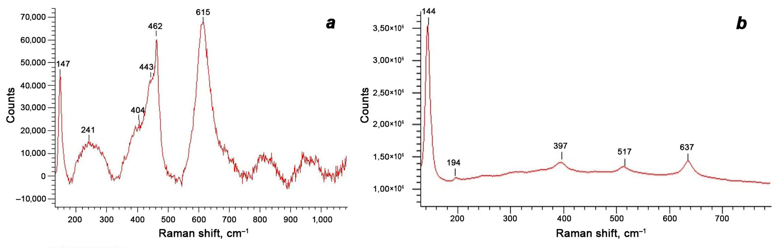
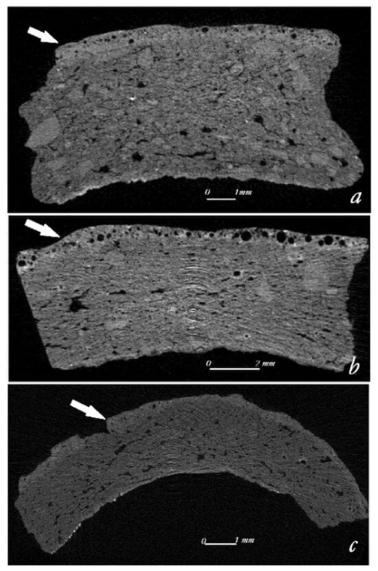
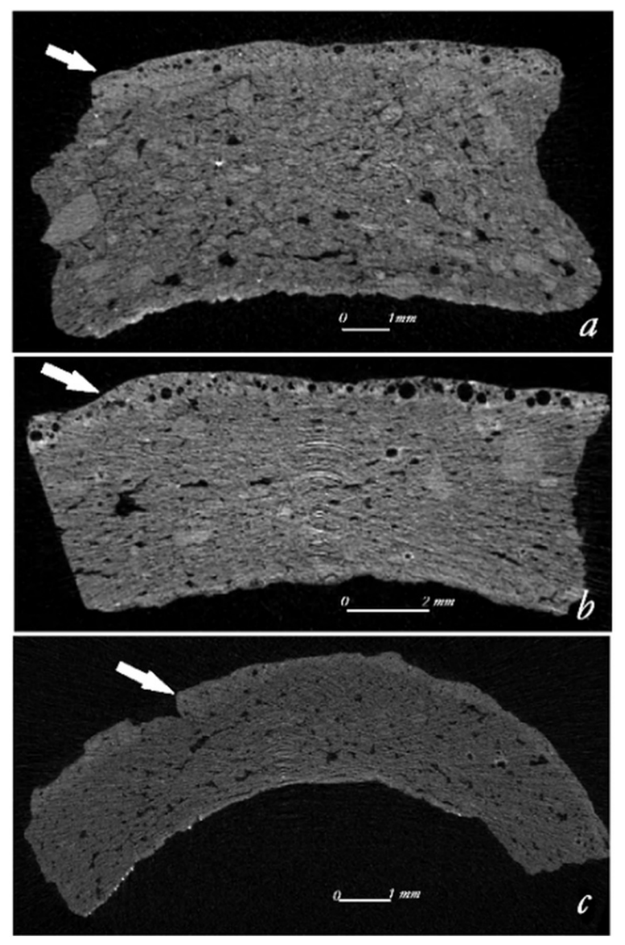
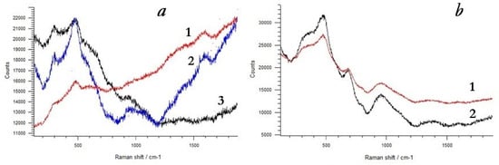
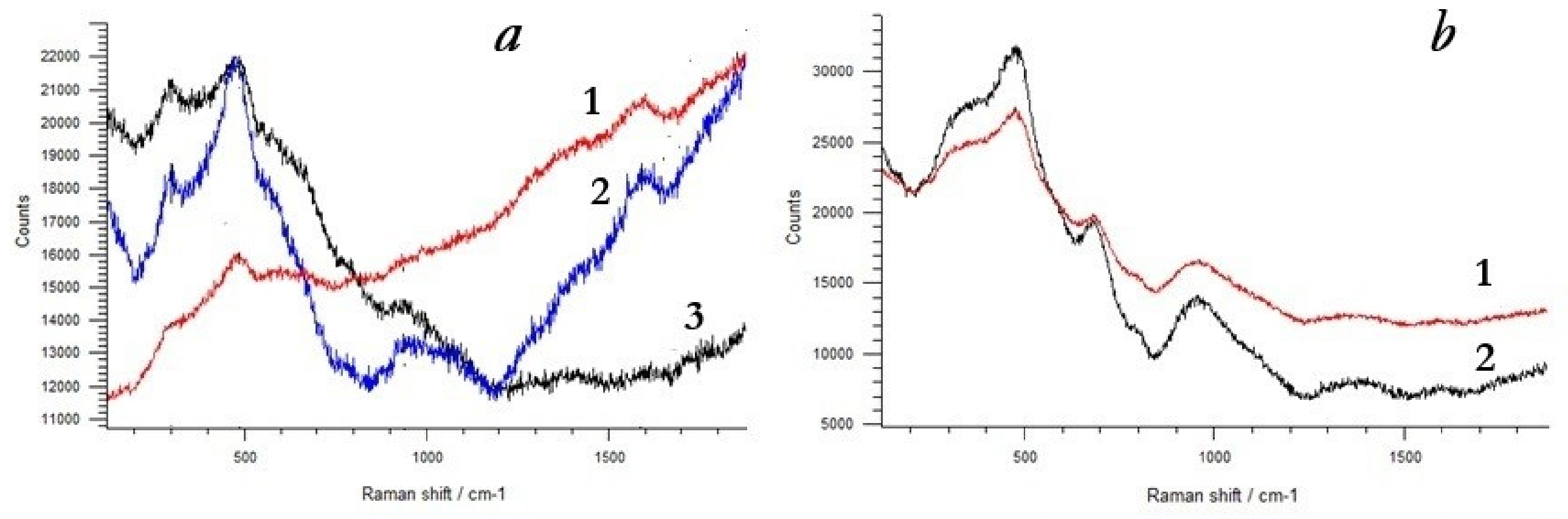
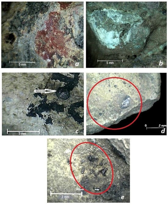
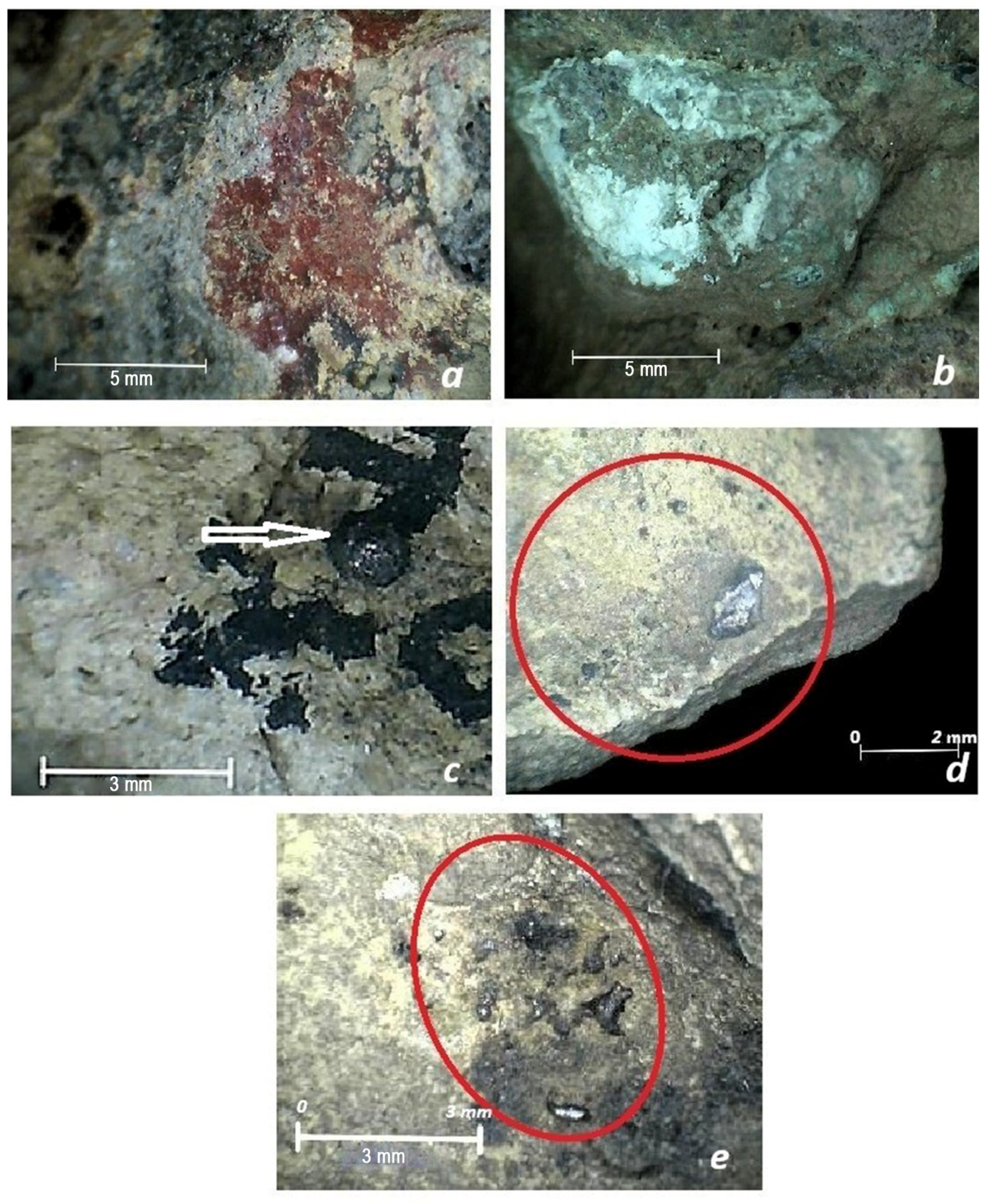
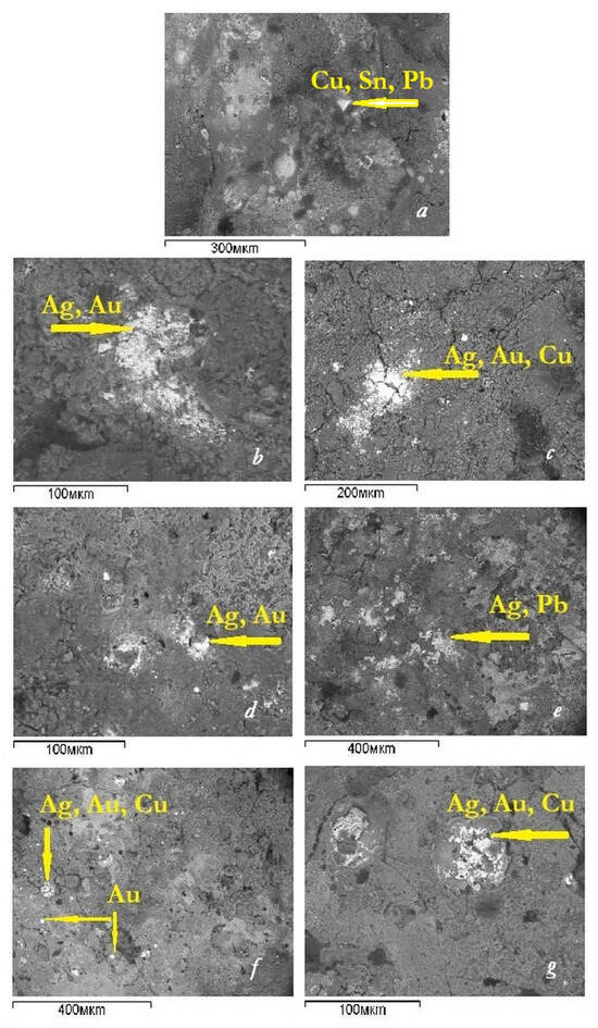
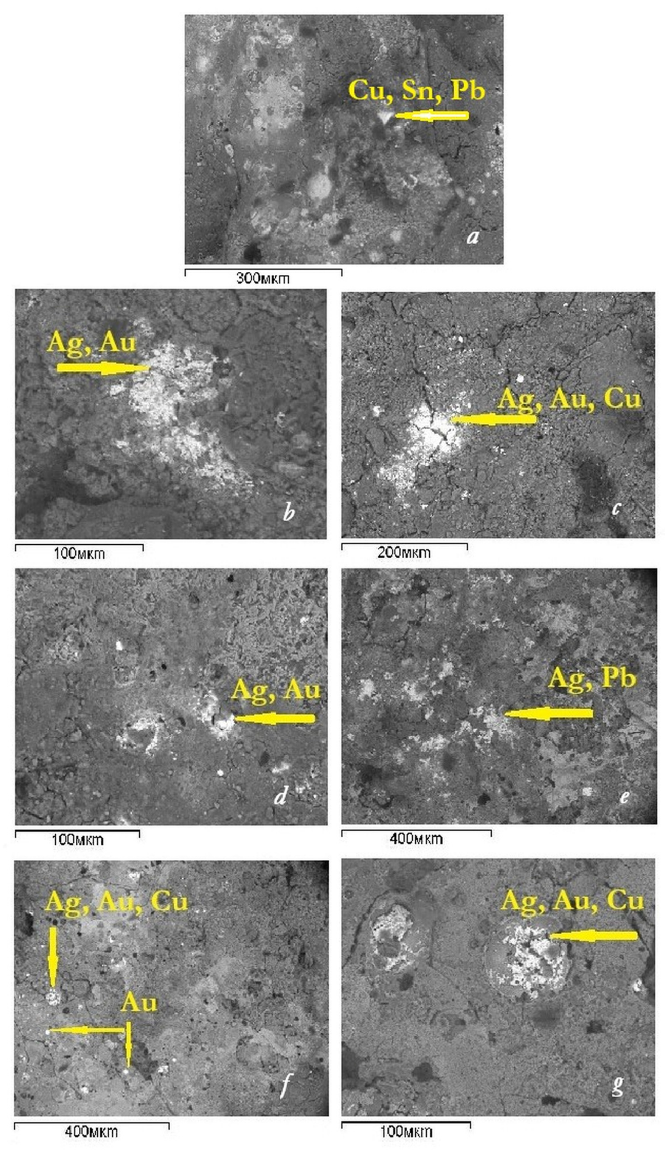
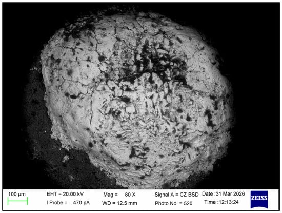
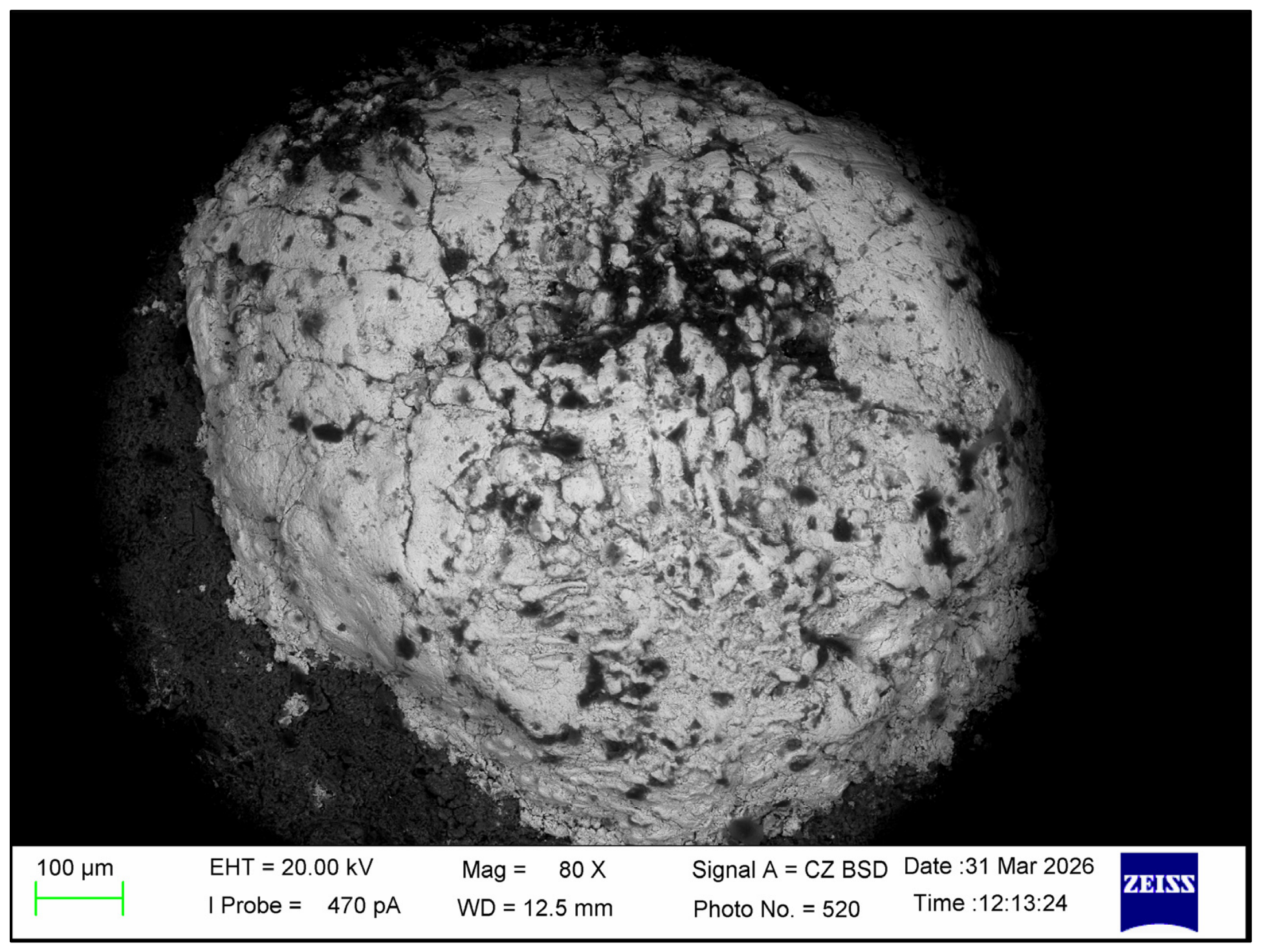
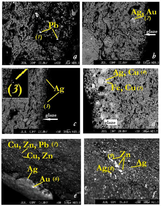
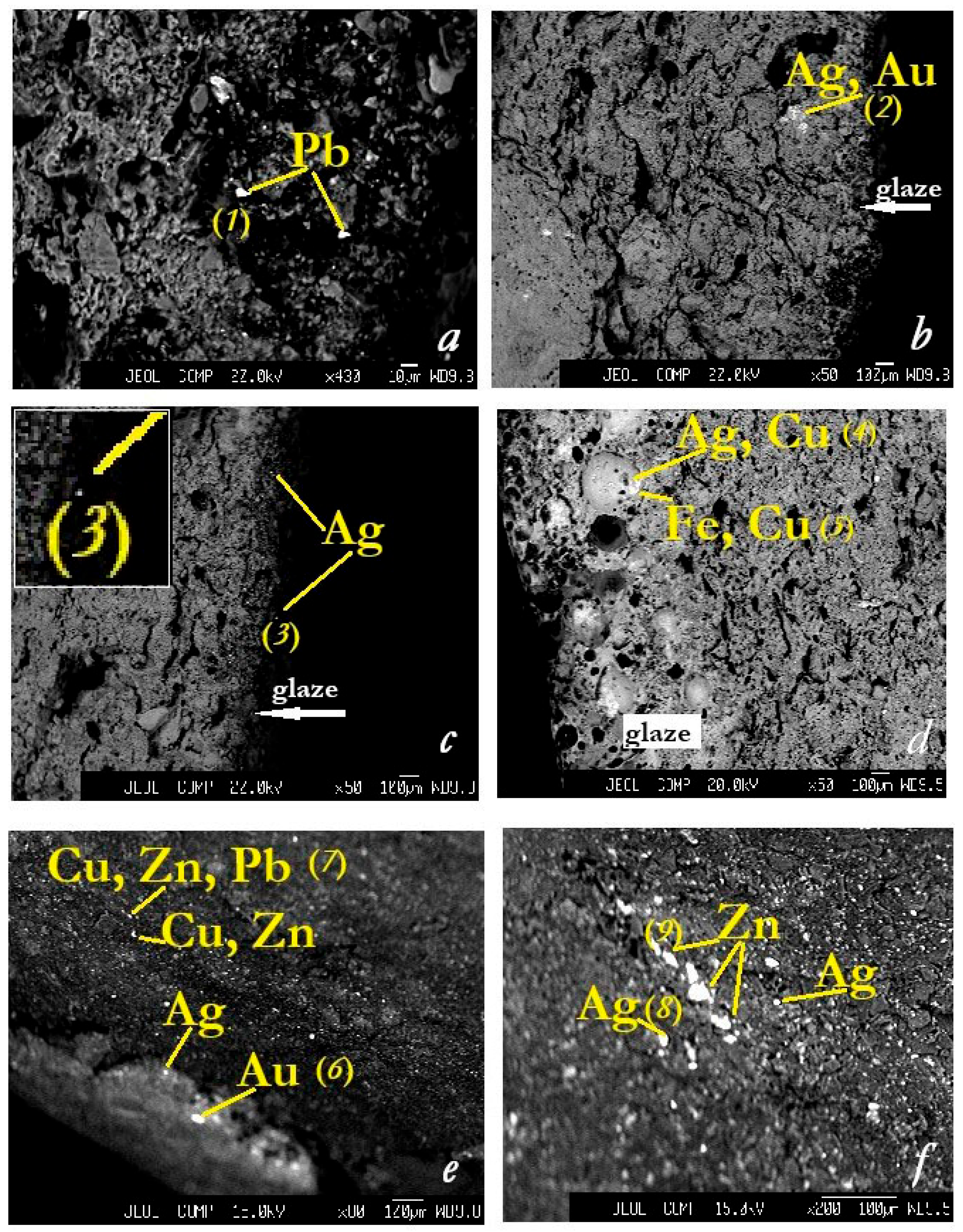
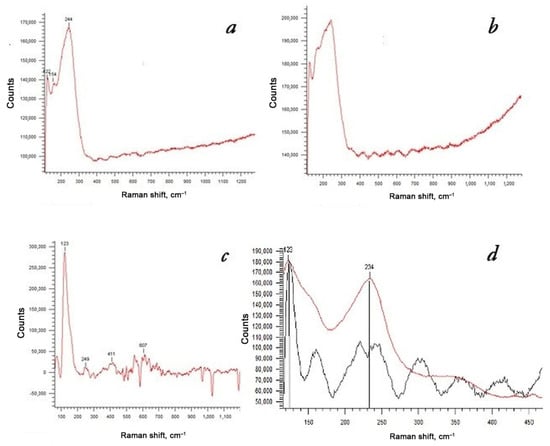
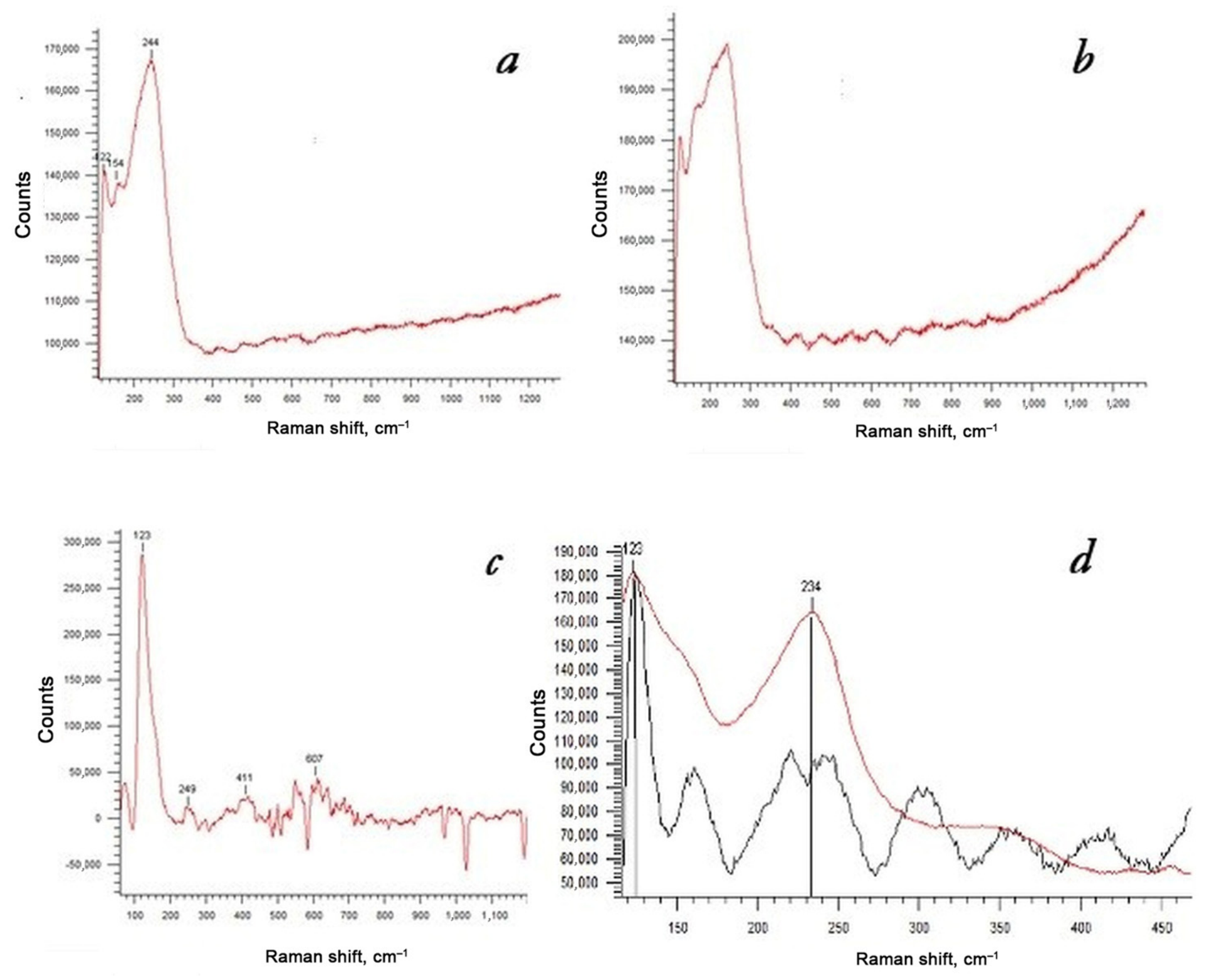
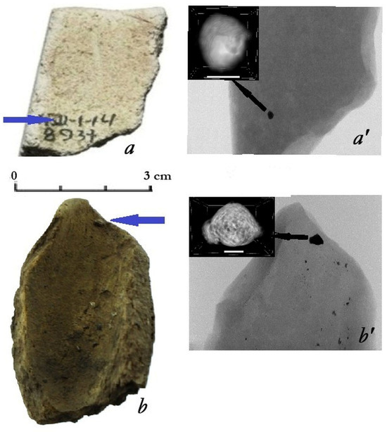
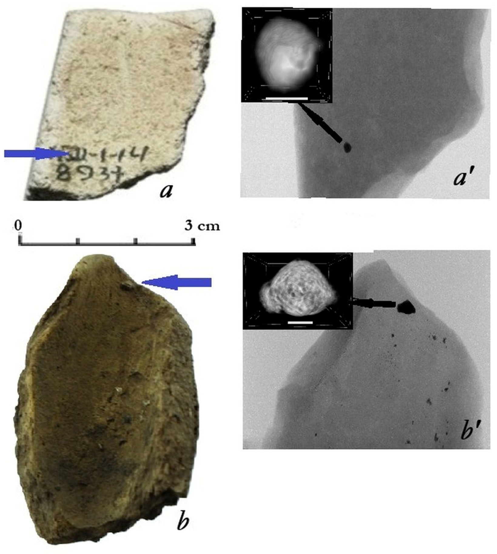
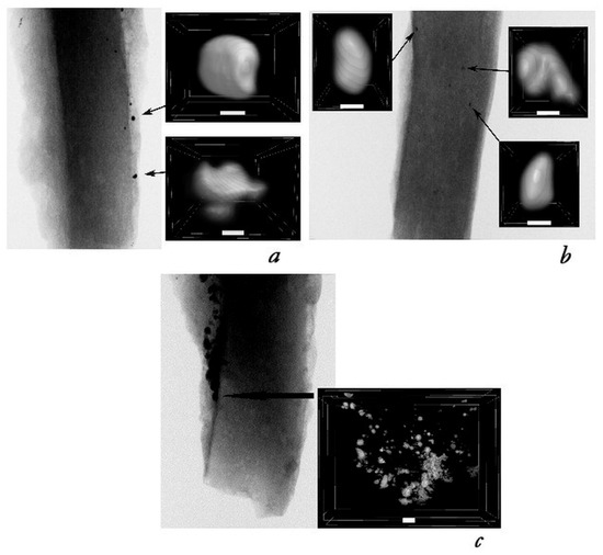
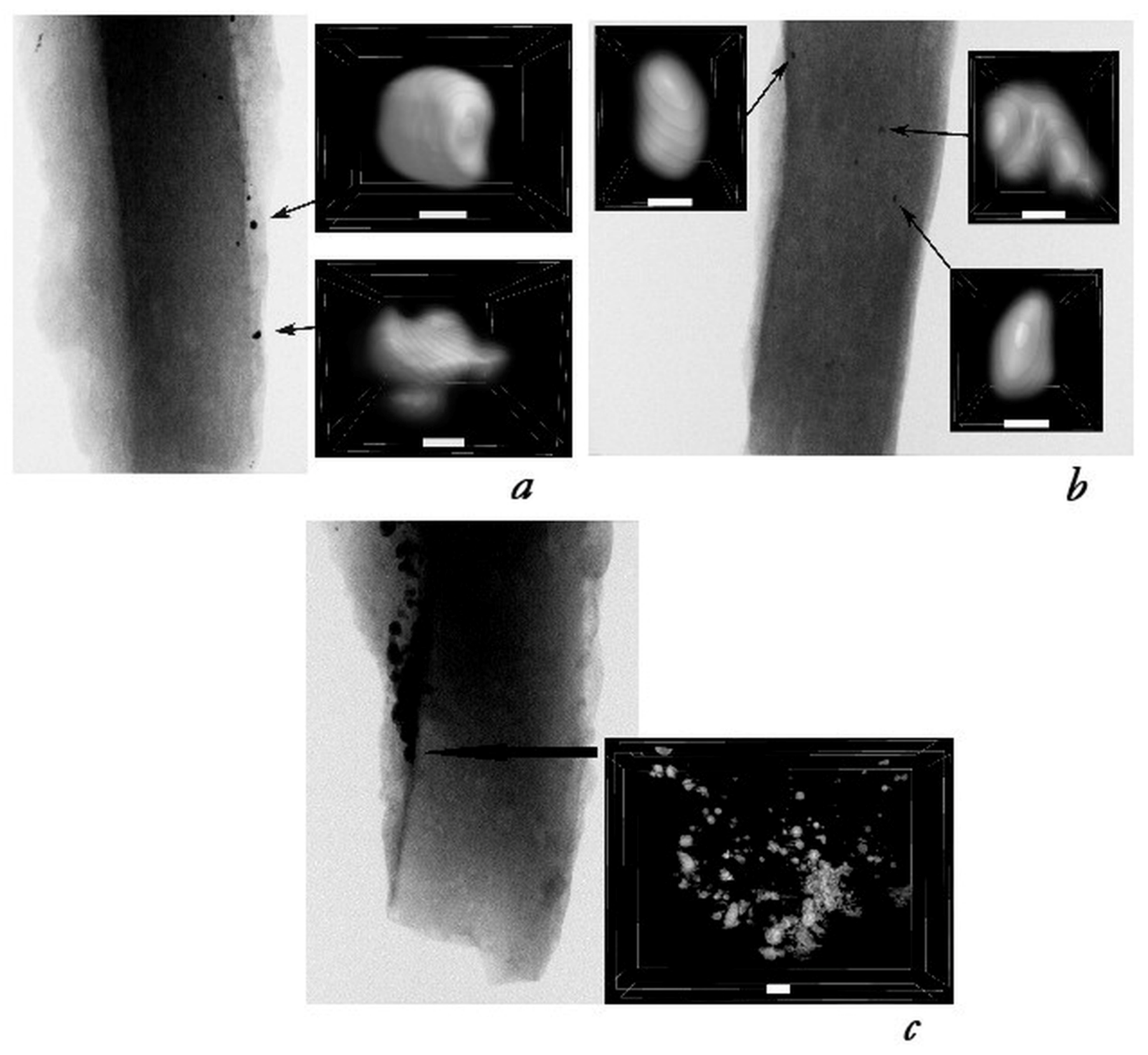

Disclaimer/Publisher’s Note: The statements, opinions and data contained in all publications are solely those of the individual author(s) and contributor(s) and not of MDPI and/or the editor(s). MDPI and/or the editor(s) disclaim responsibility for any injury to people or property resulting from any ideas, methods, instructions or products referred to in the content. |
© 2026 by the authors. Licensee MDPI, Basel, Switzerland. This article is an open access article distributed under the terms and conditions of the Creative Commons Attribution (CC BY) license.