


Abstract
Background and Clinical Significance: Adult T-cell leukemia/lymphoma (ATLL) is an aggressive mature T-cell malignancy associated with human T-lymphotropic virus type 1 (HTLV-1) and generally has poor outcomes in its acute form. Case Presentation: A 39-year-old woman originally from Jamaica presented with leukocytosis greater than 100 × 109/L, hypercalcemia, lymphadenopathy, splenomegaly, lytic osseous lesions, and multilobulated “flower cells.” Peripheral-blood flow cytometry identified an abnormal T-cell population comprising approximately 85% of events and expressing CD2, cytoplasmic CD3, CD4, CD5, CD25, bright CD15, and HLA-DR, with loss of surface CD3, CD7, CD8, and other markers. Marrow flow cytometry detected approximately 2% atypical T cells. The clinicopathologic findings supported acute ATLL; however, only a reactive HTLV-I/II screening result was available. HTLV-1-specific confirmation, proviral quantification, and clonal proviral integration analysis were not documented. Cerebrospinal fluid later contained an aberrant T-cell population. The treatment record documented one cycle each of EPOCH, Hyper-CVAD, and CHOEP. Formal response assessments and the reasons for each treatment change were unavailable. Zidovudine plus interferon-alpha began in October 2022. CHOEP was administered on 26 October 2022, and mogamulizumab was initiated on 28 October 2022, precluding independent assessment of the CHOEP response. A January 2023 marrow summary stated that no abnormal cells were seen, although the assessment method and specimen adequacy were not specified. At 41 months after diagnosis, the oncology summary described the patient as being in remission without allogeneic stem-cell transplantation. Conclusions: This case describes clinical remission during extended follow-up after overlapping multimodal therapy. It does not establish refractoriness to each chemotherapy regimen, the contribution of any individual treatment, additive or synergistic benefit, optimal treatment sequencing, or the duration of remission.
1. Introduction
Adult T-cell leukemia/lymphoma (ATLL) is a mature T-cell malignancy associated with chronic human T-lymphotropic virus type 1 (HTLV-1) infection. It is classified clinically as acute, lymphoma, chronic, or smoldering disease. The acute subtype may present with marked leukocytosis, hypercalcemia, elevated lactate dehydrogenase, organomegaly, extranodal involvement, and rapid clinical deterioration [1,2].
Aggressive ATLL remains difficult to treat. Multiagent chemotherapy may reduce tumor burden, but relapse or progression is common. International consensus recommendations support subtype-directed therapy and consideration of allogeneic hematopoietic stem-cell transplantation for eligible patients who achieve disease control [2,3]. Many patients, however, have refractory disease or other factors that preclude transplantation. A North American cohort of patients with aggressive ATLL reported a median overall survival of 5.5 months despite frequent use of EPOCH- or CHOP-based regimens [4].
Zidovudine (AZT) combined with interferon-alpha (IFN-alpha) has demonstrated activity in leukemic ATLL, and mogamulizumab, a defucosylated monoclonal antibody directed against C-C chemokine receptor 4 (CCR4), has shown activity in relapsed or refractory disease [5,6,7,8,9,10,11]. The best sequence or combination of these approaches outside endemic regions remains uncertain.
This report describes an extended clinical course in a young patient with high-burden, clinically diagnosed acute ATLL and cerebrospinal fluid involvement who received several cytotoxic, antiviral, immune-directed, and intrathecal therapies. Because formal response assessments were not available for each regimen and CHOEP and mogamulizumab were started two days apart, the case does not establish chemotherapy refractoriness or a sequence of chemotherapy failure followed by successful salvage therapy. Its contribution is a transparent description of multimodal treatment and remission documented during extended follow-up, together with the diagnostic and therapeutic limitations of the available record.
2. Case Presentation
2.1. Patient Characteristics and Presentation
A 39-year-old woman with no significant documented medical history presented in August 2022 after approximately one month of progressive back pain, dyspnea, severe nausea, fatigue, and low-grade fever. Examination showed rapidly progressive postauricular, cervical, and shoulder-region lymphadenopathy. Laboratory evaluation demonstrated a white blood cell count greater than 100 × 109/L (124.3 × 10⁹/L in the diagnostic record), normocytic anemia, preserved platelet counts, hypercalcemia of 11 mg/dL, elevated lactate dehydrogenase, and elevated liver enzymes.
The patient was originally from Jamaica and had traveled to the Bahamas one year before presentation. She had previously been sexually active. The available history did not document maternal HTLV-1 status, breastfeeding history, prior transfusion exposure, injection-drug exposure, or the HTLV status of prior sexual partners. Accordingly, the route and timing of HTLV acquisition could not be established. Her father had prostate cancer, her mother died of lung cancer, and one sister died of a brain tumor.
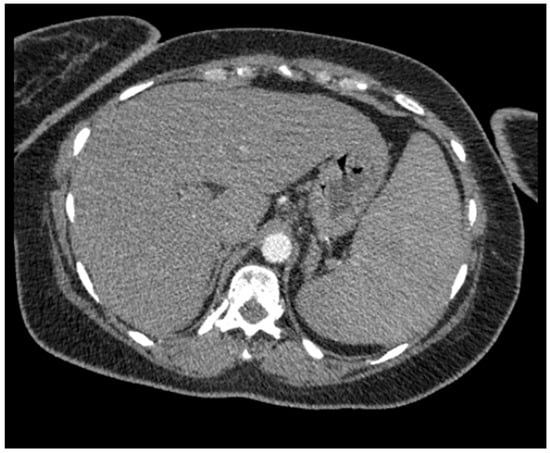
Peripheral-blood smear review showed numerous atypical lymphocytes with multilobulated nuclei, including characteristic “flower cells.” Imaging demonstrated severe splenomegaly, with a possible splenic infarct or hypoechoic splenic lesion, as well as epigastric and mediastinal lymphadenopathy and lytic lesions involving the thoracic and lumbar vertebrae. An axial staging CT image demonstrating hepatosplenomegaly is shown in Figure 1.
Figure 1.
Axial CT image from the initial staging evaluation suggesting hepatosplenomegaly.
2.2. Pathology, Immunophenotyping, and HTLV Testing
Peripheral-blood multiparameter flow cytometry identified a large abnormal T-cell population comprising approximately 85% of total events. The cells expressed CD2, cytoplasmic CD3, CD4, CD5, CD25, bright CD15, and HLA-DR. They lacked surface CD3, CD7, CD8, CD10, CD26, CD34, CD56, CD57, and CD123. The full reported flow panel included CD45, CD23, CD5, CD19, CD34, CD38, CD10, CD20, kappa, lambda, CD57, CD7, CD4, CD8, CD2, CD56, CD3, CD16, CD15, CD11b, CD117, CD13, HLA-DR, CD36, CD123, CD64, CD11c, CD33, and CD14. The archived report did not provide antibody clones, manufacturers, the instrument model, or analysis software.
Bone-marrow aspirate and biopsy were suboptimal and small, with few marrow cells and crush artifact. Marrow flow cytometry detected approximately 2% atypical lymphoid cells with a phenotype consistent with the circulating abnormal T-cell population. Immunohistochemical stains performed on the marrow biopsy were negative for PAX5, Epstein–Barr virus-encoded RNA, CD30, and ALK-1. The pathology interpretation was a T-cell neoplasm most consistent with ATLL in the setting of marked lymphocytosis, multilobulated “flower cells,” lymphadenopathy, splenomegaly, hypercalcemia, lytic lesions, and positive HTLV-I/II serology.
Fine-needle aspiration of left postauricular and right shoulder masses was positive for malignancy and showed involvement by the recently diagnosed T-cell lymphoma. Smears contained many small lymphoid cells admixed with medium-to-large atypical lymphoid cells, some with irregular nuclear contours or multilobulated nuclei. Flow cytometry of both aspirates showed an abnormal T-cell population.
The clinicopathologic findings supported ATLL. However, only a reactive HTLV-I/II screening result was available, and HTLV-1-specific confirmatory testing could not be verified. Neither proviral quantification nor analysis of clonal proviral integration was documented. These are distinct limitations: the absence of documented type-specific confirmation limits verification of HTLV-1 infection, whereas the absence of clonal integration analysis limits demonstration of a direct molecular link between an infected clone and the malignancy. Patient-specific CCR4 expression was also not assessed in the available blood, marrow, lymph-node, or skin-biopsy studies. Mogamulizumab was therefore selected on the basis of its established activity in ATLL rather than a documented patient-specific CCR4 assay. These limitations are acknowledged because positive serology alone done not demonstrate clonal proviral integration within the malignant population. The documented immunophenotypic and ancillary findings are summarized in Table 1.
Table 1.
Documented immunophenotypic and ancillary findings.
2.3. Central Nervous System Involvement and Documented Treatment Chronology
The patient was admitted to the hematology service and initially received broad-spectrum antibiotics because of concern for sepsis; cultures were negative and the antibiotics were discontinued after clinical stabilization. The record documents one EPOCH cycle administered over five days and completed on 29 August 2022, followed by one Hyper-CVAD cycle beginning on 13 September 2022. The available source materials did not include serial disease measurements, formal response assessments, or explicit reasons for changing treatment after either regimen. These therapies are therefore reported as treatment exposures rather than as proven treatment failures.
A lumbar puncture performed on 17 October 2022 showed an aberrant T-cell population in the cerebrospinal fluid. Intrathecal therapy was administered on 17 October 2022 and again on 23 February 2023. Zidovudine plus interferon-alpha was initiated on an unspecified date in October 2022. One CHOEP cycle was administered on 26 October 2022, and mogamulizumab was initiated on 28 October 2022. Because mogamulizumab began only two days after CHOEP, the response to CHOEP could not be evaluated independently, and the subsequent course cannot be interpreted as remission occurring after demonstrated CHOEP failure. Prior cytotoxic therapy may also have contributed to the later clinical course.
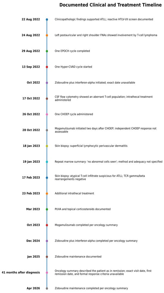
Mogamulizumab was continued through October 2023. Zidovudine plus interferon-alpha continued through December 2024, followed by zidovudine maintenance from January 2025 through April 2026 according to the oncology summary. Exact drug doses, interferon formulation, infusion schedules, dose modifications, treatment interruptions, and administration-level adverse-event data were not available in the records retrieved for this report. This limits reproducibility and precludes evaluation of a specific treatment regimen. The documented chronology is summarized in Figure 2.
Figure 2.
Documented clinical and treatment chronology. The source records did not include formal response assessments after each regimen, exact drug doses, or reasons for each treatment change. Mogamulizumab was initiated two days after CHOEP, preventing independent assessment of the CHOEP response.
2.4. Cutaneous Findings
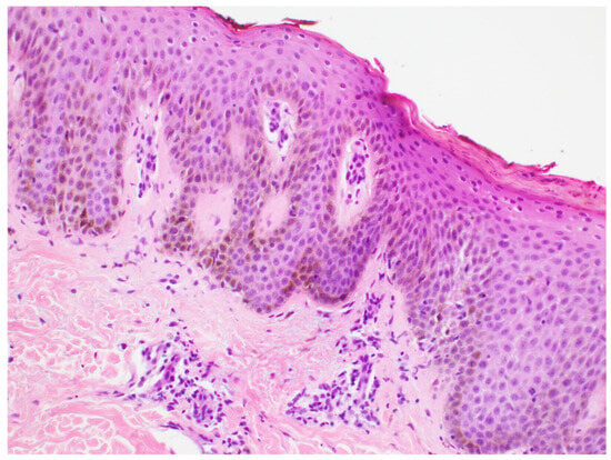
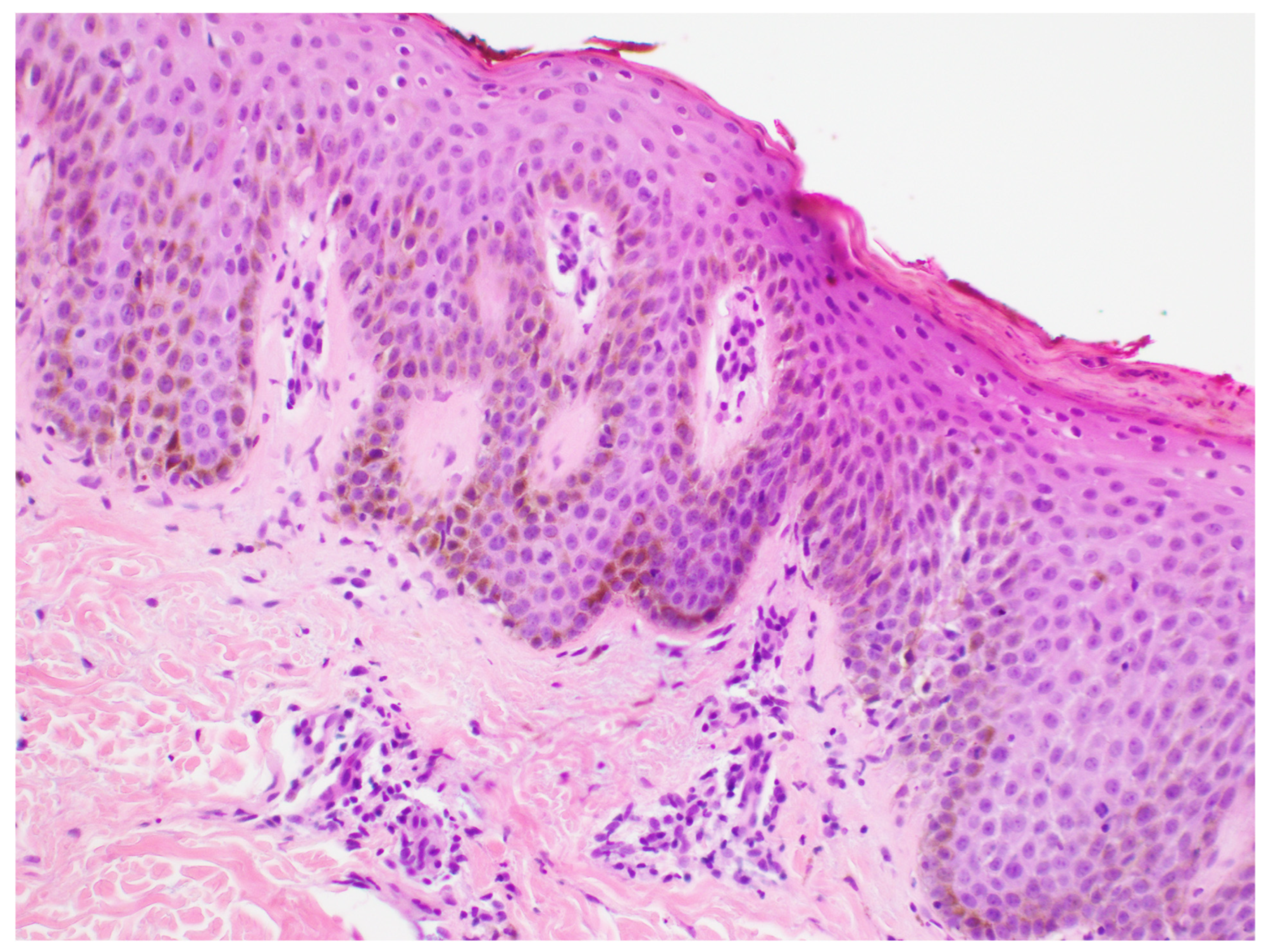
A cutaneous eruption developed during the treatment course and was managed with topical corticosteroids. A skin biopsy obtained on 18 January 2023 showed superficial lymphocytic perivascular dermatitis (Figure 3). A second skin biopsy on 17 February 2023 showed an atypical T-cell infiltrate suspicious for involvement by the patient’s known ATLL; T-cell receptor gamma- and beta-gene rearrangement studies were negative.
Figure 3.
Hematoxylin-and-eosin stain (200× magnification) showing epidermis overlying dermis with a superficial perivascular lymphocytic infiltrate. No skin-biopsy immunostain images were available.
The available skin-biopsy reports did not document immunohistochemical stains, and immunostain images were not available. A clinical photograph of the eruption was also not available in the medical record. Therefore, the manuscript includes the available hematoxylin-and-eosin image and reports the biopsy interpretation and molecular clonality results without assigning a definitive cause to the eruption. PUVA therapy and topical corticosteroids were used beginning in March 2023.
2.5. Response and Follow-Up
The oncology summary recorded a repeat bone-marrow biopsy on 19 January 2023 as showing “no abnormal cells.” The available source material did not specify whether this conclusion was based on morphology, flow cytometry, immunohistochemistry, or a combination of methods, and specimen adequacy was not documented. At 41 months after diagnosis, the oncology summary described the patient as being in remission without allogeneic stem-cell transplantation. The exact date of that visit, the date on which remission was first documented, the response criteria used, and comprehensive site-by-site assessments of previously involved blood, lymph nodes, spleen, skin, bone, and cerebrospinal fluid were not available.
The same oncology summary listed zidovudine maintenance through April 2026. Accordingly, this report describes clinical remission documented during extended follow-up rather than a 41-month duration of complete remission. Because cytotoxic, antiviral, immune-directed, and intrathecal therapies were administered sequentially and, at times, in close temporal overlap, the contribution of any individual component cannot be determined.
3. Discussion
This case describes high-burden, clinically diagnosed acute ATLL in a 39-year-old woman with leukemic disease, hypercalcemia, extensive lymphadenopathy, splenomegaly, osseous lesions, and cerebrospinal fluid involvement. The principal observation is that the oncology record described the patient as being in remission at 41 months after diagnosis following multimodal treatment. The case does not establish refractoriness to each cytotoxic regimen, a discrete sequence of chemotherapy failure followed by successful salvage therapy, or superiority of the later treatment approach.
The patient’s Jamaican origin is epidemiologically relevant because HTLV-1 is endemic in parts of the Caribbean. However, birthplace alone cannot establish the route or timing of infection. The available chart did not document maternal infection, breastfeeding history, transfusion exposure, injection-drug exposure, or the HTLV status of prior sexual partners. The manuscript therefore does not assign a route of transmission.
The diagnosis was supported by the characteristic clinical syndrome, “flower-cell” morphology, mature CD4-positive and CD25-positive T-cell immunophenotype, and involvement of blood, marrow, lymph-node aspirates, and cerebrospinal fluid. The detailed flow-cytometry results also showed loss of surface CD3 and CD7, supporting an abnormal mature T-cell population. However, only a reactive HTLV-I/II screening result was available, and HTLV-1-specific confirmatory testing could not be verified. Neither proviral quantification nor clonal proviral integration analysis was documented. These omissions are not interchangeable: type-specific confirmatory testing addresses whether HTLV-1 infection was established, whereas clonal integration analysis addresses the molecular relationship between an infected clone and the malignant population.
CCR4 is commonly expressed in ATLL and is the therapeutic target of mogamulizumab. In this patient, however, CCR4 was not included in the available flow-cytometry or immunohistochemical reports. Consequently, the clinical response cannot be correlated with measured tumor CCR4 expression. Mogamulizumab was used on the basis of its established activity in relapsed or refractory ATLL [8,9,10,11].
Aggressive ATLL is often difficult to control with chemotherapy, and high tumor burden, hypercalcemia, immunodeficiency, and multidrug resistance are associated with adverse outcomes [2]. A North American cohort reported a median overall survival of 5.5 months despite frequent use of EPOCH- or CHOP-based regimens [4]. Those general observations do not establish resistance in this individual patient. She received one cycle each of EPOCH and Hyper-CVAD, but the available records did not include objective response measurements or reasons for changing therapy. CHOEP was administered on 26 October 2022 and mogamulizumab was initiated on 28 October 2022, so an independent response to CHOEP could not be assessed. The prior chemotherapy regimens remain possible contributors to the later clinical course.
AZT and IFN-alpha have been used in ATLL for several decades, particularly in leukemic subtypes. A 2023 systematic review and meta-analysis reported an overall response rate of 67% and a complete response rate of 33% across AZT/IFN-containing regimens, while acknowledging that most evidence was observational and responses were less favorable in aggressive disease [7]. Mogamulizumab has demonstrated activity in relapsed or refractory ATLL in phase II, randomized, and observational studies [8,9,10,11]. In the present case, the overlap of cytotoxic therapy, AZT/IFN, mogamulizumab, and intrathecal treatment prevents attribution of remission to one component. A single uncontrolled case also cannot establish additive benefit, synergy, optimal sequencing, or the necessity of any individual treatment.
The cutaneous findings also required cautious interpretation. The first biopsy showed superficial lymphocytic perivascular dermatitis, whereas the second showed an atypical T-cell infiltrate suspicious for ATLL involvement. Negative T-cell receptor gamma- and beta-gene rearrangement studies did not confirm a clonal cutaneous process. Mogamulizumab-associated eruptions are recognized, but the available pathology did not include immunostains and no clinical photograph was available. The eruption therefore cannot be definitively classified as drug-related dermatitis or cutaneous ATLL involvement based on the retrieved record.
The principal strength of this report is the detailed reconstruction of a complex clinical course using the available pathology, flow-cytometry, treatment-summary, skin-biopsy, and follow-up records. The central limitations concern the evidence supporting the diagnostic and therapeutic claims: HTLV-1-specific confirmatory testing and clonal proviral integration were not documented; formal response assessments after each chemotherapy regimen were unavailable; CHOEP and mogamulizumab were initiated two days apart; exact doses and administration schedules were not available; and the date of first remission, formal response criteria, and comprehensive restaging of all previously involved sites were not documented. Additional limitations include the absence of patient-specific CCR4 testing, antibody-clone and instrument details, clinical skin photographs, and skin immunostains. These limitations require the case to be interpreted as a descriptive clinical observation rather than evidence for a reproducible regimen or causal treatment effect.
4. Conclusions
The clinicopathologic findings supported clinically diagnosed acute ATLL with cerebrospinal fluid involvement, although HTLV-1-specific confirmatory testing, proviral quantification, and clonal proviral integration were not documented. The patient received multiple cytotoxic, antiviral, immune-directed, and intrathecal therapies, some of which overlapped. At 41 months after diagnosis, the oncology summary described her as being in remission; however, the date of first remission, formal response criteria, and comprehensive site-by-site reassessment were unavailable. This case does not establish chemotherapy refractoriness, causal benefit of any individual agent, additive or synergistic effects, optimal sequencing, or the necessity of any treatment component.
Author Contributions
Writing—original draft preparation, A.K. and S.M.; writing—review and editing, P.T.; supervision, P.T. All authors have read and agreed to the published version of the manuscript.
Funding
This research received no external funding.
Institutional Review Board Statement
Ethical review and approval were not required by the Institutional Review Board of Memorial Hospital at Gulfport because this single-patient case report, based on de-identified clinical information, was exempt from IRB review under institutional policy.
Informed Consent Statement
Written informed consent was obtained from the patient for publication of this case report.
Data Availability Statement
The original contributions presented in this case report are included in the article. Additional patient-level information is not publicly available because of privacy and confidentiality requirements. Further inquiries can be directed to the corresponding author.
Acknowledgments
During preparation of this manuscript, the authors used ChatGPT 5.6 Pro for assistance with language refinement and formatting. The tool was not used to generate, alter, or analyze patient data, clinical findings, or original images. All AI-assisted text was reviewed and verified by the authors, who take responsibility for the final content.
Conflicts of Interest
The authors declare no conflicts of interest.
Abbreviations
ATLL, adult T-cell leukemia/lymphoma; HTLV-1, human T-lymphotropic virus type 1; AZT, zidovudine; IFN-alpha, interferon-alpha; CCR4, C-C chemokine receptor 4; CNS, central nervous system; EPOCH, etoposide, prednisone, vincristine, cyclophosphamide, and doxorubicin; Hyper-CVAD, hyperfractionated cyclophosphamide, vincristine, doxorubicin, and dexamethasone; CHOEP, cyclophosphamide, doxorubicin, vincristine, etoposide, and prednisone; CSF, cerebrospinal fluid; PUVA, psoralen plus ultraviolet A.
References
- Shimoyama, M. Diagnostic criteria and classification of clinical subtypes of adult T-cell leukaemia-lymphoma: A report from the Lymphoma Study Group (1984–87). Br. J. Haematol. 1991, 79, 428–437. [Google Scholar] [CrossRef] [Scilit]
- Cook, L.B.; Fuji, S.; Hermine, O.; Bazarbachi, A.; Ramos, J.C.; Ratner, L.; Horwitz, S.; Fields, P.; Tanase, A.; Bumbea, H.; et al. Revised Adult T-Cell Leukemia-Lymphoma International Consensus Meeting Report. J. Clin. Oncol. 2019, 37, 677–687. [Google Scholar] [CrossRef] [Scilit]
- Cook, L.B.; Phillips, A.A. How I treat adult T-cell leukemia/lymphoma. Blood 2021, 137, 459–470. [Google Scholar] [CrossRef] [Scilit]
- Chiu, E.; Samra, B.; Tam, E.; Baseri, B.; Lin, B.; Luhrs, C.; Gonsky, J.; Sawas, A.; Taiwo, E.; Sidhu, G. Clinical Characteristics and Outcomes of Caribbean Patients With Adult T-Cell Lymphoma/Leukemia at Two Affiliated New York City Hospitals. JCO Glob. Oncol. 2020, 6, 548–556. [Google Scholar] [CrossRef] [Scilit]
- El Hajj, H.; Hermine, O.; Bazarbachi, A. Therapeutic advances for the management of adult T cell leukemia: Where do we stand? Leuk. Res. 2024, 147, 107598. [Google Scholar] [CrossRef] [Scilit]
- Gill, P.S.; Harrington, W., Jr.; Kaplan, M.H.; Ribeiro, R.C.; Bennett, J.M.; Liebman, H.A.; Bernstein-Singer, M.; Espina, B.M.; Cabral, L.; Allen, S.; et al. Treatment of adult T-cell leukemia-lymphoma with a combination of interferon alfa and zidovudine. N. Engl. J. Med. 1995, 332, 1744–1748. [Google Scholar] [CrossRef] [Scilit]
- Shafiee, A.; Seighali, N.; Taherzadeh-Ghahfarokhi, N.; Mardi, S.; Shojaeian, S.; Shadabi, S.; Hasani, M.; Haghi, S.; Mozhgani, S.H. Zidovudine and Interferon Alfa based regimens for the treatment of adult T-cell leukemia/lymphoma (ATLL): A systematic review and meta-analysis. Virol. J. 2023, 20, 118. [Google Scholar] [CrossRef] [Scilit]
- Ishida, T.; Joh, T.; Uike, N.; Yamamoto, K.; Utsunomiya, A.; Yoshida, S.; Saburi, Y.; Miyamoto, T.; Takemoto, S.; Suzushima, H.; et al. Defucosylated anti-CCR4 monoclonal antibody (KW-0761) for relapsed adult T-cell leukemia-lymphoma: A multicenter phase II study. J. Clin. Oncol. 2012, 30, 837–842. [Google Scholar] [CrossRef] [Scilit]
- Phillips, A.A.; Fields, P.A.; Hermine, O.; Ramos, J.C.; Beltran, B.E.; Pereira, J.; Wandroo, F.; Feldman, T.; Taylor, G.P.; Sawas, A.; et al. Mogamulizumab versus investigator’s choice of chemotherapy regimen in relapsed/refractory adult T-cell leukemia/lymphoma. Haematologica 2019, 104, 993–1003. [Google Scholar] [CrossRef] [Scilit]
- Ishitsuka, K.; Yurimoto, S.; Tsuji, Y.; Iwabuchi, M.; Takahashi, T.; Tobinai, K. Safety and effectiveness of mogamulizumab in relapsed or refractory adult T-cell leukemia-lymphoma. Eur. J. Haematol. 2019, 102, 407–415. [Google Scholar] [CrossRef] [Scilit]
- Yonekura, K.; Kusumoto, S.; Choi, I.; Nakano, N.; Ito, A.; Suehiro, Y.; Imaizumi, Y.; Yoshimitsu, M.; Nosaka, K.; Ohtsuka, E.; et al. Mogamulizumab for adult T-cell leukemia-lymphoma: A multicenter prospective observational study. Blood Adv. 2020, 4, 5133–5145. [Google Scholar] [CrossRef] [Scilit]

Disclaimer/Publisher’s Note: The statements, opinions and data contained in all publications are solely those of the individual author(s) and contributor(s) and not of MDPI and/or the editor(s). MDPI and/or the editor(s) disclaim responsibility for any injury to people or property resulting from any ideas, methods, instructions or products referred to in the content. |
© 2026 by the authors. Licensee MDPI, Basel, Switzerland. This article is an open access article distributed under the terms and conditions of the Creative Commons Attribution (CC BY) license.