


Abstract
Background: Head and neck cutaneous squamous cell carcinoma (HNcSCC) presents intersecting oncologic, functional, and reconstructive challenges. Although numerous clinicopathologic factors have been associated with recurrence, metastasis, and survival, their relationship with reconstructive complexity and patient-centered outcomes remains insufficiently studied. This review aimed to examine how established oncologic risk factors might inform reconstructive planning while distinguishing measured reconstructive evidence from hypothesis-generating clinical inferences. Methods: A structured PubMed/MEDLINE search conducted through 15 July 2026 was used to identify the literature addressing clinicopathologic prognostic factors in HNcSCC. Twenty-nine prognostic publications were retained for structured charting. Additional reconstructive, functional, aesthetic, and patient-reported outcome sources were identified through reference-list screening and were used solely for narrative contextualization. Because no dedicated multi-database systematic search of reconstructive outcomes was performed, the article is presented as a structured narrative review and hypothesis-generating research framework rather than a systematic review of reconstructive evidence. Results: The prognostic literature reported associations between adverse oncologic outcomes and factors including tumor size and depth, perineural invasion, lymphovascular invasion, poor differentiation, immunosuppression, recurrent disease, positive margins, nodal involvement, and extranodal extension. However, most of these studies did not measure post-excision defect characteristics, reconstructive technique, wound complications, functional recovery, scar quality, aesthetic outcomes, or patient-reported outcomes. Direct reconstructive evidence was limited and predominantly derived from site-specific, mixed-histology, technical, or methodological publications. Consequently, clinicopathologic factors should be regarded as potential upstream variables for future investigation rather than validated predictors of reconstructive outcomes. Conclusions: Current evidence supports oncologic risk stratification more strongly than prediction of reconstructive difficulty or patient-centered outcomes in HNcSCC. Prospective studies should jointly measure patient, tumor, treatment-field, defect, reconstructive, functional, aesthetic, and patient-reported variables. The proposed framework is intended to guide such research and is not a validated clinical prediction model.
1. Introduction
Cutaneous squamous cell carcinoma (cSCC) is the second most common non-melanoma skin cancer and represents a significant public health burden due to its continuously increasing incidence worldwide [1,2]. Although the majority of lesions are successfully treated with surgical excision, a subset of tumors exhibits aggressive biological behavior characterized by local recurrence, regional lymph node metastasis, distant dissemination, and disease-specific mortality [3]. Among all anatomical locations, the head and neck region accounts for approximately 80–90% of cSCC cases because of chronic ultraviolet radiation exposure, making head and neck cutaneous squamous cell carcinoma (HNcSCC) the most clinically relevant subtype [4,5].
For patients, oncologic clearance is only one component of the outcome, because facial surgery may leave lasting functional and aesthetic consequences. A clear margin may leave a defect that crosses facial aesthetic subunits, removes cartilage or bone, exposes the orbit or parotid bed, interrupts the facial nerve, or requires staged reconstruction. In the central face, a technically viable repair may still produce nasal obstruction, ectropion, or oral incompetence. Other visible consequences can include contour asymmetry, color mismatch, a conspicuous scar, or a prolonged sequence of revisions. Oncologic and reconstructive studies have largely evolved in parallel: one predicts recurrence and survival, whereas the other describes tissue replacement, reconstructive options, and aesthetic subunits [6,7,8,9,10,11,12,13,14,15].
This separation creates a clinically important blind spot. Tumor biology may influence the anticipated extent of resection, but the actual post-excision defect is a more immediate determinant of the reconstruction that follows. Diameter, depth, perineural or parotid involvement, previous treatment, and repeated margin clearance are plausible upstream variables; however, these pathways are rarely measured together with reconstructive outcomes in the same dataset [6,7,8,9,10,11,12,13].
Therefore, this review aimed to examine the conceptual interface between established oncologic risk factors and reconstructive planning in HNcSCC. Specifically, we sought to distinguish variables supported by direct reconstructive measurements from factors whose reconstructive relevance remains inferred, identify defect- and treatment-field characteristics that should be incorporated into future studies, and propose a hypothesis-generating framework for prospective validation. The review was not intended to establish validated predictors of reconstructive complications, functional impairment, or aesthetic outcomes.
2. Materials and Methods
2.1. Review Design and Scope
This article was developed as a structured narrative review and hypothesis-generating research framework. A structured literature search was used to identify publications addressing clinicopathologic prognostic factors in HNcSCC. Reconstructive, functional, aesthetic, and patient-reported outcome sources identified through reference-list screening were used for narrative contextualization and were not treated as a systematically assembled reconstructive dataset.
The review explicitly distinguished findings measured in the original publications from author-generated clinical hypotheses. Direct evidence required that a publication measured a defect characteristic, reconstructive technique, postoperative complication, functional outcome, aesthetic outcome, revision procedure, or patient-reported outcome. When an oncologic factor was discussed as potentially relevant to reconstruction without measurement of such an endpoint, it was classified as an indirect, hypothesis-generating association.
2.2. Scope and Source-Selection Framework
The clinical scope included adults with primary, recurrent, locally advanced, or nodal metastatic HNcSCC. Publications addressing cSCC from all anatomical sites were retained when they provided transferable information on high-risk clinicopathologic factors. Mixed-histology facial or scalp cohorts were used only for reconstructive context and were labeled accordingly when cSCC-specific data could not be separated. The scope and source-handling framework is summarized in Table 1. These criteria define the boundaries of the narrative review and should not be interpreted as eligibility criteria for a comprehensive systematic review of reconstructive interventions.
Table 1.
Scope and source-handling framework used in the structured narrative review.
2.3. Literature Identification
A structured PubMed/MEDLINE search was conducted from database inception through 15 July 2026 to identify publications addressing clinicopathologic prognostic factors in HNcSCC. Eight keyword-based searches were performed separately, and the exact search phrases are reproduced verbatim in Supplementary Methods S1. The search was designed to identify oncologic prognostic literature and was not intended as a comprehensive systematic search of reconstructive outcomes.
After relevance assessment, 29 prognostic publications were retained for structured charting. Titles, abstracts, and available full texts were reviewed against the narrative scope. The process was not performed as independent duplicate screening, and no claim of exhaustive systematic coverage is made.
Additional publications addressing reconstruction, functional outcomes, aesthetic assessment, scar evaluation, and patient-reported outcomes were identified through backward reference-list screening. These sources were used only to provide narrative and methodological context. No claim is made that this contextual literature represents an exhaustive reconstructive evidence base.
2.4. Data Charting, Source Verification, and Evidence Classification
Charted domains included study design, sample size, population, clinical and histopathologic variables, measured outcomes, defect-level variables, reconstructive technique, complications, functional measures, aesthetic assessment, follow-up, and limitations. Only findings explicitly reported in the source publication were recorded as study-level results. Direct evidence required a measured defect, reconstructive, functional, complication, revision, aesthetic, or patient-reported endpoint. Potential reconstructive implications not measured by the source were classified separately as author-generated hypotheses and were not attributed to the original investigators.
2.5. Narrative Synthesis and Limitations of Appraisal
Because this article is a structured narrative review rather than a systematic review, no formal study-level risk-of-bias grading was undertaken. Study design, population, outcome measurement, confounding, generalizability, and potential cohort overlap were considered descriptively. Findings were not statistically pooled, and qualitative descriptors such as “strong” or “moderate-to-strong” were avoided because reproducible grading criteria were not available.
2.6. Use of Generative Artificial Intelligence (GenAI)
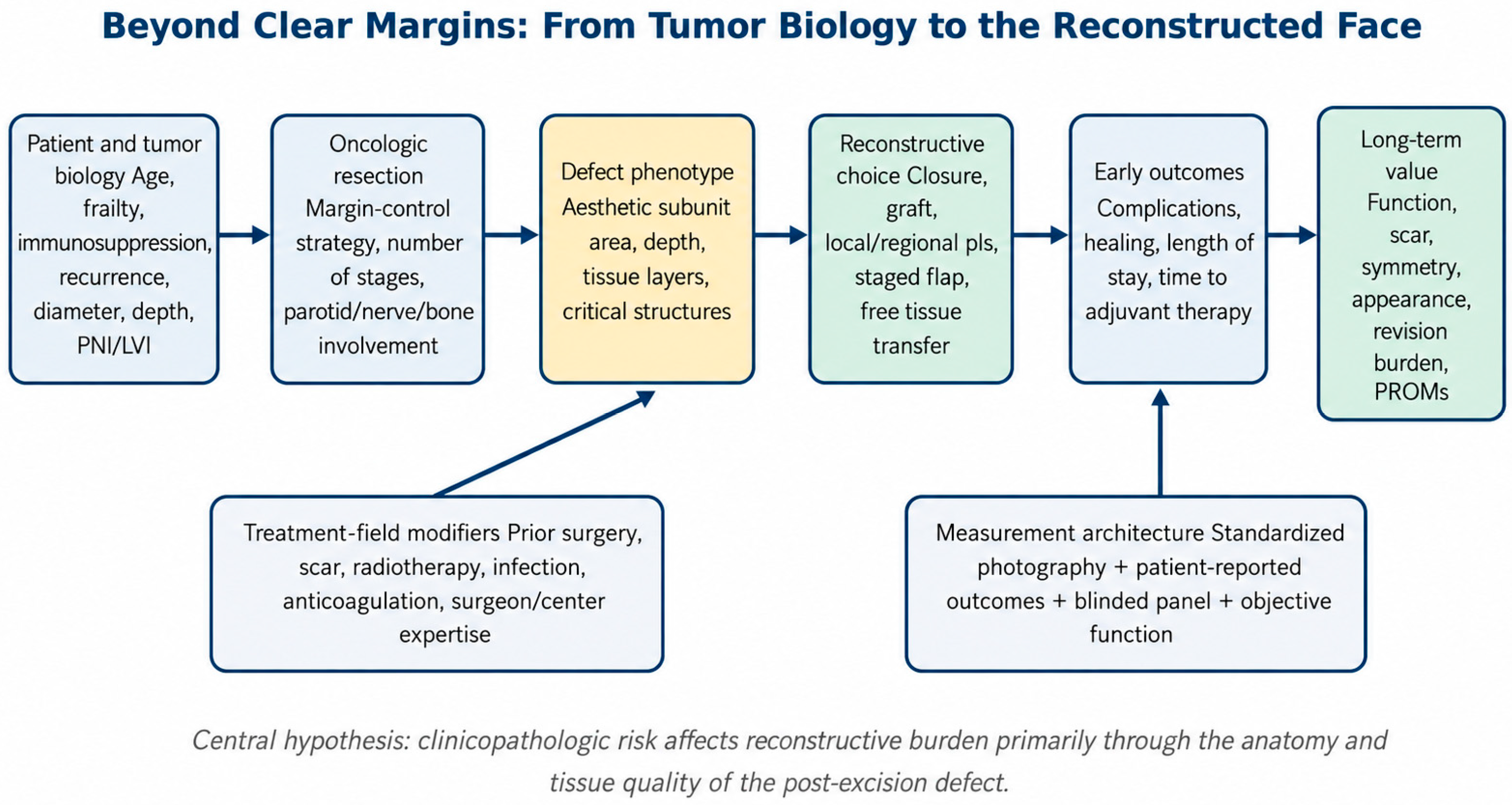
ChatGPT (OpenAI, GPT-5.5) was used to support language editing, manuscript organization, and preliminary structuring of the conceptual framework and evidence tables. It was not treated as a source of scientific evidence. During revision, study-level statements and bibliographic records retained in the reconstructive table were checked against available source records and source text, and unsupported AI-assisted statements were removed. The authors remain responsible for the interpretation, conclusions, and final manuscript. The hypothesis-generating conceptual framework is presented in Figure 1.
Figure 1.
Hypothesis-generating conceptual pathway linking clinicopathologic factors, surgical resection, post-excision defect characteristics, reconstructive strategy, postoperative complications, functional recovery, aesthetic outcome, and patient-reported outcomes in HNcSCC. The arrows represent proposed relationships for future testing and do not indicate demonstrated causality or mediation.
3. Results
3.1. Characteristics of the Evidence Base
The structured prognostic literature set comprised 29 publications addressing clinicopathologic factors and oncologic outcomes in cSCC, with particular emphasis on head and neck disease (Table S1). These publications were used to describe patient-, tumor-, and nodal-level associations with recurrence, metastasis, or survival; they were not treated as evidence of reconstructive outcomes unless such outcomes were explicitly measured.
The prognostic literature was predominantly retrospective and observational, with additional prospective cohorts, systematic reviews, a clinician survey with a literature review, and a secondary analysis of a randomized trial. Sample sizes, populations, staging systems, outcome definitions, follow-up, and multivariable adjustment varied substantially. Several large multicenter publications may also contain overlapping source cohorts and were therefore not interpreted as fully independent replications.
The revised Supplementary Materials contain 42 unique publications: 29 prognostic publications in Table S1 and 13 distinct reconstructive, functional, aesthetic, technical, or methodological sources in Table S2, with no publication repeated across the two tables. The contextual sources were classified according to whether they provided direct clinical reconstructive outcomes, mixed-histology or site-specific evidence, technical guidance, or outcome-measurement methodology.
The predominant outcomes in the prognostic set were local recurrence, nodal or distant metastasis, disease-specific survival, overall survival, and staging performance. Post-excision defect dimensions, missing tissue layers, reconstructive technique, flap or graft failure, complications, function, scar quality, aesthetic assessment, patient satisfaction, and revision burden were generally not measured. Accordingly, the review identifies an evidence gap rather than validated clinicopathologic predictors of reconstructive difficulty or outcome.
No prognostic publication was counted as direct reconstructive evidence solely on the basis of biological plausibility. Table 2 therefore separates measured oncologic associations from direct reconstructive evidence and from author-generated hypotheses. Supporting references and evidence counts are reported for each domain.
Table 2.
Summary of evidence domains and their reconstructive interpretation in HNcSCC.
3.2. Patient-Level Predictors: Age, Frailty, and Immunosuppression
Increasing age was associated with adverse tumor characteristics and reduced survival in portions of the prognostic literature, but chronological age alone should not be considered a contraindication to reconstruction. Its clinical significance is better interpreted alongside frailty, comorbidity, anesthesia tolerance, rehabilitation potential, and competing mortality. Local tissue laxity should be assessed independently of age: increased cervical skin laxity may facilitate local flap mobilization, whereas limited laxity—particularly on the scalp or in previously operated fields—may restrict primary closure and local flap advancement. These field characteristics were not measured in the prognostic studies and should be documented directly in future reconstructive cohorts.
Immunosuppression was associated with adverse oncologic outcomes across several prognostic publications. However, the retained prognostic studies did not establish that immunosuppression predicts infection, dehiscence, graft loss, flap complications, or delayed adjuvant therapy. These remain biologically plausible hypotheses that require direct prospective measurement rather than inference from recurrence or survival data.
3.3. Tumor-Level Predictors: Size, Depth, Differentiation, PNI, and LVI
Tumor diameter and invasion depth were repeatedly associated with oncologic progression. They may be investigated as upstream variables in reconstructive research, but the retained prognostic studies did not demonstrate a direct relationship with flap selection, complications, function, or aesthetics. Tumor dimensions cannot substitute for measurement of the actual post-excision defect, particularly after Mohs or staged margin-controlled surgery.
Perineural invasion illustrates the difference between oncologic and reconstructive evidence. Incidental microscopic PNI may influence adjuvant decisions, whereas clinically evident named-nerve spread may coincide with procedures involving the facial nerve, parotid, or skull base. However, the prognostic literature rarely reported nerve reconstruction, facial function, eye protection, static suspension, or patient-reported disability. Lymphovascular invasion likewise indicated aggressive biology but was not directly linked to reconstructive endpoints.
3.4. Margins, Recurrent Disease, and Treatment-Field Quality
Negative margins remain the primary oncologic endpoint. Repeated excision, delayed margin confirmation, recurrent disease, previous surgery, and previous radiotherapy may influence reconstructive planning, but their effects should be measured rather than assumed. Previous radiotherapy should not be treated as a binary variable: potential effects may vary with dose, field extent, tissue volume, fractionation, and interval since treatment. Future cohorts should record these parameters together with fibrosis, atrophy, telangiectasia, tissue mobility, recipient-vessel location, margin method, number of excision stages, timing of reconstruction, and time to adjuvant therapy.
3.5. Nodal and Parotid Disease
Nodal size, number of involved nodes, bilateral disease, extranodal extension, and parotid or cervical involvement were investigated primarily as oncologic variables. Parotidectomy and neck dissection nevertheless create a surgical context that differs substantially from a localized cutaneous defect and may involve vessel exposure, facial nerve dysfunction, contour loss, or soft-tissue volume loss. Future reconstructive studies should analyze this group separately and measure facial symmetry, dynamic and static facial function, neck contour, shoulder morbidity, and radiation-related change rather than infer these outcomes from nodal stage.
3.6. Direct Reconstructive and Aesthetic Evidence
The most informative direct aesthetic evidence came from nasal reconstruction studies rather than cSCC-specific cohorts. Mureau and colleagues assessed functional and aesthetic outcomes after nasal reconstruction [6]. Moolenburgh and colleagues evaluated patient, professional-panel, and lay-observer perspectives [7,8], demonstrating that these perspectives should be reported separately rather than treated as interchangeable measures.
Technical sources describe the aesthetic subunit principle and the restoration of nasal lining, structural support, and skin cover for complex nasal defects [9,10]. Scalp and calvarial literature describes technique selection according to defect dimensions, depth, exposed bone or dura, infection, previous irradiation, and tissue quality [11,12,13,44,45,46]. These principles are anatomically relevant, but many sources are reviews, technical frameworks, or mixed-histology cohorts rather than HNcSCC-specific comparative studies. Table 3 therefore identifies measured endpoints, evidence counts, and domains in which no direct evidence was identified.
Table 3.
Evidence-gap matrix linking clinicopathologic and defect-level variables with reconstructive, functional, aesthetic, and patient-reported outcomes.
4. Discussion
4.1. Main Findings and Interpretation
The central finding of this review is an asymmetry between oncologic prognostication and reconstructive outcome research in HNcSCC. Tumor diameter, invasion depth, differentiation, PNI, LVI, immunosuppression, recurrence, margin status, nodal burden, and extranodal extension have been investigated mainly in relation to recurrence, metastasis, or survival [16,17,18,19,20,21,22,23,24,25,26,27,28,29,30,31,32,33,34,35,36,37,38,39,40,41,42,43,47,48,49,50,51,52]. The same literature rarely records post-excision defect anatomy, reconstructive tier, number of procedures, functional recovery, scar quality, or patient-rated appearance. These variables should therefore not be described as validated predictors of reconstructive outcomes.
Oncologic risk and reconstructive burden are related but not interchangeable. We propose the post-excision defect as a conceptual intermediate variable between disease, surgical resection, and reconstructive outcome. This relationship has not been demonstrated through mediation analysis or prospective longitudinal data. The framework should therefore be interpreted as a hypothesis for future testing, with defect anatomy placed at the center of measurement rather than assumed from tumor stage alone.
4.2. Tumor Biology as an Upstream Determinant of Reconstructive Burden
Tumor size and depth are established oncologic variables [16,17,18], but their contribution to reconstructive burden remains unquantified. They should be evaluated as potential upstream indicators rather than assumed surrogates for the definitive defect. Margin-controlled excision may preserve tissue in one patient, whereas ill-defined borders, recurrent growth, or repeated re-excision may enlarge the defect in another patient with a tumor of similar clinical size. Future studies should record both preoperative tumor dimensions and postoperative defect dimensions.
PNI illustrates this distinction. Incidental microscopic PNI and clinically evident spread along a named nerve occupy different positions on the oncologic spectrum and may be associated with different surgical fields. Existing studies identify PNI as an adverse oncologic factor [16,17,18,25,26,27,29,31,32,47,48], but they seldom report facial function, corneal protection, sensory loss, nerve reconstruction, rehabilitation, or patient-reported disability. LVI and poor differentiation likewise signal aggressive biology, yet their reconstructive effects remain unmeasured. They are candidate variables for prospective research, not validated reconstructive predictors.
4.3. Patient Factors, Recurrence, and Treatment-Field Quality
Patient-level variables modify both the feasibility and the consequences of reconstruction. Increasing age is associated with adverse tumor characteristics and poorer survival in many HNcSCC cohorts, but chronological age alone is an inadequate measure of operative vulnerability. Frailty, cardiopulmonary reserve, nutritional status, diabetes, smoking, anticoagulation, functional independence, and rehabilitation potential are more likely to determine whether a prolonged microvascular procedure, a staged reconstruction, or a simpler wound strategy is appropriate. Thus, age should be interpreted as contextual information rather than as an isolated contraindication to complex reconstruction.
Immunosuppression deserves particular attention because it may influence the reconstructive pathway through two mechanisms. First, transplant recipients and patients with hematologic or treatment-related immunosuppression are more likely to develop aggressive, recurrent, and multifocal cSCC. Second, impaired immune function may plausibly increase infection, dehiscence, graft loss, delayed wound healing, and interruption of adjuvant therapy. The first pathway is well supported by oncologic evidence, whereas the second remains insufficiently quantified in HNcSCC-specific reconstructive cohorts. This distinction should be preserved in clinical interpretation: a biologically credible association is not equivalent to a measured reconstructive risk.
Recurrent tumors and previously treated fields may present scars, distorted tissue planes, reduced mobility, prior grafts or flaps, and radiation-related tissue changes. The magnitude of radiation effects is likely to depend on dose, field, fractionation, tissue volume, and time since treatment; these details were not captured by the prognostic literature. Current European guidance maintains complete oncologic excision as the primary objective [3,53]. Future reconstructive studies should additionally record margin assessment method, number of excision stages, previous surgery, radiotherapy parameters, clinically observed tissue quality, and the interval to adjuvant treatment.
4.4. Nodal and Parotid Disease as a Distinct Reconstructive Phenotype
Nodal metastasis and parotid or cervical involvement should be distinguished from localized cutaneous defects. Parotidectomy and neck dissection may involve facial nerve dysfunction, contour loss, exposure of major vessels, loss of soft-tissue volume, shoulder morbidity, or a larger adjuvant-treatment field. The staging literature provides prognostic information on nodal burden and extranodal extension [29,41,43], but reconstructive outcomes are rarely stratified by these variables. Future analyses should therefore separate localized resections from composite parotid, neck, orbital, or skull-base procedures.
4.5. Aesthetic and Functional Outcomes Require Multidimensional Assessment
The contextual reconstructive literature indicates that technical success, patient satisfaction, and observer-rated aesthetics are distinct outcomes. Nasal reconstruction studies reported differences between patient, professional-panel, and lay-observer assessments [6,7,8]. These perspectives should be reported separately. A global label such as “good cosmetic result” does not specify the measurement method and is therefore inadequate for reproducible outcome assessment.
The aesthetic subunit principle and replacement of missing lining, structural support, and skin cover are established technical concepts in nasal reconstruction [9,10]. Scalp and calvarial sources describe reconstruction according to defect size, depth, exposed bone or dura, surrounding tissue quality, and previous treatment [11,12,13,44,45,46]. These frameworks support defect-based planning, but much of the evidence is derived from mixed-histology, site-specific, retrospective, or technical sources. Anatomical transferability does not establish prognostic validity in HNcSCC.
Future studies should combine standardized clinical photography, blinded observer assessment, site-specific functional measures, complication reporting, revision burden, and patient-reported outcomes. The Nasal Obstruction Symptom Evaluation scale measures patient-perceived nasal obstruction [14], while the Patient and Observer Scar Assessment Scale permits parallel assessment of scar quality by patients and observers [15]. These outcomes should be reported separately rather than collapsed into a single success score.
4.6. Clinical Implications and a Hypothesis-Generating Research Framework
The proposed framework is intended solely to guide future research and should not be interpreted as a clinically applicable prediction model. No variables have been weighted, no scoring system or decision threshold has been defined, and predictive performance, calibration, and clinical utility have not been evaluated. Its purpose is to promote earlier multidisciplinary documentation of anticipated tissue loss, potential nerve or structural sacrifice, local tissue availability, staged reconstruction, surveillance needs, and compatibility with adjuvant treatment without compromising oncologic clearance.
Future research could evaluate two linked models rather than assume a single score. A preoperative model might examine frailty, immunosuppression, recurrence, tumor size, depth, subsite, clinical PNI, anticipated parotid or bone involvement, previous surgery, radiotherapy parameters, and local tissue mobility. A postoperative model could examine surface area, dimensions, depth, absent tissue layers, aesthetic subunits, exposed structures, margin status, and field quality in relation to technique, complications, revision, function, and aesthetics. Any model should be developed according to TRIPOD principles, assessed with PROBAST, and subjected to external validation and calibration before clinical use [54,55].
4.7. Methodological Strengths, Limitations, and Research Priorities
The primary contribution of this review is its explicit separation of measured reconstructive evidence from oncologic associations and author-generated hypotheses. This distinction reduces the risk of treating recurrence or survival factors as established predictors of flap failure, function, scar quality, or patient satisfaction. The framework is intended to make missing outcomes visible and to guide future study design.
Several limitations temper the conclusions. This was a single-database, structured narrative review rather than an exhaustive systematic search, and reconstructive sources were identified through reference-list screening. Screening and charting were not performed independently in duplicate; formal study-level risk-of-bias grading was not undertaken; and effect estimates were not quantitatively synthesized. The prognostic literature was predominantly retrospective, heterogeneous, and potentially affected by overlapping multicenter cohorts. Contextual reconstructive publications frequently included mixed histologies, selected tertiary-center populations, or technical reviews. Consequently, the proposed relationships are hypothesis-generating and should not be interpreted as validated causal or predictive effects.
The next step should be a prospective, multicenter HNcSCC reconstruction cohort with consecutive enrollment and a predefined minimum dataset. Patient variables should include frailty, comorbidity, immunosuppression, smoking, diabetes, nutritional status, and previous treatment; tumor variables should include size, depth, differentiation, PNI, LVI, recurrence, margins, nodal burden, and extranodal extension; defect variables should document dimensions, depth, aesthetic subunits, missing layers, and critical structures; and reconstructive variables should capture technique, operative stages, complications, reoperation, and time to adjuvant treatment. Standardized photographs, functional measures, scar assessment, patient satisfaction, and revision burden should be collected longitudinally. Only by measuring these domains in the same patient can the field determine whether tumor biology predicts the reconstructed face directly, indirectly through defect anatomy, or not at all.
5. Conclusions
Current HNcSCC literature supports oncologic risk stratification but does not validate clinicopathologic predictors of reconstructive difficulty, complications, functional impairment, aesthetic outcome, or patient experience. Most prognostic studies did not measure post-excision defect characteristics or reconstructive endpoints, while the available direct reconstructive literature was limited, site-specific, and frequently derived from mixed-histology or technical sources.
The post-excision defect is proposed as a conceptual intermediate variable for future investigation rather than as a demonstrated mediating phenotype. Prospective studies should jointly record standardized patient, tumor, treatment-field, defect, reconstructive, functional, aesthetic, and patient-reported variables. The framework presented here requires prospective development and external validation before any clinical application.
Supplementary Materials
The following supporting information can be downloaded at https://www.mdpi.com/article/10.3390/reports9030280/s1, Supplementary Method S1: Exact PubMed/MEDLINE keyword search phrases used to identify oncologic prognostic literature; Supplementary Table S1: structured charting of prognostic publications (n = 29); Supplementary Table S2: distinct contextual reconstructive, functional, aesthetic, technical, and methodological sources (n = 13).
Author Contributions
Conceptualization, I.-I.A. and L.V.; methodology, I.-I.A.; software, R.-G.M.; validation, I.-I.A., L.V. and A.O.; formal analysis, A.-B.I.; investigation, A.-P.C.; resources, B.M.; data curation, I.-I.A.; writing—original draft preparation, L.V.; writing—review and editing, I.-I.A.; visualization, L.V.; supervision, A.O.; project administration, I.-I.A. All authors have read and agreed to the published version of the manuscript.
Funding
This research received no external funding.
Institutional Review Board Statement
Not applicable.
Informed Consent Statement
Not applicable.
Data Availability Statement
The search phrases, structured study-charting tables, and evidence classifications created for this review are provided in the Supplementary Materials. No individual participant data were generated.
Acknowledgments
The use of generative artificial intelligence is described in Section 2.6. The authors critically reviewed and edited the final manuscript and take responsibility for its content.
Conflicts of Interest
The authors declare no conflicts of interest.
Abbreviations
The following abbreviations are used in this manuscript:
| AJCC | American Joint Committee on Cancer |
| AJCC8 | American Joint Committee on Cancer, 8th Edition |
| BWH | Brigham and Women’s Hospital |
| CC BY | Creative Commons Attribution License |
| cSCC | Cutaneous squamous cell carcinoma |
| DFS | Disease-free survival |
| DMR | Distant metastatic recurrence |
| DSS | Disease-specific survival |
| ENE | Extranodal extension |
| EADO | European Association of Dermato-Oncology |
| EADV | European Academy of Dermatology and Venereology |
| EDF | European Dermatology Forum |
| EORTC | European Organisation for Research and Treatment of Cancer |
| ESTRO | European Society for Radiotherapy and Oncology |
| ECS | Extracapsular spread |
| GenAI | Generative Artificial Intelligence |
| HNcSCC | Head and neck cutaneous squamous cell carcinoma |
| HNSCC | Head and neck squamous cell carcinoma |
| ITEM | Item-based nodal staging system |
| LVI | Lymphovascular invasion |
| MRI | Magnetic resonance imaging |
| NOSE | Nasal Obstruction Symptom Evaluation |
| OS | Overall survival |
| PET | Positron emission tomography |
| PMID | PubMed Identifier |
| PNI | Perineural invasion |
| PORT | Postoperative radiotherapy |
| POSAS | Patient and Observer Scar Assessment Scale |
| PRO | Patient-reported outcome |
| PROBAST | Prediction model Risk Of Bias ASsessment Tool |
| PROM | Patient-reported outcome measure |
| PROMs | Patient-reported outcome measures |
| RCT | Randomized controlled trial |
| SCC | Squamous cell carcinoma |
| S-ITM | Satellitosis/In-transit metastasis |
| TNM | Tumor–Node–Metastasis |
| TRIPOD | Transparent Reporting of a multivariable prediction model for Individual Prognosis Or Diagnosis |
| UEMS | European Union of Medical Specialists |
References
- Waldman, A.; Schmults, C. Cutaneous Squamous Cell Carcinoma. Hematol. Oncol. Clin. N. Am. 2019, 33, 1–12. [Google Scholar] [CrossRef] [Scilit] [PubMed]
- Que, S.K.T.; Zwald, F.O.; Schmults, C.D. Cutaneous Squamous Cell Carcinoma: Incidence, Risk Factors, Diagnosis, and Staging. J. Am. Acad. Dermatol. 2018, 78, 237–247. [Google Scholar] [CrossRef] [Scilit] [PubMed]
- Stratigos, A.J.; Garbe, C.; Dessinioti, C.; Lebbe, C.; van Akkooi, A.; Bataille, V.; Bastholt, L.; Dreno, B.; Dummer, R.; Fargnoli, M.C.; et al. European Consensus-Based Interdisciplinary Guideline for Invasive Cutaneous Squamous Cell Carcinoma: Part 2. Treatment—Update 2023. Eur. J. Cancer 2023, 193, 113252. [Google Scholar] [CrossRef] [Scilit] [PubMed]
- Desai, N.; Divatia, M.K.; Jadhav, A.; Wagh, A. Aggressive Cutaneous Squamous Cell Carcinoma of the Head and Neck: A Review. Curr. Oncol. 2023, 30, 6634–6647. [Google Scholar] [CrossRef] [Scilit] [PubMed]
- Huang, S.H.; O’Sullivan, B. Overview of the 8th Edition TNM Classification for Head and Neck Cancer. Curr. Treat. Options Oncol. 2017, 18, 40. [Google Scholar] [CrossRef] [Scilit] [PubMed]
- Mureau, M.A.M.; Moolenburgh, S.E.; Levendag, P.C.; Hofer, S.O.P. Aesthetic and functional outcome following nasal reconstruction. Plast. Reconstr. Surg. 2007, 120, 1217–1227. [Google Scholar] [CrossRef] [Scilit] [PubMed]
- Moolenburgh, S.E.; Mureau, M.A.M.; Hofer, S.O.P. Facial attractiveness and abnormality of nasal reconstruction patients and controls assessed by laypersons. J. Plast. Reconstr. Aesthet. Surg. 2008, 61, 676–680. [Google Scholar] [CrossRef] [Scilit] [PubMed]
- Moolenburgh, S.E.; Mureau, M.A.M.; Hofer, S.O.P. Aesthetic outcome after nasal reconstruction: Patient versus panel perception. J. Plast. Reconstr. Aesthet. Surg. 2008, 61, 1459–1464. [Google Scholar] [CrossRef] [Scilit] [PubMed]
- Menick, F.J. Nasal reconstruction. Plast. Reconstr. Surg. 2010, 125, 138e–150e. [Google Scholar] [CrossRef] [Scilit] [PubMed]
- Menick, F.J. Nasal reconstruction with a forehead flap. Clin. Plast. Surg. 2009, 36, 443–459. [Google Scholar] [CrossRef] [Scilit] [PubMed]
- Lin, S.J.; Hanasono, M.M.; Skoracki, R.J. Scalp and calvarial reconstruction. Semin. Plast. Surg. 2008, 22, 281–293. [Google Scholar] [CrossRef] [Scilit] [PubMed]
- Leedy, J.E.; Janis, J.E.; Rohrich, R.J. Reconstruction of acquired scalp defects: An algorithmic approach. Plast. Reconstr. Surg. 2005, 116, 54e–72e. [Google Scholar] [CrossRef] [Scilit] [PubMed]
- Seitz, I.A.; Gottlieb, L.J. Reconstruction of scalp and forehead defects. Clin. Plast. Surg. 2009, 36, 355–377. [Google Scholar] [CrossRef] [Scilit] [PubMed]
- Stewart, M.G.; Witsell, D.L.; Smith, T.L.; Weaver, E.M.; Yueh, B.; Hannley, M.T. Development and validation of the Nasal Obstruction Symptom Evaluation (NOSE) scale. Otolaryngol. Head Neck Surg. 2004, 130, 157–163. [Google Scholar] [CrossRef] [Scilit] [PubMed]
- Draaijers, L.J.; Tempelman, F.R.H.; Botman, Y.A.M.; Tuinebreijer, W.E.; Middelkoop, E.; Kreis, R.W.; van Zuijlen, P.P.M. The Patient and Observer Scar Assessment Scale: A reliable and feasible tool for scar evaluation. Plast. Reconstr. Surg. 2004, 113, 1960–1965. [Google Scholar] [CrossRef] [Scilit] [PubMed]
- Thompson, A.K.; Kelley, B.F.; Prokop, L.J.; Murad, M.H.; Baum, C.L. Risk factors for cutaneous squamous cell carcinoma recurrence, metastasis, and disease-specific death: A systematic review and meta-analysis. JAMA Dermatol. 2016, 152, 419–428. [Google Scholar] [CrossRef] [Scilit] [PubMed]
- Brantsch, K.D.; Meisner, C.; Schönfisch, B.; Trilling, B.; Wehner-Caroli, J.; Röcken, M.; Breuninger, H. Analysis of risk factors determining prognosis of cutaneous squamous-cell carcinoma: A prospective study. Lancet Oncol. 2008, 9, 713–720. [Google Scholar] [CrossRef] [Scilit] [PubMed]
- Schmults, C.D.; Karia, P.S.; Carter, J.B.; Han, J.; Qureshi, A.A. Factors predictive of recurrence and death from cutaneous squamous cell carcinoma: A 10-year, single-institution cohort study. JAMA Dermatol. 2013, 149, 541–547. [Google Scholar] [PubMed]
- Lubov, J.; Labbé, M.; Sioufi, K.; Morand, G.B.; Hier, M.P.; Khanna, M.; Sultanem, K.; Mlynarek, A.M. Prognostic factors of head and neck cutaneous squamous cell carcinoma: A systematic review. J. Otolaryngol. Head Neck Surg. 2021, 50, 54. [Google Scholar] [CrossRef] [Scilit] [PubMed] [PubMed Central]
- Leus, A.J.G.; Haisma, M.S.; Terra, J.B.; Diercks, G.F.H.; Van Kester, M.S.; Halmos, G.B.; Rácz, E.; Van Dijk, B.A.C.; Plaat, B.E.C. Age-related Differences in Tumour Characteristics and Prognostic Factors for Disease Progression in Cutaneous Squamous Cell Carcinoma of the Head and Neck. Acta Derm. Venereol. 2022, 102, 00652. [Google Scholar] [CrossRef] [Scilit] [PubMed] [PubMed Central]
- Ran, N.A.; Granger, E.E.; Brodland, D.G.; Cañueto, J.; Carr, D.R.; Carter, J.B.; Carucci, J.A.; Hirotsu, K.E.; Koyfman, S.A.; Mangold, A.R.; et al. Risk Factor Number and Recurrence, Metastasis, and Disease-Related Death in Cutaneous Squamous Cell Carcinoma. JAMA Dermatol. 2025, 161, 597–604. [Google Scholar] [CrossRef] [Scilit] [PubMed] [PubMed Central]
- de Jong, E.; Genders, R.; Harwood, C.A.; Green, A.C.; Plasmeijer, E.I.; Proby, C.; Geissler, E.; Ferrándiz-Pulido, C.; Ducroux, E.; Euvrard, S.; et al. Cumulative incidence and risk factors for cutaneous squamous cell carcinoma metastases in organ transplant recipients: The Skin Care in Organ Transplant Patients in Europe-International Transplant Skin Cancer Collaborative metastases study, a prospective multicenter study. J. Am. Acad. Dermatol. 2024, 90, 1200–1209. [Google Scholar] [CrossRef] [Scilit] [PubMed]
- Shahwan, K.T.; Walker, T.D.; Tan, A.; Ruiz, E.; Ran, N.; Granger, E.E.; Koyfman, S.; Vidimos, A.; Wysong, A.; Hirotsu, K.E.; et al. Identifying the impact of minor risk factors in Brigham and Women’s Hospital stage T1 cutaneous squamous cell carcinomas on risk of poor outcomes: A retrospective cohort study. J. Am. Acad. Dermatol. 2025, 92, 1319–1326. [Google Scholar] [CrossRef] [Scilit] [PubMed]
- Girardi, F.M.; Wagner, V.P.; Machado, C.D.C.; Wysong, A.; Ran, N.A.; Granger, E.E.; Koyfman, S.A.; Vidimos, A.T.; Carr, D.R.; Shahwan, K.T.; et al. Validation of Current Staging Systems in HNCSCC: A Multinational Cohort Study. Head Neck 2025, 47, 3385–3393. [Google Scholar] [CrossRef] [Scilit] [PubMed]
- Mooney, C.P.; Martin, R.C.W.; Dirven, R.; Ashford, B.G.; Shannon, K.; Palme, C.E.; Ngo, Q.; Wykes, J.; Davies, S.; Gao, K.; et al. Sentinel Node Biopsy in 105 High-Risk Cutaneous SCCs of the Head and Neck: Results of a Multicenter Prospective Study. Ann. Surg. Oncol. 2019, 26, 4481–4488. [Google Scholar] [CrossRef] [Scilit] [PubMed]
- Campoli, M.; Brodland, D.G.; Zitelli, J. A prospective evaluation of the clinical, histologic, and therapeutic variables associated with incidental perineural invasion in cutaneous squamous cell carcinoma. J. Am. Acad. Dermatol. 2014, 70, 630–636. [Google Scholar] [CrossRef] [Scilit] [PubMed]
- Schachtel, M.J.C.; Gandhi, M.; Bowman, J.J.; Porceddu, S.V.; Panizza, B.J. Facial nerve perineural spread from cutaneous squamous cell carcinoma of the head and neck: A single institution analysis of epidemiology, treatment, survival outcomes, and prognostic factors. Head Neck 2022, 44, 1223–1236. [Google Scholar] [CrossRef] [Scilit] [PubMed] [PubMed Central]
- Bachar, G.; Mizrachi, A.; Rabinovics, N.; Guttman, D.; Shpitzer, T.; Ad-El, D.; Hadar, T. Prognostic factors in metastatic cutaneous squamous cell carcinoma of the head and neck. Ear Nose Throat J. 2016, 95, E32–E36. [Google Scholar] [CrossRef] [Scilit] [PubMed]
- Ebrahimi, A.; Gupta, R.; McDowell, L.; Magarey, M.J.R.; Smith, P.N.; Schulte, K.M.; Perriman, D.M.; Veness, M.; Porceddu, S.; Low, T.H.; et al. Determinants of Prognosis in Head and Neck Cutaneous Squamous Cell Carcinoma with Nodal Metastases. JAMA Otolaryngol. Head Neck Surg. 2024, 150, 986–994. [Google Scholar] [CrossRef] [Scilit] [PubMed] [PubMed Central]
- Hirotsu, K.E.; Aasi, S.Z.; Samson, K.K.; Zheng, C.; Nazaroff, J.R.; Voller, L.M.; Ruiz, E.S.; Ran, N.A.; Granger, E.E.; Koyfman, S.A.; et al. Lymphovascular invasion is an independent predictor of metastasis and disease-specific death in cutaneous squamous cell carcinoma: A multicenter retrospective study. J. Am. Acad. Dermatol. 2025, 93, 368–377. [Google Scholar] [CrossRef] [Scilit] [PubMed]
- Fermi, M.; Guarino, P.; Chiari, F.; Serafini, E.; Filippini, D.M.; Ferrulli, G.; Basso, M.; Mattioli, F.; Caporale, C.D.; Marchioni, D.; et al. Survival rates in head and neck cutaneous squamous cell carcinoma with nodal metastases: A multicenter study. Eur. Arch. Otorhinolaryngol. 2026, 283, 1099–1109. [Google Scholar] [CrossRef] [Scilit] [PubMed]
- Manyam, B.V.; Garsa, A.A.; Chin, R.I.; Reddy, C.A.; Gastman, B.; Thorstad, W.; Yom, S.S.; Nussenbaum, B.; Wang, S.J.; Vidimos, A.T.; et al. A multi-institutional comparison of outcomes of immunosuppressed and immunocompetent patients treated with surgery and radiation therapy for cutaneous squamous cell carcinoma of the head and neck. Cancer 2017, 123, 2054–2060. [Google Scholar] [CrossRef] [Scilit] [PubMed]
- Bobin, C.; Ingrand, P.; Dréno, B.; Rio, E.; Malard, O.; Espitalier, F. Prognostic factors for parotid metastasis of cutaneous squamous cell carcinoma of the head and neck. Eur. Ann. Otorhinolaryngol. Head Neck Dis. 2018, 135, 99–103. [Google Scholar] [CrossRef] [Scilit] [PubMed]
- de Melo, G.M.; das Neves, M.C.; Romero, R.D.; Rosano, M.; Martines, R.T.; Takimoto, R.M.; Greggio, B.; Palumbo, M.D.N.; Brodskyn, F.; Gatti, A.P.; et al. Prognostic Factors of Locally Advanced Cutaneous Squamous Cell Carcinoma in Head and Neck Region in Transplanted Patients. Diagnostics 2026, 16, 404. [Google Scholar] [CrossRef] [Scilit] [PubMed] [PubMed Central]
- McLean, T.; Brunner, M.; Ebrahimi, A.; Gao, K.; Ch’ng, S.; Veness, M.J.; Clark, J.R. Concurrent primary and metastatic cutaneous head and neck squamous cell carcinoma: Analysis of prognostic factors. Head Neck 2013, 35, 1144–1148. [Google Scholar] [CrossRef] [Scilit] [PubMed]
- Klein, J.C.; Shahwan, K.T.; Petric, U.B.; Mallela, T.; Voller, L.; Ruiz, E.; Ran, N.; Granger, E.E.; Koyfman, S.; Vidimos, A.; et al. Impact of immunosuppression on cutaneous squamous cell carcinoma outcomes. J. Am. Acad. Dermatol. 2026, 94, 151–160. [Google Scholar] [CrossRef] [Scilit] [PubMed]
- Ebrahimi, A.; Gupta, R.; McDowell, L.; Magarey, M.J.R.; Smith, P.N.; Schulte, K.M.; Perriman, D.M.; Veness, M.; Porceddu, S.; Low, T.H.; et al. Predictors of distant metastatic recurrence in head and neck cutaneous squamous cell carcinoma with lymph node metastases treated with curative intent: A multicenter study. Head Neck 2025, 47, 254–262. [Google Scholar] [CrossRef] [Scilit] [PubMed] [PubMed Central]
- Porceddu, S.V.; Connolly, E.; Bressel, M.; Wratten, C.; Liu, H.Y.; Rischin, D. Prognostic Subgroups for Disease-Free Survival with Cutaneous Squamous Cell Carcinoma of the Head and Neck: A Secondary Analysis of a Randomized Clinical Trial. JAMA Otolaryngol. Head Neck Surg. 2025, 151, 938–945. [Google Scholar] [CrossRef] [Scilit] [PubMed] [PubMed Central]
- Luk, P.P.; Ebrahimi, A.; Veness, M.J.; McDowell, L.; Magarey, M.; Gao, K.; Palme, C.E.; Clark, J.R.; Gupta, R. Prognostic value of the 8th edition American Joint Commission Cancer nodal staging system for patients with head and neck cutaneous squamous cell carcinoma: A multi-institutional study. Head Neck 2021, 43, 558–567. [Google Scholar] [CrossRef] [Scilit] [PubMed]
- Ebrahimi, A.; Luk, P.P.; Low, H.; McDowell, L.; Magarey, M.J.R.; Smith, P.N.; Perriman, D.M.; Shadbolt, B.; Veness, M.; Gupta, R.; et al. The American Joint Committee on Cancer staging for metastatic head and neck cutaneous squamous cell carcinoma: A multi-institutional study of within-stage heterogeneity and impact on prognostic performance. Head Neck 2020, 42, 3235–3242. [Google Scholar] [CrossRef] [Scilit] [PubMed]
- Costantino, A.; Pace, G.M.; Zimello, C.; Russo, E.; Greco, A.; Polimeni, A.; Magnuson, J.S.; De Virgilio, A. Impact of Extranodal Extension in Metastatic Cutaneous Squamous Cell Carcinoma of the Head and Neck: A Systematic Review and Meta-Analysis. Head Neck 2025, 47, 3275–3284. [Google Scholar] [CrossRef] [Scilit] [PubMed]
- Ebrahimi, A.; Luk, P.P.; Low, H.; McDowell, L.; Magarey, M.J.R.; Smith, P.N.; Perriman, D.M.; Veness, M.; Gupta, R.; Clark, J.R. A critical analysis of the 8th edition TNM staging for head and neck cutaneous squamous cell carcinoma with lymph node metastases and comparison to N1S3 stage and ITEM risk score: A multicenter study. J. Surg. Oncol. 2021, 123, 1531–1539. [Google Scholar] [CrossRef] [Scilit] [PubMed]
- Ebrahimi, A.; Gupta, R.; Luk, P.; Low, T.H.; McDowell, L.; Magarey, M.J.R.; Smith, P.N.; Perriman, D.M.; Schulte, K.M.; Veness, M.; et al. Number of nodal metastases and the American Joint Committee on cancer staging of head and neck cutaneous squamous cell carcinoma: A multicenter study. Oral. Oncol. 2020, 111, 104855. [Google Scholar] [CrossRef] [Scilit] [PubMed]
- van Driel, A.A.; Mureau, M.A.M.; Goldstein, D.P.; Gilbert, R.W.; Irish, J.C.; Gullane, P.J.; Neligan, P.C.; Hofer, S.O.P. Aesthetic and oncologic outcome after microsurgical reconstruction of complex scalp and forehead defects after malignant tumor resection: An algorithm for treatment. Plast. Reconstr. Surg. 2010, 126, 460–470. [Google Scholar] [CrossRef] [Scilit] [PubMed]
- Iblher, N.; Ziegler, M.C.; Penna, V.; Eisenhardt, S.U.; Stark, G.B.; Bannasch, H. An algorithm for oncologic scalp reconstruction. Plast. Reconstr. Surg. 2010, 126, 450–459. [Google Scholar] [CrossRef] [Scilit] [PubMed]
- Chao, A.H.; Yu, P.; Skoracki, R.J.; Demonte, F.; Hanasono, M.M. Microsurgical reconstruction of composite scalp and calvarial defects in patients with cancer: A 10-year experience. Head Neck 2012, 34, 1759–1764. [Google Scholar] [CrossRef] [Scilit] [PubMed]
- Jambusaria-Pahlajani, A.; Kanetsky, P.A.; Karia, P.S.; Hwang, W.-T.; Gelfand, J.M.; Whalen, F.M.; Elenitsas, R.; Xu, X.; Schmults, C.D. Evaluation of American Joint Committee on Cancer tumor staging for cutaneous squamous cell carcinoma and a proposed alternative tumor staging system. JAMA Dermatol. 2013, 149, 402–410. [Google Scholar] [PubMed]
- Karia, P.S.; Jambusaria-Pahlajani, A.; Harrington, D.P.; Murphy, G.F.; Qureshi, A.A.; Schmults, C.D. Evaluation of American Joint Committee on Cancer, International Union Against Cancer, and Brigham and Women’s Hospital tumor staging for cutaneous squamous cell carcinoma. J. Clin. Oncol. 2014, 32, 327–334. [Google Scholar] [CrossRef] [Scilit] [PubMed]
- Watts, F.; Palme, C.E.; Porceddu, S.; Sundaresan, P.; Clark, J.R.; Gupta, R. Clinician perspectives on the factors influencing prognostic stratification by the American Joint Commission on Cancer Head and Neck Cutaneous Squamous Cell Carcinoma Staging. Surgery 2021, 170, 1467–1473. [Google Scholar] [CrossRef] [Scilit] [PubMed]
- Jambusaria-Pahlajani, A.; Jeanselme, V.; Wang, D.M.; Ran, N.A.; Granger, E.E.; Cañueto, J.; Brodland, D.G.; Carr, D.R.; Carter, J.B.; Carucci, J.A.; et al. riSCC: A personalized risk model for the development of poor outcomes in cutaneous squamous cell carcinoma. J. Am. Acad. Dermatol. 2025, 93, 73–81. [Google Scholar] [CrossRef] [Scilit] [PubMed]
- Pahalyants, V.; Jairath, N.K.; Maas, D.E.; Cheraghlou, S.; Mandal, S.; Friedman, S.; Criscito, M.C.; Lee, N.; Doudican, N.A.; Ruiz, E.S.; et al. Satellitosis or in-transit metastasis in cutaneous squamous cell carcinoma: Risk factors and the prognostic significance. J. Am. Acad. Dermatol. 2025, 93, 1432–1440. [Google Scholar] [CrossRef] [Scilit] [PubMed]
- Silberstein, E.; Sofrin, E.; Bogdanov-Berezovsky, A.; Nash, M.; Segal, N. Lymph Node Metastasis in Cutaneous Head and Neck Squamous Cell Carcinoma. Dermatol. Surg. 2015, 41, 1126–1129. [Google Scholar] [CrossRef] [Scilit] [PubMed]
- Stratigos, A.J.; Garbe, C.; Dessinioti, C.; Lebbe, C.; van Akkooi, A.; Bataille, V.; Bastholt, L.; Dreno, B.; Dummer, R.; Fargnoli, M.C.; et al. European consensus-based interdisciplinary guideline for invasive cutaneous squamous cell carcinoma: Part 1. Diagnostics and prevention—Update 2023. Eur. J. Cancer 2023, 193, 113251. [Google Scholar] [CrossRef] [Scilit] [PubMed]
- Wolff, R.F.; Moons, K.G.; Riley, R.; Whiting, P.F.; Westwood, M.; Collins, G.S.; Reitsma, J.B.; Kleijnen, J.; Mallett, S. PROBAST: A tool to assess the risk of bias and applicability of prediction model studies. Ann. Intern. Med. 2019, 170, 51–58. [Google Scholar] [CrossRef] [Scilit] [PubMed]
- Collins, G.S.; Reitsma, J.B.; Altman, D.G.; Moons, K.G.M. Transparent Reporting of a multivariable prediction model for Individual Prognosis or Diagnosis (TRIPOD): The TRIPOD statement. Ann. Intern. Med. 2015, 162, 55–63. [Google Scholar] [CrossRef] [Scilit] [PubMed]

Disclaimer/Publisher’s Note: The statements, opinions and data contained in all publications are solely those of the individual author(s) and contributor(s) and not of MDPI and/or the editor(s). MDPI and/or the editor(s) disclaim responsibility for any injury to people or property resulting from any ideas, methods, instructions or products referred to in the content. |
© 2026 by the authors. Licensee MDPI, Basel, Switzerland. This article is an open access article distributed under the terms and conditions of the Creative Commons Attribution (CC BY) license.