


Abstract
Satellite glial cells (SGCs) play a critical role in the development and maintenance of chronic pain through complex interactions with inflammatory mediators and the opioid pathway. This systematic review synthesizes recent advances in the molecular mechanisms underlying SGC activation and implications for chronic pain management, particularly in conditions associated with the dorsal root ganglia (DRG) and trigeminal ganglia (TG). A systematic search was conducted in four databases (PubMed, Embase, Scopus, and Web of Science) covering studies published between January 2004 and May 2026. After screening 111 records, 19 studies were included. This review highlights how pro-inflammatory cytokines such as IL-1β, IL-1α, and TNF-α, as well as neurotransmitters like ATP and glutamate, contribute to SGC activation, neuroinflammation, and pain modulation. It also explores the role of receptors like CXCR4, TLR4, and P2X7 in SGCs in enhancing analgesic effects and their contributions to opioid tolerance and hyperalgesia. The findings underscore the potential of targeting SGCs to improve pain management outcomes across various pain models, including neuropathic, cancer-related, and visceral pain. Despite promising insights, variability in study methodologies and the complexity of glial–neuronal interactions present challenges. Future research should focus on standardizing experimental protocols and developing targeted therapies to modulate SGC activity to offer hope for patients suffering from chronic pain, particularly in managing opioid tolerance.
1. Introduction
Pain is a major contributor to human suffering and remains one of the leading causes of medical consultation worldwide. It affects millions of people, leading to diminished quality of life, decreased economic productivity, and a significant strain on healthcare systems [1,2,3]. According to the International Association for the Study of Pain (IASP), chronic pain is defined as pain that persists or recurs for longer than three months, often outlasting the normal healing process and becoming a distinct clinical condition [4]. Chronic pain conditions, such as neuropathic pain, diabetic neuropathy, and postherpetic neuralgia, are often refractory to conventional treatment [5]. These conditions are not only debilitating but also contribute to substantial healthcare costs and loss of productivity [6].
Recent advances in neuroscience have highlighted the critical role of satellite glial cells (SGCs) in the peripheral nervous system (PNS) in pain development and maintenance [1,7,8]. These cells, which surround the sensory neurons in the dorsal root ganglia (DRG) and trigeminal ganglia (TG), have gained attention for their involvement in pain modulation through the release of pro-inflammatory cytokines and the alteration of neuronal excitability [9]. Emerging evidence suggests that SGCs may also play a crucial role in the morphine/opioid pathway, potentially influencing opioids’ efficacy and side effects [10]. This is particularly relevant for chronic pain conditions, which are associated with peripheral and central sensitization [11]. Understanding the complex interactions between SGCs and opioids offers promising avenues for developing targeted therapies that enhance pain relief while minimizing adverse effects.
Despite the growing interest in SGCs and their potential role in pain management, there remains a significant gap in the literature regarding the specific molecules involved in the morphine/opioid pathway related to SGCs and their contributions to chronic pain mechanisms. Although some studies have identified neurotransmitters, cytokines, and other molecules associated with SGC activity, the intricate web of molecular interactions and pathways underlying opioid-mediated pain modulation remains poorly understood. For instance, molecules such as ATP (adenosine triphosphate), glutamate, and pro-inflammatory cytokines have been implicated in SGC activation [12,13], but their exact roles and interactions with opioid receptors are not fully delineated. Additionally, crosstalk between SGCs and other cell types, such as microglia and macrophage-like cells, further challenges description of their role in the pain pathway [14]. This gap limits the development of targeted therapies that could modulate SGC activity and enhance the effectiveness of opioids in managing chronic pain.
This systematic review aims to analyze the current evidence on molecules associated with SGC activation and their involvement in the morphine/opioid pathway underlying chronic pain mechanisms. By integrating findings from recent studies, this review seeks to clarify the molecular interactions underlying SGC-mediated opioid response modulation and identify potential targets for developing more effective and safer analgesic therapies.
2. Methods
2.1. Study Design
This systematic review aims to synthesize current evidence on molecules associated with SGC activation and their involvement in the morphine/opioid pathway in chronic pain mechanisms. The review focuses on chronic pain conditions—specifically those linked to the DRG and TG—to provide insight into potential therapeutic targets and molecular interactions involving SGCs, and was conducted and reported in accordance with the Preferred Reporting Items for Systematic Reviews and Meta-Analyses (PRISMA 2020) guidelines. The complete PRISMA 2020 checklist and detailed search strategies used for each database are provided as Supplementary Materials.
2.2. Literature Search Strategy
A comprehensive literature search was conducted across multiple databases, including PubMed, Embase, Scopus, and Web of Science, to identify relevant studies published between January 2004 and May 2026. The search strategy utilized a combination of keywords related to SGCs, opioid pathways, and associated molecules. The core terms “satellite glia” and “satellite glial cell” were selected to maximize sensitivity for this specific glial cell population, while “morphine” and “opioid” were chosen as the primary pharmacological terms to capture both mechanistic and translational studies involving opioid-related pain modulation. The last search was performed on 8 May 2026.
The search strategy was developed to address the following guiding question: “In individuals or animal models with chronic pain, which molecules associated with SGCs modulate or interact with the opioid pathway, and what role do they play in the mechanisms of opioid-induced pain modulation?”
To ensure thorough identification of the relevant literature, slightly different search strategies were employed for each database considering the specific search functionalities and indexing systems of each platform, as shown in Table 1. Additionally, reference lists of relevant review articles were manually searched to identify additional relevant studies.
Table 1.
Search strategy according to the databases.
2.3. Inclusion and Exclusion Criteria
Studies were selected according to predefined eligibility criteria.
2.3.1. Inclusion Criteria
- Studies investigating SGC activation in relation to chronic pain mechanisms.
- Research examining the role of SGCs in the morphine/opioid pathway.
- Articles published in peer-reviewed journals between 2004 and 2026.
- Studies involving animal models, cell cultures, or human subjects related to DRG and TG.
- Articles available in English, Portuguese, or Spanish.
2.3.2. Exclusion Criteria
- Studies not directly related to SGC or chronic pain.
- Reviews, meta-analyses, editorials, and opinion pieces without original data.
- Studies focusing solely on central nervous system (CNS) glial cells.
2.4. Study Selection
All retrieved records were imported into the Rayyan platform for screening and duplicate removal. Two reviewers independently screened titles and abstracts. Discrepancies were resolved through discussion or consultation with a third reviewer. Full texts of potentially relevant studies were subsequently assessed for eligibility.
2.5. Data Extraction
Data extraction was performed independently by two reviewers using a standardized data extraction form developed for this review. Each reviewer independently extracted data from the included studies. Any discrepancies were discussed and resolved through consensus. When necessary, a third reviewer was consulted to resolve disagreements and ensure consistency in the extracted information.
Data were systematically extracted from each study using a structured extraction form. The extraction process focused on key categories.
Study identification included the study title, authors, publication year, DOI, and institutional affiliations. Information on the study’s geographical location, original language, sources of sponsorship, trial registration numbers, and any reported conflicts of interest was also documented, as well as details regarding ethics approval and the study’s primary aim.
Methodological details covered study design, including whether the research involved in vitro or in vivo models. For in vivo studies, specifics such as animal species, strain, sample size, and experimental conditions were noted. In vitro studies included details on cell types, culture conditions, and passage numbers. Interventions were thoroughly documented, focusing on the molecules studied, dosages, routes of administration, and timing and duration of exposure. Control conditions and any co-interventions applied were also recorded.
Outcome measures centered on primary and secondary outcomes relevant to SGC activation and pain modulation. This included pain-related metrics, molecular and immunological outcomes, neuroinflammatory markers, and neurophysiological changes. Methods used for outcome assessment, such as assays, imaging techniques, and statistical analyses, were also documented.
Results and conclusions were summarized to highlight key findings, including the effects of interventions on SGC pathways, neuron–glia communication, and receptor–ligand interactions. Genetic and epigenetic modifications relevant to the study objectives were also captured. Attention was given to implications for future research and limitations discussed by the authors.
2.6. Quality Assessment
Methodological quality was assessed using a modified version of the OHAT Risk of Bias Rating Tool, which applies to both human and preclinical studies [15]. The assessment considered domains such as selection bias, performance bias, detection bias, attrition bias, reporting bias, confounding bias, conflict of interest bias, and any other potential biases (OHAT Risk of Bias Rating Tool for Human and Animal Studies) [16]. Assessment was independently performed by two reviewers; disagreements were resolved through discussion until consensus was reached.
2.7. Certainty of Evidence Assessment
A formal assessment of the certainty of the evidence using the GRADE approach was not performed due to substantial methodological heterogeneity among the included studies and the predominance of preclinical experimental designs. Instead, the findings were interpreted in light of study quality, risk of bias, consistency of results, and biological plausibility.
2.8. Data Synthesis
Data synthesis was performed using a qualitative approach to identify key molecular findings, interactions, and knowledge gaps. Findings were grouped according to shared signaling pathways relevant to SGC-mediated pain modulation and opioid responses. Additional studies were considered to support interpretation and contextualize mechanisms where direct evidence was limited.
2.9. Reporting Bias Assessment
Publication bias could not be formally assessed using statistical methods (e.g., funnel plot asymmetry tests) due to insufficient quantitative data for meta-analysis.
2.10. Ethical Considerations
As this study is based exclusively on previously published data, no ethical approval was required.
3. Results
3.1. Overview of the Included Studies
A systematic literature search was conducted in four electronic databases: Scopus, PubMed, Web of Science, and Embase. Screening and study selection were performed using the Rayyan platform.
A total of 111 records were identified, including 22 from Scopus, 18 from PubMed, 30 from Web of Science, and 41 from Embase. After removal of 47 duplicate records, 64 studies remained for title and abstract screening. Of these, 42 records were excluded.
Twenty-two full-text articles were assessed for eligibility. Three studies were excluded after full-text review for the following reasons: lack of focus on SGCs (n = 1), absence of opioid- or morphine-related outcomes (n = 1), and inappropriate study design (n = 1). Ultimately, 19 studies were included in this review. A flowchart illustrating the selection process is presented in Figure 1.
Figure 1.
Flowchart of the study selection process according to PRISMA 2020 guidelines. ** Human reviewers excluded all 42 records during title and abstract screening; no automation tools were used in this process.
3.2. Period of Publication, Experimental Models, and Study Designs
The included studies covered a 20-year period (2005 to 2025). Most of the studies (n = 14; 73.7%) were performed in vivo, followed by studies that used in vivo and in vitro methods (n = 3; 15.8%); one study used in vivo and electrophysiological methods (n = 1; 5.3%), and there was one human translational study (n = 1; 5.3%). Although neuropathic pain was the most prevalent type of pain studied (n = 10; 52.6%), there was substantial variation and even greater variability in the animal pain models used, with chronic constriction injury (CCI) of the sciatic nerve being the most prevalent (n = 3; 15.8%), followed by chronic administration of morphine (n = 2; 10.5%) and plantar incision with remifentanil infusion (n = 2; 10.5%). These findings are presented in Table 2.
Table 2.
Summary of animal pain models and study designs.
3.3. Molecules Analyzed in the Included Studies
A variety of molecules were used across the included studies. As presented in Table 3, at least one inflammatory cytokine (e.g., interleukin-1 beta, IL-1β, or tumor necrosis factor-alpha, TNF-α) was used in six studies (n = 6; 31.6%); morphine in two studies (n = 2; 10.5%); nociceptive/orphanin FQ (N/OFQ), a neuropeptide that acts as a natural ligand of the opioid receptor-like 1 (ORL1/OPMR1/NOP receptor), in one study (n = 1; 5.3%); OPMR1 in one study (n = 1; 5.3%); and naloxone, an antagonist of the opioid receptors μ, δ, and κ, in one study (n = 1; 5.3%). Behavioral assays (BAs) were used in 12 studies (n = 12; 63.2%); immunohistochemistry (IHC) in 12 studies (n = 12; 63.2%); and qPCR in 7 studies (n = 7; 58.3%). SGCs were studied directly in 14 studies (n = 14; 73.68%); sensory neurons in 12 studies (n = 12; 63.2%); DRG in 14 studies (n = 14; 73.68%); and TG in 3 studies (n = 3; 15.79%).
Table 3.
Summary of molecules analyzed in each study.
3.4. Activation of SGCs and Their Modulation in Pain Pathways
The role of SGC activation in modulating pain pathways and related outcomes, the impact of interventions on SGCs, and the indicators used to provide evidence of SGC activation in each study are presented in Table 4.
Table 4.
Summary of SGC activation and its modulation in pain pathways.
3.5. SGC-Mediated Modulation of Opioid Signaling and Pain Sensitization
Table 5 shows the molecules or receptors located in SGCs that directly affected the opioid pathway in each included study, changes in opioid pathway signaling, and implications for opioid efficacy and sensitization. While IL-1β was used to evaluate opioid neurotransmission activity in four studies (n = 4; 21%), most (n = 9; 47.4%) did not evaluate a specific SGC molecule or receptor or changes in opioid neurotransmission.
Table 5.
Summary of potential involvement of SGCs in opioid-related signaling mechanisms.
3.6. Risk of Bias Assessment
The included studies’ methodological quality was assessed using the OHAT Risk of Bias Tool. Overall, most studies presented a low to moderate risk of bias across exposure and outcome assessment. However, limitations related to reporting bias, attrition bias, and insufficient methodological reporting were observed in several studies. Detailed assessments are presented in Table 6, and the complete risk of bias evaluation is available in the Supplementary Materials.
Table 6.
Summary of risk of bias assessment of included studies according to the OHAT Tool (low = definitely/probably low risk of bias; moderate = mixed classification across detection domains; high = probably high risk of bias).
4. Discussion
4.1. SGC Biology: From Homeostatic Functions to Pain Pathophysiology
SGCs are part of the PNS, which surrounds the cell bodies of sensory neurons in various ganglia, including the DRG, TG, cochlear ganglia, and vagal ganglia. These structures transmit sensory information from the periphery, face, and internal organs to the CNS [36,37,38].
SGCs demonstrate remarkable plasticity, evidenced by their ability to express typical stem cell markers, alter their morphology under in vitro conditions, and differentiate into various cell types, including neurons [39,40,41].
One of the most striking manifestations of this plasticity is the expression of undifferentiated cell markers, such as the transcription factors Sox2 and Sox10 [42,43,44,45]. Sox2, widely recognized as a stem cell marker, demonstrates the potential of SGCs to act as progenitor cells and differentiate into other cell types, such as oligodendrocytes and Schwann cells [46].
In addition to expressing stem cell markers, SGCs also exhibit a notable ability to alter their morphology when cultured under in vitro conditions. Studies show that after being isolated from their associated neurons, these cells initiate migration, losing their original shape and adopting a spindle-shaped morphology. This transformation suggests that outside of the restrictive environment of the ganglion, SGCs possess significant phenotypic plasticity [47,48,49]. This may be due to components of the extracellular matrix, such as laminin or other compounds derived from SGC–neuron interactions, although this remains under-investigated [50].
Furthermore, SGCs possess stem-cell-like characteristics and can differentiate into multiple cell types, including neurons. Research on DRG cultures reveals that these cells can differentiate into Schwann cells and functional sensory neurons, demonstrating their multipotency [40]. This multipotent differentiation capacity, as in Schwann cell precursors, highlights the importance of SGCs in nerve regeneration and tissue repair.
SGCs form the neuron–glial unit. Due to their flattened morphology, they envelop neuronal cell bodies, maintaining a narrow extracellular cleft of approximately 20 nm between their plasma membranes. The neuron–SGC unit is surrounded by basal lamina and a connective tissue capsule [51], allowing SGCs to quickly monitor and adjust the extracellular environment in response to changes in neuronal activity [51,52,53]. Within each SGC–neuron unit, glial cells are often interconnected via gap junctions primarily composed of connexins such as Cx43 and Cx32. Under physiological conditions, communicating junctions between SGCs of different SGC–neuron units are less common [54,55,56,57].
Although SGCs do not conduct action potentials, they express a variety of other ion channels essential for their role in regulating the neuronal environment. Among these are potassium channels. Neurons release potassium (K+) into the extracellular space during action potential propagation. An increase in extracellular K+ levels is believed to lower the neuronal firing threshold, leading to hyperexcitability. SGCs help sustain extracellular K+ levels by taking up excess K+. The most well-known channel for this function in SGCs is Kir4.1 (inwardly rectifying potassium channel 4.1). Recently, small conductance calcium-activated potassium channels (SK channels) have also been observed to play a role in this context [58,59,60,61,62].
In addition to ion homeostasis, SGCs contribute to neurotransmitter regulation, particularly glutamate metabolism. Through the expression of glutamine synthetase, SGCs convert glutamate into glutamine, preventing its excessive accumulation in the extracellular space and reducing the risk of excitotoxicity and neuronal damage [63,64,65,66,67].
The most studied type of communication between neurons and SGCs is purinergic signaling. Sensory neurons can release ATP, which in turn activates purinergic receptors on SGCs. Under homeostatic conditions, P2Y receptors, which are G-protein-coupled and include subtypes such as P2Y1, P2Y2, P2Y4, P2Y6, P2Y12, and P2Y13, regulate intracellular calcium levels in response to ATP released by sensory neurons [68,69,70,71,72,73]. P2X receptors, which are ion channels and include subtypes such as P2X2, P2X4, P2X5, and P2X7, enable the rapid influx of calcium and sodium ions in response to ATP. Together, these receptors help regulate neuronal excitability and the maintenance of ionic balance [70,71,74,75,76].
Gamma-aminobutyric acid (GABA) has also been shown to modulate SGC function. Released from neuronal cell bodies in response to increased extracellular K+, GABA can bind to GABAB receptors on SGCs, enhancing their K+ buffering capacity. This contributes to stabilization of the extracellular environment and helps prevent excessive neuronal firing [53,77,78,79,80]. As illustrated in Figure 2, SGCs regulate neuronal excitability through interconnected mechanisms involving gap junctions, ion buffering, neurotransmitter homeostasis, purinergic signaling, and inflammatory mediators.
Figure 2.
Representative scheme of the interaction between satellite glial cells (SGCs) and sensory neurons within sensory ganglia. SGCs envelop neuronal cell bodies and regulate neuronal excitability through multiple signaling pathways. Gap junctions mediate communication between adjacent SGCs, while Kir4.1 channels contribute to extracellular potassium buffering. Glutamate signaling occurs through NMDA and AMPA receptors, and GABA-mediated signaling occurs through GABAB receptors. ATP released into the extracellular environment activates purinergic P2X and P2Y receptors, contributing to neuron–glia communication and inflammatory signaling. Cytokines and prostaglandins released within the ganglionic microenvironment may further modulate peripheral sensitization. GLAST transporters participate in glutamate uptake and neurotransmitter homeostasis maintenance. Adapted based on current understanding of SGC–neuron communication in pain modulation.
Current evidence no longer supports the view of SGCs as passive structural elements. Instead, they are now recognized as active contributors to chronic pain initiation and maintenance. One of the first changes following injury is an impact on SGC intercommunication. Gap-junction-mediated coupling is significantly enhanced following peripheral nerve damage, with upregulation of connexins such as Cx43 and Cx26. Increased communication is no longer restricted to individual neuron–glial units but extends to adjacent units, promoting a broader network of interconnected cells. As a result, SGCs exhibit increased functional coupling, which may facilitate the spread of excitatory and inflammatory signals across neighboring neurons [81,82,83,84,85].
Alterations in K+ homeostasis also play a critical role in this context. Under conditions of injury or inflammation, SGCs exhibit reduced Kir4.1 channel expression, compromising their ability to buffer extracellular K+. The resulting accumulation of K+ lowers the neuronal firing threshold, contributing to hyperexcitability and peripheral sensitization [52,62,86].
Purinergic signaling also appears to be altered in chronic pain models, including changes in P2X7 channels. Under homeostatic conditions, when P2X7 is activated by ATP, it functions as a cation channel, enabling the influx of ions such as calcium (Ca2+) and sodium (Na+) into the cell and the efflux of K+. At low ATP concentrations, P2X7 acts solely as a cation channel. However, at high ATP concentrations, such as those released in response to nerve injury, the P2X7 receptor can not only open its ion channel but also interact with pannexin-1, leading to the formation of hemichannels in the cell membrane. These hemichannels, formed by pannexin-1, are large enough to allow the passage of larger molecules out of the cell, such as ATP, NAD+, and other signaling molecules. This dilation and the subsequent influx of large amounts of calcium into the cell trigger a cascade of cellular responses, activating signaling pathways within SGCs that can lead to the release of pro-inflammatory cytokines, which in turn may further increase the excitability of sensory neurons and perpetuate the chronic pain cycle [61,74,87,88,89,90,91,92,93,94,95,96,97].
A key concept borrowed from immunology is glial activation. SGC activation is a complex process that resembles immune system cell activation in response to danger signals. One of the primary indicators of SGC activation is elevated expression of glial fibrillary acidic protein (GFAP), a classic marker of glial reactivity [98,99,100,101,102,103]. Activated SGCs also exhibit increased sensitivity to ATP [104,105]. Another proposed mechanism of glial activation involves endothelin-1 [106]. When activated, SGCs begin to produce and release pro-inflammatory cytokines, such as TNFα, IL-1β, and IL-6 [102,107,108,109,110].
These cytokines are known to enhance neuronal excitability, contributing to peripheral sensitization and amplifying pain signals. SGCs may also play an immune role similar to that of antigen-presenting cells, as they express molecules such as MHC class II, CD40, and CD54, indicating their capacity to participate in broader immune responses [111,112].
The role of glial cells in opioid neurotransmission and the effects of analgesic opioids have also been investigated. Morphine exerts its analgesic effects primarily through activation of the mu-opioid receptor (MOR) in both the CNS and the PNS. In the CNS, MOR activation couples to inhibitory G proteins, inhibiting adenylate cyclase and reducing intracellular cyclic AMP (cAMP) levels. The reduction in cAMP levels leads to closure of voltage-dependent calcium channels at nerve terminals, decreasing the influx of calcium ions into the cells. With less calcium available, the release of excitatory neurotransmitters such as glutamate and substance P is significantly reduced. Simultaneously, morphine induces the opening of K+ channels, leading to hyperpolarization of the cell membrane, making the cell less excitable and less likely to generate action potentials [113,114,115].
In the PNS, morphine also binds to MOR on neurons. Activation of these receptors in the peripheral environment triggers a signaling cascade involving the production of nitric oxide (NO), activation of guanylate cyclase, an increase in cGMP levels, and activation of protein kinase G (PKG). This cascade results in the opening of ATP-sensitive K+ channels [116,117,118].
In addition to MOR, morphine also interacts with kappa (κ) and delta (δ) opioid receptors, which are involved in additional mechanisms of analgesia and other physiological responses. At kappa (κ) receptors, activation also leads to voltage-dependent Ca2+ channel inhibition, like mu receptors, which reduces the release of excitatory neurotransmitters and modulates pain transmission primarily in regions of the CNS associated with pain control and stress response. Furthermore, activation of kappa receptors has been associated with the induction of dysphoria and psychotomimetic effects, limiting their potential as a standalone therapeutic target for analgesia. Opioid δ receptors, on the other hand, are involved in pain modulation and neuronal plasticity. Activation of these receptors by morphine and other agonists can promote analgesia through mechanisms involving both inhibition of adenylate cyclase and activation of signaling pathways, such as mitogen-activated protein kinase (MAPK) pathways. These signaling pathways can modulate gene expression and neuronal sensitivity, influencing analgesic efficacy and adaptation to painful stimuli over time [119,120,121,122].
Beyond its direct action on neurons, morphine is metabolized in the liver into two major metabolites: morphine-3-glucuronide (M3G) and morphine-6-glucuronide (M6G). These processes are catalyzed by the enzyme UDP-glucuronosyltransferase 2B7 (UGT2B7). While M3G is inactive in terms of analgesia and may even be associated with adverse effects such as neurotoxicity, M6G has a higher affinity for mu receptors than morphine and therefore contributes prolonged analgesic effects.
The presence of transporters at the blood–brain barrier, such as those encoded by the ABCB1 gene, can influence the efficiency with which morphine and its active metabolites, such as M6G, enter and exit the brain [123]. Genetic variations in these transporters and the enzymes that metabolize morphine can result in significant variability in responses to morphine; for instance, individuals with certain variants of the CYP2D6 gene may metabolize codeine (a precursor to morphine) into morphine efficiently, directly impacting analgesic efficacy [124].
Morphine acts both centrally and peripherally, blocking the conduction of painful stimuli and providing pain relief. When morphine repeatedly binds to mu receptors, there is continuous activation of G-protein-coupled MORs, which initially promotes analgesia. However, prolonged activation leads to phosphorylation of the receptors by specific kinases, such as G-protein-coupled receptor kinases (GRKs). Phosphorylation of MORs facilitates the binding of a protein called β-arrestin to the receptor, which blocks G-protein-mediated signaling and promotes receptor internalization, removing it from the cell surface [125,126].
This process of MOR internalization, followed by degradation or recycling back to the cell membrane, reduces the number of receptors available for morphine to act on, decreasing opioid efficacy and clinically manifesting as tolerance. With prolonged morphine use, there is a shift in MOR coupling from predominantly Gi/Go protein signaling, which inhibits adenylate cyclase, to Gs protein signaling, which can increase adenylate cyclase activity and consequently elevate cAMP levels, promoting effects opposite to the initial analgesia [127,128].
Another important factor in morphine tolerance is the activation of glial cells in the CNS, such as microglia and astrocytes [129]. Morphine activates Toll-like receptor 4 (TLR4) on these cells, triggering an inflammatory response [130,131]. Activation of NLRP3 inflammatory cytokines, such as IL-1β and TNF-α, increases neuroinflammation and reduces morphine’s analgesic efficacy [132]. These cytokines can modify MOR expression and sensitivity, further promoting tolerance.
4.2. Evidence from the Included Studies
Unless otherwise indicated, the findings described in this section are derived directly from the 19 studies included in this systematic review; additional references cited for contextual or mechanistic interpretation represent the supporting background literature and were not part of the formal inclusion set. According to the included studies, SGC activation was consistently observed in response to different pain conditions. This activation leads to the release of pro-inflammatory cytokines, including, most notably, IL-1β. IL-1β acts on IL-1 receptors on neurons, promoting hyperexcitability that perpetuates chronic pain. Cytokine release forms part of a broader neuroinflammatory response that sustains pain, even in the presence of opioid analgesics [20,21,22,25].
The role of SGCs in sustaining neuroinflammation is particularly evident in opioid-induced hyperalgesia, as opioids like morphine and remifentanil can paradoxically increase pain sensitivity by activating SGCs. Morphine, for example, was shown to induce MMP-9 release from neurons; MMP-9, in turn, activates SGCs to release IL-1β. This cytokine then binds to IL-1 receptors on neurons, exacerbating pain rather than alleviating it [22,25]. This finding underscores the dual role of SGCs in both chronic pain maintenance and opioid efficacy modulation.
While generalized SGC activation has been consistently associated with chronic pain, increasing evidence suggests that specific receptor-mediated pathways may regulate neuron–glia communication within sensory ganglia. In recent years, particular attention has been directed toward chemokine, purinergic, and inflammatory signaling pathways involved in opioid-induced sensitization and maladaptive nociceptive processing.
Emerging evidence suggests that SGCs actively participate in receptor-mediated neuroimmune signaling within sensory ganglia. Rather than functioning solely as structural support cells, SGCs respond to neuronal injury, opioid exposure, and inflammatory mediators, contributing to peripheral sensitization and altered nociceptive processing. According to the studies included in this review, chemokine-, cytokine-, and purinergic-related pathways repeatedly emerge as mechanisms associated with SGC activation and opioid-related pain modulation.
Among chemokine-related mechanisms, the CXCR4/SDF1 axis appears to play an important role in neuron–glia communication during opioid-induced hyperalgesia. Wilson et al. [21] demonstrated increased SDF1/CXCL12 signaling following repeated morphine administration, accompanied by CXCR4 activation in both sensory neurons and SGCs. These findings support the hypothesis that chronic opioid exposure promotes neuroimmune amplification within the DRG microenvironment, contributing to enhanced nociceptive sensitization and reduced opioid analgesic efficacy.
Additional evidence suggests that chemokine signaling may broadly influence opioid responsiveness during neuropathic pain conditions. Experimental blockade of CCR2/CCR5 [27] and CCR3 pathways [29] reduced inflammatory cytokine expression and glial activation while enhancing morphine and buprenorphine analgesia. Although these studies did not exclusively investigate SGC-specific mechanisms, modulation of neuroimmune signaling in sensory ganglia further supports the contribution of chemokine-mediated neuron–glia interactions to impaired opioid efficacy in chronic pain.
Inflammatory signaling pathways associated with TLR4 activation have also been implicated in opioid-related neuroplasticity. Yamakita et al. [31] observed increased ERK1/2 phosphorylation and elevated expression of pro-inflammatory cytokines, including IL-1α and TNF-α, in neurons and SGCs following chronic morphine exposure. Although the precise contribution of SGC-specific TLR4 activation remains incompletely understood, these findings reinforce the notion that opioid-induced hyperalgesia involves complex neuroimmune interactions extending beyond classical neuronal opioid signaling alone.
Supporting the relevance of TNF-α-mediated inflammatory signaling in sensory ganglia, Wei et al. [18] demonstrated that peri-sciatic administration of recombinant TNF-α induced persistent mechanical allodynia accompanied by increased TNF-α and TNFR1 expression in DRG neurons and, to a lesser extent, SGCs. Furthermore, NF-κB signaling inhibition attenuated both cytokine upregulation and pain behavior, reinforcing the importance of inflammatory neuron–glia interactions in peripheral sensitization.
Beyond cytokine release, inflammatory signaling may also compromise SGCs’ homeostatic functions within sensory ganglia. Using an HSV-1-induced herpetic neuralgia model, Silva et al. [26] demonstrated that infiltrating macrophages and neutrophils promoted TNF-α-dependent Kir4.1 downregulation in SGCs, which was associated with neuronal hyperexcitability and mechanical allodynia. These findings further support the idea that SGC dysfunction contributes directly to peripheral sensitization maintenance under inflammatory conditions.
SGC activation has also been associated with stress-related chronic pain conditions. Zhang and Standifer [28] demonstrated increased GFAP expression and enhanced nociceptin/orphanin FQ signaling in sensory ganglia in a combined PTSD–headache pain model, suggesting that traumatic stress may further potentiate glial-mediated nociceptive sensitization.
Purinergic signaling likewise emerged as a recurrent pathway associated with SGC activation in chronic pain. In visceral pain models, P2X7 signaling inhibition reduced neuroinflammatory responses and attenuated pain-related behaviors [30]. Considering the established role of extracellular ATP in neuron–glia communication, these findings suggest that ATP-mediated SGC activation may contribute to peripheral sensitization amplification and inflammatory signaling maintenance within sensory ganglia. Although direct interactions between purinergic pathways and opioid signaling remain insufficiently characterized, the convergence of these mechanisms supports a relevant modulatory role for SGCs in chronic pain.
Recent evidence further suggests that transcriptional regulation within SGCs may contribute to neuropathic pain maintenance. Yuan et al. [34] demonstrated that TFAP2A upregulation increased Grin1/NR1 expression and inflammatory cytokine production in SGCs following spinal nerve ligation, contributing to neuronal hyperexcitability and persistent nociceptive sensitization.
Collectively, these studies suggest that SGCs may function as active neuroimmune modulators that shape opioid responsiveness through cytokine release, chemokine signaling, and neuron–glia communication. However, most currently available evidence is based on indirect markers of glial activation and experimental animal models. As such, the precise molecular interactions between neurons and SGCs during opioid exposure remain incompletely understood and require further investigation.
More recently, non-opioid mechanisms involving neuron–glia stress response pathways have also emerged as potential therapeutic targets. Yousuf et al. [32] demonstrated that σ2R/TMEM97 signaling modulation reduced neuropathic pain and inhibited integrated stress response pathways in sensory neurons. Interestingly, σ2R/TMEM97 expression was also identified in human nociceptors and SGCs, reinforcing the growing translational relevance of glial-associated signaling pathways in chronic pain modulation.
Recent human DRG transcriptomic and in situ hybridization studies further support the translational relevance of neuron–glial opioid signaling. Staedtler et al. [33] identified distinct nociceptive neuronal populations based on OPRM1 expression and demonstrated predominant OPRK1 expression in SGCs within human DRG tissue. These findings reinforce the possibility that SGCs may directly participate in opioid-related modulation in humans, extending observations previously described predominantly in experimental animal models.
Persistent SGC activation and their interactions with receptor-mediated pathways have profound implications for opioid tolerance and dependence. As SGCs are persistently activated [17,19,20], they maintain a neuroinflammatory environment that diminishes opioids’ effectiveness. This reduction in efficacy leads to a need for further opioids to achieve the same level of pain relief, accelerating the development of tolerance.
Studies exploring interventions that target these glial pathways offer promising strategies for mitigating opioid tolerance and dependence. For example, the use of CXCR4 antagonists has been shown to reduce the neuroinflammatory response mediated by SGCs, preserving opioids’ analgesic efficacy. Similarly, antioxidant treatments such as NAC have been shown to counteract oxidative stress and subsequent MMP-9 release from neurons, preventing SGC activation and maintaining opioid receptor sensitivity. These findings suggest that targeting the specific pathways involved in SGC activation could be a viable approach to improving opioid pain management and reducing the risks associated with long-term opioid use [21,22,25].
Despite growing evidence supporting the role of SGCs in opioid tolerance, several limitations remain. A major challenge is the heterogeneity of experimental pain models, which differ in their underlying mechanisms and may influence SGC activation pathways in different ways. Furthermore, most studies focus on acute or short-term opioid exposure, whereas the long-term effects of chronic opioid use on SGC biology are still poorly understood. Another critical gap lies in the limited exploration of multicellular interactions within the neuroimmune axis. Crosstalk between SGCs and other glial populations, such as microglia and astrocytes, as well as immune cells, including macrophages and T lymphocytes, remains insufficiently characterized.
4.3. Distinguishing Direct from Indirect Evidence of SGCs’ Involvement in Opioid Pathways
Direct and indirect evidence associating SGCs with opioid mechanisms must be differentiated and interpreted accordingly. Direct evidence arises from studies that demonstrate SGC activation through increased GFAP immunoreactivity, co-localization of different cell markers, and functional consequences of SGC–neuron interactions using specific cell markers. These include a) morphine-induced ERK1/2 phosphorylation in SGCs alongside neurons in the DRG [31]; b) gap-junction-dependent communication between activated SGCs and neurons, contributing to opioid-induced hyperalgesia [23]; c) MMP-9-mediated SGC activation leading to IL-1β release, which attenuates morphine analgesia [22,25]; and d) predominant OPRK1 expression in SGCs in human DRG tissues [33]. Indirect evidence arises from studies that infer SGC involvement in opioid neurotransmission from increased expression of inflammatory cytokines, chemokine receptor signaling (e.g., CCR2/CCR5, CCR3, CXCR4), or altered ion channel function in whole DRG tissues under opioid exposure or pain conditions without clearly differentiating SGCs, neurons, and immune cells [17,18,19,20,21,24,26,27,28,29,30,32,34,35].
4.4. Impact of the Heterogeneity of Pain Models in the Included Studies
The considerable heterogeneity across the included studies, encompassing diverse pain types (e.g., neuropathic, opioid-induced hyperalgesia, cancer-related pain, postoperative pain, visceral pain, and acute herpetic neuralgia) and 14 distinct animal models, limits comparison and generalization. Despite this variability, SGC activation emerged as a consistent feature across pain phenotypes, characterized by the release of inflammatory cytokines, altered ion channel function, and increased GFAP expression. However, methodological diversity complicates interpretation, requiring cautious evaluation when extrapolating to specific pain conditions or drawing conclusions regarding interactions between SGCs and the opioid system. The substantial methodological variability among included studies, combined with the predominance of preclinical models, limits the generalizability of findings.
4.5. Methodological Limitations and Strengths of the Included Studies
According to our evaluation, most studies (n = 13; 68.4%) exhibited low risk of selection bias, adopting adequate procedures to guarantee allocation and randomization. They also exhibited low risk of detection bias, indicating that adequate procedures for research blinding were implemented in most (n = 16; 84.2%); low risk of attrition bias (n = 14; 73.7%), indicating appropriate exclusion and dropout documentation; and high risk of reporting bias (n = 11; 57.9%), suggesting that the expected outcomes were not reported or not clearly found in most studies.
Some features that impact reproducibility must be considered when evaluating the findings, including (a) lack of power calculation [17,18,19,20,21,22,23,24,25,26,27,28]; (b) experiments only conduced on male subjects without plausible and/or clear justification [17,18,19,20,21,22,23,24,25,26,27,28,29,30,31,34,35]; (c) application of non-specific pharmacological tools without genetic validation [19,20,21,25,27,29,30]; (d) use of structural glial markers (e.g., Iba1 in cases of resident macrophages and GFAP in cases of SGCs) as evidence of functional cell activation without causal validation [22,24,26,28,31]; and (e) whole-tissue analysis, preventing the identification of cellular and molecular changes [17,18,20,25].
On the other hand, several of the included studies were strengthened by the use of advanced tools, such as chemogenetic cell targeting [31], selective scFv antibodies [35], and highly selective pharmacological ligands [32].
4.6. Perspectives
Future research should prioritize investigating unexplored pathways and molecules that may be involved in SGC activation, as well as their interactions with opioid mechanisms. For example, in addition to MMP-9, examining the role of MMP-2 in SGC activation could yield new insights. Similarly, exploring neurotrophic factors such as BDNF may reveal how SGCs influence neuronal plasticity and pain modulation during opioid exposure. Investigating epigenetic modifications in SGCs, particularly in the context of chronic opioid use and its effects on gene expression, represents another promising avenue for research. Additionally, the endocannabinoid system, known for its anti-inflammatory properties and interactions with opioids, could be relevant to SGC function [133]. Advanced techniques such as single-cell RNA sequencing could help to clarify the heterogeneity of SGCs [134], while in vivo imaging methods like two-photon microscopy [135] could provide real-time observations of SGC–neuron interactions. In addition, more human translational studies are necessary to validate preclinical findings, as evidenced by the identification of OPRK1 expression in human DRG tissues [33], a pattern that may differ from that observed in rodents. Moreover, the development of selective SGC-targeted therapies, such as gap junction modulators, P2X7 receptor antagonists, or chemokine blockers, could provide novel therapies to modulate SGC activity without interfering with opioid receptor activity, potentially preserving the analgesic effects of opioids while reducing the risk of developing hyperalgesia and tolerance. By pursuing these new directions, future research could significantly enhance our understanding of SGCs in the functioning of opioid pathways and lead to more effective therapeutic strategies.
5. Conclusions
This systematic review underscored the crucial yet often overlooked role of SGCs in modulating chronic pain and opioid pathways. To the best of our knowledge, this is the first systematic review to focus specifically on SGCs within the context of opioid modulation. Our synthesis of the recent literature shows that, while significant progress has been made in understanding the role of microglia in pain modulation, SGC involvement is increasingly being recognized. Although key players in SGC activation, including MMP-9, CXCR4, TLR2, P2X7 receptors, and the release of pro-inflammatory cytokines, such as IL-1β and TNF-α, have been identified, a vast array of other cellular pathways remains to be explored. Furthermore, investigating the specific role of SGCs presents significant challenges due to their highly plastic nature; these cells can rapidly alter their properties in vitro, making it difficult to accurately assess their functions under controlled conditions. Some limitations of the current study must be acknowledged, including the substantial heterogeneity in experimental models, differences in outcome measures, and differences in the methodological approaches across the 19 included studies, which limits our ability to draw definitive conclusions. Despite substantial advances in understanding SGC-associated neuroimmune signaling, the precise mechanisms through which these cells modulate opioid responsiveness remain poorly understood, highlighting the need for more studies using standardized methodologies, in addition to refined translational and cell-specific approaches.
Supplementary Materials
The following supporting information can be downloaded at: https://www.mdpi.com/article/10.3390/neuroglia7030026/s1; Complete assessment of the included studies, including the detailed risk-of-bias evaluation using the OHAT Risk of Bias Tool; PRISMA 2020 Checklist: Completed checklist for systematic reviews.
Author Contributions
Conceptualization, M.F.H.d.S. and L.G.A.F.; methodology, L.G.A.F., F.S.d.S. and Y.A.N.; investigation, L.G.A.F., F.S.d.S. and C.K.S.; data curation, L.G.A.F., E.V.R. and F.S.d.S.; writing (original draft preparation), L.G.A.F. and C.K.S.; writing (review and editing), M.F.H.d.S., P.G. and C.K.S.; supervision, M.F.H.d.S. and P.G. All authors have read and agreed to the published version of the manuscript.
Funding
This work was supported by the Fundação Carlos Chagas Filho de Amparo à Pesquisa do Estado do Rio de Janeiro (FAPERJ) E-26/200.325/2026—CNE, the Conselho Nacional de Desenvolvimento Científico e Tecnológico (CNPq) 314660/2023-2 Bolsa PQ, and CAPES, the Coordenação de Aperfeiçoamento de Pessoal de Nível Superior, Brazil.
Data Availability Statement
All data supporting the findings of this systematic review are available within the article and its Supplementary Materials. Additional information regarding the data extraction and analysis may be obtained from the corresponding author upon reasonable request.
Conflicts of Interest
The authors declare no conflicts of interest.
References
- Gazerani, P. Contribution of Central and Peripheral Glial Cells in the Development and Persistence of Itch: Therapeutic Im-plication of Glial Modulation. Neuroglia 2023, 4, 15–27. [Google Scholar] [CrossRef] [Scilit]
- Mills, S.E.E.; Nicolson, K.P.; Smith, B.H. Chronic pain: A review of its epidemiology and associated factors in population-based studies. Br. J. Anaesth. 2019, 123, e273–e283. [Google Scholar] [CrossRef] [Scilit] [PubMed]
- Pandelani, F.F.; Nyalunga, S.L.N.; Mogotsi, M.M.; Mkhatshwa, V.B. Chronic pain: Its impact on the quality of life and gender. Front. Pain Res. 2023, 4, 1253460. [Google Scholar] [CrossRef] [Scilit] [PubMed]
- Raja, S.N.; Carr, D.B.; Cohen, M.; Finnerup, N.B.; Flor, H.; Gibson, S.; Keefe, F.J.; Mogil, J.S.; Ringkamp, M.; Sluka, K.A.; et al. The revised International Association for the Study of Pain definition of pain: Concepts, challenges, and compromises. Pain 2020, 161, 1976–1982. [Google Scholar] [CrossRef] [Scilit] [PubMed]
- Raffaeli, W.; Tenti, M.; Corraro, A.; Malafoglia, V.; Ilari, S.; Balzani, E.; Bonci, A. Chronic Pain: What Does It Mean? A Review on the Use of the Term Chronic Pain in Clinical Practice. J. Pain Res. 2021, 14, 827–835. [Google Scholar] [CrossRef] [Scilit] [PubMed]
- Stubhaug, A.; Hansen, J.L.; Hallberg, S.; Gustavsson, A.; Eggen, A.E.; Nielsen, C.S. The costs of chronic pain—Long-term estimates. Eur. J. Pain 2024, 28, 960–977. [Google Scholar] [CrossRef] [Scilit] [PubMed]
- Hanani, M. Satellite Glial Cells in Human Disease. Cells 2024, 13, 566. [Google Scholar] [CrossRef] [Scilit] [PubMed]
- Ji, R.R.; Berta, T.; Nedergaard, M. Glia and pain: Is chronic pain a gliopathy? Pain 2013, 154, S10–S28. [Google Scholar] [CrossRef] [Scilit] [PubMed]
- Gazerani, P. Satellite Glial Cells in Pain Research: A Targeted Viewpoint of Potential and Future Directions. Front. Pain Res. 2021, 2, 646068. [Google Scholar] [CrossRef] [Scilit] [PubMed]
- Peppin, J.F.; Raffa, R.B. The “Missing Link” in the Physiology of Pain: Glial Cells. Pract. Pain Manag. 2016, 16. Available online: https://www.medcentral.com/pain/chronic/missing-link-physiology-pain-glial-cells (accessed on 22 January 2026).
- Hanani, M.; Spray, D.C. Emerging importance of satellite glia in nervous system function and dysfunction. Nat. Rev. Neurosci. 2020, 21, 485–498. [Google Scholar] [CrossRef] [Scilit] [PubMed]
- Finnerup, N.B.; Kuner, R.; Jensen, T.S. Neuropathic pain: Frommechanisms to treatment. Physiol. Rev. 2021, 101, 259–301. [Google Scholar] [CrossRef] [Scilit] [PubMed]
- Qiu, X.; Yang, Y.; Da, X.; Wang, Y.; Chen, Z.; Xu, C. Satellite glial cells in sensory ganglia play a wider role in chronic pain via multiple mechanisms. Neural Regen. Res. 2024, 19, 1056–1063. [Google Scholar] [CrossRef] [Scilit] [PubMed]
- Messlinger, K.; Balcziak, L.K.; Russo, A.F. Cross-talk signaling in the trigeminal ganglion: Role of neuropeptides and other mediators. J. Neural Transm. 2020, 127, 431–444. [Google Scholar] [CrossRef] [Scilit] [PubMed]
- Thayer, K.; Harry, J.; Shapiro, A.; Holmgren, S.; Behl, M.; Bucher, J.; Carter, G.; Hartman, P.; Henning, C. NTP Research Report on Systematic Literature Review on the Effects of Fluoride on Learning and Memory in Animal Studies. 2016. Available online: https://www.ncbi.nlm.nih.gov/books/NBK552739/ (accessed on 22 January 2026).
- OHAT Risk of Bias Rating Tool for Human and Animal Studies. 2015. Available online: https://api.semanticscholar.org/CorpusID:214690239 (accessed on 15 January 2026).
- Peters, C.M.; Ghilardi, J.R.; Keyser, C.P.; Kubota, K.; Lindsay, T.H.; Luger, N.M.; Mach, D.B.; Schwei, M.J.; Sevcik, M.A.; Mantyh, P.W. Tumor-induced injury of primary afferent sensory nerve fibers in bone cancer pain. Exp. Neurol. 2005, 193, 85–100. [Google Scholar] [CrossRef] [Scilit] [PubMed]
- Wei, X.-H.; Zang, Y.; Wu, C.-Y.; Xu, J.-T.; Xin, W.-J.; Liu, X.-G. Peri-sciatic administration of recombinant rat TNF-α induces mechanical allodynia via upregulation of TNF-α in dorsal root ganglia and in spinal dorsal horn: The role of NF-kappa B pathway. Exp. Neurol. 2007, 205, 471–484. [Google Scholar] [CrossRef] [Scilit] [PubMed]
- Chichorro, J.G.; Zampronio, A.R.; Cabrini, D.A.; Franco, C.R.C.; Rae, G.A. Mechanisms operated by endothelin ETA and ETB receptors in the trigeminal ganglion contribute to orofacial thermal hyperalgesia induced by infraorbital nerve constriction in rats. Neuropeptides 2009, 43, 133–142. [Google Scholar] [CrossRef] [Scilit] [PubMed]
- Mika, J.; Rojewska, E.; Makuch, W.; Przewlocka, B. Minocycline reduces the injury-induced expression of prodynorphin and pronociceptin in the dorsal root ganglion in a rat model of neuropathic pain. Neuroscience 2010, 165, 1420–1428. [Google Scholar] [CrossRef] [Scilit] [PubMed]
- Wilson, N.M.; Jung, H.; Ripsch, M.S.; Miller, R.J.; White, F.A. CXCR4 signaling mediates morphine-induced tactile hyperalgesia. Brain Behav. Immun. 2011, 25, 565–573. [Google Scholar] [CrossRef] [Scilit] [PubMed]
- Berta, T.; Liu, T.; Liu, Y.-C.; Xu, Z.-Z.; Ji, R.-R. Acute morphine activates satellite glial cells and up-regulates IL-1β in dorsal root ganglia in mice via matrix metalloprotease-9. Mol. Pain 2012, 8, 18. [Google Scholar] [CrossRef] [Scilit] [PubMed]
- Romero, A.; Romero-Alejo, E.; Vasconcelos, N.; Puig, M.M. Glial cell activation in the spinal cord and dorsal root ganglia induced by surgery in mice. Eur. J. Pharmacol. 2013, 702, 126–134. [Google Scholar] [CrossRef] [Scilit] [PubMed]
- Hironaka, K.; Ozaki, N.; Hattori, H.; Nagamine, K.; Nakashima, H.; Ueda, M.; Sugiura, Y. Involvement of glial activation in trigeminal ganglion in a rat model of lower gingival cancer pain. Nagoya J. Med. Sci. 2014, 76, 323–332. [Google Scholar] [PubMed]
- Liu, Y.; Ni, Y.; Zhang, W.; Sun, Y.-E.; Ma, Z.; Gu, X. N-acetyl-cysteine attenuates remifentanil-induced postoperative hyperalgesia via inhibiting matrix metalloproteinase-9 in dorsal root ganglia. Oncotarget 2017, 8, 16988–17001. [Google Scholar] [CrossRef] [Scilit] [PubMed]
- Silva, J.R.; Lopes, A.H.; Talbot, J.; Cecilio, N.T.; Rossato, M.F.; Silva, R.L.; Souza, G.R.; Silva, C.R.; Lucas, G.; Fonseca, B.A.; et al. Neuroimmune–Glia Interactions in the Sensory Ganglia Account for the Development of Acute Herpetic Neuralgia. J. Neurosci. 2017, 37, 6408–6422. [Google Scholar] [CrossRef] [Scilit] [PubMed]
- Kwiatkowski, K.; Pawlik, K.; Ciapała, K.; Piotrowska, A.; Makuch, W.; Mika, J. Bidirectional Action of Cenicriviroc, a CCR2/CCR5 Antagonist, Results in Alleviation of Pain-Related Behaviors and Potentiation of Opioid Analgesia in Rats With Peripheral Neuropathy. Front. Immunol. 2020, 11, 615327. [Google Scholar] [CrossRef] [Scilit] [PubMed]
- Zhang, Y.; Standifer, K.M. Exacerbated Headache-Related Pain in the Single Prolonged Stress Preclinical Model of Post-traumatic Stress Disorder. Cell. Mol. Neurobiol. 2021, 41, 1009–1018. [Google Scholar] [CrossRef] [Scilit] [PubMed]
- Pawlik, K.; Ciechanowska, A.; Ciapała, K.; Rojewska, E.; Makuch, W.; Mika, J. Blockade of CC Chemokine Receptor Type 3 Diminishes Pain and Enhances Opioid Analgesic Potency in a Model of Neuropathic Pain. Front. Immunol. 2021, 12, 781310. [Google Scholar] [CrossRef] [Scilit] [PubMed]
- Staal, R.G.W.; Gandhi, A.; Zhou, H.; Cajina, M.; Jacobsen, A.M.; Hestehave, S.; Hopper, A.; Poda, S.; Chandresana, G.; Zorn, S.H.; et al. Inhibition of P2X7 receptors by Lu AF27139 diminishes colonic hypersensitivity and CNS prostanoid levels in a rat model of visceral pain. Purinergic Signal. 2022, 18, 499–514. [Google Scholar] [CrossRef] [Scilit] [PubMed]
- Yamakita, S.; Fujita, D.; Sudo, K.; Ishikawa, D.; Kushimoto, K.; Horii, Y.; Amaya, F. Activation of neurons and satellite glial cells in the DRG produces morphine-induced hyperalgesia. Mol. Pain 2023, 19, 17448069231181973. [Google Scholar] [CrossRef] [Scilit] [PubMed]
- Yousuf, M.S.; Sahn, J.J.; Yang, H.; David, E.T.; Shiers, S.; Moreno, M.M.; Iketema, J.; Royer, D.M.; Garcia, C.D.; Zhang, J.; et al. Highly specific σ2R/TMEM97 ligand FEM-1689 alleviates neuropathic pain and inhibits the integrated stress response. Proc. Natl. Acad. Sci. USA 2023, 120, e2306090120. [Google Scholar] [CrossRef] [Scilit] [PubMed]
- Staedtler, E.S.; Sapio, M.R.; King, D.M.; Maric, D.; Ghetti, A.; Mannes, A.J.; Iadarola, M.J. The μ-opioid receptor differentiates two distinct human nociceptive populations relevant to clinical pain. Cell Rep. Med. 2024, 5, 101788. [Google Scholar] [CrossRef] [Scilit] [PubMed]
- Yuan, B.-T.; Li, M.-N.; Zhu, L.-P.; Xu, M.-L.; Gu, J.; Gao, Y.-J.; Ma, L.-J. TFAP2A is involved in neuropathic pain by regulating Grin1 expression in glial cells of the dorsal root ganglion. Biochem. Pharmacol. 2024, 227, 116427. [Google Scholar] [CrossRef] [Scilit] [PubMed]
- Goyal, S.; Adams, I.; Montera, M.; Zurek, N.A.; Goyal, S.; Kunamneni, A.; Westlund, K.N.; Alles, S.R.A. Physiological actions of a humanized P2X4 scFv on peripheral and central neurons in male mice with neuropathic pain. Neurobiol. Pain 2025, 18, 100198. [Google Scholar] [CrossRef] [Scilit] [PubMed]
- Andreeva, D.; Murashova, L.; Burzak, N.; Dyachuk, V. Satellite Glial Cells: Morphology, functional heterogeneity, and role in pain. Front. Cell. Neurosci. 2022, 16, 1019449. [Google Scholar] [CrossRef] [Scilit] [PubMed]
- Hanani, M.; Spray, D.C. Glial Cells in Autonomic and Sensory Ganglia. In Neuroglia; Oxford University Press: New York, NY, USA, 2014; pp. 122–134. [Google Scholar] [CrossRef] [Scilit]
- Mapps, A.A.; Thomsen, M.B.; Boehm, E.; Zhao, H.; Hattar, S.; Kuruvilla, R. Diversity of satellite glia in sympathetic and sensory ganglia. Cell Rep. 2022, 38, 110328. [Google Scholar] [CrossRef] [Scilit] [PubMed]
- Arora, D.K.; Cosgrave, A.S.; Howard, M.R.; Bubb, V.; Quinn, J.P.; Thippeswamy, T. Evidence of postnatal neurogenesis in dorsal root ganglion: Role of nitric oxide and neuronal restrictive silencer transcription factor. J. Mol. Neurosci. 2007, 32, 97–107. [Google Scholar] [CrossRef] [Scilit] [PubMed]
- Li, H.-Y.; Say, E.H.M.; Zhou, X.-F. Isolation and Characterization of Neural Crest Progenitors from Adult Dorsal Root Ganglia. Stem Cells 2007, 25, 2053–2065. [Google Scholar] [CrossRef] [Scilit] [PubMed]
- Lu, J.; Wang, D.; Xu, J.; Zhang, H.; Yu, W. New Insights on the Role of Satellite Glial Cells. Stem Cell Rev. Rep. 2023, 19, 358–367. [Google Scholar] [CrossRef] [Scilit] [PubMed]
- Britsch, S.; Goerich, D.E.; Riethmacher, D.; Peirano, R.I.; Rossner, M.; Nave, K.A.; Birchmeier, C.; Wegner, M. The transcription factor Sox10 is a key regulator of peripheral glial development. Genes Dev. 2001, 15, 66–78. [Google Scholar] [CrossRef] [Scilit] [PubMed]
- Chu, Y.; Jia, S.; Xu, K.; Liu, Q.; Mai, L.; Liu, J.; Fan, W.; Huang, F. Single-cell transcriptomic profile of satellite glial cells in trigeminal ganglion. Front. Mol. Neurosci. 2023, 16, 1117065. [Google Scholar] [CrossRef] [Scilit] [PubMed]
- Koike, T.; Wakabayashi, T.; Mori, T.; Hirahara, Y.; Yamada, H. Sox2 promotes survival of satellite glial cells in vitro. Biochem. Biophys. Res. Commun. 2015, 464, 269–274. [Google Scholar] [CrossRef] [Scilit] [PubMed]
- Mercurio, S.; Serra, L.; Nicolis, S.K. More than just stem cells: Functional roles of the transcription factor Sox2 in differentiated glia and neurons. Int. J. Mol. Sci. 2019, 20, 4540. [Google Scholar] [CrossRef] [Scilit] [PubMed]
- George, D.; Ahrens, P.; Lambert, S. Satellite glial cells represent a population of developmentally arrested Schwann cells. GLIA 2018, 66, 1496–1506. [Google Scholar] [CrossRef] [Scilit] [PubMed]
- Belzer, V.; Shraer, N.; Hanani, M. Phenotypic changes in satellite glial cells in cultured trigeminal ganglia. Neuron Glia Biol. 2010, 6, 237–243. [Google Scholar] [CrossRef] [Scilit] [PubMed]
- Poulsen, J.N.; Larsen, F.; Duroux, M.; Gazerani, P. Primary culture of trigeminal satellite glial cells: A cell-based platform to study morphology and function of peripheral glia. Int. J. Physiol. Pathophysiol. Pharmacol. 2014, 6, 1–12. [Google Scholar] [PubMed]
- Veríssimo, C.P.; Filha, L.G.A.; da Silva, F.J.M.; Westgarth, H.; Aguiar, J.D.M.C.; Pontes, B.; Moura-Neto, V.; Gazerani, P.; Dossantos, M.F. Short-Term Functional and Morphological Changes in the Primary Cultures of Trigeminal Ganglion Cells. Curr. Issues Mol. Biol. 2022, 44, 1257–1272. [Google Scholar] [CrossRef] [Scilit] [PubMed]
- Veríssimo, C.P.; Carvalho, J.d.S.; da Silva, F.J.M.; Campanati, L.; Moura-Neto, V.; Coelho-Aguiar, J.d.M. Laminin and Envi-ronmental Cues Act in the Inhibition of the Neuronal Differentiation of Enteric Glia in vitro. Front. Neurosci. 2019, 13, 914. [Google Scholar] [CrossRef] [Scilit] [PubMed]
- Pannese, E. The structure of the perineuronal sheath of satellite glial cells (SGCs) in sensory ganglia. Neuron Glia Biol. 2010, 6, 3–10. [Google Scholar] [CrossRef] [Scilit] [PubMed]
- Huang, L.Y.M.; Gu, Y.; Chen, Y. Communication between neuronal somata and satellite glial cells in sensory ganglia. GLIA 2013, 61, 1571–1581. [Google Scholar] [CrossRef] [Scilit] [PubMed]
- Qarot, E.; Guan, Y.; Hanani, M. The protective barrier role of satellite glial cells in sensory ganglia. GLIA 2024, 72, 1054–1066. [Google Scholar] [CrossRef] [Scilit] [PubMed]
- Ohara, P.T.; Vit, J.P.; Bhargava, A.; Jasmin, L. Evidence for a role of connexin 43 in trigeminal pain using RNA interference in vivo. J. Neurophysiol. 2008, 100, 3064–3073. [Google Scholar] [CrossRef] [Scilit] [PubMed]
- Procacci, P.; Magnaghi, V.; Pannese, E. Perineuronal satellite cells in mouse spinal ganglia express the gap junction protein connexin43 throughout life with decline in old age. Brain Res. Bull. 2008, 75, 562–569. [Google Scholar] [CrossRef] [Scilit] [PubMed]
- Retamal, M.A.; Riquelme, M.A.; Stehberg, J.; Alcayaga, J. Connexin43 hemichannels in satellite glial cells, can they influence sensory neuron activity? Front. Mol. Neurosci. 2017, 10, 374. [Google Scholar] [CrossRef] [Scilit] [PubMed]
- Spray, D.C.; Iglesias, R.; Shraer, N.; Suadicani, S.O.; Belzer, V.; Hanstein, R.; Hanani, M. Gap junction mediated signaling between satellite glia and neurons in trigeminal ganglia. GLIA 2019, 67, 791–801. [Google Scholar] [CrossRef] [Scilit] [PubMed]
- DosSantos, M.F.; Filha, L.G.A.; Veríssimo, C.P.; Sanz, C.K.; Gazerani, P. Presence of Small-Conductance Calcium-Activated Potassium (SK) Channels in the Central and Peripheral Nervous Systems and Their Role in Health and Disease. J. Integr. Neurosci. 2023, 22, 69. [Google Scholar] [CrossRef] [Scilit] [PubMed]
- Feng, Y.; Tang, R.; Zhang, Y.; Lin, J.; Liu, Y.; Li, Y.; Li, C.; Zhou, C.; Liu, F.; Shen, J.-F.; et al. Contribution of Inwardly Rectifying Potassium Channel 4.1 in Orofacial Neuropathic Pain: Regulation of Pannexin 3 via the Reactive Oxygen Species-Activated P38 MAPK Signal Pathway. Eur. J. Neurosci. 2024, 60, 4569–4585. [Google Scholar] [CrossRef] [Scilit] [PubMed]
- Hibino, H.; Horio, Y.; Fujita, A.; Inanobe, A.; Doi, K.; Gotow, T.; Uchiyama, Y.; Kubo, T.; Kurachi, Y.; Kurachi Ex, Y. Expression of an inwardly rectifying K channel, Kir4.1, in satellite cells of rat cochlear ganglia. Am. J. Physiol. 1999, 277, C638–C644. [Google Scholar] [CrossRef] [Scilit] [PubMed]
- Huang, B.; Zdora, I.; de Buhr, N.; Lehmbecker, A.; Baumgärtner, W.; Leitzen, E. Phenotypical peculiarities and species-specific differences of canine and murine satellite glial cells of spinal ganglia. J. Cell. Mol. Med. 2021, 25, 6909–6924. [Google Scholar] [CrossRef] [Scilit] [PubMed]
- Vit, J.P.; Ohara, P.T.; Bhargava, A.; Kelley, K.; Jasmin, L. Silencing the Kir4.1 potassium channel subunit in satellite glial cells of the rat trigeminal ganglion results in pain-like behavior in the absence of nerve injury. J. Neurosci. 2008, 28, 4161–4171. [Google Scholar] [CrossRef] [Scilit] [PubMed]
- Cho, Y.S.; Mah, W.; Youn, D.H.; Kim, Y.S.; Ko, H.G.; Bae, J.Y.; Kim, Y.S.; Bae, Y.C. Increase of glutamate in satellite glial cells of the trigeminal ganglion in a rat model of craniofacial neuropathic pain. Front. Neuroanat. 2023, 17, 1302373. [Google Scholar] [CrossRef] [Scilit] [PubMed]
- Haroon, E.; Miller, A.H.; Sanacora, G. Inflammation, Glutamate, and Glia: A Trio of Trouble in Mood Disorders. Neuropsychopharmacology 2017, 42, 193–215. [Google Scholar] [CrossRef] [Scilit] [PubMed]
- Krawczyk, A.; Mozel, S.; Rycerz, K.; Jaworska-Adamu, J.; Arciszewski, M.B. Immunoreactivity of glutamine synthetase in satellite glia around various subpopulations of lumbar dorsal root ganglia neurons in adult rats treated with monosodium glutamate. J. Chem. Neuroanat. 2023, 134, 102347. [Google Scholar] [CrossRef] [Scilit] [PubMed]
- Kung, L.H.; Gong, K.; Adedoyin, M.; Ng, J.; Bhargava, A.; Ohara, P.T.; Jasmin, L. Evidence for Glutamate as a Neuroglial Transmitter within Sensory Ganglia. PLoS ONE 2013, 8, e68312. [Google Scholar] [CrossRef] [Scilit] [PubMed]
- Wagner, L.; Warwick, R.A.; Pannicke, T.; Reichenbach, A.; Grosche, A.; Hanani, M. Glutamate release from satellite glial cells of the murine trigeminal ganglion. Neurosci. Lett. 2014, 578, 143–147. [Google Scholar] [CrossRef] [Scilit] [PubMed]
- Abbracchio, M.P.; Burnstock, G.; Boeynaems, J.M.; Barnard, E.A.; Boyer, J.L.; Kennedy, C.; Knight, G.E.; Fumagalli, M.; Gachet, C.; Jacobson, K.A.; et al. International Union of Pharmacology LVIII: Update on the P2Y G protein-coupled nucleotide recep-tors: From molecular mechanisms and pathophysiology to therapy. Pharmacol. Rev. 2006, 58, 281–341. [Google Scholar] [CrossRef] [Scilit] [PubMed]
- Ceruti, S. From astrocytes to satellite glial cells and back: A 25 year-long journey through the purinergic modulation of glial functions in pain and more. Biochem. Pharmacol. 2021, 187, 114397. [Google Scholar] [CrossRef] [Scilit] [PubMed]
- del Puerto, A.; Wandosell, F.; Garrido, J.J. Neuronal and glial purinergic receptors functions in neuron development and brain disease. Front. Cell. Neurosci. 2013, 7, 197. [Google Scholar] [CrossRef] [Scilit] [PubMed]
- Patritti-Cram, J.; Coover, R.A.; Jankowski, M.P.; Ratner, N. Purinergic signaling in peripheral nervous system glial cells. GLIA 2021, 69, 1837–1851. [Google Scholar] [CrossRef] [Scilit] [PubMed]
- Weick, M.; Cherkas, P.S.; Härtig, W.; Pannicke, T.; Uckermann, O.; Bringmann, A.; Tal, M.; Reichenbach, A.; Hanani, M. P2 receptors in satellite glial cells in trigeminal ganglia of mice. Neuroscience 2003, 120, 969–977. [Google Scholar] [CrossRef] [Scilit] [PubMed]
- Zhang, X.; Li, G. P2Y receptors in neuropathic pain. Pharmacol. Biochem. Behav. 2019, 186, 172788. [Google Scholar] [CrossRef] [Scilit] [PubMed]
- Inoue, H.; Kuroda, H.; Ofusa, W.; Oyama, S.; Kimura, M.; Ichinohe, T.; Shibukawa, Y. Functional coupling between the p2x7 receptor and pannexin-1 channel in rat trigeminal ganglion neurons. Int. J. Mol. Sci. 2021, 22, 5978. [Google Scholar] [CrossRef] [Scilit] [PubMed]
- Mut-Arbona, P.; Sperlágh, B. P2 receptor-mediated signaling in the physiological and pathological brain: From development to aging and disease. Neuropharmacology 2023, 233, 109541. [Google Scholar] [CrossRef] [Scilit] [PubMed]
- Nowodworska, A.; van den Maagdenberg, A.M.J.M.; Nistri, A.; Fabbretti, E. In situ imaging reveals properties of purinergic signalling in trigeminal sensory ganglia in vitro. Purinergic Signal. 2017, 13, 511–520. [Google Scholar] [CrossRef] [Scilit] [PubMed]
- Du, X.; Hao, H.; Yang, Y.; Huang, S.; Wang, C.; Gigout, S.; Ramli, R.; Li, X.; Jaworska, E.; Edwards, I.; et al. Local GABAergic signaling within sensory ganglia controls peripheral nociceptive transmission. J. Clin. Investig. 2017, 127, 1741–1756. [Google Scholar] [CrossRef] [Scilit] [PubMed]
- Schon, F.; Kelly, J.S. Autoradiographic localisation of [3H]GABA and [3H]glutamate over satellite glial cells. Brain Res. 1974, 66, 275–288. [Google Scholar] [CrossRef] [Scilit]
- Takeda, M.; Nasu, M.; Kanazawa, T.; Shimazu, Y. Activation of GABAB receptors potentiates inward rectifying potassium currents in satellite glial cells from rat trigeminal ganglia: In vivo patch-clamp analysis. Neuroscience 2015, 288, 51–58. [Google Scholar] [CrossRef] [Scilit] [PubMed]
- Vargas-Parada, A.; Loeza-Alcocer, E.; González-Ramírez, R.; Rodríguez-Sánchez, M.; Raya-Tafolla, G.; Florán, B.; Felix, R.; Delgado-Lezama, R. Γ-Aminobutyric acid (GABA) from satellite glial cells tonically depresses the excitability of primary afferent fibers. Neurosci. Res. 2021, 170, 50–58. [Google Scholar] [CrossRef] [Scilit] [PubMed]
- Durham, P.L.; Garrett, F.G. Emerging Importance of Neuron-Satellite Glia Interactions within Trigeminal Ganglia in Cranio-facial Pain. Open Pain J. 2010, 3, 3–13. [Google Scholar] [CrossRef] [Scilit]
- Garrett, F.G.; Durham, P.L. Differential expression of connexins in trigeminal ganglion neurons and satellite glial cells in re-sponse to chronic or acute joint inflammation. Neuron Glia Biol. 2008, 4, 295–306. [Google Scholar] [CrossRef] [Scilit] [PubMed]
- Jin, Y.Z.; Zhang, P.; Hao, T.; Wang, L.M.; Di Guo, M.; Gan, Y.H. Connexin 43 contributes to temporomandibular joint in-flammation induced-hypernociception via sodium channel 1.7 in trigeminal ganglion. Neurosci. Lett. 2019, 707, 134301. [Google Scholar] [CrossRef] [Scilit] [PubMed]
- Rawat, A.; Morrison, B.M. Metabolic Transporters in the Peripheral Nerve—What, Where, and Why? Neurotherapeutics 2021, 18, 2185–2199. [Google Scholar] [CrossRef] [Scilit] [PubMed]
- Retamal, M.A.; Alcayaga, J.; Verdugo, C.A.; Bultynck, G.; Leybaert, L.; Sáez, P.J.; Fernández, R.; León, L.E.; Sáez, J.C. Opening of pannexin- and connexin-based channels increases the excitability of nodose ganglion sensory neurons. Front. Cell. Neurosci. 2014, 8, 158. [Google Scholar] [CrossRef] [Scilit] [PubMed]
- Ohno, Y.; Kunisawa, N.; Shimizu, S. Emerging roles of astrocyte Kir4.1 channels in the pathogenesis and treatment of brain diseases. Int. J. Mol. Sci. 2021, 22, 10236. [Google Scholar] [CrossRef] [Scilit] [PubMed]
- Chen, Y.; Zhang, X.; Wang, C.; Li, G.; Gu, Y.; Huang, L.-Y.M. Activation of P2X7 receptors in glial satellite cells reduces pain through downregulation of P2X3 receptors in nociceptive neurons. Proc. Natl. Acad. Sci. USA 2008, 105, 16773–16778. [Google Scholar] [CrossRef] [Scilit] [PubMed]
- Chen, Y.; Li, G.; Huang, L.Y.M. P2X7 receptors in satellite glial cells mediate high functional expression of P2X3 receptors in immature dorsal root ganglion neurons. Mol. Pain 2012, 8, 9. [Google Scholar] [CrossRef] [Scilit] [PubMed]
- Chen, Z.; Zhang, C.; Song, X.; Cui, X.; Liu, J.; Ford, N.C.; He, S.; Zhu, G.; Dong, X.; Hanani, M.; et al. BzATP Activates Satellite Glial Cells and Increases the Excitability of Dorsal Root Ganglia Neurons In Vivo. Cells 2022, 11, 2280. [Google Scholar] [CrossRef] [Scilit] [PubMed]
- Kursewicz, C.D. The Response of Satellite Glial Cells to P2X7 Receptor Activation. Available online: http://library.ucf.edu (accessed on 1 May 2026).
- Kushnir, R.; Cherkas, P.S.; Hanani, M. Peripheral inflammation upregulates P2X receptor expression in satellite glial cells of mouse trigeminal ganglia: A calcium imaging study. Neuropharmacology 2011, 61, 739–746. [Google Scholar] [CrossRef] [Scilit] [PubMed]
- Lemes, J.B.P.; de Campos Lima, T.; Santos, D.O.; Neves, A.F.; de Oliveira, F.S.; Parada, C.A.; da Cruz Lotufo, C.M. Participation of satellite glial cells of the dorsal root ganglia in acute nociception. Neurosci. Lett. 2018, 676, 8–12. [Google Scholar] [CrossRef] [Scilit] [PubMed]
- Neves, A.F.; Farias, F.H.; de Magalhães, S.F.; Araldi, D.; Pagliusi, M.; Tambeli, C.H.; Sartori, C.R.; Lotufo, C.M.d.C.; Parada, C.A. Peripheral Inflammatory Hyperalgesia Depends on P2X7 Receptors in Satellite Glial Cells. Front. Physiol. 2020, 11, 473. [Google Scholar] [CrossRef] [Scilit] [PubMed]
- Song, J.; Ying, Y.; Wang, W.; Liu, X.; Xu, X.; Wei, X.; Ruan, X. The role of P2X7R/ERK signaling in dorsal root ganglia satellite glial cells in the development of chronic postsurgical pain induced by skin/muscle incision and retraction (SMIR). Brain Behav. Immun. 2018, 69, 180–189. [Google Scholar] [CrossRef] [Scilit] [PubMed]
- Vitureira, N.; Rafael, A.; Abudara, V. P2X7 receptors and pannexin1 hemichannels shape presynaptic transmission. Purinergic Signal. 2024, 20, 223–236. [Google Scholar] [CrossRef] [Scilit] [PubMed]
- Wilkaniec, A.; Gąssowska, M.; Czapski, G.A.; Cieślik, M.; Sulkowski, G.; Adamczyk, A. P2X7 receptor-pannexin 1 interaction mediates extracellular alpha-synuclein-induced ATP release in neuroblastoma SH-SY5Y cells. Purinergic Signal. 2017, 13, 347–361. [Google Scholar] [CrossRef] [Scilit] [PubMed]
- Zhang, Q.; Zhao, J.; Shen, J.; Zhang, X.; Ren, R.; Ma, Z.; He, Y.; Kang, Q.; Wang, Y.; Dong, X.; et al. The ATP-P2X7 Signaling Pathway Participates in the Regulation of Slit1 Expression in Satellite Glial Cells. Front. Cell. Neurosci. 2019, 13, 420. [Google Scholar] [CrossRef] [Scilit] [PubMed]
- Fischer, G.; Kostic, S.; Nakai, H.; Park, F.; Sapunar, D.; Yu, H.; Hogan, Q. Direct injection into the dorsal root ganglion: Tech-nical, behavioral, and histological observations. J. Neurosci. Methods 2011, 199, 43–55. [Google Scholar] [CrossRef] [Scilit] [PubMed]
- Konnova, E.A.; Deftu, A.F.; Chu Sin Chung, P.; Pertin, M.; Kirschmann, G.; Decosterd, I.; Suter, M.R. Characterisation of GFAP-Expressing Glial Cells in the Dorsal Root Ganglion after Spared Nerve Injury. Int. J. Mol. Sci. 2023, 24, 15559. [Google Scholar] [CrossRef] [Scilit] [PubMed]
- Mohr, K.M.; Pallesen, L.T.; Richner, M.; Vaegter, C.B. Discrepancy in the usage of gfap as a marker of satellite glial cell reactivity. Biomedicines 2021, 9, 1022. [Google Scholar] [CrossRef] [Scilit] [PubMed]
- Nascimento, D.S.M.; Castro-Lopes, J.M.; Neto, F.L.M. Satellite glial cells surrounding primary afferent neurons are activated and proliferate during monoarthritis in rats: Is there a role for ATF3? PLoS ONE 2014, 9, e108152. [Google Scholar] [CrossRef] [Scilit] [PubMed]
- Schmitt, L.I.; Leo, M.; Kutritz, A.; Kleinschnitz, C.; Hagenacker, T. Activation and functional modulation of satellite glial cells by oxaliplatin lead to hyperexcitability of sensory neurons in vitro. Mol. Cell. Neurosci. 2020, 105, 103499. [Google Scholar] [CrossRef] [Scilit] [PubMed]
- Yuan, Q.; Liu, X.; Xian, Y.F.; Yao, M.; Zhang, X.; Huang, P.; Wu, W.; Lin, Z.X. Satellite glia activation in dorsal root ganglion contributes to mechanical allodynia after selective motor fiber injury in adult rats. Biomed. Pharmacother. 2020, 127, 110187. [Google Scholar] [CrossRef] [Scilit] [PubMed]
- Feldman-Goriachnik, R.; Hanani, M. How do neurons in sensory ganglia communicate with satellite glial cells? Brain Res. 2021, 1760, 147384. [Google Scholar] [CrossRef] [Scilit] [PubMed]
- Yang, R.; Du, J.; Li, L.; Xu, X.; Liang, S. Central role of purinergic receptors with inflammation in neuropathic pain-related macrophage-SGC-neuron triad. Neuropharmacology 2023, 228, 109445. [Google Scholar] [CrossRef] [Scilit] [PubMed]
- Feldman-Goriachnik, R.; Hanani, M. The effects of endothelin-1 on satellite glial cells in peripheral ganglia. Neuropeptides 2017, 63, 37–42. [Google Scholar] [CrossRef] [Scilit] [PubMed]
- Du, J.; Yi, M.; Xi, D.; Wang, S.; Liu, B.; Shao, X.; Liang, Y.; He, X.; Fang, J.; Fang, J. Satellite glial cells drive the transition from acute to chronic pain in a rat model of hyperalgesic priming. Front. Mol. Neurosci. 2023, 16, 1089162. [Google Scholar] [CrossRef] [Scilit] [PubMed]
- Mitterreiter, J.G.; Ouwendijk, W.J.D.; van Velzen, M.; van Nierop, G.P.; Osterhaus, A.D.M.E.; Verjans, G.M.G.M. Satellite glial cells in human trigeminal ganglia have a broad expression of functional Toll-like receptors. Eur. J. Immunol. 2017, 47, 1181–1187. [Google Scholar] [CrossRef] [Scilit] [PubMed]
- Souza, G.R.; Talbot, J.; Lotufo, C.M.; Cunha, F.Q.; Cunha, T.M.; Ferreira, S.H. Fractalkine mediates inflammatory pain through activation of satellite glial cells. Proc. Natl. Acad. Sci. USA 2013, 110, 11193–11198. [Google Scholar] [CrossRef] [Scilit] [PubMed]
- Zhang, Y.; Song, N.; Liu, F.; Lin, J.; Liu, M.; Huang, C.; Liao, D.; Zhou, C.; Wang, H.; Shen, J. Activation of mitogen-activated protein kinases in satellite glial cells of the trigeminal ganglion contributes to substance P-mediated inflammatory pain. Int. J. Oral Sci. 2019, 11, 24. [Google Scholar] [CrossRef] [Scilit] [PubMed]
- van Velzen, M.; Laman, J.D.; KleinJan, A.; Poot, A.; Osterhaus, A.D.M.E.; Verjans, G.M.G.M. Neuron-Interacting Satellite Glial Cells in Human Trigeminal Ganglia Have an APC Phenotype. J. Immunol. 2009, 183, 2456–2461. [Google Scholar] [CrossRef] [Scilit] [PubMed]
- Whitaker, E.E.; Mecum, N.E.; Cott, R.C.; Goode, D.J. Novel expression of major histocompatibility complex II in dorsal root ganglion neurons attenuates paclitaxel-induced cold hypersensitivity in male and female mice Graphical abstract One Sentence Summary. bioRxiv 2023. [Google Scholar] [CrossRef] [Scilit] [PubMed]
- Pathan, H.; Williams, J. Basic opioid pharmacology: An update. Br. J. Pain 2012, 6, 11–16. [Google Scholar] [CrossRef] [Scilit] [PubMed]
- Thorn, C.F.; Klein, T.E.; Altman, R.B. Codeine and morphine pathway. Pharmacogenet. Genom. 2009, 19, 556–558. [Google Scholar] [CrossRef] [Scilit] [PubMed]
- Trescot, A.M.; Datta, S.; Lee, M.; Hansen, H. Opioid Pharmacology. Available online: www.painphysicianjournal.com (accessed on 20 December 2025).
- Barpujari, A.; Ford, N.; He, S.-Q.; Huang, Q.; Gaveriaux-Ruff, C.; Dong, X.; Guan, Y.; Raja, S. Role of peripheral sensory neuron mu-opioid receptors in nociceptive, inflammatory, and neuropathic pain. Reg. Anesth. Pain Med. 2020, 45, 907–916. [Google Scholar] [CrossRef] [Scilit] [PubMed]
- Fürst, S.; Zádori, Z.S.; Zádor, F.; Király, K.; Balogh, M.; László, S.B.; Hutka, B.; Mohammadzadeh, A.; Calabrese, C.; Galambos, A.R.; et al. On the role of peripheral sensory and gut mu opioid receptors: Peripheral analgesia and tolerance. Molecules 2020, 25, 2473. [Google Scholar] [CrossRef] [Scilit] [PubMed]
- Jeske, N.A. Dynamic opioid receptor regulation in the periphery. Mol. Pharmacol. 2019, 95, 463–467. [Google Scholar] [CrossRef] [Scilit] [PubMed]
- Al-Hasani, R.; Bruchas, M.R. Molecular mechanisms of opioid receptor-dependent signaling and behavior. Anesthesiology 2011, 115, 1363–1381. [Google Scholar] [CrossRef] [Scilit] [PubMed]
- Dietis, N.; Guerrini, R.; Calo, G.; Salvadori, S.; Rowbotham, D.J.; Lambert, D.G. Simultaneous targeting of multiple opioid receptors: A strategy to improve side-effect profile. Br. J. Anaesth. 2009, 103, 38–49. [Google Scholar] [CrossRef] [Scilit] [PubMed]
- Lamberts, J.; Traynor, J. Opioid Receptor Interacting Proteins and the Control of Opioid Signaling. Curr. Pharm. Des. 2014, 19, 7333–7347. [Google Scholar] [CrossRef] [Scilit] [PubMed]
- Reeves, K.C.; Shah, N.; Muñoz, B.; Atwood, B.K. Opioid Receptor-Mediated Regulation of Neurotransmission in the Brain. Front. Mol. Neurosci. 2022, 15, 919773. [Google Scholar] [CrossRef] [Scilit] [PubMed]
- De Gregori, S.; De Gregori, M.; Ranzani, G.N.; Allegri, M.; Minella, C.; Regazzi, M. Morphine metabolism, transport and Brain disposition. Metab. Brain Dis. 2012, 27, 1–5. [Google Scholar] [CrossRef] [Scilit] [PubMed]
- Dean, L.; Kane, M. Codeine Therapy and CYP2D6 Genotype. Available online: https://www.ncbi.nlm.nih.gov/books/ (accessed on 3 February 2026).
- Lemel, L.; Lane, J.R.; Canals, M. GRKs as Key Modulators of Opioid Receptor Function. Cells 2020, 9, 2400. [Google Scholar] [CrossRef] [Scilit] [PubMed]
- Zhang, J.; Ferguson, S.S.G.; Barak, L.S.; Bodduluri, S.R.; Laporte, S.A.; Law, P.-Y.; Caron, M.G. Role for G protein-coupled receptor kinase in agonist-specific regulation of-opioid receptor responsiveness. Proc. Natl. Acad. Sci. USA 1998, 95, 7157–7162. [Google Scholar] [CrossRef] [Scilit] [PubMed]
- Chan, P.; Lutfy, K. Molecular Changes in Opioid Addiction: The Role of Adenylyl Cyclase and cAMP/PKA System. Prog. Mol. Biol. Transl. Sci. 2016, 137, 203–227. [Google Scholar] [CrossRef] [Scilit] [PubMed]
- Traynor, J. μ-Opioid receptors and regulators of G protein signaling (RGS) proteins: From a symposium on new concepts in mu-opioid pharmacology. Drug Alcohol Depend. 2012, 121, 173–180. [Google Scholar] [CrossRef] [Scilit] [PubMed]
- Du, E.R.; Fan, R.P.; Rong, L.L.; Xie, Z.; Xu, C.S. Regulatory mechanisms and therapeutic potential of microglial inhibitors in neuropathic pain and morphine tolerance. J. Zhejiang Univ. Sci. B 2020, 21, 204–217. [Google Scholar] [CrossRef] [Scilit] [PubMed]
- Gabr, M.M.; Saeed, I.; Miles, J.A.; Ross, B.P.; Shaw, P.N.; Hollmann, M.W.; Parat, M.O. Interaction of opioids with tlr4—Mechanisms and ramifications. Cancers 2021, 13, 5274. [Google Scholar] [CrossRef] [Scilit] [PubMed]
- Zhang, P.; Yang, M.; Chen, C.; Liu, L.; Wei, X.; Zeng, S. Toll-Like Receptor 4 (TLR4)/Opioid Receptor Pathway Crosstalk and Impact on Opioid Analgesia, Immune Function, and Gastrointestinal Motility. Front. Immunol. 2020, 11, 1455. [Google Scholar] [CrossRef] [Scilit] [PubMed]
- Cai, Y.; Kong, H.; Pan, Y.B.; Jiang, L.; Pan, X.X.; Hu, L.; Qian, Y.N.; Jiang, C.Y.; Liu, W.T. Procyanidins alleviates morphine tolerance by inhibiting activation of NLRP3 inflammasome in microglia. J. Neuroinflamm. 2016, 13, 53. [Google Scholar] [CrossRef] [Scilit] [PubMed]
- Cui, D.; Zhang, Y.; Zhang, M. The effect of cannabinoid type 2 receptor agonist on morphine tolerance. IBRO Neurosci. Rep. 2024, 16, 43–50. [Google Scholar] [CrossRef] [Scilit] [PubMed]
- Tonello, R.; Silveira Prudente, A.; Hoon Lee, S.; Faith Cohen, C.; Xie, W.; Paranjpe, A.; Roh, J.; Park, C.K.; Chung, G.; Strong, J.A.; et al. Single-cell analysis of dorsal root ganglia reveals metalloproteinase signaling in satellite glial cells and pain. Brain Behav. Immun. 2023, 113, 401–414. [Google Scholar] [CrossRef] [Scilit] [PubMed]
- Pakan, J.M.P.; McDermott, K.W. A method to investigate radial glia cell behavior using two-photon time-lapse microscopy in an ex vivo model of spinal cord development. Front. Neuroanat. 2014, 8, 22. [Google Scholar] [CrossRef] [Scilit] [PubMed]
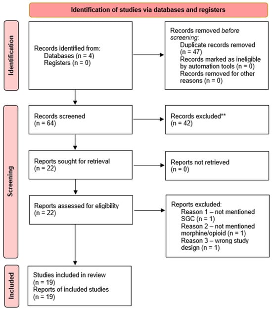
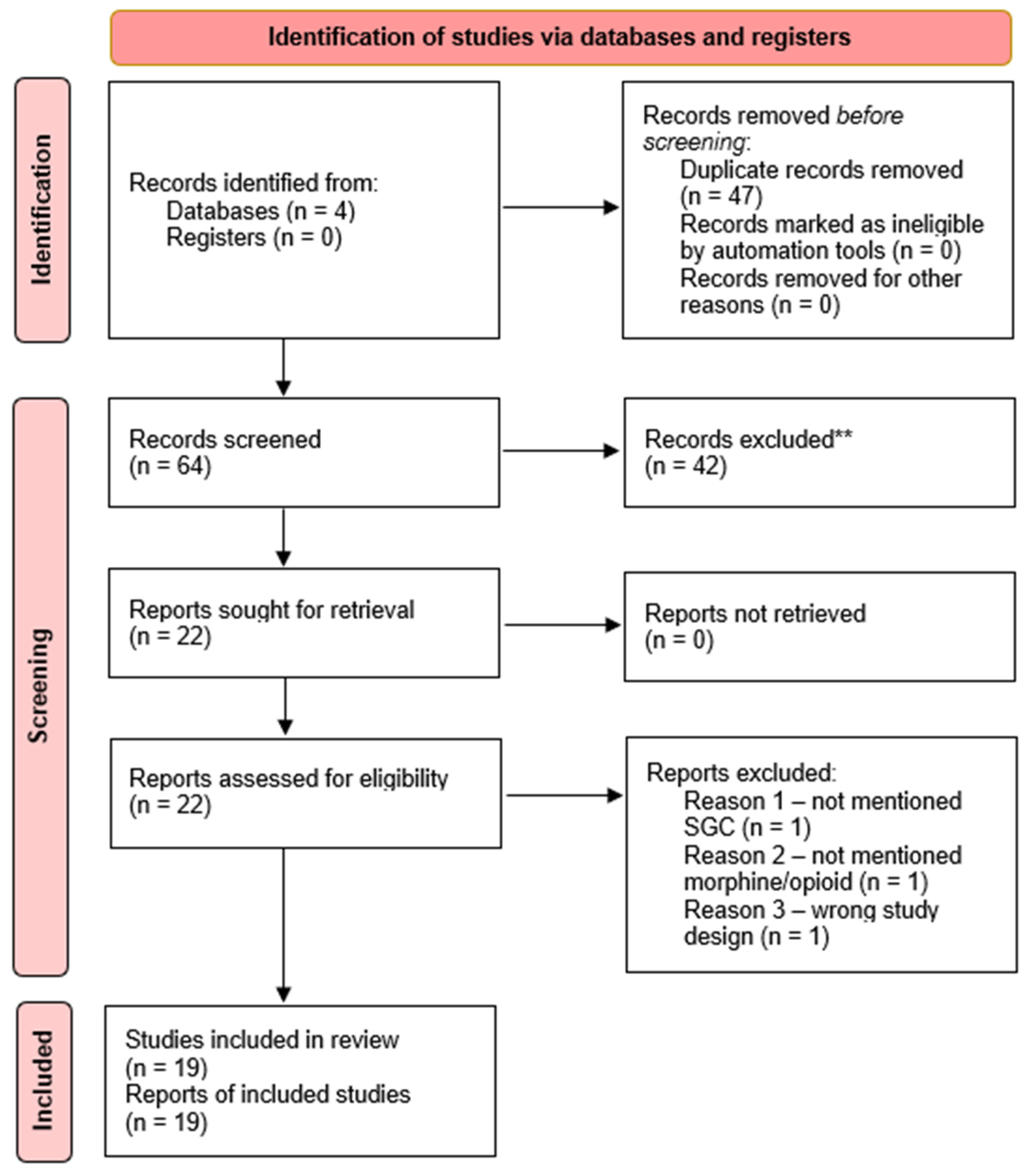
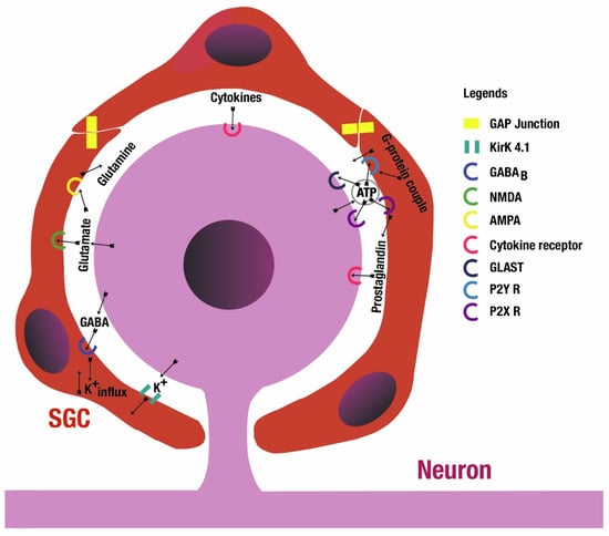
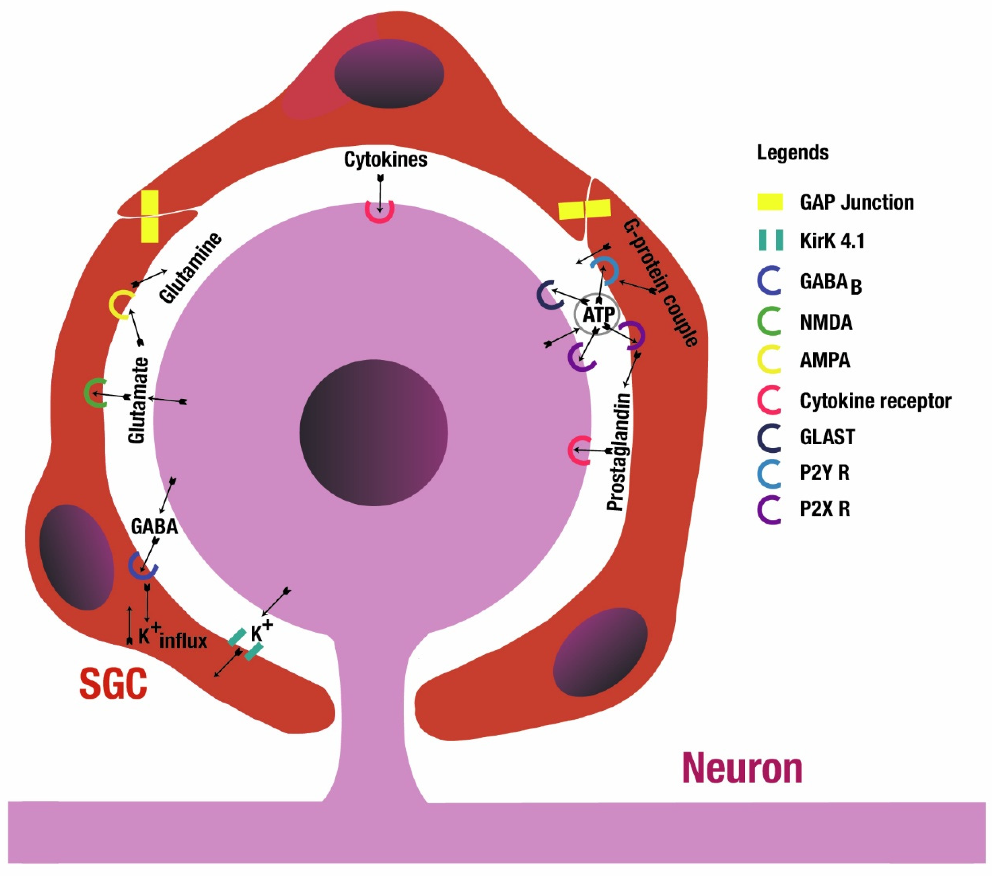

Disclaimer/Publisher’s Note: The statements, opinions and data contained in all publications are solely those of the individual author(s) and contributor(s) and not of MDPI and/or the editor(s). MDPI and/or the editor(s) disclaim responsibility for any injury to people or property resulting from any ideas, methods, instructions or products referred to in the content. |
© 2026 by the authors. Licensee MDPI, Basel, Switzerland. This article is an open access article distributed under the terms and conditions of the Creative Commons Attribution (CC BY) license.