
Review

Parkinson’s Disease, Microglia, and Extracellular Matrix Remodeling
by
Norma Serrano-García
1,†,
Alexis Ponce-Juárez
2,3,†,
Maximiliano Ganado
4,
Javier Pérez-Villavicencio
1,5 and
Moisés Rubio-Osornio
3,*
1
Department of Neurophysiology, National Institute of Neurology and Neurosurgery, Mexico City 14269, Mexico
2
Doctoral Program in Biomedical Sciences, National Autonomous University of Mexico, Mexico City 09340, Mexico
3
Department of Neurochemistry, National Institute of Neurology and Neurosurgery, Mexico City 14269, Mexico
4
Vivarium of Metropolitan Autonomous University, Xochimilco Campus, Mexico City 04960, Mexico
5
Department of Electrical Engineering, Basic Sciences and Engineering Division, Metropolitan Autonomous University, Iztapalapa Campus, Mexico City 09340, Mexico
*
Author to whom correspondence should be addressed.
These authors contributed equally to this work.
Neuroglia 2026, 7(3), 27; https://doi.org/10.3390/neuroglia7030027
Submission received: 18 June 2026 / Revised: 22 July 2026 / Accepted: 25 July 2026 / Published: 3 August 2026

Abstract

Parkinson’s disease (PD) is a progressive neurodegenerative disorder characterized by the selective loss of dopaminergic neurons in the substantia nigra pars compacta (SNpc) and the intracellular accumulation of alpha-synuclein (α-syn) aggregates. Historically, research has focused on neuronal mechanisms; however, growing evidence indicates that the progression of neurodegeneration is influenced by changes in the brain microenvironment, particularly through the dynamic interplay between microglia and the extracellular matrix (ECM). ECM in the central nervous system is an organized network of structural proteins, glycoproteins, and proteoglycans that encases neurons and glial cells, regulating processes such as synaptic stability, neural plasticity, and intercellular signaling. In PD, the aggregation of α-syn and neuronal damage induce sustained microglial activation, which can alter ECM structure. Activated microglia release proteases, including matrix metalloproteinases and cathepsins, which can degrade critical ECM components such as collagens, laminins, and proteoglycans. This remodeling can modify synaptic architecture, regulate cellular signaling, and disrupt neuron-glia interactions, fostering an environment conducive to dopaminergic degeneration. Furthermore, ECM remodeling and microglial activation exhibit regional variability within the brain. Regions notably prone to degeneration, such as the SNpc and striatum, display significant alterations in matrix organization and inflammatory activity, while other dopaminergic regions, including the ventral tegmental area, show increased resilience. We suggest that microglia-mediated ECM remodeling serves as a mechanistic link between neuroinflammation and neuronal susceptibility in PD. This review consolidates the existing knowledge on microglial modulation of ECM dynamics during neurodegeneration, explores regional differences in these processes, and evaluates their significance as possible treatment targets.

1. Introduction

Parkinson’s disease (PD) ranks as the second most common neurodegenerative disorder globally, marked by the progressive degeneration of dopaminergic neurons in the substantia nigra pars compacta (SNpc). This degeneration leads to diminished dopamine levels in basal nuclei circuits and manifests as motor symptoms, including bradykinesia, rigidity, and resting tremor [1]. The disease is marked by the intracellular accumulation of alpha-synuclein(α-syn)-rich aggregates that form Lewy bodies, leading to neuronal dysfunction and death [2,3]. Historically, PD research has concentrated on intrinsic neural processes such as oxidative stress, mitochondrial dysfunction, increased iron content, defective protein degradation, and α-syn toxicity [4,5]. Nonetheless, mounting data suggests that neurodegeneration is significantly affected by changes in the adjacent brain microenvironment [6,7]. In this scenario, glial cells and extracellular components have become essential regulators of neuronal homeostasis and disease advancement [8]. Importantly, ECM homeostasis and neuroinflammatory regulation are not exclusively microglia-dependent processes; astrocytes, endothelial cells, and other neurovascular unit components also actively contribute to the maintenance of the perineuronal and perivascular matrix, establishing a multicellular regulatory network rather than a single-cell-type axis [9]. Microglia are the intrinsic immune cells of the central nervous system (CNS), crucial for immune surveillance, debris clearance, and the regulation of synaptic plasticity [10]. Under physiological conditions, microglia perform essential homeostatic functions, including activity-dependent synaptic pruning and trophic support, which are indispensable for normal neurodevelopment and CNS maintenance. In pathogenic situations like PD, prolonged microglial activation can induce persistent neuroinflammation and lead to neuronal damage [6,11]. In addition to their immune roles, microglia are involved in the modulation of the brain’s extracellular matrix (ECM), a sophisticated network of structural proteins, glycoproteins, and proteoglycans that encases neurons and glial cells, governing cell adhesion, synaptic plasticity, and intercellular communication [12,13,14]. Persistent microglial activation during dementia may provoke substantial ECM remodeling, compromising synaptic integrity and neuronal survival [15]. Recent research indicates that the interplay between microglia and the ECM may considerably affect the selective susceptibility of dopaminergic neurons in PD [16]. This review seeks to consolidate the existing evidence on microglial modulation of ECM dynamics in neurodegenerative processes, investigate regional variations linked to these mechanisms, and assess their potential role in selective dopaminergic degeneration and therapeutic strategies.

2. The Microglia-Extracellular Matrix Axis in the Pathogenesis of Parkinson’s Disease

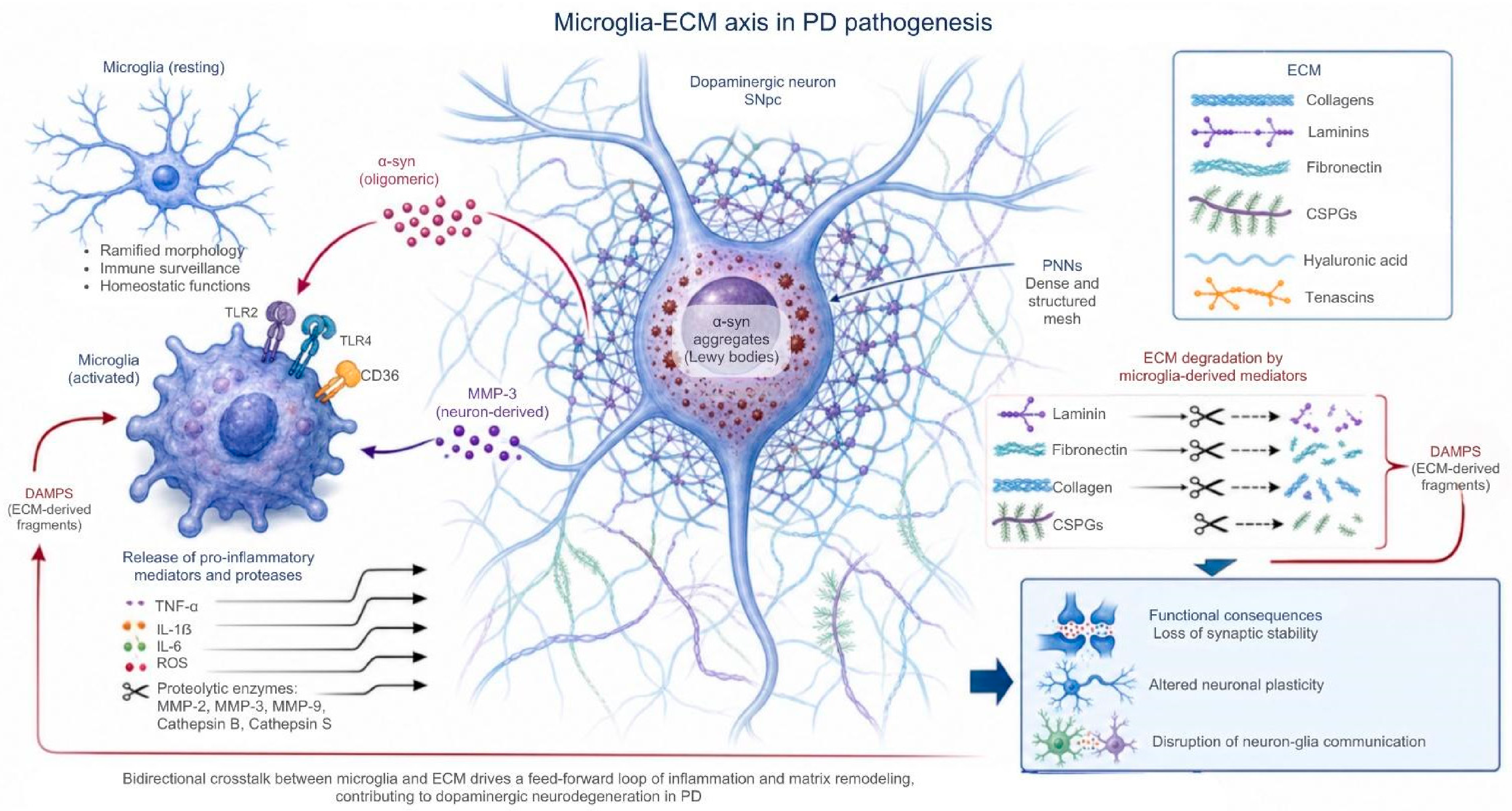
PD is characterized by the gradual degeneration of dopaminergic neurons, aggregation of α-syn, and persistent neuroinflammation [1,17,18]. Recent evidence suggests that dopaminergic neurodegeneration is significantly affected by dynamic changes in the interaction between microglia and ECM, which governs synaptic homeostasis, neuronal plasticity, and inflammatory responses [12,19,20]. Rather than a unidirectional process in which microglia solely act upon the ECM, this relationship is bidirectional: ECM composition and stiffness also modulate microglial phenotype, motility, and phagocytic capacity, establishing a continuous feedback loop between the two compartments. Microglia are the main innate immune cells of the CNS and play a dual role in Parkinson’s disease. Initially, they facilitate the removal of misfolded proteins and cellular detritus; however, prolonged activation induces chronic inflammatory responses that aggravate dopaminergic damage [21,22]. Postmortem analyses of PD patients have demonstrated elevated HLA-DR expression and altered microglial morphology in the SNpc, indicating ongoing immunological activation linked to neuronal loss [23,24]. Positron emission tomography (PET) neuroimaging investigations utilizing translocator protein (TSPO) ligands have corroborated the presence of active neuroinflammation in many brain regions of PD patients [25]. The cerebral ECM comprises glycoproteins, proteoglycans, collagens, laminins, and hyaluronic acid, which governs tissue architecture, synaptic stability, and neuronal communication [12,13]. Under healthy conditions, microglia facilitate ECM homeostasis by engaging in synaptic remodeling and modulating extracellular proteases [26,27]. Key molecular pathways mediate this interaction, including integrin signaling (particularly β1-integrins and the microglial CD11b/CD18 complex), focal adhesion kinase (FAK) activation, and CD44-hyaluronan binding, which collectively regulate microglial adhesion, migration, and mechanosensing within the ECM. In PD, the buildup of α-syn and oxidative stress disturb this equilibrium, fostering prolonged inflammatory activation [6,28]. Experimental investigations indicate that extracellular α-syn oligomers stimulate microglia via TLR2, TLR4, and CD36 receptors [29,30,31]. This activation results in the secretion of cytokines, reactive oxygen species (ROS), and proteolytic enzymes that modify ECM structure. Animal models utilizing 1-methyl-4-phenyl-1,2,3,6-tetrahydropyridine (MPTP) [32] and 6-hydroxydopamine (6-OHDA) [33] exhibit heightened reactive microglia and atypical ECM remodeling in nigrostriatal areas, indicating that ECM disruption is a crucial element of neurodegeneration [34,35,36]. Matrix metalloproteinases (MMP-2, MMP-3, and MMP-9) serve as key mediators of the microglia-ECM axis, which is involved in ECM degradation, inflammatory signaling, and disruption of the blood–brain barrier [37,38,39]. MMP-3, produced by compromised dopaminergic neurons, can activate microglia and enhance the production of TNF-α and IL-1β, perpetuating a cycle of inflammation and neuronal destruction. Moreover, ECM degradation generates damage-associated molecular patterns (DAMPs), which act on microglial cells and further exacerbate neuroinflammation (Figure 1) [32,40,41]. Pharmacological suppression of MMPs in murine models of Parkinson’s disease decreases dopaminergic neuronal degeneration and attenuates inflammatory responses [42,43]. In cerebrospinal fluid samples from patients with this disease, tissue inhibitors of MMPs are elevated [44]. Transcriptomic and proteomic analyses of tissue from PD patients and induced pluripotent stem cell models demonstrate substantial modifications in ECM-receptor interaction pathways, focal adhesion signaling, and cell adhesion molecules, such as collagens, fibronectin, laminins, and tenascins [16,45,46]. While these findings support a mechanistic association between MMP-mediated ECM remodeling and PD pathology, notably, most evidence remains correlative, derived primarily from experimental models and cross-sectional patient samples; causal directionality has not yet been established.

3. Microglia, Extracellular Matrix Reorganization, and Dissemination of Neurodegeneration

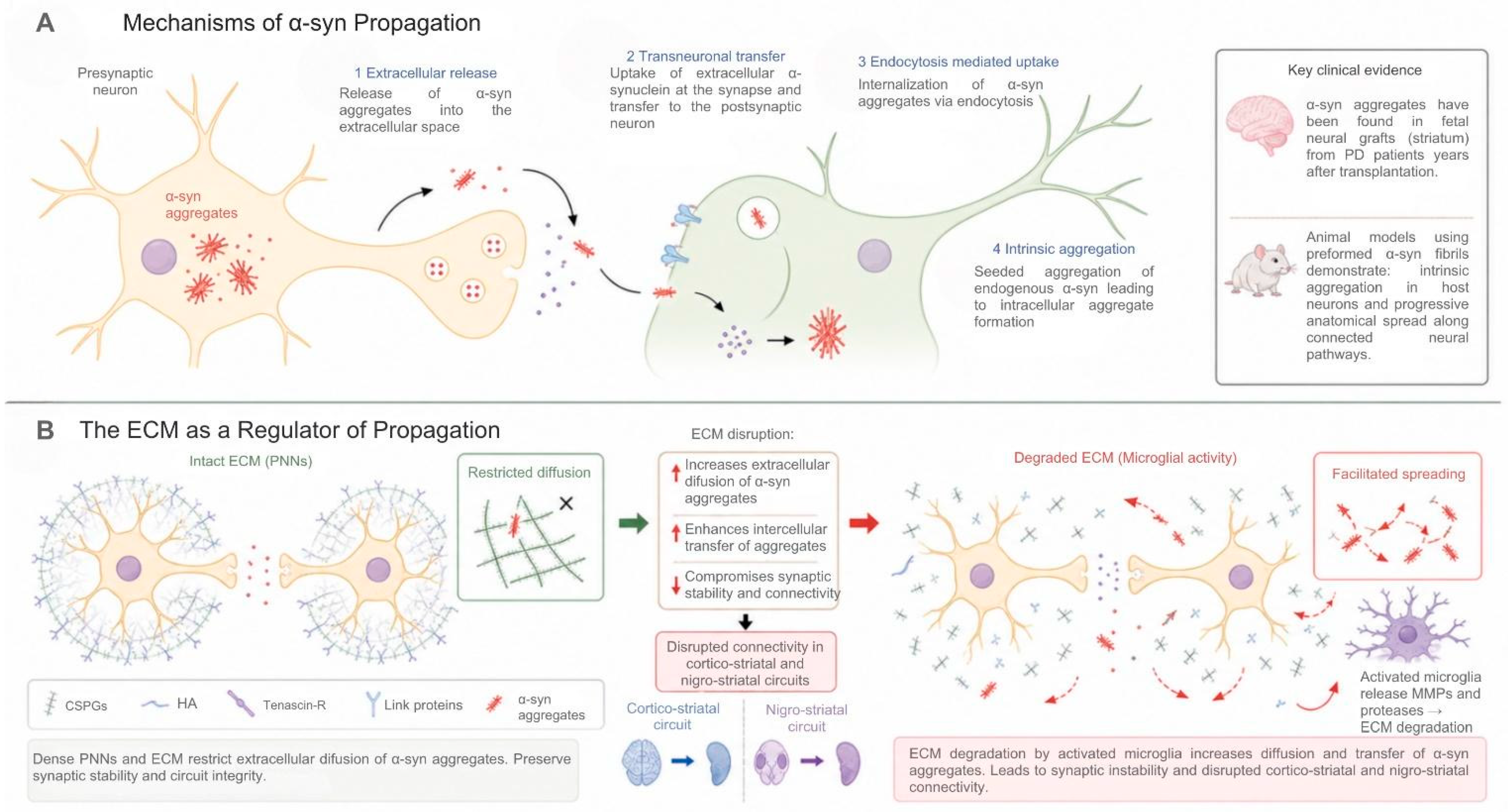
The advancement of PD entails a dynamic neurodegenerative mechanism characterized by the dissemination of α-syn pathology throughout interconnected neural circuits [47,48]. Experimental investigations indicate that α-syn aggregates can propagate by prion-like mechanisms (Figure 2A), including extracellular release, transneuronal transfer, and endocytosis-mediated uptake [49,50]. Animal models implanted with premade α-syn fibrils demonstrate intrinsic aggregation, degeneration of nigrostriatal neurons, and gradual anatomical spread of disease [51,52]. Clinical evidence corroborating this method encompasses the identification of α-syn aggregates in fetal neural grafts implanted in PD patients [53,54]. Chronic microglial activation considerably facilitates disease progression. PET imaging and postmortem analyses reveal persistent inflammatory activation in the SNpc, striatum, and other brain regions of PD patients, which correlated with elevated cytokine production and oxidative stress [25,55,56]. Extracellular α-syn activates TLR2, TLR4, and CD36 receptors in microglia, promoting release of TNF-α, IL-1β, IL-6, and ROS [57,58,59]. Activated microglia expresses proteases that degrade ECM structural components, including proteoglycans, tenascin-R, and hyaluronan [60,61]. Disruption of the ECM may enhance the diffusion and intercellular transfer of α-syn aggregation (Figure 2B), hence augmenting pathogenic connections within neuronal circuits [62]. The disruption of perineuronal nets (PNNs) undermines synaptic stability and neuronal plasticity, leading to the progressive decline of cortico-striatal and nigrostriatal connections [34,63]. Collectively, these observations support a self-perpetuating feed-forward loop in which α-synuclein aggregation triggers microglial activation, microglia-driven ECM remodeling facilitates further α-synuclein propagation between neurons, and the resulting neurodegeneration in turn amplifies local inflammatory signaling—establishing a cycle that may sustain disease progression independently of the initial triggering event.

4. Alterations in Microglial Extracellular Matrix Interactions in Parkinson’s Disease

Transcriptomic and proteomic analyses have revealed substantial modifications in ECM architecture and ECM-receptor interaction pathways in brain regions impacted by PD [16,45]. These changes include dysregulation of collagens, laminins, fibronectin, proteoglycans, and metalloproteinases involved in extracellular architecture and neuroglial signaling [62]. These alterations are particularly evident in regions with severe dopaminergic degeneration, such as the SNpc and striatum, where reactive microgliosis and remodeling of PNNs are also observed [63,64]. This bidirectional relationship extends to specific molecular mediators: TGF-β and TIMP signaling, for instance, not only regulate ECM turnover but are themselves modulated by ECM-derived cues, illustrating how matrix composition can feed back onto microglial functional states. The ECM regulates cell adhesion, microglial migration, synaptic architecture, and inflammatory signaling, all of which are disrupted during chronic neuroinflammation [65]. Experimental evidence indicates that altered microglia-ECM interactions promote a pro-inflammatory microenvironment that exacerbates dopaminergic degeneration through cytokine release, oxidative stress, and extracellular proteolysis [40,66,67].

5. Microglial Remodeling of the Extracellular Matrix During Pathological Processes

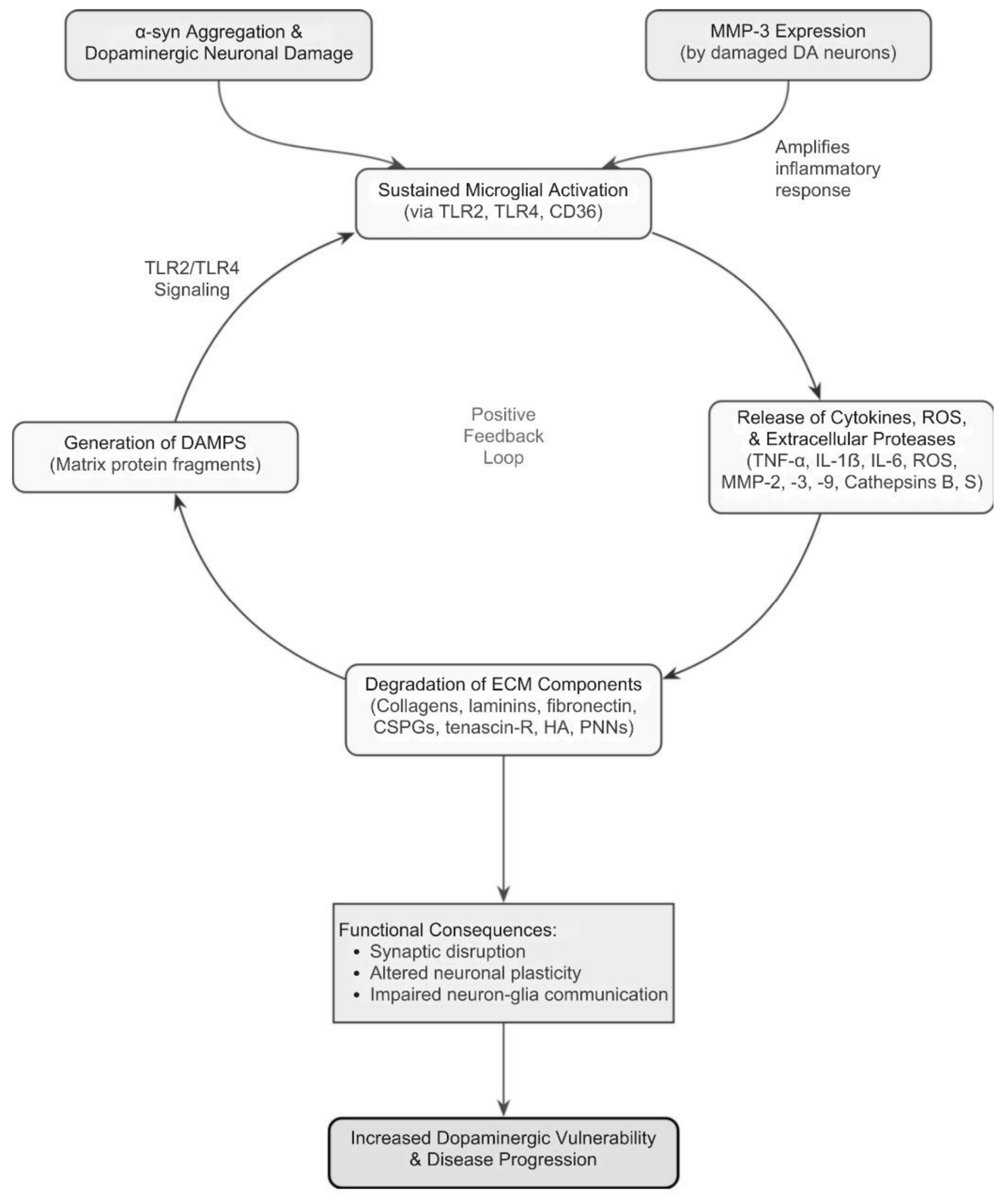
In PD, α-syn aggregation induces persistent microglial activation, one of the principal neuroinflammatory mechanisms associated with dopaminergic degeneration. Activated microglia release TNF-α, IL-1β, IL-6, ROS, and proteolytic enzymes that disrupt brain microenvironment homeostasis [17,68]. Among these enzymes are MMP-2, MMP-3, MMP-9, cathepsin B, and cathepsin S, which degrade laminin, fibronectin, collagen, and chondroitin sulfate proteoglycans [69]. Experimental studies demonstrate that fibrillar α-syn induces MMP-3 expression in both microglia and dopaminergic neurons, thereby promoting inflammatory and neurotoxic pathways [70]. Elevated levels of MMP-3 and MMP-9 have also been detected in PD patients [71,72]. This implies that ECM degradation disrupts the perineuronal environment and impairs communication between neurons and glial cells. It is important to emphasize that ECM remodeling is not limited to structural degradation via matrix metalloproteinases; it also entails functional reorganization of the extracellular environment—altering the diffusion of signaling molecules, ion buffering capacity, and synaptic stability—thereby directly influencing neuronal plasticity and glia-neuron communication. Furthermore, fragmentation of matrix proteins generates DAMPs, which can activate TLR2 and TLR4 signaling in glial cells and amplifying neuroinflammation [73]. Taken together, these findings suggest a potential positive feedback loop in which inflammation promotes ECM remodeling and ECM disruption perpetuates glial activation, although this circular causality has been inferred largely from correlative and cross-sectional data rather than longitudinal mechanistic evidence. ECM remodeling also affects synaptic organization and neuronal plasticity (Figure 3). Alterations in PNNs and integrin-mediated signaling may contribute to the progressive functional decline observed in PD [74,75].

6. Regional Differences in Dopaminergic Vulnerability

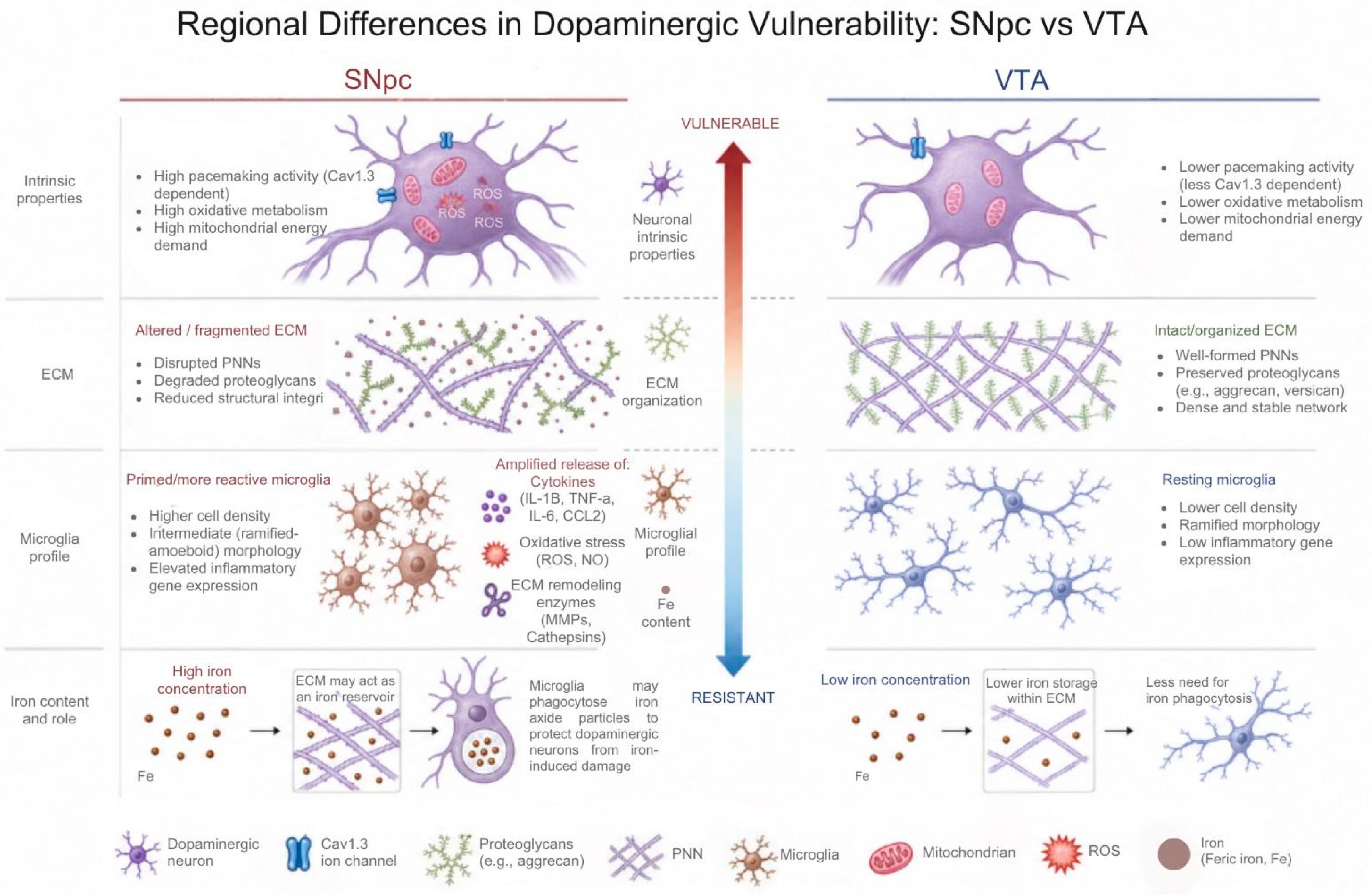
One of the defining neuropathological features of PD is the selective vulnerability of dopaminergic neurons in the SNpc, whereas neurons in the ventral tegmental area (VTA) exhibit relative resistance to degeneration [76,77]. This indicates that neurodegeneration does not depend on intrinsic neuronal properties and regional microenvironmental characteristics as independent mechanisms, but rather on their dynamic interaction, which jointly determines the cumulative vulnerability threshold of dopaminergic neurons. SNpc neurons exhibit high Cav1.3-dependent pacemaker activity, elevated oxidative metabolism, and increased mitochondrial energy demand. These intrinsic factors are associated with greater susceptibility to oxidative stress and α-syn accumulation [78,79]. In parallel, the ECM regulates trophic signaling, synaptic stability, and inflammatory responses. Regional alterations in proteoglycans, hyaluronan, tenascins, and PNNs may therefore influence neuronal resistance to inflammatory injury [80,81]. Microglia also contribute to regional vulnerability. Transcriptomic and single-cell sequencing studies reveal region-specific molecular profiles that determine differential inflammatory responses to tissue injury and α-syn aggregates [82,83]. Microglia in the substantia nigra display a more reactive basal phenotype and elevated expression of inflammatory genes [84]. Additionally, the SNpc contains relatively a high microglial density, potentially amplifying local cytokine release, oxidative stress, and ECM remodeling through metalloproteinases and cathepsins [85,86]. In contrast, the VTA appears to maintain a more stable extracellular organization and a less inflammatory microglial profile. An important aspect that differentiates the two dopaminergic regions is iron concentration. The VTA contains significantly less iron than the SNpc [87,88], which coexists with large amounts of neuromelanin, making it much more prone to cellular degeneration compared to the VTA [89,90]. To eliminate these high concentrations of this metal, the ECM may act as an iron reservoir. It has been proposed to capture iron oxide, potentially protecting dopaminergic neurons from iron-induced damage and promoting cellular repair, based on findings in other neurodegenerative diseases [91,92,93]; however, this iron-buffering role of the ECM remains an emerging hypothesis that warrants further mechanistic validation. Cumulative evidence suggests that regional differences in ECM composition and microglial states contribute substantially to the selective vulnerability of dopaminergic neurons in PD (Figure 4).

7. Therapeutic Implications of the Microglia-Extracellular Matrix Axis

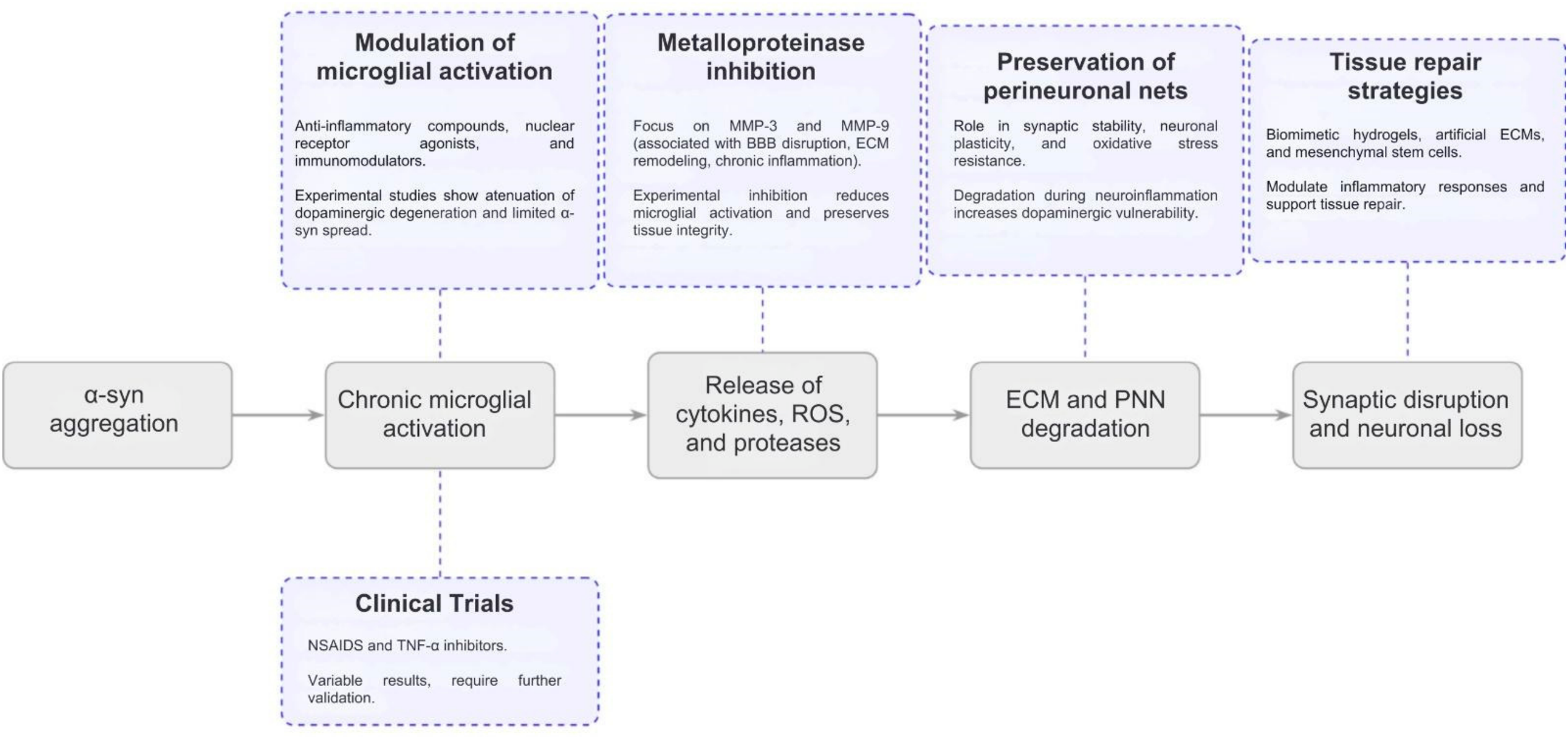
Recognition of the role of the brain microenvironment in PD has generated a major conceptual shift in therapeutic research. Traditionally, treatments focused primarily on dopamine replacement or direct neuronal protection; however, increasing evidence indicates that interactions among microglia, neuroinflammation, and ECM may be significantly influencing disease progression [94,95]. One promising therapeutic strategy involves regulating microglial activation to reduce the release of inflammatory cytokines, ROS, and proteolytic enzymes that contribute to ECM degradation and neuronal injury [96,97,98]. Experimental studies show that anti-inflammatory compounds, nuclear receptor agonists, and immunomodulators can attenuate dopaminergic degeneration and limit α-syn propagation in animal models [99,100]. Clinical trials evaluating nonsteroidal anti-inflammatory drugs and TNF-α inhibitors have produced variable results and require further validation [101,102]. The inconsistent outcomes of anti-inflammatory clinical trials in PD likely reflect several converging factors: disease heterogeneity, including variable degrees of neuroinflammation across patients and disease stages; treatment timing, since interventions initiated after substantial neurodegeneration may fail to modify an already-established disease trajectory; and the difficulty of selectively modulating detrimental microglial responses without compromising their neuroprotective and reparative functions. Another important strategy focuses on the regulation of MMP activity, particularly MMP-3 and MMP-9, which are associated with blood–brain barrier disruption, ECM remodeling, and chronic inflammation [103,104]. Experimental inhibition of MMPs reduces microglial activation and preserves tissue integrity in preclinical models [40,105]. Preservation of PNNs represents another emerging therapeutic approach. These ECM structures regulate synaptic stability, neuronal plasticity, and resistance to oxidative stress. Their degradation during neuroinflammation may increase dopaminergic vulnerability [13,106]. Recent studies have also explored tissue-repair strategies, such as biomimetic hydrogels, artificial extracellular matrices, and mesenchymal stem cells, aimed at modulating inflammatory responses and supporting tissue repair (Figure 5) [107]. Most of these strategies, including MMP modulation, PNN preservation, and ECM-restorative biomaterials, remain at preclinical or early experimental stages. Their translational potential is constrained by the difficulty of achieving spatial and temporal specificity (e.g., inhibiting pathological MMP activity without disrupting physiological ECM turnover), and by the risk of adverse effects arising from interference with microglial functions that are also required for normal tissue homeostasis and repair. In sum, the current evidence supports an integrated view of PD in which neurodegeneration arises from complex interactions among neurons, glia, and ECM components. Targeting the microglia-ECM axis may therefore represent a complementary strategy capable of slowing disease progression and preserving neuronal function.

8. Future Perspectives

Despite substantial progress in understanding the interaction between microglia and the ECM in PD, several key mechanisms remain incompletely understood. One of the principal unresolved questions concerns the precise molecular pathways through which microglia regulate ECM composition and structural organization across distinct brain regions. Clarifying these mechanisms will be essential to understanding how the brain microenvironment contributes to the selective vulnerability of dopaminergic neurons. An important area for future investigation is the functional heterogeneity of microglia throughout CNS. Recent transcriptomic and single-cell studies demonstrate that microglia exhibit region-specific molecular and metabolic profiles that vary according to anatomical location and pathological context. These regional differences may influence both the inflammatory response to α-syn accumulation and the capacity of microglia to remodel the ECM. Beyond regional microglial heterogeneity, future work should also address temporal shifts in microglial phenotype during disease progression and aging, as microglial priming and senescence may differentially influence ECM remodeling capacity over time. Another major challenge is to determine how ECM alterations affect the propagation of α-syn pathology. Because ECM regulates extracellular diffusion, synaptic connectivity, and cell–cell communication, changes in its composition may influence the intercellular transmission of aggregated proteins and the anatomical progression of neurodegeneration. Understanding these mechanisms may provide novel insights into disease progression and selective neuronal susceptibility. Emerging technologies, including spatial transcriptomics, single-cell RNA sequencing, and ECM proteomics, offer unprecedented opportunities to investigate the brain microenvironment with high spatial and molecular resolution. These approaches may facilitate the identification of novel therapeutic targets and improve our understanding of the complex interactions among microglia, ECM components, and neuronal populations during neurodegeneration. Finally, greater integration between experimental and clinical research will be necessary to translate mechanistic findings into therapeutic applications. Although animal and cellular models have provided valuable information regarding microglia-ECM interactions, many of these mechanisms still require validation in human tissues and longitudinal clinical studies. Combining experimental models, postmortem analyses, biomarker studies, and advanced neuroimaging techniques will likely provide a more comprehensive understanding of PD pathophysiology.

9. Limitations of Current Evidence

Despite increasing evidence supporting the involvement of the ECM axis in PD, several limitations currently restrict a complete understanding of its precise role in neurodegeneration. First, a substantial proportion of the available evidence derives from experimental animal models and in vitro systems, including MPTP, 6-OHDA, and α-syn fibril-based models. Although these approaches have provided important mechanistic insights, they do not fully reproduce the chronic and multifactorial nature of human PD. Consequently, caution is required when extrapolating experimental findings to the clinical setting. An additional limitation concerns the genetic models commonly used to study Parkinson’s disease (e.g., SNCA, LRRK2, PARK2 mutants), which often fail to fully recapitulate the ECM and microglial alterations observed in idiopathic disease. Human induced pluripotent stem cell (iPSC)-derived microglia and midbrain organoid systems represent a complementary translational platform, allowing for the study of human-specific microglia-ECM interactions in a genetically tractable context. Another important limitation is that much of the current evidence remains correlative rather than directly causal. Numerous transcriptomic, proteomic, and neuropathological studies demonstrate alterations in ECM organization and microglial activation in PD; however, it remains difficult to determine whether these changes constitute primary drivers of neurodegeneration or secondary consequences of neuronal injury and chronic inflammation. Similarly, alterations in ECM composition may vary substantially according to disease stage, anatomical region, and methodological approach. The intrinsic complexity and heterogeneity of the brain ECM also represent experimental and analytical methodological challenges. The ECM is a highly dynamic structure composed of multiple proteins, glycoproteins, proteoglycans, and PNNs that differ across brain regions and cellular microenvironments. Current molecular and transcriptomic approaches may not fully capture the spatial organization, biomechanical properties, or functional interactions of ECM components in vivo. In parallel, microglial heterogeneity remains incompletely understood. Recent single-cell studies demonstrate the existence of region-specific and disease-associated microglial phenotypes; however, the precise functional relevance of these subpopulations during PD progression remains under investigation. Finally, the translational application of the microglia-ECM axis is still limited by the absence of robust and specific clinical biomarkers. Although neuroimaging studies and inflammatory markers provide indirect evidence of microglial activation and ECM remodeling, reliable biomarkers capable of monitoring these processes in patients remain insufficiently validated. Additional longitudinal studies integrating experimental models, human tissue analysis, advanced imaging, and molecular profiling will therefore be necessary to clarify the clinical relevance of microglia-mediated ECM remodeling in PD.

10. Conclusions

Parkinson’s disease is a complex neurodegenerative disorder in which neuronal degeneration is strongly influenced by interactions within the brain microenvironment. Although PD has traditionally been studied from a neuron-centered perspective, increasing evidence indicates that microglia and ECM play fundamental roles in the regulation of neuroinflammation, synaptic stability, and dopaminergic vulnerability. It is important to situate this microglia-ECM axis within the broader multicellular neuroglial environment. Astrocytes contribute substantially to ECM homeostasis through the synthesis of core matrix components (e.g., tenascin-C, brevican), participate in neuroinflammatory regulation via cytokine and chemokine release, maintain glutamate clearance through EAAT1/2 transporters, and support neurovascular integrity—collectively shaping the microenvironment in which microglia-ECM interactions unfold. Microglia, often described as the resident macrophages of the CNS, support neuronal function under physiological conditions through several homeostatic mechanisms, including the regulation of neurogenesis, activity-dependent synaptic pruning, provision of trophic support, and recruitment of peripheral immune cells during injury. However, chronic microglial activation promotes sustained inflammatory responses and pathological ECM remodeling through the release of cytokines, ROS, and extracellular proteases. These alterations may disrupt neuron–glia communication, impair synaptic organization, and facilitate the progression of α-syn pathology. In parallel, regional differences in ECM composition and microglial phenotypes appear to contribute to the selective vulnerability of dopaminergic neurons observed in PD. Experimental and clinical studies demonstrate that the microglia-ECM axis is closely related to disease progression; however, further studies are needed to understand whether it is a cause of dopaminergic damage or a consequence. Consequently, therapeutic strategies aimed at modulating neuroinflammation, preserving ECM integrity, or restoring microenvironmental homeostasis may represent promising complementary approaches for slowing dopaminergic degeneration. A more comprehensive understanding of the interactions among microglia, ECM components, and neuronal circuits will be essential for developing future therapies capable not only of alleviating symptoms, but also of modifying disease progression in PD. Despite increasing evidence indicating a role for the microglia-ECM axis in PD, much of the existing research is correlational and predominantly sourced from experimental models rather than direct mechanistic evidence in patients.

Author Contributions

Investigation, Original Draft Preparation & Writing, N.S.-G.; Investigation, Original Draft Preparation & Writing, A.P.-J.; Review, Editing & Data Curation, M.G.; Review, Editing & Data Curation, J.P.-V. Conceptualization, Review & Editing M.R.-O. All authors have read and agreed to the published version of the manuscript.

Funding

This study did not receive any external financial support.

Institutional Review Board Statement

Not applicable.

Informed Consent Statement

Not applicable.

Data Availability Statement

No new data were created or analyzed in this study. Data sharing is not applicable to this article.

Acknowledgments

We sincerely thank the National Institute of Neurology and Neurosurgery for the institutional support provided throughout this work.

Conflicts of Interest

The authors state that they have no conflicts of interest to declare.

Abbreviations

α-synalpha-synuclein
Cav1.3L-type calcium channel
CNSCentral Nervous System
CSPGsChondroitin Sulfate Proteoglycans
DAMPsDamage-Associated Molecular Patterns
ECMExtracellular Matrix
EAAT1excitatory amino acid transporter 1
EAAT2excitatory amino acid transporter 2
FAKFocal adhesion kinase
HLA-DRHuman Leukocyte Antigen—DR isotype
IL-1βInterleukin-1 Beta
IL-6Interleukin-6
iPSCHuman induced pluripotent stem cell
LRRK2 Leucine-rich repeat kinase 2
MMPsMatrix Metalloproteinases
MMP-2Matrix Metalloproteinase-2
MMP-3Matrix Metalloproteinase-3
MMP-9Matrix Metalloproteinase-9
MPTP1-methyl-4-phenyl-1,2,3,6-tetrahydropyridine
PARK2gene encoding the Parkin 2 protein
PDParkinson’s Disease
PETPositron Emission Tomography
PNNsPerineuronal Nets
ROSReactive Oxygen Species
SNCAgene encoding alpha-synuclein
SNpcSubstantia Nigra Pars Compacta
TGF-βTransforming growth factor beta
TIMP Tissue Inhibitor of Metalloproteinases
TLR2Toll-Like Receptor 2
TLR4Toll-Like Receptor 4
TNF-αTumor Necrosis Factor Alpha
TLR2Toll type 2-like receptors
TLR4Toll type 4-like receptors
TSPOTranslocator Protein
VTAVentral Tegmental Area
6-OHDA6-hydroxydopamine

References

  1. Poewe, W.; Seppi, K.; Tanner, C.M.; Halliday, G.M.; Brundin, P.; Volkmann, J.; Schrag, A.E.; Lang, A.E. Parkinson disease. Nat. Rev. Dis. Prim. 2017, 3, 17013. [Google Scholar] [CrossRef] [Scilit]
  2. Kalia, L.V.; Lang, A.E. Parkinson’s disease. Lancet 2015, 386, 896–912. [Google Scholar] [PubMed]
  3. Kaya, Z.B.; Amerna, D.; Susarla, A.; Lim, M.J.; Bregendahl, M.; Sekiya, H.; DeTure, M.; Ross, O.A.; Dickson, D.W.; Boschen, S.L.; et al. Molecular profiling of alpha-synuclein pathology and seeding activity in Parkinson’s disease. Acta Neuropathol. 2026, 151, 54. [Google Scholar] [CrossRef] [Scilit] [PubMed]
  4. Michel, P.P.; Hirsch, E.C.; Hunot, S. Understanding Dopaminergic Cell Death Pathways in Parkinson Disease. Neuron 2016, 90, 675–691. [Google Scholar] [CrossRef] [Scilit] [PubMed]
  5. Zhang, N.; Yan, Z.; Xin, H.; Shao, S.; Xue, S.; Cespuglio, R.; Wang, S. Relationship among α-synuclein, aging and inflammation in Parkinson’s disease (Review). Exp. Ther. Med. 2023, 27, 23. [Google Scholar] [CrossRef] [Scilit] [PubMed]
  6. Block, M.L.; Zecca, L.; Hong, J.S. Microglia-mediated neurotoxicity: Uncovering the molecular mechanisms. Nat. Rev. Neurosci. 2007, 8, 57–69. [Google Scholar] [CrossRef] [Scilit] [PubMed]
  7. Yildirim-Balatan, C.; Fenyi, A.; Besnault, P.; Gomez, L.; Sepulveda-Diaz, J.E.; Michel, P.P.; Melki, R.; Hunot, S. Parkinson’s disease-derived α-synuclein assemblies combined with chronic-type inflammatory cues promote a neurotoxic microglial phenotype. J. Neuroinflamm. 2024, 21, 54. [Google Scholar] [CrossRef] [Scilit] [PubMed]
  8. Salter, M.W.; Stevens, B. Microglia emerge as central players in brain disease. Nat. Med. 2017, 23, 1018–1027. [Google Scholar] [CrossRef] [Scilit] [PubMed]
  9. Manu, D.R.; Slevin, M.; Barcutean, L.; Forro, T.; Boghitoiu, T.; Balasa, R. Astrocyte Involvement in Blood-Brain Barrier Function: A Critical Update Highlighting Novel, Complex, Neurovascular Interactions. Int. J. Mol. Sci. 2023, 24, 17146. [Google Scholar] [CrossRef] [Scilit] [PubMed]
  10. Zhan, Y.; Paolicelli, R.C.; Sforazzini, F.; Weinhard, L.; Bolasco, G.; Pagani, F.; Vyssotski, A.L.; Bifone, A.; Gozzi, A.; Ragozzino, D.; et al. Deficient neuron-microglia signaling results in impaired functional brain connectivity and social behavior. Nat. Neurosci. 2014, 17, 400–406. [Google Scholar] [CrossRef] [Scilit] [PubMed]
  11. Shukuri, M.; Uchino, M.; Sakamaki, T.; Onoe, S.; Hosoi, R.; Todoroki, K.; Arano, Y.; Sakai, T.; Akizawa, H.; Inoue, O. Ex vivo imaging and analysis of ROS generation correlated with microglial activation in rat model with acute neuroinflammation induced by intrastriatal injection of LPS. Biochem. Biophys. Res. Commun. 2021, 584, 101–106. [Google Scholar] [CrossRef] [Scilit] [PubMed]
  12. Frantz, C.; Stewart, K.M.; Weaver, V.M. The extracellular matrix at a glance. J. Cell Sci. 2010, 123, 4195–4200. [Google Scholar] [CrossRef] [Scilit] [PubMed]
  13. Sorg, B.A.; Berretta, S.; Blacktop, J.M.; Fawcett, J.W.; Kitagawa, H.; Kwok, J.C.; Miquel, M. Casting a Wide Net: Role of Perineuronal Nets in Neural Plasticity. J. Neurosci. 2016, 36, 11459–11468. [Google Scholar] [CrossRef] [Scilit] [PubMed]
  14. Tewari, B.P.; Woo, A.M.; Prim, C.E.; Chaunsali, L.; Patel, D.C.; Kimbrough, I.F.; Engel, K.; Browning, J.L.; Campbell, S.L.; Sontheimer, H. Astrocytes require perineuronal nets to maintain synaptic homeostasis in mice. Nat. Neurosci. 2024, 27, 1475–1488. [Google Scholar] [CrossRef] [Scilit] [PubMed]
  15. Stoyanov, S.; Sun, W.; Düsedau, H.P.; Cangalaya, C.; Choi, I.; Mirzapourdelavar, H.; Baidoe-Ansah, D.; Kaushik, R.; Neumann, J.; Dunay, I.R.; et al. Attenuation of the extracellular matrix restores microglial activity during the early stage of amyloidosis. Glia 2021, 69, 182–200. [Google Scholar] [PubMed]
  16. Chapman, M.A.; Sorg, B.A. Systematic Review of Extracellular Matrix-Related Alterations in Parkinson’s Disease. Brain Sci. 2024, 14, 522. [Google Scholar] [CrossRef] [Scilit] [PubMed]
  17. Hirsch, E.C.; Hunot, S. Neuroinflammation in Parkinson’s disease: A target for neuroprotection? Lancet Neurol. 2009, 8, 382–397. [Google Scholar] [CrossRef] [Scilit] [PubMed]
  18. Yang, L.; Guo, C.; Zhu, J.; Feng, Y.; Chen, W.; Feng, Z.; Wang, D.; Sun, S.; Lin, W.; Wang, Y. Increased Levels of Pro-Inflammatory and Anti-Inflammatory Cellular Responses in Parkinson’s Disease Patients: Search for a Disease Indicator. Med. Sci. Monit. 2017, 23, 2972–2978. [Google Scholar] [CrossRef] [Scilit] [PubMed]
  19. Tansey, M.G.; Romero-Ramos, M. Immune system responses in Parkinson’s disease: Early and dynamic. Eur. J. Neurosci. 2019, 49, 364–383. [Google Scholar] [PubMed]
  20. Wang, S.; Wang, S.; Chen, H.; Xu, J. Microglia-Neuron Crosstalk: An Intimate Molecular Conversation in Neurodegeneration. Int. J. Mol. Sci. 2026, 27, 2011. [Google Scholar] [CrossRef] [Scilit] [PubMed]
  21. Schapansky, J.; Nardozzi, J.D.; LaVoie, M.J. The complex relationships between microglia, alpha-synuclein, and LRRK2 in Parkinson’s disease. Neuroscience 2015, 302, 74–88. [Google Scholar] [CrossRef] [Scilit] [PubMed]
  22. Tansey, M.G.; Wallings, R.L.; Houser, M.C.; Herrick, M.K.; Keating, C.E.; Joers, V. Inflammation and immune dysfunction in Parkinson disease. Nat. Rev. Immunol. 2022, 22, 657–673. [Google Scholar] [CrossRef] [Scilit] [PubMed]
  23. McGeer, P.L.; Itagaki, S.; Boyes, B.E.; McGeer, E.G. Reactive microglia are positive for HLA-DR in the substantia nigra of Parkinson’s and Alzheimer’s disease brains. Neurology 1988, 38, 1285–1291. [Google Scholar] [CrossRef] [Scilit] [PubMed]
  24. Amin, J.; Holmes, C.; Dorey, R.B.; Tommasino, E.; Casal, Y.R.; Williams, D.M.; Dupuy, C.; Nicoll, J.A.R.; Boche, D. Neuroinflammation in dementia with Lewy bodies: A human post-mortem study. Transl. Psychiatry 2020, 10, 267. [Google Scholar] [CrossRef] [Scilit] [PubMed]
  25. Gerhard, A.; Pavese, N.; Hotton, G.; Turkheimer, F.; Es, M.; Hammers, A.; Eggert, K.; Oertel, W.; Banati, R.B.; Brooks, D.J. In vivo imaging of microglial activation with [11C](R)-PK11195 PET in idiopathic Parkinson’s disease. Neurobiol. Dis. 2006, 21, 404–412. [Google Scholar] [CrossRef] [Scilit] [PubMed]
  26. Crapser, J.D.; Arreola, M.A.; Tsourmas, K.I.; Green, K.N. Microglia as hackers of the matrix: Sculpting synapses and the extracellular space. Cell. Mol. Immunol. 2021, 18, 2472–2488. [Google Scholar] [CrossRef] [Scilit] [PubMed]
  27. Dityatev, A.; Schachner, M.; Sonderegger, P. The dual role of the extracellular matrix in synaptic plasticity and homeostasis. Nat. Rev. Neurosci. 2010, 11, 735–746. [Google Scholar] [CrossRef] [Scilit] [PubMed]
  28. Cardinale, A.; Calabrese, V.; de Iure, A.; Picconi, B. Alpha-Synuclein as a Prominent Actor in the Inflammatory Synaptopathy of Parkinson’s Disease. Int. J. Mol. Sci. 2021, 22, 6517. [Google Scholar] [CrossRef] [Scilit] [PubMed]
  29. Kim, C.; Ho, D.H.; Suk, J.E.; You, S.; Michael, S.; Kang, J.; Joong Lee, S.; Masliah, E.; Hwang, D.; Lee, H.J.; et al. Neuron-released oligomeric α-synuclein is an endogenous agonist of TLR2 for paracrine activation of microglia. Nat. Commun. 2013, 4, 1562. [Google Scholar] [CrossRef] [Scilit] [PubMed]
  30. Tang, Y.; Le, W. Differential Roles of M1 and M2 Microglia in Neurodegenerative Diseases. Mol. Neurobiol. 2016, 53, 1181–1194. [Google Scholar] [PubMed]
  31. Mavroeidi, P.; Xilouri, M. Neurons and Glia Interplay in α-Synucleinopathies. Int. J. Mol. Sci. 2021, 22, 4994. [Google Scholar] [CrossRef] [Scilit] [PubMed]
  32. Chung, Y.C.; Kim, Y.S.; Bok, E.; Yune, T.Y.; Maeng, S.; Jin, B.K. MMP-3 contributes to nigrostriatal dopaminergic neuronal loss, BBB damage, and neuroinflammation in an MPTP mouse model of Parkinson’s disease. Mediat. Inflamm. 2013, 2013, 370526. [Google Scholar] [CrossRef] [Scilit] [PubMed]
  33. Barata-Antunes, S.; Teixeira, F.G.; Mendes-Pinheiro, B.; Domingues, A.V.; Vilaça-Faria, H.; Marote, A.; Silva, D.; Sousa, R.A.; Salgado, A.J. Impact of aging on the 6-OHDA-induced rat model of Parkinson’s disease. Int. J. Mol. Sci. 2020, 21, 3459. [Google Scholar] [CrossRef] [Scilit] [PubMed]
  34. Freitas, A.; Aroso, M.; Barros, A.; Fernández, M.; Conde-Sousa, E.; Leite, M.; Carvalho, E.D.; Ribeiro, C.C.; Ferreira, R.; Pêgo, A.P.; et al. Characterization of the Striatal Extracellular Matrix in a Mouse Model of Parkinson’s Disease. Antioxidants 2021, 10, 1095. [Google Scholar] [CrossRef] [Scilit] [PubMed]
  35. Simola, N.; Morelli, M.; Cartam, A.R. The 6-Hydroxydopamine model of Parkinson’s disease. Neurotox. Res. 2007, 11, 151–167. [Google Scholar] [CrossRef] [Scilit] [PubMed]
  36. Freire, M.A.M.; Rocha, G.S.; Lemos, N.A.M.; Lima, R.R.; Bittar, S.; Jenkins, L.B.; Falcao, D.; Steinbusch, H.W.M.; Santos, J.R. Insights into Parkinson’s Disease Pathology Focusing on Glial Response and Apoptosis in a Classic Rat Model of Dopaminergic Degeneration. Neuroglia 2025, 6, 36. [Google Scholar] [CrossRef] [Scilit]
  37. Rosenberg, G.A. Matrix metalloproteinases and their multiple roles in neurodegenerative diseases. Lancet Neurol. 2009, 8, 205–216. [Google Scholar] [CrossRef] [Scilit] [PubMed]
  38. Annese, V.; Herrero, M.T.; Di Pentima, M.; Gomez, A.; Lombardi, L.; Ros, C.M.; De Pablos, V.; Fernandez-Villalba, E.; De Stefano, M.E. Metalloproteinase-9 contributes to inflammatory glia activation and nigro-striatal pathway degeneration in both mouse and monkey models of 1-methyl-4-phenyl-1,2,3,6-tetrahydropyridine (MPTP)-induced Parkinsonism. Brain Struct. Funct. 2015, 220, 703–727. [Google Scholar] [PubMed]
  39. Gu, H.W.; Xing, F.; Jiang, M.J.; Wang, Y.; Bai, L.; Zhang, J.; Li, T.T.; Zhang, W.; Xu, J.T. Upregulation of matrix metalloproteinase-9/2 in the wounded tissue, dorsal root ganglia, and spinal cord is involved in the development of postoperative pain. Brain Res. 2019, 1718, 64–74. [Google Scholar] [CrossRef] [Scilit] [PubMed]
  40. Kim, Y.S.; Kim, S.S.; Cho, J.J.; Choi, D.H.; Hwang, O.; Shin, D.H.; Chun, H.S.; Beal, M.F.; Joh, T.H. Matrix metalloproteinase-3: A novel signaling proteinase from apoptotic neuronal cells that activates microglia. J. Neurosci. 2005, 25, 3701–3711. [Google Scholar] [CrossRef] [Scilit] [PubMed]
  41. Cho, Y.; Son, H.J.; Kim, E.M.; Choi, J.H.; Kim, S.T.; Ji, I.J.; Choi, D.H.; Joh, T.H.; Kim, Y.S.; Hwang, O. Doxycycline is neuroprotective against nigral dopaminergic degeneration by a dual mechanism involving MMP-3. Neurotox. Res. 2009, 16, 361–371. [Google Scholar] [CrossRef] [Scilit] [PubMed]
  42. Ruan, Z.; Zhang, D.; Huang, R.; Sun, W.; Hou, L.; Zhao, J.; Wang, Q. Microglial Activation Damages Dopaminergic Neurons through MMP-2/-9-Mediated Increase of Blood-Brain Barrier Permeability in a Parkinson’s Disease Mouse Model. Int. J. Mol. Sci. 2022, 23, 2793. [Google Scholar] [CrossRef] [Scilit] [PubMed]
  43. Kim, T.; Jeon, J.; Park, J.S.; Park, Y.; Kim, J.; Noh, H.; Kim, H.S.; Seo, H. Matrix Metalloproteinase-8 Inhibitor Ameliorates Inflammatory Responses and Behavioral Deficits in LRRK2 G2019S Parkinson’s Disease Model Mice. Biomol. Ther. 2021, 29, 483–491. [Google Scholar] [CrossRef] [Scilit] [PubMed]
  44. Lorenzl, S.; Albers, D.S.; LeWitt, P.A.; Chirichigno, J.W.; Hilgenberg, S.L.; Cudkowicz, M.E.; Beal, M.F. Tissue inhibitors of matrix metalloproteinases are elevated in cerebrospinal fluid of neurodegenerative diseases. J. Neurol. Sci. 2003, 207, 71–76. [Google Scholar] [CrossRef] [Scilit] [PubMed]
  45. Rike, W.A.; Stern, S. Proteins and Transcriptional Dysregulation of the Brain Extracellular Matrix in Parkinson’s Disease: A Systematic Review. Int. J. Mol. Sci. 2023, 24, 7435. [Google Scholar] [CrossRef] [Scilit] [PubMed]
  46. Novoseletskaya, E.S.; Evdokimov, P.V.; Efimenko, A.Y. Extracellular matrix-induced signaling pathways in mesenchymal stem/stromal cells. Cell Commun. Signal. 2023, 21, 244. [Google Scholar] [CrossRef] [Scilit] [PubMed]
  47. Braak, H.; Del Tredici, K.; Rüb, U.; de Vos, R.A.; Jansen Steur, E.N.; Braak, E. Staging of brain pathology related to sporadic Parkinson’s disease. Neurobiol. Aging 2003, 24, 197–211. [Google Scholar] [CrossRef] [Scilit] [PubMed]
  48. Del Tredici, K.; Braak, H. Review: Sporadic Parkinson’s disease: Development and distribution of α-synuclein pathology. Neuropathol. Appl. Neurobiol. 2016, 42, 33–50. [Google Scholar] [CrossRef] [Scilit] [PubMed]
  49. Luk, K.C.; Kehm, V.; Carroll, J.; Zhang, B.; O’Brien, P.; Trojanowski, J.Q.; Lee, V.M. Pathological α-synuclein transmission initiates Parkinson-like neurodegeneration in nontransgenic mice. Science 2012, 338, 949–953. [Google Scholar] [CrossRef] [Scilit] [PubMed]
  50. Volpicelli-Daley, L.A.; Luk, K.C.; Patel, T.P.; Tanik, S.A.; Riddle, D.M.; Stieber, A.; Meaney, D.F.; Trojanowski, J.Q.; Lee, V.M. Exogenous α-synuclein fibrils induce Lewy body pathology leading to synaptic dysfunction and neuron death. Neuron 2011, 72, 57–71. [Google Scholar] [CrossRef] [Scilit] [PubMed]
  51. Fonseca-Ornelas, L.; Viennet, T.; Rovere, M.; Jiang, H.; Liu, L.; Nuber, S.; Ericsson, M.; Arthanari, H.; Selkoe, D.J. Altered conformation of α-synuclein drives dysfunction of synaptic vesicles in a synaptosomal model of Parkinson’s disease. Cell Rep. 2021, 36, 109333. [Google Scholar] [CrossRef] [Scilit] [PubMed]
  52. Paumier, K.L.; Luk, K.C.; Manfredsson, F.P.; Kanaan, N.M.; Lipton, J.W.; Collier, T.J.; Steece-Collier, K.; Kemp, C.J.; Celano, S.; Schulz, E.; et al. Intrastriatal injection of pre-formed mouse α-synuclein fibrils into rats triggers α-synuclein pathology and bilateral nigrostriatal degeneration. Neurobiol. Dis. 2015, 82, 185–199. [Google Scholar] [CrossRef] [Scilit] [PubMed]
  53. Kordower, J.H.; Chu, Y.; Hauser, R.A.; Freeman, T.B.; Olanow, C.W. Lewy body-like pathology in long-term embryonic nigral transplants in Parkinson’s disease. Nat. Med. 2008, 14, 504–506. [Google Scholar] [CrossRef] [Scilit] [PubMed]
  54. Li, J.Y.; Englund, E.; Holton, J.L.; Soulet, D.; Hagell, P.; Lees, A.J.; Lashley, T.; Quinn, N.P.; Rehncrona, S.; Björklund, A.; et al. Lewy bodies in grafted neurons in subjects with Parkinson’s disease suggest host-to-graft disease propagation. Nat. Med. 2008, 14, 501–503. [Google Scholar] [CrossRef] [Scilit] [PubMed]
  55. Walker, D.G.; Lue, L.F.; Serrano, G.; Adler, C.H.; Caviness, J.N.; Sue, L.I.; Beach, T.G. Altered Expression Patterns of Inflammation-Associated and Trophic Molecules in Substantia Nigra and Striatum Brain Samples from Parkinson’s Disease, Incidental Lewy Body Disease and Normal Control Cases. Front. Neurosci. 2016, 9, 507. [Google Scholar] [CrossRef] [Scilit] [PubMed]
  56. Imamura, K.; Hishikawa, N.; Sawada, M.; Nagatsu, T.; Yoshida, M.; Hashizume, Y. Distribution of major histocompatibility complex class II-positive microglia and cytokine profile of Parkinson’s disease brains. Acta Neuropathol. 2003, 106, 518–526. [Google Scholar] [CrossRef] [Scilit] [PubMed]
  57. Fellner, L.; Irschick, R.; Schanda, K.; Reindl, M.; Klimaschewski, L.; Poewe, W.; Wenning, G.K.; Stefanova, N. Toll-like receptor 4 is required for α-synuclein dependent activation of microglia and astroglia. Glia 2013, 61, 349–360. [Google Scholar] [CrossRef] [Scilit] [PubMed]
  58. Béraud, D.; Hathaway, H.A.; Trecki, J.; Chasovskikh, S.; Johnson, D.A.; Johnson, J.A.; Federoff, H.J.; Shimoji, M.; Mhyre, T.R.; Maguire-Zeiss, K.A. Microglial activation and antioxidant responses induced by the Parkinson’s disease protein α-synuclein. J. Neuroimmune Pharmacol. 2013, 8, 94–117. [Google Scholar] [CrossRef] [Scilit] [PubMed]
  59. Chung, L.Y.; Lin, Y.T.; Liu, C.; Tai, Y.C.; Lin, H.Y.; Lin, C.H.; Chen, C.C. Neuroinflammation upregulated neuronal toll-like receptors 2 and 4 to drive synucleinopathy in neurodegeneration. Front. Pharmacol. 2022, 13, 845930. [Google Scholar] [CrossRef] [Scilit] [PubMed]
  60. Yong, V.W. Metalloproteinases: Mediators of pathology and regeneration in the CNS. Nat. Rev. Neurosci. 2005, 6, 931–944. [Google Scholar] [CrossRef] [Scilit] [PubMed]
  61. Jang, D.G.; Simm, H.J.; Songm, E.K.; Kwonm, T.; Parkm, T.J. Extracellular matrixes and neuroinflammation. BMB Rep. 2020, 53, 491–499. [Google Scholar] [CrossRef] [Scilit] [PubMed]
  62. Pintér, P.; Alpár, A. The Role of Extracellular Matrix in Human Neurodegenerative Diseases. Int. J. Mol. Sci. 2022, 23, 11085. [Google Scholar] [CrossRef] [Scilit] [PubMed]
  63. Brückner, G.; Morawski, M.; Arendt, T. Aggrecan-based extracellular matrix is an integral part of the human basal ganglia circuit. Neuroscience 2008, 51, 489–504. [Google Scholar] [CrossRef] [Scilit] [PubMed]
  64. Zhang, Z.; Niu, K.; Huang, T.; Guo, J.; Xarbat, G.; Gong, X.; Gao, Y.; Liu, F.; Cheng, S.; Su, W.; et al. Microglia depletion reduces neurodegeneration and remodels extracellular matrix in a mouse Parkinson’s disease model triggered by α-synuclein overexpression. npj Park. Dis. 2025, 11, 15. [Google Scholar] [CrossRef] [Scilit] [PubMed]
  65. Samant, R.R.; Standaert, D.G.; Harms, A.S. The emerging role of disease-associated microglia in Parkinson’s disease. Front. Cell Neurosci. 2024, 18, 1476461. [Google Scholar] [CrossRef] [Scilit] [PubMed]
  66. Harms, A.S.; Ferreira, S.A.; Romero-Ramos, M. Periphery and brain, innate and adaptive immunity in Parkinson’s disease. Acta Neuropathol. 2021, 141, 527–545. [Google Scholar] [CrossRef] [Scilit] [PubMed]
  67. Wareham, L.K.; Calkins, D.J. Making tracks: Microglia and the extracellular matrix. Mol. Neurodegener. 2025, 20, 101. [Google Scholar] [CrossRef] [Scilit] [PubMed]
  68. Ishijima, T.; Nakajima, K. Inflammatory cytokines TNFα, IL-1β, and IL-6 are induced in endotoxin- stimulated microglia through different signaling cascades. Sci. Prog. 2021, 104, 368504211054985. [Google Scholar] [CrossRef] [Scilit] [PubMed]
  69. Soles, A.; Selimovic, A.; Sbrocco, K.; Ghannoum, F.; Hamel, K.; Moncada, E.L.; Gilliat, S.; Cvetanovic, M. Extracellular Matrix Regulation in Physiology and in Brain Disease. Int. J. Mol. Sci. 2023, 24, 7049. [Google Scholar] [CrossRef] [Scilit] [PubMed]
  70. Leem, Y.H.; Park, J.S.; Park, J.E.; Kim, D.Y.; Kang, J.L.; Kim, H.S. Papaverine inhibits α-synuclein aggregation by modulating neuroinflammation and matrix metalloproteinase-3 expression in the subacute MPTP/P mouse model of Parkinson’s disease. Biomed. Pharmacother. 2020, 130, 110576. [Google Scholar] [CrossRef] [Scilit] [PubMed]
  71. Lorenzl, S.; Albers, D.S.; Narr, S.; Chirichigno, J.; Beal, M.F. Expression of MMP-2, MMP-9, and MMP-1 and their endogenous counterregulators TIMP-1 and TIMP-2 in postmortem brain tissue of Parkinson’s disease. Exp. Neurol. 2002, 178, 13–20. [Google Scholar] [CrossRef] [Scilit] [PubMed]
  72. Si, X.; Dai, S.; Fang, Y.; Tang, J.; Wang, Z.; Li, Y.; Song, Z.; Chen, Y.; Liu, Y.; Zhao, G.; et al. Matrix metalloproteinase-9 inhibition prevents aquaporin-4 depolarization-mediated glymphatic dysfunction in Parkinson’s disease. J. Adv. Res. 2024, 56, 125–136. [Google Scholar] [CrossRef] [Scilit] [PubMed]
  73. Roh, J.S.; Sohn, D.H. Damage-Associated Molecular Patterns in Inflammatory Diseases. Immune Netw. 2018, 18, e27. [Google Scholar] [CrossRef] [Scilit] [PubMed]
  74. Frischknecht, R.; Gundelfinger, E.D. The brain’s extracellular matrix and its role in synaptic plasticity. Adv. Exp. Med. Biol. 2012, 970, 153–171. [Google Scholar] [CrossRef] [Scilit] [PubMed]
  75. Dankovich, T.M.; Rizzoli, S.O. The Synaptic Extracellular Matrix: Long-Lived, Stable, and Still Remarkably Dynamic. Front. Synaptic. Neurosci. 2022, 14, 854956. [Google Scholar] [CrossRef] [Scilit] [PubMed]
  76. Surmeier, D.J.; Obeso, J.A.; Halliday, G.M. Selective neuronal vulnerability in Parkinson disease. Nat. Rev. Neurosci. 2017, 18, 101–113. [Google Scholar] [CrossRef] [Scilit] [PubMed]
  77. Ni, A.; Ernst, C. Evidence That Substantia Nigra Pars Compacta Dopaminergic Neurons Are Selectively Vulnerable to Oxidative Stress Because They Are Highly Metabolically Active. Front. Cell. Neurosci. 2022, 16, 826193. [Google Scholar] [CrossRef] [Scilit] [PubMed]
  78. Pacelli, C.; Giguère, N.; Bourque, M.J.; Lévesque, M.; Slack, R.S.; Trudeau, L.É. Elevated Mitochondrial Bioenergetics and Axonal Arborization Size Are Key Contributors to the Vulnerability of Dopamine Neurons. Curr. Biol. 2015, 25, 2349–2360. [Google Scholar] [CrossRef] [Scilit] [PubMed]
  79. Verma, A.; Ravindranath, V. CaV1.3 L-Type Calcium Channels Increase the Vulnerability of Substantia Nigra Dopaminergic Neurons in MPTP Mouse Model of Parkinson’s Disease. Front. Aging Neurosci. 2020, 11, 382. [Google Scholar] [CrossRef] [Scilit] [PubMed]
  80. Chelyshev, Y.A.; Kabdesh, I.M.; Mukhamedshina, Y.O. Extracellular Matrix in Neural Plasticity and Regeneration. Cell Mol. Neurobiol. 2022, 42, 647–664. [Google Scholar] [CrossRef] [Scilit] [PubMed]
  81. Melrose, J.; Hayes, A.J.; Bix, G. The CNS/PNS Extracellular Matrix Provides Instructive Guidance Cues to Neural Cells and Neuroregulatory Proteins in Neural Development and Repair. Int. J. Mol. Sci. 2021, 22, 5583. [Google Scholar] [CrossRef] [Scilit] [PubMed]
  82. Grabert, K.; Michoel, T.; Karavolos, M.H.; Clohisey, S.; Baillie, J.K.; Stevens, M.P.; Freeman, T.C.; Summers, K.M.; McColl, B.W. Microglial brain region-dependent diversity and selective regional sensitivities to aging. Nat. Neurosci. 2016, 19, 504–516. [Google Scholar] [CrossRef] [Scilit] [PubMed]
  83. Mirzac, D.; Glaser, M.B.; Kreis, S.L.; Ringel, F.; Bange, M.; Herz, D.M.; Groppa, S.; Rotaru, L.; Almeida, V.; Blech, J.; et al. Cortical Single-Cell Primers of Abnormal Brain Activity in Parkinson’s Disease. Research 2025, 8, 0863. [Google Scholar] [CrossRef] [Scilit] [PubMed]
  84. Lawson, L.J.; Perry, V.H.; Dri, P.; Gordon, S. Heterogeneity in the distribution and morphology of microglia in the normal adult mouse brain. Neuroscience 1990, 39, 151–170. [Google Scholar] [CrossRef] [Scilit] [PubMed]
  85. Kim, Y.S.; Choi, D.H.; Block, M.L.; Lorenzl, S.; Yang, L.; Kim, Y.J.; Sugama, S.; Cho, B.P.; Hwang, O.; Browne, S.E.; et al. A pivotal role of matrix metalloproteinase-3 activity in dopaminergic neuronal degeneration via microglial activation. FASEB J. 2007, 21, 179–187. [Google Scholar] [CrossRef] [Scilit] [PubMed]
  86. Choudhury, M.E.; Ozaki, S.; Miyaue, N.; Matsuura, T.; Mikami, K.; Islam, A.; Kubo, M.; Ando, R.; Yano, H.; Kunieda, T.; et al. Chloride Intracellular Channel Protein 2 Promotes Microglial Invasion: A Link to Microgliosis in the Parkinson’s Disease Brain. Brain Sci. 2022, 13, 55. [Google Scholar] [CrossRef] [Scilit] [PubMed]
  87. Hare, D.J.; Lei, P.; Ayton, S.; Roberts, B.R.; Grimm, R.; George, J.L.; Bishop, D.P.; Beavis, A.D.; Donovan, S.J.; McColl, G.; et al. An iron–dopamine index predicts risk of parkinsonian neurodegeneration in the substantia nigra pars compacta. Chem. Sci. 2014, 5, 2160–2169. [Google Scholar] [CrossRef] [Scilit]
  88. Zhang, D.; Yao, J.; Sun, J.; Wang, J.; Chen, L.; He, H.; Wu, T. Iron accumulation in the ventral tegmental area in Parkinson’s disease. Front. Aging Neurosci. 2023, 15, 1187684. [Google Scholar] [CrossRef] [Scilit] [PubMed]
  89. Faucheux, B.A.; Martin, M.E.; Beaumont, C.; Hauw, J.J.; Agid, Y.; Hirsch, E.C. Neuromelanin associated redox-active iron is increased in the substantia nigra of patients with Parkinson’s disease. J. Neurochem. 2003, 86, 1142–1148. [Google Scholar] [CrossRef] [Scilit] [PubMed]
  90. Jakaria, M.; Cannon, J.R. Neuromelanin-induced cellular stress and neurotoxicity in the pathogenesis of Parkinson’s disease. Apoptosis 2025, 30, 2481–2494. [Google Scholar] [CrossRef] [Scilit] [PubMed]
  91. Stephenson, E.; Nathoo, N.; Mahjoub, Y.; Dunn, J.F.; Yong, V.W. Iron in multiple sclerosis: Roles in neurodegeneration and repair. Nat. Rev. Neurol. 2014, 10, 459–468. [Google Scholar] [CrossRef] [Scilit] [PubMed]
  92. Unbehau, R.; Luthringer-Feyerabend, B.J.C.; Willumeit-Römer, R. The impact of brain cell metabolism and extracellular matrix on magnesium degradation. Acta Biomater. 2020, 116, 426–437. [Google Scholar] [CrossRef] [Scilit] [PubMed]
  93. Reinert, A.; Reinert, T.; Arendt, T.; Morawski, M. High Iron and Iron Household Protein Contents in Perineuronal Net-Ensheathed Neurons Ensure Energy Metabolism with Safe Iron Handling. Int. J. Mol. Sci. 2022, 23, 1634. [Google Scholar] [CrossRef] [Scilit] [PubMed]
  94. Ghorbani, S.; Yong, V.W. The extracellular matrix as modifier of neuroinflammation and remyelination in multiple sclerosis. Brain 2021, 144, 1958–1973. [Google Scholar] [CrossRef] [Scilit] [PubMed]
  95. Nosi, D.; Lana, D.; Giovannini, M.G.; Delfino, G.; Zecchi-Orlandini, S. Neuroinflammation: Integrated Nervous Tissue Response through Intercellular Interactions at the “Whole System” Scale. Cells 2021, 10, 1195. [Google Scholar] [CrossRef] [Scilit] [PubMed]
  96. Du, L.; Zhang, Y.; Chen, Y.; Zhu, J.; Yang, Y.; Zhang, H.L. Role of Microglia in Neurological Disorders and Their Potentials as a Therapeutic Target. Mol. Neurobiol. 2017, 54, 7567–7584. [Google Scholar] [CrossRef] [Scilit] [PubMed]
  97. Jin, X.; Liu, M.Y.; Zhang, D.F.; Zhong, X.; Du, K.; Qian, P.; Gao, H.; Wei, M.J. Natural products as a potential modulator of microglial polarization in neurodegenerative diseases. Pharmacol. Res. 2019, 145, 104253. [Google Scholar] [CrossRef] [Scilit] [PubMed]
  98. Zhang, L.; Wang, Y.; Liu, T.; Mao, Y.; Peng, B. Novel Microglia-based Therapeutic Approaches to Neurodegenerative Disorders. Neurosci. Bull. 2023, 39, 491–502. [Google Scholar] [CrossRef] [Scilit] [PubMed]
  99. Gao, H.M.; Hong, J.S. Why neurodegenerative diseases are progressive: Uncontrolled inflammation drives disease progression. Trends Immunol. 2008, 29, 357–365. [Google Scholar] [CrossRef] [Scilit] [PubMed]
  100. Forloni, G. Alpha Synuclein: Neurodegeneration and Inflammation. Int. J. Mol. Sci. 2023, 24, 5914. [Google Scholar] [CrossRef] [Scilit] [PubMed]
  101. Alrouji, M.; Al-Kuraishy, H.M.; Al-Gareeb, A.I.; Saad, H.M.; Batiha, G.E. A story of the potential effect of non-steroidal anti-inflammatory drugs (NSAIDs) in Parkinson’s disease: Beneficial or detrimental effects. Inflammopharmacology 2023, 31, 673–688. [Google Scholar] [CrossRef] [Scilit] [PubMed]
  102. NINDS Exploratory Trials in Parkinson Disease (NET-PD) FS-ZONE Investigators. Pioglitazone in early Parkinson’s disease: A phase 2, multicentre, double-blind, randomised trial. Lancet Neurol. 2015, 14, 795–803. [PubMed]
  103. Behl, T.; Kaur, G.; Sehgal, A.; Bhardwaj, S.; Singh, S.; Buhas, C.; Judea-Pusta, C.; Uivarosan, D.; Munteanu, M.A.; Bungau, S. Multifaceted Role of Matrix Metalloproteinases in Neurodegenerative Diseases: Pathophysiological and Therapeutic Perspectives. Int. J. Mol. Sci. 2021, 22, 1413. [Google Scholar] [CrossRef] [Scilit] [PubMed]
  104. Narayanan, K.B. Enzyme-Based Anti-Inflammatory Therapeutics for Inflammatory Diseases. Pharmaceutics 2025, 17, 606. [Google Scholar] [CrossRef] [Scilit] [PubMed]
  105. Borjini, N.; Fernandez, M.; Giardino, L.; Sorokin, L.; Calzà, L. Pharmacological Inhibition of Microglial Proliferation Supports Blood-Brain Barrier Integrity in Experimental Autoimmune Encephalomyelitis. Cells 2025, 14, 414. [Google Scholar] [CrossRef] [Scilit] [PubMed]
  106. Fawcett, J.W.; Oohashi, T.; Pizzorusso, T. The roles of perineuronal nets and the perinodal extracellular matrix in neuronal function. Nat. Rev. Neurosci. 2019, 20, 451–465. [Google Scholar] [CrossRef] [Scilit] [PubMed]
  107. Ciuffreda, M.C.; Malpasso, G.; Chokoza, C.; Bezuidenhout, D.; Goetsch, K.P.; Mura, M.; Pisano, F.; Davies, N.H.; Gnecchi, M. Synthetic extracellular matrix mimic hydrogel improves efficacy of mesenchymal stromal cell therapy for ischemic cardiomyopathy. Acta Biomater. 2018, 70, 71–83. [Google Scholar] [CrossRef] [Scilit] [PubMed]
Figure 1. Overview of the microglia–extracellular matrix axis in Parkinson’s disease pathogenesis. In the substantia nigra pars compacta (SNpc), dopaminergic neurons accumulate intracellular α-synuclein (α-syn) aggregates, forming Lewy bodies, and release oligomeric α-syn into the extracellular space. Extracellular α-syn activates microglia through TLR2, TLR4, and CD36 receptors, inducing a transition from a surveilling (ramified) to an activated (amoeboid) phenotype. Activated microglia produce pro-inflammatory cytokines (TNF-α, IL-1β, IL-6), reactive oxygen species (ROS), and proteolytic enzymes, including matrix metalloproteinases (MMP-2, MMP-3, MMP-9) and cathepsins (cathepsin B, cathepsin S). These proteases degrade the main ECM components, including collagens, laminins, fibronectin, and chondroitin sulfate proteoglycans, as well as perineuronal nets (PNNs), highly specialized ECM structures that surround neuronal somas and regulate synaptic stability. Damaged dopaminergic neurons also express MMP-3, which further activates microglia and enhances the production of TNF-α and IL-1β. The degradation of the ECM leads to the generation of damage-associated molecular patterns (DAMPs), which act via TLR2 and TLR4 on glial cells, further exacerbating neuroinflammation. The resultant disruption of the brain microenvironment compromises synaptic architecture, neuronal plasticity, and neuron–glia communication, creating a favorable milieu for dopaminergic degeneration. Note: This schematic integrates findings from multiple experimental systems; the sequence and directionality of some interactions (e.g., the precise contribution of individual proteases to specific ECM components) remain to be validated in vivo.
Neuroglia 07 00027 g001
Figure 2. Prion-like propagation of α-syn and its facilitation by ECM remodeling. (A) Mechanisms of α-syn dissemination across synaptically connected neurons. α-Syn aggregates spread through three described routes: extracellular release, transneuronal transfer, and endocytosis-mediated uptake. Uptake of preformed fibrils by recipient neurons seeds intrinsic aggregation, initiating pathology in previously unaffected cells. This model is supported by animal studies demonstrating progressive anatomical spread of pathology following injection of preformed α-syn fibrils, and by clinical evidence showing α-syn aggregates in fetal neural grafts transplanted into PD patients. (B) ECM integrity as a regulator of α-syn propagation. Left: Intact ECM and well-formed perineuronal nets restrict extracellular diffusion and maintain synaptic stability within corticostriatal and nigrostriatal circuits. Right: Microglia-mediated degradation of proteoglycans, tenascin-R, and hyaluronan disrupts perineuronal nets and loosens the extracellular meshwork, thereby enhancing the extracellular diffusion and intercellular transfer of α-syn aggregates and contributes to progressive loss of circuit connectivity. Note: The causal contribution of ECM/PNN degradation to accelerated α-syn propagation (panel (B)) is supported primarily by in vitro and ex vivo evidence; its relevance in vivo remains to be fully established.
Neuroglia 07 00027 g002
Figure 3. Positive feed-forward loop between neuroinflammation, ECM remodeling and dopaminergic degeneration in Parkinson’s disease. The loop is initiated by α-syn aggregation and dopaminergic neuronal damage that promote sustained microglial activation via TLR2, TLR4 and CD36 receptor activation by extracellular α-syn oligomers. Activated microglia secrete TNF-α, IL-1β, IL-6, ROS and proteolytic enzymes (MMP-2, MMP-3, MMP-9, cathepsin B, cathepsin S) that degrade ECM structural proteins including collagens, laminins, fibronectin, chondroitin sulfate proteoglycans, tenascin-R, hyaluronan and perineuronal nets. Cleavage of these matrix proteins generates DAMPs that activate TLR2 and TLR4 signaling in glial cells, thus perpetuating the inflammatory loop. In parallel, damaged dopaminergic neurons express MMP-3, which directly activates microglia and amplifies TNF-α and IL-1β production, constituting an additional entry point into the cycle. Downstream consequences of sustained ECM remodeling include disruption of synaptic organization, impairment of neuronal plasticity, and compromised neuron–glia communication, collectively increasing dopaminergic vulnerability and driving disease progression Arrows indicate the proposed direction of signaling, secretion, or causal influence between the depicted components; the curved arrow denotes closure of the feed-forward loop via DAMP-mediated TLR2/TLR4 reactivation. Note: This feed-forward loop is inferred from convergent but largely correlative experimental findings; its self-perpetuating dynamics have not been directly demonstrated through longitudinal or causal experimental designs.
Neuroglia 07 00027 g003
Figure 4. Regional differences in dopaminergic vulnerability: SNpc versus VTA. SNpc (left) exhibits specific neuronal features that contribute to increased vulnerability such as Cav1.3-dependent pacemaker activity, high oxidative metabolism, and mitochondrial energy demand, which collectively foster oxidative stress and α-syn accumulation. Microglia in the SNpc displays a more reactive baseline phenotype, characterized by the upregulation of inflammatory genes, a higher cell density, and increased release of cytokines, ROS, and ECM-remodeling proteases (metalloproteinases and cathepsins). ECM organization is significantly altered in this region, with the disruption of PNNs and the breakdown of matrix architecture. Furthermore, the SNpc contains high levels of iron; the ECM has potential as an iron reservoir, and microglia has been proposed to phagocytize iron oxide, potentially protecting dopaminergic neurons from iron-related damage. The VTA (right) is relatively resilient, with a more stable extracellular environment, intact PNNs, a less inflammatory microglial profile with a ramified morphology and lower cell density, and a significantly lower iron content. Note: The proposed iron-buffering role of the ECM and its interaction with microglial iron handling remain hypothetical and are based on indirect evidence from other neurodegenerative conditions.
Neuroglia 07 00027 g004
Figure 5. Therapeutic targets along the microglia–ECM axis in PD. The pathological cascade progresses from α-syn aggregation through chronic microglial activation, release of inflammatory mediators and proteases, and ECM and PNN degradation, to synaptic disruption and neuronal loss. Therapeutic strategies are mapped to specific points of intervention. Modulation of microglial activation includes anti-inflammatory compounds, nuclear receptor agonists, and immunomodulators, which have been shown experimentally to attenuate dopaminergic degeneration and limit α-syn propagation in animal models; clinical trials with nonsteroidal anti-inflammatory drugs and TNF-α inhibitors have yielded variable results that require further validation. Inhibition of metalloproteinase activity targets MMP-3 and MMP-9, whose activity is associated with BBB disruption, ECM remodeling, and chronic inflammation; experimental inhibition reduces microglial activation and preserves tissue integrity in preclinical models. Preservation of PNNs addresses the loss of ECM structures that regulate synaptic stability, neuronal plasticity, and resistance to oxidative stress. Tissue repair strategies include biomimetic hydrogels, artificial extracellular matrices, and mesenchymal stem cells as approaches to modulate inflammatory responses and support tissue repair. Note: Except for MMP inhibition and anti-inflammatory approaches tested in clinical trials, the therapeutic strategies depicted remain experimental, and their positioning in this schematic reflects proposed rather than validated points of intervention.
Neuroglia 07 00027 g005
Disclaimer/Publisher’s Note: The statements, opinions and data contained in all publications are solely those of the individual author(s) and contributor(s) and not of MDPI and/or the editor(s). MDPI and/or the editor(s) disclaim responsibility for any injury to people or property resulting from any ideas, methods, instructions or products referred to in the content.