


Abstract
Endogenous matrix metalloproteinases (MMPs) are activated in dentin during carious lesion progression and restorative procedures, degrading the tooth-restoration interface and contributing to restoration failure. This study investigated by means of in situ zymography whether cold atmospheric plasma activation (PA) of distilled water (DW) and phosphate-buffered saline (PBS) modulates endogenous dentinal MMP activity. A Dielectric Barrier Discharge-rod source generated PA liquids, treating DW and PBS for 22 min. Chemical characterization demonstrated that PADW yielded 9.61 mg/L H2O2, 18.3 mg/L NO2−, 375.70 mg/L NO3−, and a pH of 3.2. PAPBS yielded 9.79 mg/L H2O2, 36.01 mg/L NO2−, 505.74 mg/L NO3−, and a pH of 7.13. Both liquids served as 1 min dentin pretreatments in a simulated restorative procedure using a universal adhesive and resin composite, tested after 24 h. MMP activity was assessed via in situ zymography with fluorescein-conjugated gelatin and confocal microscopy. Data were statistically analyzed (p < 0.05). PADW increased dentinal enzymatic activity, while PAPBS reduced it (p < 0.05). Non-activated PBS elicited higher baseline MMP activity than non-activated DW. The divergent responses likely reflect differences in RONS composition, pH, and initial ionic content between the two liquids. The precise mechanism underlying PA liquid interactions with dentinal MMPs warrants further investigation.
1. Introduction
Cold atmospheric plasma (CAP) technology represents a significant advancement in medical and dental applications [1,2]. CAP operates at near-room temperature, making it suitable for medical applications without causing thermal damage to tissues. Stoffels et al. [3] were pioneers in demonstrating CAP’s efficacy against oral pathogens such as “Streptococcus mutans”, a major contributor to dental caries. This discovery initiated research into promising, yet still largely unexplored, applications of CAP in dental medicine [4].
In dentistry applications, CAP sources are categorized based on their treatment modes: direct or indirect. Direct treatments involve the immediate application of plasma to target tissues or surfaces, whereas indirect treatments utilize plasma-activated media to exert their effects [5,6]. For direct treatments, Dielectric Barrier Discharge (DBD) and Atmospheric Pressure Plasma Jets (APPJ) are predominantly used. DBD plasma generates a uniform, low-temperature plasma suitable for direct application on dental surfaces and tissues, effectively used for disinfection and tissue treatment without causing thermal damage. On the other hand, APPJ produces a focused plasma plume, making it highly effective for localized treatments such as root canal disinfection and tooth whitening [6,7].
Indirect treatments primarily utilize Plasma-Activated Water (PAW) or Plasma-Activated Hydrogels. It is well known that air-operated DBD plasmas generate reactive oxygen and nitrogen species, including excited N2, atomic oxygen and N2+ species, which can subsequently interact with the liquid surface and lead to the formation of both short- and long-lived reactive species in the treated liquid [8]. Contact between CAP and liquid produces dissolved reactive oxygen and nitrogen species (RONS), retaining the antimicrobial, anti-inflammatory and healing properties of plasma [9]. H2O2, NO2−, and NO3− are among the most commonly quantified long-lived RONS in plasma-activated liquids. Unlike transient species, these compounds persist after plasma exposure and contribute to biological effects during the subsequent indirect application of the activated liquid [10].
The antimicrobial properties of PAW are particularly advantageous in dental settings where controlling pathogenic microorganisms is crucial. Studies have demonstrated that PAW effectively reduces biofilms formed by dental pathogens such as ‘Enterococcus faecalis’ and ‘Candida albicans’ [11]. This makes PAW a valuable tool in endodontic treatments by irrigating root canals and enhancing the disinfection process without the cytotoxic effects associated with traditional chemical agents. The ability of PAW to penetrate and disrupt biofilms is particularly beneficial in ensuring thorough disinfection, thereby improving the outcomes of root canal therapies.
In periodontal therapy, PAW manages periodontal diseases, characterized by inflammation and infection of the gums and supporting structures of the teeth. Beyond its well-examined antimicrobial properties, PAW also holds potential in reparative and regenerative processes across various tissues. Its anti-inflammatory properties help modulate the host response, reducing inflammation and promoting tissue regeneration, crucial for healing periodontal tissues [12,13]. By reducing the bacterial load and promoting a healthier environment, PAW aids in managing periodontal diseases and supports the healing process.
In restorative dentistry, PAW disinfects and reduces the surface tension of tooth tissues, enhancing the penetration of adhesive resins into the substrate and consequently the bonding strength and longevity of adhesives and composites [11,14,15]. However, the longevity and quality of dental adhesive restorations are also influenced by other factors, including the properties of the dentinal substrate and the impact of various elements on activating potentially detrimental proteolytic enzymes in dentin.
The activity of dentinal proteolytic enzymes, specifically matrix metalloproteinases (MMPs), plays a crucial role in adhesive dentistry by significantly impacting the durability of dental restorations. MMPs influence the degradation rate of resin–dentin interfaces, underscoring the need for strategies to regulate these enzymes and maintain the integrity of the adhesive interface [16]. To enhance the longevity of resin–dentin bonds, various experimental strategies have been developed by research groups, including: increasing the conversion rate and esterase resistance of hydrophilic adhesives [17,18,19], using collagenolytic enzyme inhibitors [20,21,22], applying collagen cross-linkers to reinforce the dentin matrix [23,24,25,26,27,28], employing ethanol wet-bonding with hydrophobic adhesive resins to improve resin infiltration [16,29], promoting biomimetic remineralization of the hybrid layer [30,31], and incorporating bioactive/antimicrobial dental materials to the adhesive procedures [32,33,34,35,36]. Based on previously collected knowledge, it can be assumed that the chemical changes introduced to PAW through plasma treatment may either enhance or inhibit enzymatic activity, potentially resulting in customized effects of PAW for dental applications.
Our study aimed to explore the previously unexamined influence of PAW treatment on MMP activity in dentin, offering insights into its potential applications in adhesive dentistry. We investigated how cold atmospheric plasma activation (PA) of distilled water (DW) and phosphate-buffered saline (PBS) affects endogenous dentinal enzymatic activity. The null hypothesis was that plasma activation of DW and PBS would not affect the endogenous enzymatic activity of dentin.
2. Materials and Methods
2.1. Dielectric Barrier Discharge (DBD) for the Production of PAW: Electrical Analysis and Chemical Characterization of RONS in PAW
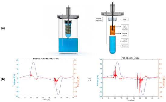
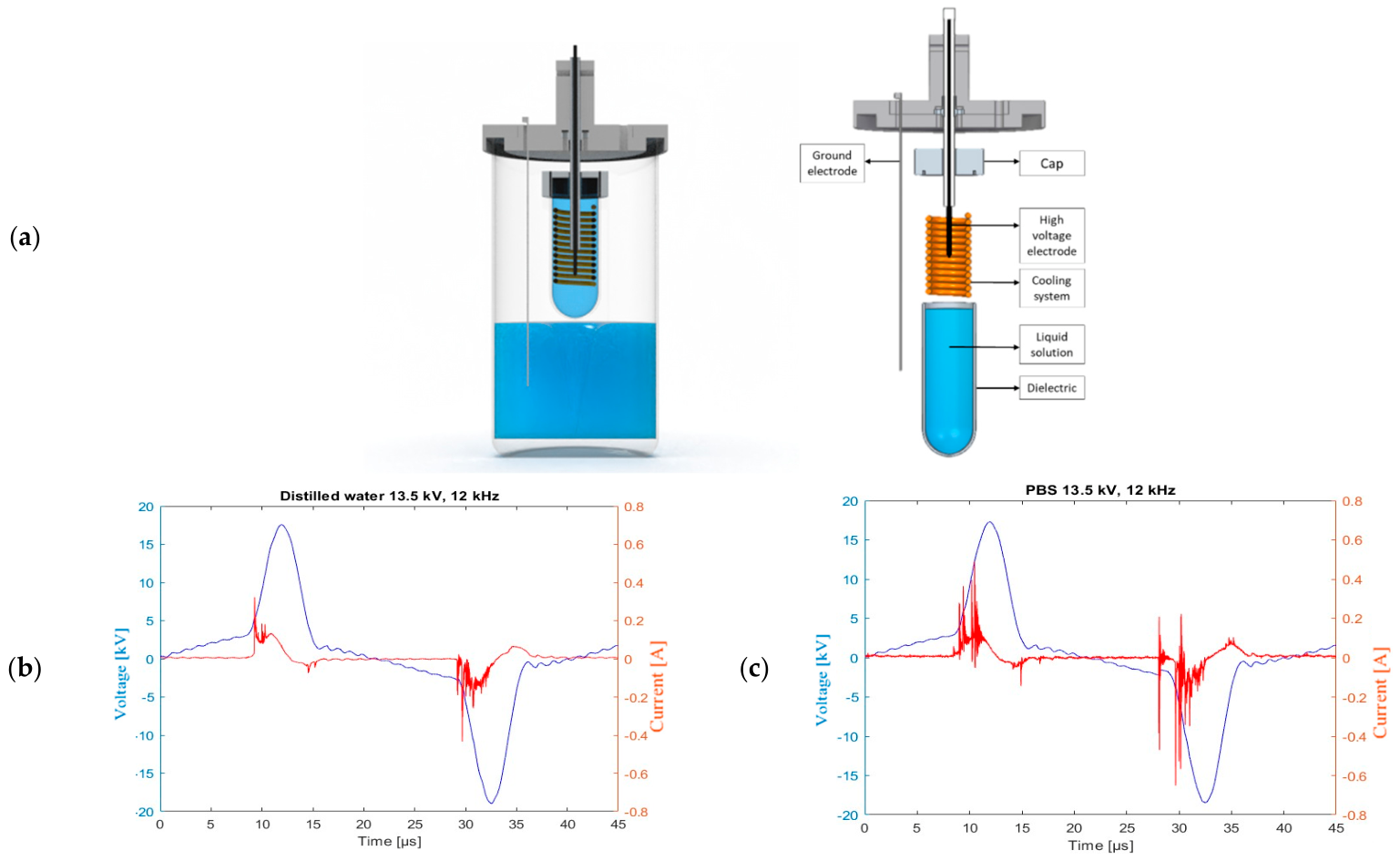
PAW was generated by exposing DW and PBS to a DBD cold plasma source. This source features a high-voltage electrode submerged in an electrically conductive solution housed within a glass tube. The DBD system consisted of a high-voltage metallic electrode immersed in an NaCl-saturated conductive solution contained inside a glass tube acting as a dielectric barrier. The glass tip was positioned above the treated liquid surface, while a grounded stainless steel cylinder was immersed in the liquid to be treated. The system includes a cooling system for the liquid solution (Figure 1). The DBD was positioned vertically 5 mm above the water surface; the liquid was grounded through the immersion of a stainless steel cylinder. Plasma was generated in an ambient air gap between the cylinder tip and the liquid surface. CAP was generated in the air gap between the DBD’s glass tip and the water surface. The high-voltage electrode was connected to a micropulsed high-voltage generator (AlmaPulse, AlmaPlasma s.r.l., Bologna, Italy), operating at 13.5 kV with a fixed frequency of 12 kHz. Voltage (V) and current (i) measurements were obtained using a high-voltage probe (P6015A, Tektronix, Beaverton, OR, USA) and a current probe (6585, Pearson Electronics, Palo Alto, CA, USA), interfaced with a digital oscilloscope (DPO4034, Tektronix, 350 MHz, 2.5 GSa s−1). The average power (P) dissipated in the discharge was calculated using the formula:
Figure 1.
(a) CAP source for PAW production; (b) voltage and current waveforms for distilled water treatment; (c) voltage and current waveforms for PBS treatment.
Concentrations of H2O2, NO2−, NO3− and pH generated in PAW by CAP treatment were quantified immediately after the treatment. The Amplex Red Hydrogen Peroxide Assay Kit (Thermo Fisher Scientific, Waltham, MA, USA) and the Nitrate/Nitrite Colorimetric Assay (ROCHE, Basel, Switzerland) were employed to measure H2O2, NO2−, and NO3−, respectively. The concentration analysis was conducted photometrically as per the manufacturer’s protocols.
2.2. PAW and PAPBS Treatments
A volume of 2 L of DW or PBS (NaCl, KCl, Na2HPO4 and KH2PO4 in water, pH 7.34) was treated with a DBD source for 22 min. During the treatment, the stirrer vortexed the water at 750 rpm.
To evaluate the effect of pH separated from the effect of plasma-generated RONS, DW was adjusted to the same pH value as PADW by dropwise addition of HCl solution (Sigma Aldrich, St. Louis, MO, USA), while PBS was used as a buffered liquid to evaluate the effect of plasma-activated water under neutral pH conditions. All experiments were conducted in ambient air under controlled laboratory conditions, at approximately 25 °C and 50% relative humidity.
2.3. In Situ Zymography of Resin–Dentin Interfaces
Healthy human third molars (n = 5) were sectioned transversally using a water-cooled slow-speed diamond saw (Micromet, Remet, Bologna, Italy) to obtain 1 mm thick sections of mid-coronal dentin. The sections were further cut into 4 parts to test all the groups on the same dental substrate and reduce the biological variability related to the substrate itself. The non-activated as well as freshly activated DW/PBS were applied on dentin as a pretreatment using a microbrush for 1 min in the simulation of a clinical dental restorative procedure using a universal adhesive resin (Scotchbond Universal Plus, Solventum; St Paul, MN, USA) in self-etch mode and a flowable resin composite. The adhesive resin and the resin composite were applied following the manufacturers’ instructions and polymerized with an LED curing light (3M Elipar, Solventum, Kowloon, Hong Kong) for 10 s and 20 s, respectively. The resin–dentin specimens were then stored in artificial saliva at 37 °C for 24 h. Afterward, the specimens were cut into 1 mm thick sticks, glued to microscope glass slides, ground down to approximately 50 μm, polished and subjected to in situ zymography, as presented in detail elsewhere [26]. Briefly, fluorescein-conjugated gelatin mixture (E-12055; Molecular Probes, Eugene, OR, USA) was placed on the specimens overnight in dark and humid conditions at 37 °C. A series of z-stack images (around 15 μm into the depth of the specimens) was made using a confocal microscope (TCS SP2 AOBS, Leica Microsystems GmbH, Wetzlar, Germany). Green fluorescence was assessed as a relative measure of enzymatic activity quantified in ImageJ v. 2.9.0 (National Institutes of Health, Bethesda, MD, USA).
2.4. Statistical Analysis
Since the data were not normally distributed (Shapiro–Wilk p < 0.05), the in situ zymography data were statistically analyzed using Kruskal–Wallis and Dunn’s post hoc tests with the significance level set at p < 0.05. The analysis was performed using SigmaPlot v. 14.0 (Systat Software GmbH, Düsseldorf, Germany).
3. Results
3.1. Electrical Analysis and Chemical Characterization of RONS in PAW
Figure 1b,c illustrates the voltage and current waveforms for the DBD source operating at 13.50 kV and 12 kHz, contingent on the treated liquid. Two voltage peaks with a maximum intensity of 17.50 kV are observed for each half period. The average discharge power, calculated directly from the voltage and current measurements, was 138.60 ± 10.55 W for DW and 154.32 ± 10.31 W for PBS. The higher average discharge power measured during PBS treatment can be attributed to its higher ionic conductivity, which modifies the electrical impedance of the plasma liquid system and increases the current flowing through the discharge [37]. The pH value measured after the treatment was 3.42 ± 0.24 for PADW and 7.11 ± 0.15 for PAPBS. The concentrations of RONS measured post-treatment in PADW were 9.63 ± 0.07 mg/L H2O2, 19.11 ± 1.20 mg/L NO2−, and 375.70 ± 7.2 mg/L NO3−. In contrast, PAPBS treatment reported RONS concentrations of 9.71 ± 0.20 mg/L H2O2, 36.1 ± 0.83 mg/L NO2−, and 505.72 ± 15.26 mg/L NO3−. To eliminate the influence of pH on the differences in the activation of MMPs between the PA and control liquids, both DW and PBS were brought to the pH of their activated counterpart by dropwise addition of HCl solution.
3.2. In Situ Zymography of Resin–Dentin Interface
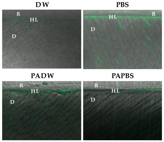
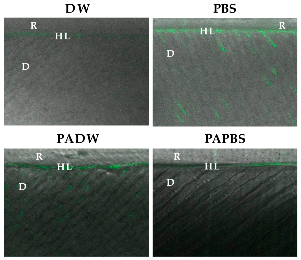
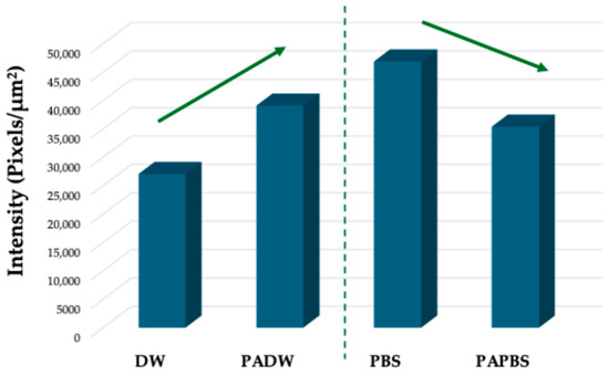
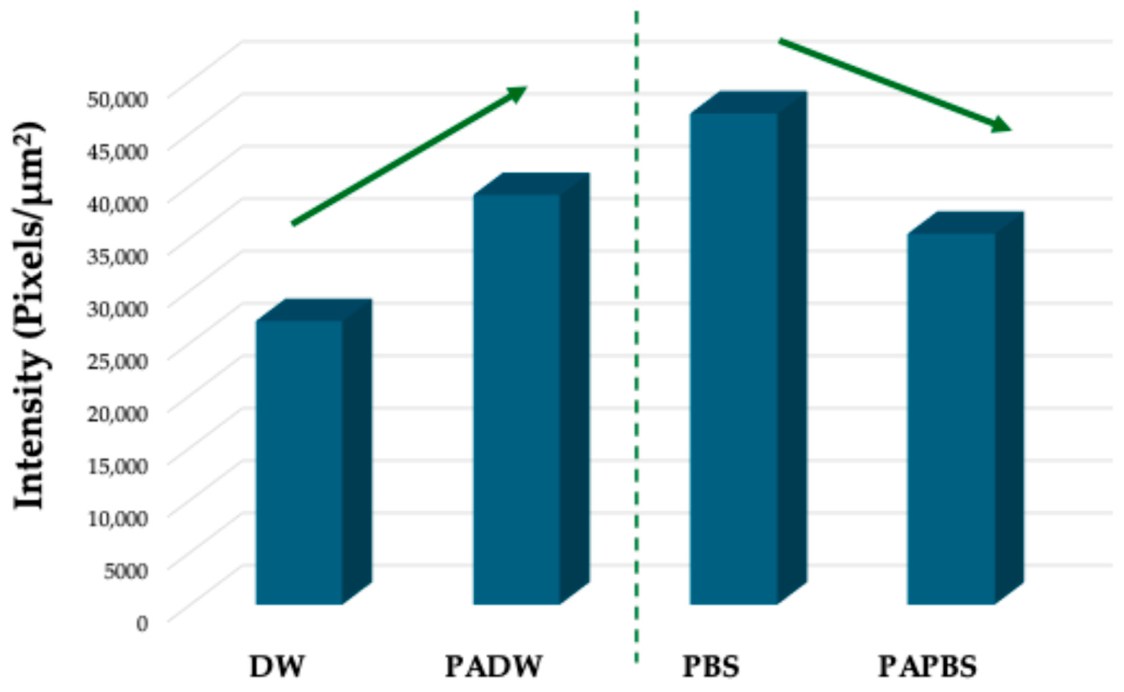
Fluorescence intensity, representing endogenous MMP activity, was presented in confocal microscope images (Figure 2), while the quantification of integrated density of the fluorescence signal was presented in Figure 3. Qualitatively, the fluorescence was distributed mainly in the hybrid layer and at a lower level in the underlying dentin. Among the non-activated liquids, PBS produced higher baseline MMP activity in dentin than non-activated DW (p < 0.001), indicating that the ionic composition of the pretreatment liquid itself influences dentinal enzymatic activity independently of plasma treatment. Plasma activation of the two liquids produced divergent effects. Pretreatment with PADW resulted in increased fluorescence relative to non-activated DW (p = 0.008), indicating that plasma activation of distilled water potentiated endogenous MMP activity in dentin. In contrast, pretreatment with PAPBS reduced fluorescence relative to non-activated PBS (p < 0.001), demonstrating that plasma activation of PBS suppressed enzymatic activity under the same experimental conditions.
Figure 2.
In situ zymography—confocal micrographs of treated dentinal samples: green fluorescence represents relative enzymatic activity of the MMPs. The fluorescence is mainly distributed in the hybrid layer and the underlying dentin. DW—distilled water; PBS—phosphate-buffered saline; PADW—plasma-activated distilled water; PAPBS—plasma-activated phosphate-buffered saline; D—dentin; HL—hybrid layer; R—resin.
Figure 3.
The graph representing the quantification of MMP activity measured in the confocal microscopy images. DW—distilled water; PBS—phosphate-buffered saline; PADW—plasma-activated distilled water; PAPBS—plasma-activated phosphate-buffered saline.
4. Discussion
As expected, air treatment of liquid-generated reactive species involves several coupled processes, including electron-induced dissociation of water and air molecules, formation and recombination of radicals, oxidation of intermediate compounds, and dissolution of gaseous nitrogen oxides into the liquid. These processes lead to the generation of short-lived species, such as OH radicals, and more stable products, including H2O2, NO2−, and NO3− [38]. Moreover, the acidification observed in PADW is related to the formation of acidic nitrogen species (HNO3 and HNO2) and to the very low buffering capacity of distilled water. In contrast, the phosphate buffer present in PBS counteracts the increase of hydrogen ion concentration, maintaining PAPBS close to neutral pH. Experimental plasma-treated liquids have shown distinct effects on the activity of MMPs in dentin, which are crucial for the degradation of resin–dentin interfaces in adhesive dentistry. Hence, the null hypothesis must be rejected.
Despite the important advances in adhesive dentistry in recent decades, adhesion to dentin still represents a series of issues that undermine its longevity [16]. While enamel is a highly mineralized tissue (96 wt%) and can be successfully etched and dried completely before adhesive procedures, enabling capillary penetration of the adhesive onto the crystal structure [39,40], the situation is more complex regarding dentin. Namely, dentin contains 20 wt% organic matter, mainly consisting of collagen type I, but also of non-collagenous proteins [41,42,43]. MMPs are among the non-collagenous proteins of the dentin matrix and have important roles during tooth formation, while becoming inactive after tooth mineralization. However, the caries process, erosion, or different dental procedures can reactivate MMPs, which then degrade the collagen matrix and can cause premature failure of a composite restoration [44]. MMP-2 and MMP-9 are gelatinases that are particularly important in the process of collagen degradation since they digest the telopeptides at the final portions of the collagen helix, enabling the cleavage of the collagen molecule by true collagenases [16]. The hydrolytic degradation of the collagen underneath the restoration enables water to enter into the resin–dentin interface, causing hydrolytic degradation of the resin, with this cascade of events eventually leading to restoration failure [45]. In situ zymography is a test that entails placement of gelatin, the substrate of MMP-2 and -9, on the resin–dentin section and allows for the digestion of gelatin in case there are active MMPs present. Since the gelatin is conjugated with fluorescein, the level of fluorescence on the confocal images represents a relative measure of MMP activity. Confocal micrographs illustrating the gelatinolytic activity of dentinal MMPs demonstrate the modifications caused by plasma-activated solutions (Figure 2 and Figure 3, respectively). However, it is important to note that in situ zymography entails the use of a gelatin substrate, which is not specific only for MMP-2 and MMP-9, and therefore we cannot claim that only these two proteases were involved in the observed fluorescence levels. Nevertheless, given their abundance in dentin extracellular matrix [46] and previous reports of the accordance of in situ zymography and gelatin zymography results (which clearly identifies pro- and active forms of MMP-2 and -9) [26,47], we can safely hypothesize that MMP-2 and -9 play an important role in the cleavage of the gelatin substrate in the present results.
Interestingly, the two liquids demonstrated differences regarding enzyme activation. Namely, while plasma activation of DW increased enzymatic activity in dentin, PBS activation reduced it (p < 0.05) (Figure 2 and Figure 3). It could, however, be noted that non-activated PBS yielded higher enzymatic activity compared to the non-activated DW (p < 0.05). These outcomes could be due to differences in the composition and the pH of the liquids used. While DW had an acidic pH, PBS had a neutral pH value, which was consistent both in the PA and in the non-activated liquids. Furthermore, PBS contained different ions and chemical compounds, such as sodium, potassium, chloride, phosphates, etc. The presence of ions and the neutral pH could have offered more physiological and therefore favorable conditions for the activity of the MMPs in the case of non-activated liquids. On the other hand, after the PA of the liquids, there is an important change, where the initially inactive DW, deprived of ions, becomes rich in RONS, activating the MMPs. It has been demonstrated that the MMPs can be activated in contact with hydrogen peroxide contained in the dental bleaching agents [47]. Conversely, the activation of PBS led to the inhibition of the MMPs. This may be due to the elevated concentration of RONS in the PAPBS, which had a twofold higher amount of NO2− and ~35% higher amount of NO3− compared to PADW. It has been demonstrated that nitrites can cause structural damage to proteins, including collagen [48]. It is worth noting that short-lived species such as OH and ONOOH decay within milliseconds or seconds, so the prolonged biological activity of PAPBS and PADW is more likely related to long-lived species and to secondary reaction products formed during the post-discharge period [49]. Moreover, PBS contains chloride ions; thus, reactive chlorine species are produced during plasma treatment. Previous studies demonstrated the formation of hypochlorite, chlorite and chlorate in plasma-treated saline solutions, although their production and stability strongly depend on the plasma source, gas composition, pH and the presence of other reactive species. These species could have contributed to the observed effects [50]. We could hypothesize that the interaction of nitrogen species also occurs with MMPs, possibly up to a point that elevates enzymatic activity; when a certain threshold is surpassed, this effect becomes deleterious to the MMPs and possibly also other proteins. It is also conceivable that plasma-treated liquids, through the generation of RONS, could induce conformational changes in MMPs embedded within the dentin matrix. RONS are known to affect protein tertiary and quaternary structures by targeting amino acid side chains or disrupting disulfide bonds. Such oxidative modifications could either activate MMPs—by promoting zymogen activation through disruption of pro-domains—or inhibit them by causing irreversible oxidative damage to catalytic domains. Therefore, RONS-mediated structural changes in MMPs could represent an additional mechanism by which plasma-treated liquids modulate matrix remodeling processes in dentin.
These findings appear to align with the principle of hormesis, which may serve as a valuable framework for optimizing plasma activation protocols across a variety of substrates. Hormesis, understood as the concept of an optimal dose–response relationship, could potentially represent a fundamental approach to identifying the most effective plasma activation settings [51]. In this study, it was observed that DW was too neutral to adequately activate the enzymes, whereas PADW and PBS provided more favorable physiological conditions for enzyme activation. In contrast, PAPBS appeared excessively potent, leading to inhibition of MMP activity. A more precise interpretation could be achieved by including measurements of tissue inhibitors of metalloproteinases (TIMPs) alongside MMPs, given that the TIMPs/MMPs ratio is a critical parameter, especially in contexts where tissue regeneration is desired. Specifically, a TIMPs/MMPs ratio greater than one is generally considered favorable for promoting tissue remodeling [52,53]. In a diabetic rat wound model, PAW treatment significantly reduced MMP-9 mRNA expression compared to untreated diabetic wounds on days III and X post-injury, while TIMP-1 mRNA—which was otherwise suppressed in the diabetic group—was relatively preserved. This shifted the MMP-9/TIMP-1 ratio downward by 6.8-fold, favoring net collagen retention over degradation [54]. Given that the dentin extracellular matrix is mostly composed of collagen type I, it is plausible that this scenario could also be translated to dentin collagen. Hence, it could be hypothesized that both PADW and PBS may create a hormetic window that supports optimal activation of tissue remodeling processes. Future studies should aim to further refine plasma activation parameters across different liquids, with the goal of identifying conditions that best facilitate tissue regeneration. The ability to modulate MMP activity through plasma-treated liquids suggests potential for these solutions in developing innovative strategies for preserving dentin integrity and improving the overall success and longevity of adhesive dental restorations.
5. Conclusions
Plasma-treated PBS and DW exhibited distinct RONS concentrations and pH profiles following plasma activation, which could partially explain their differing effects on MMP activation. In addition, the activity of MMPs is highly dependent on the presence of specific ions, suggesting that the inherent compositional differences between PBS and DW may have further contributed to the variations observed in enzymatic activity.
Author Contributions
Conceptualization, T.L.; methodology, T.M., R.L. and R.M.; validation, C.M. and U.J.; formal analysis, D.D.; investigation, R.M., C.M. and V.C.; resources, L.B.; data curation, T.M. and T.L.; writing—original draft preparation, T.M., R.M. and T.L.; writing—review and editing, R.L., D.D., L.B., U.J., C.M., V.C. and M.G.; supervision, L.B. and M.G.; funding acquisition, L.B. All authors have read and agreed to the published version of the manuscript.
Funding
The research leading to these results was funded by the European Union—NextGenerationEU through the Italian Ministry of University and Research under PNRR—M4C2-I1.3 Project PE_00000019 “HEAL ITALIA” to Lorenzo Breschi, CUP J33C22002920006. Further, this work was carried out in the frame of PlasTHER COST Action CA20114, supported by the resources from DIBINEM lab and DIN, from the University of Bologna (participants Romolo Laurita, Tijana Lainovic, Tatjana Maravic, Roberto Montalbetti).
Institutional Review Board Statement
University Ethical Committee approval (protocol N°: 71/2019/OSS/AUSLBO on 23 January 2019) was obtained for the use of human teeth.
Informed Consent Statement
Informed consent was obtained from all subjects involved in the study (tooth donors).
Data Availability Statement
The dataset is available on request from the authors.
Conflicts of Interest
The authors declare no conflicts of interest.
References
- Jungbauer, G.; Moser, D.; Müller, S.; Pfister, W.; Sculean, A.; Eick, S. The Antimicrobial Effect of Cold Atmospheric Plasma against Dental Pathogens—A Systematic Review of in-Vitro Studies. Antibiotics 2021, 10, 211. [Google Scholar] [CrossRef] [Scilit] [PubMed]
- Borges, A.C.; Kostov, K.G.; Pessoa, R.S.; De Abreu, G.M.A.; Lima, G.D.M.G.; Figueira, L.W.; Koga-Ito, C.Y. Applications of Cold Atmospheric Pressure Plasma in Dentistry. Appl. Sci. 2021, 11, 1975. [Google Scholar] [CrossRef] [Scilit]
- Stoffels, E.; Kieft, I.E.; Sladek, R.E.J.; Van Den Bedem, L.J.M.; Van Der Laan, E.P.; Steinbuch, M. Plasma Needle for in Vivo Medical Treatment: Recent Developments and Perspectives. Plasma Sources Sci. Technol. 2006, 15, S169–S180. [Google Scholar] [CrossRef] [Scilit]
- Hoffmann, C.; Berganza, C.; Zhang, J. Cold Atmospheric Plasma: Methods of Production and Application in Dentistry and Oncology. Med. Gas Res. 2013, 3, 21. [Google Scholar] [CrossRef] [Scilit] [PubMed]
- Almeida, N.D.; Klein, A.L.; Hogan, E.A.; Terhaar, S.J.; Kedda, J.; Uppal, P.; Sack, K.; Keidar, M.; Sherman, J.H. Cold Atmospheric Plasma as an Adjunct to Immunotherapy for Glioblastoma Multiforme. World Neurosurg. 2019, 130, 369–376. [Google Scholar] [CrossRef] [Scilit] [PubMed]
- Sanesi, L.; Puca, V.; Caponio, V.C.A.; Pinti, M.; Balice, G.; Femminella, B.; Paolantonio, M.; Cela, I.; Kaushik, N.K.; Choi, E.H.; et al. Disinfection of Dental Root Canals by Cold Atmospheric Plasma: A Systematic Review and Meta-Analysis of Dental Biofilm. Front. Oral Health 2024, 5, 1483078. [Google Scholar] [CrossRef] [Scilit] [PubMed]
- Santak, V.; Zaplotnik, R.; Milosevic, S.; Klaric, E.; Tarle, Z. Atmospheric Pressure Plasma Jet as an Accelerator of Tooth Bleaching. Acta Stomatol. Croat. 2014, 48, 268–278. [Google Scholar] [CrossRef] [Scilit] [PubMed]
- Rathore, V.; Patil, C.; Sanghariyat, A.; Nema, S.K. Design and Development of Dielectric Barrier Discharge Setup to Form Plasma-Activated Water and Optimization of Process Parameters. Eur. Phys. J. D 2022, 76, 77. [Google Scholar] [CrossRef] [Scilit]
- Labay, C.; Hamouda, I.; Tampieri, F.; Ginebra, M.P.; Canal, C. Production of Reactive Species in Alginate Hydrogels for Cold Atmospheric Plasma-Based Therapies. Sci. Rep. 2019, 9, 16160. [Google Scholar] [CrossRef] [Scilit] [PubMed]
- Montalbetti, R.; Machala, Z.; Gherardi, M.; Laurita, R. Production and Chemical Composition of Plasma Activated Water: A Systematic Review and Meta-analysis. Plasma Process. Polym. 2025, 22, 2400249. [Google Scholar] [CrossRef] [Scilit]
- Milhan, N.V.M.; Chiappim, W.; Sampaio, A.d.G.; Vegian, M.R.d.C.; Pessoa, R.S.; Koga-Ito, C.Y. Applications of Plasma-Activated Water in Dentistry: A Review. Int. J. Mol. Sci. 2022, 23, 4131. [Google Scholar] [CrossRef] [Scilit] [PubMed]
- Ohshima, T.; Ikawa, S.; Kitano, K.; Maeda, N. A Proposal of Remedies for Oral Diseases Caused by Candida: A Mini Review. Front. Microbiol. 2018, 9, 1522. [Google Scholar] [CrossRef] [Scilit] [PubMed]
- Qiao, D.; Li, Y.; Pan, J.; Zhang, J.; Tian, Y.; Wang, K. Effect of Plasma Activated Water in Caries Prevention: The Caries Related Biofilm Inhibition Effects and Mechanisms. Plasma Chem. Plasma Process. 2022, 42, 801–814. [Google Scholar] [CrossRef] [Scilit]
- Stasic, J.N.; Selaković, N.; Puač, N.; Miletić, M.; Malović, G.; Petrović, Z.L.; Veljovic, D.N.; Miletic, V. Effects of Non-Thermal Atmospheric Plasma Treatment on Dentin Wetting and Surface Free Energy for Application of Universal Adhesives. Clin. Oral Investig. 2019, 23, 1383–1396. [Google Scholar] [CrossRef] [Scilit] [PubMed]
- Stasic, J.N.; Pficer, J.K.; Milicic, B.; Puač, N.; Miletic, V. Effects of Non-Thermal Atmospheric Plasma on Dentin Wetting and Adhesive Bonding Efficiency: Systematic Review and Meta-Analysis. J. Dent. 2021, 112, 103765. [Google Scholar] [CrossRef] [Scilit] [PubMed]
- Breschi, L.; Maravic, T.; Mazzitelli, C.; Josic, U.; Mancuso, E.; Cadenaro, M.; Pfeifer, C.S.; Mazzoni, A. The Evolution of Adhesive Dentistry: From Etch-and-Rinse to Universal Bonding Systems. Dent. Mater. 2025, 41, 141–158. [Google Scholar] [CrossRef] [Scilit] [PubMed]
- Fugolin, A.P.; Dobson, A.; Huynh, V.; Mbiya, W.; Navarro, O.; Franca, C.M.; Logan, M.; Merritt, J.L.; Ferracane, J.L.; Pfeifer, C.S. Antibacterial, Ester-Free Monomers: Polymerization Kinetics, Mechanical Properties, Biocompatibility and Anti-Biofilm Activity. Acta Biomater. 2019, 100, 132–141. [Google Scholar] [CrossRef] [Scilit] [PubMed]
- Fugolin, A.P.; de Paula, A.B.; Dobson, A.; Huynh, V.; Consani, R.; Ferracane, J.L.; Pfeifer, C.S. Alternative Monomer for BisGMA-Free Resin Composites Formulations. Dent. Mater. 2020, 36, 884–892. [Google Scholar] [CrossRef] [Scilit] [PubMed]
- Pongprueksa, P.; Miletic, V.; Janssens, H.; Van Landuyt, K.L.; De Munck, J.; Godderis, L.; Van Meerbeek, B. Degree of Conversion and Monomer Elution of CQ/Amine and TPO Adhesives. Dent. Mater. 2014, 30, 695–701. [Google Scholar] [CrossRef] [Scilit] [PubMed]
- Loguercio, A.D.; Hass, V.; Gutierrez, M.F.; Luque-Martinez, I.V.; Szezs, A.; Stanislawczuk, R.; Bandeca, M.C.; Reis, A. Five-Year Effects of Chlorhexidine on the in Vitro Durability of Resin/Dentin Interfaces. J. Adhes. Dent. 2016, 18, 35–43. [Google Scholar] [CrossRef] [Scilit] [PubMed]
- Li, H.; Li, T.; Li, X.; Zhang, Z.; Li, P.; Li, Z. Morphological Effects of MMPs Inhibitors on the Dentin Bonding. Int. J. Clin. Exp. Med. 2015, 8, 10793–10803. [Google Scholar] [PubMed]
- Bhoopathi, P.G.; Kittappa, K.K.; Sanjeev, K.; Sekar, M. Effect of Chlorhexidine and Cystatin Incorporated Adhesives on MMPs and Cysteine Cathepsin-an in-Vitro Zymographic Analysis. J. Clin. Diagn. Res. 2018, 12, ZC01–ZC05. [Google Scholar] [CrossRef] [Scilit]
- Leme-Kraus, A.A.; Phansalkar, R.S.; dos Reis, M.C.; Aydin, B.; Sousa, A.B.S.; Alania, Y.; McAlpine, J.; Chen, S.N.; Pauli, G.F.; Bedran-Russo, A.K. Dimeric Proanthocyanidins on the Stability of Dentin and adhesive Biointerfaces. J. Dent. Res. 2020, 99, 175. [Google Scholar] [CrossRef] [Scilit] [PubMed]
- Alania, Y.; Yourdkhani, M.; Trevelin, L.; Bim-Junior, O.; Majithia, H.; Farsi, L.; Bedran-Russo, A.K. Proanthocyanidin Encapsulation for Sustained Bioactivity in Dentin Bioadhesion: A Two-Year Study. Dent. Mater. 2022, 38, 421–430. [Google Scholar] [CrossRef] [Scilit] [PubMed]
- Comba, A.; Scotti, N.; Mazzoni, A.; Maravic, T.; Ribeiro Cunha, S.; Michelotto Tempesta, R.; Carossa, M.; Pashley, D.H.; Tay, F.R.; Breschi, L. Carbodiimide Inactivation of Matrix Metalloproteinases in Radicular Dentine. J. Dent. 2019, 82, 56–62. [Google Scholar] [CrossRef] [Scilit] [PubMed]
- Maravic, T.; Breschi, L.; Comba, A.; Cunha, S.R.; Angeloni, V.; Nucci, C.; Hebling, J.; Pashley, D.; Tay, F.; Mazzoni, A. Experimental Use of an Acrolein-Based Primer as Collagen Cross-Linker for Dentine Bonding. J. Dent. 2017, 68, 85–90. [Google Scholar] [CrossRef] [Scilit] [PubMed]
- Enrich-Essvein, T.; Rodríguez-Navarro, A.B.; Álvarez-Lloret, P.; Cifuentes-Jiménez, C.; Bolaños-Carmona, M.V.; González-López, S. Proanthocyanidin-Functionalized Hydroxyapatite Nanoparticles as Dentin Biomodifier. Dent. Mater. 2021, 37, 1437–1445. [Google Scholar] [CrossRef] [Scilit] [PubMed]
- Boteon, A.P.; Kato, M.T.; Buzalaf, M.A.R.; Prakki, A.; Wang, L.; Rios, D.; Honório, H.M. Effect of Proanthocyanidin-Enriched Extracts on the Inhibition of Wear and Degradation of Dentin Demineralized Organic Matrix. Arch. Oral Biol. 2017, 84, 118–124. [Google Scholar] [CrossRef] [Scilit] [PubMed]
- Pashley, D.H.; Tay, F.R.; Carvalho, R.M.; Rueggeberg, F.A.; Agee, K.A.; Carrilho, M.; Donnelly, A.; García-Godoy, F. From Dry Bonding to Water-Wet Bonding to Ethanol-Wet Bonding. A Review of the Interactions between Dentin Matrix and Solvated Resins Using a Macromodel of the Hybrid Layer. Am. J. Dent. 2007, 20, 7–20. [Google Scholar] [PubMed]
- Sauro, S.; Osorio, R.; Watson, T.F.; Toledano, M. Influence of Phosphoproteins’ Biomimetic Analogs on Remineralization of Mineral-Depleted Resin-Dentin Interfaces Created with Ion-Releasing Resin-Based Systems. Dent. Mater. 2015, 31, 759–777. [Google Scholar] [CrossRef] [Scilit] [PubMed]
- Kim, Y.K.; Mai, S.; Mazzoni, A.; Liu, Y.; Tezvergil-Mutluay, A.; Takahashi, K.; Zhang, K.; Pashley, D.H.; Tay, F.R. Biomimetic Remineralization as a Progressive Dehydration Mechanism of Collagen Matrices—Implications in the Aging of Resin-Dentin Bonds. Acta Biomater. 2010, 6, 3729–3739. [Google Scholar] [CrossRef] [Scilit] [PubMed]
- Abuna, G.; Feitosa, V.P.; Correr, A.B.; Cama, G.; Giannini, M.; Sinhoreti, M.A.; Pashley, D.H.; Sauro, S. Bonding Performance of Experimental Bioactive/Biomimetic Self-Etch Adhesives Doped with Calcium-Phosphate Fillers and Biomimetic Analogs of Phosphoproteins. J. Dent. 2016, 52, 79–86. [Google Scholar] [CrossRef] [Scilit] [PubMed]
- Tay, F.R.; Pashley, D.H. Biomimetic Remineralization of Resin-Bonded Acid-Etched Dentin. J. Dent. Res. 2009, 88, 719–724. [Google Scholar] [CrossRef] [Scilit] [PubMed]
- Boruziniat, A.; Atoufi, A.; Chehreli, Z.; Akbari, M.; Gifani, M. Effect of Non-Vital Bleaching on the Durability of Resin-Dentin Bond with an Ethanol-Based Etch-And-Rinse Adhesive. Biomimetics 2018, 3, 35. [Google Scholar] [CrossRef] [Scilit] [PubMed]
- André, C.B.; Gomes, B.P.F.A.; Duque, T.M.; Stipp, R.N.; Chan, D.C.N.; Ambrosano, G.M.B.; Giannini, M. Dentine Bond Strength and Antimicrobial Activity Evaluation of Adhesive Systems. J. Dent. 2015, 43, 466–475. [Google Scholar] [CrossRef] [Scilit] [PubMed]
- Neves, J.G.; Marcato, P.D.; de Paula e Silva, F.W.G.; Mantovani, C.P.T.; Prado, H.S.; Aires, C.P.; Massaro, T.N.C.; Borsato, M.C. Synthesis and Characterization of an Experimental Primer Containing Chitosan Nanoparticles—Effect on the Inactivation of Metalloproteinases, Antimicrobial Activity and Adhesive Strength. Arch. Oral Biol. 2021, 127, 105148. [Google Scholar] [CrossRef] [Scilit] [PubMed]
- Wang, H.; Wandell, R.J.; Tachibana, K.; Voráč, J.; Locke, B.R. The Influence of Liquid Conductivity on Electrical Breakdown and Hydrogen Peroxide Production in a Nanosecond Pulsed Plasma Discharge Generated in a Water-Film Plasma Reactor. J. Phys. D Appl. Phys. 2019, 52, 075201. [Google Scholar] [CrossRef] [Scilit]
- Dhakal, O.B.; Dahal, R.; Iqbal, Z.; Choi, E.H. Synergistic Degradation of Tetracycline Using Plasma-Activated Persulfate: Roles of Hydroxyl and Sulfate Radicals. J. Environ. Chem. Eng. 2026, 14, 122955. [Google Scholar] [CrossRef] [Scilit]
- Münchow, E.A.; Bottino, M.C. Recent Advances in Adhesive Bonding: The Role of Biomolecules, Nanocompounds, and Bonding Strategies in Enhancing Resin Bonding to Dental Substrates. Curr. Oral Health Rep. 2017, 4, 215–227. [Google Scholar] [CrossRef] [Scilit] [PubMed]
- Cardoso, M.; de Almeida Neves, A.; Mine, A.; Coutinho, E.; Van Landuyt, K.; De Munck, J.; Van Meerbeek, B. Current Aspects on Bonding Effectiveness and Stability in Adhesive Dentistry. Aust. Dent. J. 2011, 56, 31–44. [Google Scholar] [CrossRef] [Scilit] [PubMed]
- Cui, F.-Z.; Ge, J. New Observations of the Hierarchical Structure of Human Enamel, from Nanoscale to Microscale. J. Tissue Eng. Regen. Med. 2007, 1, 185–191. [Google Scholar] [CrossRef] [Scilit] [PubMed]
- Risnes, S. Growth Tracks in Dental Enamel. J. Hum. Evol. 1998, 35, 331–350. [Google Scholar] [CrossRef] [Scilit] [PubMed]
- Nanci, A. Ten Cate’s Oral Histology: Development, Structure, and Function, 8th ed.; Elsevier: Amsterdam, The Netherlands; Mosby: Clay County, MO, USA, 2008. [Google Scholar]
- Longhi, M.; Cerroni, L.; Condò, S.G.; Ariano, V.; Pasquantonio, G. The Effects of Host Derived Metalloproteinases on Dentin Bond and the Role of MMPs Inhibitors on Dentin Matrix Degradation. Oral Implantol. 2014, 7, 71–79. [Google Scholar]
- Teshima, I. Degradation of 10-Methacryloyloxydecyl Dihydrogen Phosphate. J. Dent. Res. 2010, 89, 1281–1286. [Google Scholar] [CrossRef] [Scilit] [PubMed]
- Turco, G.; Cadenaro, M.; Maravić, T.; Frassetto, A.; Marsich, E.; Mazzoni, A.; Di Lenarda, R.; Tay, F.R.; Pashley, D.H.; Breschi, L. Release of ICTP and CTX Telopeptides from Demineralized Dentin Matrices: Effect of Time, Mass and Surface Area. Dent. Mater. 2018, 34, 452–459. [Google Scholar] [CrossRef] [Scilit] [PubMed]
- Mayer-Santos, E.; Maravic, T.; Comba, A.; Freitas, P.M.; Bueno Marinho, G.; Mazzitelli, C.; Mancuso, E.; Scotti, N.; Florenzano, F.; Breschi, L.; et al. The Influence of Different Bleaching Protocols on Dentinal Enzymatic Activity: An in Vitro Study. Molecules 2022, 27, 1684. [Google Scholar] [CrossRef] [Scilit] [PubMed]
- Paik, D.C.; Dillona, J.; Galicia, E.; David Tilson’, M. The Nitrite Collagen Reaction: Non-Enzymatic Nitration as a Model System for Age-Related Damage. Connect. Tissue Res. 2001, 42, 111–122. [Google Scholar] [CrossRef] [Scilit] [PubMed]
- Liu, Z.C.; Liu, D.X.; Chen, C.; Liu, Z.J.; Yang, A.J.; Rong, M.Z.; Chen, H.L.; Kong, M.G. Post-Discharge Evolution of Reactive Species in the Water Activated by a Surface Air Plasma: A Modeling Study. J. Phys. D Appl. Phys. 2018, 51, 175202. [Google Scholar] [CrossRef] [Scilit]
- Jirásek, V.; Lukeš, P. Formation of Reactive Chlorine Species in Saline Solution Treated by Non-Equilibrium Atmospheric Pressure He/O 2 Plasma Jet. Plasma Sources Sci. Technol. 2019, 28, 035015. [Google Scholar] [CrossRef] [Scilit]
- González-Llorente, L.; Andrés-Gasco, M.; Gil Aranda, M.A.; Rabadán-Ros, R.; Zapata-Pérez, R.; Núñez-Delicado, E.; Menéndez-Coto, N.; García-González, C.; Baena-Huerta, F.J.; Coto-Montes, A.; et al. The Hormetic Adaptative Capacity and Resilience to Oxidative Stress Is Strengthened by Exposome Enrichment with Air Cold Atmospheric Plasma: A Metabolome Targeted Follow-up Approach. Biomedicines 2025, 13, 949. [Google Scholar] [CrossRef] [Scilit] [PubMed]
- Lambert, E.; Dassé, E.; Haye, B.; Petitfrère, E. TIMPs as Multifacial Proteins. Crit. Rev. Oncol. Hematol. 2004, 49, 187–198. [Google Scholar] [CrossRef] [Scilit] [PubMed]
- Alpagot, T.; Bell, C.; Lundergan, W.; Chambers, D.W.; Rudin, R. Longitudinal Evaluation of GCF MMP-3 and TIMP-1 Levels as Prognostic Factors for Progression of Periodontitis. J. Clin. Periodontol. 2001, 28, 353–359. [Google Scholar] [CrossRef] [Scilit] [PubMed]
- Rajić, J.; Grdović, N.; Marković, A.; Škoro, N.; Dinić, S.; Uskoković, A.; Arambašić Jovanović, J.; Đorđević, M.; Sarić, A.; Vidaković, M.; et al. Plasma-Activated Water Improve Wound Healing in Diabetic Rats by Influencing the Inflammatory and Remodelling Phase. Int. J. Mol. Sci. 2025, 26, 1265. [Google Scholar] [CrossRef] [Scilit] [PubMed]
Disclaimer/Publisher’s Note: The statements, opinions and data contained in all publications are solely those of the individual author(s) and contributor(s) and not of MDPI and/or the editor(s). MDPI and/or the editor(s) disclaim responsibility for any injury to people or property resulting from any ideas, methods, instructions or products referred to in the content. |
© 2026 by the authors. Licensee MDPI, Basel, Switzerland. This article is an open access article distributed under the terms and conditions of the Creative Commons Attribution (CC BY) license.