
Article

Dental Extra-Masticatory Wear and Dental Calculus Micro-Remains as Indicators of Fibre Manipulation in the 15th–19th Century Necropolis at St. Athanasius Church, Niculițel (Romania)


by
Ozana-Maria Ciorpac-Petraru
1,
Mihaela Danu
1,*,
Ana Drob
2,
Paul-Iulian Donciu
1,
Aurel-Daniel Stănică
3 and
Luminița Bejenaru
1,4
1
Faculty of Biology, Alexandru Ioan Cuza University of Iași, 700505 Iasi, Romania
2
Arheoinvest Center, Department of Natural and Exact Sciences, Institute of Interdisciplinary Research, “Alexandru Ioan Cuza” University of Iasi, 700505 Iasi, Romania
3
“Gavrilă Simion” Eco-Museum Research Institute, 820009 Tulcea, Romania
4
“Olga Necrasov” Center of Anthropological and Biomedical Research, Romanian Academy-Iași Branch, 700481 Iasi, Romania
*
Author to whom correspondence should be addressed.
Quaternary 2026, 9(2), 25; https://doi.org/10.3390/quat9020025
Submission received: 16 January 2026 / Revised: 19 February 2026 / Accepted: 3 March 2026 / Published: 5 March 2026

Abstract

Dental wear provides valuable evidence for reconstructing past human behaviour, including diet abrasiveness and non-masticatory activities such as the use of teeth as a “third hand”. This study investigates activity-induced dental modifications (AIDMs) in two adult human skeletons recovered from a 15th–19th-century necropolis at the St. Athanasius Church in Niculițel (Tulcea County, Romania). Dental remains and associated dental calculus were examined using low- and high-magnification optical microscopy and scanning electron microscopy (SEM). Well-polished grooves with parallel striations were identified on the incisor crowns, consistent with repetitive extra-masticatory activities related to fibre drafting during spinning and textile production. Dental calculus analysis revealed the presence of plant and animal fibres, providing direct micro-contextual evidence for textile-related practices. These results offer new insights into the use of teeth as tools and contribute to the reconstruction of textile-related craft activities during the Ottoman and early modern periods in southeastern Europe.

1. Introduction

Teeth are a valuable source of information for bioarcheologists, representing a “window into the lifestyle of ancient peoples” due to the resistance of their hard tissues to postmortem changes [1,2]. In biological anthropology, teeth are used in several research areas, such as age-at-death estimation in subadults and adults using standardised methods or histological approaches [3,4], the study of pathological conditions associated with infectious diseases and nutritional deficiencies [5,6,7], dental phenetic variation in past human populations and biodistance analyses [8,9,10], and dental wear as an indicator of diet [11,12,13,14,15], as well as an indicator for non-dietary purposes (i.e., the use of teeth as “a third hand”) [16]. Occlusal dental macrowear and microwear analyses, along with special dental wear features, provide insights into the reconstruction of diet variation, food-processing techniques, extra-masticatory use of teeth, cultural modification, and occupational activities in past human lifestyles [16,17].

1.1. Activity-Induced Dental Modifications (AIDMs) in Past Human Remains

The abrasion patterns produced using teeth in occupational activities are known as activity-induced dental modifications (AIDMs) [18,19]. These modifications, such as chipping, notches, grooves, labial or buccal striations produced by instruments, and lingual surface attrition of maxillary anterior teeth—LSAMAT—provide evidence of cultural practices, and of the technical and social organisation of work, including the repetition, intensity, and embodied character of specific craft-related gestures [2,16]. The extra-masticatory uses of teeth have been highlighted in various prehistoric and historic human populations, from diverse geographic and chronological contexts, as indicators for textile-related craft activities and other non-dietary practices [2,16,20,21,22,23,24,25,26]. In this study, the term “textile-related craft activities” refers specifically to fibre manipulation and spinning-related gestures performed within a household production framework rather than to formally organised, workshop-based craft production. Among the AIDMs, occlusal, lingual, and interproximal grooves and striation, along with their orientation and morphology, localised on the anterior teeth (i.e., incisors and canines), are usually correlated with sewing, pliant cordage, and fibre manipulation of plant and animal origin, and other materials worked with their teeth being usually associated with female craft specialisations [2,17,26,27]. However, according to some authors, interproximal grooves can also be correlated with the tooth-picking behaviours using rigid “instruments” [28,29]. Given the great potential of dental calculus micro-remains studies [30], the identification of possible fibres within dental calculus could be linked to specific task activities involving the use of teeth as a third hand in textile-related craft specialisations; further bioarchaeological studies are necessary [26,31].
Regarding the approach of dental wear studies in archaeological human populations from Romania, several studies have been published addressing macrowear and microwear [12,15,32,33,34]. Among AIDMs, chipping and oblique dental wear were identified in some studies without being statistically quantified or correlated with other bioarchaeological indicators related to the use of teeth as tools [34,35,36,37]. To the best of our knowledge, in Romania, only a single case of tooth notching in a male human skeleton has been recently identified to date, in a study regarding dental wear in a 17th–19th century human population from the city of Iași [38]. Although there are many studies addressing activity-induced dental modifications in past human populations, knowledge of these features remains limited in Romania, making this study one of the first to correlate the AIDMs with micro-remains findings from human dental calculus.

1.2. Aim of the Study

The aim of this study is to identify and characterise the activity-induced dental modifications along with micro-remains analyses from dental calculus in two human skeletons discovered during the archaeological excavations at St. Athanasius Church (15th–19th century) in Niculițel (Tulcea County, Romania) using low- and high-magnification optical microscopy and scanning electron microscopy (SEM).
Bioarchaeological studies of historical-period populations contribute to the understanding of long-term patterns in human behaviour, including subsistence practices, craft activities, and the non-masticatory use of teeth. Although the individuals analysed in this study date to the late Holocene (15th–19th centuries), the combined analysis of activity-induced dental modifications and dental calculus micro-remains provides relevant information for Quaternary research concerned with reconstructing daily practices and technological behaviours. The documentation of tooth-tool use in a well-defined historical context also offers a comparative reference for similar dental wear features observed in prehistoric and protohistoric populations, supporting their interpretation within a broader temporal perspective.

2. Study Area and Archaeological Background

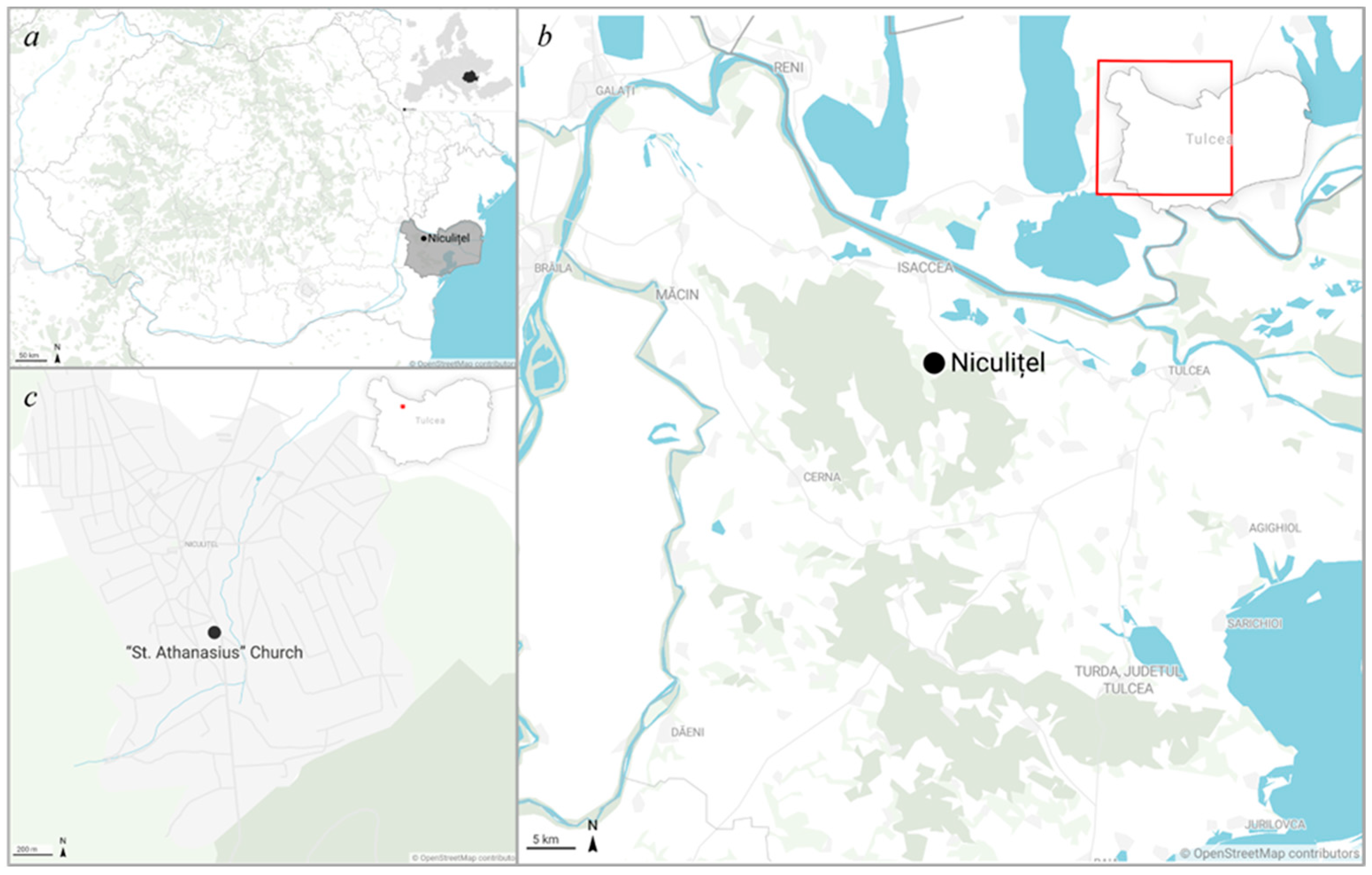
The study area is located in southeastern Romania, within the commune of Niculițel (45°10′38″ N, 28°28′38″ E), in the northern part of Tulcea County, approximately 28 km northwest of the city of Tulcea and 12 km south of the town of Isaccea (Figure 1a,b). The commune covers an area of 93.67 km2 and is located a few kilometres from the Danube, within a small depression of the Niculițel Hills.
From a geomorphological perspective, the area is characterised by a hilly relief consisting of hills and plateaus bordered by steep slopes, strongly fragmented by torrential drainage networks. These landforms, particularly developed toward the Danube sector and within the catchment areas of tributaries of the Taița River, favour active surface erosion processes affecting the soil cover [39,40,41]. Climatically, the area falls within a temperate continental regime specific to the transitional zone of southeastern Romania, characterised by hot summers with reduced precipitation and relatively mild winters marked by frequent strong winds. The mean annual temperature is approximately 11 °C, while the mean annual precipitation ranges between 415 and 500 mm [40,42].
Archaeologically, Niculițel is located in an area with a long and complex history of human occupation [43]. In the southern part of the modern village stands the St. Athanasius Church (Figure 1c), a monument constructed at the beginning of the 14th century and subsequently subjected to multiple phases of repair and enlargement during the 16th, 18th, and 19th centuries. Archaeological and architectural investigations have documented several construction phases and confirmed the continuous use of the monument by the local community until the end of the 19th century [44,45,46,47,48].
Preventive archaeological excavations carried out in 2022–2023 in the area surrounding and inside the church, in connection with conservation and restoration works, revealed an extensive medieval and modern cemetery [49]. A total of 348 archaeological complexes were documented, the majority consisting of inhumation graves, reburials, and disturbed burials. The necropolis spans a chronological interval from the 15th to the 19th centuries, and it is characterised by four burial horizons, with graves predominantly oriented west–east in accordance with Christian funerary practices.
Funerary inventories include personal adornments, clothing accessories, and a substantial numismatic assemblage dominated by Ottoman, Western, and Romanian coinage dating mainly to the 18th–19th centuries [49]. In addition, several graves yielded preserved textile remains and clothing elements, indicating that the deceased were frequently buried dressed rather than wrapped in simple shrouds [49]. These data provide the archaeological context for the osteological and dental remains analysed in this study, which derive from adult individuals buried in the cemetery during the Ottoman and early modern periods.
Five excavation units (S1–S5) were investigated during the campaigns.
Excavation unit S1 represents the most extensively documented sector of the cemetery and yielded a high density of funerary contexts, including inhumation graves, reburials, and disturbed burials, predominantly dated to the 18th–19th centuries, with isolated complexes possibly attributable to the late medieval period. The graves in S1 are mainly simple pit burials, often affected by later disturbances, and are associated with diverse funerary inventories, including coins (frequently perforated), jewellery items, clothing accessories, and, in several instances, preserved textile fragments, including fabrics incorporating metallic threads [49].
Excavation units S2 and S3 contributed additional data regarding the spatial organisation and chronological development of the cemetery [49]. S2 revealed a mixed stratigraphic sequence, including graves, reburials, and associated features, alongside non-funerary structures such as ovens and domestic installations dated mainly to the 18th century. S3 documented a high concentration of funerary complexes, predominantly dated to the 18th–19th centuries, with particularly rich funerary inventories in some graves, including abundant numismatic material, jewellery, clothing accessories, and preserved textile remains [49]. Although no dental remains from S2 and S3 were included in the present analysis, the evidence from these areas provides essential contextual information for understanding burial practices and material culture at the site.
Excavation unit S4 also documented a concentration of inhumated graves belonging to adult and juvenile individuals. Although funerary inventories in S4 are generally more limited than those recorded in S1, they include comparable categories of finds, such as coins, jewellery items, belt fittings, beads, and isolated textile fibres, indicating similar burial practices and a comparable chronological framework [49].
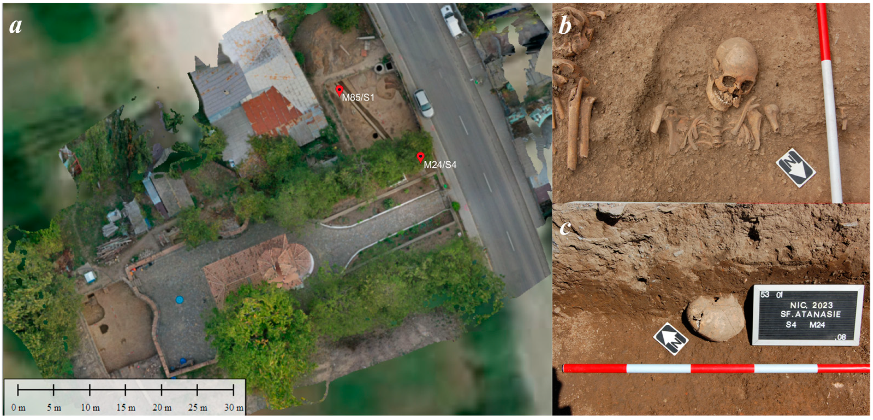
Two adult human skeletons, M85/S1 and M24/S4, exhibiting dental features of particular interest, constitute the material analysed in this study (Figure 2a–c). These individuals were recovered from simple pit graves excavated in sections S1 and S4, respectively, and are securely dated to the Ottoman and early modern periods based on stratigraphic position and associated archaeological materials. The archaeological context of S1 and S4 thus provides a well-defined funerary framework for the interpretation of the observed dental wear patterns.

3. Materials and Methods

During the 2022 archaeological excavations, 279 graves were documented, followed by an additional 69 graves in 2023. The assemblage includes remains of both adults and subadults; among the adult individuals, dentition was preserved in only 138 cases. Detailed osteological assessment and biological profile estimation are being undertaken in a separate, ongoing study and will be reported in a forthcoming publication. Of the 138 adult human skeletons, only two showed particular dental wear features. This study is based on a case-study approach focusing on two human skeletons, permitting a detailed qualitative assessment of activity-induced dental modifications and associated micro-remains. Such an approach facilitates the recognition and interpretation of discrete dental wear features that may be overlooked in larger-scale analyses. Comparable case-based studies have been successfully applied in bioarchaeological research addressing extra-masticatory dental wear and craft-related activities, particularly when supported by microscopic observations and archaeological context.

3.1. Preservation Status, Estimations of Age-at-Death and Sex

The assessment of the two human skeletons, codified as M85/S1 and M24/S4, recovered during archaeological excavations at the St. Athanasius Church necropolis in Niculițel, Tulcea County, Romania, used a methodology adhering to standard osteological protocols [4,50,51]. The preservation status was quantified using the completeness index (CI), which reports the number of recovered elements relative to a standard inventory of 28 major skeletal segments [52]. Age-at-death estimation was performed by corroborating two indicators: the degree of obliteration (synostosis) of the cranial sutures (i.e., sagittal and coronal) [53,54], and the degree of dental wear (attrition), quantified according to the methodology [55]. Sex estimation was derived from the morphological assessment of cranial sexual dimorphism, utilising criteria such as the robusticity of the glabella, supraorbital ridges, mastoid processes, and the morphology of the mental eminence [56,57].

3.2. Dental Wear Assessment

3.2.1. Stereomicroscopy

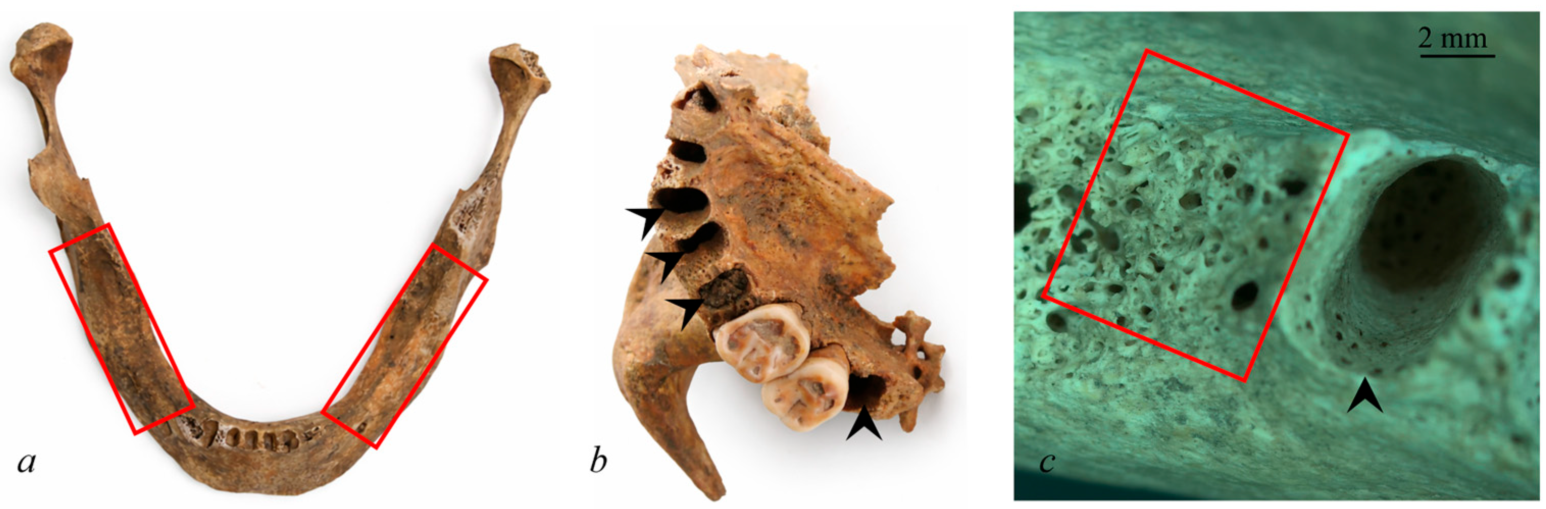
The initial stage of dental assessment was based on recording whether the teeth were present or absent antemortem or postmortem (Figure 3).
Teeth were considered antemortem lost if bone resorption was observed at the level of the dental alveoli, and postmortem lost if no indications of bone remodelling were detected at the alveolar site [7,58]. The preparation of the dental material for low-power microscopical observation involved the removal of contaminants and cleaning with 30% ethyl alcohol, following the methodology proposed by Mahoney [59]. The dental material, whether located in the dental alveoli or isolated, was examined under reflected light using a Carl Zeiss Stemi 508 stereomicroscope equipped with an Axiocam 208 camera and ZEISS ZEN lite 3.8 software (Zeiss, Oberkochen, Germany).

3.2.2. Dental Macrowear Analysis

The dental macrowear scoring was established on the extent of exposed dentine tissue on the occlusal/incisal surfaces of teeth, applying the semiquantitative system proposed by Smith [60] for the incisors (I), canines (C), and premolars (P), while for the molars (M), the scoring system proposed by Scott [61] was used. The stages of dental wear were considered in three categories: teeth with invisible or very small wear facets (Smith scale: 1–2, Scott scale: 4–9), teeth with moderately advanced wear facets (Smith scale: 3–5, Scott scale: 10–22), and teeth with highly advanced wear facets (Smith scale: 6–8, Scott scale: 23–40) [62]. Dental macrowear scoring based on the amount of dentine exposure can be visualised in the methodology proposed by Smith [60] and Scott [61].

3.2.3. Assessment of Activity-Induced Dental Modifications

The evaluation of activity-induced dental modification was performed macroscopically and microscopically using a Carl Zeiss Stemi 508 stereomicroscope and scanning electron microscopy (SEM). Only notching, grooving, and chipping have been taken into the evaluation.
Notching refers to antemortem indentations on the occlusal or incisal edges of teeth that show greater width than length, characterised by smooth, polished enamel and dentine surfaces. These notches display a vestibulo-lingual direction and are oriented perpendicularly or transversely along the mesial–distal axis of the tooth [20,24].
Grooving is defined as the occurrence of tubular channels extending across the occlusal wear plane of teeth, oriented in either a labiolingual or mesiodistal direction (occlusal grooves) [24]. These features may also be observed on interproximal tooth surfaces, specifically on the mesial or distal aspects at the cemento–enamel junction (interproximal grooves), where they generally exhibit a buccolingual orientation. Grooving can involve both the crown and the root and is present in both anterior and posterior teeth [18,19,20].
Chipping (microtrauma) refers to an irregular antemortem fracture affecting the enamel or both enamel and dentine, located on the buccal, lingual, or interproximal edges of the tooth and resulting from the application of high mechanical forces to the dental structure [20,63]. Chipping was recorded as antemortem when the affected areas displayed coloration comparable to adjacent crown surfaces and when the margins of the chipped areas were smooth [64].

3.3. Scanning Electron Microscopy

The analysis was performed using a VEGA II LSH (Tescan; model manufactured in Brno, Czech Republic) scanning electron microscope (SEM) fitted with a tungsten filament electron gun, capable of achieving a resolution of up to 3 nm at an accelerating voltage of 30 kV, and provides a magnification range between 30× and 1,000,000× in resolution mode. The accelerating voltage can vary between 200 V and 30 kV, while the scanning speed ranges from 200 ns to 10 ms per pixel, under working pressure conditions below 1 × 10⁻² Pa. The samples were examined without carbon or metal coating, as coating is a non-reversible process. Thus, the integrity of the dental specimens was preserved for both current and future analyses.

3.4. Dental Calculus Analysis

Dental calculus from M85/S1 and M24/S4 was mechanically removed using sterile dental scalers, which were cleaned between samples to prevent cross-contamination. Sampling was performed using sterile, powder-free gloves on sterile aluminium foil, and samples were transferred into sterile 2 mL Eppendorf tubes.
Sample cleaning and decalcification followed established dental calculus methodologies with adaptations [65,66,67]. Burial contaminants were removed by repeated rinsing in ultrapure water with gentle agitation, monitored under a stereomicroscope. The remaining residues were removed under a dissecting microscope using sterile, single-use needles. Samples were subsequently decalcified in 0.6% HCl until complete dissolution. After the cleaning procedure, the remaining residue was mounted on glass slides using Zeiss immersion oil. Microscopic examination was carried out with a Leica DMRB™ microscope (Leica Microsystems, Leitz Wetzlar, Germany) at a magnification of 650×. No SEM-based analyses or systematic yarn thickness measurements were undertaken; therefore, detailed correlations between textile microstructure and dental wear patterns fall beyond the scope of the present study.

4. Results

4.1. Preservation Status, Age and Sex Estimation

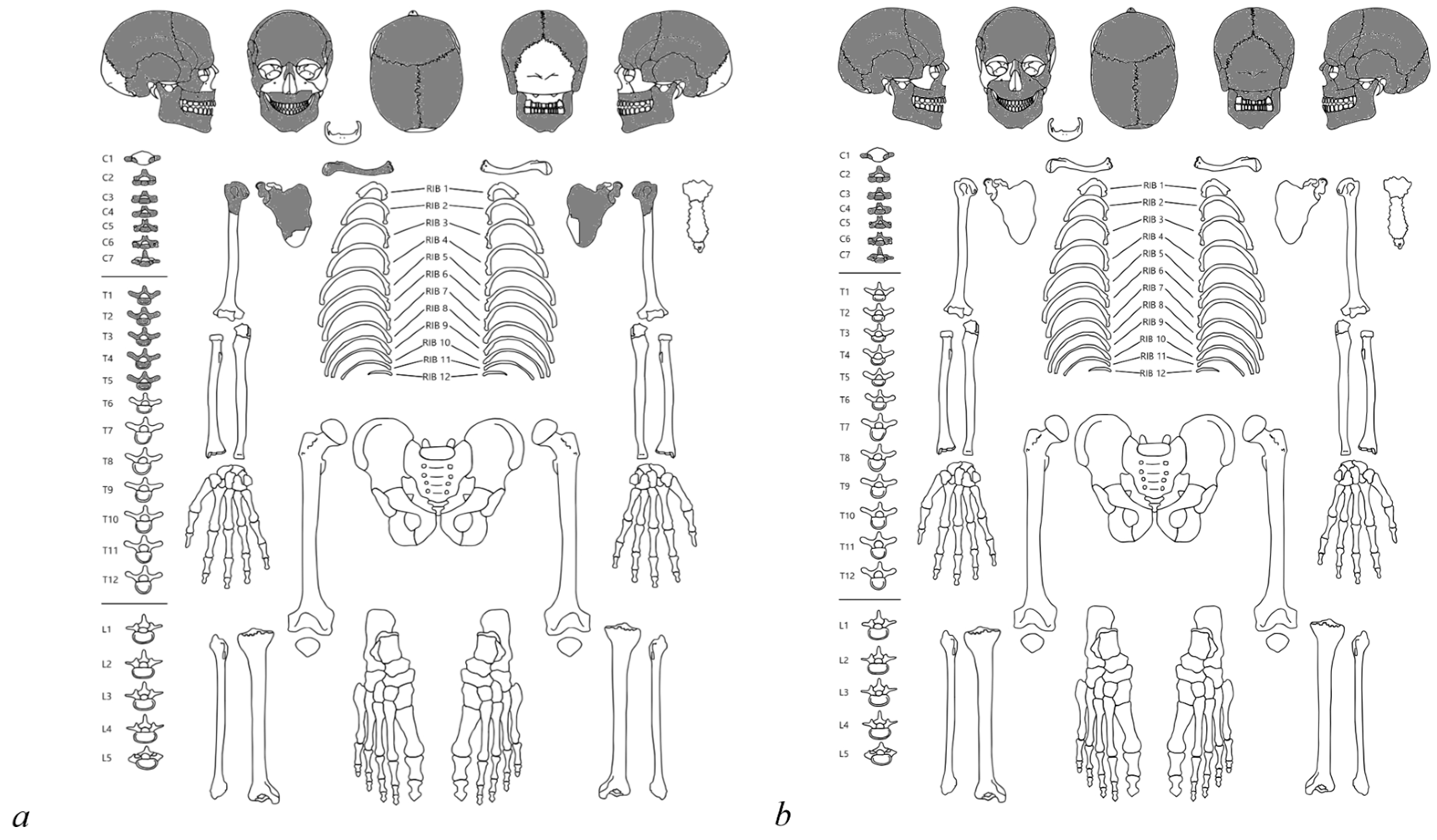
The skeleton M85/S1 presents fragmentary preservation (Figure 4a). The recovered skull bones include frontal, parietals, temporals, nasals, zygomatics, maxillae, and mandible. The post-cranial inventory is also limited, comprising cervical vertebrae (C1–C7) and thoracic (T1–T5) vertebrae, both humeral proximal epiphyses, nearly complete scapulae, and the right clavicle. The completeness index for this skeleton is CI M85/S1 = 13/28 × 100 = 46.43%. The preservation is rated as fair. The completeness is strongly biased towards the cranium and superior axial skeleton, with complete loss of the pelvic girdle and lower limbs (Table 1). There are noticeable oxidation staining marks on the temporal surfaces, suggesting the in situ presence of associated metallic artefacts. Additionally, the exocranial surface of the frontal and parietal bones shows delamination, a taphonomic alteration likely caused by environmental conditions such as soil moisture and pH fluctuations.
Estimation of age-at-death was conducted using the degree of cranial suture obliteration and dental wear. Analysis of the sagittal and coronal sutures reveals an incipient stage of endocranial and ectocranial synostosis, suggesting classification within the young adult category. The dentition is present and exhibits minimal dental wear (Grade 1), which corroborates the age estimation in the 20–30 years old. The resulting morphological data indicated a gracile cranial structure, characteristic of the female sex. Specifically, the glabella and supraorbital ridges are low-expressed, and the mastoid processes are minimally developed. Based on these cranial indicators, the skeleton M85/S1 was estimated to belong to a female.
The skeleton M24/S4 is even more fragmentary. The preserved bones are limited to skull elements (i.e., frontal, parietals, temporals, maxillae, left nasal, left zygomatic, and mandible) and the cervical vertebrae (Figure 4b). The completeness index for this skeleton is CI M24/S4 = 8/28 × 100 = 28.57% (Table 1). The preservation is classified as poor, and it is almost exclusively limited to fragmented skull elements and the cervical spine. This low CI highlights the significant limitations imposed on the analysis of biological profile.
Age-at-death estimation was derived from the analysis of the cranial sutures, which are open, without synostosis. This indicator, together with the dental wear of Grade 1, places the individual in the young adult category, with an estimated age between 20 and 30 years. Analysis of the skull morphological traits for sex estimation indicated a gracile structure with subtle relief. The glabella is low-developed, the nuchal crest is barely perceptible, and the mastoid processes are reduced. The mandible, although partial, presents a slightly acute shape at the mental eminence, which is typically associated with the female sex and is concurrently gracile. Consequently, the skeleton M24/S4 is estimated to be female.

4.2. Dental Macrowear

The dentition of the M85/S1 skeleton consisted of 24 teeth. The I1, along with all anterior maxillary teeth except for the left I2 and the right M1 molar, were lost postmortem. All mandibular anterior teeth showed moderately advanced wear (Smith score 3), while the only preserved maxillary teeth (left I2) showed a very small wear facet (Smith score 1). The maxillary premolars, along with the left P4 and right r P3, were characterised by small wear (Smith score 2). The right P4 and left P3 showed moderately advanced wear (Smith score 3), as well as the molar teeth, with Scott scores ranging from 10 to 18.
In the M24/S4 human skeleton, 21 teeth were present. Most right mandibular teeth were missing along with the bone structure, except for the I1, which was lost postmortem. The left P3-P4, the third M3, and the right I1 were also postmortem lost. Both mandibular and maxillary molars were characterised by moderately advanced occlusal wear, with scores ranging from 12 to 21. All anterior teeth (i.e., incisors and canines) showed small wear facets (Smith score 2). The same wear intensity was assigned for the P3s, while the P4s showed moderately advanced wear (Smith score 3).

4.3. Activity-Induced Dental Modifications

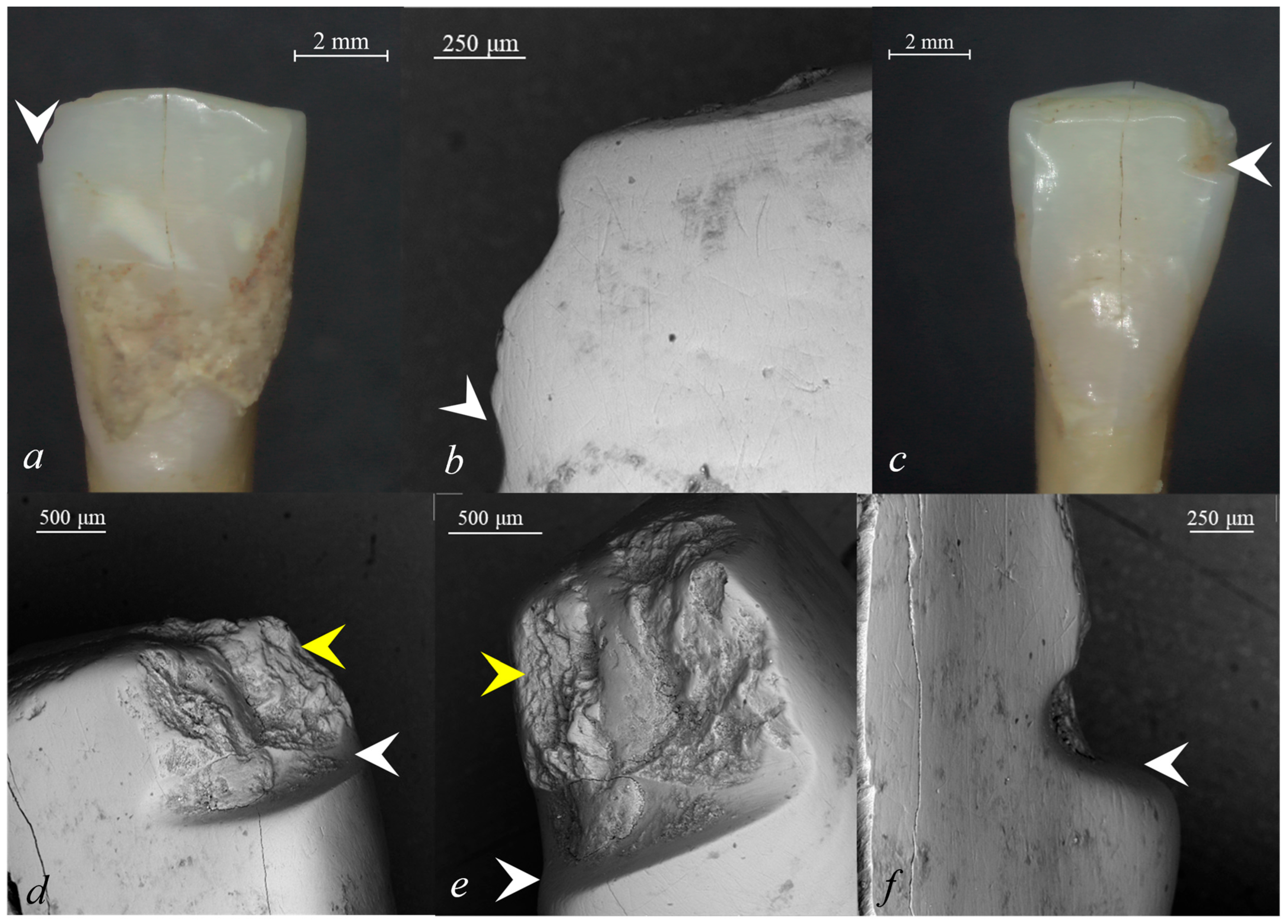
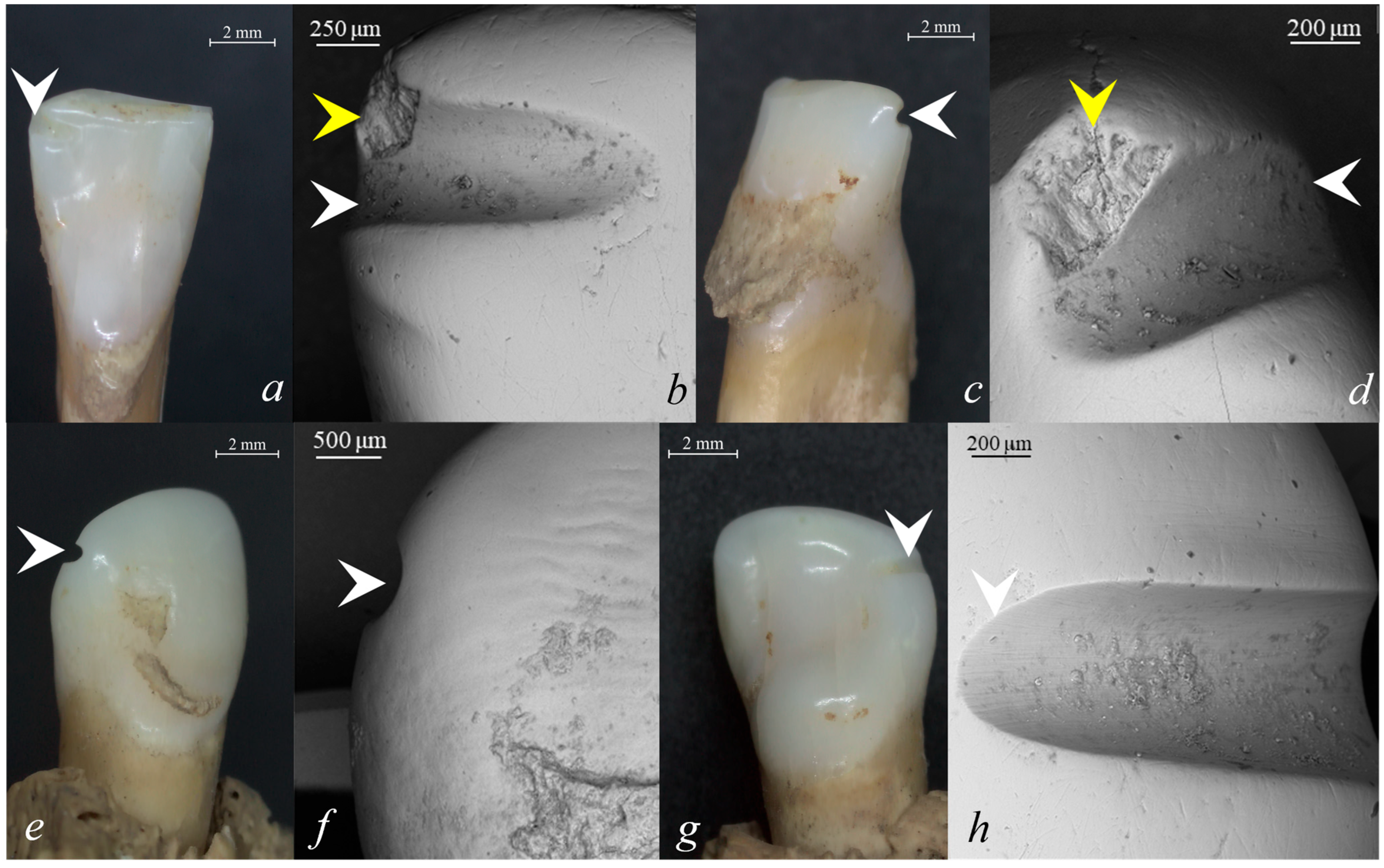
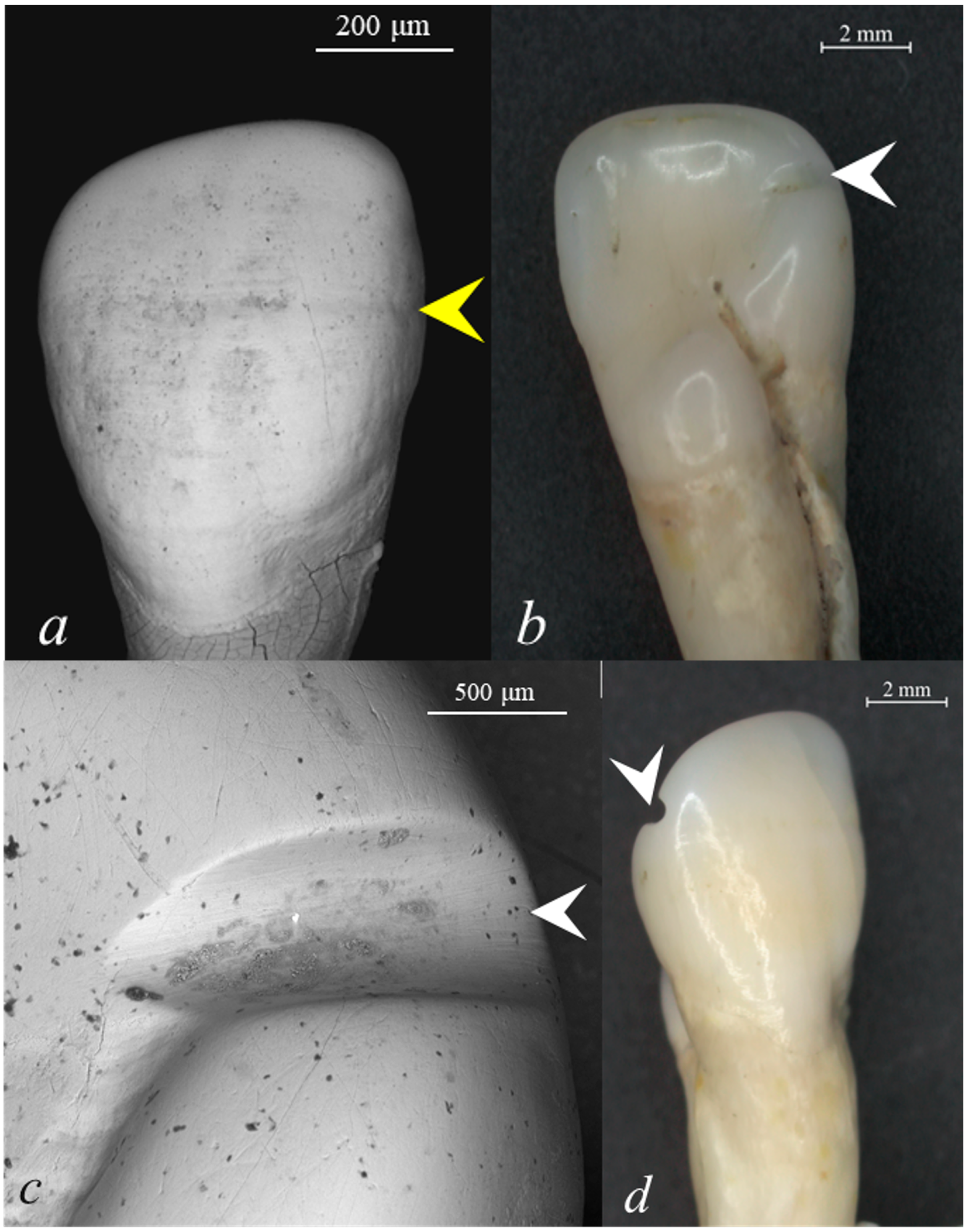
Two types of activity-induced dental modification (AIDM) features were identified in the M85/S1 dentition (female, young adult): grooves and chipping. Both I2 showed grooves in the upper third of the dental crown, on the distal surface. The grooves had a distolingual orientation, perpendicular to the cemento-enamel junction (Figure 5a–f and Figure 6a–d). The length and width of the groove on the I2 were approximately 1.5 mm and 0.5 mm, respectively (Figure 5a–f), while for the left I2, the length measured approximately 1 mm and the width approximately 0.5 mm, being reduced in size (Figure 6a–c).
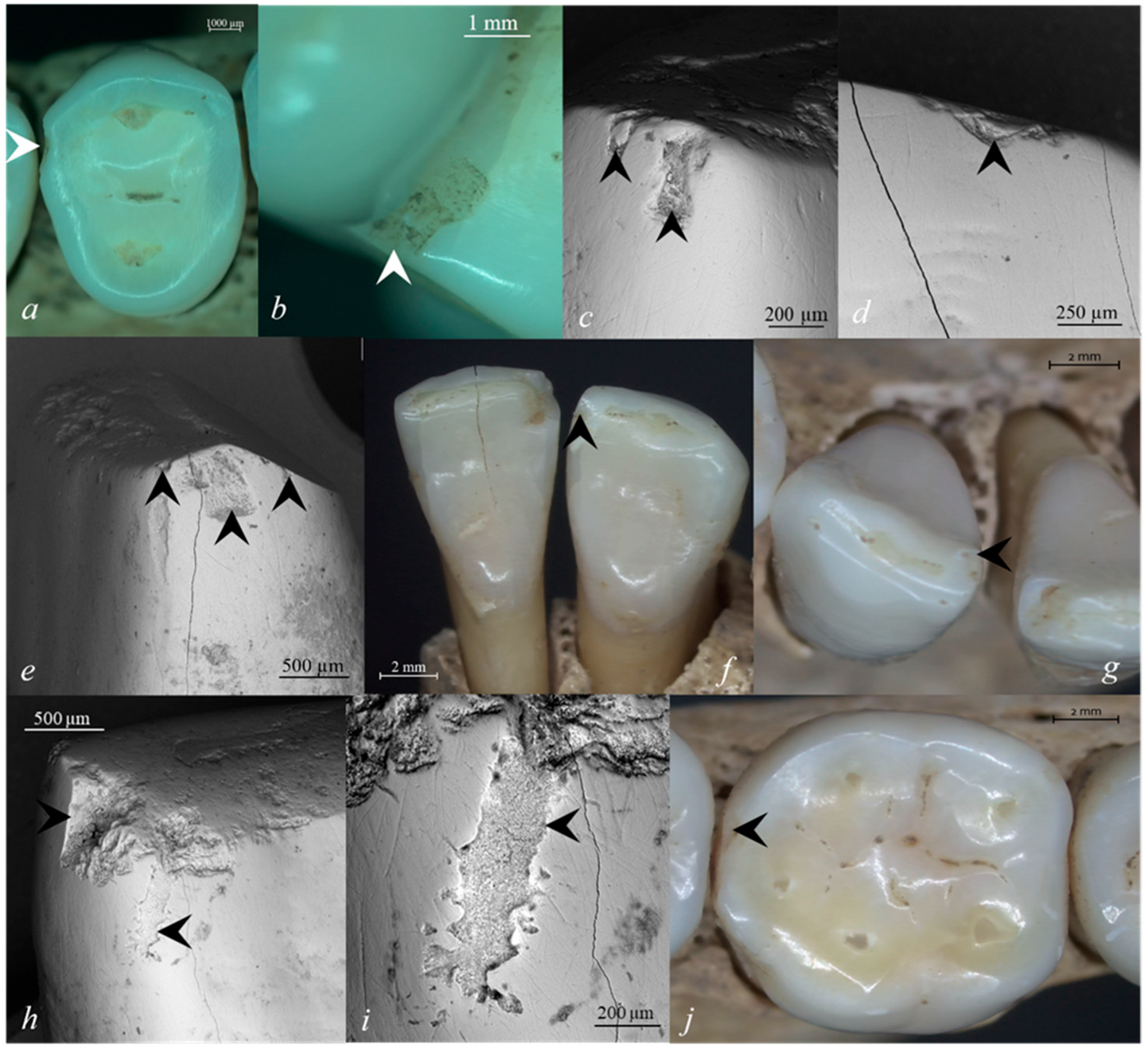
Both mandibular incisors showed chipping features adjacent to the grooves (Figure 5d,e and Figure 6b,d). Chipping was more accentuated on the right I2, affecting both enamel and dentin tissue (Figure 5d,e) compared to the antemortem fracture identified on the left I2 (Figure 6b,d). Furthermore, multiple antemortem microfractures were identified on the mesial corner (Figure 7c) and labial edge (Figure 7d) of the right I2 and on the mesial corner of the left I2 (Figure 7e). The right mandibular canine showed significant chipping on the mesial edge of the dental crown (Figure 7f–i). Around the chipped areas, several striae with vertical and oblique orientations were identified (Figure 7i). Among the posterior teeth of the M85/S1 skeleton, only the right M1 showed an enamel antemortem fracture (Figure 7j).
During the optical screening of the M24/S4 dentition (female, young adult), the I2 showed grooves in the upper third of the dental crown, on its distal surface, near the incisal corner, which had a nearly perpendicular distolingual orientation relative to the longitudinal axis of the teeth (Figure 8).
The left I2 also showed a small groove on the distal surface (Figure 6e–h). The orientation was distolingual with a width of less than 0.5 mm and a length of less than 1 mm (Figure 6e–h). Scanning electron microscopy showed numerous fine, parallel striae on the groove surfaces (Figure 5e and Figure 6b,d,h). Additionally, several striae with different orientations were observed outside the contours of the grooves on the labial surface of right I2 (Figure 5b), on the lingual surface of left I2 (Figure 6b), and on the lingual surface of the left I2 (Figure 6h).
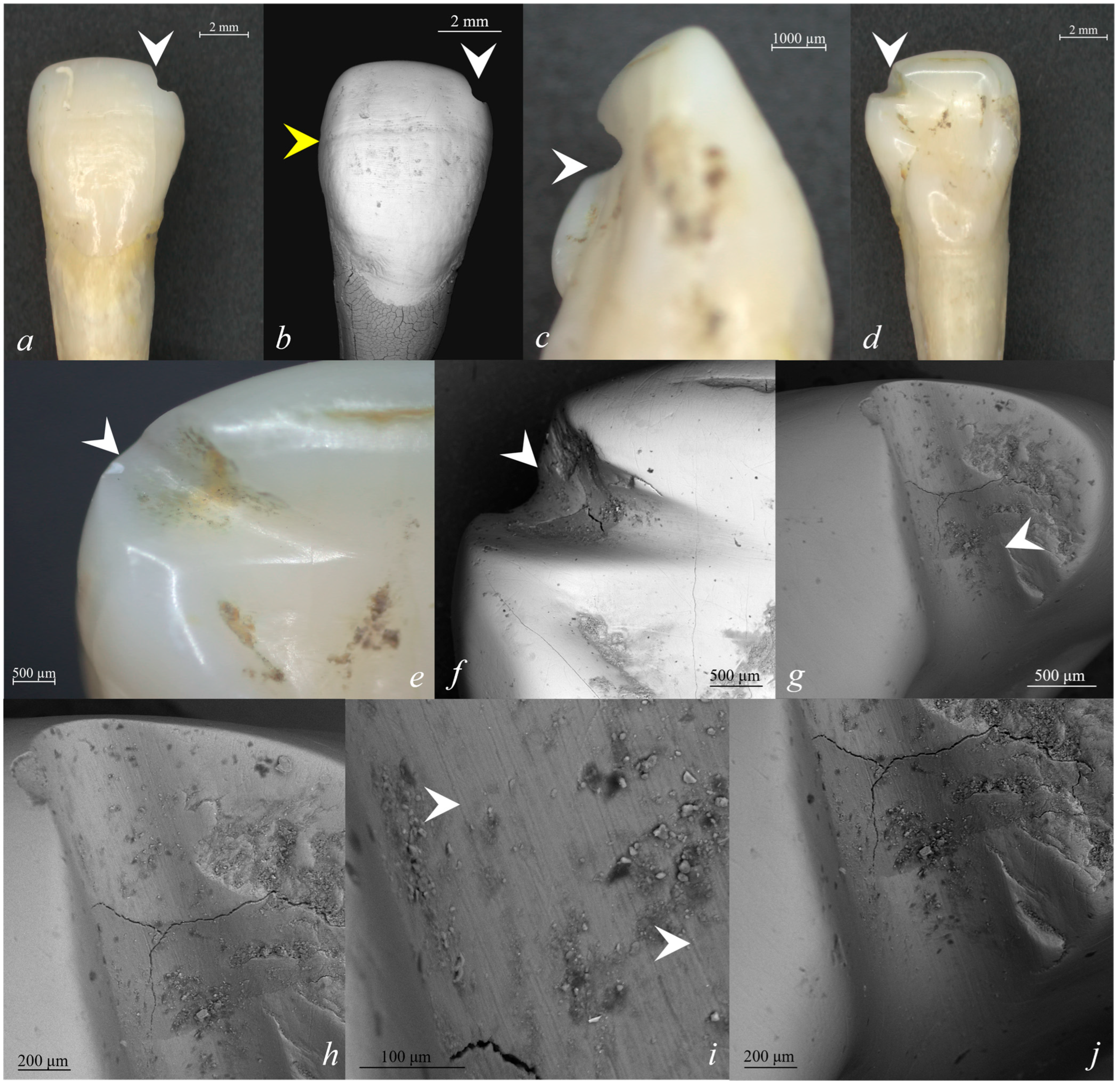
The groove identified on the right I2 is more evident in size, as it is larger in length and width (length ≈ 1.5 mm, width ≈ 1 mm) (Figure 9a–j) compared to the groove observed on the left I2 (length ≈ 1 mm, width ≈ 0.6 mm) (Figure 8b–d). There is no exposure of the pulp chamber, though greater dentine exposure could be identified in the right I2 compared with its antimere (Figure 8b,c and Figure 9e,f). When scanning electronic microscopy was performed, numerous fine parallel striae could be identified throughout the entire surface of both grooves (Figure 8c and Figure 9h–j). Beyond the contours of the grooves on the lingual surface of the incisors, several striae with different lengths and orientations could be identified (Figure 8c). During the microscopical inspection of both labial surfaces of I2, linear enamel hypoplasia was identified (Figure 8b and Figure 9b). Chipping was identified on the right I2 near the groove (Figure 9f) and on the left P3 on the distal surface (Figure 7a,b). No additional evidence of extra-masticatory dental wear was identified in the M24/S4 dentition.

4.4. Dental Calculus

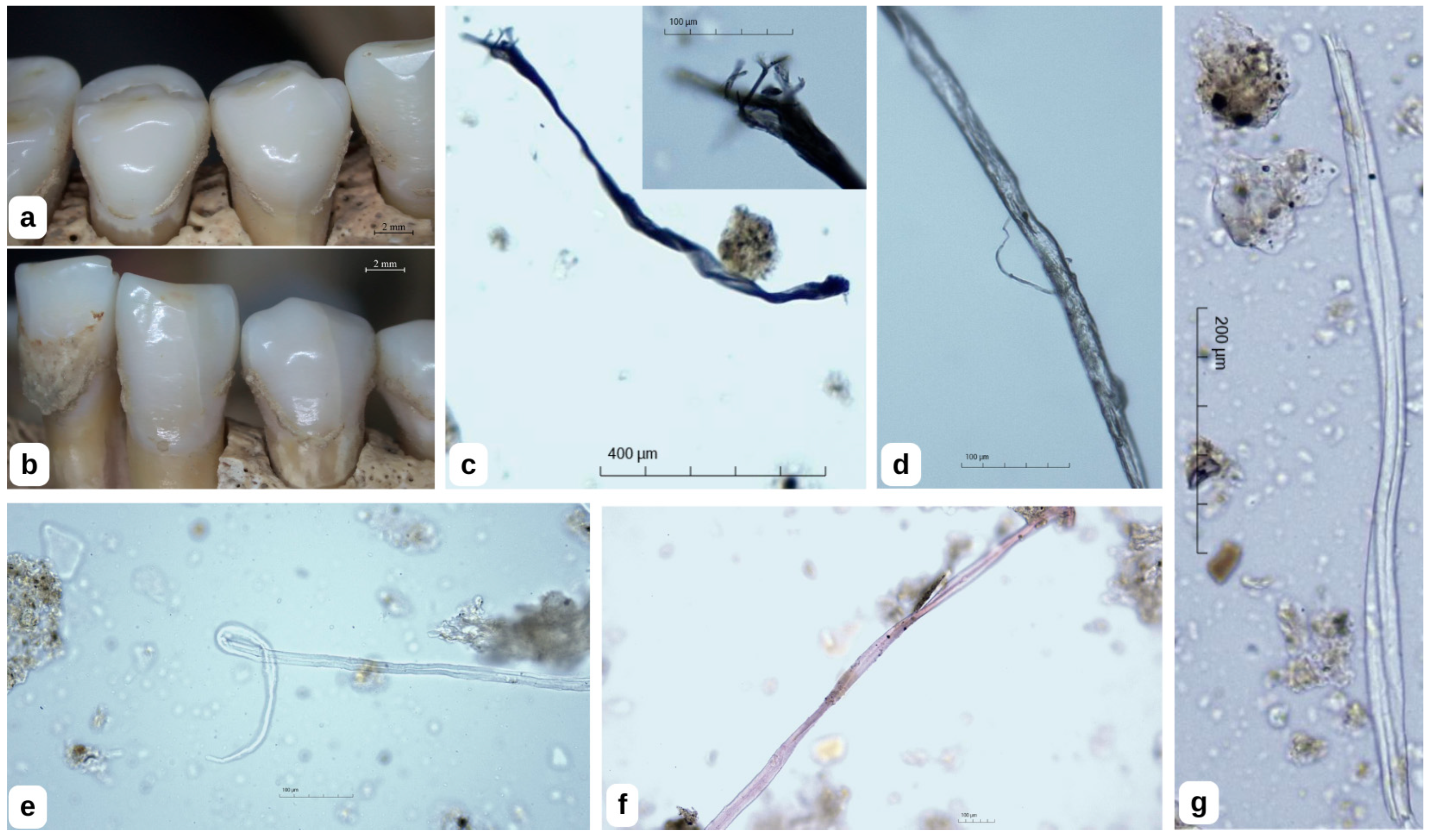
Dental calculus analysis from individuals M24/S4 and M85/S1 revealed the presence of textile fibres (Figure 10 and Figure 11).
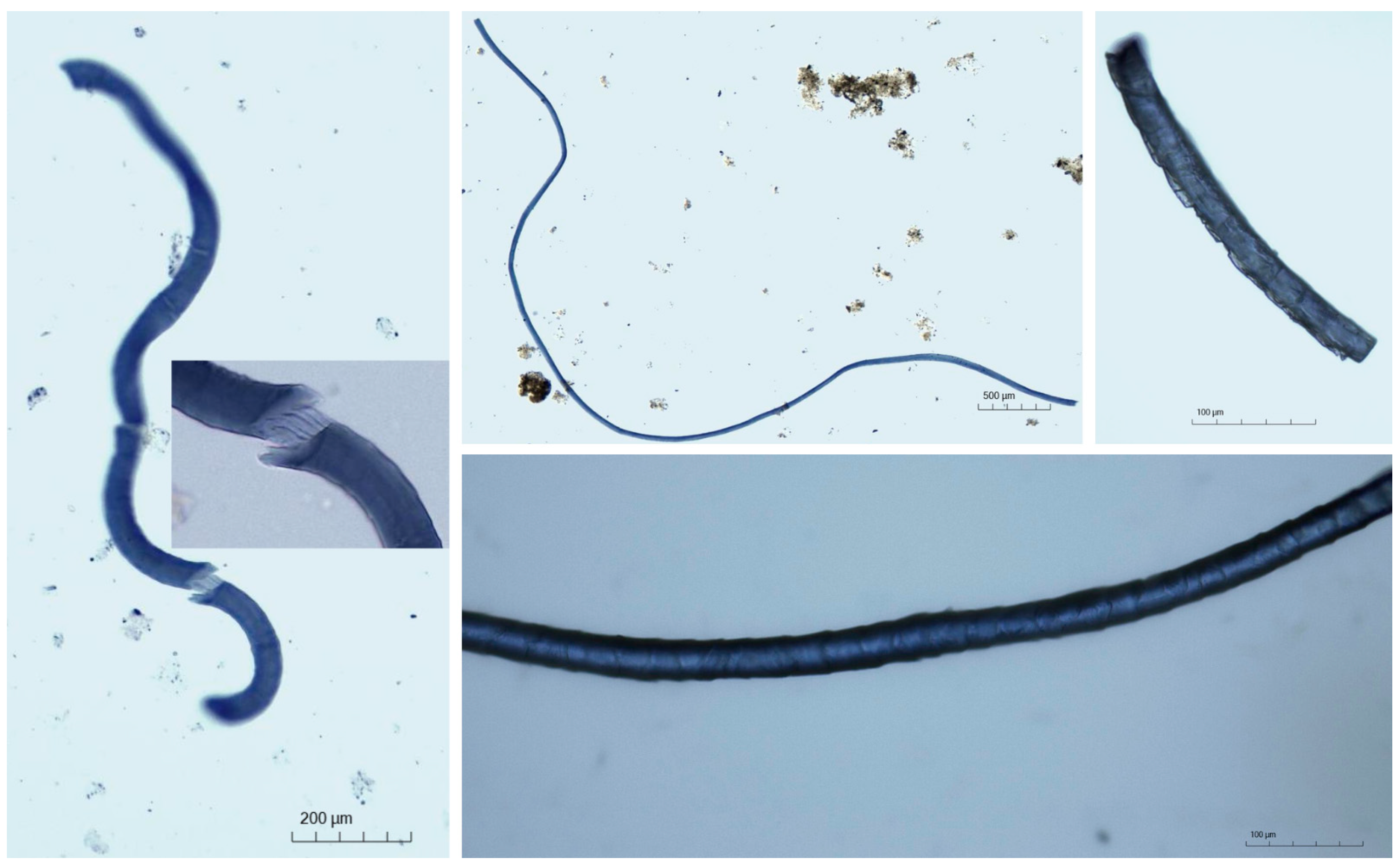
A total of 65 fibres were identified in the M24/S4 sample and 69 fibres in the M85/S1 sample. Both samples contained plant fibres, most likely cotton, hemp, and flax, as well as animal fibres identified as wool (Figure 11). Some fibres were coloured, predominantly blue, with a smaller number displaying pink and brown hues, while others were uncoloured.

5. Discussion

The two analysed skeletons (i.e., M85/S1 and M24/S4) reflect moderate-to-poor preservation, resulting in the exclusive reliance on the cranial and dental indicators for age-at-death and sex estimations. As a biological profile, both skeletons are estimated as females with the age-at-death falling within the young adult (20–30 years).

5.1. AIDMs Related to Spinning-Stage Fibre Manipulation

Both I2 and left I2 belonging to M85/S1, and the I2 belonging to M24/S4, showed grooves with a distolingual orientation characterised by smooth surfaces and parallel striations. Occlusal and non-occlusal grooves present on anterior teeth, usually incisors, are usually interpreted as forms of cordage, sinew, yarn, and different types of plant and animal fibres [2,16,24,68,69]. This type of AIDM was reported in prehistoric, medieval, and contemporary periods of archaeological populations [17,20,23,24]. These features resulted from tooth-using activities, adding to the shape of these special types of wear, being the result of everyday life activities [16]. In our study, the groove morphology is related to repetitive behaviour over an extended period and suggests size uniformity of fibres, which also indicates efficiency in craftwork. The morphology of grooves indicates small-diameter fibres that could probably be correlated with drafting during spinning, in which the fibres were pulled across the distal surface of incisors and could involve one tooth or the two antimere teeth. During spinning, controlled drafting of fibres is essential for regulating yarn thickness and maintaining uniform tension prior to twisting. In this context, the anterior teeth may serve as a temporary stabilising and tension-regulating element, allowing fibre bundles to be aligned and separated before twist insertion. The intermittent use of the mouth may also facilitate the production of finer yarn through improved control of fibre cohesion. Such biomechanically functional gestures provide a plausible explanation for the patterned abrasion observed in the present cases. In both human dentitions, the antimere teeth were involved, but to a different extent, the grooves situated on the right incisors showed increased length and width. Although our cases describe fibre manipulation during spinning by female individuals, no firm conclusions can be drawn regarding the female sex linking activities due to the limited sample size. Moreover, it is unlikely that these practices were representative of households of the period; rather, they appear to have been confined to a restricted subset of individuals.
Nevertheless, the literature to date highlights a female sex link related to fibre manipulation and spinning activities [2,68,70]. A study regarding the evidence for fibre processing in Italian Eneolithic/Bronze Age skeletal remains highlighted a sex division in fibre manipulation using teeth, with a higher frequency of AIDMs on anterior teeth in females, whereas only one male exhibited AIDMs [2]. In a study conducted by Lorkiewicz (2011) [71] on non-alimentary use of teeth in Neolithic skeletal series, 12 females out of 76 human individuals showed occlusal grooves on incisors and canines, which were probably produced by passing fibres through teeth. By contrast, only four males presented grooves on anterior teeth, suggesting a possible sex-related difference regarding fibre drafting and spinning-related activities [2]. AIDMs related to fibre drafting during spinning are typically identified on the incisors and canines, and occur less frequently on the premolars and molars [2]. Activity-induced dental modifications related to fibre manipulation tend to affect adult individuals, especially middle-aged and older adults, increasing with age, while subadults are less affected [2,71]. Grooves are associated with continuous and repetitive abrasion of enamel surfaces by soft fibres [26]. The macroscopic identification of grooves suggests that the activity was sustained over a sufficient period to allow their development [71]. Although grooves develop initially at the microscopic level in relatively shorter timescales, they provide guidance to maintain the uniformity of the fibres and further development of the macroscopic groove, which represents a cumulative result of a repetitive activity over a longer period [63].
Chipping was also identified in the two human dentitions, mainly on anterior teeth, with multiple features in the M85/S1 skeleton. The mandibular I2 incisors of the M85/S1 skeleton showed multiple chipping features near the well-delineated surface of the grooves, as well as on other tooth surfaces of the same teeth and the canine teeth. Chipping of the posterior teeth is likely associated with dietary factors such as hard food consumption and foods that contain grit introduced during preparation [20,69].
The anterior-dominant chipping patterns can be related to the use of teeth as tools, and can result from task-related activities, while chipping in posterior teeth can be linked to dietary factors [25,64,72]. The co-occurrence of chipping and atypical wear on the same tooth could indicate a common causal agent, supporting the interpretation that teeth were used as a third hand [18,19]. The interpretation must rely on archaeological and ethnographic information that supports the idea that the extra-masticatory wear was caused by fibre manipulation.

5.2. Archaeological Textile Materials

The rich archaeological discoveries from the St. Athanasius Church in the 18th–19th centuries correlate with a period of relative stability and intensified circulation of consumer goods, including high-quality textiles [49]. Although textile materials are rarely preserved in archaeological contexts due to the acidic nature of the soil and the perishable character of the material itself, the artefact collection of the Museum of History and Archaeology within ICEM Tulcea includes preserved textile or fabric remains chronologically comparable to the findings from St. Athanasius Church. These derive from the grave inventories of burials investigated at Isaccea—St. George Church, Enisala—La Biserică (18th–19th centuries) [73,74,75], and Tulcea (19th century) (unpublished data). Furthermore, during the archaeological excavations at Luncavița, Cetățuia site, the funerary inventory of a female included “a textile thread (silk?), twisted with a copper wire, which belonged either to the applied decoration of an item of dress (veil) or to a garment” [76].
Textile fibres (cotton, hemp, flax, silk, and wool) and finished textile products are explicitly documented in Ottoman customs and commercial registers of the ports of Tulcea and Isaccea during the 18th–19th centuries. The Danubian ports of Tulcea and Isaccea functioned as major commercial hubs for regional supply, both of raw materials and of processed textile goods. The structure of imports and local production reflects a mixed economy, in which everyday textiles were primarily derived from local resources, while fine and luxury textiles were imported from the Orient [77,78]. According to Gustav Adolf Ramsay, in his Travel Journal through Moldavia and Dobruja (1830), with reference to the village of Mănăstirești (Niculițel, present-day Tulcea County), “the inhabitants […] have large orchards of fruit trees and are engaged in silkworm rearing, which constitutes their principal occupation” and nearly “all the inhabitants” were involved, indicating an intensive orientation toward silk production within a mixed community [79].
Building on this documentary and archaeological framework, historical and agronomic sources allow a clearer reconstruction of the local base of textile fibre production in Dobruja during the modern period. Traditionally cultivated textile plants in the region were hemp (Cannabis sativa) and flax (Linum usitatissimum), both well-adapted to local pedoclimatic conditions and fully integrated into a rural subsistence economy [80,81].
Hemp was by far the most widespread textile plant, being consistently cultivated in villages across northern Dobruja, particularly in areas close to the Danube and its floodplains, where higher humidity favoured its development. The fibres obtained were used primarily for coarse textiles, heavy cloth, ropes, and other utilitarian products essential to everyday life. Contemporary observers explicitly describe hemp as a constant presence in Dobrujan households, embedded within a mixed agricultural economy oriented towards domestic consumption rather than market production [80,81].
Flax appears less frequently in the sources but is nevertheless documented as a stable component of local agriculture, cultivated to produce finer textiles intended mainly for clothing and household use. Although flax did not reach the scale of hemp cultivation, it is consistently included among the industrial plants of the region, indicating its functional importance within rural households [82].
Cotton is mentioned only sporadically in the historical literature and appears to have been cultivated experimentally on a limited scale, mainly in southern Dobruja, under the influence of Ottoman agricultural traditions. Cotton is reported as being cultivated within the Ottoman Empire, including Dobruja; however, this activity remained limited and non-systematic, preventing cotton from emerging as a significant component of the regional textile economy [80].
Silk and sericulture likewise occupied a marginal position. Silk is mentioned only in general terms, without any concrete observations regarding silkworm breeding or the presence of mulberry plantations in Dobruja [79]. A more detailed discussion is provided by M. D. Ionescu, who explicitly characterises sericulture as weakly represented, sporadic, and lacking economic significance; mulberry plantations are described as rare and unorganised, with no impact on local agricultural production [81]. The absence of silk, sericulture, and mulberry cultivation from comprehensive post-1878 agricultural syntheses, which otherwise provide systematic inventories of crops and industrial plants, suggests the very limited economic relevance of sericulture in the regional economy during this period [82].
Wool, obtained through sheep husbandry, constituted another essential fibre, closely linked to the pastoral economy characteristic of Dobruja. Spinning and weaving were carried out at the household level, primarily during the winter months, as complementary activities to agricultural and pastoral labour. Woollen textiles were produced almost exclusively for self-consumption and served for clothing, bedding, and other everyday necessities, without generating an organised or market-oriented production [81,82].
Overall, the available evidence indicates that textile fibre production in Dobruja relied predominantly on local resources. Hemp and flax formed the backbone of traditional textile manufacture, complemented by wool within a pastoral framework, while cotton and silk remained peripheral, experimental, or sporadic. This pattern aligns closely with the archaeological data and supports the interpretation that everyday textiles were largely produced locally, whereas fine or luxury fabrics, particularly silk, were primarily acquired through trade networks rather than local production [80,81,82].
Considering this predominantly local and household-based system of textile fibre production, the archaeological record of textiles must be evaluated with particular attention to issues of preservation and recovery. Organic fibres such as hemp, flax, wool, and cotton are inherently perishable and are therefore rarely preserved in conventional archaeological contexts, which complicates direct correlations between production, use, and material survival. In this context, the identification of a high number of textile fibres in the material analysed in the present study does not contradict these general limitations but rather highlights the role of dental calculus as a favourable micro-context for the preservation of plant and animal fibres associated with textile materials and related activities, as also documented by recent bioarchaeological research [31,83,84]. Within this methodological framework, the results presented here provide a complementary line of evidence to historical and archaeological sources, allowing aspects of textile use and exposure to be documented even in the absence of macroscopic remains.

5.3. Dental Wear and Textile Production in a Household Context

The interpretation of the dental abrasion and calculus micro-remains should be grounded primarily in the observed bioarchaeological evidence. The distolingually oriented grooves documented on the anterior teeth, characterised by smooth surfaces and parallel microstriations, together with the identification of plant and animal fibres in dental calculus, indicate repeated extra-masticatory contact with fibrous materials. The identification of plant and animal fibres in dental calculus is interpreted here primarily as micro-contextual evidence of direct exposure to textile materials and does not allow differentiation of specific technical gestures according to fibre type. These features may be interpreted as material traces consistent with fibre manipulation and support the functional interpretation proposed above.
Within a textile chaîne opératoire perspective, textile production comprises successive stages including fibre drafting during spinning, twisting, and finishing. As outlined by Andersson Strand (2012) [85], textile production involves repetitive, technically controlled gestures embedded in daily practice. In many historical and ethnographic contexts, the mouth may function intermittently as an auxiliary stabilising element during fibre drafting or thread preparation. The morphology, orientation, and microstriations of the grooves observed in the Niculițel assemblage are consistent with such repeated gestures, progressively recorded in the dental tissues through cumulative use.
The archaeological and historical data presented above indicate that textile fibre production in Dobruja during the Ottoman and early modern periods was predominantly household-based and relied largely on locally available raw materials. In this context, domestic production should not be equated with sporadic or incidental activity. Comparative studies of textile production [86,87] demonstrate that household manufacture may involve sustained time investment and technical consistency without necessarily implying formally organised workshop structures. The dental evidence documented here is compatible with repeated fibre-processing practices carried out within such a domestic framework.
The term “subsistence”, when applied to household textile production, therefore requires careful qualification. As discussed by Costin (2020) [88], the absence of architecturally distinct workshops does not by itself define the scale or organisation of production. Nevertheless, the limited sample size analysed in the present study does not permit inferences regarding production intensity, standardisation of output, or broader economic integration. The dental wear patterns and calculus micro-remains instead provide evidence at the level of individual practice, indicating that fibre manipulation formed a recurrent component of daily activities for at least a subset of individuals within the community. By situating the dental evidence within the textile chaîne opératoire and the documented household-based economy of the region, the Ottoman and early modern historical context provides an interpretative framework through which these embodied practices may be assessed. The anterior dentition thus preserves a micro-archaeological record of repetitive fibre-related activity, contributing to a more nuanced understanding of everyday textile practices during the 15th–19th centuries in southeastern Romania.

6. Conclusions

The present study documents activity-induced dental modifications in two young adult female individuals whose skeletons were discovered in the necropolis of the 15th–19th centuries at St. Athanasius Church from Niculițel (southeastern Romania). Grooves identified mainly on the distal surfaces of the second incisors, characterised by smooth walls and parallel microstriations, are consistent with repetitive non-masticatory tooth use. Their morphology and orientation indicate fibre manipulation and the use of teeth as tools in textile-related activities.
This interpretation is supported by associated antemortem chipping on anterior teeth and by the identification of plant and animal fibres (hemp, flax, cotton, and wool) preserved in dental calculus. The calculus micro-remains provide direct evidence of contact with textile materials and strengthen the functional interpretation of the observed dental wear.
The results contribute to the reconstruction of everyday practices and household-level textile production during the Ottoman and early modern periods in Dobruja. More broadly, the combined analysis of activity-induced dental modifications and dental calculus micro-remains demonstrates the potential of such evidence for investigating non-dietary behaviours relevant to Quaternary research.

Author Contributions

Conceptualization, O.-M.C.-P. and M.D.; methodology, O.-M.C.-P., M.D., P.-I.D., A.D., A.-D.S. and L.B.; software, O.-M.C.-P., M.D. and A.D.; validation, O.-M.C.-P., M.D., A.D. and L.B.; formal analysis, O.-M.C.-P., M.D., P.-I.D., A.D. and A.-D.S.; investigation, O.-M.C.-P., M.D., P.-I.D., A.D., A.-D.S. and L.B.; resources, O.-M.C.-P., M.D. and L.B.; data curation, O.-M.C.-P., M.D. and A.D.; writing—original draft preparation, O.-M.C.-P., M.D., P.-I.D., A.D., A.-D.S. and L.B.; writing—review and editing, O.-M.C.-P., M.D. and L.B.; supervision, L.B. All authors have read and agreed to the published version of the manuscript.

Funding

This research received no external funding.

Data Availability Statement

The original contributions presented in this study are included in the article. Further inquiries can be directed to the corresponding author.

Conflicts of Interest

The authors declare no conflicts of interest.

References

  1. Forshaw, R. Windows into the past: Recent scientific techniques in dental analysis. Br. Dent. J. 2024, 236, 205–211. [Google Scholar] [CrossRef]
  2. Sperduti, A.; Giuliani, M.R.; Guida, G.; Petrone, P.P.; Rossi, P.F.; Vaccaro, S.; Frayer, D.W.; Bondioli, L. Tooth grooves, occlusal striations, dental calculus, and evidence for fiber processing in an Italian eneolithic/bronze age cemetery. Am. J. Phys. Anthropol. 2018, 167, 234–243. [Google Scholar] [CrossRef]
  3. Pradeep, L.; Kokila, G.; Gopinathan, P.A.; Guruswamy, S.; Nazir, S.H.; Chatterjee, A. Age estimation with cemental annulation using light, phase contrast and polarized microscopy. J. Microsc. Ultrastruct. 2021, 9, 55–60. [Google Scholar] [CrossRef]
  4. Buikstra, J.; Ubelaker, D. Standards for Data Collection from Human Skeletal Remains: Research Seminar; Arkansas Archaeological Survey Research Series, No 44; University of Arkansas Press: Fayetteville, AR, USA, 1994. [Google Scholar]
  5. Esclassan, R.; Grimoud, A.-M.; Ruas, M.; Donat, R.; Sevin, A.; Astie, F.; Lucas, S.; Crubezy, E. Dental caries, tooth wear and diet in an adult medieval (12th–14th century) population from Mediterranean France. Arch. Oral Biol. 2009, 54, 287–297. [Google Scholar] [CrossRef]
  6. Dias, G.J.; Prasad, K.; Santos, A.L. Pathogenesis of apical periodontal cysts: Guidelines for diagnosis in palaeopathology. Int. J. Osteoarchaeol. 2007, 17, 619–626. [Google Scholar] [CrossRef]
  7. Towle, I.; Davenport, C.; Irish, J.D.; De Groote, I. High frequency of dental caries and calculus in dentitions from a British medieval town. Arch. Oral Biol. 2023, 155, 105777. [Google Scholar] [CrossRef] [PubMed]
  8. Popovici, M.; Groza, V.-M.; Ciorpac-Petraru, O.-M.; Bacumenco-Pîrnău, L.; Bilavschi, G.; Bejenaru, L. Dental phenotypic variability in an archaeological human population of the 16th–19th centuries from Iaşi city (Romania). J. Archaeol. Sci. Rep. 2026, 69, 105512. [Google Scholar] [CrossRef]
  9. Caruso, A.; Nikita, E. Association between biological kinship and lifestyle in the Hellenistic-early Roman community of Menainon, Sicily. J. Archaeol. Sci. Rep. 2025, 61, 104870. [Google Scholar] [CrossRef]
  10. Popovici, M.; Groza, V.-M.; Bejenaru, L.; Petraru, O.-M. Dental morphological variation in Chalcolithic and Bronze Age human populations from North-Eastern Romania. Ann. Anat. 2023, 245, 152015. [Google Scholar] [CrossRef]
  11. Xia, J.; Zheng, J.; Huang, D.; Tian, Z.R.; Chen, L.; Zhou, Z.; Ungar, P.S.; Qian, L. New model to explain tooth wear with implications for microwear formation and diet reconstruction. Proc. Natl. Acad. Sci. USA 2015, 112, 10669–10672. [Google Scholar] [CrossRef] [PubMed]
  12. Petraru, O.-M.; Groza, V.-M.; Lobiuc, A.; Bejenaru, L.; Popovici, M. Dental microwear as a diet indicator in the seventeenth-century human population from Iasi City, Romania. Archaeol. Anthropol. Sci. 2020, 12, 184. [Google Scholar] [CrossRef]
  13. Kaidonis, J.A. Tooth wear: The view of the anthropologist. Clin. Oral Investig. 2008, 12, 21–26. [Google Scholar] [CrossRef]
  14. Smith, A.K.; Reitsema, L.J.; Williams, F.L.E.; Boano, R.; Vercellotti, G. Sex- and status-based differences in medieval food preparation and consumption: Dental microwear analysis at Trino Vercellese, Italy. Archaeol. Anthropol. Sci. 2019, 11, 4789–4800. [Google Scholar] [CrossRef]
  15. Petraru, O.-M.; Bejenaru, L.; Popovici, M. Diet-related dental wear in archaeological human populations of Chalcolithic and Bronze Age from North-Eastern Romania. HOMO 2022, 73, 77–92. [Google Scholar]
  16. Willman, J.C.; Valera, A.C.; Silva, A.M. The embodiment of craft production in Bronze Age Portugal: Exceptional dental wear grooves in an individual from Monte do Vale do Ouro 2 (Ferreira do Alentejo, Portugal). Int. J. Osteoarchaeol. 2021, 31, 252–262. [Google Scholar] [CrossRef]
  17. Lozano, M.; Jiménez-Brobeil, S.A.; Willman, J.C.; Sánchez-Barba, L.P.; Molina, F.; Rubio, Á. Argaric craftswomen: Sex-based division of labor in the Bronze Age southeastern Iberia. J. Archaeol. Sci. 2021, 127, 105239. [Google Scholar] [CrossRef]
  18. Molnar, P. Dental wear and oral pathology: Possible evidence and consequences of habitual use of teeth in a Swedish Neolithic sample. Am. J. Phys. Anthropol. 2008, 136, 423–431. [Google Scholar] [CrossRef] [PubMed]
  19. Neves, D.; Silva, A.M.; Simões, F.; Wasterlain, S.N. More than they could chew: Activity-induced dental modifications in a Portuguese medieval–modern rural population. Int. J. Osteoarchaeol. 2022, 32, 856–865. [Google Scholar] [CrossRef]
  20. Bonfiglioli, B.; Mariotti, V.; Facchini, F.; Belcastro, M.G.; Condemi, S. Masticatory and non-masticatory dental modifications in the epipalaeolithic necropolis of Taforalt (Morocco). Int. J. Osteoarchaeol. 2004, 14, 448–456. [Google Scholar] [CrossRef]
  21. Lozano, M.; Subirà, M.E.; Aparicio, J.; Lorenzo, C.; Gómez-Merino, G. Toothpicking and periodontal disease in a Neanderthal specimen from Cova Foradà site (Valencia, Spain). PLoS ONE 2013, 8, e76852. [Google Scholar] [CrossRef]
  22. Erdal, Y.S. Occlusal grooves in anterior dentition among Kovuklukaya inhabitants (Sinop, Northern Anatolia, 10th century AD). Int. J. Osteoarchaeol. 2008, 18, 152–166. [Google Scholar] [CrossRef]
  23. Rodrigues, A.C.; Silva, A.M.; Matias, A.; Santos, A.L. Atypical dental wear patterns in individuals exhumed from a medieval Islamic necropolis of Santarém (Portugal). Anthropol. Sci. 2021, 129, 187–196. [Google Scholar] [CrossRef]
  24. Monaco, M.; Riccomi, G.; Minozzi, S.; Campana, S.; Giuffra, V. Exploring activity-induced dental modifications in medieval Pieve di Pava (central Italy, 10th-12th centuries AD). Arch. Oral Biol. 2022, 140, 105449. [Google Scholar]
  25. Walsh, S. Early evidence of extra-masticatory dental wear in a Neolithic community at Bestansur, Iraqi Kurdistan. Int. J. Osteoarchaeol. 2022, 32, 1264–1274. [Google Scholar] [PubMed]
  26. Romero, A.; Rial, R.E.B.; Maestre, F.J.J.; Herrero-Otal, M.; Cuesta-Aguirre, D.R.; Fiorin, E.; Malgosa, A.; de Miguel Ibáñez, P.; Padilla, J.A.L. Dental evidence of textile-related task activities in the Bronze Age site of Laderas del Castillo (Alicante, Spain). J. Archaeol. Sci. Rep. 2024, 60, 104818. [Google Scholar] [CrossRef]
  27. Frayer, D.W. The dental remains from Krškany (Slovakia) and Vedrovice (Czech Republic). Anthropologie (1962-) 2004, 42, 71–103. [Google Scholar]
  28. Ungar, P.S.; Grine, F.E.; Teaford, M.F.; Pérez-Pérez, A. A review of interproximal wear grooves on fossil hominin teeth with new evidence from Olduvai Gorge. Arch. Oral Biol. 2001, 46, 285–292. [Google Scholar] [CrossRef]
  29. Estalrrich, A.; Alarcón, J.A.; Rosas, A. Evidence of toothpick groove formation in Neandertal anterior and posterior teeth. Am. J. Phys. Anthropol. 2017, 162, 747–756. [Google Scholar] [CrossRef]
  30. Radini, A.; Nikita, E.; Buckley, S.; Copeland, L.; Hardy, K. Beyond food: The multiple pathways for inclusion of materials into ancient dental calculus. Am. J. Phys. Anthropol. 2017, 162, 71–83. [Google Scholar] [CrossRef]
  31. Herrero-Otal, M.; Canales, L.; Agustí, B.; Rosillo, R.; Lloret, À.B.; Barberà, A.; Palomo, A.; Piqué, R.; Terradas, X.; Malgosa-Morera, A. An Experimental and Methodological Approach of Plant Fibres in Dental Calculus: The Case Study of the Early Neolithic Site of Cova del Pasteral (Girona, Spain). Open Archaeol. 2025, 11, 20250046. [Google Scholar] [CrossRef]
  32. Petraru, O.-M.; Groza, V.-M.; Bejenaru, L. Dental macrowear as marker of diet: Considerations on the skeletal sample from the 17th century necropolis of Iaşi (Iaşi County, Romania). ARA 2018, 55, 45–54. [Google Scholar]
  33. Zejdlik, K.; Bethard, J.D.; Nyárádi, Z.; Gonciar, A. Interpreting the oral condition in medieval European populations from a bioarchaeological perspective. Dent. Anthropol. 2019, 32, 77–88. [Google Scholar]
  34. Petraru, O.-M.; Popovici, M.; Groza, V.-M.; Bacumenco-Pîrnau, L.; Cordoș, C.; Bejenaru, L. Dental macrowear evaluation of the human skeletal sample discovered at the “Vovidenia” church in Iași (România) 16th–17th centuries. Arheol. Mold. 2023, XLVI, 238–252. [Google Scholar]
  35. Tencariu, F.A.; Asăndulesei, A.; Simalcsik, A.; Brașoveanu, C.M.; Petraru, O.-M.; Bejenaru, L.; Danu, M.A.; Drob, A.; Brunchi, R.A.; Pîrnău, R.G. A bolt from the blue: Investigations of a singular Bronze Age grave from the Chalcolithic site Ruginoasa (north-eastern Romania). Archaeol. Anthropol. Sci. 2022, 14, 81. [Google Scholar] [CrossRef]
  36. Ciorpac-Petraru, O.-M.; Bejenaru, L. Srudiul uzurii dentare la seria scheletică descoperită la biserica „Sfinții Voivozi” și „Tăierea Capului Sfântului Ioan Botezătorul”. In Biserica de Lemn „Sfinții Voievozi”și „Tăierea Capului Sfântului Ioan Botezătorul”. Cercetări Arheologice; Diaconu, V., Ed.; Editura „Constantin Mătasă”: Piatra-Neamț, Romania, 2025; pp. 85–101. [Google Scholar]
  37. Radu, C.; Mihalache, I.; Fazecaș, G.; Gogâltan, F.; Kelemen, B. Bioarchaeological study of a skeleton belonging to the Suplacu group, dated in the Middle Neolithic period. ActaMP 2013, 35, 73–78. [Google Scholar]
  38. Ciorpac-Petraru, O.-M.; Popovici, M.; Groza, V.-M.; Bacumenco-Pîrnău, L.; Bilavski, G.; Bejenaru, L. Caracterizarea uzurii dentare la o serie scheletică umană din secolele XVII-XIX din orașul Iași—Studiu preliminar. In Orașe și Târguri din Moldova în Perioada de Tranziție de la Evul Mediu la Epoca Moderna; Bejenaru, L., Stanc, M.S., Bacumenco-Pîrnau, L., Eds.; Editura Mega: Cluj-Napoca, Romania, 2024; pp. 173–196. [Google Scholar]
  39. Brătescu, C. Pământul Dobrogei, în 1878–1928. In Dobrogea. Cincizeci de Ani de Viață Românescă; Bratescu, C., Georgescu, I., Eds.; Cultura Națională: București, Romania, 1928; pp. 3–65. [Google Scholar]
  40. Coteț, P.V.; Popovici, I. Județele Patriei. Județul Tulcea; Editura Academiei Republicii Socialiste România: București, Romania, 1972; p. 168. [Google Scholar]
  41. Popescu, N. Județul Tulcea. Relieful. In Enciclopedia geografică a României; Posea, G., Ed.; Editura Ştiinţifică şi Enciclopedică: București, Romania, 1982; pp. 753–754. [Google Scholar]
  42. Ciulache, S. Județul Tulcea, Clima. In Eciclopedia geografică a României; Posea, G., Ed.; Editura Ştiinţifică şi Enciclopedică: București, Romania, 1982; p. 759. [Google Scholar]
  43. Baumann, V.H. Sângele Martirilor; Editura Arhiepiscopiei Tomisului: Constanța, Romania, 2004; p. 232. [Google Scholar]
  44. Barnea, I.; Ștefănescu, Ș. Din Istoria Dobrogei; Editura Academiei Republicii Socialiste Români: București, Romania, 1971; Volume III, p. 440. [Google Scholar]
  45. Baumann, V. Observații arheologice asupra poziției şi cronologiei așezărilor romane din zona de nord a Niculiţelului. Peuce 1976, 4, 109–124. [Google Scholar]
  46. Bătrâna, L.; Bătrâna, A. Contribuţii la cunoaşterea arhitecturii medievale din Dobrogea: Biserica Sf. Atanasie din Niculiţel (jud. Tulcea). Stud. Cercet. Istor. Veche Arheol. (SCIVA) 1977, 28, 531–552. [Google Scholar]
  47. Moisescu, C. Biserica Sf. Atanasie de la Niculiţel. Mag. Istor. 1999, 7, 51–54. [Google Scholar]
  48. Moisescu, C. Noi consideraţii asupra vechimii arhitecturii bisericii Sf. Atanasie din Niculiţel ( jud. Tulcea). In Istro-Pontica: Muzeul Tulcean la 50-a Aniversare; Iacob, M., Oberländer-Tîrnoveanu, E., Topoleanu, F., Eds.; Institutul de Cercetări Eco-Muzeale „Gavrilă Simion”: Tulcea, Romania, 2000; pp. 363–370. [Google Scholar]
  49. Stănică, A.-D.; Mocanu, M.; Grumăzescu, G.; Cernamoriți, R.; Nuțu, G.; Mihail, F.; Stănescu, R.; Micu, C.; Marcu, L.; Radu, G.; et al. Niculițel, jud. Tulcea, punct „Biserica Sf. Atanasie”, str. Isaccei, nr. 100. Cron. Cercet. Arheol. România 2024, 581–587. Available online: https://cronica.cimec.ro/Public/cca_pdf/cronica-cercetarilor-arheologice-campania_2023.pdf (accessed on 21 December 2025).
  50. Acsádi, G.; Nemeskéri, J.; Balás, K. History of Human Life Span and Mortality; Akadémiai Kiadó: Budapest, Hungary, 1970. [Google Scholar]
  51. Walrath, D.E.; Turner, P.; Bruzek, J. Reliability test of the visual assessment of cranial traits for sex determination. Am. J. Phys. Anthropol. 2004, 125, 132–137. [Google Scholar]
  52. Bello, S.M.; Thomann, A.; Signoli, M.; Dutour, O.; Andrews, P. Age and sex bias in the reconstruction of past population structures. Am. J. Phys. Anthropol. 2006, 129, 24–38. [Google Scholar] [CrossRef]
  53. Ruengdit, S.; Case, D.T.; Mahakkanukrauh, P. Cranial suture closure as an age indicator: A review. Forensic Sci. Int. 2020, 307, 110–111. [Google Scholar] [CrossRef]
  54. Meindl, R.S.; Lovejoy, C.O. Ectocranial suture closure: A revised method for the determination of skeletal age at death based on the lateral-anterior sutures. Am. J. Phys. Anthropol. 1985, 68, 57–66. [Google Scholar] [CrossRef]
  55. Brothwell, D.R. Digging up Bones; Cornell University Press: New York, NY, USA, 1981. [Google Scholar]
  56. Klales, A.R. MorphoPASSE: Morphological pelvis and skull sex estimation program. In Sex Estimation of the Human Skeleton: History, Methods, and Emerging Techniques; Klales, A.R., Ed.; Academic Press: London, UK, 2020; pp. 271–278. [Google Scholar]
  57. Bass, W.M. Human Osteology: A Laboratory and Field Manual, 5th ed.; Missouri Archaeological Society: Columbia, MO, USA, 2005. [Google Scholar]
  58. Novak, M. Dental health and diet in early medieval Ireland. Arch. Oral Biol. 2015, 60, 1299–1309. [Google Scholar] [CrossRef]
  59. Mahoney, P. Dental microwear from Natufian hunter-gatherers and early Neolithic farmers: Comparisons within and between samples. Am. J. Phys. Anthropol. 2006, 130, 308–319. [Google Scholar] [CrossRef]
  60. Smith, B.H. Patterns of molar wear in hunter–gatherers and agriculturalists. Am. J. Phys. Anthropol. 1984, 63, 39–56. [Google Scholar] [CrossRef]
  61. Scott, E.C. Dental wear scoring technique. Am. J. Phys. Anthropol. 1979, 51, 213–217. [Google Scholar] [CrossRef]
  62. Tomczyk, J.; Myszka, A.; Regulski, P.; Olczak-Kowalczyk, D. Case of pulp stones and dental wear in a Mesolithic (5900±100 bc) individual from Woźna Wieś (Poland). Int. J. Osteoarchaeol. 2020, 30, 375–381. [Google Scholar] [CrossRef]
  63. Díaz-Navarro, S.; García-González, R.; Cirotto, N.; Uriarte, M.H. New insight into prehistoric craft specialisation. Tooth-tool use in the Chalcolithic burial site of Camino del Molino, Murcia, SE Spain. J. Archaeol. Sci. Rep. 2023, 50, 104066. [Google Scholar] [CrossRef]
  64. Scott, G.R.; Winn, J.R. Dental chipping: Contrasting patterns of microtrauma in Inuit and European populations. Int. J. Osteoarchaeol. 2011, 21, 723–731. [Google Scholar] [CrossRef]
  65. Warinner, C.; Rodrigues, J.F.M.; Vyas, R.; Trachsel, C.; Shved, N.; Grossmann, J.; Radini, A.; Hancock, Y.; Tito, R.Y.; Fiddyment, S. Pathogens and host immunity in the ancient human oral cavity. Nat. Genet. 2014, 46, 336–344. [Google Scholar] [CrossRef]
  66. Cristiani, E.; Radini, A.; Edinborough, M.; Borić, D. Dental calculus reveals Mesolithic foragers in the Balkans consumed domesticated plant foods. Proc. Natl. Acad. Sci. USA 2016, 113, 10298–10303. [Google Scholar] [CrossRef]
  67. Cristiani, E.; Radini, A.; Borić, D.; Robson, H.K.; Caricola, I.; Carra, M.; Mutri, G.; Oxilia, G.; Zupancich, A.; Šlaus, M. Dental calculus and isotopes provide direct evidence of fish and plant consumption in Mesolithic Mediterranean. Sci. Rep. 2018, 8, 8147. [Google Scholar] [CrossRef]
  68. Waters-Rist, A.; Bazaliiskii, V.I.; Weber, A.; Goriunova, O.I.; Katzenberg, M.A. Activity-induced dental modification in Holocene Siberian hunter-fisher-gatherers. Am. J. Phys. Anthropol. 2010, 143, 266–278. [Google Scholar] [CrossRef]
  69. Fidalgo, D.; Silva, A.M.; Porfírio, E. Non-masticatory dental wear patterns in individuals exhumed from the Middle Bronze Age rock-cut tombs of Torre Velha 3 (Serpa, Portugal). Int. J. Osteoarchaeol. 2020, 30, 13–23. [Google Scholar] [CrossRef]
  70. Romero, A.; Ramírez-Rozzi, F.V.; De Juan, J.; Pérez-Pérez, A. Diet-related buccal dental microwear patterns in Central African Pygmy foragers and Bantu-speaking farmer and pastoralist populations. PLoS ONE 2013, 8, e84804. [Google Scholar] [CrossRef]
  71. Lorkiewicz, W. Nonalimentary tooth use in the Neolithic population of the Lengyel culture in central Poland (4600–4000 BC). Am. J. Phys. Anthropol. 2011, 144, 538–551. [Google Scholar] [CrossRef] [PubMed]
  72. Garralda, M.D.; Maureille, B.; Le Cabec, A.; Oxilia, G.; Benazzi, S.; Skinner, M.M.; Hublin, J.-J.; Vandermeersch, B. The Neanderthal teeth from Marillac (Charente, southwestern France): Morphology, comparisons and paleobiology. J. Hum. Evol. 2020, 138, 102683. [Google Scholar] [CrossRef]
  73. Vasiliu, I. Cimitirul medieval de la Isaccea. Biserica Sf. Gheorghe. Peuce 1995, 11, 373–409. [Google Scholar]
  74. Mănucu-Adameșteanu, G. Aspecte ale ritului și ritualului în lumina descoperirilor din necropola medievală de la Enisala. Peuce 1984, 9, 355–362. [Google Scholar]
  75. Stănică, A.-D.; Ailincăi, S.; Ignat, A.; Zvâncă, I. Noi Descoperiri Funerare de pe Teritoriul Localităţii Enisala (com. Sarichioi, jud. Tulcea). Peuce, S.N. 3–4. 2005–2006, 317–330. Available online: https://revistapeuce.icemtl.ro/wp-content/uploads/Arhiva-Peuce-Serie-noua/03-Peuce-SN-III-IV-2005-2006/19-Stanica-Ailincai-et-alii.pdf (accessed on 15 December 2025).
  76. Micu, C.; Stănică, A.; Mihail, F.; Maille, M. Observaţii asupra unor complexe funerare descoperite la Luncaviţa, punctul Cetăţuia. Peuce 2008, 6, 331–338. [Google Scholar]
  77. Maxim, M. Țările Române și Înalta Poartă. Cadrul Juridic al Relațiilor Româno-Otomane în Evul Mediu; Editura Enciclopedică: București, Romania, 1993; p. 299. [Google Scholar]
  78. Popescu, A. Portul Tulcea în documente otomane din secolul al XVI-lea. In Istro-Pontica 2. Studii şi Comunicări de Istorie a Dobrogei. Actele Sesiunii Naţionale de Comunicări Ştiinţifice ”Istro-Pontica. Tulcea—505 ani de la prima atestare documentară”; Stănică, A.D., Micu, C., Eds.; Institutul de Cercetări Eco-Muzeale, Editura Istros: Tulcea, Romania, 2014; pp. 23–32. [Google Scholar]
  79. Cernovodeanu, P.; Feneșan, C.; Filitti, G.; Gheorghe, A.; Ionescu, A.S.; Rădulescu-Zoner, Ș.; Stroia, M.; Taftă, L.; Tomi, R. Călători Străini Despre Țările Române, S.N. 2 (1822–1830); Editura Academiei Române: București, Romania, 2005; p. 648. [Google Scholar]
  80. Ionescu, I. Escursiune Agricolă în Câmpia Dobrogei; Tiparul Tribunei Române: București, Romania, 1879; p. 7. [Google Scholar]
  81. Ionescu, M.D. Dobrogea în Pragul Veacului al XX—Lea, Geografia Matematică, Fizică, Politică, Economică și Militară; Atelierele Grafice I. V. Socecü: București, Romania, 1904; p. 740. [Google Scholar]
  82. Brătescu, C. Dobrogea agricolă 1928. In Dobrogea. Cincizeci de ani de Viață Românescă; Bratescu, C., Georgescu, I., Eds.; Cultura Națională: București, Romania, 1928; pp. 485–526. [Google Scholar]
  83. Blatt, S.H.; Redmond, B.G.; Cassman, V.; Sciulli, P.W. Dirty teeth and ancient trade: Evidence of cotton fibres in human dental calculus from Late Woodland, Ohio. Int. J. Osteoarchaeol. 2011, 21, 669–678. [Google Scholar] [CrossRef]
  84. MacKenzie, L.; Speller, C.F.; Holst, M.; Keefe, K.; Radini, A. Dental calculus in the industrial age: Human dental calculus in the Post-Medieval period, a case study from industrial Manchester. Quat. Int. 2023, 653, 114–126. [Google Scholar] [CrossRef] [PubMed]
  85. Andersson Strand, E. The Textile Chaîne Opératoire: Using a Multidisciplinary Approach to Textile Archaeology with a Focus on the Ancient Near East. Paléorient 2012, 38, 21–40. [Google Scholar] [CrossRef]
  86. Andersson Strand, E. The Common Thread: Textile Production During the Late Iron Age–Viking Age; Institute of Archaeology Report Series No. 6; Lund University: Lund, Sweden, 1999. [Google Scholar]
  87. Andersson Strand, E. Tools for Textile Production from Birka and Hedeby; Birka Studies; Almqvist & Wiksell International: Stockholm, Sweden, 2003. [Google Scholar]
  88. Costin, C.L. What Is a Workshop? In Approaches to the Analysis of Production Activity at Archaeological Sites; Hodgkinson, A.K., Tvetmarken, C.L., Eds.; Archaeopress Archaeology: Oxford, UK, 2020. [Google Scholar]
Figure 1. Location of St. Athanasius Church in Niculițel (Tulcea County, Romania): (a) Tulcea County in Romania; (b) location of Niculițel in Tulcea County; area of interest outlined in red (c) St. Athanasius Church’s location in Niculițel (map source: data wrapper: https://app.datawrapper.de), (accessed on 9 January 2026).
Quaternary 09 00025 g001
Figure 2. (a) Orthophotomap overlaid on a satellite image of the archaeological excavation at the St. Athanasius Church in Niculițel (Tulcea County, Romania), indicating the location of the S1 and S2 sections along with the subjected human skeletons; (b) in situ image of the M85/S1 human skeleton; (c) in situ image of the M24/S4 human skeleton.
Quaternary 09 00025 g002
Figure 3. Examples of antemortem tooth loss characterised by bone resorption at the level of the alveolar site (outlined in red) and postmortem tooth loss (black arrowheads): (a) mandible, (b) right maxilla, and (c) stereomicroscopic image showing antemortem tooth loss (outlined in red) and postmortem tooth loss. The illustrated cases belong to skeletal remains from the same skeletal series outside of those examined in the present study.
Quaternary 09 00025 g003
Figure 4. M85/S1 (a) and M24/S4 (b) human skeletons—osteological element inventory (adapted after: INTERPOL DVI Form—Unidentified Human Remains).
Quaternary 09 00025 g004
Figure 5. Extra-masticatory tooth use (grooves—white arrows) identified in right I2 belonging to M85/S1 human skeleton: (a) Right I2, labial view, OM; (b) groove detail on the right I2, labial view, SEM; (c) right I2, lingual view, OM; (d) groove detail on the right I2, lingual view, SEM, and chipping—yellow arrowhead; (e) groove detail on the right I2, distal view, SEM, and chipping—yellow arrowhead; (f) groove detail on the right I2, mesial view, SEM; OM—optical stereomicroscopy, and SEM—scanning electron microscopy.
Quaternary 09 00025 g005
Figure 6. Extra-masticatory tooth use (grooves—white arrows) identified in left I2 and I2 belonging to M85/S1 human skeleton: (a) Left I2, lingual view, OM; (b) groove detail on the left I2, lingual view, SEM, and chipping—yellow arrowhead; (c) left I2, distal view, OM; (d) groove detail on the left I2, distal view, SEM, and chipping—yellow arrowhead; (e) left I2, labial view, OM; (f) groove detail on the left I2, labial view, SEM; (g) left I2, lingual view, OM; (h) groove detail on the left I2, lingual view, SEM; OM—optical stereomicroscopy, and SEM—scanning electron microscopy.
Quaternary 09 00025 g006
Figure 7. Chipping identified in M24/S4 human skeleton: (a) Left P4, occlusal view OM; (b) chipping detail, mesial view, OM; M85/S1 human skeleton: (c) Right I2, mesial view, SEM; (d) labial view, SEM; (e) left I2, mesial view, SEM; (f) right mandibular C canine, lingual view, OM; (g) right mandibular C canine, occlusal view, OM; (h) chipping details, mesial view, SEM; (i) chipping details, mesial view, SEM; (j) right M1, occlusal view, OM; MO—optical stereomicroscopy, and SEM—scanning electron microscopy.
Quaternary 09 00025 g007
Figure 8. Extra-masticatory tooth use (grooves—white arrows) identified in the left I2 belonging to M24/S4 human skeleton: (a) left I2, labial view, SEM, and linear enamel hypoplasia—yellow arrowhead; (b) left I2, lingual view, OM; (c) groove detail on the left I2, lingual view, SEM; (d) left I2, distal view, MO; OM—optical stereomicroscopy, and SEM—scanning electron microscopy.
Quaternary 09 00025 g008
Figure 9. Extra-masticatory tooth use (grooves—white arrows) identified in the right I2 belonging to M24/S4 human skeleton: (a) Right I2, labial view, OM; (b) right I2, labial view, SEM, and linear enamel hypoplasia—yellow arrowhead; (c) right I2—mesial view, OM; (d) right I2, lingual view, OM; (e) groove detail on the right I2, lingual view, OM; (f) groove detail on the right I2, lingual view, SEM; (g) groove view from mesial incisal corner, SEM; (h) detail of the groove on the side oriented toward the labial surface, SEM; (i) detail of the groove showing parallel microstriae, SEM; (j) detail of the groove on the side oriented toward the lingual surface, SEM; OM—optical stereomicroscopy, and SEM—scanning electron microscopy.
Quaternary 09 00025 g009
Figure 10. A selection of textile fibres from individuals M24/S4 and M85/S1: (a,b) teeth with dental calculus; and (cg) plant fibres.
Quaternary 09 00025 g010
Figure 11. A selection of coloured animal fibres (possibly wool) from individuals M24/S4 and M85/S1.
Quaternary 09 00025 g011
Table 1. Bone inventory and completeness index of the M85/S1 and M24/S4 human skeletons (CI = completeness index; NEE = number of expected elements; NPE = number of present elements).
CategoryMajor Element/SegmentNEENPEBone Inventory
(a) M85/S1 skeleton
SkullFrontal, Parietals, Temporals 55All reported as present.
Occipital, Sphenoid 20Missing (Cranial base loss).
Mandible, Maxilla 22Present.
SpineCervical 11C1-C7 reported as present.
Thoracic 11T1-T5 reported as present.
Lumbar10Missing.
Shoulder GirdleClavicle, Scapula 43Scapulae (2), right clavicle (1).
Upper LimbHumerus, Ulna, Radius 61Only proximal humeral epiphyses (counted as 1 segment).
Pelvis and Lower LimbCoxal, Femur, Tibia 60Completely absent.
TOTALSNEE = 28NPE = 13CI = (13/28) × 100 = 46.43
(b) M24/S4 skeleton
SkullFrontal, Parietals, Temporals 55All reported as present (though fragmented).
Occipital, Sphenoid20Missing.
Mandible, Maxilla22Present (though partial).
SpineCervical11Cervical vertebrae reported as present.
Thoracic, Lumbar20Missing.
Post-Cranial SuperiorClavicle, Scapula, Upper Limb100Missing.
Pelvis and Lower LimbCoxal, Lower Limb60Completely absent.
TOTALSNEE = 28NPE = 8CI = (8/28) × 100 = 28.57
Disclaimer/Publisher’s Note: The statements, opinions and data contained in all publications are solely those of the individual author(s) and contributor(s) and not of MDPI and/or the editor(s). MDPI and/or the editor(s) disclaim responsibility for any injury to people or property resulting from any ideas, methods, instructions or products referred to in the content.