


Abstract
Background: Conventional CT–dacryocystography (CT-DCG) requires canalicular cannulation and forced contrast injection, which may distort anatomy and cause discomfort. This study describes a non-cannulated CT-DCG protocol using dropwise iodixanol 320 instillation designed to simulate physiological tear drainage. Methods: We retrospectively reviewed 80 patients (25 males, 55 females; mean age 64.3 ± 8.9 years) with suspected nasolacrimal duct obstruction. Iodixanol 320 was instilled at 2 drops/min for 10 min without cannulation. Imaging was performed on a low-dose 128-slice CT system with multiplanar reconstructions. Lacrimal patency, obstruction site, and patient discomfort (10-point VAS) were evaluated by a blinded radiologist. Results: Adequate contrast opacification was achieved in all cases. Obstruction was detected in 70/80 (87.5%) patients. Mean patient discomfort was 2.1 ± 1.3 VAS scale, lower than the values reported in the literature for conventional CT-DCG. No adverse reactions occurred. Conclusions: Non-cannulated CT-DCG with dropwise iodixanol instillation is a feasible and well-tolerated imaging technique that provides high diagnostic yield while preserving physiological tear flow. Further prospective, comparative studies are warranted to validate these findings.
1. Introduction
Epiphora, or excessive tearing, is a frequent complaint in ophthalmologic practice, often resulting from partial or complete obstruction of the lacrimal drainage system [1]. Imaging plays a pivotal role in differentiating anatomical from functional causes and in guiding surgical management [2]. Conventional CT-DCG, first introduced in the mid-20th century, remains a widely used diagnostic modality. Traditional protocols involve cannulation of the lacrimal punctum followed by injection of an iodinated contrast medium under pressure, subsequently imaged with fluoroscopy, CT, or MRI [3]. While effective in delineating the anatomy of the lacrimal system, this method is not without limitations: cannulation can cause discomfort, trauma, or false passage creation, and the forced injection of contrast may artificially distend or bypass partial obstructions, leading to potential false-negative results [4,5]. To address these drawbacks, non-cannulated approaches have been explored, wherein contrast is instilled directly into the conjunctival sac without probing the canaliculi. These methods aim to simulate physiological tear drainage while improving patient comfort [6]. Nonetheless, existing reports have predominantly used bolus instillation techniques or non-standardized contrast administration, often with high-viscosity agents such as iohexol or iopamidol, which may inadequately diffuse through narrowed or partially obstructed ducts [7]. Furthermore, the lack of controlled administration timing raises variability in diagnostic yield and limits reproducibility across studies.
Our study describes a CT-DCG protocol that addresses these limitations through two key technical features. First, we employed iodixanol 320, an iso-osmolar, low-viscosity iodinated contrast medium selected for its physicochemical properties that are suitable for diagnostic imaging of the lacrimal drainage system. Its physicochemical properties may facilitate diffusion within the lacrimal drainage system while maintaining good local tolerability. Second, we implemented a fractionated instillation method, delivering two drops per minute over a ten-minute period, allowing gradual and controlled filling of the lacrimal system. This protocol is intended to better replicate physiological tear flow, potentially reducing the risk of retrograde reflux and overdistension associated with bolus or forced injections.
By avoiding mechanical overdistension and preserving near-normal flow dynamics, this technique may improve the detection of functional or subtle partial obstructions that could be missed by standard high-pressure injections. Additionally, the non-cannulated, dropwise administration simplifies the procedure, reduces technical challenges, and improves patient tolerance, potentially broadening access to diagnostic imaging in settings where cannulation is not feasible or poorly tolerated.
In this article, we report the results of an initial clinical application of this protocol, evaluating its feasibility, diagnostic yield, and potential advantages compared with conventional approaches. We also discuss its implications for clinical practice and future research directions.
2. Materials and Methods
A retrospective study was conducted on 80 patients with suspected nasolacrimal duct obstruction between February 2020 and July 2025. After topical anesthesia, iodixanol 320 was instilled dropwise into the conjunctival sac (2 drops/min for 10 min). Iodixanol 320 (Visipaque, GE Healthcare, Milan, Italy) was diluted with sterile saline solution and instilled at room temperature. No cannulation or pressure injection was performed. Images were acquired using a Philips Incisive CT 128 scanner (120 kVp, 40–80 mAs, 1 × 0.5 mm slices, automatic exposure with precise image software “CT 5100”) (Philips, Amsterdam, The Netherlands); a volume ranging from the frontal sinus to the hard palate was acquired, and the acquired images were processed with BONE reconstruction algorithm. Post-processing was performed (axial, sagittal, oblique and coronal MPR, bone-filtered MIP series). Artificial Intelligence software “iDose4” (Precise Suite, Philips, Amsterdam, The Netherlands) was used, providing intelligent workflows from automated patient positioning to motion-free image acquisition, aiming to enhance dose precision, speed, and image quality, and reducing the number and duration of scans while maximizing image quality.
Images were reviewed by a blinded radiologist. The outcomes included patency visualization, site of obstruction, and VAS discomfort.
3. Results
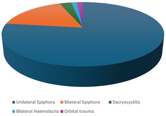
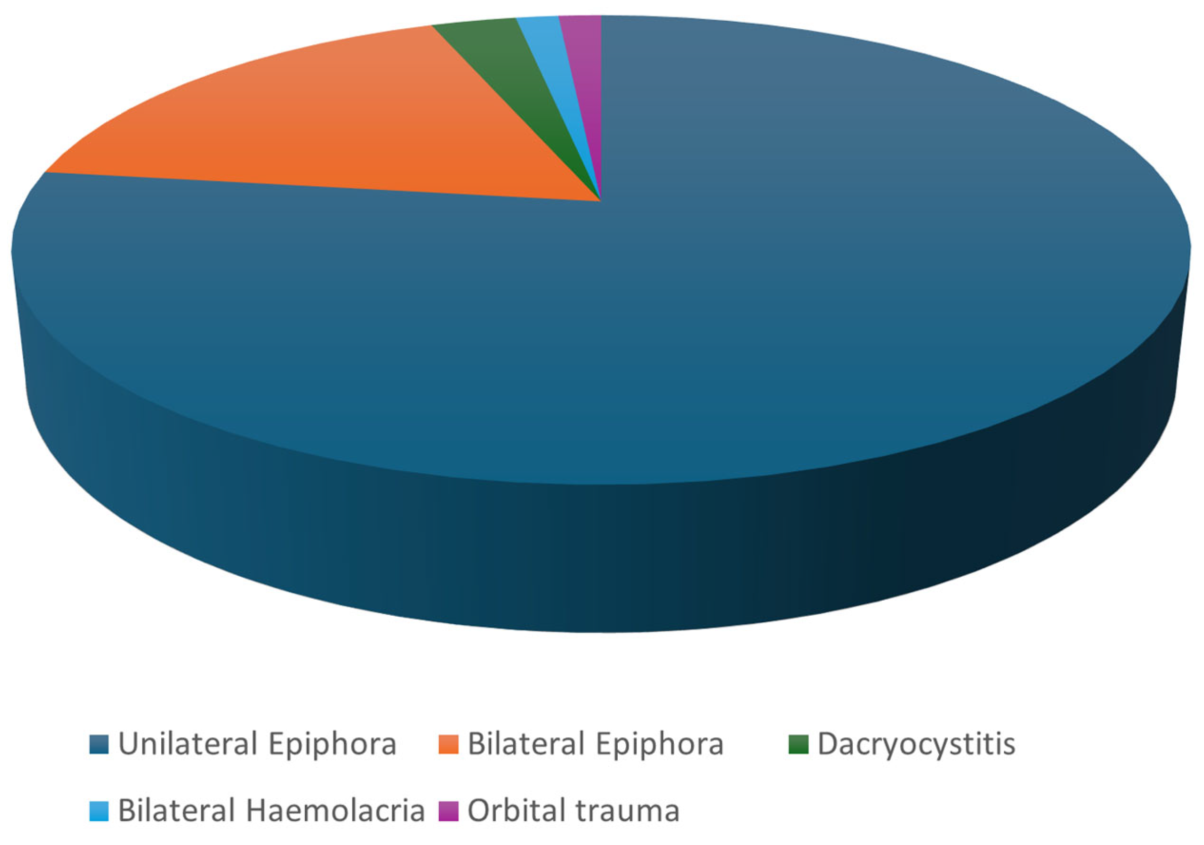
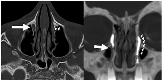
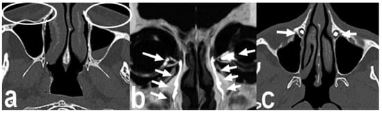
A total of 80 patients were included: 25 males and 55 females. Mean age was 64.3 years. The number and the type of diagnoses are presented in Table 1 and Figure 1. Adequate contrast opacification was achieved in all patients (Figure 2, Figure 3, Figure 4, Figure 5 and Figure 6). Obstruction was detected in 70/80 (87.5%) patients: presaccular (38), postsaccular (32), proximal duct (17), distal duct (15), and bilateral (12) (Table 2). Postsaccular obstructions were further subclassified into proximal and distal nasolacrimal duct involvement. As a result, the postsaccular category includes these sublevels and is not mutually exclusive with them, which explains why the sum of obstruction counts exceeds the total number of patients. The mean patient discomfort was 2.1 ± 1.3 on a 10-point VAS scale. No adverse events occurred.
Table 1.
Clinical diagnoses of the included patients and their relative frequencies.
Figure 1.
Graphical representation of the distribution of clinical diagnoses in the study population.
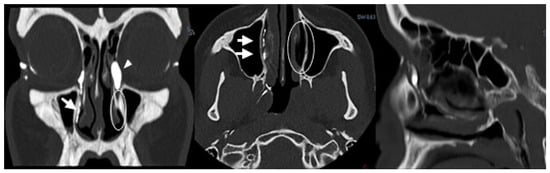
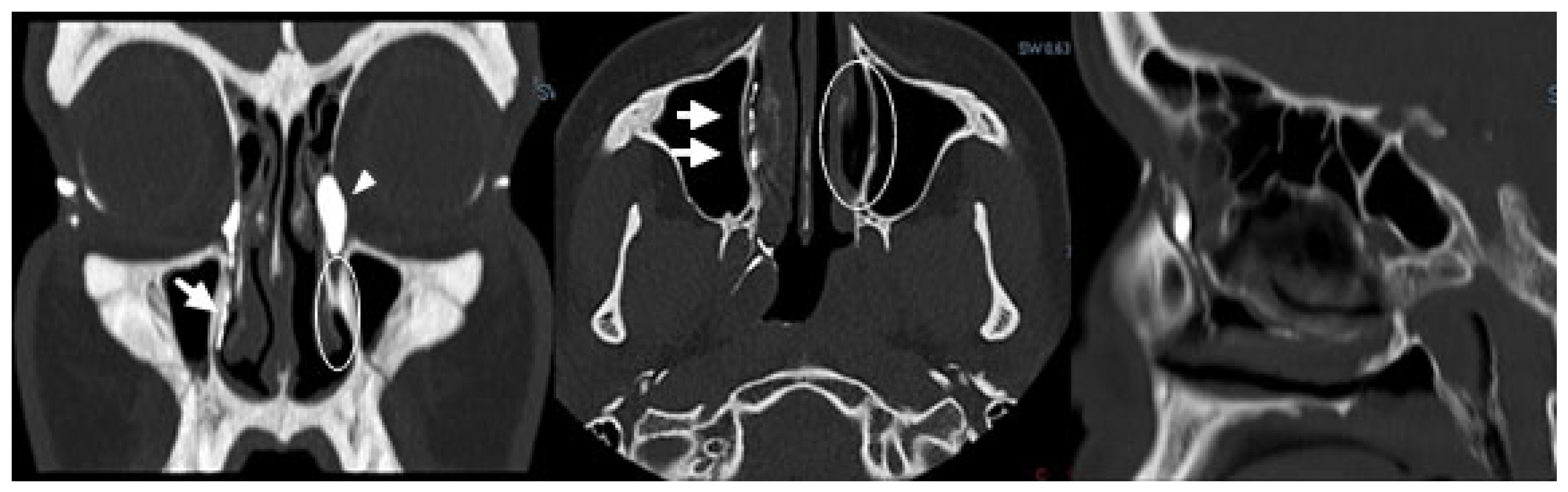
Figure 2.
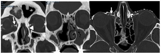
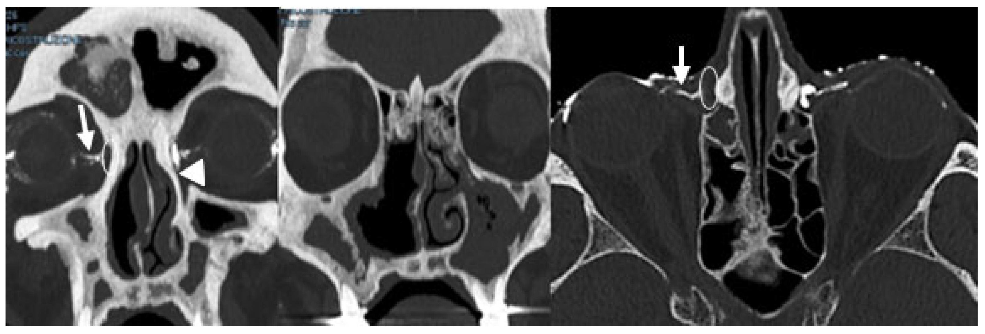
Left nasolacrimal duct obstruction with ectatic appearance of the ipsilateral lacrimal sac (arrowhead), the iodinated contrast agent opacifies the right lacrimal duct (arrows), while no opacification is observed in the left lacrimal duct (circle).
Figure 3.
Right presaccular stenosis, left juxtasaccular stenosis. The iodinated contrast agent opacifies only the right canaliculi (arrow), without filling the right lacrimal sac (circle), while it opacifies the left lacrimal sac but stops at the level of the saccular obstruction (arrowhead).
Figure 4.
Right distal nasolacrimal duct obstruction. The iodinated contrast agent opacifies the right lacrimal sac but does not opacify the entire nasolacrimal duct (arrow), while it is visible along the patent left lacrimal duct as a continuous hyperdense column (arrowheads).
Figure 5.
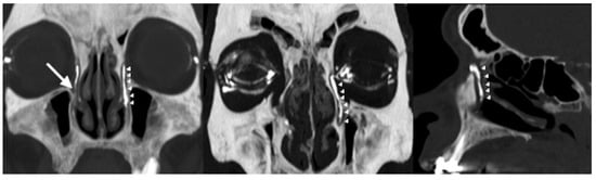
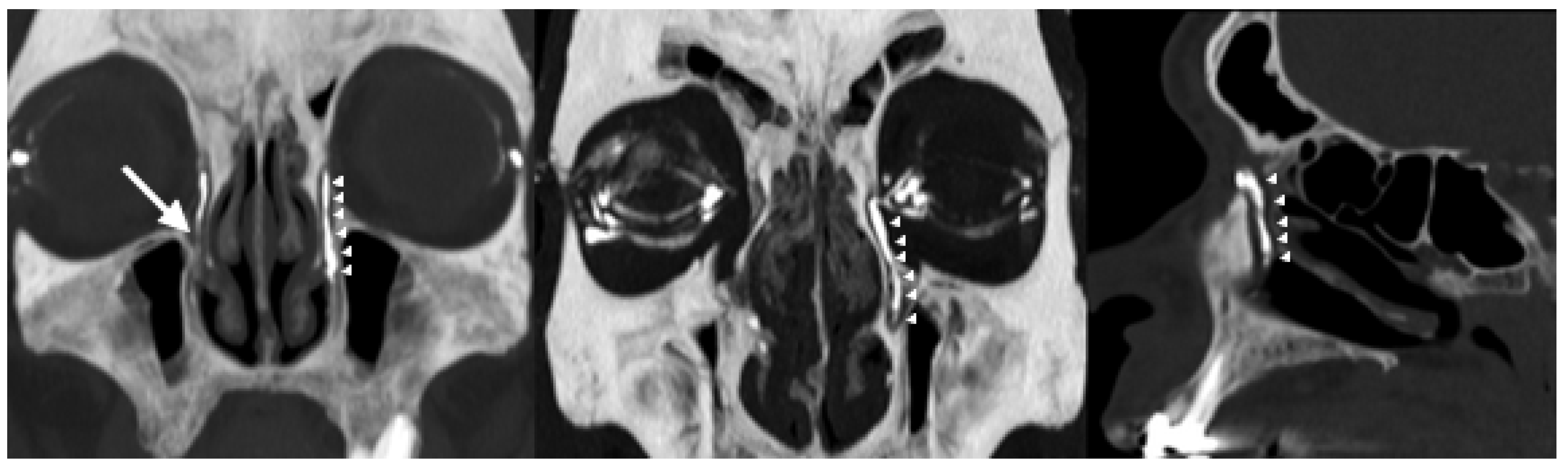
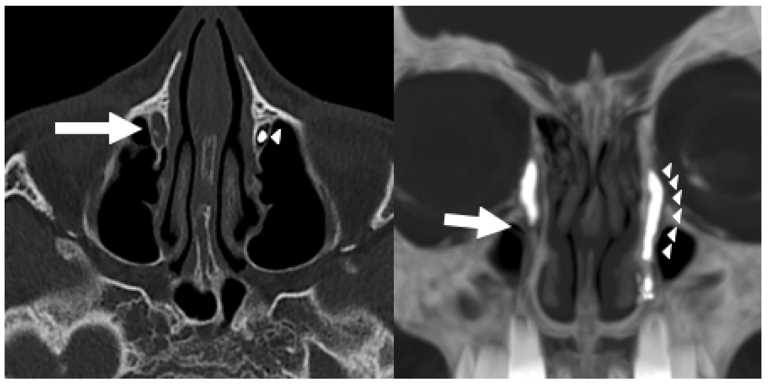
Right nasolacrimal duct obstruction at the level of the orbital plane. Right-sided image: the iodinated contrast agent is not visible at the level of the right orbital plane (arrow), with opacification limited to the lacrimal sac.) Left-side image: frontal projection showing complete opacification of the left lacrimal drainage system (arrowheads) and the right obstruction (arrow).
Figure 6.
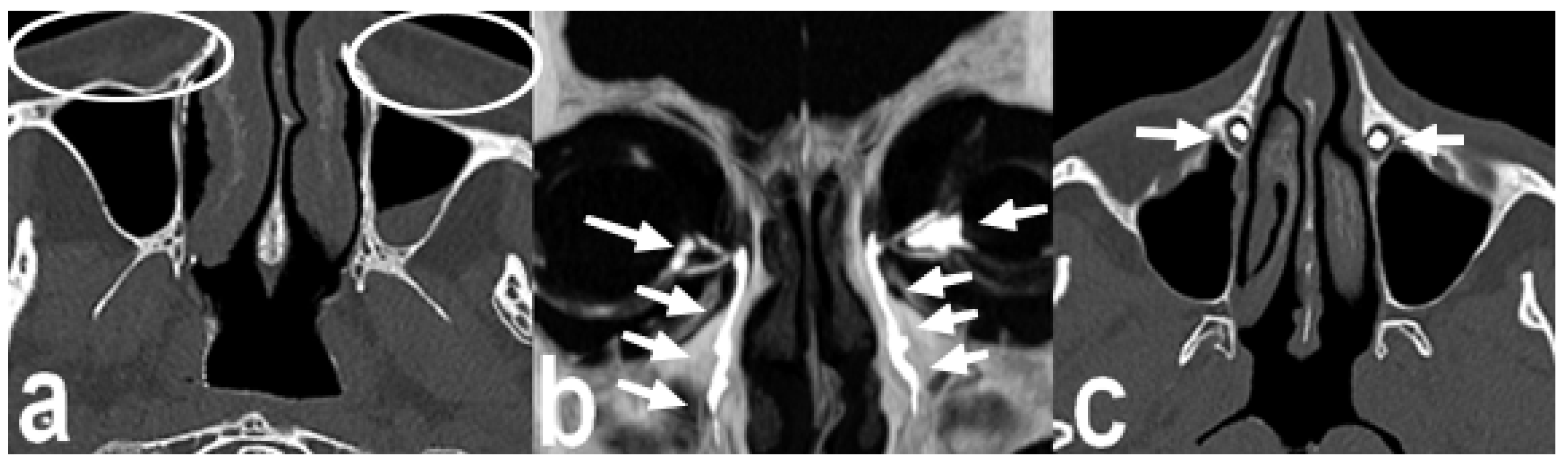
A case of suspect bilateral dacryocystitis in a patient with history of bilateral blepharoplasty; (a) swelling of orbital soft tissues (circles); (b,c) iodate contrast solution is recorded all section of the lacrimal system, ruling out the initial suspect (arrows).
Table 2.
Site of obstruction. Postsaccular obstructions include proximal and distal nasolacrimal duct involvement. Therefore, these categories are not mutually exclusive, and the total count exceeds the number of patients.
4. Discussion
The lacrimal drainage apparatus is prone to a variety of pathological conditions, including obstruction, infection, inflammation, and post-traumatic or neoplastic involvement [8]. Accurate assessment of these processes is essential for appropriate management, and imaging plays a key role in evaluating the lacrimal system and surrounding structures. However, each available imaging modality presents specific limitations.
Dacryoscintigraphy provides a physiological assessment of tear drainage, but its clinical use is limited by suboptimal spatial resolution, lack of anatomic detail, and limited availability [9]. Conventional cannulated dacryocystography, though well established, is invasive and technically demanding, requiring local anesthesia, punctal dilation, canalicular cannulation, and pressure-driven contrast injection. These steps can cause patient discomfort and carry a risk of iatrogenic trauma, false passage, or scarring of the lacrimal system. In addition, forced injection may bypass partial obstructions and produce false-negative results, while offering little information about the surrounding bone or soft tissue [2].
MRI dacryocystography has emerged as a noninvasive alternative with good soft-tissue characterization, yet it remains expensive, has long acquisition times, and provides poor depictions of bony anatomy. Moreover, motion artifacts and limited spatial resolution of small structures make MRI suboptimal for routine evaluation of the nasolacrimal ducts [3,6].
In contrast, CT–dacryocystography (CT-DCG) combines excellent bony definition with high spatial resolution and short acquisition time, making it well suited for the evaluation of both congenital and acquired nasolacrimal duct obstruction (NLDO) [10]. Our protocol further refines this approach by using a non-cannulated, dropwise instillation of an iso-osmolar, low-viscosity contrast agent (iodixanol 320) directly into the conjunctival sac. This technique is noninvasive, does not require punctal dilation or cannulation, and can be easily performed even in uncooperative or pediatric patients.
Compared with conventional cannulated CT-DCG or dacryoscintigraphy, our physiological instillation technique is rapid, well tolerated, and less costly, while maintaining excellent anatomic detail. The gradual administration (2 drops per minute for 10 min) allows progressive, physiological filling of the lacrimal drainage system, avoiding the non-physiological distension that may occur with bolus or syringe injection. This controlled delivery minimizes reflux, prevents overdistension of the canaliculi and sac, and enhances visualization of partial or functional obstructions that might otherwise remain undetected. CT-DCG with an instillation technique provides a more faithful representation of the patient’s drainage physiology and eliminates the risk of iatrogenic injury associated with cannulation [8,11].
In our experience, the use of multidetector CT (MDCT) represents an additional advantage. MDCT provides superior image quality compared with older helical systems, with higher contrast resolution and thinner sections enabling multiplanar and three-dimensional reconstructions. Moreover, the ability to cover the entire lacrimal drainage pathway—from the puncta to the inferior meatus—in a single low-dose acquisition improves workflow efficiency and diagnostic confidence.
In our series, unilateral CT-DCG was performed in all patients to shorten examination time and enhance comfort, consistent with previous authors who advised against routine bilateral studies when symptoms are unilateral. The use of iodixanol 320 as the contrast medium ensured good local tolerability and avoided chemical irritation. Iodixanol 320 is an iso-osmolar, low-viscosity iodinated contrast agent widely used in diagnostic imaging, whose physicochemical properties make it suitable for local administration with good mucosal tolerability. At the end of each examination, a brief saline instillation was performed to remove residual contrast and further improve patient satisfaction. The technique proved well tolerated, rapid, and feasible in all cases, providing accurate localization of the obstruction site along the lacrimal outflow system. The most common level of obstruction in our cohort was the presaccular segment, followed by postsaccular involvement, including proximal and distal nasolacrimal duct regions; this is consistent with prior studies on acquired NLDO.
Beyond localization, CT-DCG offers critical information about the adjacent bony structures and extrinsic causes of obstruction, including post-traumatic deformities, sinus disease, or cicatricial changes. In cases of post-traumatic NLDO, the ability to assess the lacrimal sac fossa and nasolacrimal canal is particularly valuable. Similarly, in patients with prior dacryocystorhinostomy (DCR) [12], CT-DCG can help evaluate causes of surgical failure, such as malpositioned osteotomies or restenosis of the ostium.
Our findings reaffirm that CT-DCG is an indispensable tool for both the diagnosis and management of lacrimal drainage disorders. The physiological, non-cannulated technique we propose further enhances its utility by providing an objective, reproducible, and patient-friendly approach. By maintaining natural tear dynamics and minimizing procedural discomfort, this method is potentially suitable for routine clinical use and may serve as a reference for future studies.
Nevertheless, certain limitations should be acknowledged. The present study was retrospective, included a relatively small and homogeneous population, and did not encompass complex or neoplastic lacrimal pathologies. Additionally, a direct comparison with traditional cannulated CT-DCG or dacryoscintigraphy was not performed, which limits quantitative assessment of diagnostic performance. Future prospective, comparative, and multicentric studies are warranted to confirm our preliminary observations and to standardize the protocol parameters.
In conclusion, non-cannulated CT-DCG with dropwise iodixanol instillation represents a rapid, reproducible, and well-tolerated technique for imaging the lacrimal drainage system. Its safety, high diagnostic yield, and ability to provide both functional and anatomical information make it a valuable tool in the evaluation and management of nasolacrimal duct obstruction and related disorders. This approach may also serve as a rapid preoperative imaging option for dacryocystorhinostomy planning, providing precise delineation of obstruction sites and associated bony anatomy.
5. Conclusions
A physiological, non-cannulated CT-DCG protocol with dropwise iodixanol instillation provides reproducible lacrimal system imaging with minimal discomfort and good tolerability. This technique may complement conventional cannulated methods pending further validation in prospective comparative studies.
Author Contributions
Conceptualization, G.S.; Methodology, G.L.T. and G.S.; Validation, C.M.C.; Formal analysis, F.C.; Investigation, P.A. and G.S.; Data curation, F.C.; Writing—original draft, B.G. and R.T.; Writing—review and editing, F.C.; Visualization, F.C. and G.L.T.; Supervision, G.S. All authors have read and agreed to the published version of the manuscript.
Funding
This research received no external funding. The APC was funded by Univeristà del Salento.
Institutional Review Board Statement
The study was conducted in accordance with the Declaration of Helsinki. Ethical approval was not required according to national regulations, as the study had a retrospective, non-interventional design and was based exclusively on fully anonymized data (Italian Legislative Decree No. 196/2003, as amended by Legislative Decree No. 101/2018, and Regulation (EU) 2016/679).
Informed Consent Statement
Informed consent was not needed because the data was pre-existing, anonymized (or de-identified), and used for secondary analysis.
Data Availability Statement
The data that support the findings of this study are available from the corresponding author upon reasonable request.
Conflicts of Interest
The authors declare that they have no competing interests.
References
- Mandeville, J.; Woog, J. Obstruction of the lacrimal drainage system. Curr. Opin. Ophthalmol. 2002, 13, 303–309. [Google Scholar] [CrossRef] [Scilit] [PubMed]
- Singh, S.; Ali, M.J.; Paulsen, F. Dacryocystography: From theory to current practice. Ann. Anat. Anat. Anz. 2019, 224, 33–40. [Google Scholar] [CrossRef] [Scilit] [PubMed]
- Weber, A.L.; Rodriguez-DeVelasquez, A.; Lucarelli, M.J.; Cheng, H.M. Normal anatomy and lesions of the lacrimal sac and duct: Evaluated by dacryocystography, computed tomography, and MR imaging. Neuroimaging Clin. N. Am. 1996, 6, 199–217. [Google Scholar] [CrossRef] [Scilit] [PubMed]
- Nakamura, J.; Kamao, T.; Mitani, A.; Mizuki, N.; Shiraishi, A. Accuracy of the Lacrimal Syringing Test in Relation to Dacryocystography and Dacryoendoscopy. Clin. Ophthalmol. 2023, 17, 1277–1285. [Google Scholar] [CrossRef] [Scilit] [PubMed]
- Shapira, Y.; Juniat, V.; Macri, C.; Selva, D. Syringing has limited reliability in differentiating nasolacrimal duct stenosis from functional delay. Graefes Arch. Clin. Exp. Ophthalmol. 2022, 260, 3037–3042. [Google Scholar] [CrossRef] [Scilit] [PubMed]
- Cè, M.; Grimaldi, E.; Toto-Brocchi, M.; Martinenghi, C.; Oliva, G.; Felisaz, P.F.; Schiavo, P.; Lunardi, G.; Cellina, M. Non-contrast MR dacryocystography for the evaluation of epiphora and recurrent dacryocystitis: A preliminary study. Neuroradiol. J. 2023, 36, 397–403. [Google Scholar] [CrossRef] [Scilit] [PubMed]
- Munk, P.L.; Burhenne, L.W.; Buffam, F.V.; Nugent, R.A.; Lin, D.T. Dacryocystography: Comparison of water-soluble and oil-based contrast agents. Radiology 1989, 173, 827–830. [Google Scholar] [CrossRef] [Scilit] [PubMed]
- Raghuwanshi, S.; Yadav, N.; Raghuwanshi, S.; Raghuwanshi, S. Multi-Detector CT Instillation Dacryocystography and Its Role in the Diagnosis of Lacrimal Drainage System Blocks. Indian J. Otolaryngol. Head Neck Surg. 2021, 73, 474–479. [Google Scholar] [CrossRef] [Scilit] [PubMed]
- Patnaik, C.U.; Gupta, M.A.; Nilakantan, B.A. Evaluation of Patients for Endoscopic DCR: Sac Syringing and Radionuclide Scintigraphy. Indian J. Otolaryngol. Head Neck Surg. 2019, 71, 1990–1993. [Google Scholar] [CrossRef] [Scilit] [PubMed]
- Barna, S.; Garai, I.; Kukuts, K.; Gesztelyi, R.; Toth, L.; Kemeny-Beke, A. Clinical utility of SPECT/CT and CT-dacryocystography-enhanced dacryoscintigraphy in the imaging of lacrimal drainage system obstruction. Ann. Nucl. Med. 2019, 33, 746–754. [Google Scholar] [CrossRef] [Scilit] [PubMed]
- Hamdy, H.; El-Anwar, M.W.; Mobasher, M.A.; Amin, M.I.; Abdelhamid, H. Role of CT dacryocystography in the management of lacrimal drainage system obstruction. Egypt. J. Otolaryngol. 2025, 41, 123. [Google Scholar] [CrossRef] [Scilit]
- Du, J.; Zhong, B.; Xian, J.; Ba, L.; Liu, F.; Liu, Y. Effect of revision dacryocystorhinostomy under nasal endoscopy on recrudescent dacryocystitis. Int. J. Clin. Exp. Pathol. 2017, 10, 11717–11722. [Google Scholar] [PubMed]

Disclaimer/Publisher’s Note: The statements, opinions and data contained in all publications are solely those of the individual author(s) and contributor(s) and not of MDPI and/or the editor(s). MDPI and/or the editor(s) disclaim responsibility for any injury to people or property resulting from any ideas, methods, instructions or products referred to in the content. |
© 2026 by the authors. Licensee MDPI, Basel, Switzerland. This article is an open access article distributed under the terms and conditions of the Creative Commons Attribution (CC BY) license.