


Abstract
Chrysocolla, a hydrated copper silicate mineral, is notoriously difficult to recover by conventional froth flotation due to its microporous structure, low hydrophobicity, impurity substitutions, and poor response to traditional collectors. This study evaluates the performance and adsorption mechanism of benzohydroxamic acid (BHA) as a collector for chrysocolla flotation under varying pH conditions and collector dosages. Microflotation results showed that chrysocolla recovery increased with BHA concentration, with enhanced flotation occurring at alkaline pH (8–10), consistent with BHA dissociation behavior. Zeta potential measurements indicated selective adsorption of BHA on the chrysocolla surface, while quartz showed minimal interaction, confirming collector selectivity. X-ray photoelectron spectroscopy (XPS) revealed that BHA was chemisorbed through Cu–hydroxamate complex formation. Bench-scale flotation tests on a chrysocolla ore containing 3.7% Cu produced a concentrate grading 26.7% Cu with 35.3% recovery after initial sulfide flotation. Kinetic tests indicated rapid recovery of more floatable copper phases, while scanning electron microscopy (SEM) showed preferential flotation of finer particles. Overall, the results demonstrate that BHA can effectively promote chrysocolla flotation through selective chemisorption, although high collector dosages are required due to the mineral’s high specific surface area and structural complexity.
1. Introduction
Copper is found in a variety of minerals, which can be broadly categorized into oxides, such as cuprite (Cu2O), tenorite (CuO), and melaconite (CuO). Copper can also occur as sulfide, which accounts for the majority of the copper resource, or iron sulfides such as chalcopyrite (CuFeS2), bornite (Cu5FeS4), chalcocite (Cu2S) and covellite (CuS), carbonates (azurite (Cu3(CO3)2(OH)2) and malachite (Cu2CO3(OH)2), and silicates (chrysocolla, dioptase, plancheite, and euchroite). Chrysocolla is a microporous hydrated copper hydroxy silicate with a complex structure without a fixed chemical formula due to impurity element content. It is a semi-precious jewel and has been described as a mixture of crystalline copper silicate and amorphous silicon dioxide with aluminum and trivalent iron as the most characteristic impurities. Substitution of copper by aluminum makes the chemical formula (Cu2−xAlx)H2−xSi2O5(OH)4·nH2O, and chrysocolla’s copper content decreases as its iron and/or aluminum content increases [1,2]. The CuO:SiO2 ratio was found to be approximately 1:1, even when considering substitutions of copper with other divalent or trivalent elements [3]. The mineral normally contains 10–36% copper [4] in the form of hydroxide and silicate [2]. Chemical analyses of six chrysocolla samples from different sources revealed that they contained 33–42 wt.% CuO, 36–41 wt.% SiO2, trace–7 wt.% Fe2O3, 0.5–6 wt.% Al2O3, and ≤1 wt.% of K2O, Na2O, MgO, and CaO. A negligible amount (0.1%) of PbO and ZnO was present in one sample, and the total water content of chrysocolla may reach up to 32% [3,5].
Chrysocolla is one of the most important secondary copper silicate minerals and occurs extensively in the oxidation zones or porphyry copper deposits worldwide. Due to its relatively low copper content, chrysocolla has not been a primary copper ore; however, it can contribute significantly to the overall copper recovery when present in sufficient quantities. The processing of chrysocolla often involves complex metallurgical operations due to its porous surface and varying crystallo-chemical structure [6,7]. Chrysocolla often occurs in fine-grained particles, making its liberation difficult. In addition, it usually occurs with other copper minerals, such as malachite and azurite, as well as gangue minerals such as quartz and calcite, complicating the processing flow. Moreover, the relatively low copper grade necessitates efficient recovery methods to ensure economic viability. Therefore, froth flotation is a critical step for processing chrysocolla ores.
Conventional flotation collectors such as xanthates, thiocarbamates, and fatty acids cannot effectively collect target minerals such as chrysocolla [7,8,9], cassiterite [10,11,12], and spodumene [13,14,15,16]. Chelating agents, in contrast, exhibit higher selectivity towards specific minerals due to their ability to form strong complexes with target metal ions. This selectivity can lead to cleaner concentrates and reduced reagent consumption. Chelating agents can also be more effective in treating complex mineral systems, where conventional collectors might struggle due to competing reactions. Therefore, due to their higher selectivity and efficiency, chelating agents can potentially be used to economically process low-grade ores. More importantly, some chelating agents are biodegradable and less toxic than conventional collectors, making them a more environmentally friendly option. However, despite offering several advantages, the application of chelating agents is not without challenges: chelating agents might be less stable under certain conditions, affecting their performance. Moreover, they can be more expensive than conventional collectors. Therefore, finding the optimal chelating agent and conditions for specific ores requires careful experimentation.
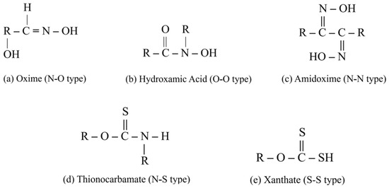
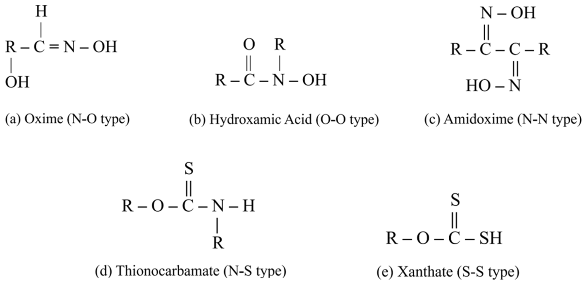
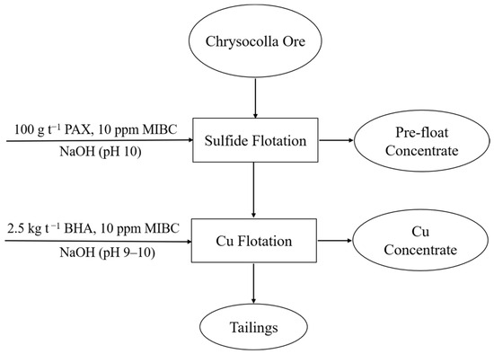
Among various chelating agents, organic reagents with the ability to form metal chelates have been found to be superior collecting agents for sulfide- and oxygen-containing copper minerals. Upon adsorption, they interact with copper ions and cover the mineral surface with insoluble hydrophobic species [17]. Fuerstenau and Herrera-Urbina suggested the typical structures of the chelate-forming collectors to be as the structural formula shown in Figure 1 [17]. The study of various organic chelating collectors suggested that the O–O type of chelating collectors exhibit a significant role in dealing with metal ions and create strong and specific interactions with copper mineral surfaces [18]. Hydroxamates (RCONHOM, where R is an alkyl, aryl, or alkylaryl group and M is an alkali or alkaline earth metal) have emerged as a promising alternative to traditional collectors in froth flotation due to their unique properties and potential for improved selectivity and efficiency [19]. Hydroxamic acids (Figure 1b) are both weak acids and weak bases which can be deprotonated from the O side or N side, producing R–CONHO– or R–COHON– (HON=RCO–) anions. These compounds contain the hydroxamic acid group (–OCNHOH), which is known for its ability to form stable complexes with metal ions. The flotation mechanism of hydroxamate is primarily based on chemisorption onto mineral surfaces. The hydroxamic acid group interacts with metal ions present on the mineral surface, forming a chelate complex, which impacts the hydrophobicity of the mineral particles. Hydroxamates have shown potential in the flotation of various minerals, including oxide minerals, such as copper oxides [20] (malachite [21,22,23,24,25,26,27] and azurite [26]), sulfides (galena [28,29] and chalcopyrite [29]), and rare earth minerals [30,31,32,33,34]. Flotation of kaolin clays with alkyl hydroxamates improved selectivity and recovery of anatase and exhibited less sensitivity to the changes in dispersant, high levels of which can depress anatase [35].
Figure 1.
Typical structures of the chelating reagents that act as flotation collectors.
Recently developed amine-based systems, including ethylene diamine phosphate (EDP) and 1,2-diaminopropane (DAP), improved chrysocolla flotation primarily by modifying surface Cu chemistry and promoting subsequent collector adsorption. EDP significantly increased chrysocolla recovery compared to conventional sulfidization–xanthate flotation by promoting sulfidization and xanthate adsorption under optimized conditions [7]. The mechanism was suggested to be formation of Cu–amine complexes, exposure of more active Cu sites, and enhanced adsorption of sulfide and xanthate species. Sulfidization increases the recovery of chrysocolla by xanthates; however, weak adsorption of the sulfide ions by the chrysocolla surface leaves a high concentration of sulfide ions in the solution as well as oxidized species such as thiosulfate, which has a depressing effect on the flotation [7,36,37]. A variety of activators investigated for chrysocolla flotation along with the references are listed by Feng et al. [2]. Although amine collectors generally exhibit limited performance [38], high recoveries of ammonia-assisted flotation were achieved through formation of chelated Cu species and modification of the chrysocolla surface chemistry through amine coordination [8]. Using hydroxamates, however, combines two attractive features. It does not require a sulfidization step, unlike EDP-based systems. Also, it can achieve high recoveries while maintaining selectivity toward quartz. Among them, n-octanohyroxamate and benzohydroxamic acid (BHA) have demonstrated complex formation for efficient recovery. Bench-scale Cu recovery with n-octanohydroxamate exceeded 97% under optimized conditions through formation of stable Cu–hydroxamate chelates on exposed Cu sites of chrysocolla [39]. However, despite the promising performance of hydroxamate collectors, the adsorption mechanism and practical flotation behavior of BHA toward chrysocolla remain insufficiently understood. This study was undertaken to assess the performance of BHA of different concentrations in the flotation of chrysocolla at different pH. It should be highlighted that due to its porous structure and large specific surface area, high concentrations of BHA were used to cover chrysocolla surface effectively.
2. Materials and Methods
2.1. Materials
Quartz mineral in this work was purchased from Ward’s Science (Rochester, NY, USA). The chrysocolla ore for microflotation tests was purchased from Excalibur Mineral Corp (Charlottesville, VA, USA). It was crushed first with a hand hammer; the richer mineral fragments were hand-picked and ground separately. Further picking by hand was carried out after pulverizing in a Siebtechnik T100 pulverizer (Siebtechnik GmbH, Mülheim, Germany). The gangue particles were removed as much as possible, so the high-grade mineral did not require further concentration. Both quartz and chrysocolla samples were screened to produce a −106 + 38 µm fraction for microflotation experiments. For zeta potential measurements, the −38 μm fractions of chrysocolla and quartz were wet-ground using a Pulverisette 6 planetary monomill (Fritsch, Idar-Oberstein, Germany) to produce very fine particles (target size: −10 μm). After screening, the surface of quartz was cleaned with 12 v/v% H2SO4 for 4 h at 60 °C, rinsed thoroughly with reverse osmosis (RO) water to remove any remaining acid, filtered, and dried at 200 °C.
The chrysocolla ore sample was obtained as a high-grade drill core from a North American deposit and prepared for further flotation test by dry-grinding down to 150 μm using a rod mill for 30 min. Potassium chloride (KCl), to be used as a background electrolyte for zeta potential measurements, was purchased from Fisher Scientific (Mississauga, ON, Canada). Hydrochloric acid (HCl) and sodium hydroxide (NaOH) for pH adjustment, and methyl isobutyl carbinol (MIBC, C6H14O) were purchased from Sigma-Aldrich (Steinheim, Germany). Commercial grade potassium amyl xanthate (PAX), purchased from Prospec Chemicals (Fort Saskatchewan, AB, Canada), was used for bench-scale flotation. For purification, 100 g of the PAX was dissolved in 1 L of 99.5% acetone (Fisher Scientific, Ottawa, ON, Canada) at 40 °C and stirred for 10 min. An undissolved PAX was settled once stirring stopped and then removed by filtration. The clear filtrate was cooled to room temperature, mixed with 2 L of petroleum ether 35/60 (Fisher Scientific, Mississauga, ON, Canada) to precipitate the PAX, which was then collected by filtering. This purification step was repeated three times to ensure a high grade. The produced PAX powder was stored in petroleum ether to prevent oxidation to dixanthogen. The purity of the product was confirmed by UV–Vis spectroscopy at 301 nm, where no other xanthate species or degradation products were detected. BHA with purity of 98% was purchased from Fisher Scientific (Ottawa, ON, Canada) and used as received.
2.2. Minerals and Ore Characterization
The particle size distribution of the minerals was determined using a Horiba LA-920 particle size analyzer (Horiba, Kyoto, Japan) Microtrac Series 5000 Sync particle size and shape analysis system (ATS Scientific Inc, Burlington, ON, Canada). The specific surface area was determined by the N2 Brunauer–Emmett–Teller (BET) technique using a TriStar II Plus surface area and porosity analyzer (Micromeritics, Norcross, GA, USA). To determine the purity of the samples, X-ray diffraction (XRD) patterns were recorded using a D2 phaser (Bruker, Karlsruhe, Germany) equipped with a LYNXEYE XE-T detector and Cu–Kα radiation (λ = 1.5418 Å). Data were collected in a 2θ range of 5 to 120° with a 0.02° step increment and 0.15 s per step. The resultant patterns were processed by DIFFRAC.EVA software (version 6.1 Bruker Corp., Karlsruhe, Germany) and compared with the PDF-5+ (version 25.0, ICDD, 2025) database to identify the main minerals present. Scanning electron microscopy (SEM) analysis was performed using a SU3500 SEM system (Hitachi, Tokyo, Japan) equipped with an 80 mm2 XMaxN silicon drift energy-dispersive spectrometer (EDS) detector (Oxford Instruments, Abingdon, UK). During operation, the deadtime was maintained close to 15%, and count rates were greater than 10,000 cps. The data were analyzed using AZtec software (version 3.0, Oxford Instruments, Abingdon, UK) to identify elements present in the sample. X-ray fluorescence (XRF) was used to determine the chemical composition of the ore sample and bench-scale flotation products. All the samples in the form of loose powder were analyzed by energy-dispersive XRF (ED-XRF) under 75 kPa He purge using an Epsilon 4 XRF spectrometer (Malvern Panalytical, Almelo, The Netherlands) coupled with the Omnian application.
2.3. Zeta Potential Determination Experiments
In order to investigate the interactions between the mineral particles and BHA in the flotation experiments, zeta potential measurements were conducted on the dispersed chrysocolla particles using a NanoBrook ZetaPlus electrophoretic analyzer (Brookhaven Instruments, Holtsville, NY, USA). An amount of 0.24 g of the mineral samples was dispersed in 600 mL of 10−3 M KCl background electrolyte. The suspension was then ultrasonicated for 45 s using a UP400S ultrasonic processor (Hielscher, Teltow, Germany). In cases where BHA was present, it was added at a dosage of 2000 kg t−1. The suspension was conditioned for 30 min before taking the first measurement. The pH of the suspensions was adjusted using NaOH and HCl solutions, and the suspensions were then allowed to equilibrate for 15 min before the measurement. The investigated pH range was from 3 to 10, which represented the widest range over which reliable and reproducible zeta potential measurements could be obtained with the available experimental setup. Measurements at higher pH values were not possible because of instrumental and suspension stability limitations. The measurements were carried out in steps of 1 pH. To avoid zeta potential hysteresis, fresh suspensions were prepared for measurements in the acidic and basic directions. For each pH condition, measurements were conducted in triplicate with each independent run comprising ten individual analyses recorded by the instrument. The reported zeta potential values represent the average of these 30 analyses for each respective pH. Throughout the test, a magnetic stirrer was used to keep the particles suspended.
2.4. Microflotation Experiments
The performance of BHA in the flotation of the minerals was evaluated by microflotation using a 170 mL modified Hallimond tube. The experiments were conducted with 1 g of the −106 + 38-μm single mineral. The sample was conditioned for 5 min with 30 mL of the collector solution following pH adjustment. In the case of collectorless tests, the samples were treated with RO water. The conditioning and then flotation tests were conducted at pH 7, 8, 9, and 10 with different concentrations of BHA. The suspension was conditioned for one more minute after the addition of a dilute solution of MIBC, which would produce a frother concentration of 15 ppm in the microflotation cell. After conditioning, the mineral suspension was transferred into the Hallimond tube, and pH-adjusted RO water was added to bring the total volume of the cell to 170 mL. The flotation tests were conducted for 1 min using an air flow rate of 40 mL min−1. The mineral particles were kept suspended throughout flotation using a magnetic stir bar and a magnetic stirrer.
2.5. Denver Flotation
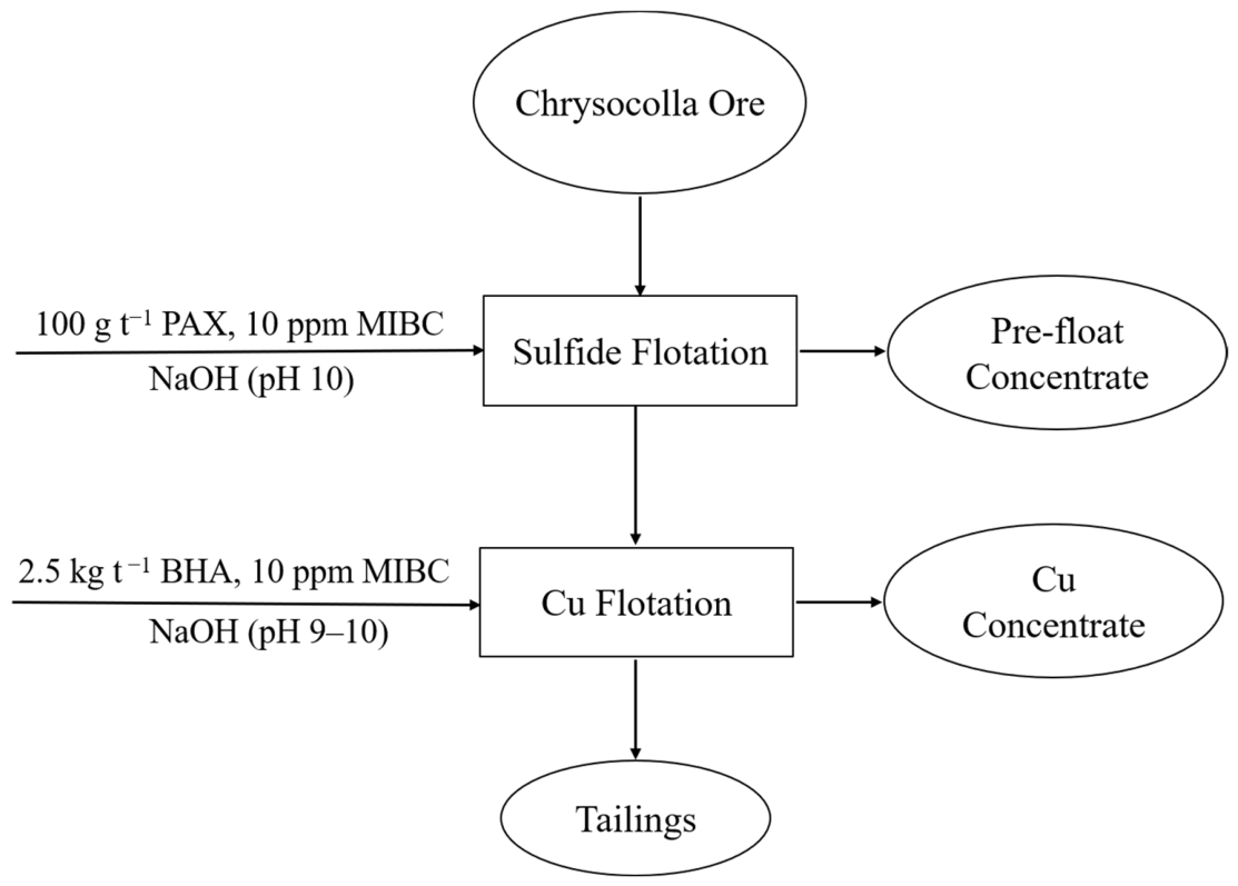
Batch flotation tests were conducted in a 1.3 L Denver flotation cell (Denver Equipment Company (Metso), Denver, CO, USA). For each test, 250 g of the ore sample was first mixed with tap water and stirred at an impeller speed of 1000 rpm. The pH was adjusted to 10 using NaOH solution. During the subsequent conditioning stage targeting the sulfide minerals, the PAX was added at a dosage of 100 g t−1 and conditioned for 1 min, followed by the addition of 10 ppm MIBC and further conditioned for 2 min. The air flow rate was regulated at 5.75 L min−1, and the froth was collected for 5 min to produce pre-float concentrates (visible, black-colored particles on the surface). After the sulfide flotation stage, the slurry was conditioned with 2.5 kg t−1 BHA for 1 min, and the pH was adjusted to 9–10 using NaOH. This was followed by the addition of 10 ppm MIBC and another 2 min of conditioning. During the subsequent chrysocolla flotation stage, the air flow was regulated at the same rate, and froth was collected for 5 min to produce copper concentrates. After each test, the flotation products were dried overnight in an oven at approximately 60 °C. All flotation experiments were performed in triplicate.
For flotation kinetics studies, the concentrates were collected at 15, 30, 60, 180, and 300 s. The flowsheet of the Denver cell flotation experiments is shown in Figure 2.
Figure 2.
Flowsheet of Denver cell flotation experiments with chrysocolla ore sample.
2.6. X-Ray Photoelectron Spectroscopy (XPS) Analyses
The adsorption of the collectors on the mineral surface was investigated using X-ray photoelectron spectroscopy (XPS) measurements before and after treating with 100 kg t−1 BHA solution for 5 min. XPS was performed using a Nexsa G2 spectrometer (Thermo Fisher Scientific, Waltham, MA, USA) equipped with an Al Kα X-ray source (1486.6 eV), applying a 400 μm spot size. The pass energy was 50.0 eV, and the energy step size was 1.000 eV and 0.100 eV for the survey and high-resolution spectra, respectively. The pass energy was adjusted to 200 eV and 20 eV for the survey and high-resolution spectra, respectively. In order to avoid charging on the surface, a flood gun was used to shoot the samples with low-energy electrons during the experiments. The collected data was processed using Avantage Data Processing software (version 6.7.0, Thermo Fisher Scientific, Waltham, MA, USA). The binding energy (BE) was calibrated using the background hydrocarbon C 1s (BE = 284.8 eV).
3. Results and Discussion
3.1. Sample Characteristics
The results of particle size analysis and BET specific surface area of the minerals prepared for zeta potential and microflotation experiments are presented in Table 1.
Table 1.
Particle size and specific surface area of the mineral samples.
The BET specific surface area of chrysocolla is considerably higher than that typically observed in many crystalline copper oxide minerals. This observation is consistent with the broad and diffuse XRD features of chrysocolla, which indicate a poor crystallinity and partially amorphous structure. The mineral is widely recognized as a highly hydrated and microporous copper silicate [3,40,41]. Reported specific surface areas of chrysocolla vary widely depending on mineral origin and crystallinity. A BET specific surface area exceeding 240 m2 g−1 has been reported for 45–74 µm chrysocolla samples [7]. The exceptionally high surface area observed in the present study likely provides a large number of adsorption sites and may permit uptake of BHA within accessible internal pores. Consequently, the microporous structure of the chrysocolla sample likely contributes to the relatively high collector dosages required to achieve substantial flotation recovery. The chemical compositions of the pure minerals and ore measured by XRF are presented in Table 2 and Table 3, respectively.
Table 2.
Elemental compositions of pure minerals.
Table 3.
Elemental composition of chrysocolla ore feed.
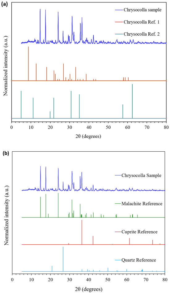
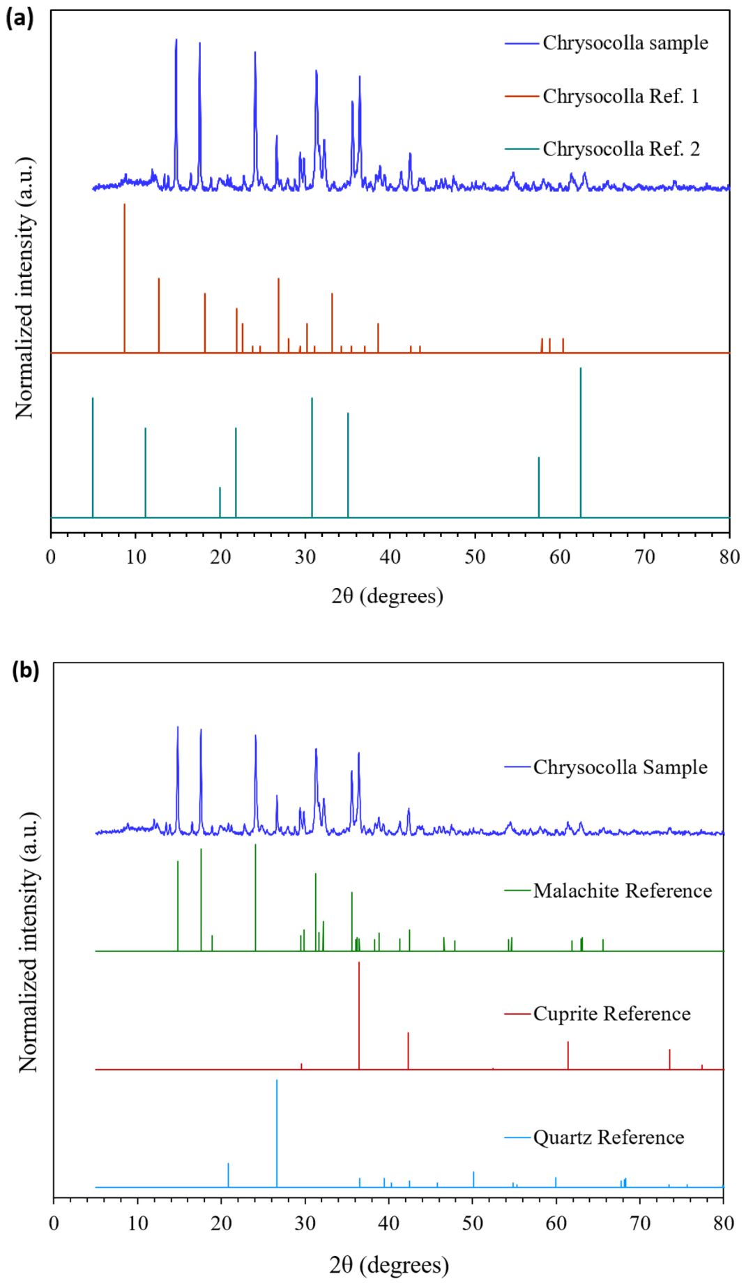
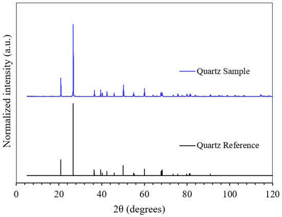
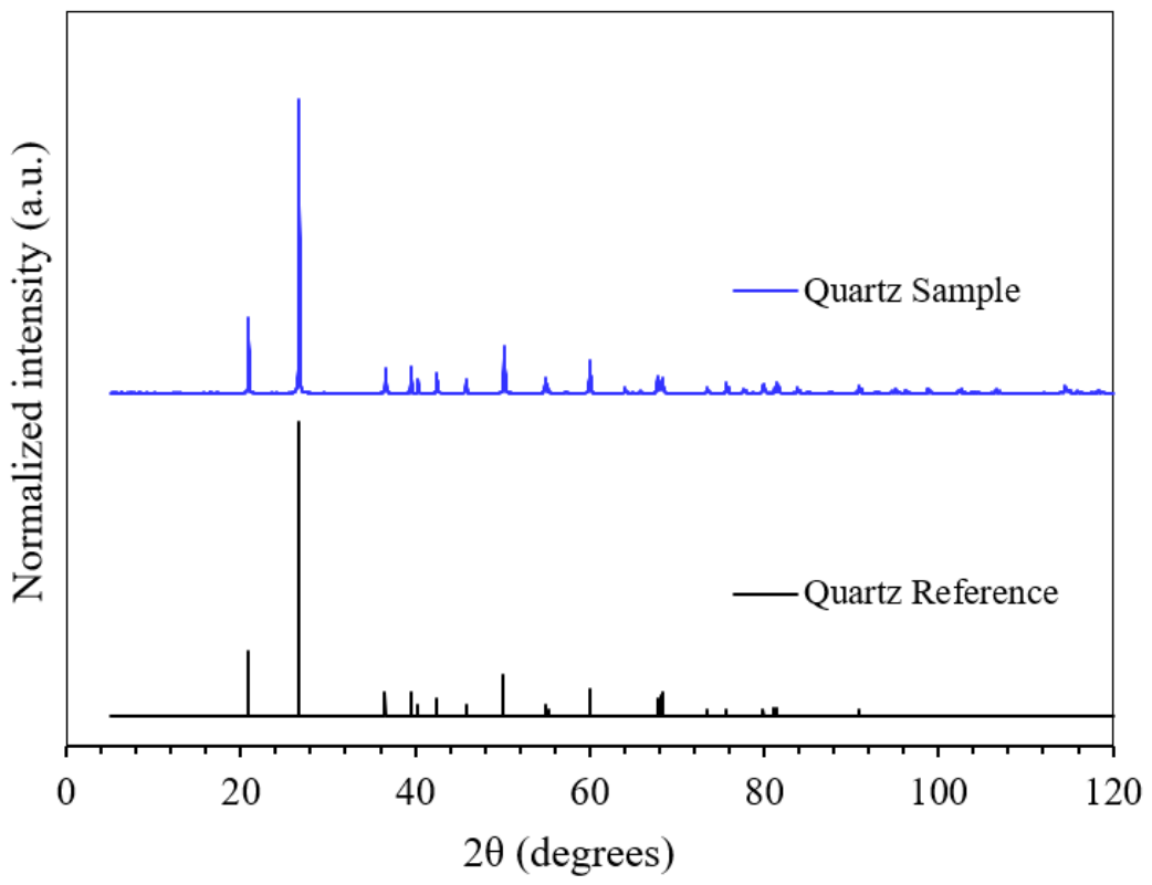
Figure 3 and Figure 4 demonstrate the XRD patterns for chrysocolla and quartz samples, respectively. Chrysocolla has multiple entries depending on its hydration state, impurities, and geological origin. This can be visualized in Figure 3a, which compares the chrysocolla sample used in this work with two references (PDF# 00-014-0685 and 00-027-0188, respectively). Chrysocolla is known to be somewhat structurally variable and often occurs as an amorphous or poorly crystalline material or as a mixture of related copper silicates [42,43]. This can lead to XRD patterns that are not always a perfect match to a single standard card (Figure 3b). The hydration level can affect the XRD pattern. Interestingly, the major peaks of the sample match well with malachite reference (PDF# 00-041-1390). This might be due to the fact that chrysocolla usually co-exists with malachite. In addition, the peaks at 36.43, 42.39, and 61.38 degrees might be attributed to cuprite (PDF# 04-007-9767). A small quantity of quartz (PDF# 00-033-1161) might also be present, considering the peaks at 26.64 degrees. The XRD pattern for the quartz sample (Figure 4) does not present any significant peaks other than those expected based on the standard, suggesting that the sample is relatively pure.
Figure 3.
XRD spectrum of the chrysocolla sample compared to (a) chrysocolla references and (b) related copper oxide and quartz references.
Figure 4.
XRD spectrum of the quartz sample compared to quartz reference.
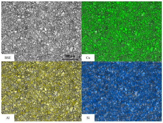
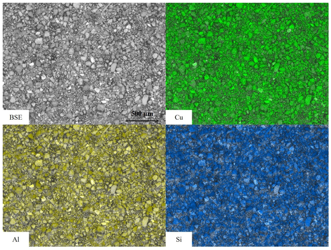
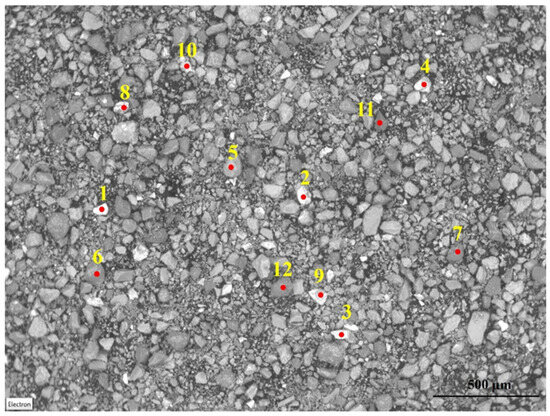
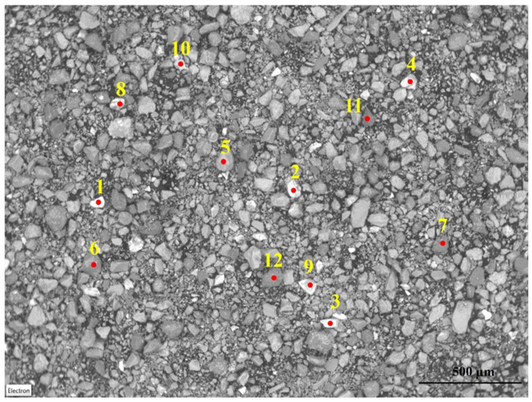
Figure 5 and Figure 6 exhibit the results of the SEM analysis of chrysocolla sample. As shown in the false-colored map scan images (Figure 5), Cu, Al, and Si are the dominant elements that could be observed in the entire area. However, particles showed different brightness, which suggests they might have different compositions. Due to this reason, in addition to map scan, point analysis was also conducted on individual particles with different brightness (categorized as “Bright” and “Dark”), as shown in Figure 6 to compare their compositions. The analysis results are compared in Table 4.
Figure 5.
False-colored SEM map scan images of the chrysocolla sample.
Figure 6.
SEM point scan of the chrysocolla sample taken using 20 kV accelerating voltage.
Table 4.
SEM point analysis results on selected particles.
As shown in Table 4, all analyzed particles contain Cu and Si at various amounts. Al was also present at different concentrations; however, in a few particles, its concentration was below the instrument detection limit. On average, the “Bright” particles contain more Cu than the “Dark” ones, and correspondingly, the Si content is significantly higher in the “Dark” particles. This explains the higher brightness since Cu has a higher atomic number than Si. The wide range of Cu contents obtained from SEM-EDS analyses demonstrates significant mineralogical heterogeneity within the chrysocolla mineral. XRD analyses identified chrysocolla, malachite, cuprite, and quartz, indicating that the analyzed particles may consist of relatively pure mineral domains or intimate intergrowths between Cu-bearing minerals and gangue phases. The brighter particles observed in the images correspond to Cu-enriched regions because of their higher average atomic number; however, the present SEM-EDS analyses do not permit discrimination between pure chrysocolla particles and intergrowths involving malachite, cuprite, and quartz. Such mineralogical heterogeneity may influence collector consumption and flotation behavior by producing particles with different surface chemistries and degrees of liberation. The relative composition of Cu, Si, and Al varies between particles. In a few particles, such as #1, other elements such as Pb are also present.
Overall, the XRD and SEM results confirm that the sample contains Cu, Si, and Al as dominant elements at various contents, which is in agreement with the expected formula of chrysocolla. In a few particles, other elements might also be present, suggesting the complex composition of the chrysocolla sample.
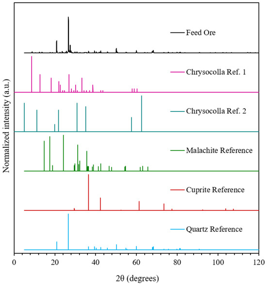
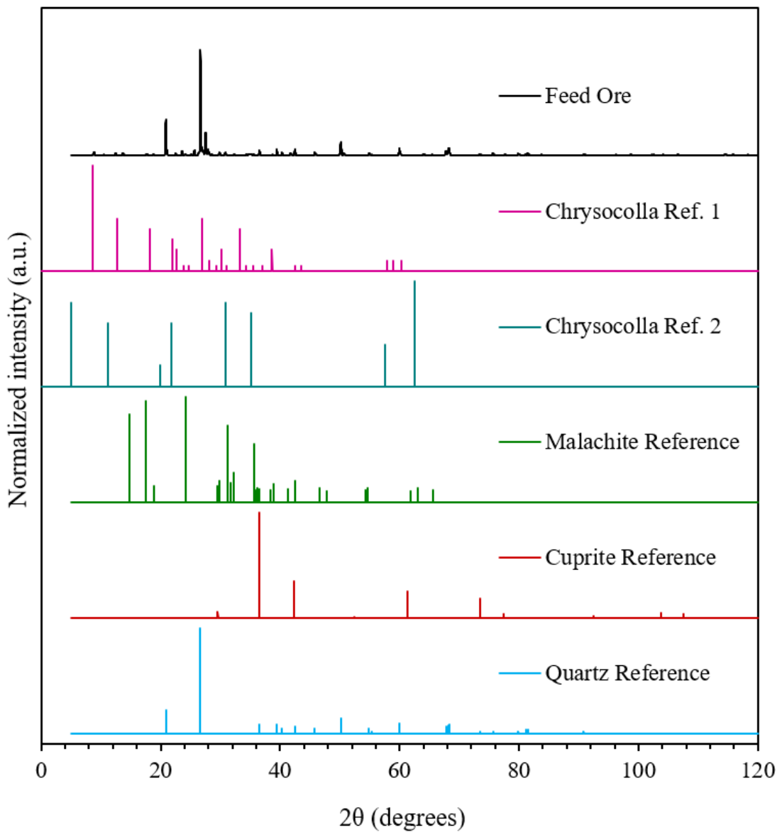
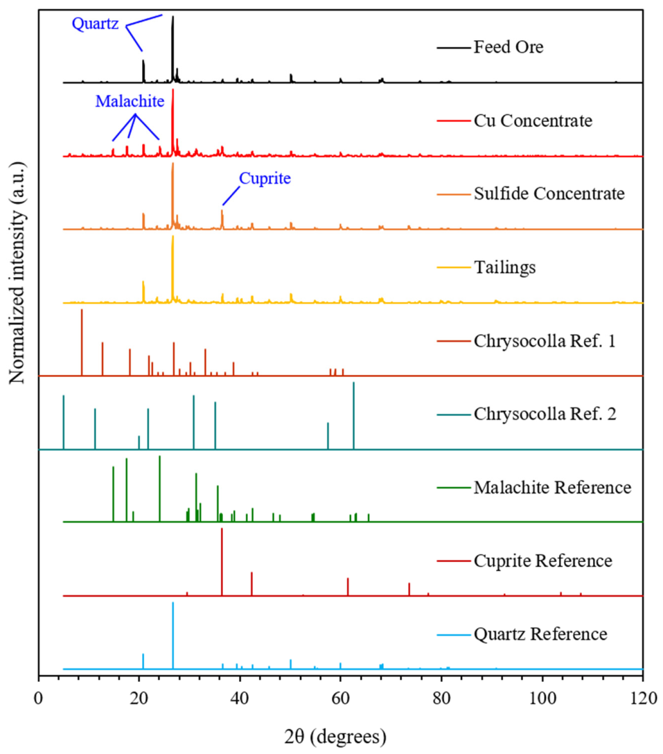
Figure 7 compares the XRD pattern of the feed ore sample with reference diffraction patterns to identify the major crystalline phases present. The results indicate that the feed sample is dominated by silicate phases such as quartz, while reference patterns for malachite and cuprite are included to illustrate the presence of Cu-bearing phases.
Figure 7.
XRD patterns of the ore and reference minerals.
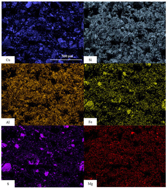
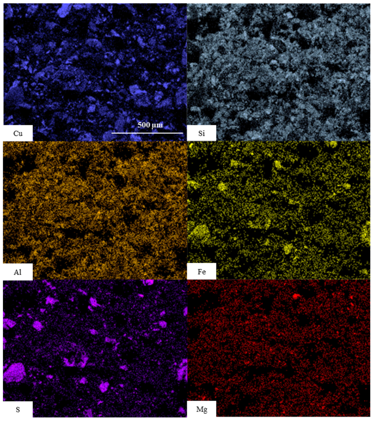
The ore sample was analyzed using scanning electron microscopy paired with energy-dispersive X-ray spectroscopy (SEM-EDS) techniques. The partitioned scans (Figure 8) indicate the presence of Cu, Si, Al, Fe, S and Mg in the ore sample. The results suggest that Cu is unevenly distributed throughout the sample and is likely concentrated in specific mineral phases that will be targeted for recovery by flotation. Si and Al are broadly distributed across the sample, while Fe is more uniformly dispersed, possibly associated with iron oxide phases or finely disseminated sulfides. S is observed in specific areas, indicating the presence of sulfide minerals. Mg shows a more diffuse and weak distribution, likely associated with gangue phases.
Figure 8.
Partitioned SEM-EDS scans of the chrysocolla ore sample.
3.2. Zeta Potential Determination Results
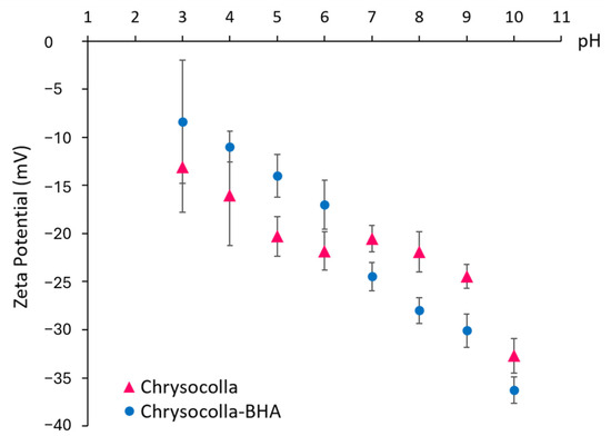
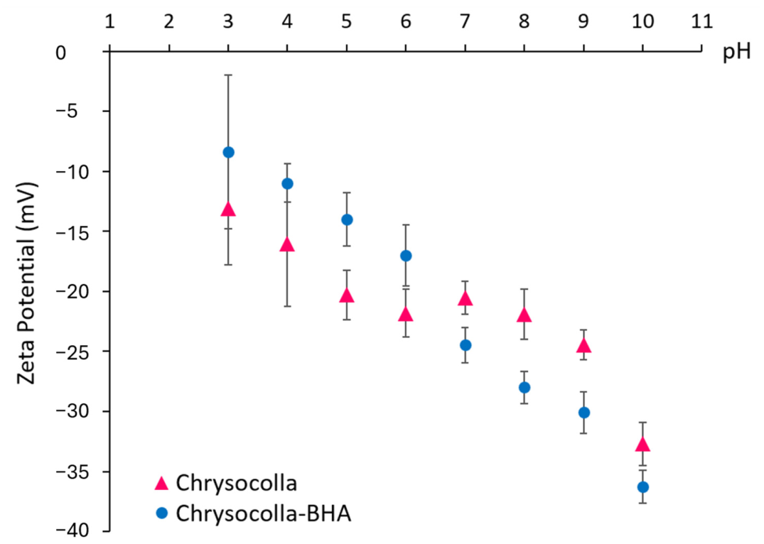
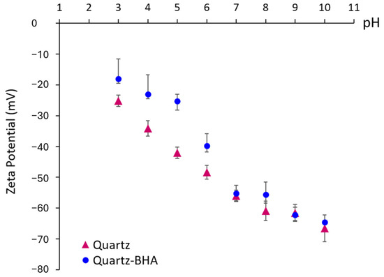
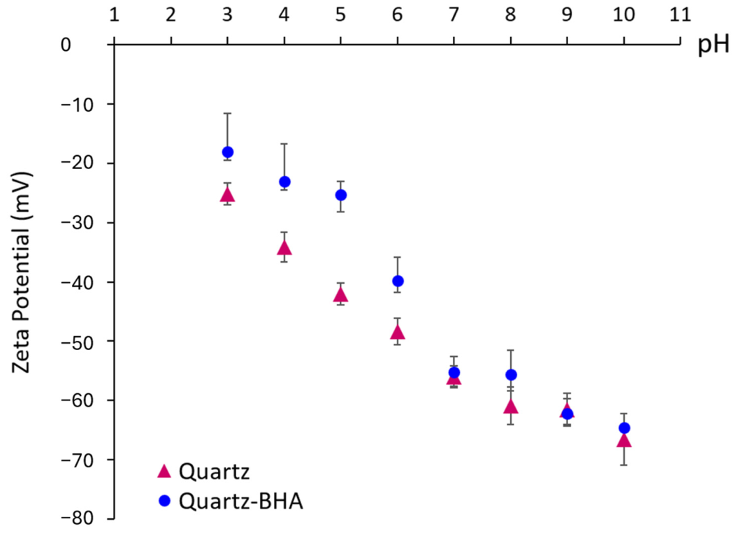
Figure 9 and Figure 10 demonstrate the zeta potential of the chrysocolla and quartz, respectively. The surface of chrysocolla has a negative zeta potential across the investigated pH range, and the zeta potential value decreases with increasing pH (Figure 9). This is in agreement with [7,44,45,46]. In the presence of BHA, the zeta potential of chrysocolla changed differently in acidic and basic ranges: in the acidic range, the shift was towards less negative values, whereas the zeta potential became more negative in the neutral and basic ranges. This change in zeta potential is possibly due to the presence of benzohydroxamate ions and the product of the dissociation of BHA [29], suggesting that BHA adsorption on chrysocolla might take place. In comparison, as shown in Figure 10, treatment with BHA has little to no effect on the zeta potential of quartz at neutral-to-basic pH. Changes in zeta potential at acidic pH could be attributed to the surface charge of quartz which becomes less negative and facilitates interaction with BHA. However, this is particularly unlikely since BHA is not ionized at this condition [29]. This also could be, as suggested by others [22,47], due to the electrical double layer compression, especially since a high concentration (2000 kg t−1) of BHA was used in this experiment.
Figure 9.
Zeta potential of chrysocolla with and without the presence of BHA.
Figure 10.
Zeta potential of quartz with and without the presence of BHA.
3.3. Microflotation Results
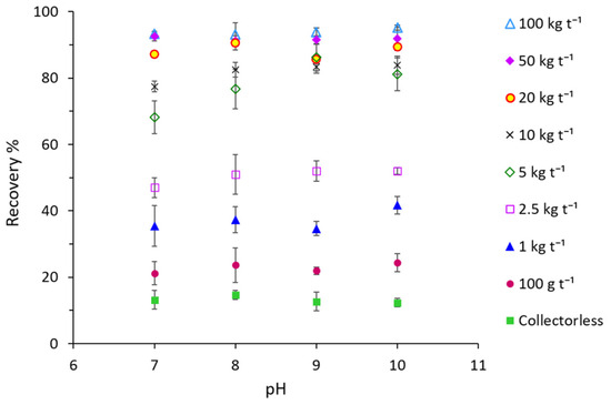
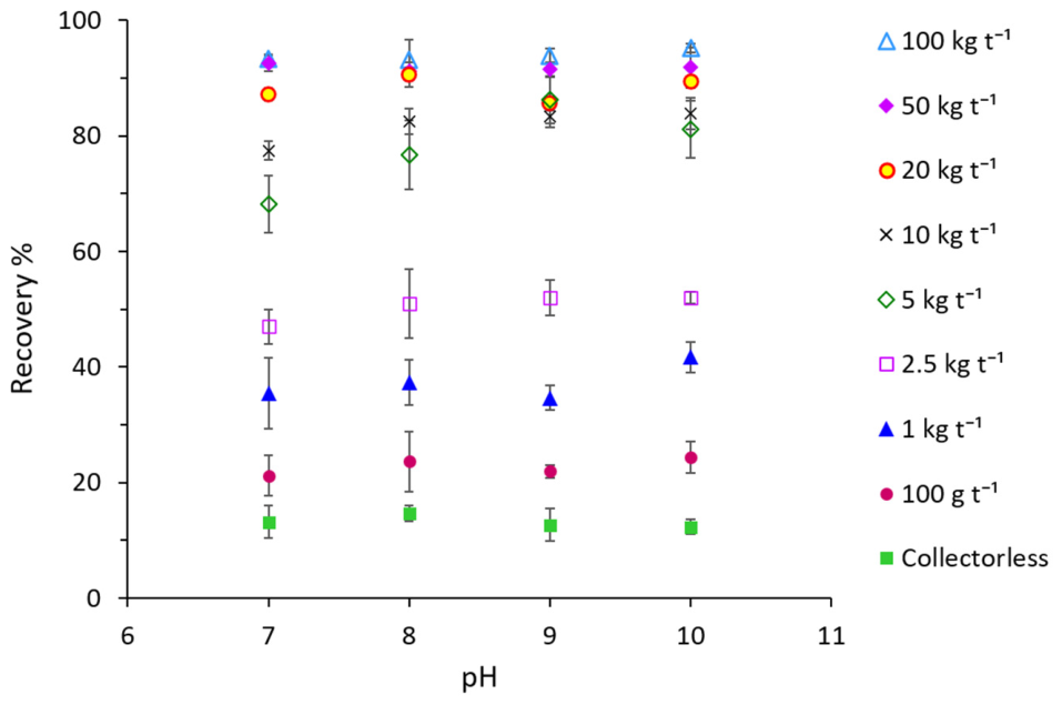
The flotation of chrysocolla and quartz was conducted to evaluate the performance of BHA at various dosages and pH. In order to obtain corroborating evidence for the formation and retention of a substantial coverage of benzohydroxamate on the mineral surface, the chrysocolla sample was treated with different concentrations of BHA, from 100 g t−1 up to 100 kg t−1, and the results are shown in Figure 11. It can be observed that the recovery of chrysocolla generally increases with an increase in BHA concentration, and significant improvement in recovery is observed at higher concentrations (e.g., 5 kg t−1 and above). It can also be seen that the recovery is generally higher at alkaline pH values (8–10) and lower at neutral pH for moderate concentrations. This might be attributed to the dissociation of BHA which occurs between pH 8 and 9 [10,29,48,49,50]. Considering the conditioning pH values of 9 and 10, there would be not much difference between the flotation recoveries for the chrysocolla samples treated with 20, 50, or 100 kg t−1 BHA.
Figure 11.
Microflotation test results of chrysocolla with different concentrations of BHA at pH 7–10.
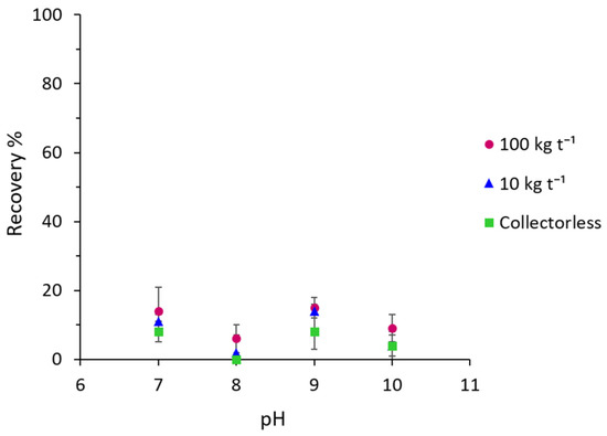
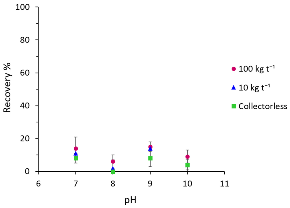
Figure 12 shows that the recovery of quartz remains at minimum at the basic pH range. There is no significant BHA adsorption on the quartz surface. When comparing the flotation recoveries, the microflotation with BHA resulted in high recovery of chrysocolla and low recovery of quartz.
Figure 12.
Microflotation test results of quartz without and with 10 and 100 kg t−1 BHA at pH 7–10.
3.4. X-Ray Photoelectron Spectroscopy (XPS) Results
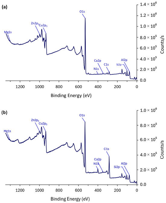
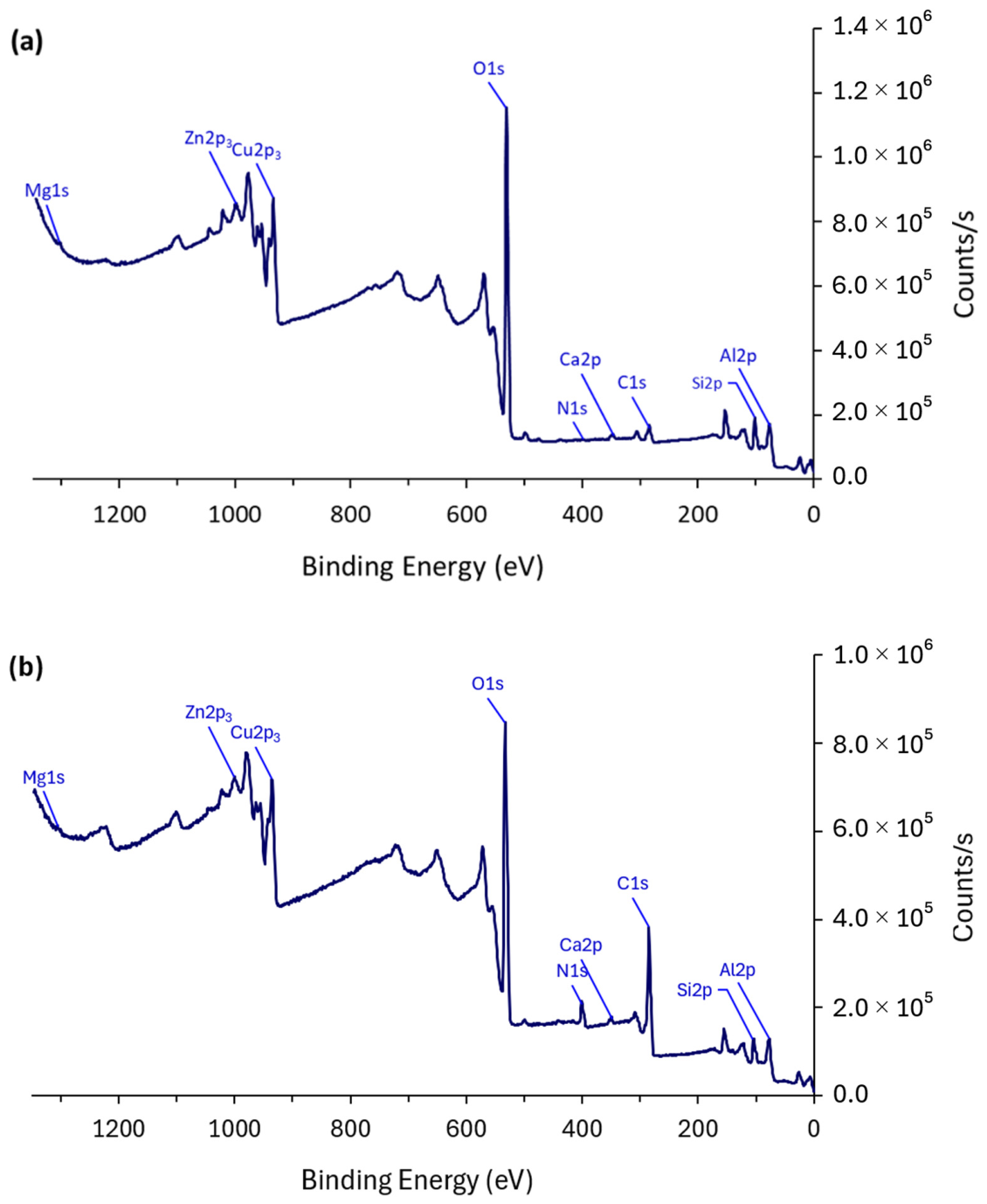
The effect of BHA collector adsorption on the minerals surfaces was examined using XPS analysis of the samples before and after conditioning with BHA. The results demonstrate the species and their chemical states on the mineral and ore surfaces. Figure 13 exhibits the survey spectra of the chrysocolla sample before (Figure 13a) and after (Figure 13b) conditioning with 100 kg t−1 of BHA at pH 9. The nitrogen peak (N 1s) shows that a very-low concentration of nitrogen is present on the surface of pristine chrysocolla, while it appears with a higher concentration on the surface of the mineral following treatment with BHA. The presence of nitrogen is the primary indicator of adsorbed hydroxamate on the chrysocolla surface, as it is the characteristic element in hydroxamate and has little to no association with pristine chrysocolla.
Figure 13.
XPS Survey spectra of chrysocolla before (a) and after (b) treatment with BHA.
Table 5 represents the surface composition of chrysocolla conditioned with 10 and 100 kg t−1 of BHA at pH 9 in comparison with the untreated mineral. Significant appearance of nitrogen concentration (4.1 at%) on the chrysocolla surface indicates successful adsorption of benzohydroxamate on the mineral surface. Interestingly, the nitrogen concentration on the sample treated with only 10 kg t−1 is much less (1.7 at%) even though a similar flotation recovery was observed.
Table 5.
Elemental quantification of the chrysocolla surface before and after treatment with BHA determined by XPS.
In addition to N, the presence of hydroxamate on chrysocolla resulted in a noticeable increase in the concentration of carbon from 7.3 (before treatment) to 14.2 (10 kg t−1 BHA) and 31.7 at% (100 kg t−1 BHA), whereas the concentrations of the other surface elements, some of which more significantly than others, decreased (Table 5). The substantial increase in C 1s concentration after BHA treatment indicates extensive organic coverage on the chrysocolla surface. However, this carbon signal should not be interpreted as arising exclusively from chemisorbed BHA. At the high collector dosage, the detected carbon may include contributions from Cu–hydroxamate surface complexes, weakly adsorbed or physisorbed BHA, residual molecular BHA retained within the porous chrysocolla structure, and minor adventitious carbon. Therefore, the increase in C 1s is considered evidence of BHA-derived organic coverage, while the chemisorption mechanism is inferred from the combined N 1s and Cu 2p spectral changes.
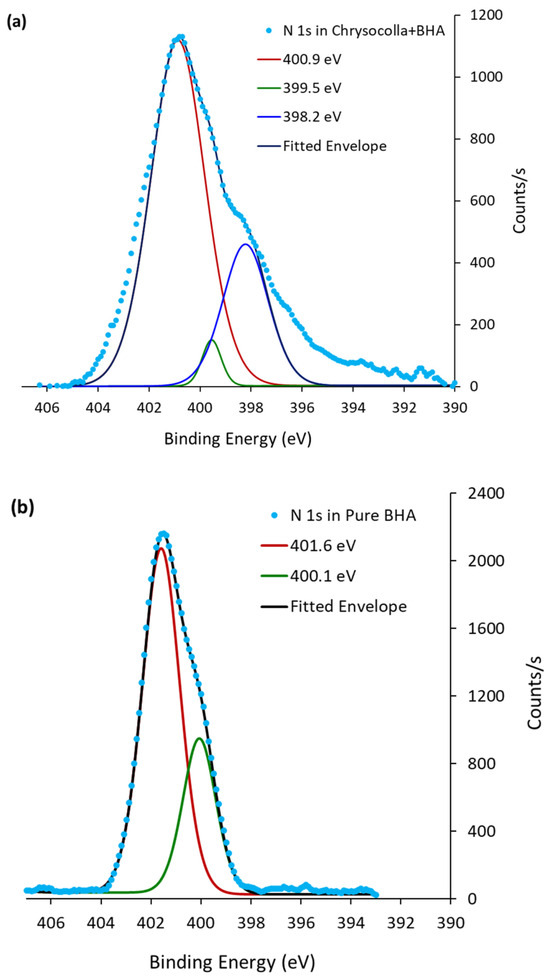
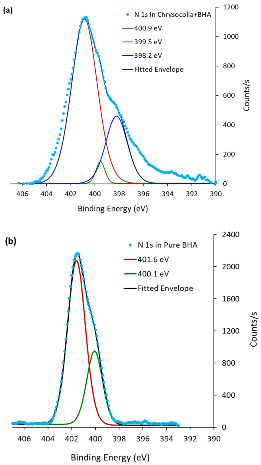
To confirm the elemental composition of N and its valence state, the high-resolution spectra of N 1s of the BHA-treated chrysocolla as well as pristine chrysocolla were recorded and processed (Figure 14). Table 6 details the fitting parameters. Peak fitting of N 1s for the surface of BHA-treated chrysocolla (Figure 14a) resulted in three peaks, two of which at 400.9 and 399.5 eV, with relative areas of 72.45% and 12.64%, respectively, correspond to the peaks at 401.6 and 400.1 eV in pure BHA (Figure 14b). A relatively intensive peak at 398.2 eV was also required to fit the spectrum. The N 1s peak at 401.6 eV could be attributed to protonated hydroxamic acid, which is close to the reported values at 401.5 [51] and 401.7 eV [29]. This characteristic peak of hydroxamates shifted to lower binding energy (400.9 eV) upon formation of chelates between BHA and the chrysocolla surface. This value is in good agreement with previous work where a peak was observed at 400.6 eV when 1,2-diaminopropane adsorbed on chrysocolla [8] and at 400.7 eV when n-octanohydroxamate chemisorbed to iron atoms [51]. The peak appeared at 400.9 and 400.6 when BHA was adsorbed on Pb2+-activated rhodochrosite and to Mn species of rhodochrosite surfaces, respectively [52]. For the BHA-treated chrysocolla, as shown in Figure 14a, the lower-intensity components at 399.5 and 398.2 eV are assigned more cautiously to minor deprotonated or weakly adsorbed nitrogen-containing species [51,52,53]. In particular, the 398.2 eV component with relative area of 14.92% may also include contributions from partial X-ray-induced degradation of adsorbed BHA [51] and is therefore not considered direct evidence of chemisorption. The chemisorption mechanism is instead inferred from the combined N 1s and Cu 2p XPS evidence. The shifts in the binding energy values are consistent with the results for protonated hydroxamate in n-octanohydroxamate [41,51], benzohydroxamic acid [29], and other aromatic hydroxamates [54].
Figure 14.
High-resolution XPS spectra for N 1s of the BHA-treated chrysocolla surface (a) and of pure BHA (b).
Table 6.
N 1s spectral fitting parameters for nitrogen species.
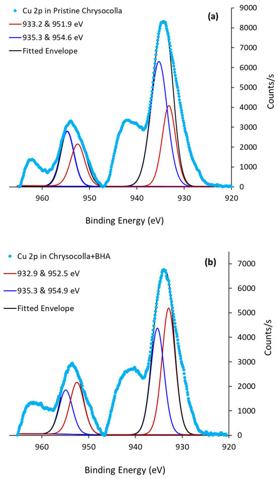
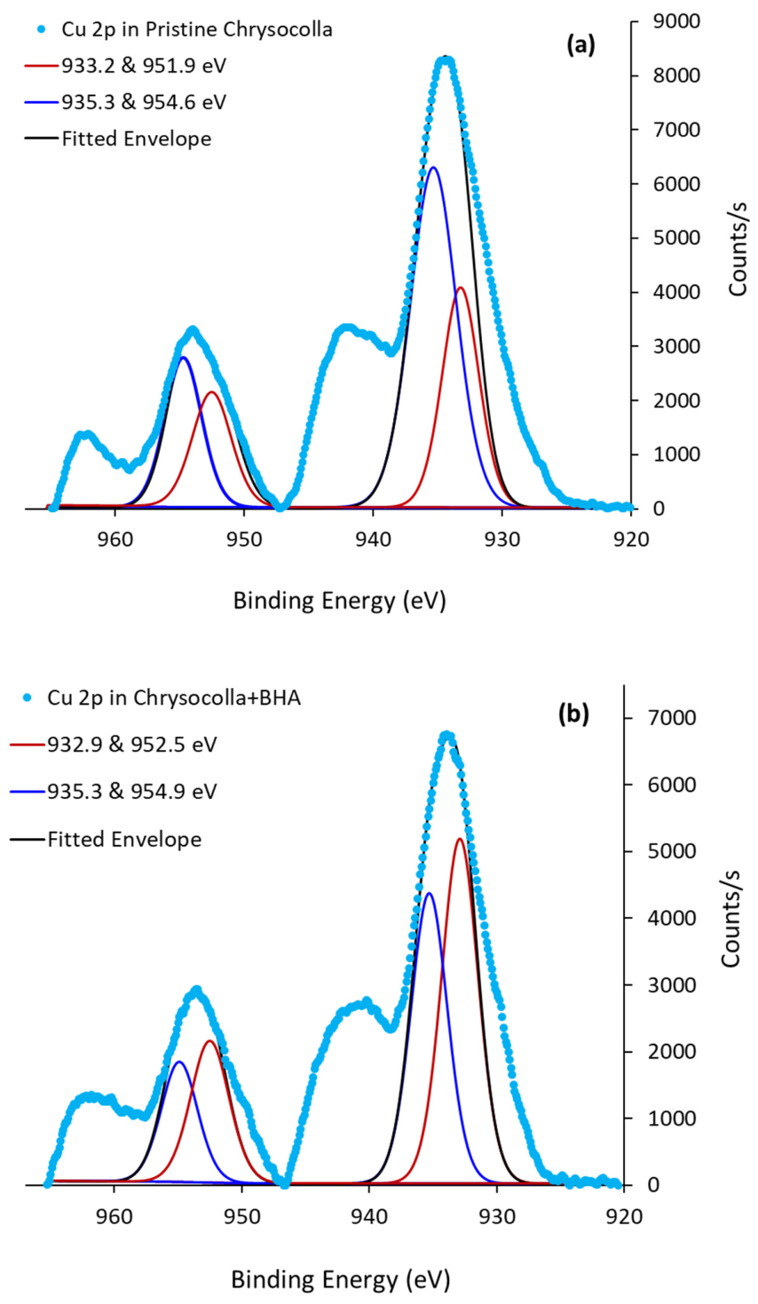
Complementary evidence for the adsorption of BHA at the mineral surface can be found in the difference between the electronic environment of copper atoms before and after treatment with BHA. The high-resolution XPS spectra of copper in chrysocolla before and after treatment with BHA are shown in Figure 15. The fitted peaks will be interpreted using the NIST XPS Database [55] and cited publications. Both spectra show fitted peaks for the Cu 2p region, resolving into components for Cu 2p3/2 and its spin–orbit split Cu 2p1/2. The Cu 2p3/2 region (around 930–935 eV) and Cu 2p1/2 region (around 950–955 eV) each contain two components, suggesting two copper species and a complex copper environment in both samples. Table 7 details the high-resolution spectra data for each fitted peak. The pristine chrysocolla shows a double at 933.2 eV and 951.9 eV (double A) for copper (Figure 15a). There is another pair of spin–orbit components at 935.3 and 954.6 eV (double B). According to the literature, both double A [7,43,56] and double B [7,54,55] are considered as chrysocolla characteristic peaks and are consistent with Cu (II) species. The peak at 933.2 eV is assigned to the copper ions in Cu(OH)2, and the peak at 935.3 eV is attributed to CuSiO3 [43]. Two strong shake-up satellites at higher binding energies relative to the main peaks are indication of the presence of Cu (II) [45], which are not observed in metallic copper and Cu (I) [57]. A similar XPS spectrum was obtained for the BHA-treated chrysocolla (Figure 15b). The fitted peaks with varying binding energies indicate the presence of copper in different oxidation states or coordination environments after the mineral has been treated with BHA. Table 7 indicates that the Cu 2p3/2 area ratio for CuSiO3·2H2O to CuO decreased from 1.9 to 0.8 upon surface treatment, which indicates that BHA adsorption modifies the relative contribution of the surface Cu environments. This suggests that BHA preferentially interacts with specific accessible Cu sites rather than uniformly complexing all Cu atoms present on the chrysocolla surface. The persistence of both Cu components after treatment further indicates that uncomplexed Cu species remain, which is expected considering the heterogeneous and microporous nature of chrysocolla and the possible presence of Cu sites embedded within silicate domains or otherwise inaccessible to BHA. Therefore, the observed change in Cu 2p peak area ratio is consistent with partial formation of Cu–hydroxamate surface complexes on accessible Cu sites, while residual uncomplexed Cu environments remain detectable by XPS. Also, the binding energy of the copper ion (Cu 2p3/2) decreased by 0.4 eV without affecting the valance state of copper, which can be due to the appearance of new Cu (II) species formed between the copper on the mineral surface and the hydroxamate chelating agent. The shape and intensity of the satellites also changed after conditioning the surface with BHA. No changes were observed in the high-resolution spectra of Si 2p, meaning that BHA did not interact with the silicon on the chrysocolla surface.
Figure 15.
High-resolution XPS spectra for Cu 2P of chrysocolla before (a) and after (b) treatment with BHA.
Table 7.
Results of the processing of the XPS high-resolution spectra for Cu 2p at the chrysocolla surface.
3.5. Denver Flotation Tests
The flotation products were characterized by XRF to determine the copper grades and calculate recoveries. The values represent the average of three experiments with 95% confidence intervals. As summarized in Table 8, the bench-scale flotation tests produced a Cu concentrate containing 26.69% Cu from a feed grading 3.67% Cu.
Table 8.
Grade and recovery of Cu in the flotation products after Denver cell flotation tests.
Although the primary objective of this work was to evaluate the flotation response of chrysocolla using BHA rather than to optimize a commercial flowsheet, the concentrate grade falls within the range commonly reported for commercial copper concentrates [58,59]. However, the overall Cu recovery indicates that a substantial fraction of Cu-bearing material remained poorly floatable under the tested conditions. The elemental compositions of the feed, pre-float concentrate, Cu concentrate, and tailings from XRF analysis are presented in Table 9, which illustrates the effectiveness of the separation. Relative to the feed, the Cu concentrate was enriched in Cu, while the SiO2 content decreased from 71.66% to 46.72%, indicating preferential recovery of Cu-bearing minerals and rejection of silicate gangue. The high SiO2 content of the tailings also demonstrates that the unrecovered material was predominantly silicate-rich. However, the bulk XRF analyses cannot distinguish whether the remaining Cu is present as liberated but poorly floatable chrysocolla, composite particles associated with gangue minerals, or other Cu-bearing phases such as malachite or cuprite. Resolving this question would require detailed mineral liberation analysis, which was beyond the scope of the present study.
Table 9.
Elemental composition of the feed and products of Denver flotation.
To further evaluate the performance of the sequential flotation flowsheet, corresponding elemental grades and recoveries are summarized in Table 10. During preparation of the revised manuscript, the flotation products were re-analyzed using XRF to establish a complete elemental mass balance for the Denver flotation tests. The original analytical dataset used for some of the previous calculations could not be fully recovered; therefore, grades and recoveries were recalculated using the newly acquired XRF measurements performed on the same archived flotation samples. Minor differences were observed relative to the originally reported values; however, these differences were small and did not influence the interpretation of the flotation behavior or the conclusions of this work.
Table 10.
Elemental grades and recoveries (mass balance) of the bench-scale flotation products by XRF analysis.
The mass balance demonstrates that the BHA flotation stage produced a Cu concentrate containing 26.69 ± 1.74% Cu, corresponding to a Cu recovery of 35.29 ± 2.98%, whereas the pre-float stage recovered and additional 6.46 ± 2.10% of the Cu at a concentrate grade of 16.39 ± 4.93%. Consequently, the overall Cu recovery reached 41.75%. The tailings contained only 1.75 ± 0.10% Cu while accounting for 58.24 ± 5.09% of the total Cu, indicating that a substantial proportion of the Cu-bearing minerals remained unrecoverable. In contrast, 96.64% of the Si reported to the tailings, confirming effective rejection of silicate gangue during flotation. Similarly, more than 95% of the Al and K also reported to the tailings, whereas sulfur was preferentially recovered into the flotation concentrates, consistent with the removal of sulfide minerals during the pre-float and subsequent Cu flotation stages.
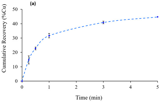
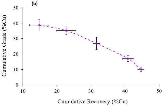
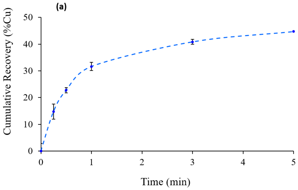
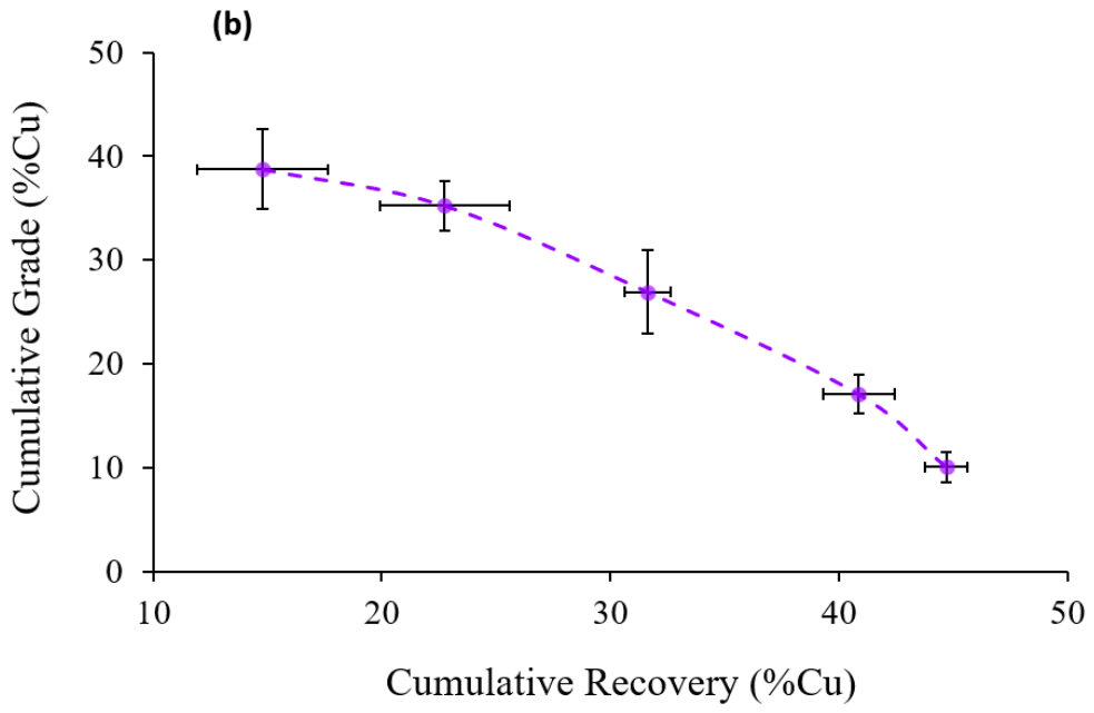
To better understand the kinetics of copper recovery, kinetic flotation experiments were also performed. The results shown in Table 11 reveal that the highest Cu grade of 39% was achieved within the first 15 s of flotation, followed by a gradual decline in grade and recovery over 5 min flotation time. The corresponding recovery curve in Figure 16a highlights the rapid initial recovery increase over the first minute followed by a gradual slowing after 3 min. The rapid recovery of a high-grade concentrate during the first 15 s indicates that only a relatively small proportion of the Cu-bearing particles exhibited excellent floatability under the investigated conditions. These particles likely possessed readily accessible surface Cu sites that promoted rapid Cu–hydroxamate complex formation and bubble attachment. As flotation progressed, the concentrate grade gradually decreased while cumulative recovery increased, suggesting that progressively less floatable Cu-bearing particles were recovered. This behavior is consistent with the mineralogical heterogeneity of the ore, which contains chrysocolla, malachite, cuprite, and quartz, together with the wide range of Cu compositions observed by SEM-EDS. Although a quantitative mineral liberation analysis was beyond the scope of this work, the flotation kinetics suggest that the ore contains Cu-bearing particles with a broad distribution of floatability rather than a uniformly responsive chrysocolla population. Figure 16b shows the grade–recovery curves of the same flotation experiments.
Table 11.
Grade and recovery of the samples after Denver cell flotation kinetic tests.
Figure 16.
Cumulative (a) Cu recovery over time and (b) Cu grade–recovery obtained from the Denver flotation experiments. Data points shown are average values with 95% confidence intervals. The dashed lines are provided as a guide to the eye.
The relatively large uncertainty associated with the Cu grade of the pre-float concentrate is attributed to the small mass and heterogeneous composition of this product. The pre-float stage recovered only a minor fraction of the feed as readily floatable sulfide material; therefore, small variations in the recovery of Cu-bearing sulfide particles or entrained material among replicate tests had a relatively large effect on the measured concentrate grade. This variability reflects the heterogeneous nature of the ore and the low mass pull of the pre-float stage.
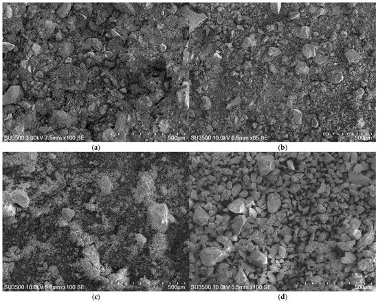
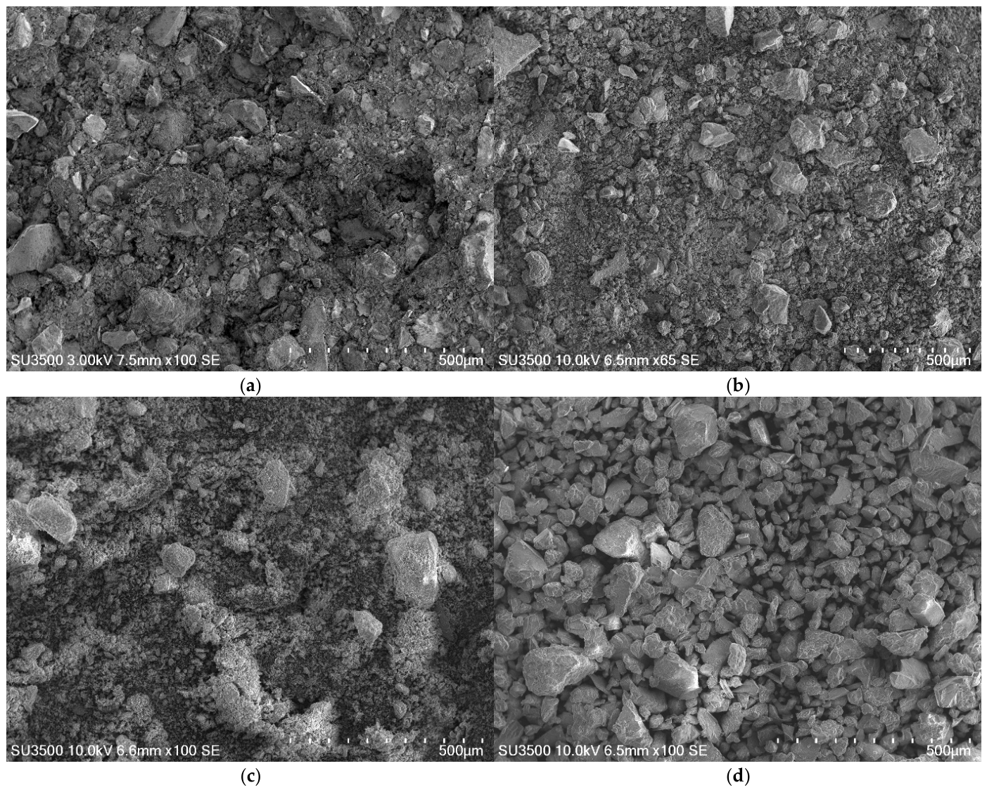
The Denver flotation products, including feed, pre-float concentrate, Cu concentrate and tails were characterized by SEM with representative results shown in Figure 17. The images reveal the differences in particle size and surface morphology among the products. In particular, the feed displays a heterogeneous mixture of coarse and fine particles (Figure 17a), whereas it qualitatively indicates the presence of relatively coarse and irregular particles in the tailings (Figure 17d). However, those observations alone cannot establish the degree of mineral liberation or the mineralogical distribution of the unrecovered Cu. Combined with XRF results, which show that the tailings are enriched in SiO2 (74%) and contain only 2.28% CuO, the results suggest that the unrecovered Cu is associated with a predominantly silicate-rich tailings product. Nevertheless, detailed mineral liberation analysis (e.g., MLA or QEMSCAN) would be required to confirm the degree of liberation and the distribution of unrecovered Cu-bearing minerals.
Figure 17.
SEM images of Denver cell flotation products showing feed (a), pre-float concentrate (b), Cu concentrate (c), and tailing (d).
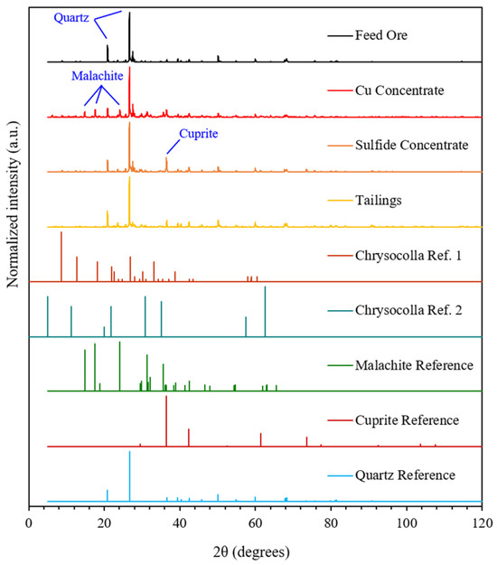
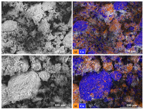
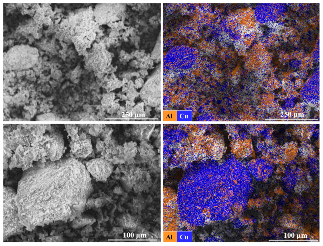
The phase identification by XRD for the feed and bench-scale flotation products shown in Figure 18 confirms the presence of malachite peaks in the Cu concentrate, consistent with successful recovery of Cu-bearing minerals. Minor reflections attributed to cuprite were also observed, while quartz peaks dominated the feed and tailing samples, indicating the predominance of silicate gangue minerals in the non-floating fraction. However, SEM-EDS analysis of the Cu concentrate in Figure 19 reveals the presence of both Cu and Al, corresponding the copper phase to chrysocolla rather than malachite.
Figure 18.
XRD results of Denver flotation products.
Figure 19.
SEM-EDS results of the Cu concentrate at different magnifications showing the presence of Cu and Al.
These results confirm that the sequential flotation process effectively separates sulfide and oxide copper minerals while substantially rejecting silicate gangue. However, the sulfur content of the pre-float concentrate was less than 1 wt% which is below the practical detection limit for minor crystalline sulfide phases by XRD. Consequently, the sulfide mineralogy of this fraction could not be conclusively identified from the XRD results.
4. Conclusions
This study provided insight into hydroxamate–chrysocolla interactions and highlighted the potential and limitations of benzohydroxamic acid (BHA) usage for oxidized copper silicate ore flotation. It has demonstrated that BHA effectively overcomes the poor flotation response of porous chrysocolla by chemisorbing as Cu–hydroxamate complexes, as confirmed by X-ray photoelectron spectroscopy (XPS). Microflotation experiments reveal that chrysocolla recovery increased (>90%) at BHA dosage of 50 and 100 kg t−1. The recovery was maximized at pH 8–10, where BHA dissociated and preferentially adsorbed on chrysocolla rather than quartz. Bench-scale Denver tests produced a Cu concentrate of 26% Cu with 35% recovery, highlighting the practical potential of BHA for oxidized copper silicate ores. Kinetic data indicated rapid flotation of fine particles, while coarse gangue remains in the tailings. The BHA dosages required to achieve maximum chrysocolla recovery in the present study are substantially higher than collector dosages typically employed in industrial flotation. This elevated reagent demand is likely related to the exceptionally high specific surface area, microporous structure, and partially amorphous mature of the investigated chrysocolla. Consequently, the dosages reported herein should not be interpreted as directly representative of industrial operating conditions but rather as those required to elucidate the adsorption behavior and maximum flotation response of this challenging mineral. From a practical perspective, such high reagent consumption would increase operating costs and could also increase the organic loading of process water, highlighting the importance of collector dosage optimization and reagent management. Future work should therefore focus on reducing BHA consumption through optimized reagent schemes, mixed collector systems, surface activation strategies, or reagent recycling while maintaining high flotation recovery.
Author Contributions
Conceptualization, S.M., O.K. and K.E.W.; Methodology, S.M., S.B., R.L., C.M., J.P., O.K. and K.E.W.; Validation, O.K.; Formal analysis, S.M., S.B., R.L., C.M. and J.P.; Investigation, S.M., S.B., R.L., C.M., J.P. and O.K.; Resources, K.E.W.; Data curation, S.M. and O.K.; Writing—original draft, S.M., S.B., R.L. and J.P.; Writing—review & editing, S.M. and K.E.W.; Visualization, K.E.W.; Supervision, K.E.W.; Project administration, K.E.W.; Funding acquisition, K.E.W. All authors have read and agreed to the published version of the manuscript.
Funding
This work was funded by the Glenn Dobby Fund for Sustainable Mineral Resources, McGill University.
Data Availability Statement
The original contributions presented in this study are included in the article. Further inquiries can be directed to the corresponding author.
Acknowledgments
The authors would like to acknowledge support from the Glenn Dobby fund for Sustainable Mineral Resources at McGill University.
Conflicts of Interest
The authors declare no conflicts of interest.
Abbreviations
The following abbreviations are used in this manuscript:
| BET | Brunauer–Emmett–Teller |
| BHA | Benzohydroxamic acid |
| ED-XRF | Energy-dispersive X-ray fluorescence |
| EDS | Energy-dispersive spectrometer |
| MIBC | Methyl isobutyl carbinol |
| PAX | Potassium amyl xanthate |
| RO | Reverse osmosis |
| SEM | Scanning electron microscopy |
| SEM-EDS | Scanning electron microscopy–energy-dispersive X-ray spectroscopy |
| XPS | X-ray photoelectron spectroscopy |
| XRD | X-ray diffraction |
| XRF | X-ray fluorescence |
References
- Frost, R.L.; Xi, Y. Is chrysocolla (Cu,Al)2H2Si2O5(OH)4·nH2O related to spertiniite Cu(OH)2?—A vibrational spectroscopic study. Vib. Spectrosc. 2013, 64, 33–38. [Google Scholar] [CrossRef] [Scilit]
- Feng, Q.; Yang, W.; Wen, S.; Wang, H.; Zhao, W.; Han, G. Flotation of copper oxide minerals: A review. Int. J. Min. Sci. Technol. 2022, 32, 1351–1364. [Google Scholar] [CrossRef] [Scilit]
- Chukhrov, F.V.; Zvyagin, B.B.; Gorshkov, A.I.; Yermilova, L.P.; Rudnitskaya, Y.S. Chrysocollas. Int. Geol. Rev. 1969, 11, 570–581. [Google Scholar] [CrossRef] [Scilit]
- Ndoro, T.O.; Witika, L.K. A Review of the Flotation of Copper Minerals. Int. J. Sci. Basic Appl. Res. 2017, 34, 145–165. [Google Scholar]
- Sun, M.-S. The nature of chrysocolla from Inspiration Mine, Arizona. Am. Mineral. 1963, 48, 649–658. [Google Scholar]
- Raghavan, S.; Fuerstenau, D.W. Characterization and pore structure analysis of a copper ore containing chrysocolla. Int. J. Miner. Process. 1977, 4, 381–394. [Google Scholar] [CrossRef] [Scilit]
- Shen, P.; Liu, D.; Xu, X.; Jia, X.; Zhang, X.; Liu, D.; Liu, R. Effect of Ethylene Diamine Phosphate on the Sulfidization Flotation of Chrysocolla. Minerals 2018, 8, 216. [Google Scholar] [CrossRef] [Scilit]
- Peng, R.; Wang, H.; Wei, Z.; Fang, J.; Zhang, X.; Shen, P.; Liu, D. Effects of 1,2-diaminopropane on the formation of chelated copper on chrysocolla surfaces and its response to flotation. Miner. Eng. 2024, 212, 108705. [Google Scholar] [CrossRef] [Scilit]
- Aplan, F.F.; Fuerstenau, D.W. The Flotation of Chrysocolla by Mercaptan. Int. J. Miner. Process. 1984, 13, 105–115. [Google Scholar] [CrossRef] [Scilit]
- Tian, M.; Hu, Y.; Sun, W.; Liu, R. Study on the mechanism and application of a novel collector-complexes in cassiterite flotation. Colloids Surf. A Physicochem. Eng. Asp. 2017, 522, 635–641. [Google Scholar] [CrossRef] [Scilit]
- Wang, X.; Li, H.; Liu, X.; Tang, Y.; Ni, C. Advances in Flotation Reagents for Cassiterite Separation: Challenges and Sustainable Solutions. Molecules 2025, 30, 2380. [Google Scholar] [CrossRef] [Scilit] [PubMed]
- Ren, Q.; Liu, P.; Yu, C.; Liu, S.; Zhang, W.; Kang, J.; Sun, W.; Tian, M. The application and adsorption selectivity of a novel collector 4-butoxy-N-hydroxybenzamide in cassiterite flotation. Appl. Surf. Sci. 2025, 679, 161272. [Google Scholar] [CrossRef] [Scilit]
- Wu, H.; Tian, J.; Xu, L.; Fang, S.; Zhang, Z.; Chi, R. Flotation and adsorption of a new mixed anionic/cationic collector in the spodumene-feldspar system. Miner. Eng. 2018, 127, 42–47. [Google Scholar] [CrossRef] [Scilit]
- Cook, B.K.; Gibson, C.E. A Review of Fatty Acid Collectors: Implications for Spodumene Flotation. Minerals 2023, 13, 212. [Google Scholar] [CrossRef] [Scilit]
- Retamal, J.I.; Robles, P.A.; Quezada, G.R.; Jeldres, R.I. Molecular Design and Spodumene Flotation—A Review. Int. J. Mol. Sci. 2024, 25, 3227. [Google Scholar] [CrossRef] [Scilit] [PubMed]
- Cook, B.K.; Aghamirian, M.; Grammatikopoulos, T.; Gibson, C.E. The effect of conditioning pH behaviour on spodumene flotation with fatty acids. Can. Metall. Q. 2024, 63, 629–645. [Google Scholar] [CrossRef] [Scilit]
- Fuerstenau, D.W.; Herrera-Urbina, R.; McGlashan, D.W. Studies on the applicability of chelating agents as universal collectors for copper minerals. Int. J. Miner. Process. 2000, 58, 15–33. [Google Scholar] [CrossRef] [Scilit]
- Necip, A.; Yekeler, H. Interfacial Mechanisms of O-O Type Chelating Collectors in the Flotation of Copper Minerals: A Density Functional Study. Manas J. Agric. Vet. Life Sci. 2025, 15, 103–112. [Google Scholar] [CrossRef] [Scilit]
- Fuerstenau, D.W.; Pradip, C. Mineral flotation with hydroxamate collectors. In Reagents in the Minerals Industry; Jones, M.J., Oblatt, R., Eds.; The Institution of Mining and Metallurgy: London, UK, 1984; pp. 161–168. [Google Scholar]
- Lee, J.S.; Nagaraj, D.R.; Coe, J.E. Practical aspects of oxide copper recovery with alkyl hydroxamates. Miner. Eng. 1998, 11, 929–939. [Google Scholar] [CrossRef] [Scilit]
- Zhang, C.; Zhou, Q.; An, B.; Yue, T.; Chen, S.; Liu, M.; He, J.; Zhu, J.; Chen, D.; Hu, B.; et al. Flotation Behavior and Synergistic Mechanism of Benzohydroxamic Acid and Sodium Butyl-Xanthate as Combined Collectors for Malachite Beneficiation. Minerals 2021, 11, 59. [Google Scholar] [CrossRef] [Scilit]
- Marion, C.; Jordens, A.; Li, R.; Rudolph, M.; Waters, K.E. An evaluation of hydroxamate collectors for malachite flotation. Sep. Purif. Technol. 2017, 183, 258–269. [Google Scholar] [CrossRef] [Scilit]
- Li, Z.; Rao, F.; Song, S.; Uribe-Salas, A.; Lopez-Valdivieso, A. Reexamining the adsorption of octyl hydroxamate on malachite surface: Forms of molecules and anions. Miner. Process. Extr. Metall. Rev. 2020, 41, 178–186. [Google Scholar] [CrossRef] [Scilit]
- Sun, X.; Huang, Y.L.; Wu, D.; Hu, B.; Zhang, M.; Tong, X. Synthesis of a novel hydroxamic acid flotation collector and its flotation separation of malachite against quartz. Physicochem. Probl. Miner. Process. 2022, 58, 149427. [Google Scholar] [CrossRef] [Scilit]
- Bulatovic, S.M. Flotation of Oxide Copper and Copper Cobalt Ores. In Handbook of Flotation Reagents: Chemistry, Theory and Practice; Bulatovic, S.M., Ed.; Elsevier: Amsterdam, The Nethelands, 2010; pp. 47–65. [Google Scholar]
- Lee, K.; Archibald, D.; McLean, J.; Reuter, M.A. Flotation of mixed copper oxide and sulphide minerals with xanthate and hydroxamate collectors. Miner. Eng. 2009, 22, 395–401. [Google Scholar] [CrossRef] [Scilit]
- Lenormand, J.; Salman, T.; Yoon, R.H. Hydroxamate Flotation of Malachite. Can. Metall. Q. 1979, 18, 125–129. [Google Scholar] [CrossRef]
- Elizondo-Álvarez, M.A.; Uribe-Salas, A.; Nava-Alonso, F. Flotation studies of galena (PbS), cerussite (PbCO3) and anglesite (PbSO4) with hydroxamic acids as collectors. Miner. Eng. 2020, 155, 106456. [Google Scholar] [CrossRef] [Scilit]
- Mohammadi-Jam, S.; Li, Z.; Rose, N.; Waters, K.E. An investigation into using benzohydroxamic acid as a collector for sulfide minerals. Physicochem. Probl. Miner. Process. 2023, 59, 175662. [Google Scholar] [CrossRef] [Scilit]
- Ren, J.; Lu, S.; Song, S.; Niu, J. A new collector for rare earth mineral flotation. Miner. Eng. 1997, 10, 1395–1404. [Google Scholar] [CrossRef] [Scilit]
- Jordens, A.; Marion, C.; Grammatikopoulos, T.; Hart, B.; Waters, K.E. Beneficiation of the Nechalacho rare earth deposit: Flotation response using benzohydroxamic acid. Miner. Eng. 2016, 99, 158–169. [Google Scholar] [CrossRef] [Scilit]
- Pavez, O.; Peres, A.E.C. Effect of sodium metasilicate and sodium sulphide on the floatability of monazite-zircon-rutile with oleate and hydroxamates. Miner. Eng. 1993, 6, 69–78. [Google Scholar] [CrossRef] [Scilit]
- Pradip; Fuerstenau, D.W. The adsorption of hydroxamate on semi-soluble minerals. Part I: Adsorption on barite, Calcite and Bastnaesite. Colloids Surf. 1983, 8, 103–119. [Google Scholar] [CrossRef] [Scilit]
- Zhang, W.; Honaker, R.Q.; Groppo, J.G. Flotation of monazite in the presence of calcite part I: Calcium ion effects on the adsorption of hydroxamic acid. Miner. Eng. 2017, 100, 40–48. [Google Scholar] [CrossRef] [Scilit]
- Yoon, R.H.; Nagaraj, D.R.; Wang, S.S.; Hildebrand, T.M. Benefication of kaolin clay by froth flotation using hydroxamate collectors. Miner. Eng. 1992, 5, 457–467. [Google Scholar] [CrossRef] [Scilit]
- Castro, S.; Soto, H.; Goldfarb, J.; Laskowski, J. Sulphidizing reactions in the flotation of oxidized copper minerals, II. Role of the adsorption and oxidation of sodium sulphide in the flotation of chrysocolla and malachite. Int. J. Miner. Process. 1974, 1, 151–161. [Google Scholar] [CrossRef] [Scilit]
- Castro, S.; Gaytan, H.; Goldfarb, J. The stabilizing effect of Na2S on the collector coating of chrysocolla. Int. J. Miner. Process. 1976, 3, 71–82. [Google Scholar] [CrossRef] [Scilit]
- Laskowski, J.; Fuerstenau, D.W.; Gonzalez, G.; Urbina, R.H. Studies on the Flotation of Chrysocolla. Miner. Process. Extr. Metall. Rev. 1985, 2, 135–155. [Google Scholar] [CrossRef] [Scilit]
- Hope, G.A.; Numprasanthai, A.; Buckley, A.N.; Parker, G.K.; Sheldon, G. Bench-scale flotation of chrysocolla with n-octanohydroxamate. Miner. Eng. 2012, 36–38, 12–20. [Google Scholar] [CrossRef] [Scilit]
- Farges, F.; Benzerara, K.; Brown, G.E., Jr. (Eds.) Chrysocolla Redefined as Spertiniite; American Institute of Physics (AIP): Melville, NY, USA, 2007; pp. 223–225. [Google Scholar] [CrossRef] [Scilit]
- Hope, G.A.; Buckley, A.N.; Parker, G.K.; Numprasanthai, A.; Woods, R.; McLean, J. The interaction of n-octanohydroxamate with chrysocolla and oxide copper surfaces. Miner. Eng. 2012, 36–38, 2–11. [Google Scholar] [CrossRef] [Scilit]
- McKeown, D.A. X-ray absorption spectroscopic study of copper in an amorphous copper silicate: Chrysocolla. J. Non-Cryst. Solids 1994, 180, 1–10. [Google Scholar] [CrossRef] [Scilit]
- Peng, R.; Wang, H.; Shen, P.; Liu, D. A novel activation scheme to improve surface floatability of chrysocolla: Mechanism and properties. J. Environ. Chem. Eng. 2025, 13, 117509. [Google Scholar] [CrossRef] [Scilit]
- Gonzalez, G.; Soto, H. The effect of thermal treatment on the flotation of chrysocolla. Int. J. Miner. Process. 1978, 5, 153–162. [Google Scholar] [CrossRef] [Scilit]
- Zuo, Q.; Wang, Z.; Wu, D.; Wen, S.; Chen, H.; Cao, J. Ammonia-induced corrosion and modification of chrysocolla surface for enhanced adsorption of sulfide species. Colloids Surf. A Physicochem. Eng. Asp. 2024, 682, 132884. [Google Scholar] [CrossRef] [Scilit]
- Liu, D.; Wang, D.; Xian, Y.; Tian, X.; Wen, S. Enhancement of xanthate adsorption on chrysocolla surface via sodium diethyldithiocarbamate (DDTC) modification. Physicochem. Probl. Miner. Process. 2022, 58, 152599. [Google Scholar] [CrossRef] [Scilit]
- Hope, G.A.; Woods, R.; Parker, G.K.; Buckley, A.N.; McLean, J. A vibrational spectroscopy and XPS investigation of the interaction of hydroxamate reagents on copper oxide minerals. Miner. Eng. 2010, 23, 952–959. [Google Scholar] [CrossRef] [Scilit]
- Elizondo-Álvarez, M.A.; Uribe-Salas, A.; Bello-Teodoro, S. Chemical stability of xanthates, dithiophosphinates and hydroxamic acids in aqueous solutions and their environmental implications. Ecotoxicol. Environ. Saf. 2021, 207, 111509. [Google Scholar] [CrossRef] [Scilit] [PubMed]
- Liu, C.; Zhang, W.; Song, S.; Li, H. Study on the activation mechanism of lead ions in wolframite flotation using benzyl hydroxamic acid as the collector. Miner. Eng. 2019, 141, 105859. [Google Scholar] [CrossRef] [Scilit]
- Xu, L.; Tian, J.; Wu, H.; Lu, Z.; Yang, Y.; Sun, W.; Hu, Y. Effect of Pb2+ ions on ilmenite flotation and adsorption of benzohydroxamic acid as a collector. Appl. Surf. Sci. 2017, 425, 796–802. [Google Scholar] [CrossRef] [Scilit]
- Parker, G.K.; Buckley, A.N.; Woods, R.; Hope, G.A. The interaction of the flotation reagent, n-octanohydroxamate, with sulfide minerals. Miner. Eng. 2012, 36–38, 81–90. [Google Scholar] [CrossRef] [Scilit]
- Cui, K.; Jin, S.; Duan, N. Insights into the adsorption mechanism of benzohydroxamic acid in the flotation of rhodochrosite with Pb2+ activation. Powder Technol. 2023, 427, 118705. [Google Scholar] [CrossRef] [Scilit]
- Kehrer, M.; Duchoslav, J.; Hinterreiter, A.; Cobet, M.; Mehic, A.; Stehrer, T.; Stifter, D. XPS investigation on the reactivity of surface imine groups with TFAA. Plasma Processes Polym. 2019, 16, 1800160. [Google Scholar] [CrossRef] [Scilit]
- Wang, H.; Zhong, H.; Li, F. Utilization of aromatic hydroxamates with different carbon chain lengths for pyrochlore, calcite, feldspar, and quartz flotation. Colloids Surf. A Physicochem. Eng. Asp. 2024, 682, 132810. [Google Scholar] [CrossRef] [Scilit]
- Naumkin, A.V.; Kraut-Vass, A.; Gaarenstroom, S.W. NIST X-Ray Photoelectron Spectroscopy Database, NIST Standard Reference Database Number 20; National Institute of Standards and Technology: Gaithersburg, MD, USA, 2000.
- Frost, R.L.; Xi, Y.; Wood, B.J. Thermogravimetric analysis, PXRD, EDX and XPS study of chrysocolla (Cu,Al)2H2Si2O5(OH)4·nH2O-structural implications. Thermochim. Acta 2012, 545, 157–162. [Google Scholar] [CrossRef] [Scilit]
- Watts, J.F.; Wolstenholme, J. An Introduction to Surface Analysis by XPS and AES; John Wiley & Sons Ltd: West Sussex, UK, 2003; p. 288. [Google Scholar]
- The World Copper Factbook 2025. International Copper Study Group. 2025. Available online: https://icsg.org/copper-factbook/ (accessed on 10 June 2026).
- Wills, B.A.; Finch, J. Wills’ Mineral Processing Technology: An Introduction to the Practical Aspects of Ore Treatment and Mineral Recovery, 8th ed.; Butterworth-Heinemann: Waltham, MA, USA, 2016. [Google Scholar]

Disclaimer/Publisher’s Note: The statements, opinions and data contained in all publications are solely those of the individual author(s) and contributor(s) and not of MDPI and/or the editor(s). MDPI and/or the editor(s) disclaim responsibility for any injury to people or property resulting from any ideas, methods, instructions or products referred to in the content. |
© 2026 by the authors. Licensee MDPI, Basel, Switzerland. This article is an open access article distributed under the terms and conditions of the Creative Commons Attribution (CC BY) license.