


Abstract
Objective: Evaluating the in vitro marginal adaptation of conventional, flowable and restorative bulk-fill resin composites placed in large class II cavities with supra- and sub-gingival margins, using a bulk-fill or layering approach, before and after thermo-mechanical loading (TML) simulating parafunctional forces. A total of 40 prepared teeth were divided and assigned to each of the five experimental groups. In group 1, restorations were made of layered high-viscosity conventional composite (Tetric EvoCeram); in groups 2 and 3 restorations were made of a high-viscosity bulk-fill composite (Tetric Powerfill) applied in one (group 2) or three layers (group 3); in groups 4 and 5 restorations were made of a flowable bulk-fill composite (SDRflow) applied in one (group 4) or two layers (group 5), underneath a layer of high-viscosity composite (Ceram-X Spectra ST). The same adhesive (OptiBond FL) was used in all groups. All specimens were submitted to a TML comprising a loading phase of 250,000 cycles at 100 N combined to 1675 thermal cycles (5 to 55 °C). The proximal tooth-restoration interfaces were analyzed quantitatively by SEM, prior and after TML. Results: Repeated measures ANOVA followed by Fisher’s LSD (Least Significant Difference) post hoc tests served for comparing inter-group marginal adaptation percentages between the pre- (T0) and post- (T1) loading conditions and intra-group marginal adaptation percentages. The lowest pre-loading values were for the cervical dentin adaptation ranging from 94.79% (SDRflow layered) to 66.06% (Tetric Powerfill layered) while the post-loading values of continuous cervical dentin adaptation varied from 61.20% (SDRflow layered) to 33.36% (SDRflow Monolayer). TML with higher axial forces led to a marked reduction in continuous adaptation at enamel or dentin margins in all groups. Overall, the low-viscosity bulk-fill SDRflow layers showed the best behavior while other products showed varying levels of degradation. Conclusions–Clinical significance: Simulated bruxism loading conditions induced severe marginal adaptions of class II composite restorations, which could potentially impact their lifespan.
1. Introduction
The progressive growth in bulk-fill resin composite use in posterior teeth was largely supported by reduced application time [1] and numerous in vitro and in vivo studies confirming their increased polymerization depth and lower volumetric polymerization shrinkage and stresses [2,3,4,5]. However, a reduction in volumetric shrinkage was proven to be significant only for high-viscosity bulk-fill materials, while low viscosity bulk-fill shows rather comparable behavior to conventional flowable resin composites [2,3,4]. The specific improvement in volumetric shrinkage stress of bulk-fill composites appeared to be mainly correlated to their specific matrix technology [5,6,7,8].
There is also clinical evidence showing that flowable bulk-fill composites perform equally to traditional, layered class I and II restorations over short-to-medium time terms [9,10,11,12]. However, data confirming satisfactory behavior over 5Y are scarce, especially for high-viscosity bulk-fill materials. Additionally, many authors confirmed the limitations of short-term follow-ups and risk of bias found in many of those studies. On the contrary, stronger evidence confirms the acceptable long-term performance of layered traditional composite restorations [13,14,15,16]. In view of their specific physic-chemical features, data and clinical validation would be needed for the use of bulk-fill technologies in bruxers and for biomechanically weakened teeth such as large and deep cavities or cracked and non-vital teeth. Owing to the high prevalence of bruxism and clenching [17,18,19], their potential impact on restoration behavior and failure patterns is of noteworthy interest. However, due to the known boundaries of clinical studies, namely the unavoidable impact of confounding factors [20,21,22] and inherent difficulty in organizing long-term prospective studies, an in vitro testing approach would therefore be of interest for an initial investigation of the aforementioned specific biomechanical and restorative configurations [23,24,25,26,27,28].
In vitro testing of marginal adaptation appears a valid approach to investigate and compare the impact of bulkfill and conventional resin composites under higher forces to understand if and how the difference in composite technology [2,3,4,5,6,7,8] might affect restoration quality. In particular, the E-modulus [2,5] has potential to influence marginal and internal adaptation through its impact on stress absorption and restored tooth deformation during functional and particularly parafunctional loading.
Extensive research data were published regarding functional and parafunctional forces as well as other bio-mechanical parameters of interest to simulate some specific oral strains in vitro with reasonably good relevance. Biting force during bruxism, measured in vivo using intra-oral sensors, presented values with large intra-individual variations (300 to 1300 N), higher forces being reached in males [29]. Interestingly, it was also shown that biting forces during bruxism can occasionally exceed those exerted voluntarily [30,31,32]. Also considering the distribution of biting forces, the inherent variation in jaw position during bruxism events (unilateral or bilateral contacts) explains the large range of pressure or force per tooth; the reported amplitude actually ranges from 10 to about 700 N [33,34,35,36]. When usual biting forces were measured per contact, pressures reached lower values, from about 38 to 50 N [33,34,35,36,37] which corresponds to the standard loading values applied in marginal analysis studies [24,25]. When it comes to the number of bruxism events, their average occurrence was reported to be of 3.6 per hour [31] with large variations reported [38,39]; these parafunctional phases last several seconds (av. 7.1 s to 15.4) [31,38] with several contacts per event. Using the aforementioned data and 3.5 average contacts per bruxism event over 8 h daily sleeping time (generating about 100 tooth-to-tooth contacts), 250,000 thermo-mechanical cycles hypothetically represent about 7Y of parafunctional activities, possibly more for patients having shorter sleep patterns. Overall, the aforementioned data confirmed that the usual testing parameters of marginal analysis studies and their results analysis had to be adjusted to assess the behavior and potential degradation of restoration submitted to parafunctional forces.
The null hypothesis of this in vitro study was that the restorative material (conventional nano-hybrid, bulk-filling flowable or high-viscosity resin composites) and application protocol (layering or bulk-filling) do not influence the marginal adaptation of supra- and sub-gingival class II restorations after thermo-occlusal stressing simulating non-physiological, parafunctional forces.
2. Materials and Methods
2.1. Specimen Preparation and Group Allocation
Forty freshly extracted human third molars were collected for this study. The inclusion criteria required complete root formation, absence of anatomical or size abnormalities and absence of carious lesions or extraction-related defects near the future restoration margins. As the teeth used in this project could not be linked to specific patients, ethical approval by the Geneva Ethics Committee was not required (Art. 2 et al., LRH).
The teeth were assigned to five experimental groups (n = 8 per group) according to size (small, medium or large) and anatomical type (maxillary or mandibular), as described in the restorative procedures section. Specimens were stored in saline solution at 5 °C from extraction until the start of the experiment.
Each tooth was stabilized using a small increment of light-curing composite on a brass holder (I.S.T. Innovative Technology GmbH Ebnat-Kappel, Switzerland) positioned in a custom-made device to control both central and horizontal axes. The roots were embedded up to 3 mm below the cemento-enamel junction (CEJ) in self-curing acrylic resin (Technovit 4071; Kulzer, Hanau, Germany) to ensure stability.
To facilitate restorative material placement, improve adaptation and reduce finishing time, an impression of each tooth was made prior to cavity preparation using a putty silicone (Memosil 2, Heraeus Kulzer, Hanau, Germany) placed in a plastic tube (2.5 cm diameter). After setting, the silicone mold was removed and sectioned bucco-lingually to produce mesial and distal indices, serving as a matrix substitute.
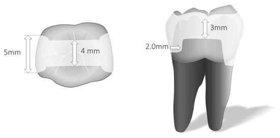
Standardized class II mesio–occluso–distal (MOD) cavities with parallel walls were prepared using a parallelometer to ensure uniform cavity dimensions and geometry. Proximal margins were located 1 mm above the CEJ mesialy and 1 mm below the CEJ distally. The cavities measured 5.0 mm in width and 2.0 mm in depth at the bottom of the proximal box, and 4.0 mm in width and 3.0 mm in depth at the occlusal isthmus (Figure 1).
Figure 1.
Cavity design.
Cavity preparation was performed with cylindrical burs with slightly rounded tips (80 µm grit) under copious water spray. Finishing was performed using fine-grit diamond burs of the same shape (40 µm grit) under water cooling. A 1.0 mm bevel was then placed along all enamel margins using a fine flame-shaped diamond bur (40 µm grit). All preparation and finishing procedures were performed under an operating microscope at 8–10× magnification.
2.2. Restorative Procedures
2.2.1. Adhesive Application
All cavity surfaces were treated using a three-step etch-and-rinse adhesive system (Optibond FL Unidose, Kerr, Bensheim, Germany). Selective enamel and dentin etching were performed for 30 s and 10 s, respectively. After thorough rinsing and gentle water removal by suction, the primer and adhesive were applied following the manufacturer’s instructions and light-cured for 20 s at an irradiance of 1250 mW/cm2 (SmartLite Pro, Dentsply, Schaan, Liechtenstein).
2.2.2. Restorative Protocol
The cavities were restored using different composite materials and layering techniques as follows:
Group 1 (control): Multilayered restorations with Tetric EvoCeram (Ivoclar, Schaan, Liechtenstein).
Proximal boxes were first filled with a 1.5 mm horizontal cervical layer, followed by two vertical layers (1.5–2 mm thick). The occlusal isthmus was restored with two oblique layers (facial and lingual).
Groups 2 and 3: Bulk-fill or layered restorations using Tetric Powerfill (Ivoclar). In group 2, a single increment of Tetric Powerfill was used to fill the entire cavity. In group 3, a trilaminar approach was employed, consisting of two horizontal increments in the proximal area and a final occlusal layer to complete the restoration.
Groups 4 and 5: Bulk-fill or layered restorations using SureFil SDRflow (Dentsply Sirona, Schaan, Liechtenstein) covered with an occlusal layer of Ceram.X Spectra ST (Dentsply Sirona). In group 4, the cavity was restored with a single bulk increment of SureFil SDRflow up to 1.5–2.0 mm below the occlusal surface. In group 5, a bilaminar base technique was applied, with a first horizontal increment in the proximal box and a second increment extending to 1.5–2 mm below the occlusal surface, followed by a single occlusal increment of Ceram.X.
The average depth of cavities below the CEJ was approximately 5 mm, measured from the proximal ridge. In groups 2 and 4, the thickness of proximal increments on the dentin side ranged from 3 to 3.5 mm for SureFil SDRflow and up to 5 mm for Tetric Powerfill, both within published light-curing efficiency limits (4). Increment thickness was verified for each specimen using a caliper.
All composite layers were light-cured for 20 s in continuous mode using the SmartLite Pro curing unit (1250 mW/cm2) according to the manufacturer’s recommendations. Proximal cavities were restored using the silicone indices fabricated before cavity preparation. To improve clinical relevance, indices were coated with aluminum foil to simulate light reflection from typical sectional metal matrices.
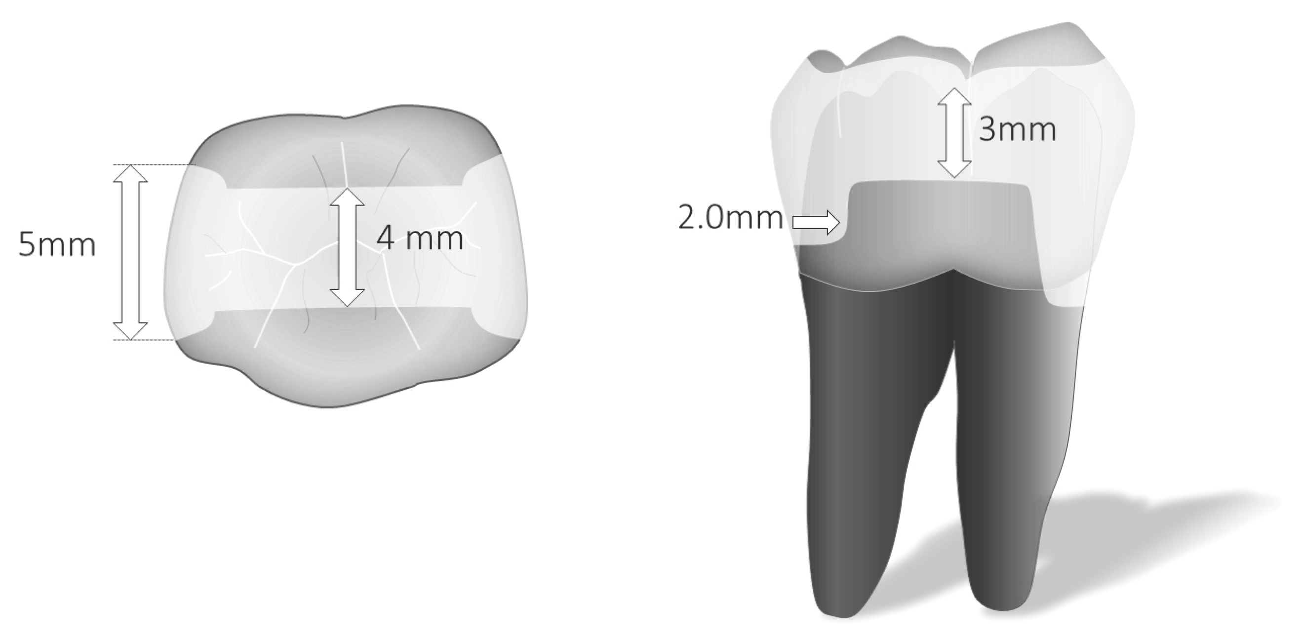
The final occlusal layer was carved and shaped with hand instruments prior to polymerization to minimize polymerization stress. Restorations were finished and polished immediately using fine diamond burs (40 µm grit) for occlusal surfaces, followed by silicone polishing points under water cooling (Brownies, Shofu Vernon, CA, USA). Proximal surfaces were finished with sequential abrasive disks of decreasing grit (Sof-Lex Pop-On XT: coarse to extra-fine; 3M). A summery of the workflow is presented in Figure 2.
Figure 2.
Study workflow.
2.3. Thermo-Mechanical Loading
Fatigue testing was performed one week after restoration placement and finishing, following a standardized protocol [24,25]. Each specimen underwent thermo-mechanical cycling using a chewing simulator (Chewing Simulator CS-4.8, SD Mechatronik, Vaterstetten, Germany) for 250,000 mechanical cycles at 100 N axial load applied to the central occlusal surface at 1.2 Hz frequency. These parameters correspond to approximately seven years of simulated parafunctional activity [31,33,34,35,36,37,38,39].
A “low-impact” chewing mode was selected to mimic the natural masticatory pattern. Additionally, specimens underwent 1675 thermal cycles between 5 °C and 55 °C. Prior to testing, a 0.5 mm rubber disk was placed beneath each sample to simulate periodontal ligament stress absorption during loading.
2.4. Specimen Evaluation
Before and after fatigue testing, restoration margins were cleaned with a brush and fine pumice, followed by a brief 5 s surface etching using diluted phosphoric acid gel (35–37% H3PO4 diluted threefold with distilled water; Ultra-Etch, Ultradent, South Jordan, UT, USA). Impressions were taken using polyvinyl siloxane (President Light Body, Coltene/Whaledent), and gold-sputtered epoxy resin replicas (Epofix; Struers Ballerup, Denmark ) were prepared for scanning electron microscopy (SEM) analysis.
Marginal adaptation was assessed using SEM (Sigma 300 VP; Zeiss Oberkochen, Germany) at 200× magnification, following established evaluation criteria [24,39]. Margins were classified as either continuous (defect-free) or defective (including overfilling, underfilling, marginal gaps or fractures). Evaluated areas included proximal vertical enamel margins (both sides), cervical enamel (above the CEJ) and cervical dentin margins (below the CEJ) (Figure 1). Occlusal margins were excluded from analysis. Higher magnifications were used when necessary to ensure assessment accuracy.
Marginal adaptation was expressed as the percentage of continuous (defect-free) margin length relative to the total segment length in each evaluated zone. Table 1 presents an overview of the study workflow.
Table 1.
Products under investigation (ok).
2.5. Statistical Analysis
SEM results were analyzed using parametric statistical tests. Repeated-measures ANOVA followed by Fisher’s Least Significant Difference (LSD) post hoc test was applied to compare inter-group and intra-group marginal adaptation percentages before (T0) and after (T1) loading. Normality and homoscedasticity assumptions were verified using the Shapiro–Wilk and Bartlett’s chi-square tests, respectively.
The statistical power of the repeated-measures ANOVA exceeded 0.7, confirming adequate sample size for valid inferences.
3. Results
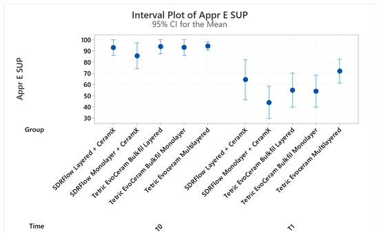
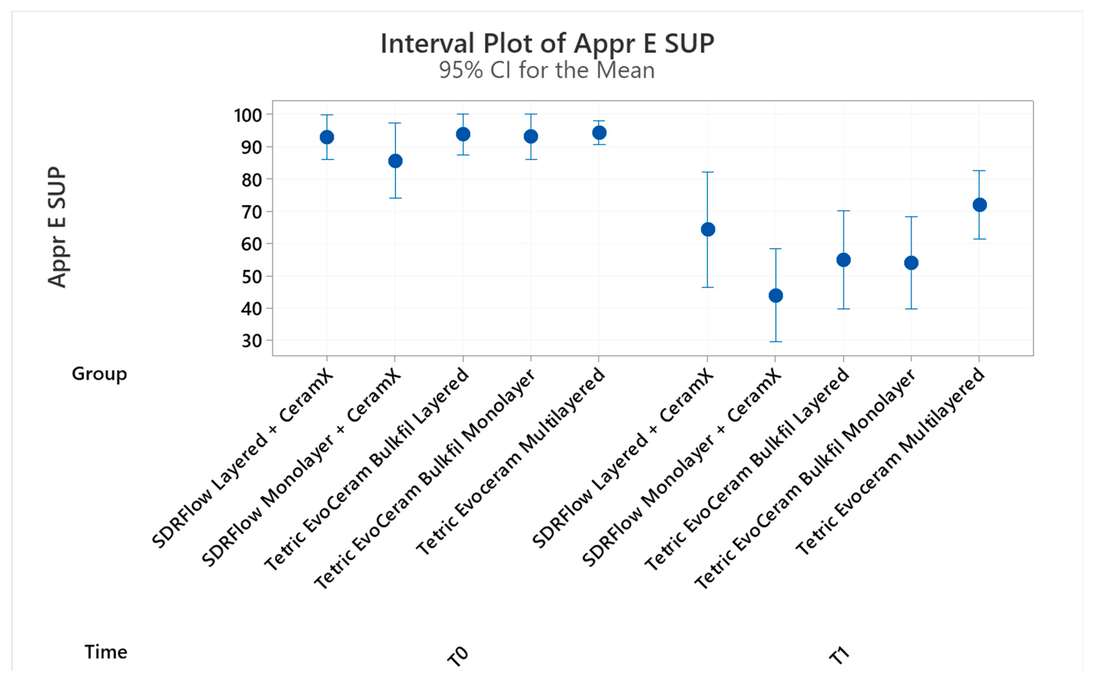
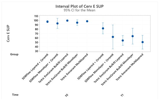
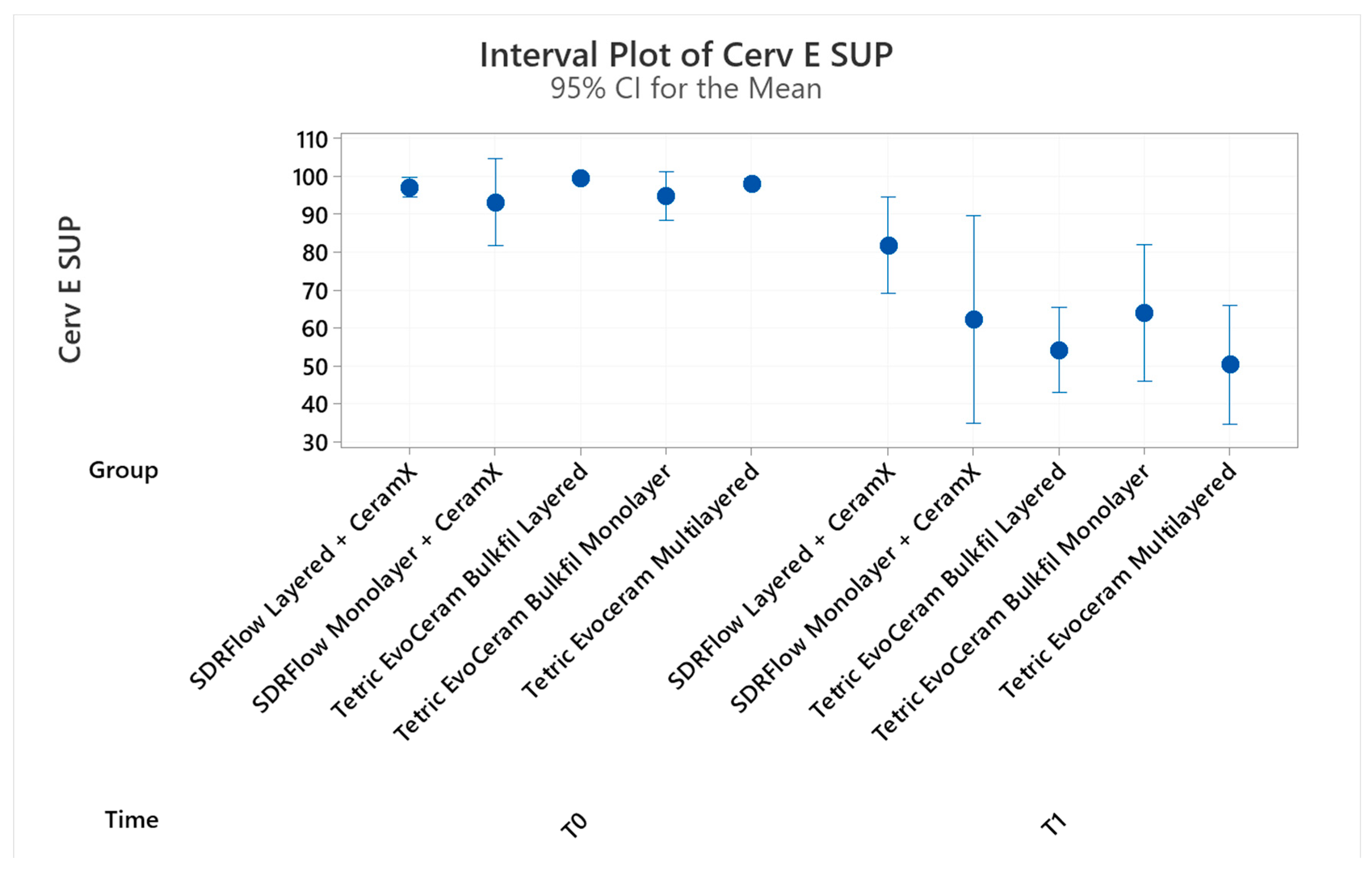
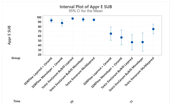
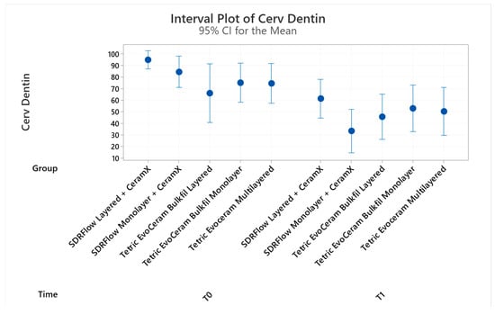
The results of continuous marginal adaptation before (T0) and after thermo-mechanical loading (T1) are presented in Figure 3, Figure 4, Figure 5 and Figure 6 and their statistical analysis in Table 2, Table 3, Table 4 and Table 5. The non-continuous, defective margins, were chiefly cohesive micro-fractures of marginal enamel or adhesive failures with gap formation at dentin interfaces.
Figure 3.
Continuous adaptation of approximal enamel margins (supra-gingival restoration side).
Figure 4.
Continuous adaptation of cervical enamel margins (supra-gingival restoration side).
Figure 5.
Continuous adaptation of approximal enamel margins (sub-gingival restoration side).
Figure 6.
Continuous adaptation of cervical dentin margins (sub-gingival restoration side).
Table 2.
Grouping Information (Fisher’s LSD Method and 95% Confidence) for supra-gingival, approximal enamel; means that do not share a letter are significantly different.
Table 3.
Grouping Informatio (Fisher’s LSD Method and 95% Confidence) for supra-gingival, cervical enamel; means that do not share a letter are significantly different.
Table 4.
Grouping Information (Fisher’s LSD Method and 95% Confidence) for sub-gingival, approximal enamel; means that do not share a letter are significantly different.
Table 5.
Grouping Information (Fisher’s LSD Method and 95% Confidence) for sub-gingival, cervical dentin; means that do not share a letter are significantly different.
The pre-loading values of continuous enamel adaptation varied from 94.26% (Tetric EvoCeram layered) to 85.65% (SDRflow Monolayer) in proximal margins of the supra-gingival side, from 96.95% (Tetric Powerfill layered) to 87.48% (SDRflow Monolayer) in proximal margins of the sub-gingival side and from 99.36% (Tetric Powerfill layered) to 93.12% (SDRflow Monolayer) in cervical margins. No statistical difference in enamel adaption was reported at T0 between the products and applications methods at any of the three enamel sections analyzed. The pre-loading values of continuous cervical dentin adaptation varied from 94.79% (SDRflow layered) to 66.06% (Tetric Powerfill layered); the continuous adaptation of Tetric Powerfill layered appeared significantly inferior to SDRflow layered.
The post-loading values of continuous enamel adaptation varied from 71.96% (Tetric EvoCeram layered) to 43.91% (SDRflow Monolayer) in proximal margins of the supra-gingival side, from 74.67% (Tetric EvoCeram layered) to 46.39% (Tetric Powerfill layered) in proximal margins of the sub-gingival side and from 81.74% (SDRflow layered) to 50.38% (Tetric EvoCeram layered in cervical margins. In both proximal enamel margins (supra- and sub-gingival restoration sides), Tetric EvoCeram layered presented an adaptation significantly better than SDRflow non-layered or Tetric Powerfill layered or not. On the contrary, in cervical enamel, SDRflow layered showed a proportion of continuous margins superior to all other groups. The post-loading values of continuous cervical dentin adaptation varied from 61.20% (SDRflow layered) to 33.36% (SDRflow Monolayer); the continuous adaptation of SDRflow layered was significantly superior to its non-layered counterpart.
A significant drop in continuous adaptation was then observed in both enamel and dentin margins after 250,000 thermo-mechanical cycles simulating about seven years of para-functional forces, with greater impact at restoration dentin interface. Tetric EvoCeram layered showed the highest proportion of post-loading defect-free margins in proximal enamel while it had the lowest in cervical enamel. SDRflow layered restorations behaved similarly to those layered with Tetric EvoCeram in proximal enamel and also showed the best scores of continuous adaptation in cervical enamel and dentin. Restorations made with Tetric Powerfill behaved overall less satisfactorily, with no significant advantage of the layering approach under such testing conditions.
4. Discussion
In vitro marginal adaptation of adhesive class II restorations submitted to thermo-mechanical stressing has been widely used as a preclinical testing method or for identifying the influence of single material or protocol variables that are difficult to isolate in an in vivo environment [20,21,22]. In the present study, the potential interference of confounding factors specific to such in vitro protocol was also addressed through various modifications of the basic in vitro protocol [40]. This comprised an equal distribution of extracted teeth (molars) based on their size, anatomy (upper or lower), the use of a mechanical device controlling bur position and generating identical cavity dimensions and the placement of a 0.5 mm rubber disk of selected e-modulus below samples to emulate the periodontal ligament dumping effect. These modifications were implemented with aim of reducing uncontrolled variability in marginal adaptation outcomes (likely induced by tooth anatomy and cavity size disparities) and enhancing the relevance of this in vitro protocol as a pre-clinical testing method.
The timeframe correlation between in vitro marginal adaptation studies and clinical service is of main interest when analyzing the results of this study. However, it has rarely been addressed in the literature [24,41] and is based on a correlation between in vivo and in vitro wear data, using fatigue devices comparable to the one utilized in the present study. Those reports provided an estimate of 250,000 yearly cycles with a range of 120,000 to 300,000. Today, a well-documented estimate of daily masticatory and deglutition cycles as well as sleep bruxism events is available and it is possible to confirm the aforementioned clinical correlation for both functional and parafunctional activities. Considering masticatory function, the number of cycles may vary with gender, age and dental conditions (fully dentated or not and type of prosthesis) [42,43,44,45]. Depending on food consistency, the number of tooth-to-tooth contacts, which occur only at the end of the masticatory cycle, has shown some discrepancies among individuals [46]. However, if cumulating over a year an estimated 45 min of daily, continuous chewing activity and integrating the known, average chewing cycle duration and frequency [35,47,48,49,50], it provides about 125 effective tooth-to-tooth contacts per day, or 45,000 yearly chewing cycles, as a meaningful reference. Then, considering an average 580 daily swallowing events [51], a total of approximately 250,000 yearly functional cycles is obtained. Since occlusal forces at swallowing can reach up to 40% of maximal biting forces [52], it seems pertinent to consider their major contribution in the yearly masticator cycles calculation. Thus, the reference used in previous studies across several decades [24,25,40,53] is chiefly validated, confirming also that applying a significantly lower number of functional cycles (e.g., lower than 250,000) might be of limited clinical relevance for this in vitro model.
Voluntary maximal biting forces per contact showed large inter-individual variation (10 to 700 N), with a number of contacts increasing with clenching intensity and average values reported from 38 to 50 N (canines to molars) [33,34,35,36,37]. These average values correspond to the loading parameters applied (usually 50 N) in most marginal analysis studies [24,40]. Owing to the fact that forces exerted during bruxism can reach or even exceed maximal biting forces [30,31,32] and that in masticatory simulation devices, load is applied through a single cusp in the tooth central fossa, a force of 100 N was considered appropriate to simulate clenching and bruxism. Considering the number of bruxism events and contacts per event, an average of 100 tooth-to-tooth contacts per day can be computed [31,39], suggesting that 250,000 thermo-mechanical cycles hypothetically simulate a much longer period of parafunctions (about 7Y) than its counterpart for masticatory and deglutition functions (1 year only). The aforementioned literature data confirms that usual testing parameters of marginal analysis studies and their results’ interpretation have to be adjusted to provide a realistic clinical counterpart, which is often not properly indicated, thus creating a significant bias.
The pre-loading results here are consistent with a previous study using the same products and restorative protocols [40]. They revealed no statistical difference between products or application protocols at enamel margins while in dentin, gap formation was significantly higher for layered Tetric Powerfill as compared to SDRflow layered. It otherwise proved again that a good control of polymerization and mechanical finishing stresses can be attained with various resin composite systems and layering or bulk-fill protocols [32,40,54,55,56,57]. After the simulation of parafunctional conditions, all groups presented a drop in continuous adaptation values. The variation for enamel margins proved significant in all groups on the supra-gingival side of restorations, but for SDRflow layered in cervical enamel on the sub-gingival side, only SDRflow layered showed no significant drop for proximal enamel margins while in dentin, all variations proved significant except for the Tetric EvoCeram bulk-fill layer which had the lowest adaptation before thermo-mechanical stressing. Then, exposed to higher loading conditions, the three different products and two application protocols showed rather distinct behaviors at both enamel and dentin margins. In approximal enamel, the conventional Tetric EvoCeram layered behaved more satisfactorily than restorative or non-layered flowable bulk-fill systems. In cervical enamel margins, layered SDRflow showed a very good resistance to stress, significantly superior to all other groups. Although its superiority was less pronounced in cervical dentin margins, the same product exhibited a better adaptation than the other groups with the exception of the Tetric EvoCeram bulk-fill used in a single increment approach. These findings are in agreement with a previous study that comprehensively analyzed the stress behavior of various composite technologies [58].
When comparing the behavior of similar products and restorative protocols under average masticatory load conditions, all other testing conditions being otherwise equal, increased forces (simulating bruxism) influenced their respective performance and resistance to marginal degradation differently. Previous results emulating normal function suggested overall a superiority of high viscosity resin composites (traditional or bulk-fill) at enamel margins, while the flowable bulk-fill material resisted better to degradation at cervical margins [40]. In such conditions, this difference in reaction to simulated masticatory stresses was attributed to their distinctive physical characteristics, in particular their E-modulus [5,6,7,8]. The present results partially confirmed the previous study’s outcome with an improvement in restoration adaption to enamel and dentin with layered flowable bulk-fill composites, while the restorative bulk-fill products showed no definite advantage of layering, with even slightly inferior performance in both dentin and enamel cervical margins. In enamel, the potential advantage of using a stiffer restorative material was partially evidentiated with satisfactory behavior of the layered conventional composite, as opposed to the restorative bulk-fill product. The interpretation of the dissimilar behavior of tested products and application protocols under different loading conditions is likely to be found in a compound effect of different material parameters such as volumetric shrinkage, inherent composite technology stiffness and depth-correlated conversion rate. For instance, when using micro-hardness as an indicator of conversation rate and material stiffness, SDRflow shows no variation up to 6 mm while it decreases to about 80% of surface values for Tetric bulk-fill brands as of 4 to 5 mm, suggesting a possible change in stress absorption capacity of the later product in a monolayer approach [59,60,61]. Depending on the loading magnitude, a lower stiffness of material at the bottom of proximal boxes could then confirm or on the contrary, antagonize the positive effect of layering. When analyzing the behavior of SDRflow, its high volumetric polymerization shrinkage has potential to generate more internal and interfacial stresses with the true bulk-fill protocol as compared to the layered configuration, potentially causing more defects under increasing loading conditions [4,5]. In this study, correlation between polymerization and load-induced tooth-restoration interfacial stresses combined with a material’s inherent stiffness and conversion rate appeared as likely influential parameters for marginal adaption, keeping in mind the rather complex behavior of tooth-restoration under varying thermo-mechanical loading.
Some limitations have to be considered as our research is an in vitro study and further research is however needed to confirm these results and, in particular, to ascertain the mechanism of restoration–degradation, the interplay of conversion rate and E-modulus parameters and the way the deleterious effect of high loading stresses could be eventually counteracted.
5. Conclusions
This in vitro trial investigated the behavior of standardized medium–large direct class II restorations with supra- and sub-CEJ margins under parafunctional loading conditions.
The present results revealed a significant drop in restoration marginal adaptation at both enamel and dentin margins after a simulated bruxism period of 7Y. Overall, the low-viscosity bulk-fill SDRflow layer showed the best behavior while other products showed varying levels of degradation, possibly influenced by a combined influence of conversion rate and E-modulus.
The null study hypothesis was then to be rejected, confirming the influence of both resin composite technology and placement protocols on the marginal adaptation of posterior composite restoration in an in vitro environment.
Author Contributions
Conceptualization, D.D.; methodology, D.D. and S.A.; software, P.T.G.; validation, P.T.G. and S.A.; formal analysis, I.R.; investigation, P.T.G.; resources, D.D.; data curation, E.D.B.; writing—original draft preparation, D.D.; writing—review and editing, S.A.; visualization, S.A.; supervision, D.D.; project administration, P.T.G. All authors have read and agreed to the published version of the manuscript.
Funding
This research received no external funding.
Data Availability Statement
The original contributions presented in this study are included in the article. Further inquiries can be directed to the corresponding author.
Conflicts of Interest
The authors declare no conflicts of interest.
References
- Bellinaso, M.D.; Soares, F.Z.M.; Rocha, R.O. Do bulk-fill resins decrease the restorative time in posterior teeth? A systematic review and meta-analysis of in vitro studies. J. Investig. Clin. Dent. 2019, 10, e12463. [Google Scholar] [CrossRef] [Scilit] [PubMed]
- Kim, R.J.; Kim, Y.J.; Choi, N.S.; Lee, I.B. Polymerization shrinkage, modulus, and shrinkage stress related to tooth-restoration interfacial debonding in bulk-fill composites. J. Dent. 2015, 43, 430–439. [Google Scholar] [CrossRef] [Scilit]
- Meereis, C.T.W.; Münchow, E.A.; de Oliveira da Rosa, W.L.; da Silva, A.F.; Piva, E. Polymerization shrinkage stress of resin-based dental materials: A systematic review and meta-analyses of composition strategies. J. Mech. Behav. Biomed. Mater. 2018, 82, 268–281. [Google Scholar] [CrossRef] [Scilit] [PubMed]
- Rizzante, F.A.P.; Mondelli, R.F.L.; Furuse, A.Y.; Borges, A.F.S.; Mendonça, G.; Ishikiriama, S.K. Shrinkage stress and elastic modulus assessment of bulk-fill composites. J. Appl. Oral Sci. 2019, 27, e20180132. [Google Scholar] [CrossRef] [Scilit]
- Leprince, J.G.; Palin, W.M.; Vanacker, J.; Sabbagh, J.; Devaux, J.; Leloup, G. Physico-mechanical characteristics of commercially available bulk-fill composites. J. Dent. 2014, 42, 993–1000. [Google Scholar] [CrossRef] [Scilit]
- Czasch, P.; Ilie, N. In vitro comparison of mechanical properties and degree of cure of bulk fill composites. Clin. Oral Investig. 2013, 17, 227–235. [Google Scholar] [CrossRef] [Scilit]
- Ilie, N.; Bucuta, S.; Draenert, M. Bulk-fill Resin-based Composites: An In Vitro Assessment of Their Mechanical Performance. Oper. Dent. 2013, 38, 618–625. [Google Scholar] [CrossRef] [Scilit]
- Papadogiannis, D.; Tolidis, K.; Gerasimou, P.; Lakes, R.; Papadogiannis, Y. Viscoelastic properties, creep behavior and degree of conversion of bulk fill composite resins. Dent. Mater. 2015, 31, 1533–1541. [Google Scholar] [CrossRef] [Scilit]
- Arbildo-Vega, H.I.; Lapinska, B.; Panda, S.; Lamas-Lara, C.; Khan, A.S.; Lukomska-Szymanska, M. Clinical Effectiveness of Bulk-Fill and Conventional Resin Composite Restorations: Systematic Review and Meta-Analysis. Polymers 2020, 12, 1786. [Google Scholar] [CrossRef] [Scilit] [PubMed]
- Kunz, P.V.M.; Wambier, L.M.; Kaizer, M.D.R.; Correr, G.M.; Reis, A.; Gonzaga, C.C. Is the clinical performance of composite resin restorations in posterior teeth similar if restored with incremental or bulk-filling techniques? A systematic review and meta-analysis. Clin. Oral Investig. 2022, 26, 2281–2297. [Google Scholar] [CrossRef] [Scilit] [PubMed]
- Cidreira Boaro, L.C.; Pereira Lopes, D.; de Souza, A.S.C.; Lie Nakano, E.; Ayala Perez, M.D.; Pfeifer, C.S.; Gonçalves, F. Clinical performance and chemical-physical properties of bulk fill composites resin -a systematic review and meta-analysis. Dent. Mater. 2019, 35, e249–e264. [Google Scholar] [CrossRef] [Scilit]
- Veloso, S.R.M.; Lemos, C.A.A.; de Moraes, S.L.D.; do Egito Vasconcelos, B.C.; Pellizzer, E.P.; de Melo Monteiro, G.Q. Clinical performance of bulk-fill and conventional resin composite restorations in posterior teeth: A systematic review and meta-analysis. Clin. Oral Investig. 2019, 23, 221–233. [Google Scholar] [CrossRef] [Scilit]
- Demarco, F.F.; Corrêa, M.B.; Cenci, M.S.; Moraes, R.R.; Opdam, N.J. Longevity of posterior composite restorations: Not only a matter of materials. Dent. Mater. 2012, 28, 87–101. [Google Scholar] [CrossRef] [Scilit]
- Beck, F.; Lettner, S.; Graf, A.; Bitriol, B.; Dumitrescu, N.; Bauer, P.; Moritz, A.; Schedle, A. Survival of direct resin restorations in posterior teeth within a 19-year period (1996–2015): A meta-analysis of prospective studies. Dent. Mater. 2015, 31, 958–985. [Google Scholar] [CrossRef] [Scilit]
- Borgia, E.; Baron, R.; Borgia, J.L. Quality and Survival of Direct Light-Activated Composite Resin Restorations in Posterior Teeth: A 5- to 20-Year Retrospective Longitudinal Study. J. Prosthodont. 2019, 28, e195–e203. [Google Scholar] [CrossRef] [Scilit] [PubMed]
- Heintze, S.D.; Loguercio, A.D.; Hanzen, T.A.; Reis, A.; Rousson, V. Clinical efficacy of resin-based direct posterior restorations and glass-ionomer restorations—An updated meta-analysis of clinical outcome parameters. Dent. Mater. 2022, 38, e109–e135. [Google Scholar] [CrossRef] [Scilit] [PubMed]
- Manfredini, D.; Winocur, E.; Guarda-Nardini, L.; Paesani, D.; Lobbezoo, F. Epidemiology of bruxism in adults: A systematic review of the literature. J. Orofac. Pain 2013, 27, 99–110. [Google Scholar] [CrossRef] [Scilit] [PubMed]
- Manfredini, D.; Restrepo, C.; Diaz-Serrano, K.; Winocur, E.; Lobbezoo, F. Prevalence of sleep bruxism in children: A systematic review of the literature. J. Oral Rehabil. 2013, 40, 631–642. [Google Scholar] [CrossRef] [Scilit]
- Melo, G.; Duarte, J.; Pauletto, P.; Porporatti, A.L.; Stuginski-Barbosa, J.; Winocur, E.; Flores-Mir, C.; De Luca Canto, G. Bruxism: An umbrella review of systematic reviews. J. Oral Rehabil. 2019, 46, 666–690. [Google Scholar] [CrossRef] [Scilit]
- Ernest, P.; Jandrain, B.; Scheen, A.J. Strengths and weaknesses of randomized clinical trials: Evolving changes according to personalized medicine. Rev. Med. Liege 2015, 70, 232–236. [Google Scholar]
- Flecha, O.D.; Douglas de Oliveira, D.W.; Marques, L.S.; Gonçalves, P.F. A commentary on randomized clinical trials: How to produce them with a good level of evidence. Perspect. Clin. Res. 2016, 7, 75–80. [Google Scholar] [CrossRef] [Scilit]
- Opdam, N.J.M.; Collares, K.; Hickel, R.; Bayne, S.C.; Loomans, B.A.; Cenci, M.S.; Lynch, C.D.; Correa, M.B.; Demarco, F.; Schwendicke, F.; et al. Clinical studies in restorative dentistry: New directions and new demands. Dent. Mater. 2018, 34, 1–12. [Google Scholar] [CrossRef]
- Luescher, B.; Lutz, F.; Oschenbein, H.; Muehlemann, H.R. Microleakage and marginal adaptation in conventional and adhesive Class II restorations. J. Prosthet. Dent. 1977, 37, 300–309. [Google Scholar] [CrossRef] [Scilit] [PubMed]
- Krejci, I.; Heinzmann, J.L.; Lutz, F. Verschleiss von Schmelz, Amalgam und ihrer Schmelz-Antagonisten im Computer gesteuerte Kausimulator. Schweiz. Monatschr. Zahmed. 1990, 100, 1285–1291. [Google Scholar]
- Krejci, I.; Lutz, F. In-vitro test results of the evaluation of dental restoration systems. Correlation with in-vivo results. Schweiz. Monatsschr. Zahnmed. 1990, 100, 1445–1449. [Google Scholar]
- DeLong, R.; Douglas, W.H. An artificial oral environment for testing dental materials. IEEE Trans. Biomed. Eng. 1991, 38, 339–345. [Google Scholar] [CrossRef] [Scilit]
- Dietschi, D.; Argente, A.; Krejci, I.; Mandikos, M. In vitro performance of Class I and II composite restorations: A literature review on nondestructive laboratory trials—Part I. Oper. Dent. 2013, 38, E166–E181. [Google Scholar] [CrossRef] [Scilit]
- Dietschi, D.; Argente, A.; Krejci, I.; Mandikos, M. In vitro performance of Class I and II composite restorations: A literature review on nondestructive laboratory trials—Part II. Oper. Dent. 2013, 38, E182–E200. [Google Scholar] [CrossRef] [Scilit]
- Lantada, A.D.; Carlos González Bris, C.G.; Morgad, P.L.; Maudes, J.S. Novel System for Bite-Force Sensing and Monitoring Based on Magnetic Near Field Communication. Sensors 2012, 12, 11544–11558. [Google Scholar] [CrossRef] [Scilit]
- Waltimo, A.; Nyström, M.; Könönen, M. Bite force and dentofacial morphology in men with severe dental attrition. Scand. J. Dent. Res. 1994, 102, 92–96. [Google Scholar] [PubMed]
- Nishigawa, K.; Bando, E.; Nakano, M. Quantitative study of bite force during sleep associated bruxism. J. Oral Rehabil. 2001, 28, 485–491. [Google Scholar] [CrossRef] [Scilit] [PubMed]
- Calderon Pdos, S.; Kogawa, E.M.; Lauris, J.R.; Conti, P.C. The influence of gender and bruxism on the human maximum bite force. J. Appl. Oral Sci. 2006, 14, 448–453. [Google Scholar] [CrossRef] [Scilit] [PubMed]
- Hidaka, O.; Iwasaki, M.; Saito, M.; Morimoto, T. Influence of clenching intensity on bite force balance, occlusal contact area, and average bite pressure. J. Dent. Res. 1999, 78, 1336–1344. [Google Scholar] [CrossRef] [Scilit]
- Ferrario, V.F.; Sforza, C.; Serrao, G.; Dellavia, C.; Tartaglia, G.M. Single tooth bite forces in healthy young adults. J. Oral Rehabil. 2004, 31, 18–22. [Google Scholar] [CrossRef] [Scilit]
- Lujan-Climent, M.; Martinez-Gomis, J.; Palau, S.; Ayuso-Montero, R.; Salsench, J.; Peraire, M. Influence of static and dynamic occlusal characteristics and muscle force on masticatory performance in dentate adults. Eur. J. Oral Sci. 2008, 116, 229–236. [Google Scholar] [CrossRef] [Scilit]
- Hattori, Y.; Satoh, C.; Kunieda, T.; Endoh, R.; Hisamatsu, H.; Watanabe, M. Bite forces and their resultants during forceful intercuspal clenching in humans. J. Biomech. 2009, 42, 1533–1538. [Google Scholar] [CrossRef] [Scilit] [PubMed]
- Kumagai, H.; Suzuki, T.; Hamada, T.; Sondang, P.; Fujitani, M.; Nikawa, H. Occlusal force distribution on the dental arch during various levels of clenching. J. Oral Rehabil. 1999, 26, 932–935. [Google Scholar] [CrossRef] [Scilit]
- Gu, W.; Yang, J.; Zhang, F.; Yin, X.; Wei, X.; Wang, C. Efficacy of biofeedback therapy via a mini wireless device on sleep bruxism contrasted with occlusal splint: A pilot study. J. Biomed. Res. 2015, 29, 160–168. [Google Scholar] [CrossRef] [Scilit]
- Robin, O.; Claude, A.; Gehin, C.; Massot, B.; McAdams, E. Recording of bruxism events in sleeping humans at home with a smart instrumented splint. Cranio 2022, 40, 14–22. [Google Scholar] [CrossRef] [Scilit]
- Dietschi, D.; Askari, M.; Rossier, I.; Caseiro, L.; Krejci, I.; Leprince, J.G.; Di Bella, E.; Ardu, S. Marginal Adaptation of In Vitro Class II Restorations Made Out of Bulk or Conventional Composite Using Single- or Multi-Layered Techniques. Materials 2023, 16, 6325. [Google Scholar] [CrossRef] [Scilit]
- Sakaguchi, R.L.; Douglas, W.H.; DeLong, R.; Pintado, M.R. The wear of a posterior composite in an artificial mouth: A clinical correlation. Dent. Mater. 1986, 2, 235–240. [Google Scholar] [CrossRef] [Scilit]
- Shinogaya, T.; Bakke, M.; Thomsen, C.E.; Vilmann, A.; Sodeyama, A.; Matsumoto, M. Effects of ethnicity, gender and age on clenching force and load distribution. Clin. Oral Investig. 2001, 5, 63–68. [Google Scholar] [CrossRef] [Scilit]
- Gibbs, C.H.; Anusavice, K.J.; Young, H.M.; Jones, J.S.; Esquivel-Upshaw, J.F. Maximum clenching force of patients with moderate loss of posterior tooth support: A pilot study. J. Prosthet. Dent. 2002, 88, 498–502. [Google Scholar] [CrossRef] [Scilit]
- Bakke, M. Bite Force and Occlusio. Semin. Orthod. 2006, 12, 120–126. [Google Scholar] [CrossRef] [Scilit]
- Roldán, S.; Buschang, P.H.; Isaza Saldarriaga, J.F.; Throckmorton, G. Reliability of maximum bite force measurements in age-varying populations. J. Oral Rehabil. 2009, 36, 801–807. [Google Scholar] [CrossRef] [Scilit]
- Wilson, E.M.; Green, J.R.; Weismer, G. A kinematic description of the temporal characteristics of jaw motion for early chewing: Preliminary findings. J. Speech Lang. Hear. Res. 2012, 55, 626–638. [Google Scholar] [CrossRef] [Scilit]
- Lassauzay, C.; Peyron, M.A.; Albuisson, E.; Dransfield, E.; Woda, A. Variability of the masticatory process during chewing of elastic model foods. Eur. J. Oral Sci. 2000, 108, 484–492. [Google Scholar] [CrossRef] [Scilit] [PubMed]
- Lavigne, G.J.; Kato, T.; Kolta, A.; Sessle, B.J. Neurobiological mechanisms involved in sleep bruxism. Crit. Rev. Oral Biol. Med. 2003, 14, 30–46. [Google Scholar] [CrossRef] [Scilit] [PubMed]
- Anderson, K.; Throckmorton, G.S.; Buschang, P.H.; Hayasaki, H. The effects of bolus hardness on masticatory kinematics. J. Oral Rehabil. 2002, 29, 689–696. [Google Scholar] [CrossRef] [Scilit] [PubMed]
- Lepley, C.R.; Throckmorton, G.S.; Ceen, R.F.; Buschang, P.H. Relative contributions of occlusion, maximum bite force, and chewing cycle kinematics to masticatory performance. Am. J. Orthod. Dentofac. Orthop. 2011, 139, 606–613. [Google Scholar] [CrossRef] [Scilit] [PubMed]
- Bulmer, J.M.; Ewers, C.; Drinnan, M.J.; Ewan, V.C. Evaluation of Spontaneous Swallow Frequency in Healthy People and Those With, or at Risk of Developing, Dysphagia: A Review. Gerontol. Geriatr. Med. 2021, 27, 23337214211041801. [Google Scholar] [CrossRef] [Scilit] [PubMed]
- Gibbs, C.H.; Mahan, P.E.; Lundeen, H.C.; Brehnan, K.; Walsh, E.K.; Holbrook, W.B. Occlusal forces during chewing and swallowing as measured by sound transmission. J. Prosthet. Dent. 1981, 46, 443–449. [Google Scholar] [CrossRef] [Scilit] [PubMed]
- Roggendorf, M.J.; Krämer, N.; Appelt, A.; Naumann, M.; Frankenberger, R. Marginal quality of flowable 4-mm base vs. conventionally layered resin composite. J. Dent. 2011, 39, 643–647. [Google Scholar] [CrossRef] [Scilit]
- Campos, E.A.; Ardu, S.; Lefever, D.; Jassé, F.F.; Bortolotto, T.; Krejci, I. Marginal adaptation of class II cavities restored with bulk-fill composites. J. Dent. 2014, 42, 575–581. [Google Scholar] [CrossRef] [Scilit]
- Al-Harbi, F.; Kaisarly, D.; Bader, D.; El Gezawi, M. Marginal Integrity of Bulk Versus Incremental Fill Class II Composite Restorations. Oper. Dent. 2016, 41, 146–156. [Google Scholar] [CrossRef] [Scilit]
- Shahidi, C.; Krejci, I.; Dietschi, D. In Vitro Evaluation of Marginal Adaptation of Direct Class II Composite Restorations Made of Different “Low-Shrinkage” Systems. Oper. Dent. 2017, 42, 273–283. [Google Scholar] [CrossRef] [Scilit]
- Dietschi, D.; Curto FDDr Di Bella, E.; Krejci, I.; Ardu, S. In vitro evaluation of marginal adaptation in medium- and large size direct class II restorations using a bulk-fill or layering technique. J. Dent. 2021, 115, 103828. [Google Scholar] [CrossRef] [Scilit]
- Rosatto, C.M.; Bicalho, A.A.; Veríssimo, C.; Bragança, G.F.; Rodrigues, M.P.; Tantbirojn, D.; Versluis, A.; Soares, C.J. Mechanical properties, shrinkage stress, cuspal strain and fracture resistance of molars restored with bulk-fill composites and incremental filling technique. J. Dent. 2015, 43, 1519–1528. [Google Scholar] [CrossRef] [Scilit]
- Flury, S.; Peutzfeldt, A.; Lussi, A. Influence of increment thickness on microhardness and dentin bond strength of bulk fill resin composites. Dent. Mater. 2014, 30, 1104–1112. [Google Scholar] [CrossRef] [Scilit]
- Rodrigues, J.A.; Tenorio, I.P.; Mello, G.B.R.; Reis, A.F.; Shen, C.; Roulet, J.F. Comparing depth-dependent curing radiant exposure and time of curing of regular and flow bulk-fill composites. Braz. Oral Res. 2017, 31, e65. [Google Scholar] [CrossRef] [Scilit] [PubMed]
- Modena, R.A.; Sinhoreti, M.A.C.; Palin, W.; Cavalcante, L.M.; Schneider, L.F. Light and viscosity effects on the curing potential of bulk-fill composites placed in deep cavities. Odontology 2021, 109, 874–883. [Google Scholar] [CrossRef] [Scilit] [PubMed]

Disclaimer/Publisher’s Note: The statements, opinions and data contained in all publications are solely those of the individual author(s) and contributor(s) and not of MDPI and/or the editor(s). MDPI and/or the editor(s) disclaim responsibility for any injury to people or property resulting from any ideas, methods, instructions or products referred to in the content. |
© 2025 by the authors. Licensee MDPI, Basel, Switzerland. This article is an open access article distributed under the terms and conditions of the Creative Commons Attribution (CC BY) license (https://creativecommons.org/licenses/by/4.0/).