


Abstract
Stable and unstable isotopes of the heavy noble gas xenon find use in various medical applications. However, apart from Xe, used for Single Photon Emission Computed Tomography, radioactive isotopes of xenon are currently complicated to obtain in small quantities. With the GAMMA-MRI project in mind, we investigated a thermal sublimation generator of the long-lived excited state (isomer) Xe. This production method utilized the decay of I, obtained commercially from a hospital supplier in the form of NaI powder. Heat treatments of the NaI powder and cryogenic trapping of released Xe allowed us to collect up to 88% of the produced xenon. Our method provides an isomeric mixture of Xe and Xe. With improvements in scalability and chemical purification, this method could be a cost-effective source of Xe for small-scale experiments.
1. Introduction
Stable and radioactive isotopes of xenon are used in large quantities in medical imaging and Nuclear Magnetic Resonance (NMR) applications [1,2,3,4,5]. Xenon has several important qualities [1]. First, it is inert, and does not interact chemically with the research subject, or sample [1]. Second, the amplitude of the NMR signal acquired from xenon can be increased by several orders of magnitude if xenon is hyperpolarized by colliding it with optically pumped alkali atoms using Spin Exchange Optical Pumping (SEOP) [6]. In addition, xenon passively crosses the blood brain barrier, and can serve as a contrast agent to image the uptake of the inhaled gas into brain tissue. This can be used effectively for stroke diagnosis for instance [7]. The most widely used stable isotopes of xenon are the nuclear ground states of Xe and Xe. They are typically used in NMR studies of materials, and for Magnetic Resonance Imaging (MRI) of the lungs [8,9,10,11]. Unstable Xe is used in Single Photon Emission Computed Tomography (SPECT) for the diagnosis of pulmonary diseases [12,13]. The physical half-life of Xe (5.3 d) makes it easy for radiopharmaceutical suppliers to manufacture and deliver it [13].
In the GAMMA-MRI project [14], we aim at developing a new imaging technique based on the MRI response from polarized unstable, SPECT-compatible nuclei. Our first test cases are radioactive xenon isomers, in particular the long-lived Xe, Xe, and Xe isomers. Optimising SEOP polarization of these isomers is one of the key components of the GAMMA-MRI project. For this purpose, it is advantageous to use the isomer that has the longest half-life, and that can be produced in an affordable way.
Here, we explore a generator of Xe ( d), via decay of a I source (d) obtained commercially from a hospital supplier. I, like Xe in its ground state, is in high demand by hospitals and thus can be procured from radiopharmaceutical companies [15,16]. Both I and Xe can be produced from U fission, upon U irradiation with neutrons in nuclear reactor facilities: and [17]. To isolate and collect these isotopes, the U target is dissolved, and the solution is separated into cells containing specific chemically active elements [18]. This approach offers several benefits: a flexible delivery schedule (medical I is available d/yr), established supply routes (to hospitals), and a relatively low cost for samples at the delivery point.
Xe generators based on I have been investigated before. For instance, P. Bedrossian [19] used a palladium metal surface to adsorb I from a solution. That generator was then connected to a vacuum-tight setup placed vertically: with generator atop and a set of glass spheres underneath. Produced Xe was collected in the bottom-most glass sphere immersed in the liquefied air, and the sphere was separated by sealing it off with a flame torch. In [20], a Xe generator is described, where a I solution was precipitated onto a fibreglass filter as palladium(II) iodide PdI, inside a commercially available syringe filter. The syringe was connected to a carrier gas which, after the desired quantity of Xe was produced, would sweep the Xe from the syringe filter into another apparatus for -ray spectroscopy. Both of these generators reported near unity Xe production efficiency. Finally, in a more recent publication [21], the production of Xe from I is mentioned but no details are given. However, to the best of our knowledge, Xe production from NaI powder has not been reported yet.
Here, we fill this gap and report on Xe production from a NaI powder. A solid state sample is preferred for the GAMMA-MRI project. Specifically, it should introduce less water into the SEOP cell than a NaI solution. It is important for successful polarization to eliminate all sources of water that could oxidize rubidium used in the SEOP process. The objective of the present work was thus to establish the most suitable conditions to extract Xe from a NaI salt, and to optimize the collection efficiency.
2. Materials and Methods
Production of Xe from I decay involved the following procedure: (i) procurement of the I source in the form of powder encapsulated in a gelatin shell, (ii) -ray spectroscopy of the I source upon arrival at CERN, (iii) NaI powder transfer from its gelatin shell in a quartz tube and -ray spectroscopy of both constituents, (iv) placement of the powder in the experimental installation for decay, (v) collection of Xe from the NaI powder at ambient temperature, or after heating, and finally (vi) Xe -ray spectroscopy. Three MBq NaI samples were purchased from Curium Pharma (Curium Pharma, Paris, France)—a supplier who could regularly deliver a NaI source at high radionuclidic purity (see Section 2.2). The samples were processed according to the procedure described above.
2.1. Characteristics of the Parent Source
The parent nucleus for the Xe generator is I. The following characteristics were considered: the half-life and the branching ratio for the transition to Xe, the isotopic and chemical purity of I, the state of I (solid or liquid), its availability, the associated radiological risks, and finally the cost of the I source and of its delivery.
I (d) is a beta-gamma (, ) ray emitter, which decays to the isomer Xe (d), and to the ground state Xe (stable). While the dominant part of decays of I feeds the ground state Xe, a small fraction of = 1.09(9)% feeds the isomer Xe [22].
The amount of collected Xe nuclei is, thus, the result of two competing processes, a source term from the decay of I, and a sink term due to Xe decay. The mathematical model describing isotopes in the decay chain (I > Xe > Xe) as a function of time is Bateman’s equation [23], which for I gives:
with (t) activity of Xe at time t, the number of I nuclei at , decaying into (t) Xe nuclei at the rate in time t and with the branching ratio . Additionally, Xe decays into stable Xe at the rate in time t. The second term describes the decay of Xe present in the system at moment .
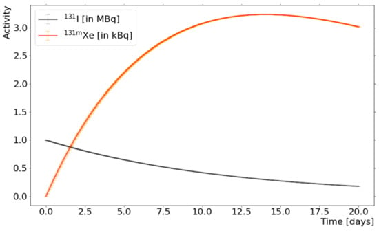
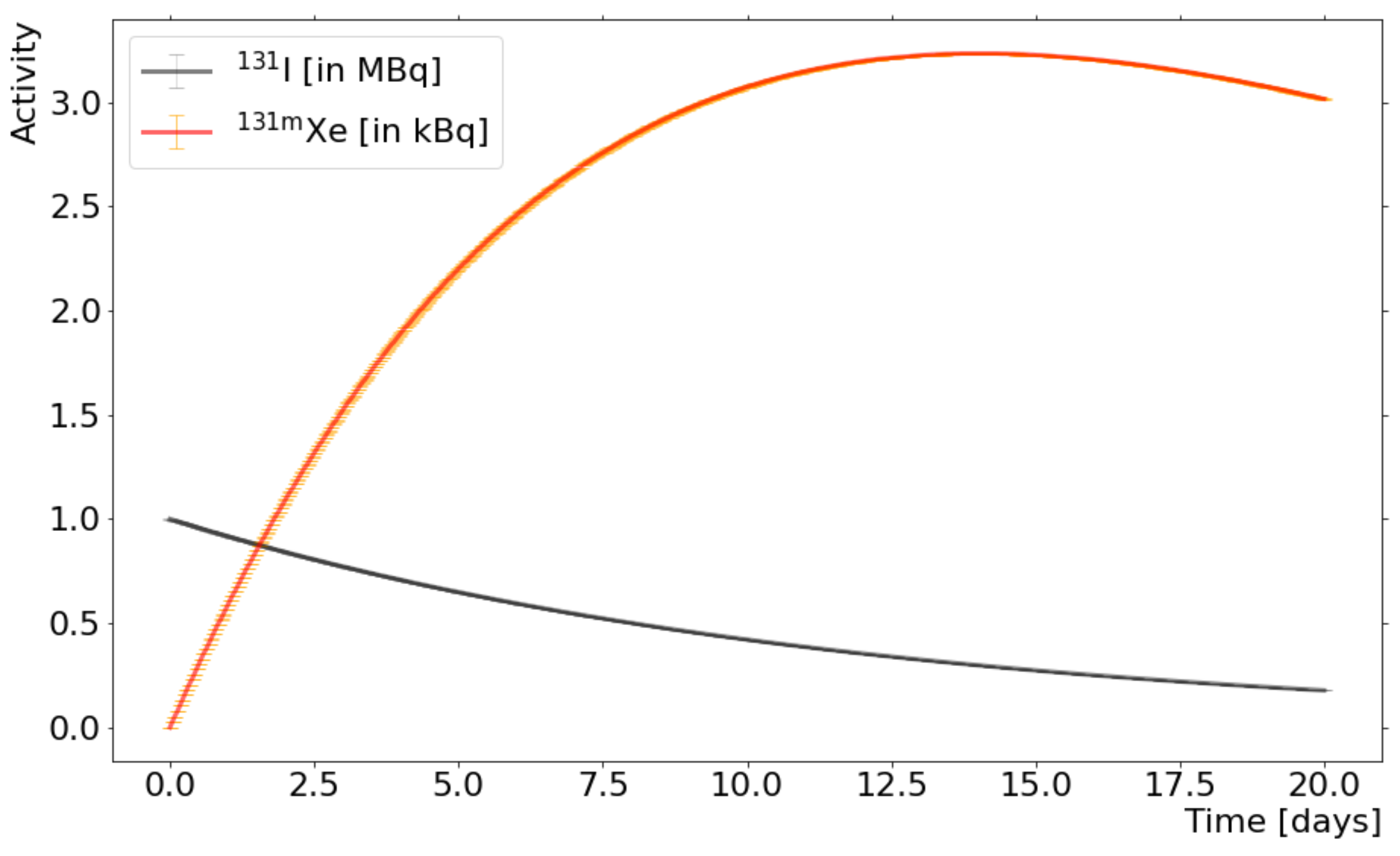
Figure 1 illustrates Equation (1) assuming and the initial activity of I (0) = 1 MBq. The figure presents the activity in function of time: the decay of I (activity in black, uncertainty in grey) in MBq, and the production and decay of Xe (activity in red, uncertainty in yellow) in kBq. The error bars comprise the errors of the decay constant and branching ratio. The optimum waiting time, when the daughter activity is maximal, can be calculated analytically by finding the maximum of function in Equation (1). After transformation, the equation reads:
and = 14.0 days. The maximum Xe activity is 3.23(1) kBq and it corresponds to 0.324(1)% of the initial activity of the I source placed in the experimental setup.
Figure 1.
Calculated Xe activity (red) vs. time. A source of I (activity in black, uncertainty in grey) decays into Xe (activity in red, uncertainty in yellow), which itself decays into Xe (stable, not shown). The error bars comprise the errors of the decay constant and branching ratio. The maximum activity plateau for Xe is at day 14 (Equation (2)).
2.2. I Source Manufacturing
Radiopharmaceutical preparation standards are defined in European Pharmacopoeia [24], and implemented by all manufacturers in Europe. They define the quality of I medication, including the chemical and radionuclidic purity, and define accepted physical forms and dosage. The radionuclidic purity of I is % with I, I and other impurities %.
The I bulk solution is delivered on a regular basis to our supplier Curium Pharma by the processors of reactor-irradiated targets, which extract the I from targets. The solution already includes excipients, added to prevent the escape of significant amounts of dissolved volatile iodine [25]. In addition, those excipients are necessary to adjust the pH of the I bulk solution, and to flavour the medicine for patients. For our application however, the additional ingredients can indirectly affect the purity of the collected Xe, as detailed in Section 3.
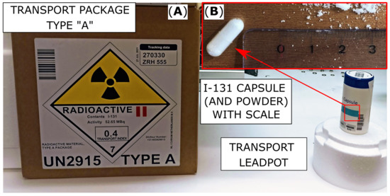
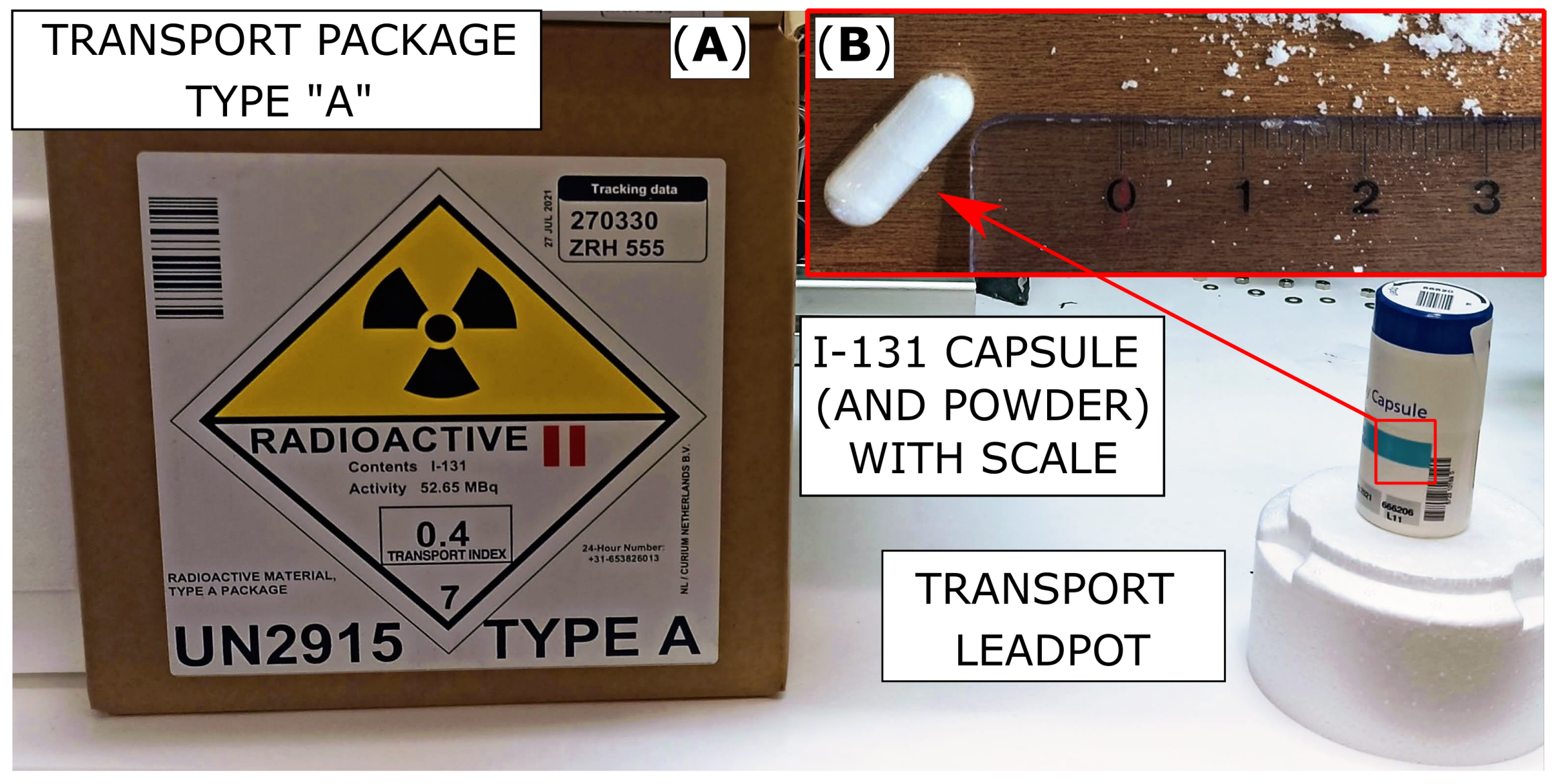
Curium Pharma provides I directly to hospitals and other partners (e.g., CERN) in a liquid NaI solution (shipped in a glass vial), or in the solid state (NaI powder in a gelatin capsule). NaI in the capsule format used in this work is prepared by adding a small volume of the solution to a NaI powder placed in a gelatin shell. Figure 2 shows one such capsule and the powder, in scale.
Figure 2.
Standard elements of the transport packaging of the radiopharmaceutical I. (A) Type-A package. The lead container, held securely in polystyrene foam inside a cardboard box. (B) Gelatin capsule and NaI powder.
2.3. Purchase of 131I and Radioactive Decay
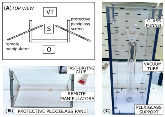
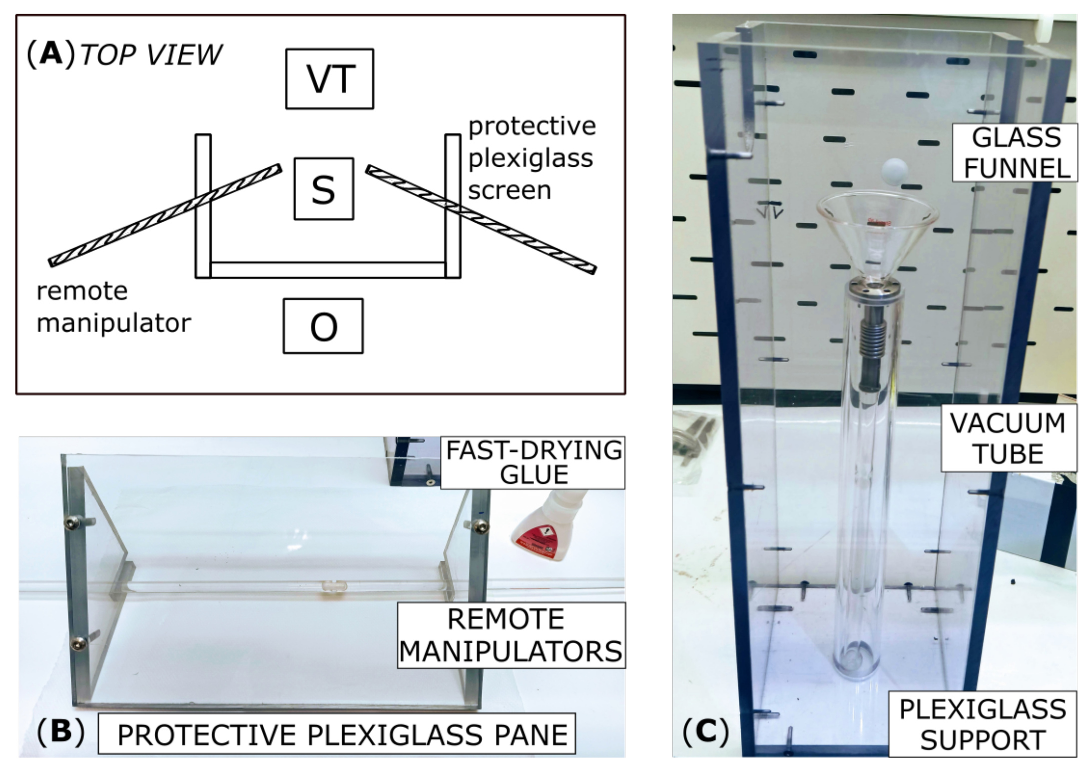
For every experiment, a single capsule containing NaI was delivered to CERN in a lead pot in a type-A package (Figure 2). The manufacturer specifies the activity to 10%, i.e., MBq. Upon arrival, the actual I activity in the capsule was determined with a n-type calibrated Extended Range (XtRa) Coaxial Ge detector (model GX6020, Canberra, Montigny-Le-Bretonneux, France) with a thin carbon window. As an acquisition system, we used a multichannel analyzer model ASPEC-927 and Maestro-32 software (AMETEK ORTEC, Oak Ridge, TN, USA). Remote manipulators with machined grooves were used to open the gelatin shell, and to transport its content into a dedicated borosilicate glass tube. Figure 3 presents the tools used for the safe handling of the capsule. These were necessary to avoid exposure to a large radiation dose received directly to the operator’s fingers.
Figure 3.
(A) Schematic of the opening setup. Operator (O) stands behind the protective panel and manipulates the source (S) with remote manipulators. The source is transferred to a glass tube that is then installed in the vacuum-tight experimental setup (labelled vacuum tube or VT in the schematic). (B) The tools for safe handling of the source included: the acrylic remote manipulators with grooves machined to the size of the gelatin shell, the acrylic shielding panel (thickness 1 cm) stopping beta particles from the I source, and the fast-drying glue necessary for the rapid attachment of the gelatin shell to the grooves in the acrylic manipulators. (C) The acrylic support for the vacuum tube: a borosilicate glass annealing tube with a CF 16 flange. A glass funnel aids powder transfer and minimizes transfer losses.
Using the acrylic manipulators with a drop of fast drying glue UHU solvent-free Flex Tube to attach the gelatin shell, the capsule was open into two halves. With the acrylic manipulators, the NaI powder was then transferred from the gelatin capsule via a glass funnel into the borosilicate glass annealing tube (labelled VT or vacuum tube in Figure 3). -ray spectroscopy on the annealing vial containing the powder provided a measurement of the activity of the transferred powder, and of the potential losses due to capsule manipulations, as detailed in Section 3.1. Next, the tube was installed in the experimental setup for xenon production.
2.4. I Extraction and Xe Collection
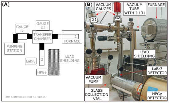
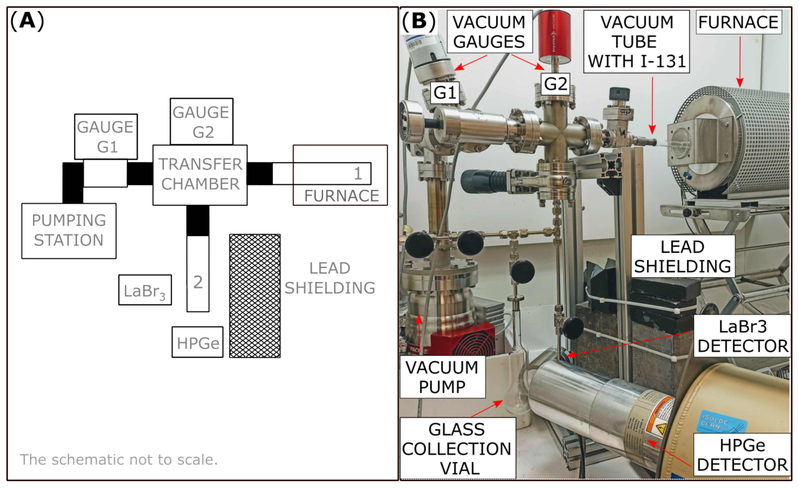
The setup for Xe collection (Figure 4) consisted of ultrahigh vacuum (UHV) elements (CF flanges, needle valves and gate valves). In addition, the setup contained the annealing tube with NaI and a collection vial for Xe, both made of borosilicate glass.
Figure 4.
(A) A schematic of the extraction setup. The NaI source (labelled 1) is in the glass annealing tube inside the furnace. The collection vial for Xe is labelled 2. Black rectangles denote valves dividing the installation into separate cells. The full-range vacuum gauge (labelled G1) was mounted at the interface between the pumping station and the transfer chamber. The capacitance diaphragm vacuum gauge (labelled G2) was mounted to the transfer chamber (B) A photograph of the experimental installation for xenon extraction and collection.
An UHV valve was mounted between the annealing tube and rest of the setup. The tip of the tube which contained the transferred NaI powder was inserted into a furnace equipped with temperature stability controls (measured temperature drift K). The NaI powder was heated to up to C to allow for xenon to diffuse out of the powder. Each heating treatment lasted one hour, with increasing temperatures for consecutive treatments. The collection vial was mounted vertically in the setup. The free space below it was used to place a container with liquid nitrogen—a cryotrap—for Xe collection. Collections were performed after every one-hour-long heat treatment.
The experimental setup was evacuated to 10mbar with a turbomolecular pump, and an oil-free scroll pump. Two pressure gauges were integrated into the system to cover the pressure range from UHV to atmospheric pressure. A full-range vacuum gauge (labelled G1 in Figure 4) housing two sensors—a Pirani gauge and a cold-cathode ionization gauge—was installed between the pumping system and the chamber in contact with xenon. This location was optimal for a wide range of vacuum regimes, but it was not used in the presence of xenon, due to the ionic pumping effect. A capacitance diaphragm vacuum gauge (labelled G2 in Figure 4) was installed at the interface between the gas diffusing from the annealing vial and the collection vial. That gauge measured the direct force on the diaphragm, and could consequently measure the pressure independently of gas type and concentration. In addition, it did not pump xenon like an ionization gauge would.
After each collection, the valve to the collection vial was closed and the cryotrap was removed to allow in situ -ray spectroscopy to characterize how collections depended on the temperature of the heating cycles. We used two -ray detectors. The first one was a 2 × 2 inch LaBr(Ce) crystal encapsulated in aluminium. Thin aluminium housing and a glass light guide allowed for acquisition of 29.8 keV X-rays from decay of Xe to the ground state (branching ratio 29.3%). The latter was a n-type Standard Electrode Coaxial Ge (SEGe) detector, model GC7020 (Canberra), with an aluminum window blocking X-ray radiation (including at 29.3 keV), but sensitive to higher energy radiation (Xe: 163.9 keV and I: 364.5 keV). We used the CAEN DT5730 (8 Channel 14 bit 500 MS/s Digitizer) with the PHA firmware and Compass software [26]. The distance between the center axis of the collection vial and the detectors was 55 mm and 80 mm, respectively, for the LaBr(Ce) and the HPGe detector. A 10-cm-thick lead shielding was placed between the NaI source and the detectors to reduce the I background, and to prevent saturating the HPGe preamplifier due to high count rates from the iodine source. The NaI source was at the distance of about 60 cm from the detectors.
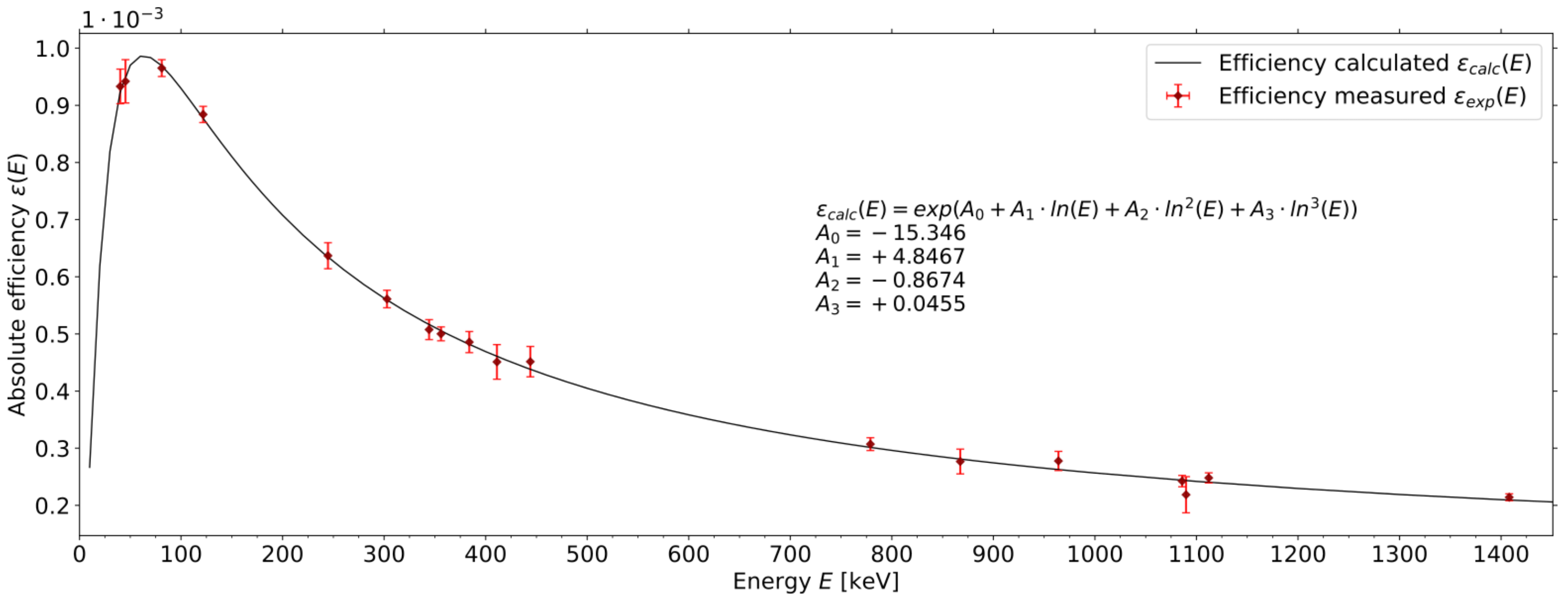
After completion of the heat treatment cycles, the collection vial with xenon was detached from the experimental setup, and transported to a second -ray spectroscopy station, with the Canberra n-type XtRa Ge detector, model GX6020, for a posteriori precise determination of activity, without the I background (as detailed in Section 3.1). The sources of Eu and Ba with known absolute activities were used for the energy calibration and for the determination of the absolute detection efficiency at energies MeV. These calibrated sources were measured at the source-detector distance d equal 50 cm (used for -ray spectroscopy of Xe collection vial) and 154 cm (used for -ray spectroscopy of high-activity NaI powder). See Figure 5 for the absolute efficiency plotted in function of the energy for cm.
Figure 5.
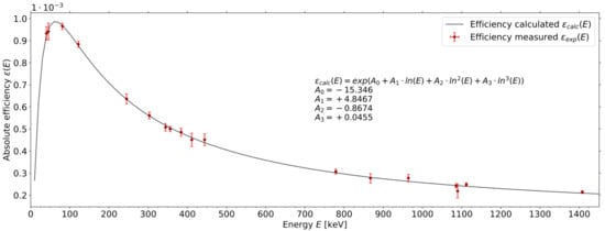
Absolute efficiency for n-type Extended Range Coaxial Ge detector (XtRa), model GX6020 at the distance of 50 cm from the source. Measured efficiency (in red) was plotted based on Eu and Ba measurements. A polynomial of 3rd degree (in black) was fitted to the experimental data. The equation and fit parameters are enclosed in Figure.
The coefficient of determination for the fitted 3rd degree polynomial was 0.996. The absolute efficiency curve in Figure 5 presents a well-known knee around 100 keV. This effect was accounted for by fitting to the dataset that included 4 rays in the 40–130 keV energy range. The accuracy determined as was calculated for the energy range near radiation characteristic for Xe (163.9 keV) and I (364.5 keV). That accuracy for selected Eu and Ba -ray peaks equaled , , , and . It confirmed good accuracy of the fit in the energy region characteristic for -radiation from I and Xe decay. The absolute efficiencies for 163.9 keV from Xe and for 364.5 keV I were listed in Table 1.
Table 1.
Absolute efficiencies for 163.9 keV and 364.5 keV (Canberra XtRa detector, model GX6020).
In activity calculations, transmission through glass and air were taken into account. Attenuation of 364.5 keV -ray in the NaI powder was neglected due to the high porosity of the powder and inhomogeneous spread of I onto NaI matrix (from the drop casting technique used for bulk I solution). This attenuation coefficient was determined to be, at most 2% given the NaI grain size of mm radius (path of X-ray).
3. Results
3.1. Efficiency of Xe Generator
Three types of -ray spectroscopy measurements (see Section 2) were taken and used for the calculations of: activity of the NaI capsule, activity of the NaI powder transferred to the vial, and activity of the collected Xe. Analyzed spectra consisted of background counts and counts in the area of interest. To derive the activity of samples, the number of counts in the peak areas was calculated using ORTEC-MAESTRO Multichannel Analyzer [27]. Linear background subtraction and Gaussian fitting were applied in regions of interest (ROIs).
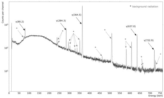
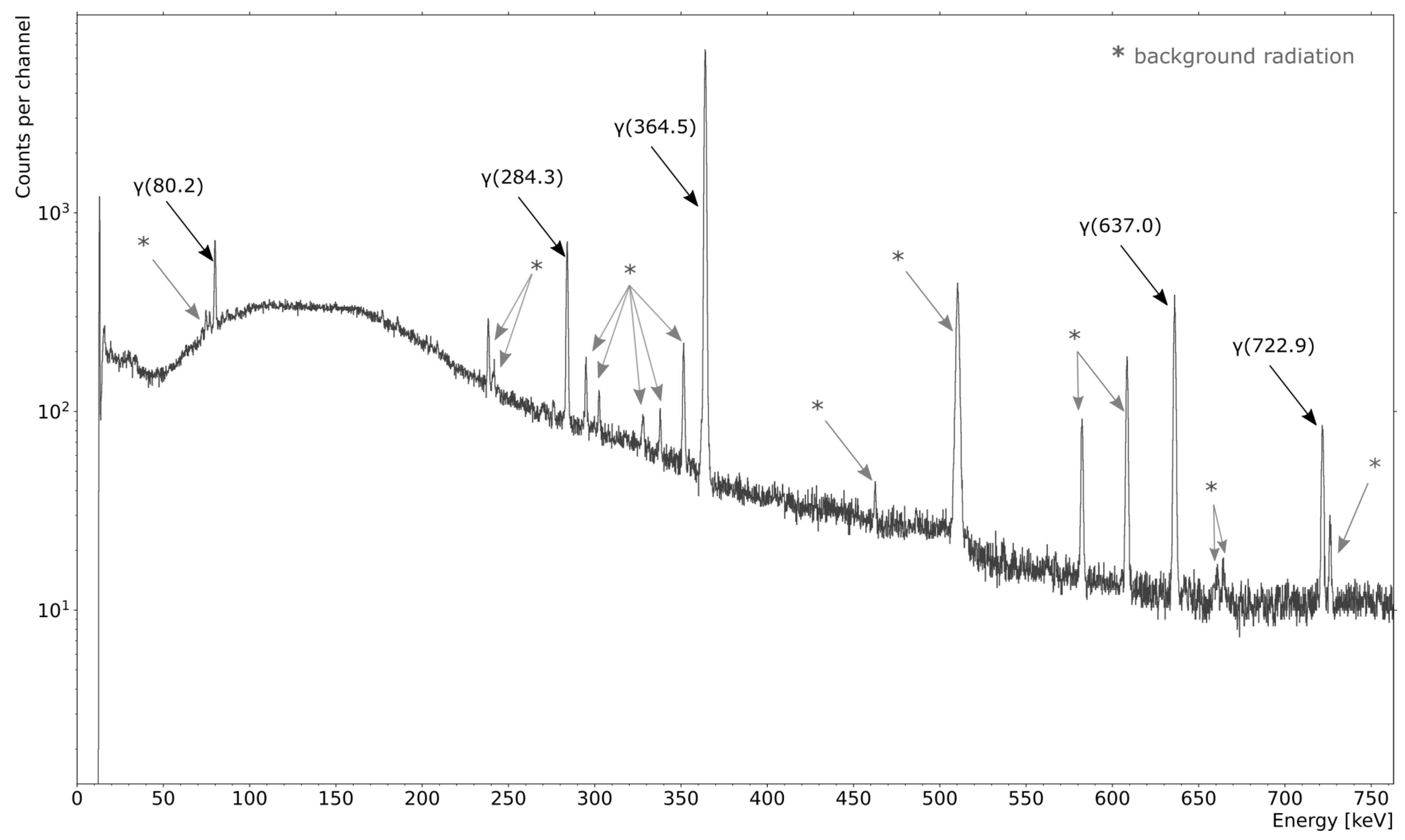
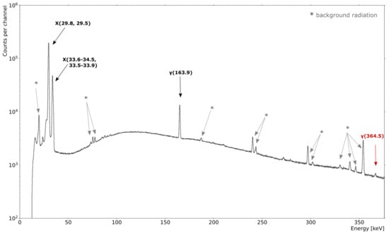
A typical spectrum is shown in Figure 6 for the transferred I powder and in Figure 7 for the collected Xe. In Figure 6, the main -ray transitions for I are marked and labelled: 364.5 keV (branching ratio 81.5%), 637.0 keV (7.16%), 284.3 keV (6.12%), 80.2 keV (2.6%), and 722.9 keV (1.8%) [22]. The -ray decay branch at 364.5 keV was used for the determination of the I activity. The sample was also verified for presence of impurities (mostly I and I), and no impurities were found.
Figure 6.
-ray spectroscopy of the I powder after the transfer into the annealing vial, recorded in the ISOLDE experimental hall. The radiation characteristic of I is labelled in black. These are lines at: 364.5 keV (81.5%), 637.0 keV (7.16%), 284.3 keV (6.12%), 80.2 keV (2.6%), and 722.9 keV (1.8%). The background radiation is marked with grey stars in the plot.
Figure 7.
-ray spectrum of the collected Xe sample, recorded in the ISOLDE experimental hall. The decay radiation characteristic of Xe is labelled in black. The -ray transition is at 163.9 keV (1.95%). The X-rays are at: 29.8 keV (29.3%), 29.5 keV (15.8%), 33.6–34.5 keV (10.5%), 33.6–33.9 keV (8.5%). The main -ray peak of I decay (364.5 keV) is labelled and marked in red. The background radiation is marked with grey stars.
The -ray lines marked grey and starred in Figure 6 and Figure 7 belong to the natural background gamma-radiation: Cs (661.66 keV), Tl (277.4 keV, 510.8 keV, 583.2 keV), Bi (727.3 keV), Pb (238.6 keV, 300.1 keV), Bi (609.3 keV, 665.5 keV), Pb (74.8 keV, 77.1 keV, 242.0 keV, 295.2 keV, 351.9 keV), Ac (209.3 keV, 270.2 keV, 327.5 keV, 328.0 keV, 338.3 keV, 463.0 keV, 509.0 keV, 726.9 keV), and electron–positron annihilation peak at 511 keV. They are present mainly due to concrete in any building and due to minerals’ concentration in a geographical location [28].
For Xe activity determination in the spectrum of the collection vial, the -ray transition of 163.9 keV (1.95% branching) was used. The transition to the ground state proceeds mostly via emission of conversion electrons: 29.8 keV (29.3%), 29.5 keV (15.8%), 33.6–34.5 keV (10.5%), 33.6–33.9 keV (8.5%) [22]. The measurement for Figure 7 was taken in a low-activity environment, free of I source. This low background baseline allowed for the precise determination of the radionuclidic purity of Xe (see Section 3.3).
Table 2 summarizes the results of experiments with three batches of NaI and collected Xe.
Table 2.
Efficiency of Xe production and collection from decay of I.
Column 2 is the measured activity of NaI capsule in MBq upon delivery of the sample to CERN-ISOLDE. The value for each experiment is in line with the ordered activity (50(5) MBq). Column 3 is the time between the end of manufacturing (EOM) of the NaI capsule, reported by Curium Pharma, and the -ray spectroscopy of NaI capsule at CERN-ISOLDE. It allowed us to determine the initial activity of NaI in the capsule, and the production of Xe prior to placing the powder in the experimental setup.
Column 4 (transfer rate ) is the percentage of delivered I activity that got transferred to the annealing tube. It was calculated based on two -ray spectroscopy measurements: of the I capsule, and of the transferred I powder. The value of varies, as it depends on the NaI saturation of the NaI powder and gelatin shell with the I bulk solution during the capsule’s preparation process. For instance, for experiment ID 1, a significant percentage of I (about 36%) was in the gelatin shell, which was cast aside.
Columns 5–7 relate to Xe activity. Column 5 details the activity of Xe expected at the end of collection (EOC). The value of Xe activity at the EOC depends on Xe production rate from mother nucleus I, rate of Xe decay and on the losses of Xe due to the room-temperature diffusion out of powder in the time T between EOM and delivery/placement in the experimental system (Column 3). The value in Column 5 was each time calculated in two steps using Equation (1). Firstly, to calculate —the number of Xe nuclei produced in NaI powder in the time T: . Secondly, to calculate Xe activity at EOC (Column 5), using , where equals the value in Column 2, and . describes the room-temperature diffusion (loss) of Xe from NaI powder prior to placement in the experimental setup (in time T). The value could not be measured directly, because upon delivery the saturation of powder with Xe was unknown (and impossible to measure in situ, due to high I activity). Therefore, the value of was determined at the end of three experiments as the one providing the best agreement between the calculated and measured activity of collected Xe for all 3 samples (defined by the residuals between calculated and measured values listed in Table 2, Columns 5 and 6). Precisely, we iterated over values of to find a minimal value of standard deviation function applied to the dataset of efficiency values for all three experiments. The minimum of that function (and the smallest residuals) was obtained for . The value was in agreement with the -ray spectroscopy measurement of the room-temperature Xe collection—the first collection prior to the series of the high temperature extractions and successive collections. Like other in situ collection measurements, this -ray spectroscopy measurement was performed with the n-type SEGe detector, model GC7020 (Canberra).
Column 6 presents the measured Xe activity at EOC. The measurements were taken without the iodine background, and after a complete cycle of heat treatments (see Figure 7). Finally, Column 7 specifies the collection efficiency as a ratio between the measured Xe (Column 6) and the determined Xe activity at EOC (Column 5). The collection efficiency was 85% on average.
3.2. Collection Efficiency as a Function of Temperature
One of the objectives of this work was to establish the most suitable conditions to extract Xe from a NaI salt and, as a result, to minimize the workload and time needed to achieve an optimized collection. For this purpose, the collection efficiency as a function of the heating temperature was studied. I was heated up to 400 C in several steps. The measurements of sample no. 1 were done in a more conservative range of temperatures (up to 300 C) than for the consecutive samples (up to 400 C).
Samples 1 and 2 were heated multiple times to temperatures between 40 C and 100 C over a course of 14 days. For sample 3, the experiment’s protocol was improved and all collections were executed successively on day 14 with each temperature threshold reached only once. Since the setup was not evacuated in between collections, we were not concerned with the loss of xenon outside the measurement system (parameter ) during the experiment.
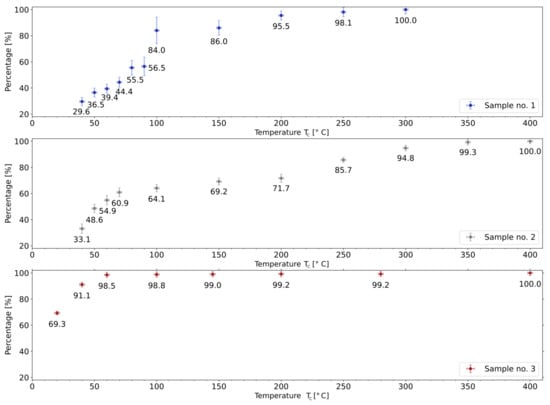
Figure 8 presents—for each NaI sample and as a function of the extraction temperature (x-axis)—the cumulative percentages of Xe extracted up until a given temperature with respect to the total collected activity of Xe (y-axis). For samples 1 and 2, the value at each point in the plot is the sum of the multiple activity increments (with the activity value adapted using the decay law) contributing to the collection up to a given temperature, divided by the measured end-product Xe activity and expressed as a percentage. For sample 3, it is the cumulative activity of Xe obtained from single collections up to a given temperature, divided by the measured activity of Xe end-product and expressed as a percentage.
Figure 8.
Percentage of Xe activity collected up until temperature normalized to the total collected Xe activity.
For practical reasons, the sample was not replaced between heating cycles at different temperatures. Therefore, the thermal diffusion coefficient could not be derived. However, the collected data allowed to establish that the temperature threshold for Xe diffusion out of NaI powder is below or at room temperature. For heat treatments up to 100 C: 84%, 64%, and 99% of all collected Xe was measured for samples 1–3, respectively. In addition, in the first collection—at room temperature—for sample 3, 69% of total collected Xe activity was measured. Consequently, for a time-saving Xe collection operation with relatively minimal losses it would be sufficient to heat the NaI sample to 200 C.
3.3. Radionuclidic Purity of Generated Xe
Before utilizing the collected Xe it has to be assured that the potential residues of I, i.e., the “breakthrough” of this sublimation generator, remains below an acceptable value. Internal exposure to I comprises of the uptake of I by thyroid gland and an increased risk of thyroid disease, including thyroid cancer [29].
A useful quantity to characterise the possible I contamination of Xe sample is the minimum detectable activity (MDA), here expressed in Bq, at a specified confidence level. It is usually calculated at the 95% confidence level, which means there is a 95% certainty that the activity above MDA threshold would be detected. Calculations of MDA are based on Currie’s derivation for single measurements [30]. With 95% level of confidence, the simplified formula for 364.5 keV emission from I reads as follows [31]:
where is the one-sided confidence factor at 95% confidence (), t is the time of acquisition, is the standard deviation of the background collected at time t, d is the detection efficiency for the -ray peak at 364.5 keV, and y is the -ray intensity per decay for the -ray peak at 364.5 keV.
Table 3 presents, for each of the three experiments sorted by ID in Column 1, the following results: MDA of I (Column 2), the I activity determined from the area under the peak at 364.5 keV (Column 3), the detected presence or absence of radiation from I in the spectrum (Column 4) based on the comparison of Columns 2 and 3, and the ratio of I residual activity to Xe activity, expressed in % (Column 5).
Table 3.
Minimum detectable activity of I in the Xe collection sample.
As shown in Table 3, for all experiments the counts in the peak area at 364.5 keV were above the MDA limit. Thus, with 95% confidence, I was present in Xe sample and equal 186(50) Bq, 37(13) Bq and 56(9) Bq, for ID 1, 2 and, 3 respectively. The ratio of I activity to Xe activity for three experiments was: 0.19%, 0.03%, and 0.03%. The elevated ratio for the first experiment might come from the residual activity of iodine that remained in the setup from earlier preparatory experiments.
With respect to chemical purity of the collection, some of the stable excipients initially present in NaI sample would decompose in the high temperature and would be introduced to the collection vial with the end product. These comprise oxides of carbon (0.8 mmol), phosphorus (0.6 mmol), and sulphur (0.15 mmol). The quantities of stable excipients present after thermal decomposition were determined analytically based on the list—provided by Curium Pharma [25]—of ingredients of the supplied source.
4. Discussion
The purpose of this research was to study the generation of the long-lived excited state Xe by thermal sublimation, from the decay of a commercially obtained I solid-state source.
-ray spectroscopy showed that with consecutive heat treatments between 40 C and 400 C, up to 88% of the determined produced Xe can be routinely collected. The remaining Xe is possibly trapped in the NaI powder, which starts to sinter at C. This hypothesis could not be verified with -ray spectroscopy because of the high background from the NaI. In addition, up to 69% of the total Xe can already be collected at room temperature.
The presented sublimation generator represents a possible way to obtain low activity batches of Xe with high radionuclidic purity throughout the year, using the well-established distribution network of I capsules. The analysis of radionuclidic purity shows that in the end-product vial with Xe, the ratio of I residual activity to collected Xe activity was 0.03% for two samples and 0.19% for one sample—from the first experiment that was possibly contaminated by the earlier preparatory experiments. The generally accepted activity range employed for the inhalation of another xenon gas agent —Xe— by an average patient (70 kg) is 74–1110 MBq for the pulmonary function imaging and 370–1110 MBq for cerebral blood flow imagining [32,33,34]. Tolerable residual level of I present in the gas cylinder with Xe radiopharmaceutical is 0.01% of total activity present in the sample [32,33,34], thus the maximum value would be 111 kBq. The necessary Xe activity in GAMMA-MRI project is: 50–100 MBq and 10–30 MBq, respectively for the polarization optimization and for proof-of-principle experiments in a preclinical MRI device built within the GAMMA-MRI project [35]. Given the experimentally derived ratio I to Xe equal to 0.03% (see Table 3), the residual activity of I would be at maximum 30 kBq for the polarization optimization and 9 kBq for proof-of-principle experiments in a preclinical MRI device. In the scope of next experiments taking place in Switzerland, an additional consideration is to follow the authorization limits (LA) listed in the Swiss Radiological Protection Ordinance [36], which are: 9 GBq for Xe and 0.5 MBq for I.
However, despite high radionuclidic purity, some of the stable excipients added to samples are present in the collection vial with the end product. Thus, purification methods of the end product might have to be implemented prior to deployment for the GAMMA-MRI project. A practical limitation is that collection of Xe would have to be planned 13–15 days in advance of a subsequent experiment to allow for I decay and production of sufficient amounts of Xe. Thus, an experimental setup needs to be dedicated solely to this goal for a whole period of approximately two weeks.
Finally, several fundamental limitations of the detailed method have to be kept in mind that de facto preclude a significant upscaling of this method. First, the maximum recovered activity of Xe is about 300 times smaller than the I activity of the generator, even assuming lossless transfer of NaI powder to the experimental setup. Further, one has to consider that the I mother has fivefold higher -ray dose rate per Bq than the Xe daughter, and several orders of magnitude higher radiotoxicity per Bq than Xe. Consequently, upscaling this method to large-scale generators without process automation is undesirable from ALARA considerations, and could run into licensing issues.
5. Conclusions and Outlook
An affordable and accessible production method of small activities of the long-lived Xe isomer via radioactive decay of commercially available I was investigated. Our thermal sublimation generator is dedicated to supplying the long-lived Xe for optimization tests of laser polarization in the GAMMA-MRI project. Once larger activity batches of Xe are required for a regular clinical use, the preferred option would be a centralized production in dedicated facilities with centralized quality control. Xe can also be produced by thermal neutron capture on enriched Xe samples and the activities and radionuclidic purity achievable in reactor irradiations will be discussed in a forthcoming article [37].
Author Contributions
Conceptualization: R.B.J., M.K., U.K., K.K.; methodology: K.K.; software: K.K., R.L., S.G.P.; validation: M.K., U.K., K.K., R.L., S.G.P.; formal analysis: K.K., R.L., S.G.P.; investigation: M.J.C., K.K., R.L., S.G.P.; resources: N.A., M.B., M.K., K.K., R.L., S.G.P.; data curation: K.K., R.L., S.G.P.; writing—original draft preparation: K.K.; writing—review and editing: R.B.J., M.K., U.K., K.K., R.L., S.G.P.; visualization: K.K.; supervision: R.B.J., M.K.; project administration: R.B.J., M.K.; funding acquisition: R.B.J., M.K. All authors have read and agreed to the published version of the manuscript.
Funding
The GAMMA-MRI project has received funding from the European Union’s Horizon 2020 research and innovation programme under grant agreement No. 964644 (GAMMA-MRI). The authors also wish to acknowledge support via the Swiss Excellence Government Scholarship, the CERN Medical Application Fund (GAMMA-MRI), and the Romanian IFA grant CERN/ISOLDE.
Institutional Review Board Statement
Not applicable.
Informed Consent Statement
Not applicable.
Data Availability Statement
Data supporting the reported results can be found at: https://cernbox.cern.ch/index.php/s/Eo9CTu5LOo5bx8O (accessed on 16 September 2022). The detailed study is available on request from the corresponding author.
Acknowledgments
We thank B. Karg, S. Warren, and members of SY-STI-RBS, in particular B. Crepieux, E. Barbero, J. Ballof, and S. Rothe, for discussions, assistance in designing the initial prototype and manufactured parts of the setup, M. Wuillemin for his input on iodine production, J.A. Ferreira Somoza for discussion on Xe cryogenic trapping, J. Schell for assistance in scheduling the experiments. We thank A. Dorsival, E. Aubert, P. Bertreix, N. Menaa, A.L. Boscher, and other members of HSE-RP for their support during project execution.
Conflicts of Interest
The authors declare no conflict of interest.
References
- Oros, A.M.; Shah, N.J. Hyperpolarized xenon in NMR and MRI. Phys. Med. Biol. 2004, 49, R105. [Google Scholar] [CrossRef] [Scilit] [PubMed]
- Crișan, G.; Moldovean-Cioroianu, N.S.; Timaru, D.G.; Andrieș, G.; Căinap, C.; Chiș, V. Radiopharmaceuticals for PET and SPECT Imaging: A Literature Review over the Last Decade. Int. J. Mol. Sci. 2022, 23, 5023. [Google Scholar] [CrossRef] [Scilit] [PubMed]
- Zheng, Y.; Zhou, Z. SPECT and PET in Vascular Dementia. In PET and SPECT in Neurology; Springer International Publishing: New York, NY, USA, 2021; pp. 563–575. [Google Scholar]
- Wisser, D.; Hartmann, M. 129Xe NMR on Porous Materials: Basic Principles and Recent Applications. Adv. Mater. Interfaces 2021, 8, 2001266. [Google Scholar] [CrossRef] [Scilit]
- Boventi, M.; Mauri, M.; Simonutti, R. 129Xe Xe: A Wide-Ranging NMR Probe for Multiscale Structures. Appl. Sci. 2022, 12, 3152. [Google Scholar] [CrossRef] [Scilit]
- Walker, T.G.; Happer, W. Spin-exchange optical pumping of noble-gas nuclei. Rev. Mod. Phys. 1997, 69, 629–642. [Google Scholar] [CrossRef] [Scilit]
- Rao, M.R.; Norquay, G.; Stewart, N.J.; Wild, J.M. Measuring 129Xe transfer across the blood-brain barrier using MR spectroscopy. Magn. Reson. Med. 2021, 85, 2939. [Google Scholar] [CrossRef] [Scilit] [PubMed]
- Berthault, P.; Boutin, C. Biosensing and study of biological cells using hyperpolarized 129Xe. In Hyperpolarized Xenon-129 Magnetic Resonance: Concepts, Production, Techniques and Applications; The Royal Society of Chemistry: London, UK, 2015; pp. 261–271. [Google Scholar]
- Stupic, K.F.; Cleveland, Z.I.; Pavlovskaya, G.E.; Meersmann, T. Hyperpolarized 131Xe NMR spectroscopy. J. Magn. Reson. 2011, 208, 58–69. [Google Scholar] [CrossRef] [Scilit] [PubMed]
- Kimura, A.; Imai, H.; Fujiwara, H. Continuous flow and dissolved phase 129Xe NMR/MRI for quantification in preclinical study as well as materials science. In Hyperpolarized Xenon-129 Magnetic Resonance: Concepts, Production, Techniques and Applications; The Royal Society of Chemistry: London, UK, 2015; pp. 301–316. [Google Scholar]
- Wang, L.Q. Hyperpolarized 129Xe NMR in materials sciences: Pore structure, interconnectivity and functionality. In Hyperpolarized Xenon-129 Magnetic Resonance: Concepts, Production, Techniques and Applications; The Royal Society of Chemistry: London, UK, 2015; pp. 142–163. [Google Scholar]
- Yokoe, K.; Satoh, K.; Yamamoto, Y.; Nishiyama, Y.; Asakura, H.; Haba, R.; Ohkawa, M. Usefulness of 99mTc-Technegas and 133Xe dynamic SPECT in ventilatory impairment. Nucl. Med. Commun. 2006, 27, 887–892. [Google Scholar] [CrossRef] [Scilit] [PubMed]
- Ziessman, H.A.; O’Malley, J.P.; Thrall, J.H. (Eds.) Chapter 10—Pulmonary System. In Nuclear Medicine, 4th ed.; W.B. Saunders: Philadelphia, PA, USA, 2014; pp. 204–226. [Google Scholar]
- GAMMA-MRI Project Website. Available online: https://gamma-mri.eu (accessed on 30 August 2022).
- The Supply of Medical Radioisotopes: Interim Report of the OECD/NEA High-Level Group on Security of Supply of Medical Radioisotopes; Technical Report; Organisation for Economic Co-Operation and Development, Nuclear Energy Agency (NEA): Paris, France, 2010.
- Manual on Therapeutic Uses of Iodine-131, Practical Radiation Safety Manual No. 6; Technical Report; International Atomic Energy Agency (IAEA): Vienna, Austria, 1996.
- Molybdenum-99 for Medical Imaging; Technical Report; National Academies of Sciences, Engineering, and Medicine: Washington, DC, USA, 2016.
- Salacz, J. Reprocessing of irradiated 235U for the production of 99Mo, 131I and 133Xe radioisotopes. Fission molybdenum for medical use. In Proceedings of the Technical Committee Meeting Organized by the IAEA, Vienna, Austria, 3–6 November 1987; International Atomic Energy Agency (IAEA): Vienna, Austria, 1989. [Google Scholar]
- Bedrossian, P.; Tóth, G.; Zsinka, L. Herstellung von tragerarmem 131mXe durch eine Adsorptionsmethode. Isot. Isot. Environ. Health Stud. 1968, 4, 117–118. [Google Scholar] [CrossRef] [Scilit]
- McFarland, R. An Improved Generator of 131mXe. Int. J. Appl. Radiat. Isot. 1974, 25, 567–568. [Google Scholar] [CrossRef] [Scilit]
- Zheng, Y.; Miller, G.W.; Tobias, W.A.; Cates, G.D. A method for imaging and spectroscopy using γ-rays and magnetic resonance. Nature 2016, 537, 652–655. [Google Scholar] [CrossRef] [Scilit] [PubMed]
- Khazov, Y.; Mitropolsky, I.; Rodionov, A. Nuclear Data Sheets for A = 131. Nucl. Data Sheets 2006, 107, 2715–2930. [Google Scholar] [CrossRef] [Scilit]
- Bateman, H. The solution of a system of differential equations occurring in the theory of radioactive transformations. Proc. Camb. Philos. Soc. 1910, 15, 423–427. [Google Scholar]
- Nahler, G. European Pharmacopoeia (Eur Ph). In Dictionary of Pharmaceutical Medicine; Springer: Berlin/Heidelberg, Germany, 2009; p. 69. [Google Scholar]
- Kulesz, K.; (CERN, Geneva, Switzerland); Wuillemin, M.; (B.E. Imaging AG, Schwyz, Switzerland). Private communication, 2021.
- Specification Sheet of DT5730/DT5730S 8 Channel 14 bit 500 MS/s Digitizer from CAEN. Available online: https://www.caen.it/products/dt5730/ (accessed on 30 August 2022).
- Livi, R. MAESTRO Multi-Channel Analyzer Software User’s Manual; AMETEK ORTEC Inc.: Vienna, Austria, 2007. [Google Scholar]
- Raeside, D.E.; Brnetich, J. A High-Resolution γ-ray Spectrum of Background Radiation. Am. J. Phys. 2005, 39, 1396. [Google Scholar] [CrossRef] [Scilit]
- Committee on Thyroid Screening Related to I-131 Exposure, Institute of Medicine; Committee on Exposure of the American People to I-131 from the Nevada Atomic Bomb Tests, National Research Council. Health Risks of I-131 Exposure. In Exposure of the American People to Iodine-131 from Nevada Nuclear-Bomb Tests: Review of the National Cancer Institute Report and Public Health Implications; National Academies Press: Washington, DC, USA, 1999; Chapter 3; pp. 45–55. [Google Scholar]
- Currie, L.A. Limits for Qualitative Detection and Quantitative Determination: Application to Radiochemistry. Anal. Chem. 1968, 40, 586–593. [Google Scholar] [CrossRef] [Scilit]
- Mirion Technologies (Canberra). Spectrum Analysis Manual. 2017. Available online: https://www.canberra.com/literature/fundamental-principles/pdf/Spectrum-Analysis.pdf (accessed on 1 August 2022).
- Curium US LLC. Xenon Xe-133 Gas Factsheet; Curium US LLC: Saint Louis, MO, USA, 2018. [Google Scholar]
- Nordion Inc. Xenon Xe-133 Gas Factsheet; Nordion Inc.: Ottawa, ON, Canada, 2016. [Google Scholar]
- Lantheus Medical Imaging Inc. Xenon Xe-133 Gas Factsheet; Lantheus Medical Imaging Inc.: North Billerica, MA, USA, 2018. [Google Scholar]
- GAMMA-MRI Collaboration. Gamma-MRI: The Future of Molecular Imaging; Grant Agreement ID: 964644; European Commission: Brussels, Belgium, 2021.
- Le Conseil Fédéral Suisse. Ordonnance du 26 Avril 2017 sur la Radioprotection (ORaP). Etat le 1er Janvier 2022; The Swiss Federal Council: Bern, Switzerland, 2022.
- Kulesz, K.; Chojnacki, M.J.; Köster, U.; Crepieux, B.; Jolivet, R.B.; Korgul, A.; Lica, R.; Michelon, I.; Murawski, L.; Prokopowicz, R.; et al. CERN: Geneva, Switzerland, 2022; (manuscript in preparation; to be submitted Applied Radiation and Isotopes).

Publisher’s Note: MDPI stays neutral with regard to jurisdictional claims in published maps and institutional affiliations. |
© 2022 by the authors. Licensee MDPI, Basel, Switzerland. This article is an open access article distributed under the terms and conditions of the Creative Commons Attribution (CC BY) license (https://creativecommons.org/licenses/by/4.0/).