


Abstract
Farming models profoundly influence the biological traits of aquatic organisms. This study compared muscle nutrients, muscle transcriptome, and gut microbiota of Pelodiscus sinensis reared under rice-field and pond conditions. Compared with the pond group (Group P), the rice-field group (Group R) exhibited significantly lower linoleic acid (LA) and higher arachidonic acid (ARA), indicating an altered muscle fatty acid profile. Transcriptomic analysis further showed that differentially expressed genes were mainly involved in fatty acid metabolic processes. For gut microbiota, Group R had a higher relative abundance of Firmicutes_C at the phylum level, and Acidaminococcus was uniquely detected at the genus level. These findings reveal that rice-field culture drives distinct muscle fatty acid profiles and gut microbial characteristics in P. sinensis, providing a theoretical basis for understanding the effects of rice-field culture on turtle nutrition and intestinal health.
Keywords:
Pelodiscus sinensis; rice-field modes; pond culture modes; muscle; transcriptome; gut microbiota Key Contribution:
Multi-omics analysis revealed that rice-field culture remodels P. sinensis muscle fatty acid profiles and elevates the gut abundance of Acidaminococcus. An exploratory gut–muscle association linking this genus to lipid metabolic genes was observed, which may correlate with the altered flesh quality under paddy cultivation.
1. Introduction
The rice–fish co-culture system efficiently exploits rice-field habitats and water resources, and reduces and lessens dependence on synthetic chemical fertilizers and pesticides [1]. This cultivation mode stabilizes yields of both rice and aquatic commodities and elevates the quality of agricultural produce, which has facilitated its widespread implementation and intensive research worldwide over recent decades [1,2,3,4]. Rice–turtle co-culture is a classic ecological integrated aquaculture system originating in China with a long developmental history. This model makes full use of paddy field space and water resources to realize complementary symbiosis between rice and soft-shelled turtles, and has been widely promoted across major rice-producing regions in recent decades. Compared with intensive single breeding, it reduces chemical input and improves aquatic product quality, attracting extensive research interest from agricultural and aquaculture researchers globally [4].
Pelodiscus sinensis is China’s top commercially farmed aquatic reptile, with an annual industry output of over 20 billion CNY and products exported to East and Southeast Asia; other edible reptiles such as Mauremys mutica only occupy a tiny market share. Its carapace serves as a classic traditional Chinese medicinal ingredient, while modern research confirms turtle tissues contain antioxidant and immunomodulatory bioactive substances for nutritional and medical development [2,5]. Existing research concerning P. sinensis has largely centered on disease prevention and feed formulation optimization [5,6,7], as well as physiological adaptations to multiple stressors such as cold challenge and fasting stress [8,9,10]. The rice–turtle integrated culture system features high stability and eco-friendliness [11]. This production system lowers breeding expenditures, elevates cultivation productivity, suppresses field pests and pathogens, and alleviates environmental contamination derived from intensive aquaculture [12]. Furthermore, the integrated mode helps maintain the natural behavioral traits and flesh quality of farmed P. sinensis, thereby generating substantial economic, ecological and commercial profits. Such integrated farming systems have drawn growing attention from global aquaculture researchers dedicated to developing sustainable, high-quality aquaculture techniques [13]. Nevertheless, relevant multi-omics-based research profiling the discrepancies of P. sinensis reared under rice–turtle co-culture remains scarce, and the findings from the present work could provide valuable references for similar paddy-based reptile cultivation practices worldwide.
Dramatic advances in transcriptomic approaches have unlocked unprecedented possibilities for unraveling sophisticated biological regulatory mechanisms [14]. Within aquaculture research, transcriptome sequencing has been widely adopted to dissect growth performance, reproductive physiology, disease resistance, and environmental acclimation across diverse aquatic taxa [15]. This analytical platform permits the profiling of tissue-specific gene expression under defined ambient conditions, promotes functional annotation of differentially expressed genes (DEGs), and sheds critical light on the modulation governing the nutritional traits of muscle tissue [16].
Gut microbiota, defined as a vital invisible organ closely associated with the nutrition, digestion, immunity and growth of aquatic animals, acts as an essential indicator of host health and maintains a stable symbiotic relationship with the host [17]. The rice–aquaculture integrated system shapes more diverse intestinal microbial communities and complex interspecific interaction networks in cultured animals by providing abundant ecological niches and nutrient sources; meanwhile, microorganisms play an indispensable role in regulating aquaculture systems and the physiological status of cultured organisms [18]. Notably, recent studies have confirmed that muscle mass is tightly correlated with intestinal microbiota composition [19]. The dynamic stability of microbial communities is crucial for sustaining intestinal health and meat quality in aquatic animals [20].
Therefore, this study adopted a multi-omics framework integrating intestinal microbiomics, muscle transcriptomics and muscle nutritional profiling to characterize P. sinensis reared under conventional pond and rice–turtle co-culture systems, aiming to unravel the complex interactions linking ambient environment, intestinal microbiota, host gene expression and muscle physiology.
2. Materials and Methods
2.1. Animals
P. sinensis individuals were sourced from Leqiao Family Farm (Zhuji City, Zhejiang Province, China). All animal procedures were conducted in compliance with protocols approved by the Committee of Laboratory Animal Experimentation at Zhejiang Academy of Agricultural Sciences (Permit No. 25ZALAS42). The 90-day trial was conducted from 16 July to 15 October 2025 at the farm’s rice-fishery integrated base. Each group contained three independent culture units, with three rice-field units for Group R and three pond units for Group P. There was no water exchange between different culture units, although the culture units were not physically separated by independent spatial partitions. Each 80 m2 rice-field unit had a 60–80 cm deep annular trench, and each 100 m2 pond maintained a water depth of 80–100 cm. Two-year-old juvenile turtles with an initial body weight of 320.00 ± 12.65 g were evenly stocked at 1 individual per square meter, with a 1:1 female-to-male ratio. All turtles were fed a commercial soft-shelled turtle feed (Tianma, Fuqing, China) twice daily at 08:00 and 15:00, with the feeding ration set at 3% of body weight to unify feeding regimes for both groups. Stable water quality suitable for turtle growth was maintained throughout the experiment: water temperature 27.5 ± 1.8 °C, dissolved oxygen 5.2–6.8 mg/L, pH 7.2–7.8, ammonia nitrogen < 0.2 mg/L, and nitrite < 0.05 mg/L. Lodging-resistant rice was planted in rice-field units without pesticides or chemical fertilizers, providing natural food sources such as benthic organisms and fallen rice grains. At the end of the trial, the final body weight reached 1016.67 ± 74.48 g for Group P and 983.00 ± 29.93 g for Group R, with survival rates of 93.75% and 95.12%, respectively.
2.2. Sample Collection
Six turtles were sampled per group (two individuals per replicate, three replicates per group). Prior to tissue collection, turtles were anesthetized in tricaine methanesulfonate (MS-222, Sigma-Aldrich, St. Louis, MO, USA). The turtles were immediately dissected under ice-cold conditions to collect intestinal and muscle tissues. Muscle samples harvested right after dissection were snap-frozen with liquid nitrogen instantly, then preserved at −80 °C pending subsequent detection. These muscle samples were used for the determination of hydrolyzed amino acids, free amino acids, minerals, and fatty acids, as well as transcriptome sequencing. Posterior intestinal contents from each individual were homogenized separately in liquid nitrogen without pooling, and preserved at −80 °C for subsequent gut microbiota sequencing and analysis. All samples were subjected to independent DNA extraction and individual library construction to guarantee the reliability of microbial diversity and differential taxa analyses.
2.3. Muscle Nutrients
To quantify total bound amino acids from tissue specimens, 0.1 g lyophilized sample was transferred into airtight glass tubes containing 10 mL 6 mol/L hydrochloric acid. The sealed vessels were kept at a constant temperature of 110 °C for a complete 24 h hydrolysis period. Once the mixture cooled to room temperature, ultrapure water was added to bring the total liquid volume up to 50 mL. One milliliter of the diluted digest was collected and fully evaporated at 60 °C; the residual solids were redissolved with 2 mL dilute 0.02 mol/L HCl before filtration via a 0.45 μm filter membrane. We adopted a Hitachi L-8900 amino acid analyzer (Hitachi High-Technologies Corporation, Tokyo, Japan) fitted with sodium citrate ion-exchange chromatography. Quantification relied on post-column ninhydrin color reaction, and all instrument operations strictly followed the official user guide supplied by the manufacturer.
Free amino acid measurement followed a classic analytical protocol reported in prior research. In brief, 0.2 g freeze-dried tissue samples were mixed with 10% sulfosalicylic acid solution (Sigma-Aldrich, St. Louis, MO, USA) (1.2 mL for plasma samples, 1.5 mL for solid tissue). The mixture was fully homogenized or vortex-mixed for 15 min, then centrifuged at 12,000× g and 4 °C for 15 min. The collected supernatant passed through a 0.22 μm filter and was loaded onto the same amino acid device (Agilent Technologies, Santa Clara, CA, USA) for analysis. All amino acid determinations were carried out with three parallel replicates for every single sample.
Contents of mineral elements were determined by inductively coupled plasma mass spectrometry (ICP-MS). All digestion and measurement procedures strictly followed the standard operation specifications. Fatty acid profiles were characterized by gas chromatography-mass spectrometry (GC-MS) fitted with a DB-23 capillary column (Agilent Technologies, Santa Clara, CA, USA) (60 m × 250 μm × 0.15 μm). High-purity helium was used as the carrier gas at a constant flow rate of 1.0 mL/min. The column heating program was set as follows: hold at 80 °C for 1 min, ramp to 180 °C at 20 °C/min and hold for 8 min, then increase to 280 °C at 5 °C/min and hold for 3 min.
2.4. Transcriptome Sequencing Analysis
Total RNA was extracted from turtle muscle specimens with TRIzol reagent (Invitrogen, Carlsbad, CA, USA) supplied by Invitrogen, operating strictly in line with the official experimental manual provided by the manufacturer. After RNA purification, two approaches were adopted to evaluate its purity and intactness: an Agilent 2100 Bioanalyzer instrument (Agilent Technologies Santa Clara, CA, USA) was used for primary quantification and integrity screening, while 1.5% agarose gel electrophoresis served as supplementary quality validation. RNA samples that met all quality thresholds were delivered to Shanghai Personal Biotechnology Co., Ltd, Shanghai China. to carry out transcriptome library construction and high-throughput sequencing.
Sequencing raw outputs were exported as standard FASTQ files. The fastp software (version 0.22.0) was applied to trim sequencing adapters and discard low-quality short reads [15]. We evaluated sequencing reliability by calculating Q20, Q30 values and overall GC composition of processed sequences. Clean high-quality reads were aligned to the reference genome of Pelodiscus sinensis (GenBank accession No. GCF_000230535.1) utilizing HISAT2 v2.2.1 [21]. The DESeq2 package was adopted to screen differentially expressed genes (DEGs); transcripts with FDR below 0.05 and absolute log2 (fold change) ≥ 1 were defined as genes with remarkably divergent expression levels. Functional enrichment analyses of all screened DEGs were implemented, relying on two mainstream bioinformatics databases, namely Gene Ontology (GO) and KEGG pathway database (https://www.kegg.jp, accessed on 27 May 2026).
2.5. Gut Microbiota Analysis
Total microbial DNA was extracted from intestinal content samples using the DNeasy PowerSoil Kit (Qiagen, Hilden, Germany) according to the manufacturer’s instructions. The concentration, purity, and integrity of the extracted DNA were assessed using a NanoDrop 2000 spectrophotometer (Thermo Fisher Scientific, Waltham, MA, USA) and agarose gel electrophoresis. The V3–V4 hypervariable regions of the 16S rRNA gene were amplified with barcode-labeled universal primer pair 341F (5′-ACTCCTACGGGAGGCAGCA-3′) and 806R (5′-GGACTACHVGGGTWTCTAAT-3′). All degenerate bases in the primers were defined following the standard IUPAC nucleotide codes: W = A/T, V = A/C/G, and H = A/C/T. The PCR amplification was performed under the following conditions: initial denaturation at 95 °C for 3 min, followed by 27 cycles of denaturation at 95 °C for 30 s, annealing at 55 °C for 30 s, and extension at 72 °C for 45 s, with a final extension at 72 °C for 10 min. High-throughput sequencing was conducted on the Illumina MiSeq PE300 platform by Shanghai Personal Biotechnology Co., Ltd. (Shanghai, China), with an average sequencing depth of 50,000 clean reads per sample. Raw sequence reads were processed using QIIME2 (v2024.5). The DADA2 plugin was applied for denoising, read merging and error correction to generate amplicon sequence variants (ASVs). ASVs were taxonomically classified against the Greengenes2 database (version 2022.10) with a confidence threshold of 0.7. Rarefaction curve analysis and Good’s coverage calculation were conducted to evaluate sequencing sufficiency and data completeness for all samples. All samples were rarefied to 10,000 sequences before diversity analysis, and Good’s coverage across all samples ranged from 0.9972 to 0.9997, indicating adequate sequencing depth. The LEfSe analysis was performed to screen differential bacterial taxa with a threshold of absolute LDA score > 2.0 and p < 0.05. Further microbial diversity and functional analyses were performed using the online platform Personalbio Genes Cloud Platform (Shanghai, China https://www.genescloud.cn/login, accessed 15 April 2026).
2.6. Correlation Analysis
The relationships between significantly changed gut bacterial taxa and differentially expressed genes were analyzed via Spearman’s correlation coefficients. Correlation results were presented as heatmaps with hierarchical clustering dendrograms based on pairwise correlation values. Coefficients close to +0.5 were defined as strong positive correlations, and those near −0.5 as strong negative correlations, whereas values near zero indicated no significant linear correlation.
2.7. Statistical Analysis
All statistical analyses were performed using SPSS 27.0 (IBM Corporation, Armonk, NY, USA). Prior to comparison, normality and homogeneity of variance were examined for all datasets. Independent-samples t-test was applied for pairwise comparisons between groups, with multiple testing correction conducted to reduce statistical bias. The overall beta-diversity differences in gut microbial communities were assessed via permutational multivariate analysis of variance (PERMANOVA) based on Bray–Curtis distance with 999 permutations, with outputs including pseudo F = 469.5418, R2 = 0.9791, and permutation-based p = 0.001. Homogeneity of multivariate dispersion (PERMDISP) with 999 permutations was further tested. Spearman’s correlation analysis was performed using the online OmicShare platform (https://www.omicshare.com/tools/home/report/reporticabg.html; accessed 28 April 2026), and false discovery rate (FDR)-adjusted p-values were adopted to eliminate false-positive results. All data are presented as mean ± SE. Statistical significance was defined as p < 0.05, and highly significant difference was set at p < 0.01.
3. Results
3.1. Muscle Nutritional Profiling
Turtles from Group R had markedly reduced contents of hydrolyzed amino acids in muscle relative to Group P, as summarized in Table 1. Among essential amino acids, Ile and Phe concentrations decreased significantly (p < 0.05), whereas Leu and Lys showed highly significant reductions (p < 0.01). For semi-essential amino acids, His and Arg declined significantly (p < 0.05). In the non-essential amino acid fraction, significant reductions were observed for Glu and Gly (p < 0.05), while Ala and Ser exhibited extremely significant decreases (p < 0.01). For free amino acid composition, the sweet-tasting amino acid Gly showed a significantly lower concentration in Group R (p < 0.05) (Table 2). In terms of muscle mineral elements, no statistically significant intergroup difference was detected between Group R and Group P (Table 3).
Table 1.
Hydrolyzed amino acid composition in muscle of P. sinensis (mg/g wet weight).
Table 2.
Free amino acid composition in muscle of P. sinensis (mg/g wet weight).
Table 3.
The comparison of mineral element contents in muscle of P. sinensis (wet weight).
Fatty acid profiling of turtle muscle samples identified 19 measurable fatty acid species, including six saturated fatty acids (SFA), five monounsaturated fatty acids (MUFA) and eight polyunsaturated fatty acids (PUFA) (Table 4). Contents of pentadecanoic acid (C15:0) and heptadecanoic acid (C17:0) were markedly elevated in Group R relative to Group P (p < 0.01). Similarly, Group R exhibited significantly higher concentrations of palmitoleic acid (C16:1) and oleic acid (C18:1n-9) (p < 0.01). Furthermore, α-linolenic acid (C18:3n-3) and arachidonic acid (C20:4n-6) accumulated at substantially greater levels in Group R (p < 0.01), while linoleic acid (C18:2n-6) was significantly downregulated.
Table 4.
The comparison of fatty acid content in muscle of P. sinensis (wet weight) (μg/g).
3.2. Transcriptome Analysis of Muscle
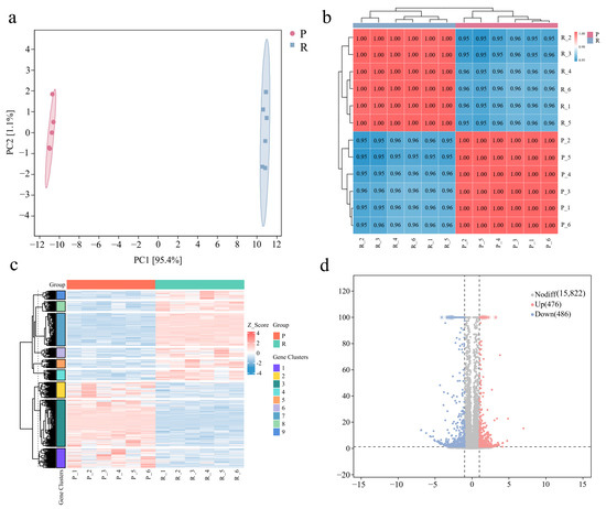
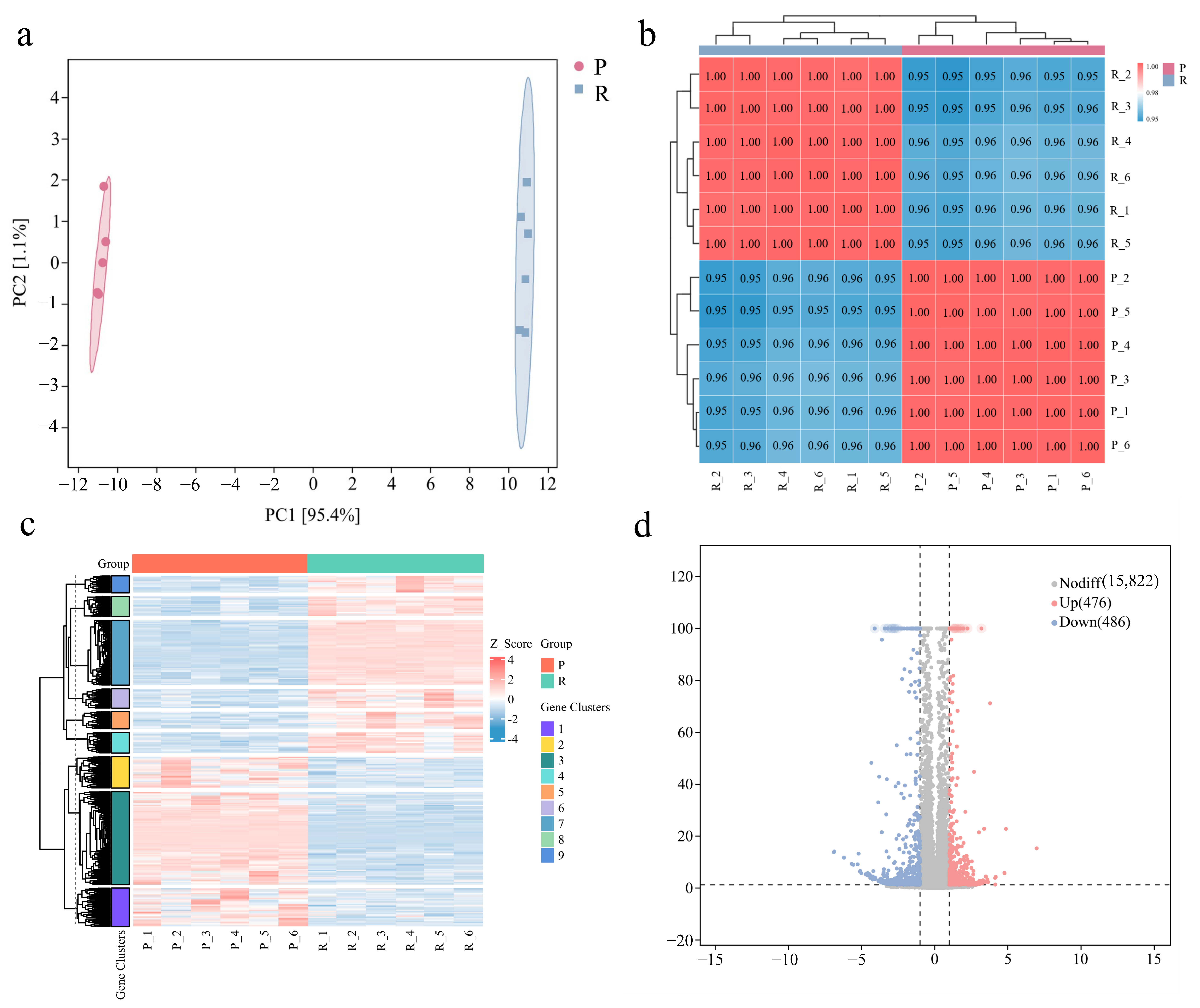
Muscle tissues of P. sinensis were harvested for transcriptome sequencing. Raw reads per sample ranged from 36,031,148 to 54,771,892, and quality trimming yielded clean reads varying between 35,661,210 and 54,191,126. All samples exhibited consistent GC contents of 47.18–47.73% with Q30 values exceeding 96.56% (Table 5). Principal component analysis (PCA) showed specimens within each group clustered closely, whereas obvious separation existed between the two experimental treatments (Figure 1a). Pairwise correlation results are displayed in Figure 1b, while the global distribution of DEGs is presented in Figure 1c. The comparison between Group P and Group R uncovered 962 DEGs in Group R, consisting of 476 upregulated and 486 downregulated transcripts (Figure 1d).
Table 5.
RNA-seq data from P. sinensis muscle.
Figure 1.
Transcriptomic data analysis between Group P and Group R. (a) Principal component analysis (PCA); (b) correlation analysis between biological replicate samples; (c) heat map of clustering analysis of differentially expressed genes. The vertical dashed lines on the hierarchical clustering tree represent the cutoff thresholdfor grouping genes into distinct gene clusters; (d) volcano plot of differentially expressed genes.
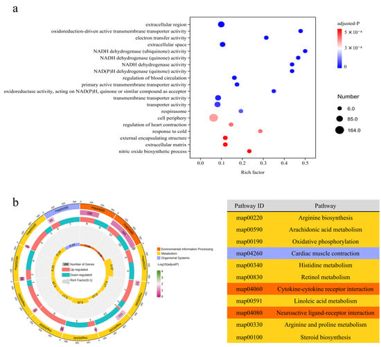
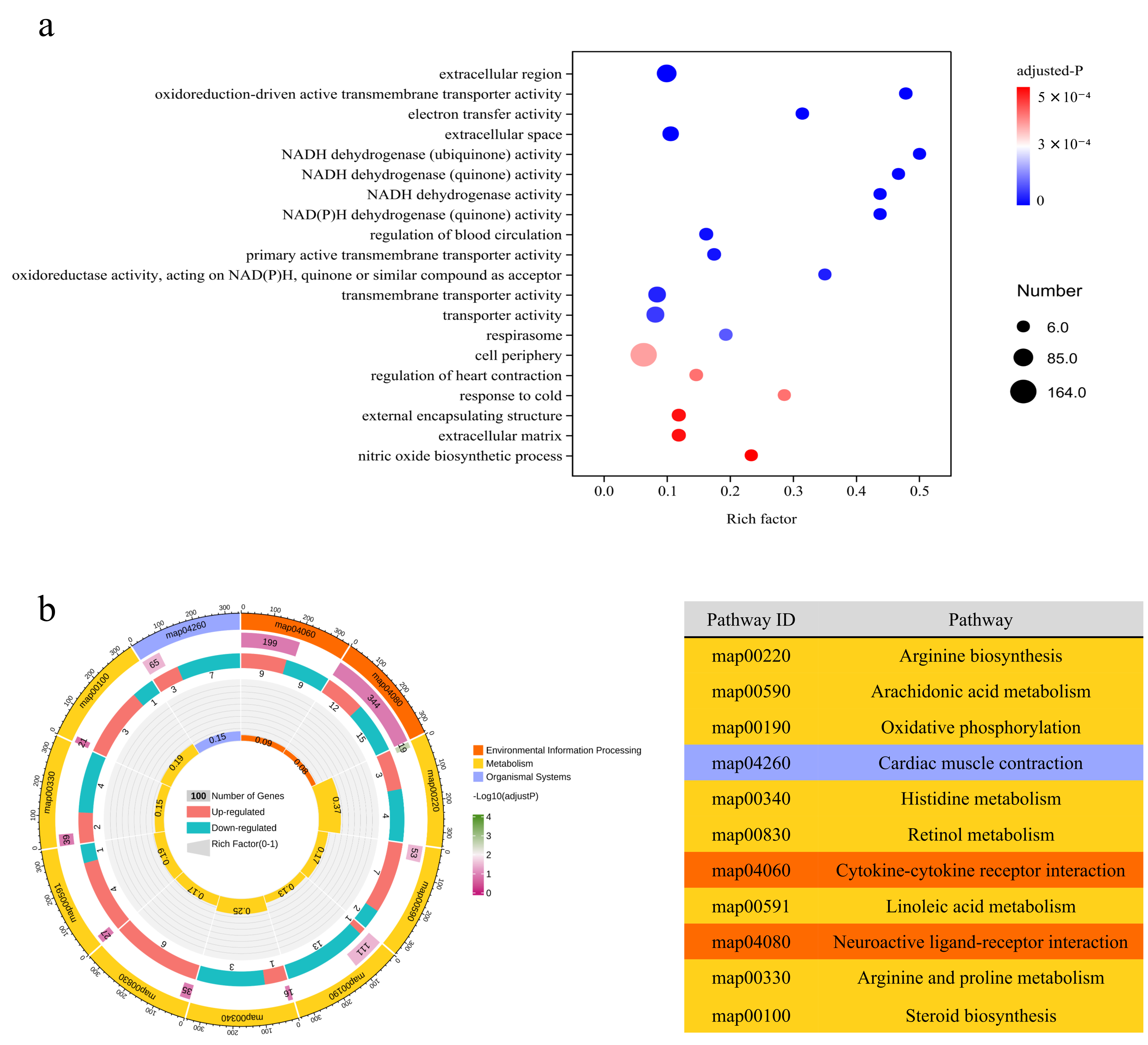
Pairwise contrast between Group P and Group R yielded six top-ranked cellular component (CC) GO terms screened by adjusted p-values: extracellular region (GO: 0005576), extracellular space (GO: 0005615), respirasome (GO: 0070469), cell periphery (GO: 0071944), external encapsulating structure (GO: 0030312), and extracellular matrix (GO: 0031012). For molecular function (MF) category, three enriched GO entries were captured from this intergroup comparison, namely oxidoreduction-driven active transmembrane transporter activity (GO: 0015453), electron transfer activity (GO: 0009055), and NADH dehydrogenase (ubiquinone) activity (GO: 0008137). Four GO entries were also screened within the biological process (BP) category: regulation of blood circulation (GO: 1903522), regulation of heart contraction (GO: 0008016), response to cold (GO: 0009409), and nitric oxide biosynthetic process (GO: 0006809) (Figure 2a).
Figure 2.
Transcriptome GO and KEGG Analysis Chart. (a) GO enrichment map of differentially expressed genes; (b) multidimensional enrichment cycle diagram of KEGG pathway enrichment analysis. Labels formatted as “Map + numerals” correspond to KEGG pathway identifiers. This enrichment plot is structured with four concentric rings. (1) The outermost ring displays categories of enriched pathways, and its outer scale quantifies gene counts; distinct colors distinguish different functional categories. (2) The second ring illustrates the total background genes within each category alongside associated Q or p values. Longer bars stand for greater gene quantities, while shades of red deepen for lower p values. (3) The third ring contains bar segments reflecting the ratio of upregulated versus downregulated genes, with exact numerical values labeled at the bottom. (4) The innermost ring presents the Rich Factor for each pathway category, calculated as the count of differentially expressed genes divided by the total background genes assigned to the category.
Results of KEGG pathway analysis are presented in Figure 2b. The 11 top pathways prioritized by adjusted p-values were predominantly involved in metabolic processes, including arginine biosynthesis (map:00220), arachidonic acid metabolism (map: 00590), histidine metabolism (map: 00340), and linoleic acid metabolism (map: 00591). According to the KEGG annotation results, nine DEGs (seven upregulated and two downregulated) linked to fatty acid metabolism were identified (Table 6).
Table 6.
List of the genes that were differentially expressed in muscle of P. sinensis.
3.3. Analysis of Gut Microbiome
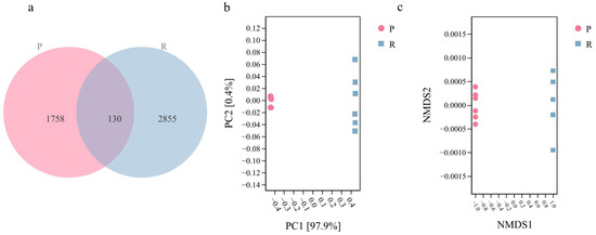
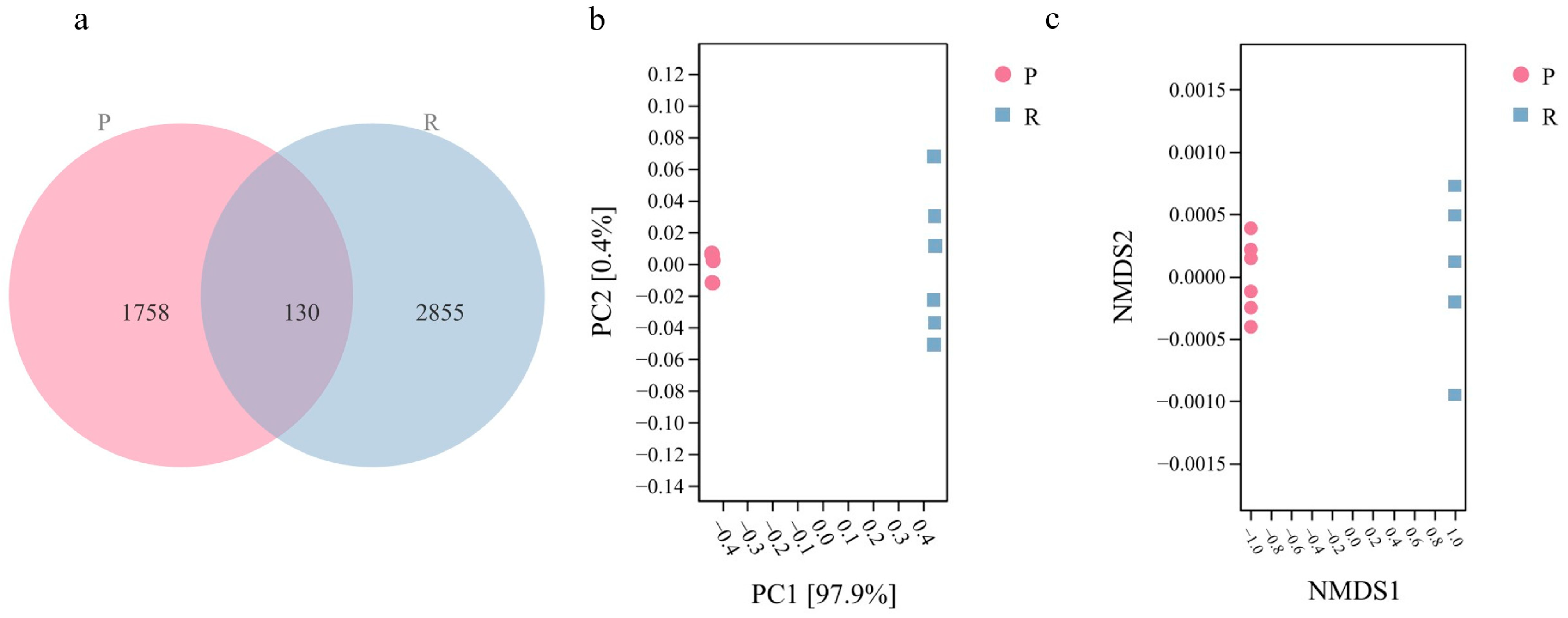
Analysis of obtained ASVs revealed that Group R and Group P harbored 2985 and 1888 representative ASVs, respectively. A total of 130 ASVs were shared by the two groups, whereas 2855 and 1758 ASVs were uniquely detected in Group R and Group P, respectively (Figure 3a). Outcomes from PCoA and NMDS analyses exhibited obvious separation between Group P and Group R samples, demonstrating marked discrepancies in intestinal microbial community composition. By comparison, biological replicates within each treatment clustered tightly, reflecting high similarity in community configuration (Figure 3b,c). PERMANOVA verified significant intergroup divergence in gut microbial community composition. Further PERMDISP analysis revealed significant differences in multivariate dispersion between groups (F = 91.3295, p = 0.001).
Figure 3.
Changes in gut microbial β-diversity and community composition between Group P and Group R. (a) Venn plot showing exclusive and overlapping amplicon sequence variants (ASVs) detected across groups; (b) β-diversity displayed via PCoA; (c) β-diversity displayed via NMDS.
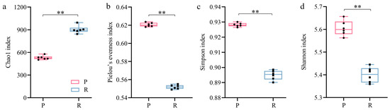
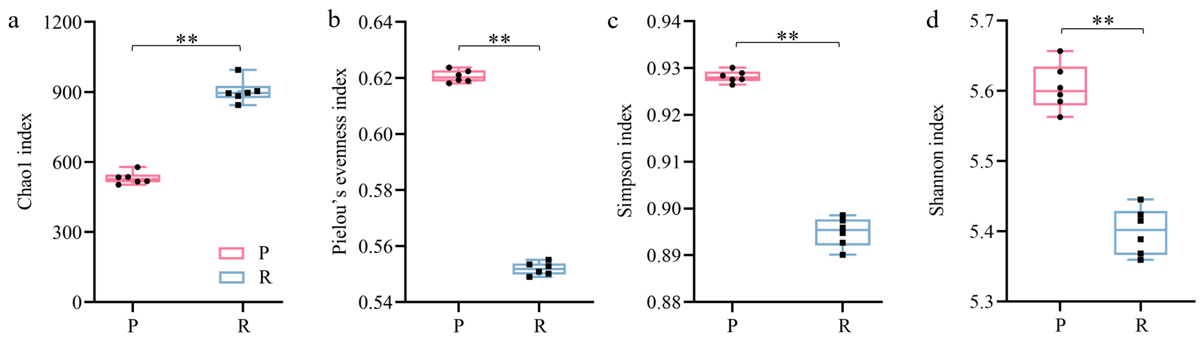
Community richness and alpha diversity were quantified via Chao 1, Pielou_e, Simpson, and Shannon indices between the two experimental groups. The Chao 1 index was markedly elevated in Group R relative to Group P (p < 0.01) (Figure 4).
Figure 4.
Changes in gut microbial α-diversity indices between Group P (P) and Group R (R). (a) Chao 1 index; (b) Pielou’s evenness index; (c) Simpson index; (d) Shannon index. Statistical significance was indicated by ** (p < 0.01).
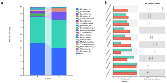
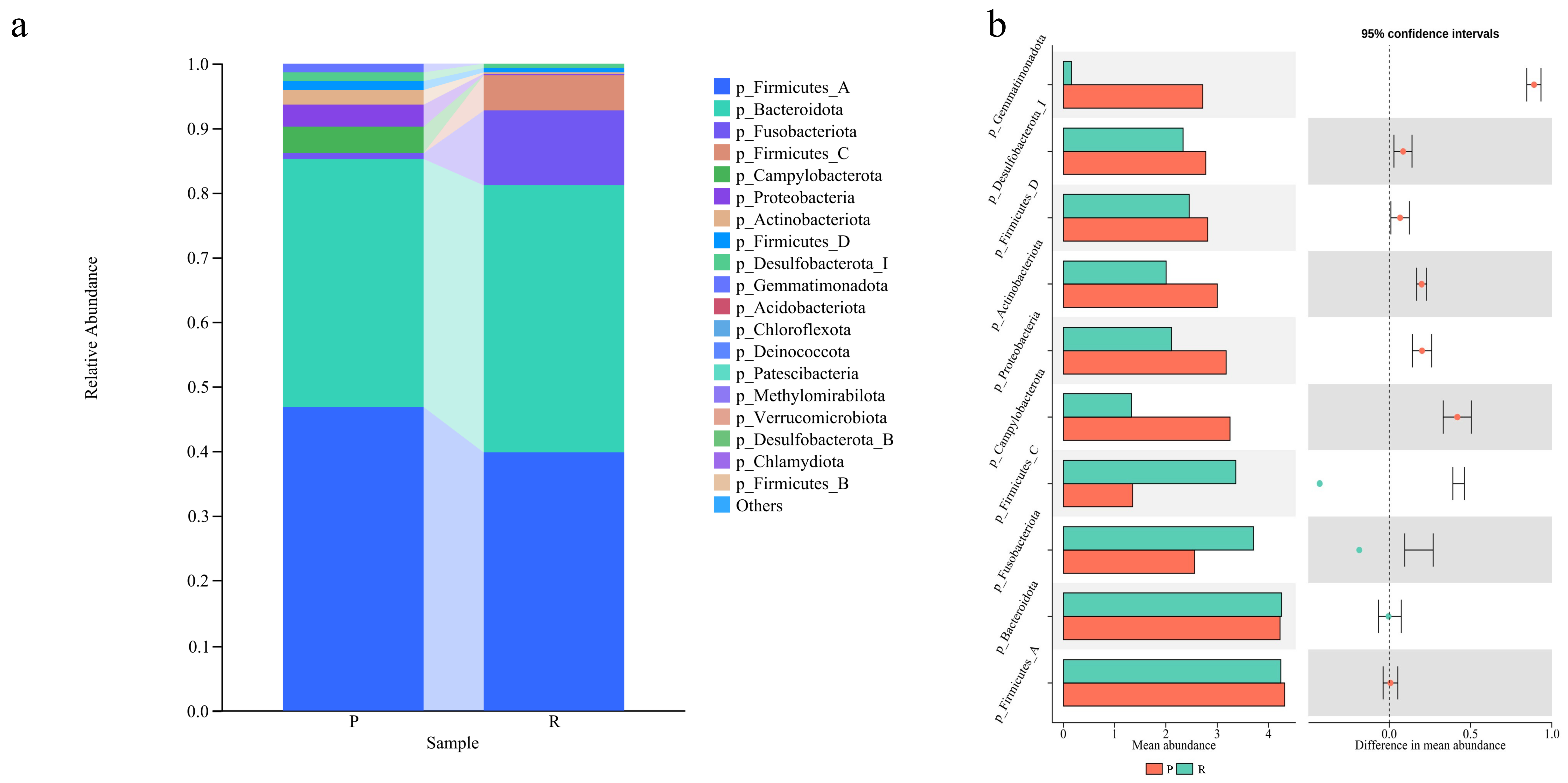
Bar charts were applied to profile intestinal bacterial compositions to further dissect the responses of gut microbiota to disparate aquaculture regimes. At the phylum taxonomic level, Firmicutes_A and Bacteroidota dominated the intestinal microbiota of both experimental groups. Group R exhibited elevated relative abundance of Firmicutes_C, while Actinobacteriota was enriched predominantly in Group P (Figure 5a). Intergroup discrepancies in the relative abundance of phylum-level taxa are summarized in Figure 5b. In detail, the relative abundances of Firmicutes_C and Bacteroidota increased significantly in Group R (p < 0.01).
Figure 5.
Intergroup comparison of gut microbiota relative abundance at the phylum taxonomic level between Group P (P) and Group R (R). (a) Bar graph displaying the top 20 phyla ranked by relative abundance; (b) differences in phylum-level relative abundance of intestinal microbial assemblages between Group P (P) and Group R (R).
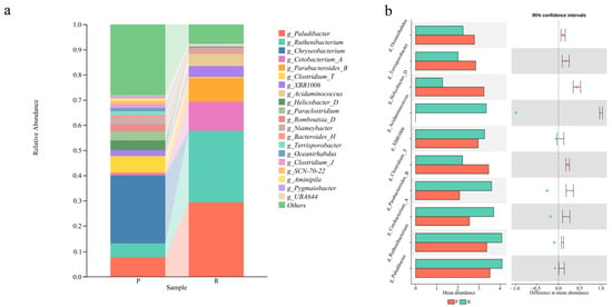
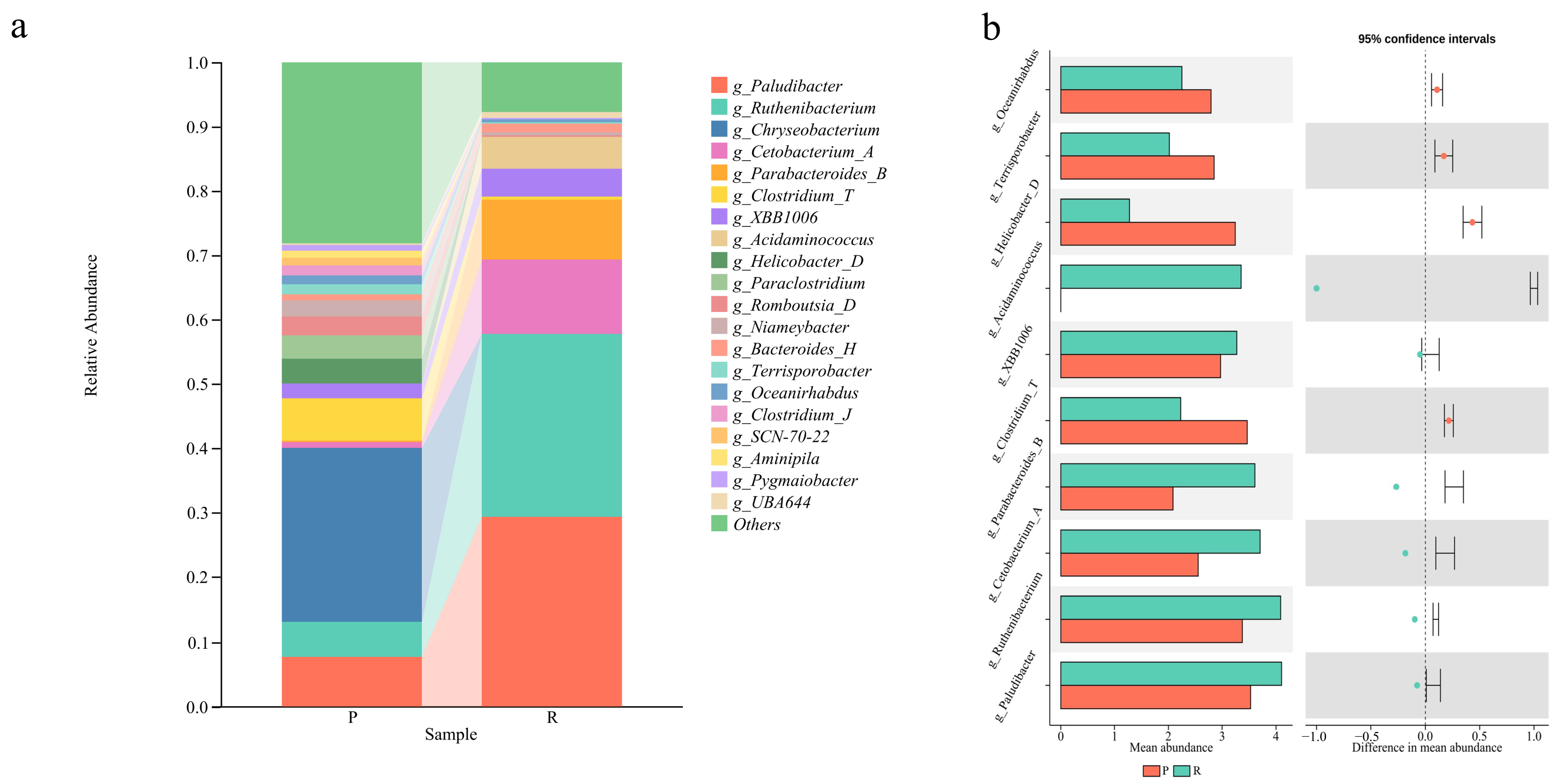
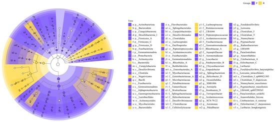
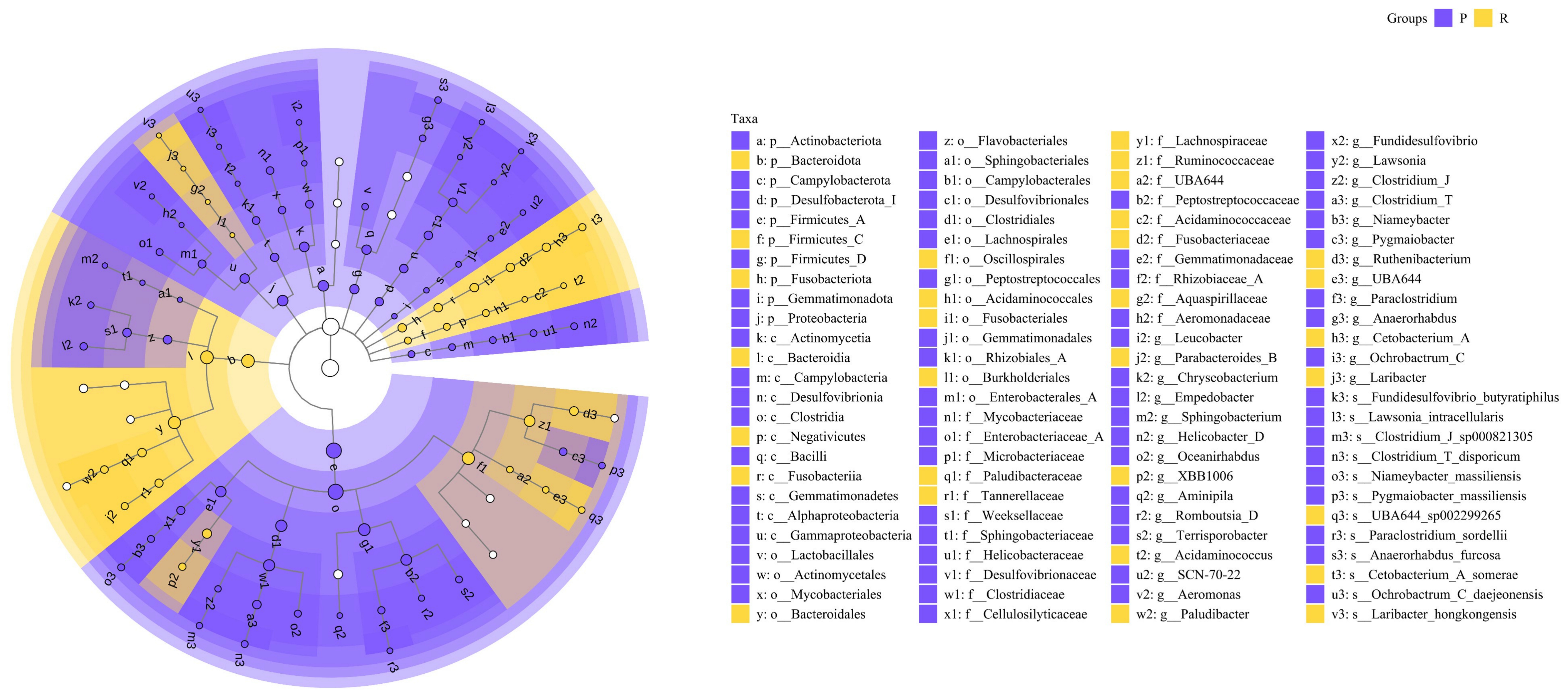
At the genus level, Paludibacter and Ruthenibacterium displayed higher relative abundances in Group R, whereas Chryseobacterium predominated in Group P (Figure 6a). Intergroup variations in genus-level bacterial relative abundance are presented in Figure 6b. In particular, the relative abundances of Clostridium_T, Parabacteroides_B, Cetobacterium_A, Ruthenibacterium, and Paludibacter were significantly elevated in Group R (p < 0.01). Notably, Acidaminococcus was exclusively detected in Group R and undetectable in Group P. LEfSe analysis identified 187 differentially abundant taxa (the top 100 taxa are illustrated in Figure 7), defined by an absolute LDA score (log10) > 2.0. Genera including Acidaminococcus, Ruthenibacterium, and Paludibacterium were significantly enriched in the intestinal tract of P. sinensis from Group R.
Figure 6.
Intergroup comparison of gut microbiota relative abundance at the genus taxonomic level between Group P (P) and Group R (R). (a) Bar graph displaying the top 20 genera ranked by relative abundance; (b) differences in genus-level relative abundance of intestinal microbial assemblages between Group P (P) and Group R (R).
Figure 7.
LEfSe analysis of bacterial taxa distinguishing Group P (P) and Group R (R).
3.4. Correlation Analysis Between Gut Microbiome and Differentially Expressed Genes
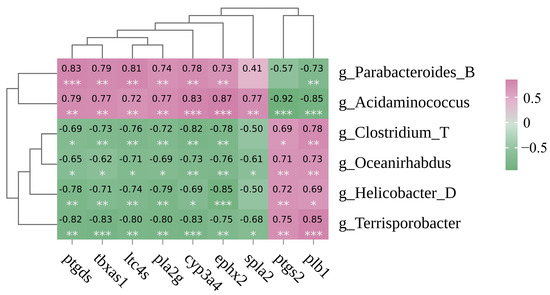
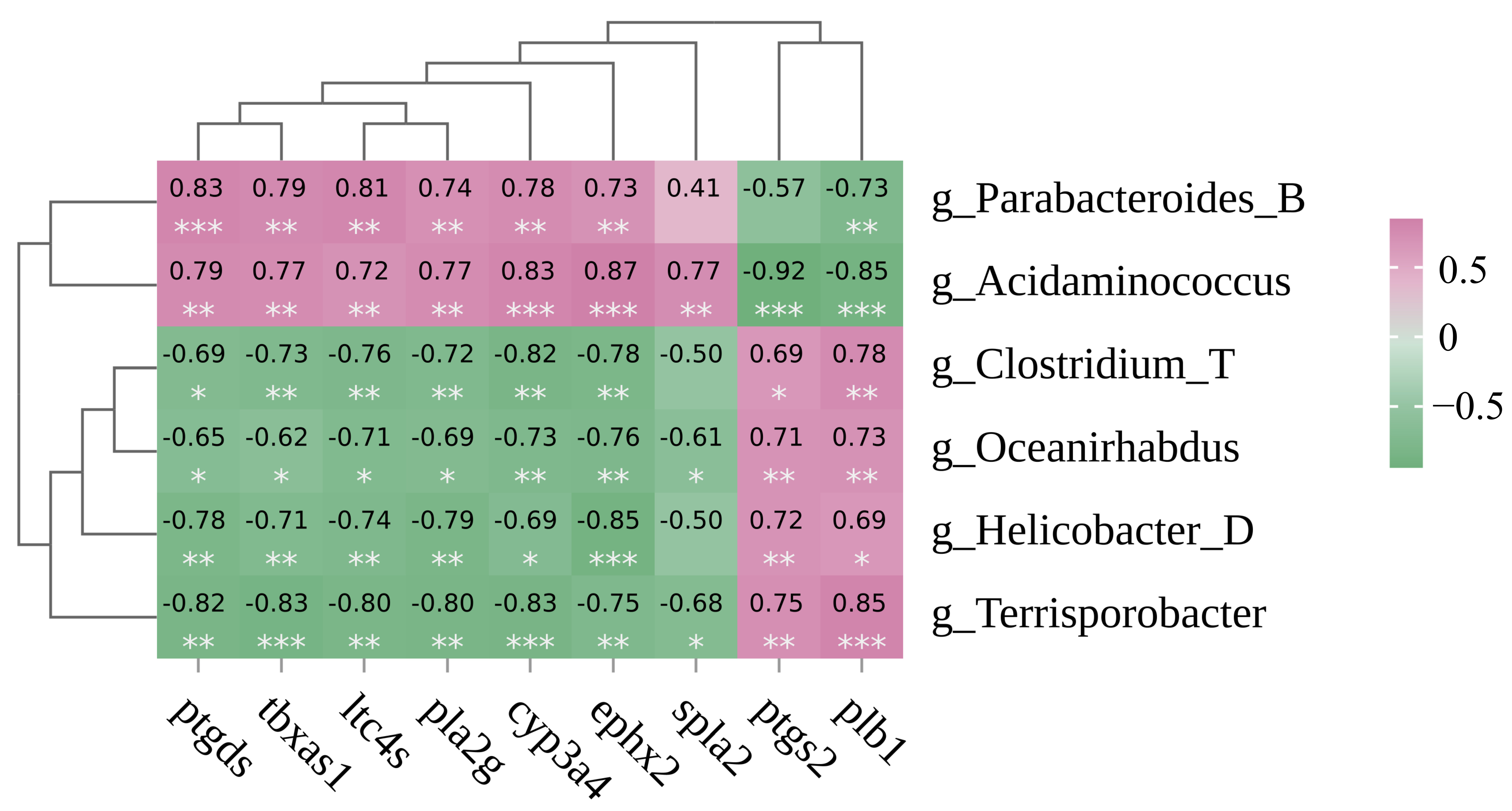
Acidaminococcus and Terrisporobacter exhibited significant correlations with all screened DEGs (p < 0.05) (Figure 8). In detail, Acidaminococcus was positively correlated with upregulated DEGs but negatively correlated with downregulated DEGs. By contrast, Terrisporobacter showed inverse correlation patterns: negative associations with upregulated DEGs and positive associations with downregulated DEGs.
Figure 8.
Heatmap representation of the Spearman correlation coefficient between fatty acid metabolism—associated gut microbiota and DEGs were significantly enriched in fatty acid metabolic pathways. Red denotes positive correlations, while green marks negative correlations (* (p < 0.05), ** (p < 0.01), *** (p < 0.001)).
4. Discussion
Different culture regimes exert prominent impacts on the muscle nutritional composition and quality of aquatic animals; specifically, the stable environmental conditions under conventional pond farming may reduce locomotor energy expenditure and thereby promote intramuscular amino acid accumulation [3]. By contrast, the complex habitat and fluctuating environmental conditions in rice fields may increase metabolic and stress consumption of P. sinensis, promoting protein and amino acid catabolism and reducing their deposition in muscle [4]. Globally, abundant overseas investigations have explored lipid regulation, intestinal microbiota and nutritional traits of cultured aquatic organisms, providing fundamental theoretical support for integrated aquaculture research. We compared our findings with domestic and overseas rice–aquaculture related studies to comprehensively contextualize our data, and the summarized shared and divergent traits provide global references for understanding paddy-driven phenotypic regulation in P. sinensis [22,23,24,25]. Results of the present study from muscle amino acid analysis indicated that several amino acids had lower contents in the rice-field group. Broadly, P. sinensis cultured in rice-field and pond systems both possessed excellent edible nutritional value and great development potential. Mineral elements act as critical markers for estimating the nutritional quality of aquatic organisms [26]. No notable variations in mineral composition and concentrations of P. sinensis were uncovered when comparing Group P against Group R. The contents of Ca, Na, and Fe were relatively higher in Group R, which may be attributed to the enhanced feed utilization efficiency under rice-field environmental conditions and consequently promoted greater mineral deposition in the body [26,27]. PUFAs are functional fatty acids that exert beneficial effects on human health [14]. Linoleic acid (LA) and ALA compete for the same metabolic enzymes participating in PUFA biosynthesis. LA acts as the key precursor of ω6 polyunsaturated fatty acids and is further converted into n-6 long-chain PUFA (LC-PUFA) such as arachidonic acid (ARA) [22,28,29]. In general, the rice-field environment modulated the overall n-6 PUFA metabolic flow, ultimately achieving a more balanced and optimized fatty acid profile [4,30]. Consistent with these previous reports, our study exhibited a similar metabolic pattern. It was presumed that the rice-field environment activated the linoleic acid and arachidonic acid metabolic pathways in P. sinensis muscle. Such activation could accelerate the conversion of substrate LA into downstream n-6 LC-PUFA, which presumably contributed to the notable reduction in LA and remarkable accumulation of ARA in Group R, and such fatty acid variation corresponds to distinct edible nutritional characteristics of P. sinensis muscle.
Fatty acid metabolism within P. sinensis is governed by an elaborate network connecting numerous metabolic cascades. Multiple DEGs were annotated to muscle fatty acid metabolism and significantly enriched in arachidonic acid metabolism (map: 00590) and linoleic acid metabolism (map: 00591), among which cyp3a4 was identified as a differentially expressed gene. The cytochrome P450 (CYP) superfamily consists of heme-linked proteins that exert pivotal biological functions in modulating the metabolism of endogenous compounds including hormones and vitamins [23]. As a key CYP family member, cyp3a4 participates in lipid turnover regulation and is tightly correlated with fatty acid metabolic homeostasis and nutritional quality traits in aquatic species [24,31,32].
Gut microbial communities are widely recognized to participate in regulating gut immune status, nutrient catabolism and host physiological conditions across aquatic animals, and the intestine acts as the primary organ for digestion and absorption as well as a critical mucosal immune barrier of P. sinensis [33,34]. In this study, higher relative abundances of Firmicutes_C and Bacteroidota were observed in Group R relative to Group P. Firmicutes and Bacteroidota are dominant intestinal taxa in healthy aquatic organisms, and previous independent studies have documented their potential roles linked to host physiological status, immune-related pathways and intestinal microecological balance [35]. Functionally, Firmicutes and Bacteroidetes participate in the degradation and fermentation of dietary fiber and carbohydrates to generate short-chain fatty acids (SCFAs) [25]. Based on the published literature [30,35], such SCFAs may be associated with mucosal barrier integrity, suppression of opportunistic pathogens and intestinal microecological balance, yet relevant immune and mucosal indicators were not quantified in the present study to verify these correlations in rice–turtle co-culture. Meanwhile, the synergistic dominance of Firmicutes and Bacteroidetes in Group R also enhances the efficiency of nutrient digestion and energy utilization, laying a microecological foundation for efficient substance metabolism [36,37]. These results implied that the rice-field culture environment may alter the intestinal microbial composition of P. sinensis. At the genus level, Group R harbored a relatively higher abundance of Cetobacterium. As a beneficial genus, Cetobacterium can improve the intestinal health of aquatic animals and inhibit the proliferation of potential pathogens to a certain extent [38,39]. Moreover, the enrichment of Cetobacterium may be associated with the improved growth performance of aquatic animals, which has also been documented in previous studies [38,39,40]. Acidaminococcus could take advantage of amino acids as growth substrates and modulate the metabolism of lipids, carbohydrates and amino acids [41]. In the present study, Acidaminococcus was exclusively detected in Group R. The specific enrichment of this genus further indicated that the rice-field environment could reshape the metabolic functional profile of intestinal microbiota. It was inferred that Acidaminococcus might participate in the decomposition and transformation of dietary nutrients, coordinate lipid and amino acid metabolism, and thereby modulate the growth characteristics and nutritional metabolic pattern of P. sinensis under rice-field culture. Integrated correlative analysis across muscle amino acid and fatty acid phenotypes, muscle transcriptome and intestinal microbiota reveals coordinated multi-dimensional signatures distinguishing rice-field and pond-reared P. sinensis, forming a linked gut–muscle associative network rather than isolated independent results [4]. Consistent with published rice–turtle co-culture studies [12], paddy culture alters muscle polyunsaturated fatty acid profiles including linoleic acid and arachidonic acid, and comparable fatty acid shifts have also been documented in other ecological aquaculture research. Transcriptomic results further complement these nutritional differences: DEGs linked to arachidonic acid and linoleic acid metabolic pathways were differentially expressed between two culture groups, and these lipid-related transcriptional signatures displayed significant statistical correlations with the rice-enriched genus Acidaminococcus in the gut [13]. Prior microbiota studies on rice–aquaculture systems also verified that paddy habitats remodel intestinal bacterial community structure and enrich Firmicutes-related beneficial taxa with lipid fermentation capacity, which aligns with our taxonomic differential analysis results [12,13]. It should be explicitly noted that all cross-layer connections observed herein only reflect statistical associations rather than confirmed causal regulatory effects; definite molecular interactions between intestinal microbes and host lipid gene expression require further functional validation in follow-up trials. Overall, the rice-field culture corresponded to shifts in the intestinal microbiota composition, which may correlate with altered microbial nutrient metabolism profiles; such microbial variations might further link to changes in muscle nutritional composition and fatty acid features of P. sinensis.
The significant correlation between gut Acidaminococcus and muscle cyp genes suggests a potential association within the gut–muscle association, which may correlate with the muscle nutritional profile of rice-field-cultured P. sinensis. Specifically, Acidaminococcus was uniquely enriched in Group R and showed significant correlations with fatty acid metabolism-related genes, with positive correlations to upregulated fatty acid metabolic genes and negative correlations to downregulated ones. Collectively, the observed association between Acidaminococcus and fatty acid metabolic genes offers a preliminary molecular clue regarding the distinct nutritional quality of rice-field-cultured P. sinensis. These findings suggest that environmental shifts shaping intestinal microbiota may have correlative connections with aquatic product quality through host-microbe interactive profiles. It is also worth noting that regional cultural and bioethical differences exist regarding the consumption of reptile meat, as varying public perceptions of reptile sentience shape local market demand for softshell turtle products, a factor that indirectly affects the global market layout of eco-cultured aquatic reptiles.
5. Conclusions
This study compared muscle amino acid and fatty acid profiles, muscle transcriptomes, and intestinal microbiota of P. sinensis reared in pond and rice-field systems, and identified interrelated correlative signatures across multi-layer datasets linked to the modified nutritional traits of rice–turtle co-culture. The results showed that rice-field culture decreased muscle linoleic acid (LA) content while increasing arachidonic acid (ARA) concentration. Transcriptomic screening revealed that differentially expressed genes were mainly enriched in arachidonic acid metabolism (map:00590) and linoleic acid metabolism (map:00591). Notably, P. sinensis reared in rice-field systems showed elevated relative abundances of intestinal Firmicutes_C and Acidaminococcus, which exhibited potential correlative links to intestinal microbial compositional balance. Collectively, rice-field co-culture corresponded to shifts in muscle nutritional components and fatty acid profiles of P. sinensis. These correlative observations hint at possible distinctions in muscle nutrient composition under paddy cultivation, though further verification is required to confirm relevant physiological effects. Taken together, these results reveal preliminary associations between rice-field culture and turtle intestinal and muscle metabolic traits, and additional research is needed to clarify underlying interactive mechanisms before the wide application of this integrated aquaculture model.
Author Contributions
Writing—original draft, funding acquisition and methodology, F.H.; writing—original draft, software and formal analysis, K.G.; methodology and writing—review and editing, X.L.; resources and investigation, L.Q.; methodology and data curation, J.T.; data curation and formal analysis, Q.J.; data curation, L.J.; resources, J.X.; methodology, Y.Z.; project administration, writing—review and editing, Q.Y. All authors have read and agreed to the published version of the manuscript.
Funding
This research was funded by the Local Cooperation Project of the Zhejiang Academy of Agricultural Sciences and Shaoxing Agricultural Bureau of Green Production Technology for Rice-Shrimp-Turtle Products (grant number ZJTY2024-N-285), and the Project of Aquatic Industry Technology Extension Service Team of Zhejiang Province (2026–2027).
Institutional Review Board Statement
The animal study was reviewed and approved by the Committee of Laboratory Animal Experimentation at Zhejiang Academy of Agricultural Sciences (Permit No. 25ZALAS42, approved on 8 July 2025).
Data Availability Statement
The original contributions presented in this study are included in the article. Further inquiries can be directed to the corresponding author.
Conflicts of Interest
The authors declare no conflicts of interest.
References
- Gong, Y.T.; Luo, M.K.; Zhang, X.B.; Qin, N.; Zhu, W.B.; Wang, L.M.; Fu, J.J.; Dong, Z.J. Evaluation of muscle quality and movement-related traits in two common carp strains under a rice-fish farming system. Aquacul. Int. 2025, 33, 309. [Google Scholar] [CrossRef] [Scilit]
- Ding, X.; Jin, F.; Xu, J.W.; Zhang, S.L.; Chen, D.X.; Hu, B.J.; Hong, Y.J. The impact of aquaculture system on the microbiome and gut metabolome of juvenile Chinese softshell turtle (Pelodiscus sinensis). iMeta 2022, 1, e17. [Google Scholar] [CrossRef] [Scilit] [PubMed]
- Fan, H.J.; Ye, J.; Zhou, F.; Su, W.F.; Wang, X.N.; Bu, W.C.; Bei, Y.J.; Chen, J.Y.; Guo, S.Q.; Zheng, B.; et al. Comparative evaluation of nutritional quality and flavor characteristics for Acrossocheilus fasciatus muscle in different aquaculture systems. Food Chem. 2026, 502, 147758. [Google Scholar] [CrossRef] [Scilit] [PubMed]
- Qi, M.; Wang, Y.; Hu, L.L.; Chen, G.M.; Zheng, T.L.; Ding, X.Y.; Bei, Y.J.; Tang, J.J.; Ma, W.J.; Chen, X. Investigation of plasticity in morphology, organ traits and nutritional composition in Chinese soft-shelled turtle (Pelodiscus sinensis) under different culturing modes. Fishes 2025, 10, 89. [Google Scholar] [CrossRef] [Scilit]
- Zhou, T.; Wu, W.Y.; Xue, M.Y.; Zhou, Y.; Liang, H.W.; Liu, W. Antioxidant capacity and disease resistance enhanced by dietary d-glucuronolactone supplementation in Chinese soft-shelled turtles (Pelodiscus sinensis). Antioxidants 2025, 14, 534. [Google Scholar] [CrossRef] [Scilit] [PubMed]
- Wu, Y.R.; Zhuang, T.X.; Jiang, L.L.; Jin, S.F.; Zhu, Y.K.; Ge, C.T.; Gao, Y.L. Sanghuangporus vaninii extract alleviates the retinoic acid-inducible gene-I-like receptors (RLRs) signaling pathway mediated antiviral response in Chinese soft-shelled turtle (Pelodiscus sinensis). Aquaculture 2026, 611, 743024. [Google Scholar] [CrossRef] [Scilit]
- Wang, H.T.; Shi, P.; Liao, Y.H.; Liu, S.Y.; Li, C.X.; Huang, J.M.; Liu, Y.S.; Bu, R.P. Effects of heterologous protein substitutions of fishmeal on the culture of the Chinese soft-shelled turtle (Pelodiscus sinensis): A review and meta-analysis. Aquac. Res. 2026, 2026, 7482637. [Google Scholar] [CrossRef] [Scilit]
- Ji, L.Q.; Shi, Q.; Chen, C.; Liu, X.L.; Zhu, J.X.; Hong, X.Y.; Wei, C.Q.; Zhu, X.P.; Li, W. Biochemical, histological, and multi-omics analyses reveal the molecular and metabolic mechanisms of cold stress response in the Chinese soft-shelled turtle (Pelodiscus sinensis). Biology 2025, 14, 55. [Google Scholar] [CrossRef] [Scilit] [PubMed]
- Shi, X.Y.; Guo, Q.Q.; Li, X.C.; Li, T.Q.; Li, T.; Li, T.Y.; Zhang, P.Y.; Li, Z.; Liu, H.Y. Metabolic responses to starvation in the soft-shelled turtle (Pelodiscus sinensis) revealed by integrated metabolome and transcriptome analysis. Comp. Biochem. Phys. D 2025, 55, 101486. [Google Scholar] [CrossRef] [Scilit] [PubMed]
- Ding, Y.J.; Dai, F.; Su, L.J.; Huang, Q.Y.; Shen, F.A.; Qian, G.Y.; Ge, C.T.; Xu, J.H. Effects of chronic ammonia nitrogen stress on intestinal barrier function and microbial composition of Chinese soft-shelled turtle (Pelodiscus sinensis). Aquaculture 2026, 614, 743459. [Google Scholar] [CrossRef] [Scilit]
- Wu, B.L.; Wu, C.C.; Huang, L.; Chen, J.; Chen, X.J.; He, J.X. Adaptation and feeding preference of Chinese soft-shelled turtle (Pelodiscus sinensis) to different feeding levels in rice-turtle - turtle co-culture system. Aquacult. Rep. 2024, 38, 102281. [Google Scholar] [CrossRef] [Scilit]
- Wang, R.; Ma, W.W.; Wu, D.; Zhang, Y.; Ma, X.H.; Lv, G.D.; Ding, J.L.; Fu, Z.Q.; Chen, C.; Huang, H. Soil bacterial community composition in rice-turtle coculture systems with different planting years. Sci. Rep. 2023, 13, 22708. [Google Scholar] [CrossRef] [Scilit] [PubMed]
- Wu, B.L.; Huang, L.; Wu, C.C.; Chen, J.; Chen, X.J.; He, J.X. Comparative analysis of the growth, physiological responses, and gene expression of Chinese Soft-Shelled Turtles Cultured in Different Modes. Animals 2024, 14, 962. [Google Scholar] [CrossRef] [Scilit] [PubMed]
- Hou, M.D.; Sun, W.B.; Ma, Y.; Ye, H.; Zhai, X.L.; Xue, Y.; Tang, R.J.; Teng, S.J.; Wu, R.H.; Luo, H. Comparative analysis for nutrients, flavor compounds, and lipidome revealed the edible value of pond-cultured male Pelodiscus sinensis with different ages. Food Chem. 2024, 454, 139795. [Google Scholar] [CrossRef] [Scilit] [PubMed]
- Wu, Y.Y.; Xiao, P.F.; Sha, H.; Luo, X.Z.; Zou, G.W.; Liang, H.W. Transcriptome analysis reveals the potential key genes in nutritional deposition in the common carp (Cyprinus carpio). Animals 2024, 14, 1939. [Google Scholar] [CrossRef] [Scilit] [PubMed]
- Tan, D.; Li, E.D.; Xiong, S.J.; Sun, Y.; Cheng, W.B.; Su, Y.; Lu, Y. Transcriptomic and metabolomic analyses reveal the attenuating role of cordycepin and Cordyceps militaris extract on acute liver injury induced by LPS in piglets. Animals 2024, 14, 2873. [Google Scholar] [CrossRef] [Scilit] [PubMed]
- Yang, S.; Duan, Y.L.; Zhang, J.; Zhou, J.; Liu, Y.; Du, J.; Zhao, L.L.; Du, Z.J.; Han, S.S. Observational comparisons of intestinal microbiota characterizations, immune enzyme activities, and muscle amino acid compositions of loach in paddy fields and ponds in Sichuan Province. Appl. Microbiol. Biot. 2017, 101, 4775–4789. [Google Scholar] [CrossRef] [Scilit] [PubMed]
- Shi, J.H.; Li, L.; Xie, D.B.; Yu, L.; Zhang, L.; Xu, Q.Y.; Zhao, J.H.; Li, B.; Liu, H.R. Odontobutis potamophila gut microbiota divergence between two culture modes: Implications for mode selection. Aquacul. Int. 2026, 34, 55. [Google Scholar] [CrossRef] [Scilit]
- Zhou, F.; Bu, W.C.; Fan, H.J.; Guo, S.R.; Qi, M.; Yao, G.H.; Bei, Y.J.; Huang, Y.F.; Zhu, S.C.; Ding, X.Y.; et al. A comparative study on the muscle and gut microbiota of Opsariichthys bidens from rice field and pond culture breeding modes. Metabolites 2024, 14, 443. [Google Scholar] [CrossRef] [Scilit] [PubMed]
- Zhu, X.C.; Nie, M.; Sun, N.; Zhang, Y.Z.; Sun, M.X.; Li, C.L.; Jiang, Q.; Wei, H.; Li, Y.D.; Hu, Q.B.; et al. Comparative analysis of crab growth performance, enzyme activity, and microbiota between rice-crab coculture and pond farming systems. Front. Vet. Sci. 2025, 12, 1571454. [Google Scholar] [CrossRef] [Scilit] [PubMed]
- Huang, H.; Liu, X.; Sun, H.; Xiao, Y.; He, Y.F.; Chen, Y.J.; Lin, S.M. Impacts of structurally distinct starches on growth, short-chain fatty acid production, hepatic lipid metabolic homeostasis, and inflammatory response in largemouth bass (Micropterus salmoides). Anim. Feed Sci. Tech. 2026, 334, 116655. [Google Scholar] [CrossRef] [Scilit]
- Sprague, M.; Xu, G.; Betancor, M.B.; Olsen, R.E.; Torrissen, O.; Glencross, B.D.; Tocher, D.R. Endogenous production of n-3 long-chain PUFA from first feeding and the influence of dietary linoleic acid and the α-linolenic: Linoleic ratio in Atlantic salmon (Salmo salar). Br. J. Nutr. 2019, 122, 1091–1102. [Google Scholar] [CrossRef] [Scilit] [PubMed]
- Lee, B.Y.; Kim, D.H.; Kim, H.S.; Kim, B.M.; Han, J.; Lee, J.S. Identification of 74 cytochrome P450 genes and co-localized cytochrome P450 genes of the CYP2K, CYP5A, and CYP46A subfamilies in the mangrove killifish Kryptolebias marmoratus. BMC Genom. 2018, 19, 7. [Google Scholar] [CrossRef] [Scilit] [PubMed]
- Poon, K.L.; Wang, X.G.; Ng, A.S.; Goh, W.H.; McGinnis, C.; Fowler, S.; Carney, T.J.; Wang, H.S.; Ingham, P.W. Humanizing the zebrafish liver shifts drug metabolic profiles and improves pharmacokinetics of CYP3A4 substrates. Arch. Toxicol. 2017, 91, 1187–1197. [Google Scholar] [CrossRef] [Scilit] [PubMed]
- Marcharla, E.; Vishnuprasadh, A.; Gnanasekaran, L.; Vinayagam, S.; Sundaram, T.; Ganesan, S. The role of functional feed in modulating fish gut microbiome to enhance resistance against aquaculture pathogens. Probiotics Antimicro. 2026, 18, 3010–3039. [Google Scholar] [CrossRef] [Scilit] [PubMed]
- Wang, B.K.; Wang, Y.H.; Lv, J.; Zhang, L.; Zhang, K.S.; Yang, X.L.; Wang, Z.P.; Zhang, C.N.; Zhang, Q. Comparative analysis of nutritional ingredients of muscle in Pseudobagrus ussuriensis under pond and wild conditions. J. Food Compos. Anal. 2025, 146, 107954. [Google Scholar] [CrossRef] [Scilit]
- Wang, Z.X.; Zheng, J.S.; Pu, D.C.; Li, P.Y.; Wei, X.L.; Li, D.S.; Gao, L.H.; Zhai, X.L.; Zhao, C.H.; Du, Y.D. Comparative evaluation of nutritional quality and flavor characteristics for Micropterus salmoides muscle in different aquaculture systems. Food Chem. X 2024, 24, 101787. [Google Scholar] [CrossRef] [Scilit] [PubMed]
- Mori, T.A. Marine OMEGA-3 fatty acids in the prevention of cardiovascular disease. Fitoterapia 2017, 123, 51–58. [Google Scholar] [CrossRef] [Scilit] [PubMed]
- Nejatian, M.; Yazdi, A.P.G.; Fattahi, R.; Saberian, H.; Bazsefidpar, N.; Assadpour, E.; Jafari, S.M. Improving the storage and oxidative stability of essential fatty acids by different encapsulation methods; a review. Int. J. Biol. Macromol. 2024, 260, 129548. [Google Scholar] [CrossRef] [Scilit] [PubMed]
- Zhao, Y.F.; Zhou, W.Z.; Li, M.Y.; Zhang, Y.N.; Lv, W.W.; Huang, W.W.; Yang, H.; Yuan, Q.; Li, M.Y. Effects of different farming models on muscle quality, intestinal microbiota diversity, and liver metabolism of rice field eel (Monopterus albus). Foods 2025, 14, 2383. [Google Scholar] [CrossRef] [Scilit] [PubMed]
- He, X.N.; Zhang, J.J.; Jiang, W.D.; Wu, P.; Liu, Y.; Ren, H.M.; Jin, X.W.; Shi, H.Q.; Feng, L.; Zhou, X.Q. 4-Methylesculetin alleviated aflatoxin B1-induced liver injury and ferritinophagy through AMPK- TOR-Ulk axis in grass carp (Ctenopharyngodon idella). Anim. Nutr. 2025, 22, 230–241. [Google Scholar] [CrossRef] [Scilit] [PubMed]
- Yang, X.X.; Gong, J.P.; Zhang, X.M.; Zhang, W.; Li, D.Y.; Lin, J.J.; Li, X.Y.; Chai, Y.; Liu, J.F. The responses of the growth, cytochrome P450 isoenzymes activities and the metabolomics in earthworms to sublethal doses of dichlorvos in soil. Ecotoxicol. Environ. Saf. 2021, 207, 11547. [Google Scholar] [CrossRef] [Scilit] [PubMed]
- Guo, J.L.; Xu, Y.; Chen, Q.; Zhang, H.Q. Integrative analysis of transcriptomics and gut microbiome in grass carp under alkalinity stress. Comp. Biochem. Phys. D 2026, 57, 101698. [Google Scholar] [CrossRef] [Scilit] [PubMed]
- Yu, H.Y.; Dong, S.J.; Wang, L.X.; Liu, S.L. Integrated gut microbiome and metabolome analysis in largemouth bass (Micropterus salmoides) following viral infection. Animals 2026, 16, 752. [Google Scholar] [CrossRef] [Scilit] [PubMed]
- Sun, Y.G.; Zhang, S.S.; Nie, Q.X.; He, H.J.; Tan, H.Z.; Geng, F.; Ji, H.H.; Hu, J.L.; Nie, S.P. Gut Firmicutes: Relationship with dietary fiber and role in host homeostasis. Crit. Rev. 2023, 63, 12073–12088. [Google Scholar] [CrossRef] [Scilit] [PubMed]
- Kanika, N.H.; Liaqat, N.; Chen, H.F.; Ke, J.; Lu, G.Q.; Wang, J.; Wang, C.H. Fish gut microbiome and its application in aquaculture and biological conservation. Front. Microbiol. 2025, 15, 1521048. [Google Scholar] [CrossRef] [Scilit] [PubMed]
- Chen, Z.L.; Ma, L.X.; Chen, S.; Huang, Y.; Qin, Z.D.; Lin, L.; Shi, F. Effects of enrofloxacin and povidone-iodine on immunity, the intestinal microbiome and transcriptome of juvenile grass carp (Ctenopharyngodon idella). J. Fish Biol. 2025, 107, 201–214. [Google Scholar] [CrossRef] [Scilit] [PubMed]
- Zhou, W.; Xie, M.X.; Xie, Y.D.; Liang, H.; Li, M.; Ran, C.; Zhou, Z.G. Effect of dietary supplementation of Cetobacterium somerae XMX-1 fermentation product on gut and liver health and resistance against bacterial infection of the genetically improved farmed tilapia (GIFT, Oreochromis niloticus). Fish Shellfish Immunol. 2022, 124, 332–342. [Google Scholar] [CrossRef] [Scilit] [PubMed]
- Wang, M.; Yi, M.M.; Zhang, Z.Y.; Fan, Z.J.; Jiang, L.J.; Liu, Z.G.; Wang, Z.; Lu, M.X.; Cao, J.M.; Ke, X.L. Effect of different feeding habits on metabolomics profiles and microbiota composition and functions of tilapia gut. Aquac. Res. 2024, 2024, 5545159. [Google Scholar] [CrossRef] [Scilit]
- Jia, S.L.; Gao, Q.; Chen, X.M.; Yao, G.H.; Zhao, Y.T.; Meng, Q.H.; Xiang, X.W.; Ding, X.Y.; Zeng, H.; Zhou, F. Comparative evaluation of growth performance, flesh quality, and gut microbiota among three strains of largemouth bass (Micropterus salmoides). Aquacult. Rep. 2025, 45, 103134. [Google Scholar] [CrossRef] [Scilit]
- Shen, Y.Y.; Wu, Y.Y.; Wang, Y.Q.; Li, L.H.; Li, C.S.; Zhao, Y.Q.; Yang, S.L. Contribution of autochthonous microbiota succession to flavor formation during Chinese fermented mandarin fish (Siniperca chuatsi). Food Chem. 2021, 348, 129107. [Google Scholar] [CrossRef] [Scilit] [PubMed]

Disclaimer/Publisher’s Note: The statements, opinions and data contained in all publications are solely those of the individual author(s) and contributor(s) and not of MDPI and/or the editor(s). MDPI and/or the editor(s) disclaim responsibility for any injury to people or property resulting from any ideas, methods, instructions or products referred to in the content. |
© 2026 by the authors. Licensee MDPI, Basel, Switzerland. This article is an open access article distributed under the terms and conditions of the Creative Commons Attribution (CC BY) license.