Article

Regulatory Effects of Graded Dietary Aqueous Chlorella Extract on Intestinal Histomorphology, Microbial Homeostasis, Immune and Antioxidant Functions in Litopenaeus vannamei

1
Tianjin Key Laboratory of Aqua-Ecology & Aquaculture, Fisheries College, Tianjin Agricultural University, Tianjin 300392, China
2
Key Laboratory of Smart Breeding (Co-Construction by Ministry and Province), Ministry of Agriculture and Rural Affairs, Tianjin Agricultural University, Tianjin 300392, China
*
Author to whom correspondence should be addressed.
Fishes 2026, 11(8), 482; https://doi.org/10.3390/fishes11080482
Submission received: 28 July 2026 / Revised: 11 August 2026 / Accepted: 14 August 2026 / Published: 18 August 2026
(This article belongs to the Special Issue Advances in the Application of Microalgae in Aquaculture)

Abstract

Aqueous Chlorella extract (CE) is a functional feed additive derived from widely cultivated microalgae. It is rich in proteins, polysaccharides, pigments, polyunsaturated fatty acids, vitamins and other bioactive compounds. CE possesses great potential to improve the health status and physiological performance of aquatic animals. This study investigated the effects of dietary CE on inflammatory response, non-specific immunity, antioxidant capacity, lipid metabolism, immune gene expression, intestinal histomorphology and intestinal microbiota in Litopenaeus vannamei. Shrimp were fed diets containing 0%, 1%, or 5% CE for 60 days. The 1% CE treatment produced stronger responses in several non-specific immune and antioxidant-related indicators and induced broader changes in immune-related gene expression. In contrast, the 5% CE treatment showed more pronounced changes in inflammation-related factors, lipid metabolism, and intestinal morphology. CE supplementation also altered intestinal microbial composition and predicted microbial functions, with different response patterns between the two inclusion levels. A distinctive feature of this study is the integrated identification of inclusion-level-specific response patterns across host physiological and intestinal microbial endpoints, rather than a simple increase in effects with increasing CE inclusion. Overall, the 1% treatment was more closely associated with immune and antioxidant-related responses, whereas the 5% treatment was more closely associated with anti-inflammatory, lipid-regulatory, and intestinal effects. These findings demonstrate the potential of CE as a functional aquafeed additive for improving aquatic animal health and provide a scientific basis for its application in sustainable shrimp aquaculture.
Key Contribution: This study assessed the effects of dietary CE on lipid metabolism, non-specific immunity, antioxidant capacity, inflammation and intestinal health in Litopenaeus vannamei. Supplementation with 1% CE enhanced phenoloxidase, lysozyme, total antioxidant capacity and immune-related gene expression, whereas 5% CE more effectively improved lipid metabolism, suppressed pro-inflammatory responses, increased sod expression and maintained intestinal integrity. Both levels altered the gut microbiota and enhanced functions related to nutrient metabolism and environmental adaptation. These findings suggest that CE has potential for application as a functional feed additive in shrimp aquaculture.

1. Introduction

Chlorella is a group of unicellular green microalgae suitable for large-scale cultivation. Owing to its rapid growth, high nutritional density, and abundance of bioactive compounds, including chlorophylls, carotenoids, polysaccharides, phenolic compounds, bioactive peptides, and polyunsaturated fatty acids, Chlorella is considered an important resource for the development of food, feed, and functional products [1]. Its biomass contains high-quality protein, essential amino acids, polyunsaturated fatty acids, vitamins, minerals, chlorophyll, carotenoids, polysaccharides, and phenolic compounds, supporting its broad application potential in the food, feed, and pharmaceutical industries [2]. In particular, pigments, including chlorophylls, lutein, neoxanthin, and β-carotene, are associated with the antioxidant and antimicrobial activities of Chlorella extracts, whereas Chlorella-derived polysaccharides exhibit immunomodulatory activity [3,4]. Therefore, the extraction and enrichment of these bioactive components represent an effective strategy for improving their utilization. CE are bioactive fractions obtained from Chlorella biomass through aqueous or solvent-assisted extraction. Specifically, 90% (v/v) aqueous ethanol has been demonstrated to be an effective solvent for the simultaneous extraction of carotenoids and chlorophylls from Chlorella vulgaris by pressurized liquid extraction [5]. Among them, Chlorella-derived polysaccharides can activate macrophage-related signaling pathways and regulate immune responses, further demonstrating the potential value of CE in functional applications [6]. CE contains proteins and peptides, phenolic compounds, carotenoids, and other functional metabolites. These concentrated bioactive fractions possess antioxidant and antimicrobial activities [7,8]. In aquatic animal nutrition, Chlorella and its extracts have been applied as protein sources, pigment sources and functional feed additives. Relevant studies have demonstrated that they can enhance growth performance, feed utilization, antioxidant capacity, non-specific immunity and anti-stress ability of fish and crustaceans [9,10,11]. Particularly in L. vannamei, dietary supplementation with Chlorella improves growth performance, specifically enhances trypsin and amylase activities, ameliorates immune and antioxidant parameters, and increases the tolerance of shrimp to hypoxia, ammonia nitrogen stress, and Vibrio parahaemolyticus infection [12,13,14]. Accordingly, CE as a natural functional feed additive, with both nutritional value and biological activity, exhibits promising application prospects in improving shrimp health and facilitating environmentally friendly and efficient aquaculture.
L. vannamei exhibits fast growth, strong environmental resilience, high feed utilization efficiency and favorable suitability for intensive culture. These traits make it one of the most widely cultivated crustaceans worldwide and establish its vital status in global shrimp aquaculture [13]. Nevertheless, the continuous expansion of farming scale and intensification have exacerbated prominent challenges including high stocking density, unstable water environments and frequent disease outbreaks, with bacterial and viral diseases emerging as major constraints hindering the sustainable development of shrimp aquaculture [14]. Unlike vertebrates such as fish, shrimp lack adaptive specific immunity and rely solely on innate immune defense mechanisms including humoral and cellular immunity, which means non-specific immune capacity is critical for sustaining shrimp health and disease resistance [15]. Under practical farming conditions, environmental stressors, such as hypoxia, ammonia nitrogen and nitrite trigger oxidative stress in shrimp, disrupt the redox homeostasis of key organs including hepatopancreas and intestine, and further impair immune function and metabolic balance [16]. Specifically, water quality stress represented by nitrite not only damages the intestinal mucosal structure of L. vannamei, but also induces oxidative stress, inflammatory responses and intestinal dysbiosis, impairs intestinal barrier function and weakens the stress resistance of shrimp [17]. The intestine acts as an essential organ responsible for nutrient absorption, immune defense and internal microecological balance maintenance in shrimp, and intact intestinal structure and stable microbiota serve as the foundation of shrimp health. Existing research has proven that dietary-resistant starch supplementation improves intestinal health in L. vannamei by optimizing intestinal mucosal morphology, regulating intestinal microbial composition, and enhancing digestion and immunity [18]. Accordingly, enhancing shrimp immune and antioxidant capacity, alleviating inflammatory responses and stabilizing intestinal microbiota via nutritional feed regulation constitutes a crucial strategy to safeguard shrimp health under intensive farming systems. Relevant studies have illustrated that immunostimulants such as β-1,3-glucan stabilize intestinal microbiota, relieve intestinal inflammation, elevate immune and antioxidant capacity, and optimize intestinal physiological status of shrimp [19]. Meanwhile, dietary supplementation with a phytobiotic-based additive enhances the resistance of L. vannamei to Vibrio parahaemolyticus challenge by increasing hemolymph phenoloxidase, lysozyme, alkaline phosphatase, and superoxide dismutase activities, improving hepatopancreas and intestine morphology, and modulating the intestinal microbiota [20]. Collectively, developing safe and efficient natural functional feed additives is of great significance for elevating immune and antioxidant status, maintaining intestinal health and advancing environmentally sustainable shrimp aquaculture of L. vannamei.
This study aimed to investigate the effects of dietary CE supplementation at different inclusion levels on immunity, antioxidant capacity and intestinal health, lipid metabolism, and intestinal microbial homeostasis in L. vannamei. We hypothesized that appropriate dietary inclusion levels of CE could boost immune capacity and antioxidant status, improve intestinal morphology and barrier function, regulate lipid metabolism, and sustain intestinal microbial homeostasis. Through expression analysis, intestinal microbiota profiling, and histological examination, the present study was designed to elucidate the mechanisms underlying the effects of CE on shrimp immunity, metabolism and intestinal health. This study provides preliminary evidence on the differential responses of L. vannamei to the two tested CE inclusion levels and supports further evaluation of CE as a functional feed additive in shrimp diets. The distinctive contribution of this study is the integrated comparison of two CE inclusion levels across physiological, molecular, intestinal, and microbiota-related endpoints, thereby revealing inclusion-level-specific response patterns in L. vannamei.

2. Materials and Methods

2.1. Diet Preparation and Rearing Experimental Procedure

CE was included in the basal diet at 0%, 1%, and 5% of the total diet dry weight (w/w), while sodium alginate was added at 2% of the total diet dry weight (w/w) as a binder. All ingredients were fully mixed and processed into 1.2 mm diameter pellets, which were dried in darkness and refrigerated for later use. The feeding trial was conducted in the recirculating aquaculture system of Tianjin Key Laboratory of Aquatic Ecology and Aquaculture, Tianjin Agricultural University, using juvenile shrimp obtained from Tianjin Haile aquaculture Cooperative (Tianjin, China). Prior to the formal trial, shrimp underwent a two-week acclimation period with basal feeding and a 24 h fasting treatment. A total of 450 juvenile shrimp (initial weight: 4.33 ± 1.67 g) were randomly assigned to three groups with three replicate tanks per group (120 cm × 120 cm × 80 cm, 1.0 t water volume, and 50 shrimp per tank) and fed corresponding experimental diets for 60 days. The rearing conditions were maintained as follows: water temperature 28–30 °C, salinity ≤ 0.5, dissolved oxygen > 6 mg/L, pH 7.6–8.1, and ammonia nitrogen < 0.1 mg/L. Shrimp were fed four times daily (at 7:00, 12:00, 17:00 and 22:00 respectively) and at a daily ration of 5–6% of body weight. The daily ration was divided equally among the four feedings, corresponding initially to approximately 1.25–1.50% of shrimp biomass per feeding and the feeding amount was adjusted timely according to the feeding status of shrimp. Mortality was recorded throughout the experiment.
At the end of the trial, six shrimp at the intermolt stage (stage C) were sampled per tank for collection of hepatopancreas, hemolymph and intestinal tissues. Tissues from three shrimp were pooled into a single sample, a total of 6 samples per group, snap-frozen in liquid nitrogen and preserved at −80 °C pending analysis. Hepatopancreas samples were analyzed for non-specific immune indices, inflammatory factors and immune gene expression. Hemolymph samples were used for lipid metabolism measurement, and intestinal samples for intestinal microbiota analysis.
The CE used in the present study was an aqueous extract of Chlorella vulgaris prepared under conditions comparable to those described by Savvidou et al. This type of extract is characterized by phenolic compounds, chlorophylls a, b, and c, and carotenoids, particularly lutein, astaxanthin, and β-carotene [21]. CE was supplemented into the basal diet at levels of 0%, 1% and 5% while 2% sodium alginate was added as a binder during diet preparation, and all ingredients were thoroughly mixed to form pellets with a diameter of 1.2 mm before being dried under dark conditions and stored in a refrigerator for subsequent use. Basal diet formulation is shown in Table 1.

2.2. Sample Analysis

Lipid metabolism. The serum levels of high-density lipoprotein cholesterol (HDL-C, A112-1-1), low-density lipoprotein cholesterol (LDL-C, A113-1-1), triglycerides (TG, A110-1-1) and total cholesterol (T-CHO, A111-1-1) were all determined using assay kits provided by Nanjing Jiancheng Bioengineering Institute (Nanjing, China) in accordance with the manufacturer’s protocols.
Inflammatory factors. The contents of interleukin-1β (IL-1β, H002-1-2), interleukin-10 (IL-10, H009-1-2), tumor necrosis factor-α (TNF-α, H052-1-2) and tumor necrosis factor-β (TNF-β, H052-2-2) in the hepatopancreas were measured using kits purchased from Nanjing Jiancheng Bioengineering Institute (Nanjing, China) following the instructions strictly.
Non-specific immunity and antioxidant indices. The contents of alkaline phosphatase (ALP, A059-2-2), acid phosphatase (ACP, A060-2-2), lysozyme (LZM, A050-1-1), polyphenol oxidase (PO, H247-1-2), superoxide dismutase (SOD, A001-3-2), malondialdehyde (MDA, A003-1-2), total antioxidant capacity (T-AOC, A015-2-1), glutathione peroxidase (GSH-PX, A005-2-1) and peroxidase (POD, A084-1-1) in the hepatopancreas were determined using assay kits from Nanjing Jiancheng Bioengineering Institute (Nanjing, China) in accordance with the manufacturer’s operating instructions.

2.3. Effects of CE on Hepatopancreas-Related Gene Expression in L. vannamei

Total RNA was isolated from hepatopancreas by the Trizol method. RNA purity (absorbance ratio) and concentration were detected using an ultra-micro spectrophotometer (ThermoFisher, Thermo Fisher Scientific, Wilmington, DE, USA). cDNA was synthesized with a TaKaRa reverse transcription kit and preserved at −20 °C for qPCR. Primer information is shown in Table 2. Gene expression was measured by quantitative real-time PCR and calculated according to the 2 −ΔΔCt method.

2.4. Preparation and Observation of Intestinal Tissue Sections from L. vannamei

Midgut tissues fixed with 4% paraformaldehyde underwent serial dehydration in graded ethanol, clearing with xylene and paraffin embedding. Sections of 5 μm thickness were prepared using a paraffin microtome, stained with hematoxylin and eosin (HE), and mounted with neutral balsam. Stained sections were observed and imaged under an optical microscope.

2.5. 16 S rRNA Sequencing and Intestinal Microbiota Analysis

Total DNA was extracted from intestinal samples of L. vannamei. The concentration, purity and integrity of extracted DNA were subsequently determined. The V4 region of the bacterial 16S rRNA gene was amplified using universal primers 341F and 806R. Purification, quantification and library construction were performed on amplified products prior to Illumina paired-end sequencing. Raw sequences were processed via quality control, paired-end read merging, chimera removal and valid sequence filtering. Operational taxonomic unit (OTU) clustering was conducted with a 97% sequence similarity threshold. Taxonomic annotation was assigned against the SILVA database. Rarefaction was performed on the OTU table. The numbers of OTUs, Good’s coverage, ACE, Shannon, Simpson and Pielou’s evenness index were calculated to evaluate the richness and diversity of intestinal microbiota in each group. Microbial structural differences among treatments were compared at the OTU level. Relative abundances at phylum, class, family and genus taxonomic levels were calculated, and dominant microbial composition was visualized via bar charts. Finally, Tax4Fun2 (v1.1.5) software was applied to predict potential functions and metabolic pathways of intestinal microbiota in L. vannamei.

2.6. Statistical Analysis

All experimental data were given as mean ± standard deviation (SD). The statistics of the experimental data were performed using SPSS 26.0 software, analyzed by one-way ANOVA, and multiple comparisons were performed using Duncan’s method, with p < 0.05 indicating a significant difference.

3. Results

3.1. Effects of Dietary CE Supplementation at Different Levels on Lipid Metabolism in L.vannamei

The effects of dietary CE supplementation at different levels on lipid metabolism-related indices of L. vannamei are presented in Figure 1. The content of LDL in shrimp showed an overall downward trend with the increase in CE addition level, and the 5% CE group had significantly lower LDL content than other groups (p < 0.05), with a reduction of 35% compared with the control group. The HDL content increased continuously as the CE inclusion level rose and reached the maximum value in the 5% CE group, which was increased by 28.57% and 87.5% relative to the 1% CE group and control group respectively. The TG content decreased firstly and then increased with increasing CE dosage. Both the 1% and 5% CE groups had markedly lower TG levels than the control group (p < 0.05), decreasing by 23.53% and 22.35% respectively, and no significant difference was found between the two groups (p > 0.05). The CHO content also showed a trend of initial decrease and subsequent increase and reached the lowest level significantly in the 1% CE group (p < 0.05).

3.2. Effects of Different Proportions of CE Supplemented in Diets on Non-Specific Immunity and Antioxidant Indices of L. vannamei

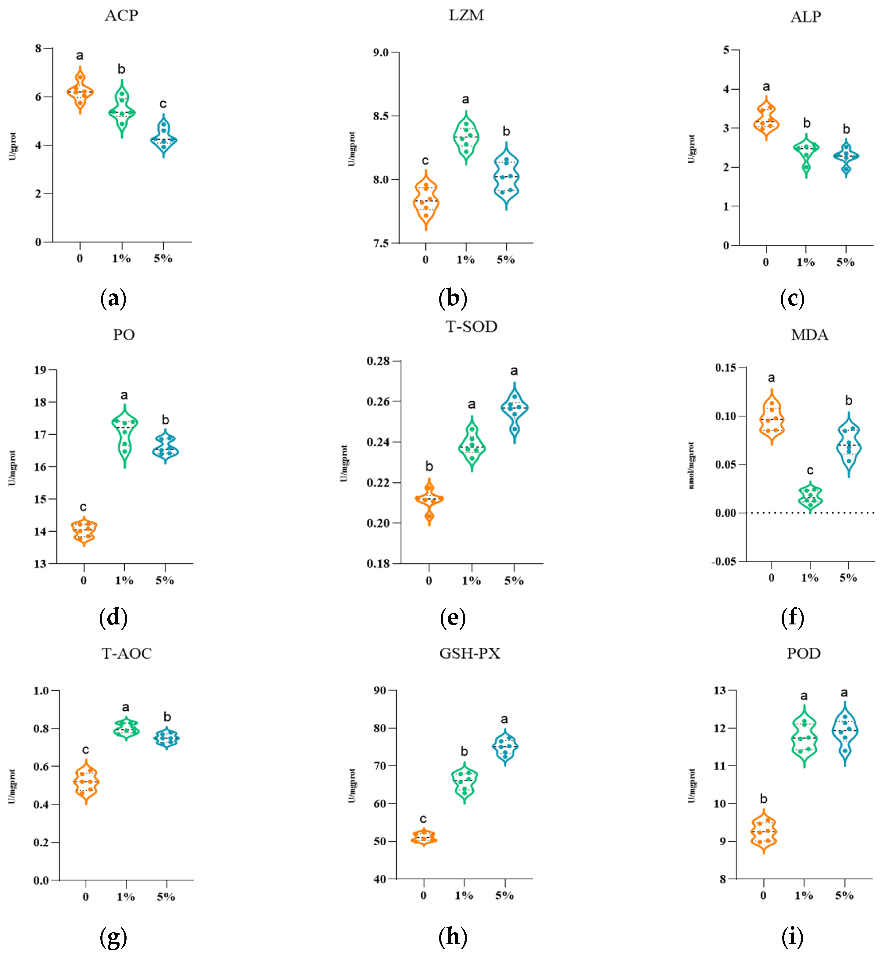
The effects of dietary CE supplementation at different levels on non-specific immune indices of L. vannamei are shown in Figure 2. The results indicated that ALP activity decreased with the increase in CE dosage. Both the 1% and 5% CE groups exhibited significantly lower ALP activity than the control group (p < 0.05), and no significant difference was found between the two groups (p > 0.05). ACP activity declined continuously with rising dietary CE levels, and the 5% CE group had notably lower ACP activity than all other groups (p < 0.05), with a decrease of 36.9% compared with the control group. Both PO and LZM activities increased firstly and then decreased as CE inclusion levels rose, and their values in the 1% CE group were significantly higher than those in other groups (p < 0.05). The results showed that the activities of T-SOD and POD increased with the rising CE inclusion level. These values were significantly higher in the 1% and 5% CE groups than those in the control group (p < 0.05), and no significant difference was detected between the two treatment groups. GPX−PX activity also showed an increasing trend, and its level in the 5% CE group was remarkably higher than that in other groups (p < 0.05). MDA content decreased first and then increased. The MDA contents were 0.016 nmol/mg and 0.071 nmol/mg in the 1% and 5% CE groups, which were both significantly lower than 0.097 nmol/mg in the control group (p < 0.05). T−AOC activity increased initially and then decreased with increased CE dosage, reaching the maximum value in the 1% CE group, which was significantly higher than that in the control group and 5% CE group.

3.3. CE Supplementation Modulates Inflammatory Responses in L. vannamei

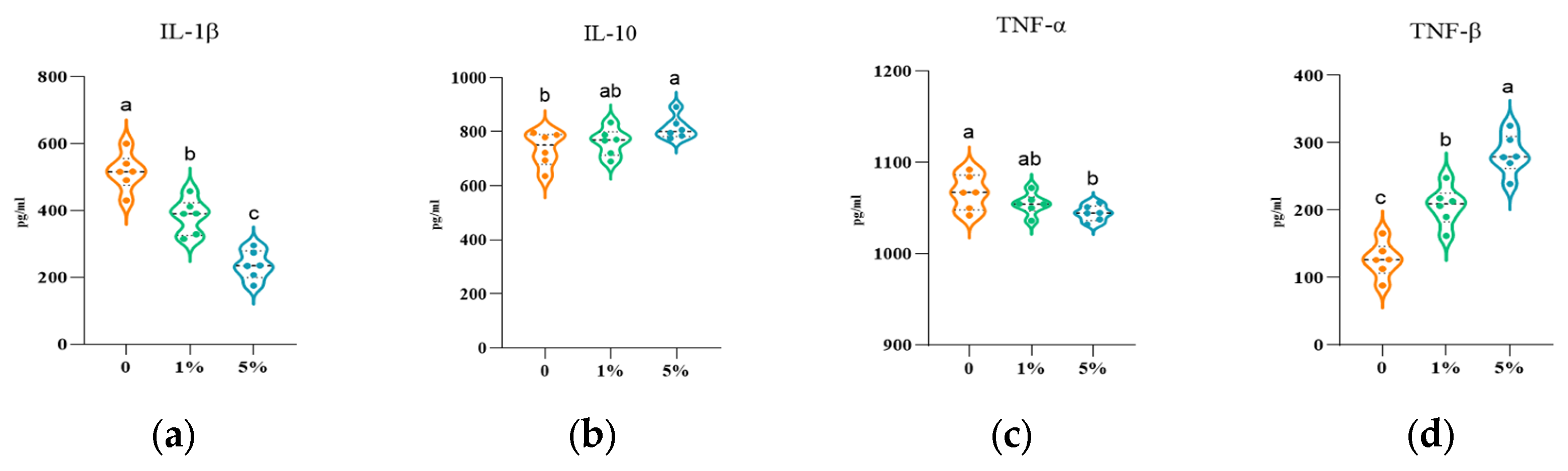
The regulation of inflammatory factors in L. vannamei by dietary CE supplementation is shown in Figure 3. In terms of anti-inflammatory factors, the relative expression level of IL-10 in the 5% CE group was significantly higher than that in the control group (p < 0.05); the 1% CE group showed a higher value than the control group with no significant difference. Compared with the control group, the relative expression of IL-10 in the 5% CE group increased by 10.59%. The relative expression of TNF−β presented an upward trend with the increasing dietary CE level, and the 5% CE group was significantly higher than the 1% CE group and the control group (p < 0.05). For pro-inflammatory factors, the relative expression of IL−1β in the 5% CE group was markedly lower than that in the control group and 1% CE group (p < 0.05). The 5% CE group had the lowest relative expression of TNF−α, which was significantly lower than that of the control group (p < 0.05).

3.4. Effects of Dietary Supplementation with Different Proportions of CE on the Expression of Immune-Related Genes in L. vannamei

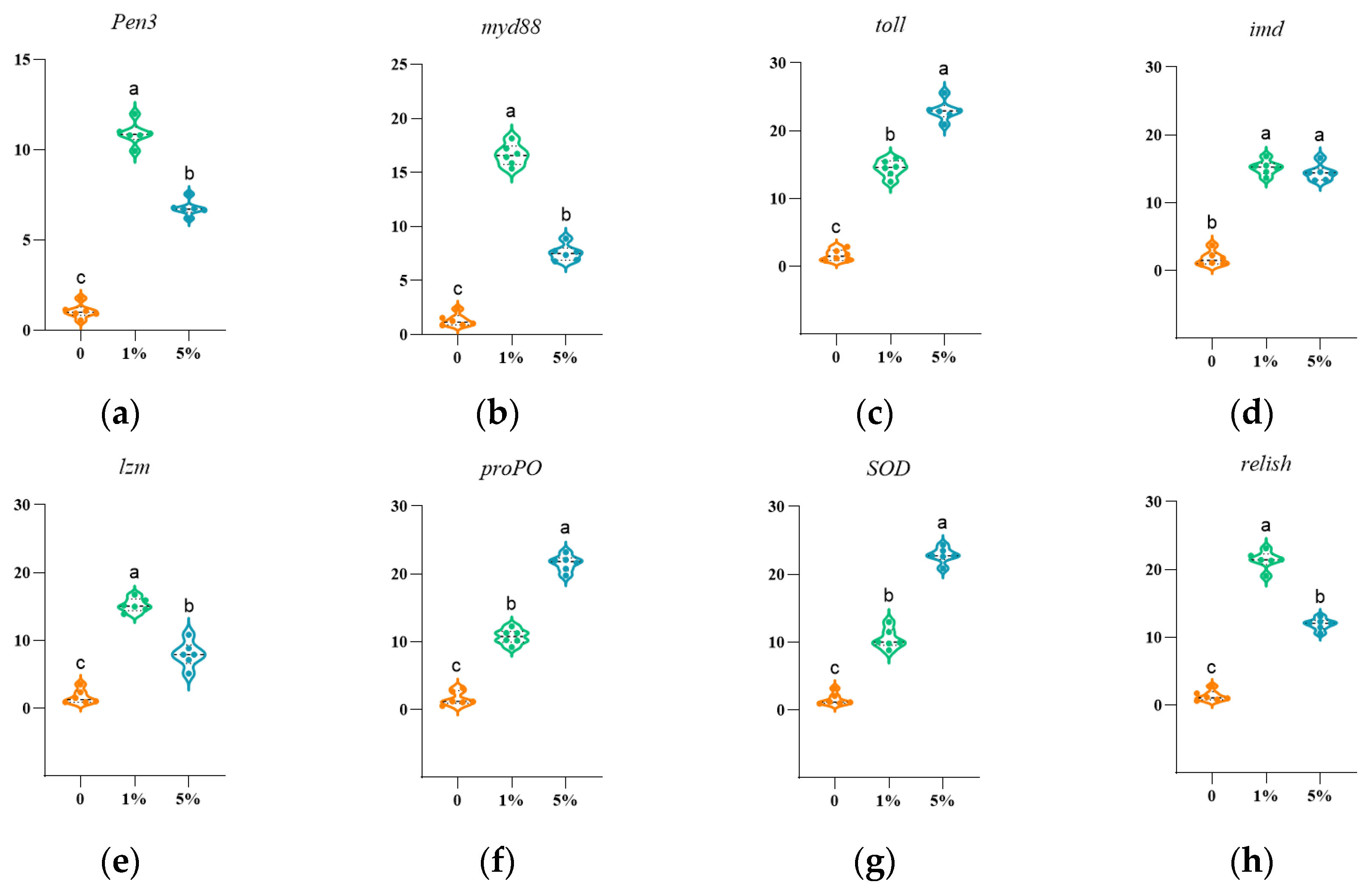
After the feeding trial, the relative expression levels of immune-related genes were determined in L. vannamei fed diets containing graded levels of CE. Significant differences in the mRNA abundances of pen3, myd88, toll, imd, lzm, propo, sod and relish were observed among dietary CE treatments (Figure 4). Compared with the control group, the relative expression of pen3, myd88, toll, imd, lzm, propo and relish was markedly upregulated in the 1% CE group (p < 0.05), with higher values than those obtained in the 5% CE group. Meanwhile, the 5% CE group exhibited significantly elevated sod expression relative to the control (p < 0.05), and its expression level was also higher than that in the 1% CE group.

3.5. Effects of Dietary CE Supplementation on Intestinal Histological Structure in L. vannamei

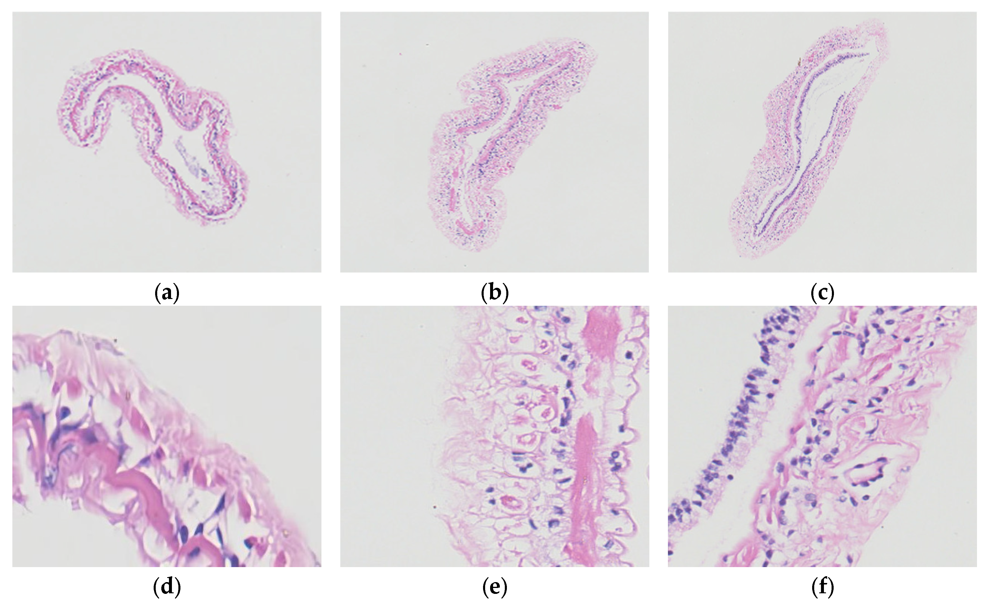
Observation on intestinal histological sections revealed that the midgut tissues of L. vannamei from the control, 1% CE and 5% CE groups maintained intact histological architecture, with the intestinal wall consisting of simple columnar epithelium, connective tissue and muscular layers from the inner to outer side, and no severe pathological lesions, including epithelial exfoliation, tissue necrosis or inflammatory exudation, were detected across all groups (Figure 5). In the control group, partial intestinal epithelial cells were loosely arranged with indistinct cellular boundaries and mild cytoplasmic swelling; slight thickening of partial mucosal layers, blurred texture of lamina propria connective tissue and mild tissue edema were also observed, accompanied by highly coiled and densely packed intestinal mucosal folds and a relatively narrow intestinal lumen. Compared with the control group, shrimp in the 1% CE group possessed intact and regularly arranged intestinal epithelial cells with mildly widened intercellular gaps but without obvious exfoliation or necrosis, and their intestinal mucosal folds presented natural morphology with moderate fold depth and quantity as well as well−preserved lumen structure. The 5% CE group exhibited favorable intestinal structural integrity characterized by tightly arranged epithelial cells with uniform and distinct nuclei, absent cellular degeneration, necrosis or exudation, evenly thickened mucosal layers and consistent tissue staining; intestinal mucosal folds in this group were relatively flat with low coiling degree and an expanded intestinal lumen. Collectively, intestinal tissues of shrimp from all three groups retained fundamental structural integrity, among which the 5% CE group displayed superior performance in epithelial cell arrangement, mucosal integrity and luminal morphology, whereas mild irregular epithelial arrangement and partial mucosal thickening were found in the control group.

3.6. Effects of Dietary Supplementation with Different Proportions of CE on Intestinal Microbiota of L. vannamei

Alpha diversity analysis. As shown in Table 3, dietary supplementation with different levels of CE affected the α−diversity indices of L. vannamei. The ACE and Chao indices in the 1% CE group were significantly lower than those in the other groups (p < 0.05), whereas no significant difference was observed between the control group and the 5% CE group (p > 0.05). The Shannon and Shannon-even indices of the control group were significantly higher than those of the 1% CE and 5% CE groups (p < 0.05), while there was no significant difference between the 1% CE group and the 5% CE group (p > 0.05).
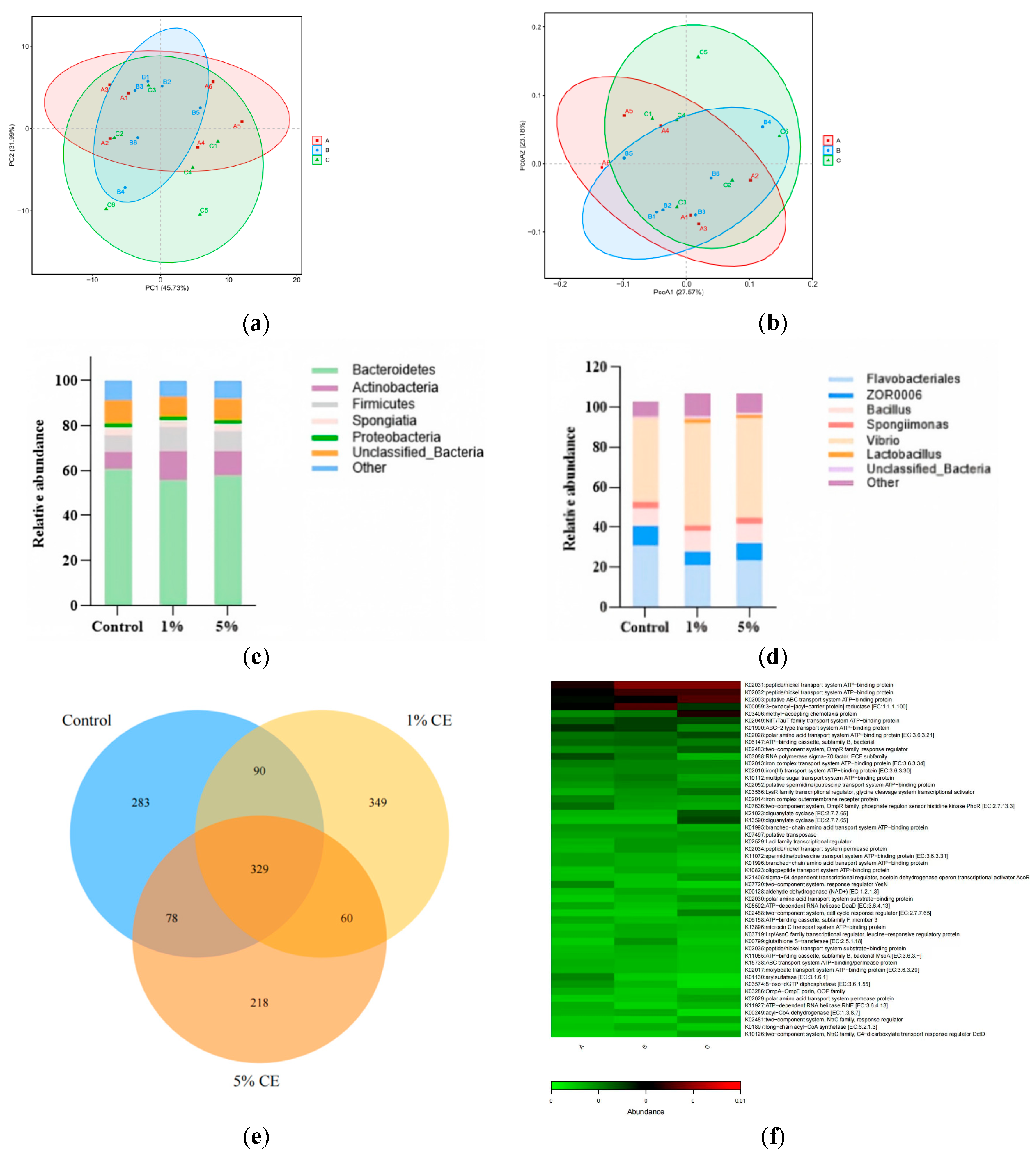
Venn and β-diversity. As shown in Figure 6e, the numbers of operational taxonomic units (OTUs) in the control, 1% CE and 5% CE groups were 780, 828 and 685, respectively, showing an initial increase followed by a decrease. A total of 329 shared OTUs were detected across all three groups, accounting for 42.18%, 39.73% and 48.03% of the total OTUs in control, 1% CE and 5% CE groups in turn. The numbers of unique OTUs in the three groups were 283, 349 and 218, which occupied 36.28%, 42.15% and 31.82% of their respective total OTUs. The PCoA results (Figure 6b) showed that the contribution rates of PCoA1 and PCoA2 were 27.57% and 23.18%, respectively, with a cumulative contribution rate of 50.75%. Meanwhile, the PCA plot illustrating the intestinal microbial community composition of Litopenaeus vannamei was constructed using principal component PC1 as the x−axis and principal component PC2 as the y−axis. Figure 6a revealed that samples from the control group and the 5% CE group were relatively scattered. Samples of the 1% CE group were mainly distributed near the intermediate region, indicating differences in the intestinal microbial structure of L. vannamei among different CE supplementation levels.
Relative species abundance. In this study, the relative abundance of microorganisms in all samples was analyzed and compared at the phylum and genus levels. As presented in Figure 6c, the OTUs of intestinal microbiota in L. vannamei were mainly composed of Bacteroidetes, Actinobacteria, Firmicutes, Porifera and Proteobacteria after dietary supplementation with different levels of CE. Bacteroidetes, Actinobacteria and Firmicutes were the predominant phyla. Compared with the control group, the relative abundance of Bacteroidetes decreased by 7.89% in the 1% CE group and 4.57% in the 5% CE group. The relative abundance of Actinobacteria increased by 66.79% and 40.70% in the 1% CE group and 5% CE group, respectively. Meanwhile, the relative abundance of Firmicutes rose by 48.85% in the 1% CE group and 21.34% in the 5% CE group.
As shown in Figure 6d, the dominant genera at the genus level were Vibrio, Flavobacteriales, Bacillus and other bacterial genera. Compared with the control group, the relative abundance of Vibrio increased in both the 1% CE and 5% CE groups. The relative abundance of Flavobacteriales decreased by 31.88% and 17.68% in the 1% CE and 5% CE groups, respectively. The relative abundance of Bacillus increased by 17.82% and 9.42% in the two CE supplemented groups, and the relative abundance of other bacterial genera was also elevated in all treatment groups.
Functional analysis of intestinal microbiota. Tax4Fun2 prediction indicated differences in the potential functional profiles of the intestinal microbiota among control, 1% CE, and 5% CE groups (Figure 6f). Compared with the control group, both CE−supplemented groups showed higher predicted abundances of functions related to substance transport. These pathways include peptide/nickel transport system, ABC transport system, amino acid transport system, iron transport system and sugar transport system. These results suggested potential differences in microbial functions associated with the transport of peptides, amino acids, carbohydrates, and metal ions. The 1% CE group showed higher predicted abundances of functions related to peptide/nickel transport and fatty acid metabolism. In contrast, the 5% CE group showed higher predicted abundances of functions related to environmental sensing and signal regulation, including chemotactic proteins, two-component regulatory systems, and diguanylate cyclase. High−dose CE further modulates colonization, motility and environmental adaptability of intestinal microbiota. Overall, CE supplementation modulates nutrient metabolism, transmembrane transport and signal response of intestinal microbes. It thereby improves intestinal microecology and gut health of L. vannamei.

4. Discussion

4.1. Regulatory Effects of CE on Lipid Metabolism and Cholesterol Homeostasis in L. vannamei

Dietary cholesterol participates in lipid absorption and transport. It also acts as a critical precursor for cell membrane construction and molting-related steroid hormone synthesis. Previous studies confirm shrimp lipids circulate in hemolymph as lipoproteins. Lipids such as cholesterol are mainly stored in the hepatopancreas. Changes in hemolymph LDL, HDL, TG and CHO serve as key phenotypic markers to evaluate how feed additives regulate lipid absorption, transport and deposition [22]. In this study, CE supplementation generally decreased LDL and increased HDL levels. LDL transports cholesterol from the hepatopancreas to peripheral tissues, while HDL mediates cholesterol reverse transport and subsequent metabolism in the hepatopancreas [23]. Thus, the simultaneous decrease in LDL and increase in HDL suggests that CE may enhance cholesterol reverse transport and limit excessive lipid accumulation in the hemolymph. TG levels were reduced in 1% and 5% CE groups, respectively, indicating a relatively stable triglyceride-lowering effect. This finding is consistent with previous studies showing that Chlorella can reduce serum and hepatopancreatic lipid levels and promote lipid excretion in high−fat diet models [24]. CE may therefore regulate triglyceride metabolism by reducing intestinal lipid absorption or enhancing lipid utilization and excretion. CHO exhibited a nonlinear response, reaching its lowest level in the 1% CE group, whereas the 5% CE treatment produced more pronounced changes in LDL and HDL. These results indicate that the optimal CE inclusion level may differ among lipid−related indicators. Similarly, dietary phytosterols and other bioactive compounds derived from algae or plants have been reported to regulate cholesterol transport and deposition in shrimp [25]. Mechanistically, CE has been shown to activate AMPK−α while suppressing SREBP−1c, HMG−CoA, and ACC expression in high−fat models [26]. These regulatory effects suggest that CE may improve lipid homeostasis by promoting energy metabolism and inhibiting fatty acid and cholesterol synthesis. The changes in lipid-related indicators may be partly associated with the phenolic compounds present in CE. In L. vannamei, dietary dihydromyricetin reduced T−CHO, TG, and LDL levels by promoting fatty acid β−oxidation and triglyceride catabolism [27]. These findings suggest that the phenolic constituents of CE may have contributed to the regulation of LDL, TG, and CHO levels observed in the present study. In conclusion, dietary CE effectively improves lipid metabolism in L. vannamei. It reduces LDL and TG, elevates HDL, and lowers CHO at low supplementation levels. CE has promising application potential in functional shrimp feeds to regulate lipid metabolism and protect hepatopancreas health.

4.2. Regulatory Effects of CE on Non-Specific Immunity and Antioxidant Capacity of L. vannamei

L. vannamei is an invertebrate. Its host defense mainly relies on non-specific immune processes mediated by hemocytes. These processes include phagocytosis, encapsulation, agglutination, bactericidal activity and humoral immune enzymes [28]. Among immune factors, the proPO activating cascade serves as a key pathway against pathogen invasion in crustaceans. Higher PO activity usually indicates stronger melanization, pathogen recognition and pathogen clearance capacity [29]. In this study, the 1% CE group showed significantly higher PO and LZM activities than the control and 5% CE groups. Moderate CE supplementation increased PO and LZM activities, suggesting enhanced humoral immune-related activity and potential immunomodulatory effects in shrimp. Consistent with our results, treatment with Gracilaria tenuistipitata extract elevates PO, SOD and lysozyme levels in L. vannamei. Treated shrimp also exhibit faster immune recovery under ammonia nitrogen stress [30]. In contrast, ALP activity decreased with increasing CE levels, while ACP activity was lowest in the 5% CE group. However, these decreases do not necessarily indicate immune suppression. Previous studies have shown that elevated ALP and ACP activities during Vibrio infection may be associated with tissue damage, infection stress, or increased lysosomal enzyme release rather than improved immune function [31]. Therefore, the concurrent increase in PO and LZM and decrease in phosphatase activities in the 1% CE group may reflect a more stable immune status rather than excessive immune stimulation. By contrast, the 5% CE group had lower PO and LZM activities than the 1% CE group. The different responses observed at 1% and 5% CE indicate that the effects of CE on non−specific immune-related indicators may depend on the inclusion level. Carrageenan immunostimulation research supports this pattern. Shrimp immune parameters rise to a plateau and then decline after stimulation. This change reflects intrinsic immune homeostasis regulation [32]. Antioxidant defense is closely linked to non-specific immunity, as excessive reactive oxygen species can induce lipid peroxidation and impair immune function. CE supplementation enhanced the activities of T-SOD, POD, and GSH−PX while reducing MDA levels, suggesting improved free−radical scavenging and reduced lipid peroxidation. Comparable antioxidant effects have been observed in L. vannamei supplemented with Gracilaria verrucosa [33]. The higher GSH-PX activity observed in the 5% CE group suggests a stronger response to this specific antioxidant enzyme. Published data confirm shrimp GPX genes participate in oxidative stress response and pathogen resistance [34]. This evidence highlights the value of elevated GSH−PX for antioxidant and immune homeostasis maintenance. Nevertheless, T-AOC peaked in the 1% CE group and dropped in the 5% CE group. High−dose CE boosts individual antioxidant enzymes but fails to raise total antioxidant capacity synchronously. Studies on plant composite additives draw similar conclusions. Optimal inclusion levels outperform high doses for immune enzyme activity, hepatopancreas and intestinal health, and anti-vibrio capacity [35]. Carotenoids present in CE, such as astaxanthin and lutein, may also contribute to its antioxidant and immunomodulatory effects. Previous studies have shown that dietary astaxanthin supplementation increases SOD, CAT, GPx, T-AOC, LZM, and PO activities while reducing MDA levels in L. vannamei. Dietary lutein may exert similar effects by modulating lipid peroxidation and Relish pathway−associated inflammatory responses [36,37]. These findings suggest that carotenoids in CE may be among the active constituents contributing to the improved antioxidant status and altered inflammatory signaling observed in the present study. In summary, CE produced inclusion-level-dependent changes in immune and antioxidant−related indicators. The 1% CE treatment showed stronger responses in PO, LZM, and T−AOC, whereas the 5% CE treatment produced the highest GSH−PX activity. Among the two tested CE inclusion levels, the 1% CE treatment produced stronger responses in several immune and antioxidant−related indicators under the present experimental conditions.

4.3. Effects of CE on the Expression of Inflammatory Factors and Immune Homeostasis in L. vannamei

Changes in inflammatory factor expression serve as a key indicator to evaluate immune homeostasis in L. vannamei. Crustaceans lack adaptive immunity and rely mainly on innate immunity to recognize and resist pathogens [38]. Thus, the function of feed additives is not limited to immune enhancement. It is more critical to explore their capacity to balance pro-inflammatory and anti-inflammatory responses and reduce tissue damage caused by persistent inflammation. Existing studies confirm that functional feed additives can regulate inflammatory responses in L. vannamei. For instance, andrographolide suppresses the upregulation of TNF−α and IL−1β triggered by Vibrio harveyi infection and alleviates tissue injury [39]. Previous research has used immune−related genes including IL-1β, IL−10 and TNF−α to assess dietary effects in L. vannamei. These works highlight the vital roles of IL family genes in innate immune regulation of invertebrates [40]. In the present study, 5% CE supplementation significantly upregulated the anti-inflammatory factor IL−10, indicating an enhanced capacity to control inflammatory responses. IL−10 is a critical anti-inflammatory cytokine that restrains inflammatory reactions and reduces host tissue damage [41]. The 5% CE group exhibited elevated levels of TNF−β. Concurrent increases in IL−10 indicated that 5% CE could induce the expression of immunoregulatory cytokines. More importantly, the concentrations of pro−inflammatory factors IL−1β and TNF−α decreased significantly in the 5% CE group. These results suggest that the 5% CE treatment was associated with a more pronounced anti−inflammatory profile. Studies on aquatic animals report consistent regulatory patterns as follows: functional feed additives upregulate Il−10 and downregulate TNF−α [42], matching our data. Another study on macroalgae diets for L. vannamei states optimal dietary supplementation reduces intestinal expression of inflammation−related genes (TNF−α, IL−1β, IL−6, and IL−8) after WSSV challenge [43]. These findings support the use of reduced pro-inflammatory gene expression as an indicator of the anti−inflammatory potential of functional feed ingredients. In conclusion, appropriate CE supplementation modulates inflammatory factor expression in L. vannamei. It mitigates excessive pro-inflammatory responses and maintains immune homeostasis. These findings suggest that CE holds potential as an immunomodulatory feed additive for shrimp diets.

4.4. Regulatory Effects of Dietary CE Supplementation at Different Inclusion Levels on the Expression of Immune−Related Genes in L. vannamei

L. vannamei lacks a canonical adaptive immune system. It relies entirely on innate immune defenses against pathogens, including pattern recognition, signal transduction, antimicrobial peptide synthesis, prophenoloxidase cascade and antioxidant responses. In the present study, 1% CE supplementation broadly upregulated the immune-related genes pen3, myd88, toll, imd, lzm, propo, and relish, with generally stronger responses than those observed at 5% CE. These results suggest that moderate CE supplementation is associated with the coordinated upregulation of genes involved in innate immune recognition, signaling, and effector functions. Myd88 encodes a critical adaptor protein in the Toll signaling cascade. Zhang et al. [44] demonstrated LvMyD88 responds to lipopolysaccharide, bacterial and viral challenges. Its overexpression triggers antimicrobial peptide genes in shrimp. Thus, the concurrent upregulation of toll and myd88 suggests that 1% CE may be associated with the transcriptional modulation of the Toll–MyD88 signaling axis. Similarly, the elevated expression of imd and relish indicates that the IMD–Relish pathway may also be transcriptionally responsive to CE supplementation. Wang et al. [45] validated LvIMD induces the expression of antimicrobial peptides such as penaeidin. The parallel upregulation of imd, relish, and pen3 is consistent with a coordinated transcriptional response involving immune signaling and antimicrobial effector-related genes. Li et al. [46] reported L. vannamei Toll4 activates Dorsal to induce lysozyme and strengthen pathogen resistance. This finding provides mechanistic context for the concurrent upregulation of toll and lzm observed in the present study. Beyond antimicrobial peptide and lysozyme-related genes, the elevated expression of proPO suggests that 1% CE may influence the prophenoloxidase system at the transcriptional level. Histological expression studies confirm PEN-3, lysozyme and proPO are mainly synthesized by circulating and tissue−infiltrating hemocytes. These molecules form core components of shrimp humoral immunity [47]. Therefore, the simultaneous upregulation of pen3, lzm, and proPO suggests that CE may modulate multiple immune effector-related genes rather than affecting a single immune marker. Licona−Jain et al. [48] found approximately 1.1% marine yeast supplementation elevates immune parameters, including LYZ, SOD and CAT, and modulates transcription of PEN, LYZ and proPO. This is consistent with the broader and more coordinated transcriptional response of immune-related genes observed at the lower CE inclusion level in the present study. In contrast, the 5% CE treatment was more strongly associated with sod upregulation, whereas the 1% CE treatment induced broader changes in genes related to immune recognition, signaling, and effector functions. Ghaffarizadeh et al. [49] similarly reported nonlinear dose responses in shrimp supplemented with selenium nanoparticles, with excessive inclusion impairing some immune and antioxidant parameters. These findings indicate that the beneficial effects of functional feed additives do not necessarily increase linearly with dosage. Overall, under the present experimental conditions, the 1% CE treatment induced broader upregulation of immune−related genes than the 5% CE treatment. The coordinated transcriptional changes in toll, myd88, imd, relish, pen3, lzm, and proPO indicate that 1% CE has potential to modulate innate immune recognition, signaling, and effector-related processes in L. vannamei.

4.5. Effects of Graded Dietary CE Supplementation on Intestinal Health in L. vannamei

The intestine serves as the primary site for nutrient digestion and absorption in L.vannamei. It also acts as a critical physical barrier against dietary irritants and aquatic pathogenic microbes. Epithelial integrity, mucosal completeness and intestinal patency directly reflect how feed additives modulate shrimp intestinal health. Previous studies confirm Chlorella reshapes beneficial intestinal microbes and regulates digestion, immunity and nutrient utilization. Chlorella−derived compounds stabilize intestinal microenvironment [50]. Midgut tissues of the control, 1% CE and 5% CE groups shared the following identical basic structures: simple columnar epithelium, connective tissue and muscular layers. No severe epithelial shedding, tissue necrosis or inflammatory exudation was observed. CE within the tested dosages caused no obvious intestinal histological damage to shrimp. By contrast, partial epithelial cells in the control group were loosely arranged with indistinct cell boundaries. Mild cytoplasmic swelling, blurred lamina propria texture and tissue edema were also detected. Basal diet alone induced slight sub-health or stress in intestinal mucosa. Compared with the control, epithelial cells in the 1% CE group arranged more regularly. Minor widening of intercellular spaces existed without shedding or necrosis. Mucosal folds possessed moderate depth and abundance, and intestinal lumens remained intact. Low−dose CE stabilized mucosal structure without abnormal tissue hyperplasia. Consistent with our observation, Chen et al. reported combined mannan–oligosaccharide and Bacillus licheniformis supplementation raised the number and height of intestinal folds and optimized short−chain fatty acid profiles in white shrimp [51]. Functional feed additives restore mucosal architecture by improving gut microecology and epithelial nutrition. More prominent histological improvements were detected in the 5% CE group. Epithelial cells tightly aligned with uniform, well−defined nuclei. No cellular degeneration, necrosis or inflammatory exudate occurred. The mucosa exhibited even thickness and homogeneous staining. A total of 5% CE better sustained epithelial homeostasis and mucosal barrier integrity. These findings agree with Fang et al.’s results. Dietary 5% filamentous microalgae Klebsormidium sp. significantly increased midgut mucosal thickness and fold height in L. vannamei [52]. Microalgal fatty acids, pigments, polysaccharides and other bioactive compounds jointly nourish and protect intestinal epithelium. Astaxanthin has been reported to preserve intestinal epithelial integrity in L. vannamei under environmental stress [53]. Accordingly, carotenoids in CE may have contributed to the improved mucosal organization and intestinal integrity observed in the 5% CE group. Yao et al. found dietary Schizochytrium in low−fishmeal diets lengthened and widened intestinal folds and thickened muscular layers. The additive alleviated swelling of endoplasmic reticulum and mitochondria in epithelial cells [54]. Algal supplements remodel intestinal histology and regulate membrane lipid composition, organelle balance and oxidative stress. The 5% CE group displayed relatively flat mucosal folds with reduced convolution and expanded intestinal lumens. This phenomenon partially matched Abdel−Rahim et al.’s report as follows: dietary sargassum and nucleotides elevated epithelial cell counts and widened intestinal cavities [55]. No degeneration, necrosis or exudation was found in the 5% CE group. The enlarged lumen likely originated from relieved mucosal edema and abnormal contraction, facilitating chyme transit and stabilizing intestinal morphology. In summary, CE supplementation did not disrupt the fundamental midgut structure of L. vannamei. The 1% CE group mainly preserved mucosal morphology and alleviated disorganized epithelium. The 5% CE group generated the following superior effects: regular epithelial arrangement, relieved cytoplasmic and tissue swelling, intact mucosal continuity and optimized lumen structure. Moderately elevated CE inclusion strengthens the structural stability of shrimp intestinal mucosa.
Intestinal microbiota rapidly responds to shifts in dietary composition. Analysis of community structure and potential functions helps clarify whether CE improves shrimp health by regulating intestinal microenvironment in L. vannamei. Published studies confirm lipid sources and lipid profiles in feeds alter intestinal bacterial communities of L. vannamei. Different feed ingredients exert selective pressure on gut microbiota [56]. In this trial, the ACE and Chao indices in the 1% CE group were significantly lower than those in the control and 5% CE groups, indicating reduced intestinal microbial richness. In contrast, the 5% CE group maintained richness comparable to the control. Both CE groups showed significantly lower Shannon and Shannon-even indices than the control, suggesting decreased microbial diversity and evenness. For the 1% CE group, the reduction in diversity may have resulted from declines in both richness and evenness, whereas in the 5% CE group it was mainly associated with less even distribution of bacterial abundance. This pattern may reflect the selective enrichment of certain taxa. Similarly, Qiao et al. found that only an appropriate β−glucan level improved microbial diversity in L. vannamei, while higher levels produced no comparable benefit [57]. These findings indicate that CE-associated changes in the intestinal microbiota differed between the two tested inclusion levels.
In terms of microbial composition, CE reduced the relative abundance of Bacteroidetes in this trial. Actinobacteria and Firmicutes rose markedly instead. The 1% CE group showed more dramatic shifts than the 5% CE group. CE supplementation was associated with changes in the relative abundance of several Gram−positive bacterial taxa. These shifts may be related to the availability of polysaccharides, proteins, and other components in CE. Duan et al. supplemented Clostridium butyricum in diets for L. vannamei. The treatment elevated the abundance of beneficial taxa including Firmicutes and Bacillus. It also strengthened intestinal microbial capacity to metabolize carbohydrates, amino acids and polymers [58]. Their findings align with our data. Bacillus abundance increased by 17.82% and 9.42% in the two CE groups. Functions related to peptide, amino acid, carbohydrate and ABC transport systems were enriched accordingly. CE boosts nutrient transport and utilization by enriching certain Firmicutes species. Imaizumi et al. [59] validated dietary Bacillus amyloliquefaciens reshapes intestinal microbial structure of L. vannamei. The supplementation improved shrimp survival after challenge with pathogenic Vibrio parahaemolyticus. The increased relative abundance of Bacillus may represent a potentially favorable microbial response because some members of this genus have probiotic properties.
On the other hand, the relative abundance of Vibrio increased in both CE groups. This shift cannot be directly interpreted as impaired intestinal health. Vibrio is a ubiquitous genus in marine aquaculture environments and the intestines of L. vannamei [60]. This genus contains both potential pathogenic species and non−pathogenic strains that decompose organic matter and utilize nutrients [61]. Variations in genus−level relative abundance alone cannot accurately evaluate its actual impacts on host health. Chang et al. [62] reported both pathogenic and non-pathogenic Vibrio parahaemolyticus alter intestinal microbial diversity of white shrimp. Only pathogenic infection induced remarkable enrichment of Vibrio and Photobacterium, accompanied by suppressed normal metabolic functions. In the present trial, elevated Vibrio abundance was detected alongside higher proportions of Bacillus, Actinobacteria and Firmicutes. Functional pathways for the transport of amino acids, carbohydrates, peptides and metal ions were also enhanced. The microbial shifts reflected community rearrangement driven by nutritional components of CE, rather than uncontrolled proliferation of pathogenic bacteria. Collectively, dietary CE remodels the composition and metabolic potential of intestinal microbiota in L.vannamei. Its main effects include rebalancing dominant microbial taxa and boosting nutrient transport capacity.
Tax4Fun2 functional prediction demonstrates dietary CE reshapes intestinal microbial composition and elevates microbial functions linked to nutrient utilization and environmental adaptation in L. vannamei. Compared with the control group, both CE groups showed enhanced functions of peptide/nickel transport system, ABC transport system, amino acid transport system, iron transport system and sugar transport system. CE improves the capacity of intestinal microbiota to digest and utilize dietary nutrients. Gao et al. identified abundant functional genes related to nutrient digestion in shrimp intestinal microbes. Certain Vibrio and Pseudoalteromonas strains participate in the degradation and utilization of nutrients [63]. Combined with the increased relative abundance of Bacillus, Actinobacteria and Firmicutes in this trial, proteins, polysaccharides and other bioactive compounds in CE provide suitable substrates for intestinal microbes. These substrates stimulate the proliferation of taxa responsible for nutrient transport and metabolism. Distinct functional discrepancies existed between two CE dosages. The 1% CE group was enriched in pathways of peptide/nickel transport and fatty acid metabolism, facilitating the utilization of nitrogen-containing substrates and lipids. The 5% CE group further upregulated functions of chemotactic proteins, two-component regulatory systems and diguanylate cyclase. High−dose CE strengthens microbial sensing, response and colonization under variable intestinal nutrient conditions. The 5% CE group possessed higher ACE and Chao indices than the control, as well as a greater Shannon index than the 1% CE group. A total of 5% CE preserves microbial richness and exerts comprehensive regulatory effects on nutrient transport and environmental adaptation. Tax4Fun2 outputs only predict potential functions instead of actual gene expression levels [64]. Nevertheless, simultaneous enrichment of diverse transport, metabolic and signal regulatory pathways supports the conclusion. CE optimizes intestinal microecology of L. vannamei by boosting microbial nutrient utilization and environmental adaptability.

4.6. Comprehensive Evaluation of Dietary CE Supplementation in L. vannamei

The present results revealed that the 1% CE group exhibited superior PO and LZM activities, higher T−AOC levels, and upregulated transcription of immune-related genes including toll, imd and proPO. Meanwhile, MDA content decreased significantly. These findings suggest that the 1% CE treatment was associated with favorable changes in several innate immune-related indicators and may contribute to the maintenance of antioxidant and redox homeostasis. In comparison, the 5% CE group achieved more prominent effects in reducing LDL and TG concentrations and elevating HDL levels as well as GPX-PX activity. This treatment upregulated IL−10 and TNF−β and suppressed IL−1β and TNF−α, thereby stabilizing inflammatory responses. Furthermore, shrimp fed 5% CE displayed tightly arranged intestinal epithelial cells and intact mucosal architecture and luminal morphology. Microbial functions associated with substance transport, environmental sensing and signal regulation were also enriched in the intestine. It follows that CE exerts physiological regulation through synergistic modulation rather than altering a single biomarker. CE coordinates lipid transport, antioxidant defense, immune signaling and intestinal microenvironment to sustain physiological homeostasis of white shrimp. From an aquaculture production perspective, the coordinated improvements in antioxidant defense, innate immune responses, inflammatory regulation, and intestinal barrier integrity observed in the present study may contribute to enhanced physiological resilience of L. vannamei under routine culture stressors. Supporting the broader aquaculture relevance of C. vulgaris−derived products, our previous study using a hot−water extract showed that 1% dietary supplementation enhanced immune and antioxidant-related responses and attenuated the physiological stress response of L. vannamei under non-ionic ammonia exposure [65]. Together, these findings suggest that C. vulgaris−derived extracts have practical potential as functional feed additives for maintaining shrimp health under variable aquaculture conditions. Nevertheless, bacterial challenge, long-term survival, and performance under commercial culture conditions were not directly evaluated in the present study and warrant further investigation.
Existing studies have demonstrated that natural bioactive substances such as Haematococcus pluvialis and phlorotannins improve antioxidant and anti-inflammatory status, facilitate metabolic regulation and maintain intestinal structural integrity in L. vannamei [66,67]. Glutathione supplementation enhances systemic antioxidant capacity and optimizes intestinal morphology [68]. Tributyrin and compound probiotics also elevate shrimp health by modulating immune responses, intestinal microbiota and tissue structure [69,70]. Accordingly, these findings partially confirm our hypothesis that CE affects immune function, antioxidant capacity, lipid metabolism and intestinal health depending on its dietary inclusion level. Taken together, these findings support the potential physiological and immunomodulatory value of CE in L. vannamei. Under the present experimental conditions, the 1% CE treatment produced stronger responses in several immune and antioxidant-related indicators, whereas the 5% CE treatment showed more pronounced changes in inflammatory-, lipid–metabolism-, and intestinal health-related indicators.

5. Conclusions

This 60-day feeding trial illustrated regulatory effects of graded dietary CE on immune homeostasis, oxidative status, lipid metabolism and intestinal microenvironment in L. vannamei. Low−dose CE mainly boosts shrimp innate immune responses and antioxidant defense. It also enriches beneficial microbes and their nutrient metabolic functions. High-dose CE generates stronger effects on inhibiting inflammation, optimizing lipid transport and utilization, and stabilizing intestinal histological structure and microecology. Based on the composition of a comparable 90% aqueous-ethanol extract of C. vulgaris, the 1% and 5% diets were estimated to provide approximately 182 and 912 mg GAE kg−1 of total phenolics, 99 and 496 mg kg−1 of total carotenoids, 34 and 170 mg kg−1 of lutein, and 4.3 and 21.5 mg kg−1 of astaxanthin, respectively. Collectively, CE coordinates immunity, antioxidant capacity, metabolism and intestinal health to maintain physiological homeostasis of white shrimp. Functional priorities differ between two inclusion dosages. Appropriate CE levels should be selected according to practical breeding demands. The 1% CE dosage works better for enhancing immunity and antioxidant capacity. The 5% CE dosage presents prominent advantages in anti-inflammation, lipid regulation and intestinal protection.

Author Contributions

Conceptualization, X.J. and D.Z.; Methodology, A.P., W.Y. and X.J.; Validation, A.P. and W.Y.; Formal Analysis, A.P., D.Z. and W.Y.; Investigation, X.J., D.Z. and P.S.; Resources, X.J., P.S. and W.Z.; Data Curation, A.P., W.Y. and P.S.; Writing—Original Draft Preparation, A.P.; Writing—Review and Editing, X.J., D.Z., A.P. and W.Z.; Visualization, A.P. and W.Y.; Supervision, X.J. and W.Z.; Project Administration, X.J.; Funding Acquisition, X.J. All authors have read and agreed to the published version of the manuscript.

Funding

This work was supported by the National Natural Science Foundation of China (No. 32303038), National key research and development program of China (2024YFD2401803), and Tianjin mariculture industry technology system innovation team construction project (ITTMRS2026000).

Institutional Review Board Statement

The Animal Ethics Committee’s scope of approval is limited to studies involving vertebrates (Laboratory Animal Act, Chapter 5Article 25). Our study is on invertebrate species and does not need ethical approval in China. The study was conducted in accordance with the local legislation and institutional requirements.

Data Availability Statement

The original contributions presented in this study are included in the article. Further inquiries can be directed to the corresponding author.

Acknowledgments

We are grateful to the Tianjin Agricultural University, Fisheries College, Tianjin Key Laboratory of Aqua-Ecology and Aquaculture for providing technical assistance.

Conflicts of Interest

The authors declare that they have no known competing financial interests or personal relationships that could have appeared to influence the work reported in this paper.

References

  1. Safi, C.; Zebib, B.; Merah, O.; Pontalier, P.-Y.; Vaca-Garcia, C. Morphology, composition, production, processing and applications of Chlorella vulgaris: A review. Renew. Sustain. Energy Rev. 2014, 35, 265–278. [Google Scholar] [CrossRef] [Scilit]
  2. Mendes, A.R.; Spínola, M.P.; Lordelo, M.; Prates, J.A.M. Chemical compounds, bioactivities, and applications of Chlorella vulgaris in food, feed and medicine. Appl. Sci. 2024, 14, 10810. [Google Scholar] [CrossRef] [Scilit]
  3. Plaza, M.; Santoyo, S.; Jaime, L.; Avalo, B.; Cifuentes, A.; Reglero, G.; Reina, G.G.-B.; Señoráns, F.J.; Ibáñez, E. Comprehensive characterization of the functional activities of pressurized liquid and ultrasound-assisted extracts from Chlorella vulgaris. LWT-Food Sci. Technol. 2012, 46, 245–253. [Google Scholar] [CrossRef] [Scilit]
  4. Hsu, H.Y.; Jeyashoke, N.; Yeh, C.H.; Song, Y.-J.; Hua, K.-F.; Chao, L.K. Immunostimulatory bioactivity of algal polysaccharides from Chlorella pyrenoidosa activates macrophages via Toll-like receptor 4. J. Agric. Food Chem. 2010, 58, 927–936. [Google Scholar] [CrossRef] [Scilit] [PubMed]
  5. Cha, K.H.; Lee, H.J.; Koo, S.Y.; Song, D.G.; Lee, D.U.; Pan, C.H. Optimization of pressurized liquid extraction of carotenoids and chlorophylls from Chlorella vulgaris. J. Agric. Food Chem. 2010, 58, 793–797. [Google Scholar]
  6. Qi, J.; Kim, S.M. Characterization and immunomodulatory activities of polysaccharides extracted from green alga Chlorella ellipsoidea. Int. J. Biol. Macromol. 2017, 95, 106–114. [Google Scholar]
  7. Ahmad, M.T.; Shariff, M.; Md Yusoff, F.; Goh, Y.M.; Banerjee, S. Applications of microalga Chlorella vulgaris in aquaculture. Rev. Aquac. 2020, 12, 328–346. [Google Scholar] [CrossRef] [Scilit]
  8. Ma, M.; Hu, Q. Microalgae as feed sources and feed additives for sustainable aquaculture: Prospects and challenges. Rev. Aquac. 2024, 16, 818–835. [Google Scholar] [CrossRef] [Scilit]
  9. Bai, S.C.; Koo, J.W.; Kim, K.W. Effects of Chlorella powder as a feed additive on growth performance in juvenile Korean rockfish, Sebastes schlegeli (Hilgendorf). Aquac. Res. 2001, 32, 92–98. [Google Scholar] [CrossRef] [Scilit]
  10. Safari, O.; Paolucci, M.; Motlagh, H.A. Dietary supplementation of Chlorella vulgaris improved growth performance, immunity, intestinal microbiota and stress resistance of juvenile narrow clawed crayfish, Pontastacus leptodactylus Eschscholtz, 1823. Aquac. 2022, 554, 738138. [Google Scholar] [CrossRef] [Scilit]
  11. Abdel-Tawwab, M.; Mousa, M.A.A.; Mamoon, A.; Abdelghany, M.F.; Abdel-Hamid, E.A.; Abdel-Razek, N.; Ali, F.S.; Shady, S.H.; Gewida, A.G. Dietary Chlorella vulgaris modulates the performance, antioxidant capacity, innate immunity, and disease resistance capability of Nile tilapia fingerlings fed on plant-based diets. Anim. Feed. Sci. Technol. 2022, 283, 115181. [Google Scholar] [CrossRef] [Scilit]
  12. Pakravan, S.; Akbarzadeh, A.; Sajjadi, M.M.; Hajimoradloo, A.; Noori, F. Chlorella vulgaris meal improved growth performance, digestive enzyme activities, fatty acid composition and tolerance of hypoxia and ammonia stress in juvenile Pacific white shrimp Litopenaeus vannamei. Aquac. Nutr. 2018, 24, 594–604. [Google Scholar] [CrossRef] [Scilit]
  13. Liao, I.C.; Chien, Y.H. The pacific white shrimp, Litopenaeus vannamei, in Asia: The world’s most widely cultured alien crustacean. In The Wrong Place-Alien Marine Crustaceans: Distribution, Biology and Impacts; Springer: Dordrecht, The Netherlands, 2011; pp. 489–519. [Google Scholar]
  14. Flegel, T.W. Historic emergence, impact and current status of shrimp pathogens in Asia. J. Invertebr. Pathol. 2012, 110, 166–173. [Google Scholar] [CrossRef] [Scilit] [PubMed]
  15. Tassanakajon, A.; Somboonwiwat, K.; Supungul, P.; Tang, S. Discovery of immune molecules and their crucial functions in shrimp immunity. Fish Shellfish Immunol. 2013, 34, 954–967. [Google Scholar] [CrossRef] [Scilit] [PubMed]
  16. Han, S.; Wang, M.; Liu, M.; Wang, B.-J.; Jiang, K.-Y.; Wang, L. Comparative sensitivity of the hepatopancreas and midgut in the white shrimp Litopenaeus vannamei to oxidative stress under cyclic serious/medium hypoxia. Aquaculture 2018, 490, 44–52. [Google Scholar] [CrossRef] [Scilit]
  17. Duan, Y.; Zhong, G.; Nan, Y.; Yang, Y.; Xiao, M.; Li, H. Effects of nitrite stress on the antioxidant, immunity, energy metabolism, and microbial community status in the intestine of Litopenaeus vannamei. Antioxidants 2024, 13, 1318. [Google Scholar] [CrossRef] [Scilit] [PubMed]
  18. Duan, Y.; Wang, Y.; Liu, Q.; Dong, H.; Li, H.; Xiong, D.; Zhang, J. Changes in the intestine microbial, digestion and immunity of Litopenaeus vannamei in response to dietary resistant starch. Sci. Rep. 2019, 9, 6464. [Google Scholar] [CrossRef] [Scilit] [PubMed]
  19. Shen, K.; Bao, L.; Liu, M.; Lei, W.; Zhou, Q.; Ding, J.; Fang, P.; Hu, B.; Wen, C.; Kumar, V.; et al. Dietary supplementation of β-1, 3-glucan improves the intestinal health of white shrimp (Litopenaeus vannamei) by modulating intestinal microbiota and inhibiting inflammatory response. Front. Immunol. 2023, 14, 1119902. [Google Scholar] [CrossRef] [Scilit] [PubMed]
  20. Ma, Q.; Zhao, G.; Liu, J.; Chen, I.-T.; Wei, Y.; Liang, M.; Dai, P.; Nuez-Ortin, W.G.; Xu, H. Effects of a phytobiotic-based additive on the growth, hepatopancreas health, intestinal microbiota, and Vibrio parahaemolyticus resistance of Pacific white shrimp, Litopenaeus vannamei. Front. Immunol. 2024, 15, 1368444. [Google Scholar] [CrossRef] [Scilit] [PubMed]
  21. Savvidou, M.G.; Georgiopoulou, I.; Antoniou, N.; Tzima, S.; Kontou, M.; Louli, V.; Fatouros, C.; Magoulas, K.; Kolisis, F.N. Extracts from Chlorella vulgaris protect mesenchymal stromal cells from oxidative stress induced by hydrogen peroxide. Plants 2023, 12, 361. [Google Scholar] [CrossRef] [Scilit] [PubMed]
  22. Yepiz-Plascencia, G.; Vargas-Albores, F.; Higuera-Ciapara, I. Penaeid shrimp hemolymph lipoproteins. Aquaculture 2000, 191, 177–189. [Google Scholar] [CrossRef] [Scilit]
  23. Limwachirakhom, R.; Triwutanon, S.; Zhang, Y.; Jintasataporn, O. Effects of dietary lysophospholipids on the performance of Pacific white shrimp (Litopenaeus vannamei) fed fish oil-and energy-reduced diets. Front. Mar. Sci. 2025, 12, 1624057. [Google Scholar] [CrossRef] [Scilit]
  24. Georgiopoulou, I.; Tzima, S.; Pappa, G.D.; Louli, V.; Voutsas, E.; Magoulas, K. Experimental Design and Optimization of Recovering Bioactive Compounds from Chlorella vulgaris through Conventional Extraction. Molecules 2022, 27, 29. [Google Scholar] [CrossRef] [Scilit] [PubMed]
  25. Chen, Y.; Pan, Z.; Li, X.; Yao, X.; He, G.; Xie, S. Evaluation of phytosterols as an alternative to cholesterol in practical diets on growth and nonspecific immunity of Litopenaeus vannamei. Aquac. Nutr. 2023, 2023, 7825559. [Google Scholar] [CrossRef] [Scilit] [PubMed]
  26. Wan, X.; Li, T.; Liu, D.; Chen, Y.; Liu, Y.; Liu, B.; Zhang, H.; Zhao, C. Effect of marine microalga Chlorella pyrenoidosa ethanol extract on lipid metabolism and gut microbiota composition in high-fat diet-fed rats. Mar. Drugs 2018, 16, 498. [Google Scholar] [CrossRef] [Scilit] [PubMed]
  27. Lu, M.; Liu, R.; Chen, Z.; Su, C.; Pan, L. Effects of dietary dihydromyricetin on growth performance, antioxidant capacity, immune response and intestinal microbiota of shrimp (Litopenaeus vannamei). Fish Shellfish Immunol. 2023, 142, 109086. [Google Scholar] [CrossRef] [Scilit] [PubMed]
  28. Vazquez, L.; Alpuche, J.; Maldonado, G.; Agundis, C.; Pereyra-Morales, A.; Zenteno, E. Immunity mechanisms in crustaceans. Innate Immun. 2009, 15, 179–188. [Google Scholar] [CrossRef] [Scilit] [PubMed]
  29. Amparyup, P.; Charoensapsri, W.; Tassanakajon, A. Prophenoloxidase system and its role in shrimp immune responses against major pathogens. Fish Shellfish Immunol. 2013, 34, 990–1001. [Google Scholar] [CrossRef] [Scilit] [PubMed]
  30. Chen, Y.Y.; Chen, J.C.; Lin, Y.C.; Yeh, S.T.; Huang, C.L. White shrimp Litopenaeus vannamei that have received Gracilaria tenuistipitata extract show early recovery of immune parameters after ammonia stressing. Mar. Drugs 2015, 13, 3606–3624. [Google Scholar] [CrossRef] [Scilit] [PubMed]
  31. Shakweer, M.S.; Elshopakey, G.E.; Abdelwarith, A.A.; Younis, E.M.; Davies, S.J.; Elbahnaswy, S. Comparison of immune response of Litopenaeus vannamei shrimp naturally infected with Vibrio species, and after being fed with florfenicol. Fishes 2023, 8, 148. [Google Scholar] [CrossRef] [Scilit]
  32. Chen, Y.Y.; Chen, J.C.; Lin, Y.C.; Putra, D.F.; Kitikiew, S.; Li, C.C.; Hsieh, J.F.; Liou, C.H.; Yeh, S.T. Shrimp that have received carrageenan via immersion and diet exhibit immunocompetence in phagocytosis despite a post-plateau in immune parameters. Fish Shellfish Immunol. 2014, 36, 352–366. [Google Scholar] [CrossRef] [Scilit] [PubMed]
  33. Elshopakey, G.E.; Abdelwarith, A.A.; Younis, E.M.; Davies, S.J.; Elbahnaswy, S. Alleviating effects of Gracilaria verrucosa supplement on non-specific immunity, antioxidant capacity and immune-related genes of pacific white shrimp (Litopenaeus vannamei) provoked with white spot syndrome virus. BMC Vet. Res. 2024, 20, 487. [Google Scholar] [CrossRef] [Scilit] [PubMed]
  34. Fan, J.; Li, B.; Hong, Q.; Yan, Z.; Yang, X.; Lu, K.; Chen, G.; Wang, L.; Chen, Y. A glutathione peroxidase gene from Litopenaeus vannamei is involved in oxidative stress responses and pathogen infection resistance. Int. J. Mol. Sci. 2022, 23, 567. [Google Scholar] [CrossRef] [Scilit] [PubMed]
  35. Du, Y.; Hu, X.; Chen, J.; Xu, W.; Li, H.; Chen, J. Investigation of the effects of cup plant (Silphium perfoliatum L.) on the growth, immunity, gut microbiota and disease resistance of Penaeus vannamei. Fish. Shellfish Immunol. 2023, 135, 108631. [Google Scholar]
  36. Eldessouki, E.A.A.; Diab, A.M.; Selema, T.A.M.A.; Sabry, N.M.; Abotaleb, M.M.; Khalil, R.H.; El-Sabbagh, N.; Younis, N.A.; Abdel-Tawwab, M. Dietary astaxanthin modulated the performance, gastrointestinal histology, and antioxidant and immune responses and enhanced the resistance of Litopenaeus vannamei against Vibrio harveyi infection. Aquac. Int. 2022, 30, 1869–1887. [Google Scholar] [CrossRef] [Scilit]
  37. Fang, H.; He, X.; Zeng, H.; Liu, Y.; Tian, L.; Niu, J. Replacement of astaxanthin with lutein in diets of juvenile Litopenaeus vannamei: Effects on growth performance, antioxidant capacity, and immune response. Front. Mar. Sci. 2021, 8, 803748. [Google Scholar] [CrossRef] [Scilit]
  38. Huang, Z.; Aweya, J.J.; Zhu, C.; Tran, N.T.; Hong, Y.; Li, S.; Yao, D.; Zhang, Y. Modulation of crustacean innate immune response by amino acids and their metabolites: Inferences from other species. Front. Immunol. 2020, 11, 574721. [Google Scholar] [CrossRef] [Scilit] [PubMed]
  39. Yin, X.; Zhuang, X.; Luo, W.; Liao, M.; Huang, L.; Cui, Q.; Huang, J.; Yan, C.; Jiang, Z.; Liu, Y.; et al. Andrographolide promote the growth and immunity of Litopenaeus vannamei, and protects shrimps against Vibrio alginolyticus by regulating inflammation and apoptosis via a ROS-JNK dependent pathway. Front. Immunol. 2022, 13, 990297. [Google Scholar] [CrossRef] [Scilit] [PubMed]
  40. Hu, F.; Wang, Y.; Hu, J.; Bao, Z.; Wang, M. Comparative study of the impact of dietary supplementation with different types of CpG oligodeoxynucleotides (CpG ODNs) on enhancing intestinal microbiota diversity, antioxidant capacity, and immune-related gene expression profiles in Pacific white shrimp (Litopenaeus vannamei). Front. Immunol. 2023, 14, 1190590. [Google Scholar] [CrossRef] [Scilit] [PubMed]
  41. Saraiva, M.; O’garra, A. The regulation of IL-10 production by immune cells. Nat. Rev. Immunol. 2010, 10, 170–181. [Google Scholar] [CrossRef] [Scilit] [PubMed]
  42. Zhang, C.; Liu, Y.; Yao, C.; Zhang, J.; Wang, Y.; Liu, J.; Hong, Y.; Mai, K.; Ai, Q. Effects of supplemental fulvic acid on survival, growth performance, digestive ability and immunity of large yellow croaker (Larimichthys crocea) larvae. Front. Physiol. 2023, 14, 1159320. [Google Scholar] [CrossRef] [Scilit] [PubMed]
  43. Niu, J.; Xie, J.J.; Guo, T.Y.; Fang, H.H.; Zhang, Y.M.; Liao, S.Y.; Xie, S.W.; Liu, Y.J.; Tian, L.X. Comparison and evaluation of four species of macro-algaes as dietary ingredients in Litopenaeus vannamei under normal rearing and WSSV challenge conditions: Effect on growth, immune response, and intestinal microbiota. Front. Physiol. 2019, 9, 1880. [Google Scholar] [CrossRef] [Scilit] [PubMed]
  44. Zhang, S.; Li, C.Z.; Yan, H.; Qiu, W.; Chen, Y.-G.; Wang, P.-H.; Weng, S.-P.; He, J.-G. Identification and function of myeloid differentiation factor 88 (MyD88) in Litopenaeus vannamei. PLoS ONE 2012, 7, e47038. [Google Scholar] [CrossRef] [Scilit] [PubMed]
  45. Wang, P.H.; Gu, Z.H.; Huang, X.D.; Liu, B.-D.; Deng, X.-X.; Ai, H.-S.; Wang, J.; Yin, Z.-X.; Weng, S.-P.; Yu, X.-Q.; et al. An immune deficiency homolog from the white shrimp, Litopenaeus vannamei, activates antimicrobial peptide genes. Mol. Immunol. 2009, 46, 1897–1904. [Google Scholar] [CrossRef] [Scilit] [PubMed]
  46. Li, C.; Li, H.; Yin, B.; Sheng, W.; Fu, Q.; Bang, X.; Kai, L.; He, J. RNAi screening identifies a new Toll from shrimp Litopenaeus vannamei that restricts WSSV infection through activating Dorsal to induce antimicrobial peptides. PLoS Pathog. 2018, 14, e1007109. [Google Scholar] [CrossRef] [Scilit] [PubMed]
  47. Wang, Y.C.; Chang, P.S.; Chen, H.Y. Tissue expressions of nine genes important to immune defence of the Pacific white shrimp Litopenaeus vannamei. Fish Shellfish Immunol. 2007, 23, 1161–1177. [Google Scholar] [CrossRef] [Scilit] [PubMed]
  48. Licona-Jain, A.; Campa-Córdova, Á.; Luna-González, A.; Racotta, I.S.; Tello, M.; Angulo, C. Dietary supplementation of marine yeast Yarrowia lipolytica modulates immune response in Litopenaeus vannamei. Fish Shellfish Immunol. 2020, 105, 469–476. [Google Scholar] [CrossRef] [Scilit] [PubMed]
  49. Ghaffarizadeh, A.; Sotoudeh, E.; Mozanzadeh, M.T.; Sanati, A.M.; Ghasemi, A. Supplementing dietary selenium nano-particles increased growth, antioxidant capacity and immune-related genes transcription in Pacific whiteleg shrimp (Penaeus vannamei) juveniles. Aquac. Rep. 2022, 25, 101215. [Google Scholar] [CrossRef] [Scilit]
  50. Chen, J.; Wang, H.; Yuan, H.; Hu, N.; Zheng, Y.; Tan, B.; Shi, L.; Zhang, S. Tapping Chlorella vulgaris potential for enhanced growth, immunity, digestion, microbiota, and immunometabolism in Litopenaeus vannamei feeding across varied salinities. Aquaculture 2024, 581, 740469. [Google Scholar] [CrossRef] [Scilit]
  51. Chen, M.; Chen, X.Q.; Tian, L.X.; Liu, Y.-J.; Niu, J. Beneficial impacts on growth, intestinal health, immune responses and ammonia resistance of pacific white shrimp (Litopenaeus vannamei) fed dietary synbiotic (mannan oligosaccharide and Bacillus licheniformis). Aquac. Rep. 2020, 17, 100408. [Google Scholar] [CrossRef] [Scilit]
  52. Fang, H.H.; Zhuang, Z.X.; Huang, L.D.; Zhao, W.; Niu, J. Dietary klebsormidium sp. Supplementation improves growth performance, antioxidant and anti-inflammatory status, metabolism, and mid-intestine morphology of litopenaeus vannamei. Front. Nutr. 2022, 9, 857351. [Google Scholar] [CrossRef] [Scilit] [PubMed]
  53. Song, G.; Zhao, Y.; Lu, J.; Liu, Z.; Quan, J.; Zhu, L. Effects of astaxanthin on growth performance, gut structure, and intestinal microorganisms of Penaeus vannamei under microcystin-LR stress. Animals 2024, 14, 58. [Google Scholar] [CrossRef] [Scilit] [PubMed]
  54. Yao, X.; Lin, Y.; Shi, M.; Chen, L.; Qu, K.; Liu, Y.; Tan, B.; Xie, S. Effect of Schizochytrium limacinum supplementation to a low fish-meal diet on growth performance, lipid metabolism, apoptosis, autophagy and intestinal histology of Litopenaeus vannamei. Front. Mar. Sci. 2022, 9, 1090235. [Google Scholar] [CrossRef] [Scilit]
  55. Abdel-Rahim, M.; Bahattab, O.; Nossir, F.; Al-Awthan, Y.; Khalil, R.H.; Mohamed, R. Dietary supplementation of brown seaweed and/or nucleotides improved shrimp performance, health status and cold-tolerant gene expression of juvenile whiteleg shrimp during the winter season. Mar. Drugs 2021, 19, 175. [Google Scholar] [CrossRef] [Scilit] [PubMed]
  56. Zhang, M.; Sun, Y.; Chen, K.; Yu, N.; Zhou, Z.; Chen, L.; Du, Z.; Li, E. Characterization of the intestinal microbiota in Pacific white shrimp, Litopenaeus vannamei, fed diets with different lipid sources. Aquaculture 2014, 434, 449–455. [Google Scholar] [CrossRef] [Scilit]
  57. Qiao, Y.; Zhou, L.; Qu, Y.; Lu, K.; Han, F.; Li, E. Effects of different dietary β-glucan levels on antioxidant capacity and immunity, gut microbiota and transcriptome responses of white shrimp (Litopenaeus vannamei) under low salinity. Antioxidants 2022, 11, 2282. [Google Scholar] [CrossRef] [Scilit] [PubMed]
  58. Duan, Y.; Wang, Y.; Dong, H.; Ding, X.; Liu, Q.; Li, H.; Zhang, J.; Xiong, D. Changes in the intestine microbial, digestive, and immune-related genes of Litopenaeus vannamei in response to dietary probiotic Clostridium butyricum supplementation. Front. Microbiol. 2018, 9, 2191. [Google Scholar] [CrossRef] [Scilit] [PubMed]
  59. Imaizumi, K.; Tinwongger, S.; Kondo, H.; Hirono, I. Analysis of microbiota in the stomach and midgut of two penaeid shrimps during probiotic feeding. Sci. Rep. 2021, 11, 9936. [Google Scholar] [CrossRef] [Scilit] [PubMed]
  60. de Souza Valente, C.; Wan, A.H.L. Vibrio and major commercially important vibriosis diseases in decapod crustaceans. J. Invertebr. Pathol. 2021, 181, 107527. [Google Scholar] [CrossRef] [Scilit] [PubMed]
  61. Fan, L.; Wang, Z.; Chen, M.; Qu, Y.; Li, J.; Zhou, A.; Xie, S.; Zeng, F.; Zou, J. Microbiota comparison of Pacific white shrimp intestine and sediment at freshwater and marine cultured environment. Sci. Total Environ. 2019, 657, 1194–1204. [Google Scholar] [CrossRef] [Scilit] [PubMed]
  62. Chang, Y.T.; Ko, H.T.; Wu, P.L.; Kumar, R.; Wang, H.-C.; Lu, H.-P. Gut microbiota of Pacific white shrimp (Litopenaeus vannamei) exhibits distinct responses to pathogenic and non-pathogenic Vibrio parahaemolyticus. Microbiol. Spectr. 2023, 11, e01180-23. [Google Scholar] [CrossRef] [Scilit] [PubMed]
  63. Gao, S.; Pan, L.; Huang, F.; Song, M.; Tian, C.; Zhang, M. Metagenomic insights into the structure and function of intestinal microbiota of the farmed Pacific white shrimp (Litopenaeus vannamei). Aquaculture 2019, 499, 109–118. [Google Scholar] [CrossRef] [Scilit]
  64. Wemheuer, F.; Taylor, J.A.; Daniel, R.; Johnston, E.; Meinicke, P.; Thomas, T.; Wemheuer, B. Tax4Fun2: Prediction of habitat-specific functional profiles and functional redundancy based on 16S rRNA gene sequences. Environ. Microbiome 2020, 15, 11. [Google Scholar] [CrossRef] [Scilit] [PubMed]
  65. Zhang, D.; Shi, S.; Jia, X.; Zhou, W.; Sun, X.; Han, C.; Lu, Y. Effects of dietary hot water extracts of Chlorella vulgaris on muscle component, non-specific immunity, antioxidation, and resistance to non-ionic ammonia stress in Pacific white shrimp Litopenaeus vannamei. Front. Mar. Sci. 2024, 11, 1431852. [Google Scholar] [CrossRef] [Scilit]
  66. Fang, H.; Zhuang, Z.; Huang, L.; Niu, J.; Zhao, W. A newly isolated strain of Haematococcus pluvialis GXU-A23 improves the growth performance, antioxidant and anti-inflammatory status, metabolic capacity and mid-intestine morphology of juvenile Litopenaeus vannamei. Front. Physiol. 2022, 13, 882091. [Google Scholar] [CrossRef] [Scilit] [PubMed]
  67. Wu, Z.; Wu, W.; Yang, S.; Cheng, F.; Lv, J.; Shao, Y.; Tang, X.; Li, E.; Zhao, Q. Safety evaluation and effects of dietary phlorotannins on the growth, health, and intestinal microbiota of Litopenaeus vannamei. Fish Shellfish Immunol. 2024, 150, 109569. [Google Scholar] [CrossRef] [Scilit] [PubMed]
  68. Wang, X.; Xu, W.; Zhou, H.; Zhang, Y.; Gao, W.; Zhang, W.; Mai, K. Reduced glutathione supplementation in practical diet improves the growth, anti-oxidative capacity, disease resistance and gut morphology of shrimp Litopenaeus vannamei. Fish Shellfish Immunol. 2018, 73, 152–157. [Google Scholar] [CrossRef] [Scilit] [PubMed]
  69. Ding, Y.; Liu, X.; Chen, Z.; Su, C.; Pan, L. Effects of tributyrin on the growth performance, immune response and intestinal microbiota of shrimp (Litopenaeus vannamei). Aquaculture 2022, 559, 738370. [Google Scholar] [CrossRef] [Scilit]
  70. Xie, J.J.; Liu, Q.; Liao, S.; Fang, H.H.; Yin, P.; Xie, S.W.; Tian, L.X.; Liu, Y.J.; Niu, J. Effects of dietary mixed probiotics on growth, non-specific immunity, intestinal morphology and microbiota of juvenile pacific white shrimp, Litopenaeus vannamei. Fish Shellfish Immunol. 2019, 90, 456–465. [Google Scholar] [CrossRef] [Scilit] [PubMed]
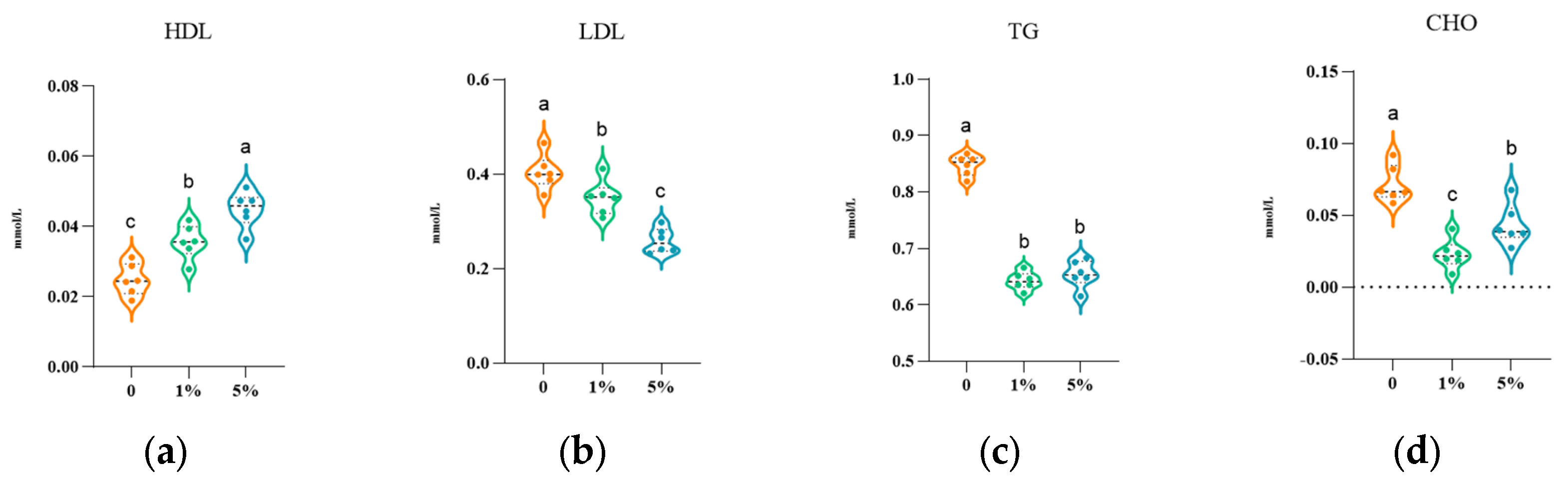
Figure 1. Changes in the levels of lipid metabolism indicators HDL (a), LDL (b), TG (c), and CHO (d) in the serum of L. vannamei fed diets supplemented with different levels of CE. Different letters above the violins indicate significant differences among treatments (p < 0.05).
Fishes 11 00482 g001
Figure 2. Changes in the levels of non-specific immune indicators ACP (a), LZM (b), ALP (c), PO (d), T−SOD (e), MDA (f), T-AOC (g), GSH−PX (h) and POD (i) in the hepatopancreas of L. vannamei fed diets supplemented with different levels of CE.
Fishes 11 00482 g002
Figure 3. Changes in the contents of IL−1β (a), IL−10 (b), TNF−α (c), and TNF−β (d) in the hepatopancreas of L. vannamei fed diets containing different levels of CE.
Fishes 11 00482 g003
Figure 4. Effects of dietary supplementation with different levels of CE on immune−related genes Pen3 (a), myd88 (b), toll (c), imd (d), lzm (e), propo (f), sod (g) and relish (h) in the hepatopancreas of L. vannamei.
Fishes 11 00482 g004
Figure 5. Effects of dietary CE supplementation on intestinal histological structure in L. vannamei (10 × 10/20 × 10). (a) Control group, 10 × 10; (b) 1% CE group, 10 × 10; (c) 5% CE group, 10 × 10; (d) control group, 20 × 10; (e) 1% CE group, 20 × 10; (f) 5% CE group, 20 × 10.
Fishes 11 00482 g005
Figure 6. PC analysis plot (a) and PCoA analysis plot (b) of L. vannamei. A: control group; B: 1% CE group; C: 5% CE group. Relative abundance of species at (c) phylum level and (d) genus level in L. vannamei; Venn diagram of intestinal microbial OTUs in L. vannamei (e); heatmap of functional annotation clustering by Tax4Fun2 (f).
Fishes 11 00482 g006
Table 1. Ingredient levels of the experiment diet (dry matter).
IngredientContentIngredientContent
Fish meal22.0 Vitamin premix0.2
Peanut meal18.0 Mineral premix0.5
Soybean meal12.0 Ca(H2PO4)21.5
Squid visceral ointment5.0 Vitamin C es1ter0.1
Shrimp shell powder5.0 Cholesterol0.2
Wheat flour31.0 NaCl0.2
Fish oil3.0 Choline chloride0.3
Lecithin1.0 Total100
Notes: Vitamin premix contains the following per kg: VA 8000 IU, VD3 4000 IU, VE 60 mg, VK3 20 mg, VB1 10 mg, VB2 30 mg, VB6 16 mg, calcium pantothenate 50 mg, folic acid 5 mg, biotin 0.16 mg, nicotinic acid 60.3 mg, VB12 0.03 mg, and inositol 300 mg; 2. Mineral premix contains the following per kg: MgSO4·H2O 60 mg, KCl 450 mg, Met-Cu 15 mg, FeSO4·H2O 5 mg, ZnSO4·H2O 50 mg, Ca(IO3)2 0.3 mg, Met-Co 0.8 mg, and NaSeO3 0.018 mg.
Table 2. Primer sequence of immune-related factor genes.
Primer(5′→ 3′)Length/bpReference Sequence
pen3-FCACCCTTCGTGAGACCTTTG141XM_027360479.2
pen3-RAATATCCCTTTCCCACGTGAC
myd88-FGCTGTTCCACCGCCATTT119JX073568.1
myd88-RGCATCATAGTGCTGTAGTCCAAGA
toll-FTGAGAGATGCCCACTGCCTG160XM_070131812.1
toll-RCACTTGAAGGTTTGTGAGGGAG
imd-FATACATCCTGCCGTTGCCGA107XM_027365703.2
imd-RCCGAGATGGGTTCCCTTGTT
lzm-FGTTCCGATCTGATG TCCGATG117XM_027375669.2
lzm-RAAGCCACCCAGGCAGAATAG
proPO-FCATCACTGACCTGGAAATCTG181XM_027381766.1
proPO-RGTAAGGGAAGTTGACGCTGT
SOD-FAGGGCTTCCATTAACAAC87XM_070119527.1
SOD-RCCGCCTCAACCAACTTCT
relish-FGAGTCCGCTCAGCAGTAACACAAG117XM_027357250.2
relish-RCAGCATCAACAAGCATACGCACAC
Table 3. Statistical table of alpha diversity indices. Different superscript letters within the same column indicate significant differences among treatments (p < 0.05).
GroupAceChaoShannonShannon-Even
Control855.45 ± 27.13 b826.84 ± 29.46 b1.72 ± 0.12 b0.26 ± 0.02 b
1% CE739.11 ± 26.41 a706.24 ± 24.49 a1.47 ± 0.09 a0.23 ± 0.02 a
5% CE872.71 ± 30.48 b847.84 ± 32.94 b1.51 ± 0.11 a0.22 ± 0.02 a
Disclaimer/Publisher’s Note: The statements, opinions and data contained in all publications are solely those of the individual author(s) and contributor(s) and not of MDPI and/or the editor(s). MDPI and/or the editor(s) disclaim responsibility for any injury to people or property resulting from any ideas, methods, instructions or products referred to in the content.