


Abstract
C-type lectins (CTLs) are important pattern recognition receptors (PRRs) involved in innate immune defense in crustaceans. In this study, a novel C-type lectin, designated LvCTL, was identified and functionally characterized from Litopenaeus vannamei. Sequence analysis revealed that the open reading frame (ORF) of LvCTL is 495 bp in length and encodes a 164-amino acid polypeptide containing a typical C-type lectin-like domain but lacking the classical carbohydrate-binding motifs. Notably, two novel EPF motifs were identified in the CTLD region, suggesting a potential non-canonical ligand recognition pattern. Phylogenetic analysis revealed that LvCTL clustered with other crustacean CTLs. Tissue expression analysis revealed that LvCTL exhibited the highest transcript level in the hepatopancreas. After immune stimulation, LvCTL expression in the hepatopancreas was significantly regulated by LPS, Vibrio parahaemolyticus, Staphylococcus aureus, WSSV and Poly(I: C), indicating its involvement in antimicrobial immune responses. RNA interference assays showed that knockdown of LvCTL significantly increased the bacterial burden and reduced the survival rate of shrimp after V. parahaemolyticus infection. Furthermore, recombinant LvCTL displayed broad bacterial binding activity to Gram-negative and Gram-positive bacteria and induced calcium-dependent agglutination of V. parahaemolyticus and S. aureus. Taken together, these results demonstrate that LvCTL functions as an important immune recognition molecule and contributes to antibacterial defense against V. parahaemolyticus in L. vannamei. These findings broaden our understanding of the functional diversity of shrimp CTLs and their contributions to crustacean innate immunity.
Keywords:
C-type lectins (CTLs); Vibrio parahaemolyticus; Litopenaeus vannamei; agglutination; immune response Key Contribution:
LvCTL is a hepatopancreas-predominant C-type lectin containing non-canonical EPF motifs that exhibits broad bacterial binding and calcium-dependent agglutination activities. Functional analyses reveal that LvCTL promotes bacterial clearance and enhances shrimp resistance to Vibrio parahaemolyticus infection.
1. Introduction
As a commercially important crustacean, the Pacific white shrimp (Litopenaeus vannamei) contributes to global aquaculture and contributes substantially to shrimp production and economic value worldwide [1]. Nevertheless, the rapid growth of shrimp aquaculture has led to frequent outbreaks of infectious disease, such as Vibrio parahaemolyticus and white spot syndrome virus (WSSV), resulting in substantial economic losses [2,3,4]. Among them, acute hepatopancreatic necrosis disease (AHPND) is mainly caused by highly pathogenic Vibrio parahaemolyticus carrying the binary toxins PirAvp and PirBvp [5,6]. Because shrimp do not possess antibody-mediated adaptive immunity, they rely primarily on innate immune mechanisms to defend against invading pathogens [7,8]. Therefore, identifying key innate immune molecules and clarifying their functions are essential for understanding shrimp host–pathogen interactions and developing new strategies for disease control.
C-type lectins (CTLs) belong to the lectin superfamily and are characterized by a conserved C-type lectin-like domain (CTLD) which mediates ligand binding [9]. The typical CRD consists of approximately 110–130 amino acid residues and adopts a double-loop architecture stabilized by two conserved disulfide bonds, enabling calcium-dependent carbohydrate recognition [10,11]. The EPN and QPD motifs are conserved amino acid motifs for carbohydrate ligand recognition and determining ligand binding specificity [12]. Moreover, a variety of non-canonical motifs, such as EPA, EPD, QPG, QAP, WND, WYD, WNH and MND, have been identified in invertebrate CTLs, suggesting that these motif variations may broaden their ligand recognition capacity [9]. Taken together, these motif variations may increase the structural diversity of CTLs and support their functional diversification in different immune processes.
Accumulating evidence demonstrates that CTLs are extensively participated in innate immune response of invertebrates, including phagocytosis, agglutination, opsonization, activation of the proPO cascade, and modulation of immune responses [10,13]. In L. vannamei, LvLTLC1 can agglutinate Gram-negative bacteria (Vibrio and Pseudomonas) as well as Gram-positive bacteria (Staphylococcus aureus) [14]. In echinoderms, the Apostichopus japonicus C-type lectin AjCTL-1 binds to peptidoglycan, promotes bacterial agglutination, and enhances the phagocytic activity of coelomocytes [15]. Additionally, LvCTL1 was reported to bind to structure proteins of WSSV (such as VP24, VP26, and VP28) [16]. Moreover, some crustacean CTLs can regulate antimicrobial peptide expression, promote hemocyte phagocytosis and participate in the prophenoloxidase cascade, thereby coordinating both cellular and humoral immune responses [17,18]. Together, these findings demonstrate that invertebrates CTLs contribute substantially to pathogen sensing, microbial aggregation, and the modulation of immune responses.
Here, we identified a previously uncharacterized CTL, termed LvCTL in L. vannamei. Its sequence features, conserved domain composition and phylogenetic relationship were subsequently characterized. In addition, we detected its expression profile following microbial stimulation, explored its role against V. parahaemolyticus in vivo in shrimp, and assessed its binding activity and agglutination toward bacteria. These findings provide new evidence for the functional diversity of shrimp CTLs and contribute to a better understanding of the host defense mechanisms against V. parahaemolyticus infection.
2. Materials and Methods
2.1. Shrimp
Healthy shrimp (~5 g) were obtained from a commercial shrimp farm in Rizhao City, China. Prior to the experiment, randomly sampled individuals were examined for the presence of V. parahaemolyticus as previously reported [19]. The shrimp were acclimated for 7 days in an aerated recirculating aquaculture system at 28 °C and 25‰ salinity and were fed a commercial diet before experimental treatment.
2.2. Bacteria
The bacterial strains have been previously characterized and are preserved in our laboratory [20]. And the Vibrio parahaemolyticus strains contain virulence genes PirABvp, stored in our laboratory. All bacterial strains were grown overnight in the Luria–Bertani (LB) medium at 30 °C or 37 °C. Bacterial cultures were harvested during the exponential growth phase by centrifugation, washed with phosphate-buffered saline (PBS), and then resuspended in fresh PBS for subsequent assays.
2.3. Sequence Analysis of LvCTL
The coding sequence of LvCTL (accession number PZ493557) was amplified using specific primers designed based on the L. vannamei transcriptome database (Table 1) [21]. The signal peptide and domain analysis were generated by SMART 7.0 [22]. The three-dimensional structural model was predicted by SWISS-MODEL 4.0. Multiple sequence alignment was conducted by GENEDOC 2.7 software, and the phylogenetic relationships were inferred through a neighbor-joining (NJ) tree using MEGA X 10.0 software [23].
Table 1.
Summary of primers used in this study.
2.4. Expression Pattern of LvCTL
For tissue distribution analysis, shrimp samples including eyestalk, stomach, hepatopancreas, epidermis, antenna, gill, heart, appendage, intestine, muscle, nerve, and hemocytes were aseptically collected from 15 healthy shrimp (each sample comprised 5 shrimp). For the immune challenge, each shrimp received a 50 μL PBS injection including LPS (5 μg/shrimp), Poly (I: C) (5 μg/shrimp), V. parahaemolyticus (containing virulence genes PirABvp), 105 CFU/mL S. aureus, or WSSV in 50 μL PBS. Hepatopancreas of challenged shrimps were collected at 0, 4, 8, 12, 24, 36, 48, 72 h post injection, pooled from 15 shrimp (each sample comprised 5 shrimp). Quantitative real-time reverse transcription PCR (qPCR) was conducted as described previously [24]. Relative LvCTL transcript abundance was determined using the 2−ΔΔCt method, with L. vannamei EF-1α (GU136229) serving as the internal reference gene using the gene-specific primer (Table 1).
2.5. RNA Interference
DsRNA-LvCTL (dsLvCTL) and dsRNA-GFP (with dsGFP as a control) were generated by in vivo transcription with a T7 RiboMAX Express RNAi System kit (Promega, Shanghai, China), as previously reported [25], using the gene-specific primer (Table 1). The lengths of LvCTL and dsGFP were 495 and 504 bp, respectively. The integrity of dsRNA was verified by 1.5% agarose gel electrophoresis, and the concentration was determined using a NanoDrop 2000 spectrophotometer (Thermo Fisher Scientific, Waltham, MA, USA). For RNA interference, shrimp in the experimental group were injected with dsLvCTL (10 μg per shrimp in 50 μL PBS), with dsGFP or PBS as a control, respectively. After 48 h, hepatopancreas tissues were sampled from the five shrimp per group to determine the RNA interference efficiency by qPCR.
2.6. Survival Rate
For the survival assay, shrimp were challenged with 1 × 10⁵ CFU of V. parahaemolyticus (containing virulence genes PirABvp) at 48 h after dsLvCTL administration, while control shrimp received an equal volume of PBS. Shrimp were randomly assigned to the experimental groups. For the sampling experiment, each group was maintained in a separate tank containing 30 shrimp. An additional independent tank containing 30 shrimp per group was used for the survival assay. Mortality was recorded every 4 h, and survival curves were compared using the Mantel–Cox log-rank test in GraphPad Prism 8.0. Bacterial loads were quantified as described previously [19].
2.7. Expression and Purification of Recombinant Proteins
The LvCTL coding sequence lacking the N-terminal signal peptide and termination codon was amplified with gene-specific primers and cloned into the pET-32a(+) expression vector (Merck Millipore, Darmstadt, Germany). The resulting recombinant construct was subsequently introduced into Escherichia coli Rosetta (DE3) cells (TransGen Biotech, Beijing, China). Recombinant protein expression was induced with 0.1 mM isopropyl β-D-1-thiogalactopyranoside (IPTG, Beyotime Biotechnology, Shanghai, China) at 16 °C for 12 h. The bacterial cells were subsequently collected by centrifugation and lysed by sonication. After centrifugation, the soluble supernatant was recovered, and the recombinant protein was purified by Ni-NTA affinity chromatography with an elution buffer (0.5 M NaCl, 20 mM Tris–Cl pH 7.4, and 300 mM imidazole) following an established protocol [26], and the thioredoxin (TRX), as a control protein, expressed and purified under the same conditions as rLvCTL.
2.8. Western Blotting
The purified recombinant LvCTL was characterized by SDS–PAGE and Western blotting following previously established procedures [27]. Protein samples (1 μg) were separated by 10% SDS–PAGE and electrotransferred onto PVDF membranes under the set conditions (200 mA, 2 h). After blocking with 5% nonfat milk (Beyotime Biotechnology) prepared in TBST for 1 h, the membranes were incubated with a mouse anti-6×His antibody (Cell Signaling Technology, Danvers, MA, USA) diluted 1:1000 at 25 °C for 1 h. Following three washes with TBST, the membranes were incubated with horseradish peroxidase (HRP)-conjugated goat anti-mouse IgG (H+L) secondary antibody (Promega, Shanghai, China) at a dilution of 1:2000. The immunoreactive bands were detected using the SuperSignal West Pico PLUS (Thermo Fisher Scientific).
2.9. Bacterial Binding Activity of rLvCTL
The bacterial binding capacity of rLvCTL was evaluated according to a previously established protocol [28]. In brief, bacteria were grown to the logarithmic phase and adjusted to 1 × 10⁸ CFU/mL. The bacterial suspension (200 μL) was then mixed with 5 μg rLvCTL and incubated with shaking for 2 h at 25 °C, while an equivalent amount of TRX protein was used as the negative control. After washing six times with PBS, the bacterial pellet was subsequently recovered and analyzed by Western blotting with an anti-6×His antibody.
2.10. The Bacterial Agglutination Assay of rLvCTL
Bacterial agglutination activity was examined according to a previously described method [29]. Briefly, an aliquot of FITC-labeled bacterial suspension (50 μL) was combined with the same volume of rLvCTL, TRX protein, or a control TBS–Ca2+ buffer (50 mM Tris-HCl, 100 mM NaCl, 10 mM CaCl2, pH 7.4) containing or not containing 4 mM EDTA. The bacterial cells were subsequently fixed in 4% paraformaldehyde (Beyotime Biotechnology), washed with PBS, transferred onto glass slides, and then examined by confocal laser scanning microscopy (Leica TCS-SP5, Wetzlar, Germany).
2.11. Statistical Analysis
Data are presented as the mean ± standard deviation (SD). Differences between two groups were evaluated using Student’s t-test.
3. Results
3.1. Sequence Characteristics of LvCTL
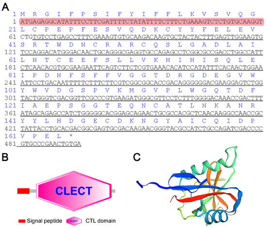
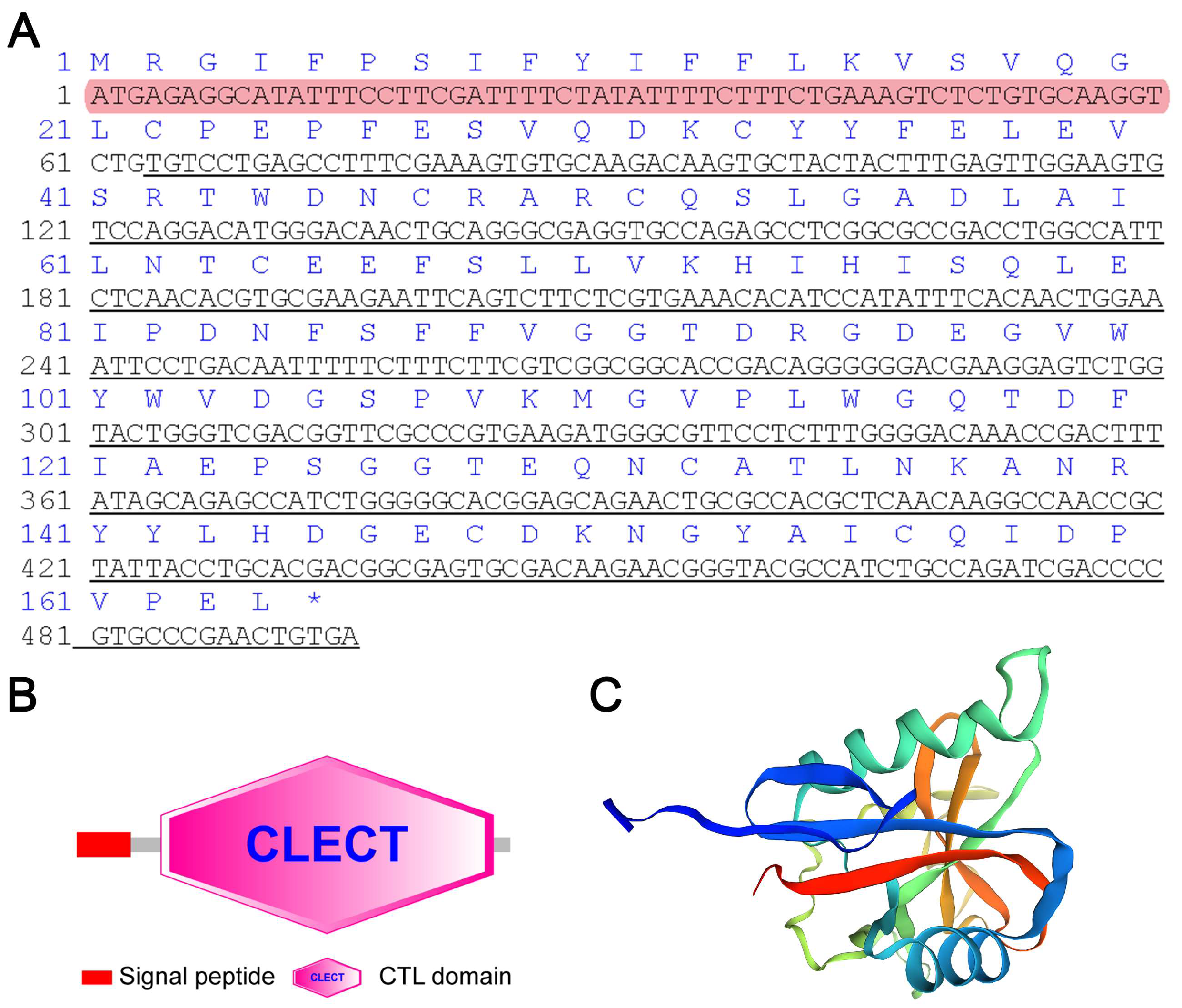
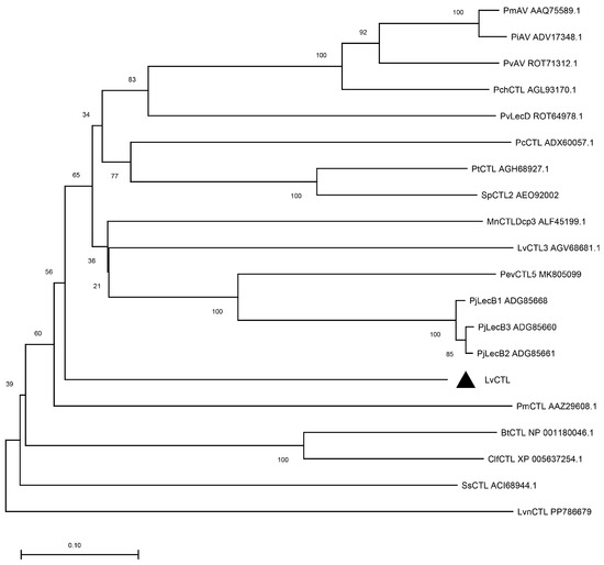
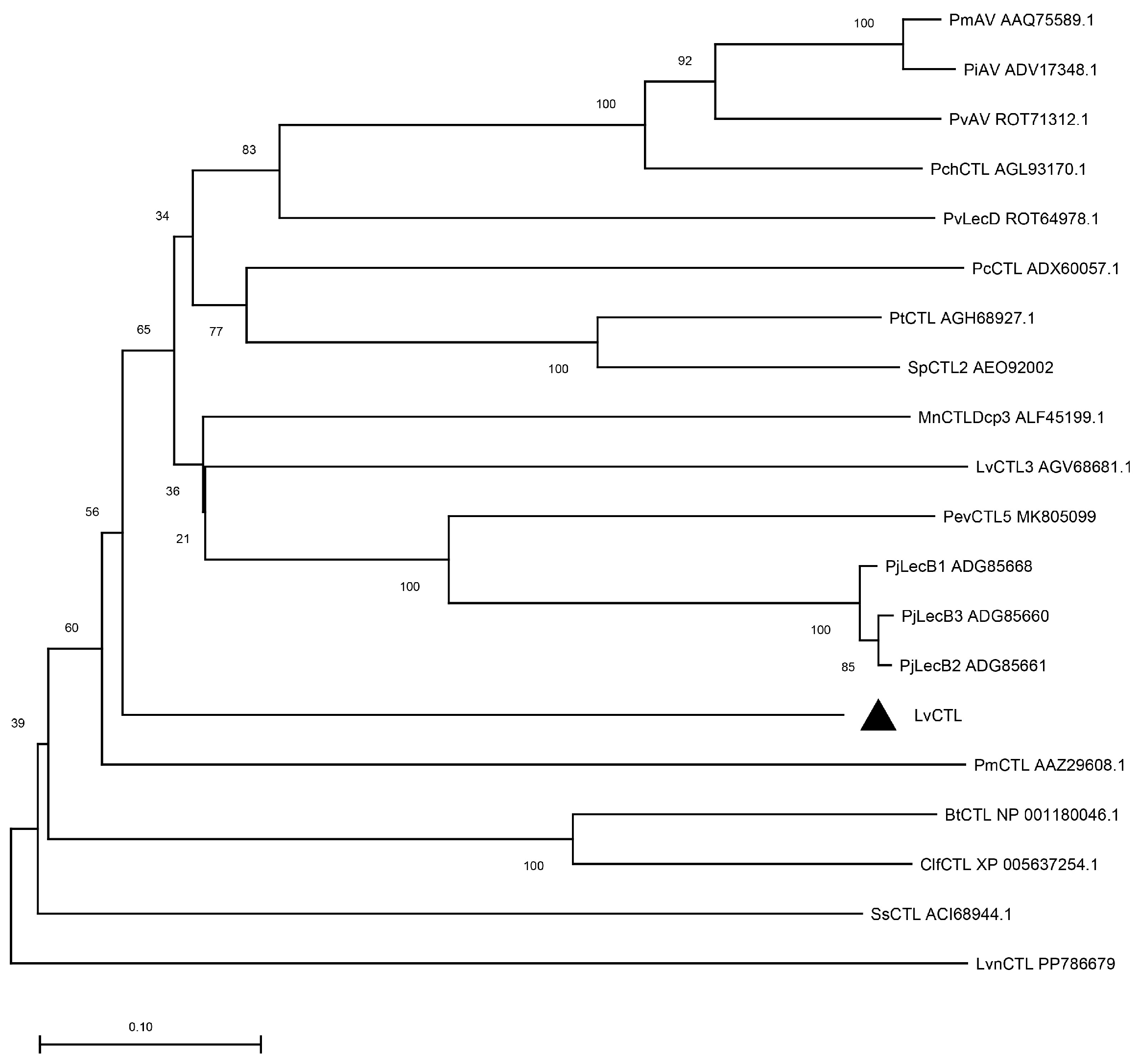
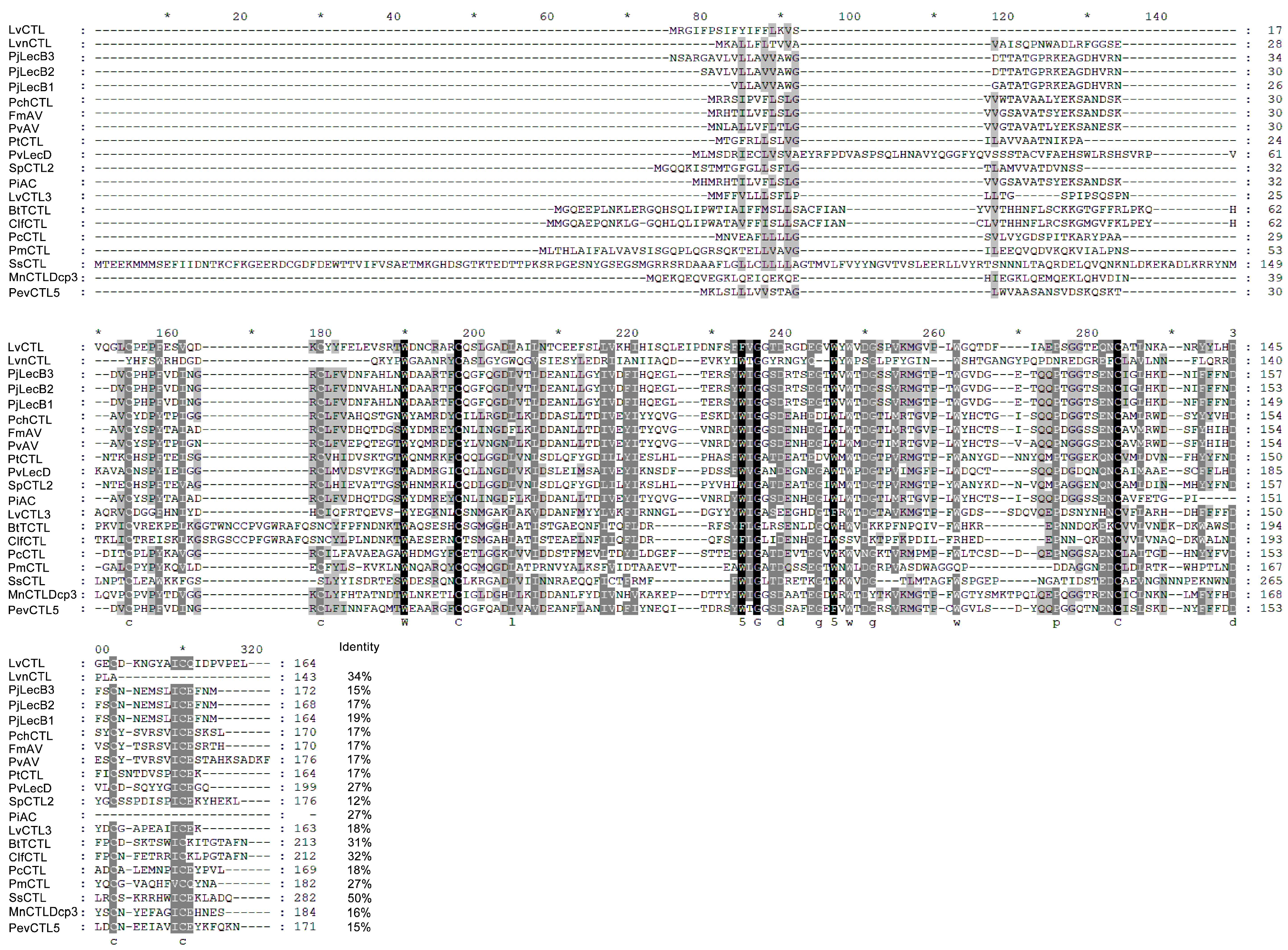
The open reading frame (ORF) of LvCTL (the nucleotide sequence has been deposited in GenBank under accession number PZ493557) was 495 bp in length and encoded a protein of 164 amino acids, with a predicted molecular weight of 18.58 kDa and a theoretical isoelectric point of 4.42. Sequence analysis showed that LvCTL contained an N-terminal signal peptide and a conserved C-type lectin-like domain (CTLD) (Figure 1A,B). Notably, LvCTL lacked the canonical QPD, EPN, and WND motifs associated with carbohydrate recognition and Ca2+ coordination. Instead, two unique EPF motifs were identified in this region, suggesting that they may function as non-canonical motifs involved in carbohydrate recognition (Figure 1A). The predicted three-dimensional structure showed that LvCTL possessed a typical CTL-like fold (Figure 1C). Phylogenetic analysis indicated that LvCTL was grouped within the invertebrate CTL clade (Figure 2). Sequence comparison revealed that LvCTL exhibited 12–50% amino acid identity with CTL homologs identified in other species, with the lowest identity to Scylla paramamosain CTL2 (12%, AEO92002) and the highest identity to Salmo salar CTL (50%, ACI68944.1) (Figure 3).
Figure 1.
Nucleotide and deduced amino acid sequence of LvCTL. (A) The ORF of the nucleotide sequence is shown in upper-case letters; the amino acid sequence is represented with one-letter codes above the nucleotide sequence. The signal peptide is shown in red, and the CLECT domain is shown in black underline. * indicates the termination codon. (B) Schematic representation of the LvCTL protein, including a signaling peptide and a conserved CLECT domain. (C) The three-dimensional model was generated by homology modeling using the SWISS-MODEL.
Figure 2.
Phylogenetic tree analysis of LvCTL. The phylogenetic tree was constructed with MEGA X with the neighbor-joining method, and the bootstrap was based on 1000 replications. The scale bar indicates the number of amino acid substitutions per site.
Figure 3.
Amino acid sequence alignment of LvCTL and its homologs. Multiple sequence alignment of CTL proteins, highly conserved amino acid residues and cysteine residues are highlighted in black. Grey color reflects amino acid conservation, darker shades indicate higher conservation. * represents a 20‑amino‑acid interval.
3.2. Expression Profile of LvCTL
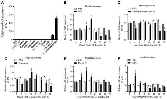
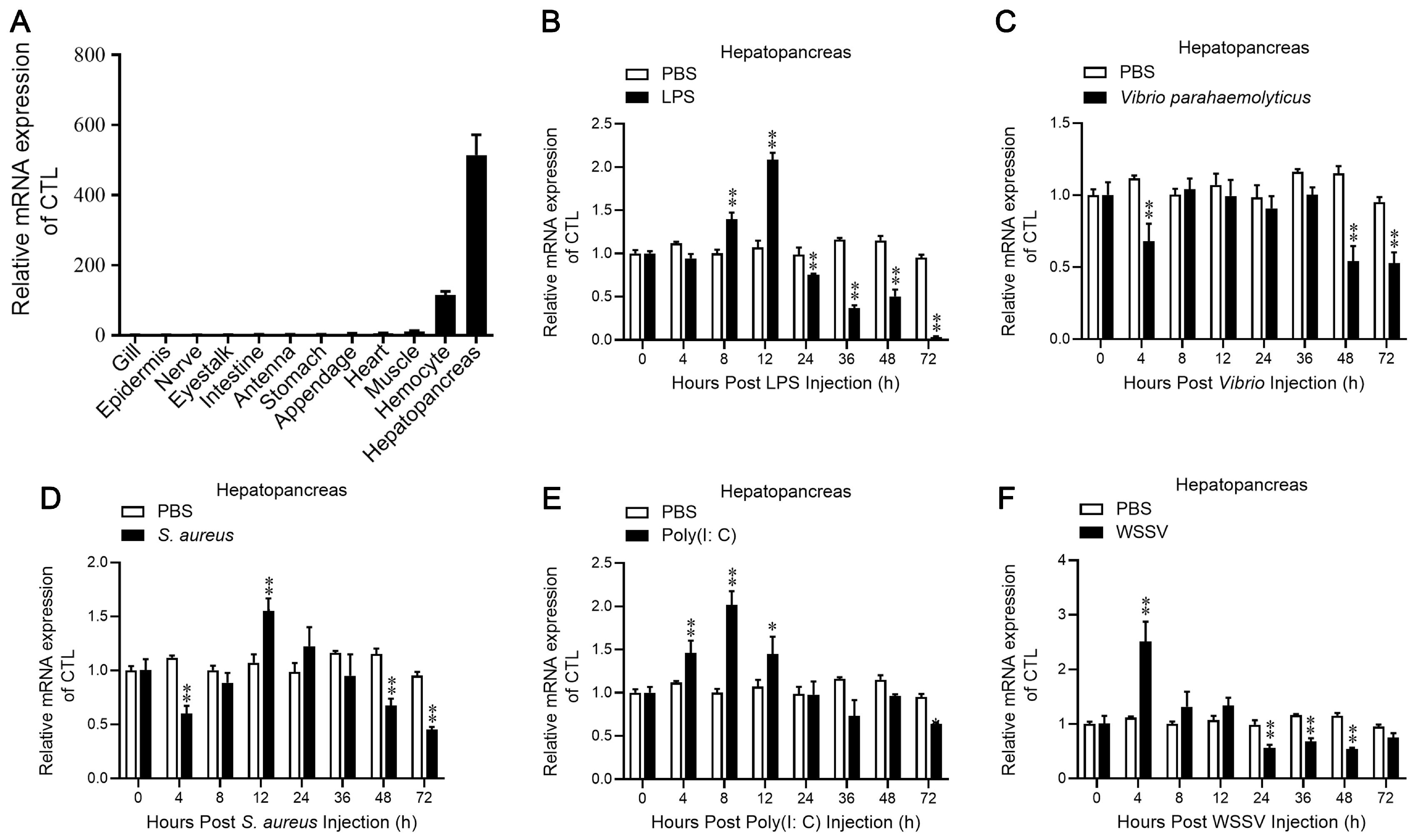
LvCTL transcripts were detected across all tissues examined, with the greatest abundance observed in the hepatopancreas, followed by hemocytes, whereas LvCTL transcripts were present at comparatively low levels in the gills (Figure 4A). To explore the participation of LvCTL in immune defense, its expression profiles were analyzed in the hepatopancreas post different challenges. Following LPS injection, the expression of LvCTL was significantly increased at 12 h (Figure 4B). Following V. parahaemolyticus infection, LvCTL expression was significantly reduced at 48 and 72 hpi (Figure 4C). Following S. aureus challenge, LvCTL transcription showed a transient increase at 12 hp (Figure 4D). In response to Poly (I: C) stimulation, the expression of LvCTL was significantly increased at 4–12 h (Figure 4E). WSSV challenge elicited a marked increase in LvCTL expression, with the strongest induction observed at 4 hpi (Figure 4F). Collectively, these observations indicate that LvCTL is preferentially expressed in immune-associated tissues and may contribute to antimicrobial defense in shrimp.
Figure 4.
Expression of LvCTL analysis by qPCR. (A) Expression profile of LvCTL in L. vannamei tissues analyzed by qPCR. Data were presented as fold values relative to the expression level in gills (set as 1.0). (B–F) Temporal expression patterns of LvCTL in the hepatopancreas after stimulation with LPS (B), V. parahaemolyticus (C), S. aureus (D), Poly (I: C) (E), and WSSV (F). Relative mRNA expression levels of LvCTL were determined by qPCR and normalized to the EF-1α gene. The statistical significance was calculated using Student’s t-test. *, p < 0.05 and **, p < 0.01. All experiments were done three times and yielded similar results.
3.3. Involvement of LvCTL in Antibacterial Immunity
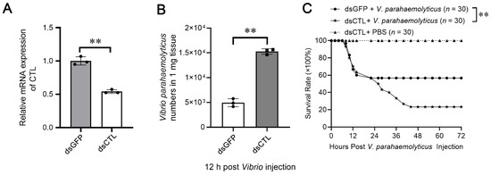
To investigate the role of LvCTL in antibacterial immunity, RNAi was performed using dsLvCTL; dsGFP was used as a control. The expression of LvCTL was significantly reduced after dsLvCTL treatment, confirming the successful knockdown of LvCTL (Figure 5A). At 12 h after V. parahaemolyticus challenge, shrimp treated with dsLvCTL exhibited a significantly greater bacterial burden than those receiving dsGFP (Figure 5B). Consistently, LvCTL knockdown markedly decreased the survival rate following V. parahaemolyticus challenge, whereas the PBS group showed no mortality during the experiment (30 individuals per group) (Figure 5C). Collectively, these results suggest that LvCTL contributes to the immune defense of L. vannamei against V. parahaemolyticus infection.
Figure 5.
The involvement of LvCTL in anti-V. parahaemolyticus immunity. (A) The knockdown efficiency of LvCTL was confirmed by qPCR in hepatopancreas after dsLvCTL treatment. dsGFP was used as the control. (B) The number of V. parahaemolyticus in hepatopancreas post treatment with LvCTL was analyzed by qPCR. The statistical significance was calculated using Student’s t-test. **, p < 0.01 by two-tailed unpaired Student’s t-test. (C) The survival rates of V. parahaemolyticus-infected shrimp (30 individuals per group) after treatment with dsLvCTL were analyzed by the Kaplan–Meier method. All experiments were done three times and yielded similar results.
3.4. Binding Activity of LvCTL Toward Bacteria
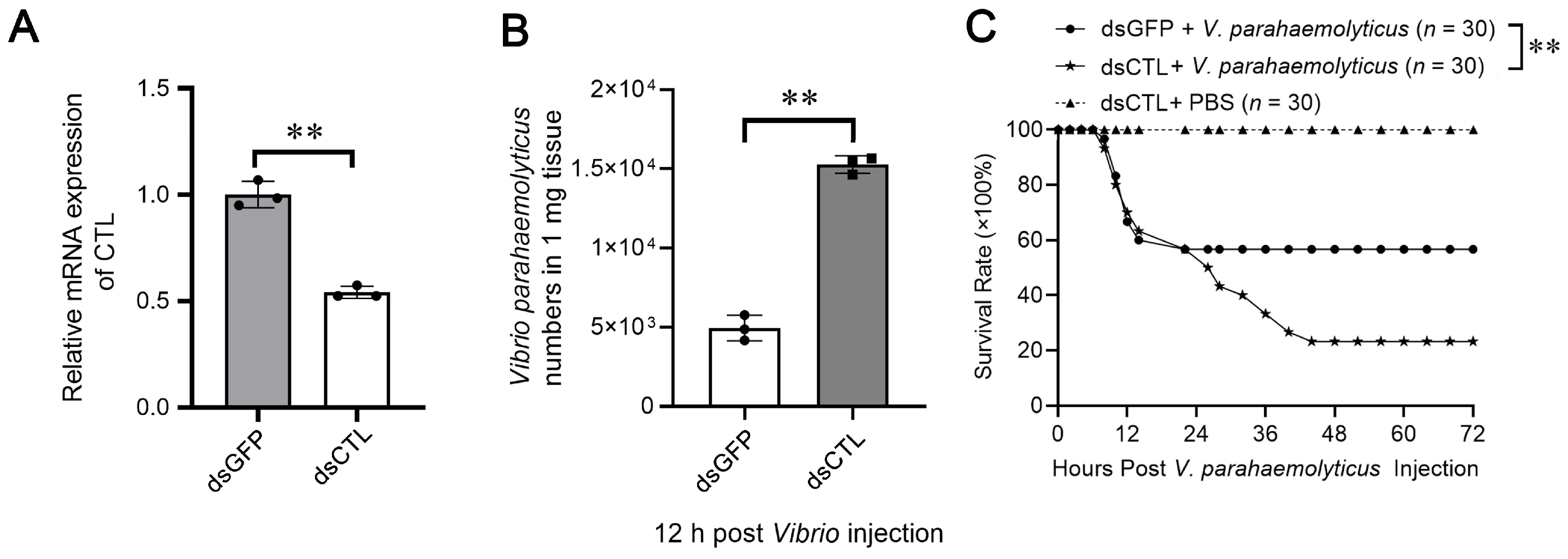
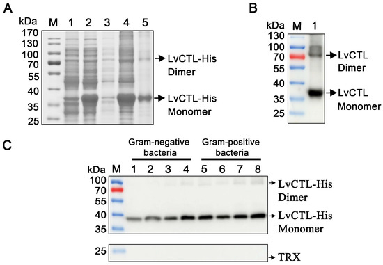
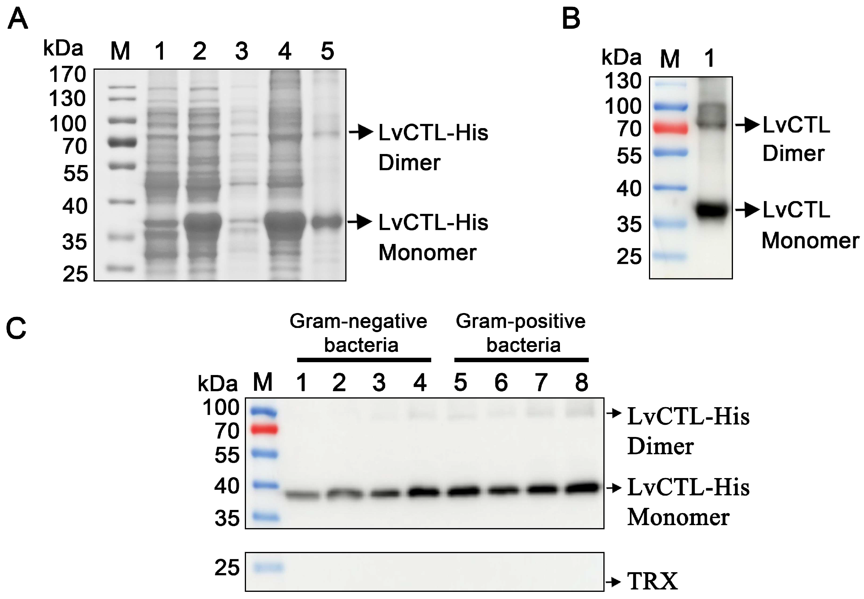
To explore its functional properties, recombinant LvCTL-His was successfully expressed and purified, with two major bands corresponding to the predicted monomeric and dimeric forms (Figure 6A). In addition to the major band corresponding to the LvCTL monomer (~35 kDa), a weaker band of approximately 70 kDa was also observed. Based on its apparent molecular weight, this band is likely to represent a dimeric form of recombinant LvCTL. Western blotting further confirmed the specificity of the monomeric and dimeric forms of LvCTL protein (Figure 6B). To evaluate its bacterial binding activity, LvCTL-His was incubated with different Gram-negative and Gram-positive bacteria. LvCTL-His was detected in the bacterial pellets of all tested strains (Figure 6C). And the TRX tag alone showed no detectable binding to the tested bacteria, confirming that the observed bacterial binding activity was attributable to recombinant LvCTL rather than the TRX fusion tag. These results indicate that LvCTL possesses broad bacterial binding activity and may participate in pathogen recognition during immune responses.
Figure 6.
The binding activity of LvCTL towards bacteria. (A) SDS-PAGE analysis of recombinant LvCTL expression and purification. Lane 1, uninduced E. coli transformed with LvCTL; Lane 2, induced E. coli transformed with LvCTL; Lane 3, supernatant of ultrasonically lysed E. coli expressing LvCTL; Lane 4, precipitate of lysed E. coli expressing LvCTL; Lane 5, purified recombinant LvCTL protein (black arrow). The recombinant protein was detected mainly as monomer and a dimer. (B) Western blotting analysis of purified recombinant LvCTL using anti-6 × His antibody. Lane 1, LvCTL protein. (C) Bacterial binding activities of rLvCTL. Lanes 1–4 represent the Gram-negative bacteria, including V. parahaemolyticus, Escherichia coli, Aeromonas hydrophila, and Pseudomonas aeruginosa, respectively. Lanes 5–8 represent the Gram-positive bacteria, including S. aureus, Enterococcus faecalis, Bacillus subtilis, and Micrococcus luteus, respectively. All experiments were done three times and yielded similar results.
3.5. Microbial Agglutination Activity of LvCTL
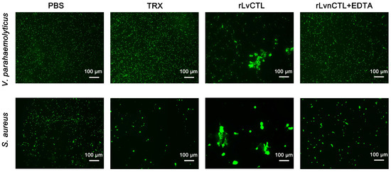
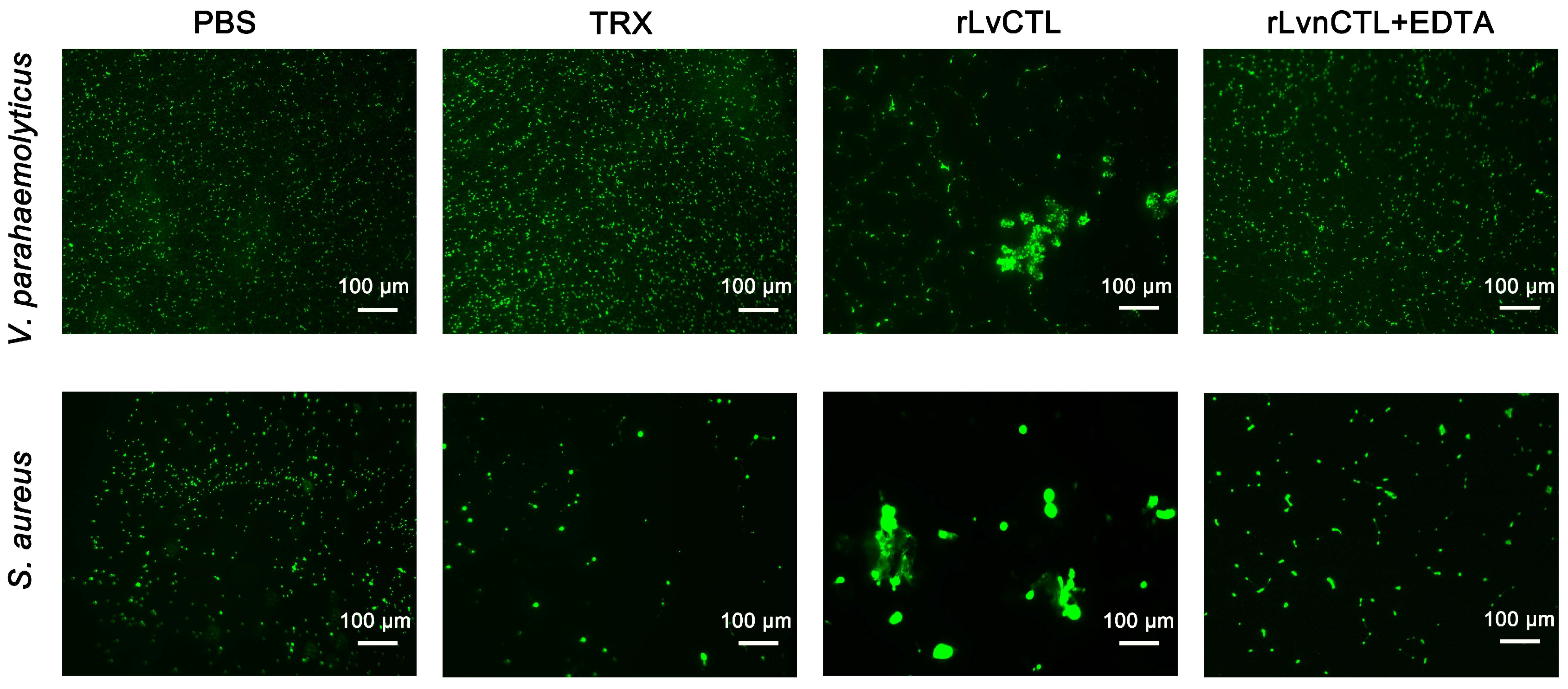
To determine whether LvCTL could agglutinate bacteria, FITC-labeled V. parahaemolyticus and S. aureus were incubated with rLvCTL in the presence or absence of Ca2+ (10 mM). Compared with the PBS and TRX control groups, rLvCTL induced obvious aggregation of both Gram-negative V. parahaemolyticus and Gram-positive S. aureus. Notably, the EDTA (4 mM) largely reduced the agglutination effect (Figure 7). These findings suggest that LvCTL mediates bacterial agglutination in a Ca2+-dependent manner.
Figure 7.
The agglutination ability of LvCTL towards FITC-labeled bacteria. The agglutination activity of recombinant LvCTL against V. parahaemolyticus and S. aureus were observed by fluorescence microscopy. FITC-labeled V. parahaemolyticus and S. aureus were treated with rLvCTL or TRX (as a control) in Ca2+-containing TBS buffer (10 mM) with or without EDTA (4 mM). Scale bar, 100 μm. All experiments were done three times and yielded similar results.
4. Discussion
C-type lectins function as key pattern recognition receptors (PRRs) and play central functions in invertebrate innate immunity [30]. Here, a previously uncharacterized C-type lectin, designated LvCTL, was identified and functionally characterized in L. vannamei. LvCTL showed typical features of the C-type lectin superfamily and exhibited high homology with crustacean CTLs. We characterized its tissue-specific expression pattern and explored the role of LvCTL in immune recognition and antibacterial defense.
In classical CTLs, carbohydrate specificity is often associated with conserved motifs (such as EPN, QPD, and WND) within the carbohydrate recognition region [31]. For example, the EPN motif is generally related to mannose-type ligand recognition, and the QPD motif is associated with galactose-type ligand recognition, whereas the WND motif is responsible for calcium ion coordination [12,32]. Notably, the CTLD of LvCTL lacked the conserved QPD, EPN, and WND motifs typically associated with carbohydrate recognition and Ca2+ coordination; instead, two EPF motifs were identified in this region. Similar variations in the canonical motifs, such as EPK, EPD, EPQ, EPS, QPN and QPT, have also been reported in shrimp CTLs, suggesting that crustacean CTLs may have undergone extensive diversification in their carbohydrate recognition sites [9,33,34]. Therefore, the presence of EPF motifs in LvCTL may indicate a non-canonical recognition pattern, which could allow LvCTL to recognize a broader or different spectrum of microbial surface glycans. However, their contribution to ligand recognition and immune function remains unclear. Future studies employing molecular docking, site-directed mutagenesis, carbohydrate-binding assays, and structural analyses will be required to determine whether these atypical motifs confer altered ligand specificity or recognition properties.
Analysis of tissue-specific expression revealed that LvCTL transcripts were most abundant in the hepatopancreas, a key organ involved in innate immune responses, and is known to synthesize multiple immune-related factors, including lectins, antimicrobial peptides and other humoral immune molecules [35,36]. Previous studies have shown that shrimp CTLs are mainly expressed in the hepatopancreas and hemocytes and participated in antimicrobial and antiviral responses [37]. It is worth noting that some studies reported on L. vannamei and Procambarus clarkii showed that some CTLs are mainly distributed in the stomach tissue and involved in the immune response to bacteria [38,39]. After immune stimulation, LvCTL expression was significantly regulated by V. parahaemolyticus, suggesting that LvCTL may be involved in host defense during V. parahaemolyticus infection, especially in the hepatopancreas, where pathogen-induced tissue damage and immune regulation frequently occur.
The in vivo RNA interference assay provided direct evidence for the protective role of LvCTL against V. parahaemolyticus. After LvCTL knockdown, the number of V. parahaemolyticus in hepatopancreas was significantly increased, and the survival rate was markedly reduced, indicating that LvCTL contributes to bacterial clearance and host resistance. Similarly, knockdown of LvCTL4, LvCTL5, or Fenneropenaeus merguiensis FmLC6 significantly increased shrimp mortality after bacterial infection [17,40,41]. Moreover, recombinant LvCTL displayed broad-spectrum binding capacity against bacteria and mediated pronounced agglutination of V. parahaemolyticus and S. aureus [34]. Although LvCTL knockdown resulted in increased bacterial burden and reduced shrimp survival, the underlying mechanisms remain to be elucidated. Future studies should investigate whether LvCTL contributes to host defense by directly promoting bacterial clearance, enhancing phagocytosis, or regulating the expression of downstream immune effectors, such as antimicrobial peptides and other immune-related genes.
Agglutination of bacteria is an important immune mechanism through which lectins can immobilize invading microorganisms, enhance pathogen aggregation, and promote the clearance of them by immune cells [14,29,42]. Similar calcium-dependent bacterial binding and agglutinating activities have been reported for other invertebrate CTLs, such as Urechis unicinctus CTL6, which exhibits calcium-dependent broad-spectrum bacterial binding capacity [43], and GCTL from Marsupenaeus japonicus, which mediates Ca2+-dependent agglutination of bacteria [31]. In the presence of EDTA, LvCTL-mediated aggregation disappeared or weakened, further indicating that calcium remains essential for LvCTL’s bacterial aggregation activity even without the classic EPN/QPD/WND domain. This suggests that LvCTL may utilize a nonclassical calcium-dependent mechanism to recognize microbial surface components, but the specific mechanism requires further investigation in the future.
In summary, our findings demonstrate that LvCTL is a newly identified hepatopancreas-predominant CTL, including a conserved CTLD and non-canonical EPF motifs, and that it plays a significant role against V. parahaemolyticus. LvCTL exerted important functions in anti-V. parahaemolyticus immune response. Collectively, these findings provide additional evidence for the structural and functional diversity of shrimp CTLs and their roles in antibacterial immunity.
5. Conclusions
In this work, we identified and functionally characterized a previously uncharacterized C-type lectin, designated LvCTL, that is predominantly expressed in the hepatopancreas. LvCTL contains a conserved C-type lectin-like domain but lacks the classical QPD, EPN and WND motifs, whereas two non-canonical EPF motifs were identified within this region. LvCTL was responsive to multiple microbial challenges, and its knockdown increased bacterial loads and reduced shrimp survival following V. parahaemolyticus infection. Moreover, recombinant LvCTL displayed broad bacterial binding activity and mediated calcium-dependent agglutination of V. parahaemolyticus and Staphylococcus aureus. These findings demonstrate that LvCTL is an important immune recognition molecule involved in antibacterial defense and shed new light on the structural and functional diversity of crustacean C-type lectins.
Author Contributions
N.F.: Validation, Investigation, Methodology, Formal analysis, Writing—review and editing. M.X.: Funding acquisition, Validation, Software, Formal analysis. Y.W.: Methodology, Formal analysis. L.Y.: Methodology, Formal analysis. D.W.: Methodology, Software. T.F.: Methodology, Formal analysis. B.X.: Conceptualization, Investigation, Methodology, Formal analysis, Writing—review and editing. S.N.: Funding acquisition, Conceptualization, Methodology, Software, Data curation, Formal analysis, Writing—review and editing. All authors have read and agreed to the published version of the manuscript.
Funding
The authors declare that this study received funding from the Shandong Provincial Natural Science Foundation (ZR2024QC228, ZR2026QC1342), Rizhao Municipal Natural Science Foundation (RZ2022ZR14), and Innovative Team Projects of Scientific Research in Rizhao Polytechnic (2025KC03). The funder was not involved in the study design, collection, analysis, interpretation of data, the writing of this article or the decision to submit it for publication.
Institutional Review Board Statement
Ethical review and approval were waived for this study due to shrimp not being subject to ethical requirements.
Data Availability Statement
The original contributions presented in this study are included in the article. Further inquiries can be directed to the corresponding authors.
Conflicts of Interest
The author Di Wang was employed by the Rizhao Hengtai Agricultural Technology Co., Ltd. The remaining authors declare that the research was conducted in the absence of any commercial or financial relationships that could be construed as a potential conflict of interest.
References
- Leape, J.; Micheli, F.; Tigchelaar, M.; Allison, E.H.; Basurto, X.; Bennett, A.; Bush, S.R.; Cao, L.; Crona, B.; DeClerck, F.; et al. The Vital Roles of Blue Foods in the Global Food System. In Science and Innovations for Food Systems Transformation; von Braun, J., Afsana, K., Fresco, L.O., Hassan, M.H.A., Eds.; Springer: Cham, Switzerland, 2023; pp. 401–419. [Google Scholar]
- Asche, F.; Anderson, J.L.; Botta, R.; Kumar, G.; Abrahamsen, E.B.; Nguyen, L.T.; Valderrama, D. The economics of shrimp disease. J. Invertebr. Pathol. 2021, 186, 107397. [Google Scholar] [CrossRef] [Scilit] [PubMed]
- Niu, S.; Zhu, Y.; Geng, R.; Luo, M.; Zuo, H.; Yang, L.; Weng, S.; He, J.; Xu, X. A novel chitinase Chi6 with immunosuppressive activity promotes white spot syndrome virus (WSSV) infection in Penaeus vannamei. Fish Shellfish Immunol. 2023, 132, 108450. [Google Scholar] [CrossRef] [Scilit] [PubMed]
- Xu, Y.; Sun, J.; Hu, J.; Bao, Z.; Wang, M. Characterization and Preliminary Application of a Novel Lytic Vibrio parahaemolyticus Bacteriophage vB_VpaP_SJSY21. Int. J. Mol. Sci. 2023, 24, 17529. [Google Scholar] [CrossRef] [Scilit] [PubMed]
- Han, J.E.; Tang, K.F.J.; Tran, L.H.; Lightner, D.V. Photorhabdus insect-related (Pir) toxin-like genes in a plasmid of Vibrio parahaemolyticus, the causative agent of acute hepatopancreatic necrosis disease (AHPND) of shrimp. Dis. Aquat. Org. 2015, 113, 33–40. [Google Scholar] [CrossRef] [Scilit] [PubMed]
- Lai, H.C.; Ng, T.H.; Ando, M.; Lee, C.T.; Chen, I.T.; Chuang, J.C.; Mavichak, R.; Chang, S.H.; Yeh, M.D.; Chiang, Y.A.; et al. Pathogenesis of acute hepatopancreatic necrosis disease (AHPND) in shrimp. Fish Shellfish Immunol. 2015, 47, 1006–1014. [Google Scholar] [CrossRef] [Scilit] [PubMed]
- Chen, Y.H.; He, J.G. Effects of environmental stress on shrimp innate immunity and white spot syndrome virus infection. Fish Shellfish Immunol. 2019, 84, 744–755. [Google Scholar] [CrossRef] [Scilit] [PubMed]
- Li, C.; Weng, S.; He, J. WSSV-host interaction: Host response and immune evasion. Fish Shellfish Immunol. 2019, 84, 558–571. [Google Scholar] [CrossRef] [Scilit] [PubMed]
- Viana, J.T.; dos Santos Rocha, R.; Maggioni, R. Structural and functional diversity of lectins associated with immunity in the marine shrimp Litopenaeus vannamei. Fish Shellfish Immunol. 2022, 129, 152–160. [Google Scholar] [CrossRef] [Scilit] [PubMed]
- Mayer, S.; Raulf, M.-K.; Lepenies, B. C-type lectins: Their network and roles in pathogen recognition and immunity. Histochem. Cell Biol. 2017, 147, 223–237. [Google Scholar] [CrossRef] [Scilit] [PubMed]
- Brown, G.D.; Willment, J.A.; Whitehead, L. C-type lectins in immunity and homeostasis. Nat. Rev. Immunol. 2018, 18, 374–389. [Google Scholar] [CrossRef] [Scilit] [PubMed]
- Drickamer, K.; Taylor, M.E. Recent insights into structures and functions of C-type lectins in the immune system. Curr. Opin. Struct. Biol. 2015, 34, 26–34. [Google Scholar] [CrossRef] [Scilit] [PubMed]
- Miyake, Y. Classification of C-type lectins and recognition of pathogens. Microbiol. Immunol. 2025, 69, 257–269. [Google Scholar] [CrossRef] [Scilit] [PubMed]
- Tian, Y.; Chen, T.; Huang, W.; Luo, P.; Huo, D.; Yun, L.; Hu, C.; Cai, Y. A new L-type lectin (LvLTLC1) from the shrimp Litopenaeus vannamei facilitates the clearance of Vibrio harveyi. Fish Shellfish Immunol. 2018, 73, 185–191. [Google Scholar] [CrossRef] [Scilit] [PubMed]
- Wei, X.; Liu, X.; Yang, J.; Wang, S.; Sun, G.; Yang, J. Critical roles of sea cucumber C-type lectin in non-self recognition and bacterial clearance. Fish Shellfish Immunol. 2015, 45, 791–799. [Google Scholar] [CrossRef] [Scilit] [PubMed]
- Zhao, Z.-Y.; Yin, Z.-X.; Xu, X.-P.; Weng, S.-P.; Rao, X.-Y.; Dai, Z.-X.; Luo, Y.-W.; Yang, G.; Li, Z.-S.; Guan, H.-J. A novel C-type lectin from the shrimp Litopenaeus vannamei possesses anti-white spot syndrome virus activity. J. Virol. 2009, 83, 347–356. [Google Scholar] [CrossRef] [Scilit] [PubMed]
- Luo, M.; Yang, L.; Wang, Z.-a.; Zuo, H.; Weng, S.; He, J.; Xu, X. A novel C-type lectin with microbiostatic and immune regulatory functions from Litopenaeus vannamei. Fish Shellfish Immunol. 2019, 93, 361–368. [Google Scholar] [CrossRef] [Scilit] [PubMed]
- Junkunlo, K.; Prachumwat, A.; Tangprasittipap, A.; Senapin, S.; Borwornpinyo, S.; Flegel, T.W.; Sritunyalucksana, K. A novel lectin domain-containing protein (LvCTLD) associated with response of the whiteleg shrimp Penaeus (Litopenaeus) vannamei to yellow head virus (YHV). Dev. Comp. Immunol. 2012, 37, 334–341. [Google Scholar] [CrossRef] [Scilit] [PubMed]
- Li, H.; Li, Q.; Wang, S.; He, J.; Li, C. Ammonia nitrogen stress increases susceptibility to bacterial infection via blocking IL-1R–Relish axis mediated antimicrobial peptides expression in shrimp. Aquaculture 2023, 563, 738934. [Google Scholar] [CrossRef] [Scilit]
- Xiao, B.; Wang, Y.; Xian, D.; Fan, T.; He, J.; Li, C. Identification of a Double-beta-Defensin with Multiple Antimicrobial Activities in a Marine Invertebrate. J. Immunol. 2023, 210, 1324–1337. [Google Scholar] [CrossRef] [Scilit] [PubMed]
- Li, C.; Weng, S.; Chen, Y.; Yu, X.; Lu, L.; Zhang, H.; He, J.; Xu, X. Analysis of Litopenaeus vannamei transcriptome using the next-generation DNA sequencing technique. PLoS ONE 2012, 7, e47442. [Google Scholar] [CrossRef] [Scilit] [PubMed]
- Schultz, J.; Milpetz, F.; Bork, P.; Ponting, C.P. SMART, a simple modular architecture research tool: Identification of signaling domains. Proc. Natl. Acad. Sci. USA 1998, 95, 5857–5864. [Google Scholar] [CrossRef] [Scilit] [PubMed]
- Kumar, S.; Stecher, G.; Li, M.; Knyaz, C.; Tamura, K. MEGA X: Molecular evolutionary genetics analysis across computing platforms. Mol. Biol. Evol. 2018, 35, 1547–1549. [Google Scholar] [CrossRef] [Scilit] [PubMed]
- Zuo, H.; Liao, A.; Shen, S.; Wang, Y.; Yang, X.; Weng, S.; He, J.; Xu, X. microRNA-34 mediates a negative feedback loop in the JAK-STAT pathway to attenuate immune overactivation in an invertebrate. Cell Commun. Signal. 2025, 23, 228. [Google Scholar] [CrossRef] [Scilit] [PubMed]
- Xiao, B.; Chen, S.; Wang, Y.; Liao, X.; He, J.; Li, C. Heat Shock Factor regulation of antimicrobial peptides expression suggests a conserved defense mechanism induced by febrile temperature in arthropods. eLife 2026, 13, RP101460. [Google Scholar] [CrossRef] [Scilit]
- Xiao, B.; Wang, Y.; He, J.; Li, C. Febrile Temperature Acts through HSP70-Toll4 Signaling to Improve Shrimp Resistance to White Spot Syndrome Virus. J. Immunol. 2024, 213, 1187–1201. [Google Scholar] [CrossRef] [Scilit] [PubMed]
- Xiao, B.; Hou, D.; Pan, J.; Kang, F.; Wang, Y.; He, J.; Li, C. Heat shock protein 70 (HSP70) regulates innate immunity and intestinal microbial homeostasis against Vibrio parahaemolyticus in shrimp. Aquaculture 2025, 596, 741814. [Google Scholar] [CrossRef] [Scilit]
- Xiao, B.; Pan, J.; Zheng, F.; Kang, F.; Wang, Y.; He, J.; Li, C. Identification and functional characterization of a novel transmembrane protein LvLRRm1 containing leucine-rich repeats in shrimp Litopenaeus vannamei. Fish Shellfish Immunol. 2025, 165, 110507. [Google Scholar] [CrossRef] [Scilit] [PubMed]
- Liang, Z.; Yang, L.; Zheng, J.; Zuo, H.; Weng, S.; He, J.; Xu, X. A low-density lipoprotein receptor (LDLR) class A domain-containing C-type lectin from Litopenaeus vannamei plays opposite roles in antibacterial and antiviral responses. Dev. Comp. Immunol. 2019, 92, 29–34. [Google Scholar] [CrossRef] [Scilit] [PubMed]
- Patnaik, B.B.; Baliarsingh, S.; Sarkar, A.; Hameed, A.S.; Lee, Y.S.; Jo, Y.H.; Han, Y.S.; Mohanty, J. The role of pattern recognition receptors in crustacean innate immunity. Rev. Aquac. 2024, 16, 190–233. [Google Scholar] [CrossRef] [Scilit]
- Alenton, R.R.R.; Koiwai, K.; Miyaguchi, K.; Kondo, H.; Hirono, I. Pathogen recognition of a novel C-type lectin from Marsupenaeus japonicus reveals the divergent sugar-binding specificity of QAP motif. Sci. Rep. 2017, 7, 45818. [Google Scholar] [CrossRef] [Scilit] [PubMed]
- Veldhuizen, E.J.; van Eijk, M.; Haagsman, H.P. The carbohydrate recognition domain of collectins. FEBS J. 2011, 278, 3930–3941. [Google Scholar] [CrossRef] [Scilit] [PubMed]
- Wang, X.-W.; Wang, J.-X. Diversity and multiple functions of lectins in shrimp immunity. Dev. Comp. Immunol. 2013, 39, 27–38. [Google Scholar] [CrossRef] [Scilit] [PubMed]
- Li, M.; Li, C.; Ma, C.; Li, H.; Zuo, H.; Weng, S.; Chen, X.; Zeng, D.; He, J.; Xu, X. Identification of a C-type lectin with antiviral and antibacterial activity from pacific white shrimp Litopenaeus vannamei. Dev. Comp. Immunol. 2014, 46, 231–240. [Google Scholar] [CrossRef] [Scilit] [PubMed]
- Wang, Y.-C.; Chang, P.-S.; Chen, H.-Y. Tissue expressions of nine genes important to immune defence of the Pacific white shrimp Litopenaeus vannamei. Fish Shellfish Immunol. 2007, 23, 1161–1177. [Google Scholar] [CrossRef] [Scilit] [PubMed]
- Vogt, G. Functional cytology of the hepatopancreas of decapod crustaceans. J. Morphol. 2019, 280, 1405–1444. [Google Scholar] [CrossRef] [Scilit] [PubMed]
- Sun, Y.-D.; Fu, L.-D.; Jia, Y.-P.; Du, X.-J.; Wang, Q.; Wang, Y.-H.; Zhao, X.-F.; Yu, X.-Q.; Wang, J.-X. A hepatopancreas-specific C-type lectin from the Chinese shrimp Fenneropenaeus chinensis exhibits antimicrobial activity. Mol. Immunol. 2008, 45, 348–361. [Google Scholar] [CrossRef] [Scilit] [PubMed]
- Niu, S.; Fu, N.; Wang, Y.; Xing, M.; An, M.; Xu, H.; Xiao, B.; Song, W.; Zeng, Y.; Borras-Hidalgo, O. Novel stomach-predominant C-type lectin (LvnCTL) triggers immune response against Vibrio parahaemolyticus and WSSV in shrimp Litopenaeus vannamei. Fish Shellfish Immunol. 2026, 171, 111158. [Google Scholar] [CrossRef] [Scilit] [PubMed]
- Zhang, X.-W.; Han, K.-K.; Yang, C.-H.; Xia, X.-H.; Ren, Q.; Zhang, H.-W. A stomach-specific lectin plays a crucial role in the innate immunity of red swamp crayfish, Procambarus clarkii. Fish Shellfish Immunol. 2020, 103, 285–292. [Google Scholar] [CrossRef] [Scilit] [PubMed]
- Li, H.; Chen, Y.; Li, M.; Wang, S.; Zuo, H.; Xu, X.; Weng, S.; He, J.; Li, C. A C-type lectin (LvCTL4) from Litopenaeus vannamei is a downstream molecule of the NF-κB signaling pathway and participates in antibacterial immune response. Fish Shellfish Immunol. 2015, 43, 257–263. [Google Scholar] [CrossRef] [Scilit] [PubMed]
- Runsaeng, P.; Kwankaew, P.; Utarabhand, P. FmLC6: An ultimate dual-CRD C-type lectin from Fenneropenaeus merguiensis mediated its roles in shrimp defense immunity towards bacteria and virus. Fish Shellfish Immunol. 2018, 80, 200–213. [Google Scholar] [CrossRef] [Scilit] [PubMed]
- Cui, M.; Zheng, G.; Wang, S.; Liang, J.; Xu, X.; Wu, J.; Xu, D. Characterization of a C-type lectin (OnCLEC10A) in Oreochromis niloticus functioning as a pattern recognition receptor with antibacterial properties. Fish Shellfish Immunol. 2026, 175, 111407. [Google Scholar] [CrossRef] [Scilit] [PubMed]
- Qiu, Y.; Shi, C.; Gao, J.; Qin, X.; Jiao, X.; Zhang, J. UuCTL6, a novel C-type lectin of Urechis unicinctus, agglutinates various bacteria and promotes immune defense against bacterial infection. Dev. Comp. Immunol. 2026, 179, 105613. [Google Scholar] [CrossRef] [Scilit] [PubMed]

Disclaimer/Publisher’s Note: The statements, opinions and data contained in all publications are solely those of the individual author(s) and contributor(s) and not of MDPI and/or the editor(s). MDPI and/or the editor(s) disclaim responsibility for any injury to people or property resulting from any ideas, methods, instructions or products referred to in the content. |
© 2026 by the authors. Licensee MDPI, Basel, Switzerland. This article is an open access article distributed under the terms and conditions of the Creative Commons Attribution (CC BY) license.