


Abstract
Salinity is a key environmental survival factor for all aquatic organisms, especially migratory species. The masu salmon (Oncorhynchus masou) is a representative migratory fish species. Following the freshwater parr stage, anadromous masu salmon briefly inhabit brackish water and transition before migrating to the ocean. To demonstrate the physiological responses of masu salmon (length: 8 ± 0.5 cm, water temperature: 10 ± 0.5 °C) to variations in salinity, we carried out three gradual transfer experiments (gradual daily increases direct transfer experiment of 3.2 (D10), 1.6 (D20), and 1.1 (D30) ppt until reaching 32 ppt) and one (immediate transfer to 32 ppt on day 0) as domestication regimens for masu salmon. The results indicated the following: (1) In the gradual transfer experiment group, growth performance, along with ion and hormone indicators, suggested that the D30 treatment group of anadromous masu salmon exhibited a high level of adaptability. (2) In the direct transfer experiment, in addition to the activity of antioxidant enzymes, both ion concentrations and hormone indicators returned to a stable state within 7 days. Our findings provide a scientific protocol for salinity regulation during the artificial propagation of masu salmon and establish critical acclimation parameters for land-based recirculating aquaculture systems aimed at marine salmonid farming, thereby highlighting their practical value.
Keywords:
Oncorhynchus masou; salinity; anadromous; direct transfer experiment; gradual transfer experiment Key Contribution:
Through physiological analysis, the optimal seaward transfer timing for cultured masu salmon (Oncorhynchus masou) was determined, thereby providing a robust scientific foundation for aquaculture management and stock enhancement programs.
1. Introduction
Migratory fish species (such as Atlantic salmon and Pacific salmon) undertake migrations of hundreds of kilometers throughout their life cycles [1,2]. Salinity acclimation is a core physiological process for such fish species to complete their life cycles [3], and their osmoregulatory ability is particularly important for future environmental changes [4]. Masu salmon (Oncorhynchus masou) are a typical anadromous species. Anadromous individuals go through a process called parr-smolt transformation. After this change, they migrate downstream to the ocean. Resident individuals take a different path. They stay in freshwater or estuarine areas for their entire lives. These residents do not undergo smoltification. They also never make the downstream journey to the sea [5,6,7,8,9]. Currently, wild populations of this species face significant threats, including habitat fragmentation and overfishing [10,11]. This species is ecologically and economically important. Understanding its acute and chronic salinity acclimation mechanisms is essential—both to reveal how osmoregulatory plasticity enables the freshwater-to-seawater transition (informing conservation), and to define its upper salinity tolerance limits (guiding aquaculture acclimation). Yet current knowledge remains limited: the mechanisms by which these fish acclimate to salinity changes—particularly the temporal dynamics of their osmoregulatory responses—remain poorly understood.
In masu salmon, the timing of metamorphosis initiation directly affects post-metamorphic survival and subsequent performance during marine life stages [12]. However, the optimal window for initiating this transition in artificial rearing systems remains insufficiently characterized. September offers a strategic time point for salinity acclimation experiments. By this month, juveniles have completed their initial freshwater growth and begun preparatory physiological changes associated with smoltification—a process that typically culminates in the following spring. Conducting controlled salinity challenges during this window facilitates early detection of osmoregulatory capacity, particularly gill Na+/K+-ATPase (NKA) activity, a well-established indicator of seawater tolerance that remains responsive to salinity variation during this developmental phase [13,14]. Additionally, September-based investigations provide insight into the metabolic costs of osmoregulation prior to winter energy storage. Previous studies have shown that juveniles acclimated to intermediate salinity (11 ppt) maintain normal growth, while exposure to higher salinity (22 ppt) may suppress growth, likely due to increased energy expenditure on ion regulation [14]. Characterizing these physiological responses in September is therefore essential for refining hatchery release protocols. Selecting individuals with enhanced seawater tolerance during this period can improve smolt survival during spring migration [12]. Thus, salinity acclimation studies conducted in September not only advance understanding of the physiological mechanisms underlying early marine acclimation in masu salmon but also offer practical implications for aquaculture and stock enhancement programs.
Bony fish have developed diverse acclimatory responses to fluctuations in environmental factors [15]. Dynamic monitoring of hematological parameters—including ion concentrations, hormone levels, and stress biomarkers—offers a robust and reliable methodology for assessing fish tolerance to salinity fluctuations [16]. Following prolonged acclimation (≥5 months), the initial growth inhibition induced by the high-salinity environment gradually diminishes, and the specific growth rate (SGR) recovers to— and in some cases exceeds—the levels observed in freshwater populations [17]. During the gradual acclimation to seawater, fish shift from actively absorbing ions and excreting water to taking in large amounts of water and excreting excessive ions to maintain osmotic balance [18]. Using juvenile pink salmon (Oncorhynchus gorbuscha) breeding in coastal areas as an example, they achieve optimal growth and survival at 15 ppt salinity [19], indicating that moderate salinity increases within the tolerance range enhance fish growth. Hormonal regulation plays an essential role in acclimation to seawater environments. Numerous studies confirm that growth hormone (GH) is central to seawater acclimation, while prolactin (PRL) primarily regulates freshwater acclimation [20,21,22]. In salmonid species, cortisol facilitates seawater acclimation by upregulating gill Na+/K+-ATPase activity, whereas prolactin (PRL) promotes freshwater acclimation by decreasing branchial ion permeability [23,24,25]. Although individual hormone functions are relatively well understood, their temporal dynamics and interactions during gradual salinity acclimation remain unclear. Thus, detecting key hormones is crucial for elucidating salt tolerance mechanisms and identifying optimal acclimation strategies.
Unlike chronic acclimation, acute salinity stress triggers a rapid activation of ionoregulatory mechanisms in fish, characterized by a swift and pronounced elevation in plasma osmotic pressure over a short time frame, along with significant increases in the plasma concentrations of sodium [Na+], chloride [Cl−], and potassium [K+] ions [26]. For example, within 6 h following the transfer of Atlantic salmon to seawater, plasma [Na+] concentration increases linearly, whereas [Cl−] concentration exhibits a curvilinear rise [16]. At the same time, acute exposure to seawater induces a rapid elevation in plasma cortisol (COR) levels within 1–3 h, thereby stimulating gill Na+/K+-ATPase activity and enhancing ionoregulatory capacity to mitigate hyperosmotic stress [23,24,25]. However, how varying acclimation rates affect plasma ion dynamics in masu salmon during gradual salinity acclimation remains unclear. Thus, measuring [Na+], [Cl−], and [K+] is critical for assessing osmoregulatory capacity and identifying the optimal acclimation strategy.
Environmental stressors such as salinity fluctuations can disrupt the redox balance in fish. This disruption often induces excessive production of reactive oxygen species (ROS) and leads to oxidative damage. Superoxide dismutase (SOD) and catalase (CAT) are key antioxidant enzymes. They work together to scavenge ROS and help maintain cellular homeostasis [27]. Prolonged salinity stress significantly elevates ROS, malondialdehyde, and other oxidative stress markers; upon stress relief, these levels gradually return to baseline [28,29]. This pattern suggests acute stress triggers rapid compensatory responses, whereas long-term acclimation entails systemic physiological reorganization [30]. Salinity stress has been shown to regulate antioxidant enzyme activity in salmoniform fish—for instance, Atlantic salmon mount antioxidant defense responses under environmental disturbance [28]. Thus, SOD and CAT serve as effective biomarkers for assessing fish responses to salinity stress. This study analyzes oxidative stress profiles in masu salmon under different salinity domestication regimes to identify the optimal strategy.
This study investigated the critical developmental stages of anadromous masu salmon by focusing on salinity variations. Through acute and chronic salinity acclimation experiments, we aimed to clarify the physiological mechanisms by which this species responds to changes in salinity, thereby providing scientific evidence to enhance survival rates during migration and offering technical support for restoring masu salmon genetic resources. These comprehensive investigations aimed to bridge critical knowledge gaps in the osmoregulatory physiology of masu salmon while establishing robust technical frameworks for (i) evidence-based seawater transition protocols; (ii) sustainable culture methodologies for migratory ecotypes; (iii) innovative germplasm resource management, collectively advancing both fundamental ichthyological understanding and applying conservation aquaculture to this ecologically vulnerable salmonid species.
2. Materials and Methods
2.1. Fish Material and Rearing Conditions
The study was conducted at the Liaoning Provincial Key Laboratory for Hydrobiology at Dalian Ocean University, China, following standard operating procedures approved by Animal Research and Ethics Committee of Dalian Ocean University (No. DLOU2024001). All masu salmon used were from the same batch provided by Mijiang Salmon Breeding Station (Jilin Province, Yanbian, China). A total of 1000 masu salmon were randomly transferred to 18 indoor tanks (volume, 100 L; height, 1.10 m; upper diameter, 0.50 m; lower diameter, 0.50 m) containing tap water (water temperature:10 ± 0.5 °C; pH: 7.9 ± 0.2). pH was measured using a multifunctional water quality monitor (AZ Instrument Company, Shenzhen, China). All environmental parameters were standardized and optimized for all tanks. Replicate tanks were independently equipped with a recirculating water supply, UV light, and temperature control system (Dalian Huixin Titanium Equipment Development Co. Ltd., Dalian, China). Salmon were approximately 1 year of age and had a mean length of 6.5 ± 0.5 cm. The experimental fish were kept under laboratory conditions (temperature: 10 ± 0.5 °C; salinity: 0 ppt; natural photoperiod) at our facility (38°52′34″ N, 121°33′38″ E) until they were fully differentiated into various life histories. This study focuses exclusively on anadromous individuals as the target population and excludes resident individuals from analysis [8]. This research lasted for approximately 9 weeks, from the beginning of September 2023 to the beginning of November.
This study design consists of two parts: a gradual transfer experiment and a direct transfer experiment. The gradual transfer experiment includes three groups: D10 group (reaching the target salinity of 32 ppt within 10 days), D20 group (reaching the target salinity of 32 ppt within 20 days), and D30 group (reaching the target salinity of 32 ppt within 30 days). Each group has three replicates. The experimental water was prepared by mixing tap water with seawater and was calibrated for salinity using a salinity meter (YD-100, Hangzhou Qiwei Instrument Co., Ltd., Hangzhou, China). Salinity domestication was carried out in a step-by-step method. The D10 group was increased 3.2 ppt every day in each test group until it reached the target salinity, the D20 group was increased 1.6 ppt every day in each test group until it reached the target salinity, and the D30 group was increased 1.1 ppt every day in each test group until it reached the target salinity. A total of 270 healthy fish were selected for the experiment and randomly and evenly assigned to nine water tanks, with three tanks constituting one experimental group (Figure S1). Ten fish in each tank were individually marked with fluorescent tags (Qingdao Haixing Instrument Co., Ltd., Qingdao, China) to facilitate the monitoring of growth parameters. Subsequently, each group was continuously cultured for 30 days under a target salinity of 32 ppt. Specifically, the D10 group concluded the experiment on Day 40, the D20 group on Day 50, and the D30 group on Day 60—counting from the initiation of the experiment in the laboratory. A direct transfer experiment was carried out using three identical water tanks, each housing 30 fish, resulting in a total of 90 fish. The three replicate tanks initially maintained in freshwater had samples collected on day 0, after which the fish were immediately transferred to seawater with a salinity of 32 ppt in the corresponding three replicate tanks (Figure S2).
The experimental fish were fed twice a day with commercial salmon feed (ALLER FUTURA EX; crude protein > 58%, crude fat > 17%, ash < 12%, water content < 8.5%, and amino acids > 3.3%). We monitored the water temperature (10 ± 0.5 °C) and dissolved oxygen concentration (>6 mg/L) daily, and measured ammonia nitrogen levels (<0.01 mg/L) on a weekly basis. Assessment of dissolved oxygen was performed using a multifunctional water quality monitor (AZ Instrument Company, Shenzhen, China). Ammonia nitrogen concentrations were determined using the salicylic acid method with an ammonia nitrogen detection kit (Zhejiang Lohand Environment Technology Co., Ltd., Hangzhou, China). Water was gradually exchanged at a rate of 20% per day to maintain optimal water quality. All fish were maintained under natural photoperiod conditions. At the beginning of the study, the salmon were approximately half past 1 year old, with a mean length of 8 ± 0.5 cm.
2.2. Sample Collection
In the gradual transfer experiment, we sought to determine the optimal acclimation period for anadromous masu salmon to seawater salinity by comparing the osmoregulatory capacity of individuals transferred to seawater at different time points. In the direct transfer experiment, our objective was to evaluate the effects of sudden salinity changes on the anadromous masu salmon.
2.2.1. Gradual Transfer Experiment
In the D10 treatment group, blood samples were collected from three non-identified fish in each tank on days 0, 10, and 40. For D20 treatment, blood samples were collected from three non-identified fish per tank on days 0, 20, and 50. For D30 treatment, blood samples were collected from three non-identified fish per tank on days 0, 30, and 60. Fluorescence was assessed based on visual observation following exposure to ultraviolet light. Since the three experimental groups served as mutual controls and exhibited no significant differences in sample weight and length on day 0, the subsequent experimental analyses excluded data from day 0.
On the day of the sampling time point, blood samples were collected from nine fish in each experimental group (three fish per tank, with three tanks per group). Prior to sampling, all fish were fasted for 24 h. The fish were then anesthetized by immersion in a bath solution containing 100 mg/L MS-222. Approximately one minute after exposure, the fish lost equilibrium and ceased swimming. Using a syringe with a specification of 0.45 × 16 mm (RWLB), blood was drawn via caudal puncture, yielding approximately 1 mL per fish. To prevent coagulation, each syringe was pre-rinsed with 0.1 mL of sodium heparin solution (500 U/mL). Blood samples were immediately transferred into microcentrifuge tubes and centrifuged at 2780× g for 20 min at 4 °C. Following centrifugation, the plasma supernatant was carefully aspirated and collected. Ion and hormone analyses were performed within 24 h of sample collection; the remaining plasma was aliquoted and stored at −80 °C for subsequent analyses. The time interval between fish retrieval from the net and blood collection was strictly limited to within 3 min.
Prior to growth data collection, individuals bearing fluorescent markers were first identified under UV illumination. Fish were then anesthetized with 10 mg/L MS-222 until loss of equilibrium was observed. Digital images were acquired using ImageJ 1.50i software (NIH, Bethesda, MD, USA), and morphological observations were documented as shown in Figure S3. Subsequently, standard body length was measured from digital images using ImageJ, and body weight was determined using a high-precision electronic balance. Upon completion of all measurements, fish were gently returned to the experimental tank and monitored until full recovery of equilibrium and normal swimming behavior was confirmed.
2.2.2. Direct Transfer Experiment
The experiment was initiated on 6 September 2023, and direct seawater transfer was conducted on the same day. 6 September 2023 marked the 0 h of the experiment. At 0, 1, 2, 4, and 7 days, three fish were randomly selected from each tank for blood sampling. Prior to sampling, all fish were fasted for 24 h. The fish were then anesthetized by immersion in a bath solution containing 100 mg/L MS-222. Approximately one minute after exposure, the fish lost equilibrium and ceased swimming. Using a syringe with a specification of 0.45 × 16 mm (RWLB), blood was drawn via caudal puncture, yielding approximately 1 mL per fish. Each syringe contained 0.1 mL of heparin solution (500 U/mL) to prevent coagulation. The collected blood samples were immediately transferred to microcentrifuge tubes and centrifuged at 2780× g for 20 min. The concentrations of ions and hormones, as well as the activities of antioxidant enzymes, were all analyzed within 24 h. The remaining plasma samples were stored at −80 °C for subsequent analytical assays. The time interval between fish retrieval from the net and blood collection was strictly limited to within 3 min.
2.3. Analysis
2.3.1. Measuring Growth Data
Daily survival rates were recorded. Fluorescently labeled fish were collected every 10 days, during which they were anesthetized prior to weighing and photographed to document their morphological characteristics. The fish were then returned to the tank. ImageJ software (version 1.8) (Figure S3) was utilized to measure various parameters of masu salmon, including total, body, and fork lengths.
2.3.2. Measurement of Plasma Ion Levels
Plasma levels of [Na+], [Cl−], and [K+] were measured using an enzyme-labeled instrument (Infinite® 200 PRO NanoQuant, TECAN, Männedorf, Switzerland). Plasma [Na+], [Cl−], and [K+] concentrations were determined using a commercial assay kit from Nanjing Jiancheng Bioengineering Institute (Nanjing, China). The turbidity uniformity induced by 6-antimony-potassium hydroxide and plasma [Na+] in the presence of a dispersant was assessed at 620 nm wavelength. The concentration of plasma [Cl−] was determined by treating it with mercury thiocyanate to form colored complexes, which were measured at 436 nm wavelength. The concentration of plasma [K+] was evaluated through the reaction between plasma potassium ions and a protein precipitant using sodium tetraphenylboron (NA-TPB) in an alkaline medium, and measurements were taken at a wavelength of 440 nm. All measurements were performed in triplicate to ensure accuracy. The samples were prepared according to the manufacturer’s instructions.
2.3.3. Measurement of Hormone Levels
COR, GH, and PRL concentrations in plasma were analyzed using a commercial ELISA kit (ELISA KIT; Jiningshiye, Shanghai, China). At 450 nm wavelength, COR, GH, and PRL concentrations in plasma samples were quantified using a calibration (standard) curve derived from a competitive (inhibitory) reaction between pre-coated COR, GH, or PRL and COR, GH, or PRL present in the plasma samples. All samples were prepared according to the manufacturer’s instructions.
The official website of the reagent kit manufacturer is provided below:
https://www.shjning.com/goods.php?id=40402 (accessed on 25 August 2023).
https://www.shjning.com/goods.php?id=40401 (accessed on 25 August 2023).
https://www.shjning.com/goods.php?id=40400 (accessed on 25 August 2023).
2.3.4. Measurement of Antioxidant Activity
Superoxide dismutase (SOD) and catalase (CAT) activities were determined using an enzyme-labeled instrument (Infinite® 200 PRO NanoQuant). SOD and CAT activities were measured using commercial assay kits (Nanjing Jiancheng Bioengineering Institute, Nanjing, China). SOD activity was quantified at 550 nm wavelength. The underlying principle involved the xanthine and xanthine oxidase reaction system, which catalyzed the production of superoxide anion radicals (O2−). The generated superoxide anions facilitated the oxidation of hydroxylamine, resulting in the formation of nitrite. In the presence of a chromogenic reagent, nitrite exhibited a purple-red coloration. CAT activity was assessed at a wavelength of 240 nm, based on the principle that CAT present in red blood cells or tissues could directly decompose its substrate, hydrogen peroxide (H2O2), under specific conditions. This enzymatic reaction gradually reduced the concentration of H2O2 in the reaction solution, leading to a corresponding decrease in absorbance. All measurements were performed at least in triplicate. All samples were prepared according to the manufacturer’s instructions.
2.4. Data Analysis
Growth in terms of SGR, condition factor (CF) and percent weight gain (WG) was calculated as follows:
All data were presented as mean ± SE, with a sample size of nine individuals. Comparisons of plasma ion levels (plasma [Na+], [Cl−], and [K+]), hormone levels (plasma COR, GH, and PRL), and antioxidant activity (SOD and CAT) were performed using analysis of variance (ANOVA). Prior to statistical analyses, raw data were tested for homogeneity of variance using Levene’s test, and the Shapiro–Wilk test was used to assess the normality of the data distribution. All statistical analyses were conducted using IBM Statistical Package for the Social Sciences software (version 29.0), and figures were generated using Origin software (version 2024).
3. Results
3.1. Gradual Transfer Experiment
3.1.1. Growth and Condition
Survival rates at the end of the experiment were 86.67% for D10, 80.00% for D20, and 86.67% for D30 (Table 1). The effects of seawater entry timing on growth were shown in Table 2. Gradual seawater entry over a longer period resulted in more efficient growth. The D30 treatment led to the most efficient growth (one-way ANOVA: p < 0.001). WG, SGR, and CF increased with longer acclimation time. These values were highest in the D30 group. They were significantly higher than in other treatments (p < 0.001).
Table 1.
The number of anadromous masu salmon that survived both during and after the salinity increase acclimation in the gradual transfer experiment was determined. The groups were: D10 (reaching 32 ppt within 10 days), D20 (within 20 days), and D30 (within 30 days).
Table 2.
Growth performance of the anadromous masu salmon varies with the time of entry into the seawater salinity. The groups were: D10 (reaching 32 ppt within 10 days), D20 (within 20 days), and D30 (within 30 days). Values accompanied by different letters indicate statistically significant difference (p < 0.05).
3.1.2. Ion Levels
In all three treatment groups, plasma ion levels changed significantly over time during both the salinity elevation and salinity stabilization periods. Plasma [Cl−] and [Na+] were substantially lower in D20 and D30 groups than in the D10 group during the salinity elevation period, while there was no significant difference in [K+] (Table 3). During the salinity stabilization period, plasma [Cl−] and [Na+] were significantly lower in the D20 group compared with D10 and D30 groups (one-way ANOVA: p < 0.05). Plasma [K+] in the D30 group was significantly lower than that in the D10 and D20 groups (one-way ANOVA: p < 0.001).
Table 3.
Plasma ion levels in each group during the salinity increase and after 30 days of continuous culture after reaching. Values accompanied by different letters indicate statistically significant difference (p < 0.05).
3.1.3. Hormone Levels
During the salinity increase acclimation, GH levels in the D10 group were higher than those in both the D20 and D30 groups. COR levels in the D20 group were significantly lower than those in the D10 and D30 groups, whereas PRL levels in the D30 group were significantly higher than those in the D10 and D20 groups.
After the salinity increase acclimation, GH levels in the D30 group were marginally higher than those in the other two groups. COR levels remained lowest in the D20 group, while PRL levels peaked in the D30 group.
The specific values are presented in Table 4.
Table 4.
Plasma hormone levels in each group during the salinity increase and after 30 days of continuous culture after reaching. Values accompanied by different letters indicate statistically significant difference (p < 0.05).
3.2. Direct Transfer Experiment
3.2.1. Mortality
There was no mortality during the last 4 days of seawater exposure (10–13 September). However, 25 fish died on 7 and 8 September (Table 5).
Table 5.
The number of deaths in the rapid transfer experiment.
3.2.2. Ion Levels
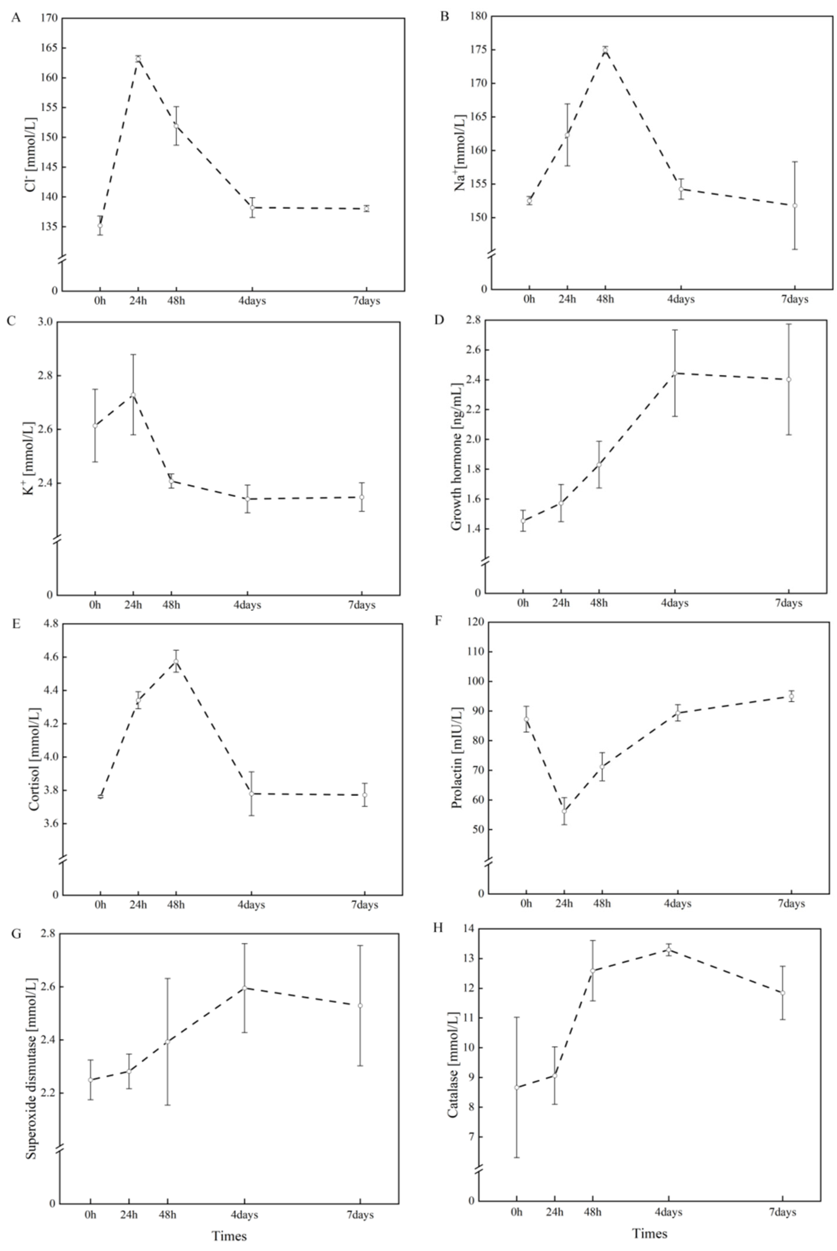
Following seawater exposure, various plasma parameters exhibited significant changes. Plasma [Cl−] concentrations showed an increasing trend, peaking on day 1 post-exposure (163.16 mmol/L) before largely returning to control levels (Figure 1A). Similarly, plasma [Na+] concentrations increased after seawater exposure, reaching a maximum (174.94 mmol/L) on day 2 (Figure 1B). In contrast, plasma [K+] concentrations decreased following seawater exposure, reaching their lowest (2.34 mmol/L) point on day 4 (Figure 1C).
Figure 1.
Plasma [Cl−], [Na+], [K+], GH, COR, PRL, SOD, CAT of freshwater masu salmon after 7 days exposure to rapid salinity change. (A) Mean ± SE values for plasma Na+ across the rapid salinity change. (B) Mean ± SE values for plasma Cl− across the rapid salinity change. (C) Mean ± SE values for plasma K+ across the rapid salinity change. (D) Mean ± SE values for plasma GH across the rapid salinity change. (E) Mean ± SE values for plasma COR across the rapid salinity change. (F) Mean ± SE values for plasma PRL across the rapid salinity change. (G) Mean ± SE values for plasma SOD across the rapid salinity change. (H) Mean ± SE values for plasma CAT across the rapid salinity change.
From day 4 to day 7, the levels of all ions remained relatively stable.
3.2.3. Hormone Levels
Following seawater exposure, plasma GH levels increased progressively, reaching a peak of 2.44 ng/mL on day 4. Plasma COR concentrations peaked at 4.57 mmol/L on day 2 post-exposure and then gradually declined. In contrast, plasma PRL exhibited an opposite trend, decreasing to a minimum of 56.22 mIU/L on day 1 before gradually increasing thereafter (Figure 1D).
From day 4 to day 7, all hormone levels remained relatively stable.
3.2.4. Antioxidant Activity
4. Discussion
This study systematically evaluated the effects of salinity on growth and development, stress responses, and hematological physiological parameters in masu salmon, through comparative analyses of gradual transfer experiments and direct transfer experiments. Gradual transfer experiment group indicated that, at a constant temperature of 10 °C, a gradual increase in salinity—D30 group (reaching the target salinity of 32 ppt within 30 days)—represented the optimal protocol for seawater transfer in masu salmon. In the direct transfer experiment, in addition to the activity of antioxidant enzymes, both ion concentrations and hormone indicators returned to a stable state within 7 days. These findings suggest that an extended, progressive salinity acclimation phase in estuarine environments prior to oceanic migration significantly enhances the physiological preparedness and seawater tolerance of masu salmon.
The growth and development of fish are closely related to their living environment, and salinity is an important environmental factor [31]. In masu salmon, significantly higher weight gain (WG), specific growth rate (SGR), protein efficiency ratio (PER), and condition factor (CF) were observed in individuals subjected to a gradual daily salinity increase of 1.1 ppt until reaching 32 ppt, compared to those experiencing more rapid increments of 1.6 or 3.2 ppt daily. These findings demonstrate that masu salmon successfully acclimate to seawater when the salinity transition occurs at a gradual rate of 1.1 ppt per day. The metabolic burden imposed by elevated salinity is well documented, with hypersaline conditions compromising growth performance, osmoregulatory capacity, physiological homeostasis, and immune function in species acclimated to freshwater [8,32]. This physiological cost stems from increased energy allocation toward maintaining ionic and osmotic balance, thereby limiting resources available for somatic growth [33]. These findings align with those reported in prior studies. A gradual transition from freshwater to seawater has been demonstrated to enhance physiological plasticity in fish, thereby improving their capacity for salinity acclimation [32,34].
The ability to maintain plasma ion concentrations within a narrow range is essential for fish to tolerate salinity changes when they migrate to new environments [35]. This is particularly important for euryhaline species such as salmonids, which can survive in saline environments and conditions approaching freshwater osmolarity [36]. Seawater-acclimated fish must counteract passive ion gain and water loss by drinking seawater and actively excreting excess [Na+] and [Cl−] through the gills [21]. Our results indicate that in the group subjected to a daily salinity increase of 1.6 ppt until reaching 32 ppt, plasma [Cl−] and [Na+] concentrations were significantly lower than those in groups with daily increases of 3.2 ppt and 1.1 ppt, suggesting that a rate of 1.6 ppt/ day may be optimal for regulating [Na+] and [Cl−] balance. However, since no significant differences were observed between day 20 and day 30 in the group with a daily increase of 1.1 ppt, the optimal salinity acclimation rate likely falls within the range of 1.1 to 1.6 ppt/ day. During the salinity increase acclimation period, plasma [K+] concentrations showed no significant response and are therefore not discussed in detail. After the salinity increase acclimation, plasma [K+] concentrations in the group subjected to a daily increase of 1.1 ppt were significantly lower than those in the groups with daily increases of 3.2 ppt and 1.6 ppt. Collectively, these findings indicate that gradually increasing salinity to 32 ppt at 1.1 ppt/day is the most effective acclimation strategy—well-acclimated fish efficiently excrete excess seawater ions and maintain stable osmolality post-transfer [17].
In the direct transfer experiment, a sharp increase in salinity led to elevated mortality of masu salmon, indicating an imbalance in their osmoregulatory capacity at that stage. However, by the later stage of the experiment, mortality had dropped to nearly zero, demonstrating that the masu salmon had gradually acclimated to the seawater environment and, within seven days, through self-regulation, progressively restored a stable osmoregulatory capacity [37,38].
The temporal changes in plasma ion concentrations in masu salmon after seawater entry reflect a complex physiological transition—from acute stress response to functional remodeling of the branchial ion transport system. In direct-transfer experiments, plasma [Na+] peaks at 48 h post-transfer, largely due to delayed activation of active [Na+] excretion. Although initial seawater ingestion causes a rapid rise in [Na+], branchial Na+/K+-ATPase (NKA) takes time to reach maximal secretory capacity—consistent with photoperiod-regulated, gradual NKA upregulation [39,40]. Consequently, [Na+] declines only after full NKA activation. In contrast, plasma [Cl−] peaks earlier, at 24 h, as [Cl−] enters the blood via passive diffusion through apical CFTR channels in gill ionocytes [40]. This process initiates faster than NKA- driven active excretion, yielding an earlier [Cl−] peak than [Na+]. Plasma [K+] shows a transient rise at 24 h, followed by a sustained decline below control levels—due to persistent NKA activity: for every 3 [Na+] ions extruded, 2 [K+] ions are imported [41]. During seawater acclimation, elevated branchial NKA activity shuttles [K+] from plasma into cells to maintain intracellular [K+] homeostasis, thereby reducing extracellular [K+]. Hence, plasma [K+] rises transiently then falls steadily in direct-transfer experiments, but remains unchanged during gradual transfer. In the latter, basolateral [K+] channels (e.g., Kir) or K+–Cl− cotransporters in gill ionocytes likely support transcellular [K+] transport [42]. Furthermore, plasma [K+] in sexually mature juvenile Atlantic salmon (Salmo salar) remains stable after 24 h of seawater exposure, indicating effective [K+] homeostasis at this developmental stage or under such acclimation [43]. These compensatory mechanisms likely offset the NKA- mediated intracellular [K+] shift, maintaining plasma [K+] stability.
Pituitary hormones, including PRL, GH, and COR, are responsible for endocrine regulation, metabolic processes, and growth and reproductive development in fishes [44]. The critical roles of GH and PRL in seawater and freshwater acclimation have been well established [45]. COR levels increase significantly in response to various physical, chemical, and biological stressors, and are widely used as indicators of acute stress [46,47]. In the gradual transfer experiment, COR levels in the group exposed to a daily salinity increase of 1.6 ppt until reaching 32 ppt were significantly lower than those in the 3.2 and 1.1 ppt groups. PRL levels in the group exposed to a daily salinity increase of 1.1 ppt until reaching 32 ppt were significantly higher than those in the 1.6 and 3.2 ppt groups. This indicates that the 3.2 ppt daily increase was the least favorable for growth and hormonal regulation in masu salmon.
The experimental results revealed that plasma prolactin levels remained consistently highest in the D30 treatment group throughout the duration of the experiment. In contrast, cortisol levels in the D30 group were intermediate—falling between those observed in the D10 and D20 groups—and exhibited a statistically significant negative correlation with prolactin levels. These findings corroborate the well-established antagonistic relationship between prolactin and cortisol in teleost osmoregulation: prolactin promotes freshwater acclimation while suppressing seawater acclimation, whereas cortisol facilitates seawater acclimation [39,48]. The persistently high prolactin expression coupled with a relatively moderate cortisol response in the D30 group suggests that under this salinity condition, the fish may exhibit reduced osmoregulatory capacity in seawater or experience endocrine dysregulation, resulting in insufficient suppression of prolactin secretion [49]. Future studies will further investigate the regulatory mechanisms underlying prolactin secretion.
In masu salmon, GH levels rise early during salinity increase. They then fall once the fish have fully acclimated. This pattern reflects the hormone’s dual roles. GH helps fish acclimate to seawater at first [14]. It boosts Na+/K+-ATPase activity in the gills. It also stimulates the growth of chloride cells. Together, these changes build the ability to excrete excess ions [50]. Fish in the 3.2 ppt daily increment group faced the strongest osmotic challenge. Their GH levels were the highest among all groups. This response likely maximized the development of their ion transport systems. After full acclimation, GH levels dropped sharply. Sustained high GH is no longer needed once gill function stabilizes [51]. Keeping it elevated would waste energy and might slow down growth [52]. Reducing GH helps conserve energy. It also allows the GH/IGF-I axis to take over. This shift promotes IGF-I synthesis and supports further growth [40]. This careful regulation is a hallmark of salmonid endocrinology. GH surges when rapid seawater acclimation is critical. It then declines to redirect resources toward growth. During the direct transfer experiment, GH, COR, and PRL reached their maximum or minimum values on day 4 after seawater exposure and remained stable from days 4 to 7, consistent with previous findings [18,53,54].
When exposed to adverse environmental stimuli, fish produce large amounts of ROS. SOD and CAT are key components of the antioxidant defense system in fish. They can intercept ROS produced by cells exposed to environmental stressors, thereby preventing cellular damage [55]. This mechanism enhances their resistance to oxidative stress [56,57,58]. In this study, plasma SOD activity in masu salmon was temporarily inhibited by salinity stress but increased during the acclimation period. SOD activity reached a maximum 4 days after seawater exposure and then declined, indicating an effective antioxidant response to rapid salinity changes. Conversely, salinity stress did not temporarily inhibit plasma CAT activity. The observed increases in CAT and SOD activities suggest that acute salinity exposure induces a transient stress state in masu salmon, potentially leading to oxidative damage [58].
Our results help clarify an important idea in salmon biology. Marine acclimation and smoltification are two different processes. They are not the same thing. Smoltification is a set of changes that prepare the fish for life in the sea. These changes happen in the fish’s body, behavior, and hormones. Photoperiod, or the length of daylight, mainly triggers them. Key signs include a silvery body color and higher NKA activity in the gills. The main point of these changes is to build a foundation. This foundation helps the fish later adjust to salty water [39,59]. Our experimental results demonstrate that direct transfer of the fish to the marine environment led to a significant increase in mortality. We also monitored their physical condition during the first four days post-transfer. Several key physiological parameters exhibited a marked upward trend—most notably, plasma concentrations of sodium and chloride ions. This pattern provided critical insight: although the fish displayed morphological features characteristic of a “pre-migratory state”, indicating readiness for ocean entry, their physiological status had not yet fully transitioned to support normal marine life. This finding points to a key difference. Smoltification gives the fish the basic setup and starting signals for moving to the sea. But actually living in the ocean takes more. It needs another set of changes. These are active adjustments in the body’s physiology and chemistry. They happen on their own schedule. And they are not done just because smoltification is finished.
Overall, although varying salinities altered the blood chemical composition of masu salmon, they did not adversely affect normal growth. This resilience likely reflects the strong innate ability of the species to regulate its internal osmotic balance, thereby highlighting its suitability to saline environments.
5. Conclusions
Through systematic salinity gradient experiments, this study demonstrated the physiological acclimation mechanisms of Oncorhynchus masou under varying salinity conditions. The findings of this study have significant theoretical and practical implications. The gradual transfer experiment group indicated that, at a constant temperature of 10 °C, a gradual increase in salinity—in the D30 group (reaching the target salinity of 32 ppt within 30 days)—represented the optimal protocol for seawater transfer in masu salmon. In the direct transfer experiment, in addition to the activity of antioxidant enzymes, both ion concentrations and hormone indicators returned to a stable state within 7 days. This study established two distinct modes of salinity regulation: gradual salinity gradient changes and acute salinity fluctuations. By measuring key physiological indicators, including plasma ion concentration, hormone levels, and antioxidant enzyme activity, we systematically clarified the regulatory strategies employed by masu salmon in response to salinity stress. These results provide a precise implementation plan for managing salinity during the artificial breeding of masu salmon and establishing critical domestication parameters for land-based recirculating aquaculture systems for marine salmon farming. From an ecological perspective, demonstrating salinity acclimation mechanisms offers novel insights into the recovery of the upstream spawning fish populations. By simulating natural salinity trajectories, the marine acclimation capacity of artificially bred juvenile fish can be enhanced, which is crucial for supporting declining populations of wild masu salmon. Furthermore, this study serves as a methodological reference for the domestication of other economically important salmonids in seawater environments, thereby promoting precision and standardization in marine fish farming practices.
Supplementary Materials
The following supporting information can be downloaded at: https://www.mdpi.com/article/10.3390/fishes11030179/s1, The Supplementary Materials comprise three sections: (i) the experimental design for the gradual transfer experiment; (ii) the experimental design for the direct transfer experiment; and (iii) the methodology employed to obtain data on weight gain (WG), condition factor (CF), and specific growth rate (SGR). Figure S1: During the gradual transfer experiment, fish were assigned to three treatment groups (D10, D20, and D30). Each group consisted of three replicate buckets, with 30 anadromous masu salmon per bucket. The three groups served as mutual controls. Figure S2: In the direct transfer experiment, a total of 90 anadromous individuals were subjected to abrupt salinity change. Baseline samples were collected from all fish in freshwater at 0 h to serve as the control. Subsequently, the fish were rapidly transferred to seawater, and additional samples were collected at 24 h, 48 h, 4 days, and 7 days post-transfer. Notably, this experimental design did not include a separate, parallel control group maintained in freshwater throughout the duration of the experiment. Figure S3: The images were analyzed using ImageJ software to quantify the total length, fork length, and caudal peduncle length of the fish. Subsequently, weight gain (WG), specific growth rate (SGR), and condition factor (CF) were calculated.
Author Contributions
X.Y. and S.S. conceived this study, designed this experiment, and wrote the manuscript. S.S., Y.G. and D.Y. performed this experiment. S.S. and J.L. analyzed the data. S.S. prepared the tables and figures. H.N. is coordinating sample acquisition. All authors have read and agreed to the published version of the manuscript.
Funding
This work was funded by the Natural Science Foundation of Liaoning Province (2024-MS-178), and the Science and Technology Development Program of Jilin Province (20220202068NC).
Institutional Review Board Statement
All experimental protocols were approved by Animal Research and Ethics Committee of Dalian Ocean University (No. DLOU2024001, approval date 8 January 2019).
Data Availability Statement
The original contributions presented in this study are included in the article and Supplementary Material. Further inquiries can be directed to the corresponding author.
Acknowledgments
We thank C. Liu, S. Liang, Y. Pan, G. Hu and H. Dong for technical assistance during the work.
Conflicts of Interest
The authors declare no conflicts of interest.
References
- Scanlan, M.M.; Putman, N.F.; Pollock, A.M.; Noakes, D.L.G. Magnetic map in nonanadromous Atlantic salmon. Proc. Natl. Acad. Sci. USA 2018, 115, 201807705. [Google Scholar] [CrossRef] [Scilit] [PubMed]
- Ueda, H. Sensory mechanisms of natal stream imprinting and homing in Oncorhynchus spp. J. Fish Biol. 2019, 95, 293–303. [Google Scholar] [CrossRef] [Scilit] [PubMed]
- Mozanzadeh, M.T.; Safari, O.; Oosooli, R.; Mehrjooyan, S.; Najafabadi, M.Z.; Hoseini, S.J.; Saghavi, H.; Monem, J. The effect of salinity on growth performance, digestive and antioxidant enzymes, humoral immunity and stress indices in two euryhaline fish species: Yellowfin seabream (Acanthopagrus latus) and Asian seabass (Lates calcarifer). Aquaculture 2021, 534, 736329. [Google Scholar] [CrossRef] [Scilit]
- Cao, Q.; Li, J.; Sun, Y.; Geng, D.; Chu, P.; Wang, H.; Yin, S. Time-course studies of osmoregulatory responses in different salinities for the marbled eel (Anguilla marmorata). Aquaculture 2021, 540, 736699. [Google Scholar] [CrossRef] [Scilit]
- Futamura, R.; Morita, K.; Kanno, Y.; Kumikawa, S.; Matsuoka, Y.; Okuda, A.; Sugiyama, H.; Takahashi, H.; Uchida, J.; Kishida, O. Size-dependent growth tactics of a partially migratory fish before migration. Oecologia 2022, 198, 371–379. [Google Scholar] [CrossRef] [Scilit]
- Schwabl, H.; Groothuis, T. Maternal Effects on Behavior. In Reviews in Encyclopedia of Animal Behavior, 2nd ed.; Choe, J.C., Ed.; Elsevier Ltd.: Amsterdam, The Netherlands, 2019; pp. 483–494. [Google Scholar]
- Kuroki, M.; Tamate, T.; Morita, K. An additional life-history tactic of masu salmon: Migration of parr to coastal habitats. Ecol. Freshw. Fish 2020, 29, 495–501. [Google Scholar] [CrossRef] [Scilit]
- Sun, S.; Pan, Y.; Hu, P.; Dong, H.; Chen, C.; Liu, S.; Yin, X. Acute Salinity Challenges in Oncorhynchus masou Under Warm Temperature: Differential Responses of Osmolality, Plasma Ions, Hormone Levels, and Immune Parameters. Aquac. Res. 2025, 2025, 1889801. [Google Scholar] [CrossRef] [Scilit]
- Liang, S.; Song, B.; Chong, X.; Zheng, W.; Nie, H.; Yin, X. Phylogenetic position and genetic diversity of masu salmon Oncorhynchus masou in the Mijiang River, China. Ichthyol. Res. 2023, 70, 402–405. [Google Scholar] [CrossRef] [Scilit]
- Lenders, H.; Chamuleau, T.; Hendriks, A.; Lauwerier, R.; Leuven, R.; Verberk, W. Historical rise of waterpower initiated the collapse of salmon stocks. Sci. Rep. 2016, 6, 29269. [Google Scholar] [CrossRef] [Scilit]
- Disco, N. Losing the Salmon: Technology, Science, and Ecological Decline on the Rhine. Technol. Cult. 2026, 67, 11–40. [Google Scholar] [CrossRef] [Scilit]
- Uematsu, Y.; Abe, M.; Yada, T.; Matsuda, H.; Munakata, A. Exploration of selection methods for optimal juveniles in hatchery-reared masu salmon (Oncorhynchus masou). J. Fish Biol. 2025; ahead of print. [CrossRef] [Scilit] [PubMed]
- Jiang, L.; Hu, L.; Huang, S.; Wang, W.; Yu, K.; Zhang, L. Industrialized Breeding and Cultivation Techniques for Rainbow Trout Fry. Sci. Fish Farming 2017, 11, 8–9. (In Chinese) [Google Scholar] [CrossRef]
- Hayashi, M.; Maruoka, S.; Oikawa, J.; Ugachi, Y.; Shimizu, M. Effects of Acclimation to Diluted Seawater on Metabolic and Growth Parameters of Underyearling Masu Salmon (Oncorhynchus masou). Zool. Sci. 2021, 38, 513–522. [Google Scholar] [CrossRef] [Scilit] [PubMed]
- Herrera, M.; Salamanca, N.; Ferrer, J.F.; De La Rosa, I. Use of tissue chromogranin A as chronic and acute stress marker in fish. Aquaculture 2023, 565, 739120. [Google Scholar] [CrossRef] [Scilit]
- Ciavoni, E.; Schrama, J.W.; Radhakrishnan, G.; Sæle, Ø.; Prabhu Philip, A.J. Salinity induced changes in the progression of water, ion and nutrient fluxes along the gastrointestinal tract of Atlantic salmon smolt (Salmo salar). Aquaculture 2024, 580, 740331. [Google Scholar] [CrossRef] [Scilit]
- Xiong, Y.; Dong, S.; Huang, M.; Li, Y.; Wang, X.; Wang, F.; Ma, S.; Zhou, Y. Growth, osmoregulatory response, adenine nucleotide contents, and liver transcriptome analysis of steelhead trout (Oncorhynchus mykiss) under different salinity acclimation methods. Aquaculture 2020, 520, 734937. [Google Scholar] [CrossRef] [Scilit]
- Flores, A.M.; Shrimpton, J.M. Differential physiological and endocrine responses of rainbow trout, Oncorhynchus mykiss, transferred from fresh water to ion-poor or salt water. Gen. Comp. Endocrinol. 2012, 175, 244–250. [Google Scholar] [CrossRef] [Scilit]
- Beacham, T.D. Early survival and growth of pink salmon (Oncorhynchus gorbuscha) in fresh and saline water. Aquaculture 1992, 106, 151–159. [Google Scholar] [CrossRef] [Scilit]
- Rhee, J.-S.; Kim, B.-M.; Seo, J.S.; Kim, I.-C.; Lee, Y.-M.; Lee, J.-S. Cloning of growth hormone, somatolactin, and their receptor mRNAs, their expression in organs, during development, and on salinity stress in the hermaphroditic fish, Kryptolebias marmoratus. Comp. Biochem. Physiol. Part A Mol. Integr. Physiol. 2012, 161, 436–442. [Google Scholar] [CrossRef] [Scilit]
- Mancera, J.M.; Carrión, R.L.; del Río, M.D.P.M. Osmoregulatory action of PRL, GH, and cortisol in the gilthead seabream (Sparus aurata L.). Gen. Comp. Endocrinol. 2002, 129, 95–103. [Google Scholar] [CrossRef] [Scilit]
- Aruna, A.; Nagarajan, G.; Chang, C.F. The acute salinity changes activate the dual pathways of endocrine responses in the brain and pituitary of tilapia. Gen. Comp. Endocrinol. 2015, 211, 154–164. [Google Scholar] [CrossRef] [Scilit] [PubMed]
- Cao, Y.; Tveten, A.-K.; Stene, A. Establishment of a NonInvasive Method for Stress Evaluation in Farmed Salmon Based on Direct Fecal Corticoid Metabolites Measurement. Fish Shellfish Immunol. 2017, 66, 317–324. [Google Scholar] [CrossRef] [Scilit] [PubMed]
- O’Toole, C.; White, P.; Thomas, K.; O’Maoiléidigh, N.; Fjelldal, P.; Hansen, T.; Graham, C.; Brophy, D. Effects of Temperature and Feeding Regime on Cortisol Concentrations in Scales of Atlantic Salmon Post-Smolts. J. Exp. Mar. Biol. Ecol. 2023, 569, 151955. [Google Scholar] [CrossRef] [Scilit]
- Pankhurst, N.; Ludke, S.; King, H.R.; Peter, R. The Relationship Between Acute Stress, Food Intake, Endocrine Status and Life History Stage in Juvenile Farmed Atlantic Salmon, Salmo salar. Aquaculture 2008, 275, 311–318. [Google Scholar] [CrossRef] [Scilit]
- Al-Jandal, N.J.; Wilson, R.W. A comparison of osmoregulatory responses in plasma and tissues of rainbow trout (Oncorhynchus mykiss) following acute salinity challenges. Comp. Biochem. Physiol. A Mol. Integr. Physiol. 2011, 159, 175–181. [Google Scholar] [CrossRef] [Scilit]
- Olsvik, P.; Kristensen, T.; Waagbø, R.; Rosseland, B.; Tollefsen, K.-E.; Baeverfjord, G.; Berntssen, M. mRNA expression of antioxidant enzymes (SOD, CAT and GSH-Px) and lipid peroxidative stress in liver of Atlantic salmon (Salmo salar) exposed to hyperoxic water during smoltification. Comp. Biochem. Physiol. Part C Toxicol. Pharmacol. 2005, 141, 314–323. [Google Scholar] [CrossRef] [Scilit]
- Li, X.; Shen, Y.; Bao, Y.; Wu, Z.; Yang, B.; Jiao, L.; Zhang, C.; Tocher, D.R.; Zhou, Q.; Jin, M. Physiological responses and adaptive strategies to acute low-salinity environmental stress of the euryhaline marine fish black seabream (Acanthopagrus schlegelii). Aquaculture 2022, 554, 738117. [Google Scholar] [CrossRef] [Scilit]
- Xu, M.; Zhang, C.; Qi, Q.; Wang, R.; Zhang, S.; Yan, R.; Li, B.; Li, S. Effects of salinity stress on anxiety behavior and antioxidant capability of guppy (Poecilia reticulata). Ecotoxicology 2023, 32, 598–605. [Google Scholar] [CrossRef] [Scilit]
- Mubarik, M.S.; Asad, F.; Zahoor, M.K.; Abid, A.; Ali, T.; Yaqub, S.; Ahmad, S.; Qamer, S. Study on survival, growth, haematology and body composition of Cyprinus carpio under different acute and chronic salinity regimes. Saudi J. Biol. Sci. 2019, 26, 999–1002. [Google Scholar] [CrossRef] [Scilit]
- Liu, J.; Ai, T.; Yang, J.; Shang, M.; Jiang, K.; Yin, Y.; Gao, L.; Jiang, W.; Zhao, N.; Ju, J.; et al. Effects of Salinity on Growth, Digestive Enzyme Activity, and Antioxidant Capacity of Spotbanded Scat (Selenotoca multifasciata) Juveniles. Fishes 2024, 9, 309. [Google Scholar] [CrossRef] [Scilit]
- van Muilekom, D.; Mueller, J.; Lindemeyer, J.; Schultheiß, T.; Maser, E.; Seibel, H.; Rebl, A.; Schulz, C.; Goldammer, T. Salinity change evokes stress and immune responses in Atlantic salmon with microalgae showing limited potential for dietary mitigation. Front. Physiol. 2024, 15, 1338858. [Google Scholar] [CrossRef] [Scilit]
- Nuic, B.; Bowden, A.; Franklin, C.E.; Cramp, R.L. Atlantic salmon Salmo salar do not prioritize digestion when energetic budgets are constrained by warming and hypoxia. J. Fish Biol. 2024, 104, 1718–1731. [Google Scholar] [CrossRef] [Scilit] [PubMed]
- Kültz, D. Physiological mechanisms used by fish to cope with salinity stress. J. Exp. Biol. 2015, 218, 1907–1914. [Google Scholar] [CrossRef] [Scilit] [PubMed]
- Stich, D.; Zydlewski, G.; Zydlewski, J. Physiological preparedness and performance of Atlantic salmon Salmo salar smolts in relation to behavioural salinity preferences and thresholds. J. Fish Biol. 2016, 88, 595–617. [Google Scholar] [CrossRef] [Scilit] [PubMed]
- Kaivarainen, E.; Rendakov, N.; Efremov, D.; Nemova, N. Na+/K+-ATPase Activity in Smolts of Pink Salmon Oncorhynchus gorbuscha (Walbaum, 1792) from the White Sea Exposed to Fresh, Estuarine, and Sea Water. Dokl. Biol. Sci. 2021, 501, 201–205. [Google Scholar] [CrossRef] [Scilit]
- McCormick, S.; Moyes, C.; Ballantyne, J. Influence of Salinity on the Energetics of Gill and Kidney of Atlantic Salmon (Salmo salar). Fish Physiol. Biochem. 1989, 6, 243–254. [Google Scholar] [CrossRef] [Scilit]
- Prunet, P.; Boeuf, G. Plasma Prolactin Level During Transfer of Rainbow Trout (Salmo gairdneri) and Atlantic Salmon (Salmo salar) From Fresh Water to Sea Water. Aquaculture 1985, 45, 167–176. [Google Scholar] [CrossRef] [Scilit]
- Suzuki, S.; Takahashi, E.; Nilsen, T.; Kaneko, N.; Urabe, H.; Ugachi, Y.; Yamaha, E.; Shimizu, M. Physiological changes in off-season smolts induced by photoperiod manipulation in masu salmon (Oncorhynchus masou). Aquaculture 2020, 526, 735353. [Google Scholar] [CrossRef] [Scilit]
- Mackie, P.; Gharbi, K.; Ballantyne, J.; McCormick, S.; Wright, P. Na+/K+/2Cl− cotransporter and CFTR gill expression after seawater transfer in smolts (0+) of different Atlantic salmon (Salmo salar) families. Aquaculture 2007, 272, 625–635. [Google Scholar] [CrossRef] [Scilit]
- Tipsmark, C.; Madsen, S.; Seidelin, M.; Christensen, A.; Cutler, C.; Cramb, G. Dynamics of Na+,K+,2Cl− cotransporter and Na+,K+-ATPase expression in the branchial epithelium of brown trout (Salmo trutta) and atlantic salmon (Salmo salar). J. Exp. Zool. 2002, 293, 106–118. [Google Scholar] [CrossRef] [Scilit]
- Kiilerich, P.; Kristiansen, K.; Madsen, S.S. Cortisol regulation of ion transporter mRNA in Atlantic salmon gill and the effect of salinity on the signaling pathway. J. Endocrinol. 2007, 194, 417–427. [Google Scholar] [CrossRef] [Scilit] [PubMed]
- Rydevik, M.; Borg, B.; Haux, C.; Kawauchi, H.; Björnsson, B.T. Plasma growth hormone levels increase during seawater exposure of sexually mature Atlantic salmon parr (Salmo salar L.). Gen. Comp. Endocrinol. 1990, 80, 9–15. [Google Scholar] [CrossRef] [Scilit] [PubMed]
- Hu, Q.; Qin, Q.; Xu, S.; Zhou, L.; Xia, C.; Shi, X.; Zhang, H.; Jia, J.; Ye, C.; Yin, Z.; et al. Pituitary Actions of EGF on Gonadotropins, Growth Hormone, Prolactin and Somatolactins in Grass Carp. Biology 2020, 9, 279. [Google Scholar] [CrossRef] [Scilit] [PubMed]
- Yuan, M.; Jia, Q.; Wang, T.; Lu, Q.; Tang, L.; Wang, Y.; Lu, W. Dynamic responses of prolactin, growth hormone and their receptors to hyposmotic acclimation in the olive flounder Paralichthys olivaceus. Gen. Comp. Endocrinol. 2017, 254, 8–13. [Google Scholar] [CrossRef] [Scilit]
- Kammerer, B.D.; Cech, J.J., Jr.; Kültz, D. Rapid changes in plasma cortisol, osmolality, and respiration in response to salinity stress in tilapia (Oreochromis mossambicus). Comp. Biochem. Physiol. A Mol. Integr. Physiol. 2010, 157, 260–265. [Google Scholar] [CrossRef] [Scilit]
- Chang, R.J.A.; Celino-Brady, F.T.; Seale, A.P. Changes in cortisol and corticosteroid receptors during dynamic salinity challenges in Mozambique tilapia. Gen. Comp. Endocrinol. 2023, 342, 114340. [Google Scholar] [CrossRef] [Scilit]
- Martine, A.; Graham, Y.; Patrick, P.; Carl, B.S. Plasma prolactin and cortisol concentrations during salinity challenges of coho salmon (Oncorhynchus kisutch) at smolt and post-smolt stages. Aquaculture 1990, 91, 359–372. [Google Scholar] [CrossRef] [Scilit]
- Ágústsson, T.; Sundell, K.; Sakamoto, T.; Ando, M.; Björnsson, B.T. Pituitary gene expression of somatolactin, prolactin, and growth hormone during Atlantic salmon parr–smolt transformation. Aquaculture 2003, 222, 229–238. [Google Scholar] [CrossRef] [Scilit]
- Nakajima, T.; Shimura, H.; Yamazaki, M.; Fujioka, Y.; Ura, K.; Hara, A.; Shimizu, M. Lack of hormonal stimulation prevents the landlocked Biwa salmon (Oncorhynchus masou subspecies) from adapting to seawater. Am. J. Physiol.-Regul. Integr. Comp. Physiol. 2014, 307, R414–R425. [Google Scholar] [CrossRef] [Scilit]
- Sakamoto, T.; McCormick, S.D. Prolactin and growth hormone in fish osmoregulation. Gen. Comp. Endocrinol. 2006, 147, 24–30. [Google Scholar] [CrossRef] [Scilit]
- Young, G.; Björnsson, B.T.; Prunet, P.; Lin, R.J.; Bern, H.A. Smoltification and seawater adaptation in coho salmon (Oncorhynchus kisutch): Plasma prolactin, growth hormone, thyroid hormones, and cortisol. Gen. Comp. Endocrinol. 1989, 74, 335–345. [Google Scholar] [CrossRef] [Scilit] [PubMed]
- Ojima, D.; Pettersen, R.J.; Wolkers, J.; Johnsen, H.K.; Jørgensen, E.H. Growth hormone and cortisol treatment stimulate seawater tolerance in both anadromous and landlocked Arctic charr. Comp. Biochem. Physiol. Part A Mol. Integr. Physiol. 2009, 153, 378–385. [Google Scholar] [CrossRef] [Scilit]
- Breves, J.P.; Hasegawa, S.; Yoshioka, M.; Fox, B.K.; Davis, L.K.; Lerner, D.T.; Takei, Y.; Hirano, T.; Grau, E.G. Acute salinity challenges in Mozambique and Nile tilapia: Differential responses of plasma prolactin, growth hormone and branchial expression of ion transporters. Gen. Comp. Endocrinol. 2010, 167, 135–142. [Google Scholar] [CrossRef] [Scilit]
- Chang, C.H.; Mayer, M.; Rivera-Ingraham, G.; Blondeau-Bidet, E.; Wu, W.Y.; Lorin-Nebel, C.; Lee, T.H. Effects of temperature and salinity on antioxidant responses in livers of temperate (Dicentrarchus labrax) and tropical (Chanos chanos) marine euryhaline fish. J. Therm. Biol. 2021, 99, 103016. [Google Scholar] [CrossRef] [Scilit]
- Saoud, I.P.; Kreydiyyeh, S.; Chalfoun, A.; Fakih, M. Influence of salinity on survival, growth, plasma osmolality and gill Na+-K+-ATPase activity in the rabbitfish Siganus rivulatus. J. Exp. Mar. Biol. Ecol. 2007, 348, 183–190. [Google Scholar] [CrossRef] [Scilit]
- Gonzalez, P.; Dominique, Y.; Massabuau, J.C.; Boudou, A.; Bourdineaud, J.P. Comparative effects of dietary methylmercury on gene expression in liver, skeletal muscle, and brain of the zebrafish (Danio rerio). Environ. Sci. Technol. 2005, 39, 3972–3980. [Google Scholar] [CrossRef] [Scilit] [PubMed]
- Cohen-Sánchez, A.; Box, A.; Valencia, J.M.; Pinya, S.; Tejada, S.; Sureda, A. Exploring the impact of high salinity and parasite infection on antioxidant and immune systems in Coris julis in the Pityusic Islands (Spain). Sci. Total Environ. 2024, 951, 175848. [Google Scholar] [CrossRef] [Scilit] [PubMed]
- Ugachi, Y.; Kitade, H.; Takahashi, E.; Suzuki, S.; Hayashi, M.; Yamada, T.; Cui, W.; Shimizu, M. Size-driven parr-smolt transformation in masu salmon (Oncorhynchus masou). Sci. Rep. 2023, 13, 16643. [Google Scholar] [CrossRef] [Scilit]

Disclaimer/Publisher’s Note: The statements, opinions and data contained in all publications are solely those of the individual author(s) and contributor(s) and not of MDPI and/or the editor(s). MDPI and/or the editor(s) disclaim responsibility for any injury to people or property resulting from any ideas, methods, instructions or products referred to in the content. |
© 2026 by the authors. Licensee MDPI, Basel, Switzerland. This article is an open access article distributed under the terms and conditions of the Creative Commons Attribution (CC BY) license.