


Abstract
This pioneering study investigates bacteria isolated from marine pelagic fish, fishing vessels, and gear surfaces, focusing on the variability in biofilm formation across different substrates, media, and cultivation conditions. Bacteria from fish intestines, skin, and gills, including spoilage organisms and potential fish and human pathogens, can contaminate vessel surfaces, gear, and containers and may act as microbial reservoirs and transmission vectors. In this study, biofilm formation was evaluated at air–liquid interfaces and on submerged plastic, metal, and glass surfaces under various incubation temperatures and media. Vibrio spp. were isolated both from fishing nets and fish gills, particularly Vibrio alginolyticus, V. gigantis, and V. pelagius. Although V. harveyi was examined as a representative vibrio, it did not form a biofilm on polypropylene. Photobacterium damselae subsp. damselae, Pseudomonas fragi, P. gessardii, Psychrobacter spp., and Rothia endophytica showed a strong affinity for stainless steel. Overall adhesion regardless of media type was highest for P. gessardii, followed by P. damselae and Aeromonas veronii, which adhered strongly to steel, glass, and polypropylene; however, only P. gessardii also adhered well to polystyrene, an important finding because these are known fish and human pathogens. These results highlight species-dependent biofilm triggers and their substantial variability and provide guidance for standardized marine biofilm protocols.
Key Contribution:
To date, there is no single method or unified framework that provides consistent results for different bacteria or for a single species that would be suitable for a range of applications and purposes. This work is, thus, conceived as a guiding principle for testing biofilm formation of marine/fish/pathogenic bacteria on various surfaces. It also demonstrates that bacterial biofilms formed on fishing boat surfaces harbor bacterial communities similar to those isolated from anchovy and sardine catches. These biofilms may act as microbial reservoirs, facilitating bacterial transfer and potential cross-contamination within the fishing and aquaculture environments.
1. Introduction
Bacterial biofilms consist of structured, surface-attached sessile bacterial communities stabilized in self-produced extracellular polymeric substances (EPSs). EPSs comprise polysaccharides, glycoproteins, proteins, glycolipids, extracellular nucleic acids, and components of lysed bacteria, which act as a biofilm adhesive for surface attachment and provide nutrient supply and protection [1,2]. EPS protects the embedded multispecies bacterial cells from external elements, atmospheric and aquatic factors, as well as antimicrobial agents [3]. Not only do biofilms enhance the ability of bacterial communities to adapt to adverse conditions but they also enable them to enter dormancy in unfavorable conditions, thus allowing pathogens to survive in a hostile environment [4]. Biofilm development generally involves several stages: (1) reversible adhesion of bacteria to the surface; (2) production of a monolayer on the surface by bacterial adhesins, as well as production of a gel-like extracellular matrix; (3) formation of multilayer microcolonies and production of EPS; (4) biofilm maturation and formation of typical polysaccharide structures; and (5) detachment and dissemination of bacterial cells, with a return to the planktonic form [4].
Bacterial biofilms occur on all kinds of natural and engineered surfaces, existing in submerged habitats as well as on liquid surfaces. Biofilms develop easily at interfaces, including solid/liquid interfaces in humid or aqueous environments, as well as liquid/gas or liquid/liquid interfaces of different densities [5]. Light, irradiance, flow velocity and temperature affect phototrophic biofilms in aquatic systems [2]. In the marine environment, the adhesion to surfaces starts shortly after immersion; bacterial colonization starts after an hour; and colonization by unicellular eukaryotes, such as diatoms, yeasts and protozoa, occurs several days post-immersion [6,7]. The characteristics of the substratum are an important factor for the rate and extent of bacterial attachment, as rougher and more hydrophobic materials will develop biofilms more rapidly. In addition, the characteristics of the cell surface also matter, as the presence of flagella, pili, fimbriae or glycocalyx may affect the rate of attachment [8].
In intertidal zones, biofilms offer particular protection to microorganisms against temporary stress caused by UV exposure, intermittent dehydration, and limited access to nutrients [6,9]. In such environments, cells experience rapid cold/warm temperatures, high/low salinity, dried/immersed conditions, irradiance and pH variations [10,11]. Similar conditions may be expected on the surfaces of fishing vessels and their fishing gear, which may act as a microbial repository and vector for the transfer of potentially pathogenic microorganisms. Bacteria are typically found in aquatic animals’ intestinal tracts and on outer surfaces, such as skin, shell and gills. Fish also carry indigenous microbiota, which include spoilage bacteria and, in rare cases, potential human pathogens [5]. On fishing vessels, they come into contact with surfaces of fishing and handling gear, containers, and storage surfaces. Contamination can occur in both directions through water, personnel, and inadequate cleaning of surfaces [12].
Therefore, with the aim of investigating typical surfaces of an operating fishing vessel for bacterial contamination and biofilm production, we identified cultivated bacterial species from the vessel’s plastic, wooden, glass and steel surfaces, as well as from the pelagic fish hauled on board. We tested the identified bacterial species for biofilm production and quantified their biofilm production. Biofilm formation was investigated at the air–liquid interface and in immersed conditions on surfaces of plastic, metal and glass objects. Bacteria were grown and analyzed in 12 different culture media at three incubation temperatures in biological and technical replicates. Comprehensive testing was performed on 10 selected bacterial species, with a greater emphasis on human and aquaculture/marine fish bacterial pathogens.
2. Materials and Methods
2.1. Sample Collection
Surfaces of a pelagic fishing seine-haul boat operating in the Eastern Adriatic Sea were swabbed. At the same time, swabs were taken from the gills of fished anchovies (Engraulis encrasicolus) and sardines (Sardina pilchardus), hauled by the fishing crew. Gills were selected instead of skin due to being less prone to cross-contamination between individual fish specimens for investigations of microbial diversity between samples and seasons [13]. Samples were collected from various surfaces: plastic (dry fishing net); steel (refrigeration machine for netted fish, hand-railings and ship hull); glass windows; and wood (hand-railings, helm and ropes). Fish and fishing nets were always sampled as the primary goal, and other surfaces were sampled once or twice. Samples were collected in the spring and fall of two years during the operational period of the seiner, when fully staffed and engaged. A sampling area of 10 cm2 was defined with a sterile template where possible, while round and uneven surfaces were swabbed without a template but with the approximation of a 10 cm2 area. Samples were swabbed from surfaces that come into contact with fish, human skin, seawater, aerosol, light, and rain (except the refrigeration surfaces, which are not under the influence of the weather). Samples were aseptically swabbed with a sterile cotton swab with an applicator stick and inserted into the SRK Neutralizing Transport Medium (Copan, Zagreb, Croatia).
All experiments were conducted as per the guidelines of the Institutional Animal Ethics Committee of the Ruđer Bošković Institute, Zagreb, Croatia. However, fish used in this study were captured for commercial purposes. Therefore, use of these animals in research does not require ethical approval.
2.2. Sample Analyses
Samples were transported to the laboratory in plastic tubes stored in a portable cooler. The analyses commenced 48 h after swabbing. Samples were streaked onto Zobell Marine Agar (HiMedia, Einhausen, Germany) and incubated at 25 °C for 48–72 h. Representative colonies deriving from each of the surfaces were subcultured on fresh medium until purity was attained. Colonies were subsequently Gram-stained and subjected to standard physiological, morphological, biochemical tests and identification by MALDI-TOF MS. Bacterial isolates were stored via cryopreservation in marine broth supplemented with 15% (v/v) glycerol at −80 °C.
2.3. MALDI-TOF MS Identification
Three biological replicates of each isolate were applied on a 96-spot polished stainless-steel target plate. All samples were prepared using the on-target extraction method as previously described [14]. All procedures were conducted in duplicates or triplicates. The on-target extraction comprised a smear of a colony loopful from each selected strain on a target plate (Bruker Daltonics, Bremen, Germany). Then, 1 μL of 70% formic acid (Kemika, Zagreb, Croatia) was added to each spot overlying the bacterial colony and left to dry at room temperature. To each spot, 1 μL of MALDI matrix was added and allowed to air dry at 22 °C (saturated solution of α-cyano-4-hydroxycinnamic acid in 50% acetonitrile and 2.5% trifluoroacetic acid (Bruker Daltonics)). For measuring the mass spectra, a bench-top Bruker Microflex LT mass spectrometer equipped with the Bruker Biotyper 3.0 software (Bruker Daltonics) was used, according to the manufacturer’s instructions. The calibration was conducted with Escherichia coli DH5 alpha spiked with two additional pure proteins (RNAse A and myoglobin) covering a mass range 4–17 kDa. Ions were captured in the positive linear mode, and positive ions were extracted at accelerated voltage of 20 kV. Spectra with the sum of the respective ions were obtained by 240 laser shots in different regions of every target plate spot. All spectra obtained were compared to the reference mass spectra in the database. A log score of <1.700 was considered unreliable, a score of 1.700 to 1.999 indicated probable identification to the genus level, a score of 2.000 to 2.299 indicated probable species identification, while a log score of 2.300 to 3.000 indicated highly probable species-level identification.
2.4. Bacterial Biofilm Formation
Biofilm formation was evaluated aerobically at 14, 22 and 35 °C in 96-well flat-bottomed, clear, polystyrene microtiter plates (Deltalab, Barcelona, Spain). Twelve different broths were made using 4 base media: Zobell Marine broth (ZMB, HiMedia), M9 (Milipore, Zug, Switzerland) modified through supplementation with casamino acids and 1% (w/v) glucose, Mueller-Hinton (MHB, Merck, Darmstadt, Germany) and tryptic soy (TSB, Biolife, Monza, Italy). TSB broth had 9 modifications: 3 pH levels were prepared for unmodified TSB, TSB supplemented with 1.5% (w/v) NaCl and 10% TSB (nutrient depletion to 10% with NaCl correction to basal level). Adjustments to pH were performed by titration with 1M NaOH/HCl to 6.3 for acidic and 8.3 for alkaline, while for neutral pH, no modification was necessary (pH 7.2–7.3).
Microtiter plate procedure based on crystal violet staining of total biomass (cells and matrix) was adapted from Stepanović et al. [15]. Briefly, strains were first grown on tryptic soy agar (TSA, Biolife) aerobically at 22 °C overnight. A loopful of pure colonies was suspended in 500 µL of PBS, from which 50 µL was added to 5 mL of TSB in 15 mL Falcon tubes and cultivated with shaking (200 rpm) overnight. Fresh exponential growth was diluted 1:40 by transferring 5 µL to freshly prepared microtiter plates with 195 µL of sterile media in wells. Tests were performed in at least technical duplicates with two biological replicates (different days). After incubation, the contents of the wells were discarded, and wells were washed 3× with 300 μL of sterile phosphate-buffered saline (PBS) to remove planktonic growth. After washing, the plates were inverted and remaining attached bacteria were heat-fixed at 60 °C for 1 h. Fixed biofilm was stained with 200 µL of 0.1% (v/v) filtered crystal violet for 15 min. The stain was washed by 3× immersion in water, and plates were left inverted to dry overnight. The stain was eluted from the attached cells with 150 μL (for biofilm at submerged surface only) of 95% ethanol per well for 30 min at room temperature and then scanned at 590 nm wavelength (OD590) with Infinite 200 Pro microplate reader (Tecan, Grödig, Austria). Another 50 µL of ethanol was then added and scanned after another 30 min to include the surface of the air–liquid interface as well. The choice of the endpoint generally depends on the aims and purposes and appropriate context. For instance, biofilm on submerged surface only is more relevant for fish, aquaculture tubes and materials that are continuously submerged (e.g., bone implants in human medicine). In turn, air–liquid interface would be preferred for materials that are partly submerged or afloat (e.g., catheters in human medicine) or certain ecological contexts such as inter-tidal zones. The cutoff value (ODc) was established in every experimental run and for each microtiter plate separately. Negative controls, blank media, were used to determine the ODc by adding 3 standard deviations to the average of all replicates. According to the ratio of OD590 of samples and ODc, the strain is categorized as follows: no biofilm producer (≤1), weak biofilm producer (>1 and ≤2), moderate biofilm producer (>2 and ≤4) and strong biofilm producer (>4). While these cutoffs are arbitrary, fixed OD590 is not advised, and the ODratio approach is the most widely used and considered the best choice [15]. Similarly, the ratio of planktonic growth (OD600 nm) was used for semi-quantitative evaluation of the overall bacterial growth in a comparable manner. Control strains for quality assurance were Pseudomonas aeruginosa PAO1 and S. aureus DSM 1104.
The surfaces of different materials tested for biofilm formation of selected bacteria on objects submerged in three different broths (TSB, TSB + 1.5% NaCl, and ZMB) and on their air–liquid interfaces included galvanized steel, stainless steel, glass, polypropylene and polystyrene. These objects (except polystyrene microtiter plates) were uniformly cut glass slides, galvanized steel bolts, stainless-steel L-shaped tie-angles, and polypropylene balls, inserted in the conical Falcon tubes of 50 mL capacity (Corning, Corning, NY, USA) or 6-well microtiter plates, and were performed in accordance with the method for microtiter plates.
Of the identified bacterial communities, 10 representative bacterial strains were selected for qualitative and quantitative biofilm formation assays: Aeromonas veronii, Bacillus megaterium, Exiguobacterium mexicanum, Kocuria oceani, Photobacterium damselae subsp. damselae (P. damselae), Pseudomonas fragi, P. gessardii, Psychrobacter spp., Rothia endophytica, and Vibrio harveyi. Pathogens of fish and humans, and food spoilage species, were of primary interest, and a minority of non-pathogenic species were included for more ecological relevance. The rationale for selection of particular strains for further testing of biofilm was as follows: A. veronii: isolated only from fish has zoonotic potential; B. megaterium: one of the principal model bacteria among Gram-positives, isolated from metal and wood surfaces; E. mexicanum: has abilities to survive in temperature extremes, isolated from fish and fishing tanks; K. oceani: associated with urinary tract infections in humans, mesophilic bacterium from the deep sea, isolated only from wood surfaces; P. damselae: marine fish pathogen, isolated only from fish; P. fragi: isolated from fish and metal, food spoilage bacterium; P. gessardii: isolated only from fish, also found in mineral-rich waters; Psychrobacter spp.: isolated from fish, metal, plastic and wood surfaces, found in many niches of marine and terrestrial environments; R. endophytica: isolated only from plastic surfaces, may cause diseases in humans; V. harveyi: isolated only from fish, has zoonotic potential.
2.5. Scanning Electron Microscopy
For scanning electron microscopy, samples were first sequentially dehydrated by submersion in 30% and 50% (v/v) ethanol for 1 h and then left overnight in 70% ethanol. Next day, samples were submerged in 80% ethanol for 1 h and then in 90% ethanol for another hour. Finally, samples were twice submerged in 100% ethanol for 1 h. Dehydration of samples was performed at 4 °C. After dehydration, the samples were attached to the specimen holder using conductive double-sided adhesive carbon tape and coated with a thin conductive layer of gold using Sputter Coater SC7620 (Quorum Technologies Ltd., Lewes, England). This conductive layer reduces beam stir due to sample charging and improves imaging quality. Scans were made using the Axia™ ChemiSEM™ Scanning Electron Microscope equipped with an energy-dispersive X-ray spectroscopy (EDS) system (Thermo Fisher Scientific, Waltham, MA, USA).
2.6. Data Analysis
All exploratory and statistical analyses and visualizations were performed using R v4.5.2 (R: A language and environment for statistical computing. R Core Team (2025), URL: https://www.R-project.org/) in RStudio IDE v2025.09.2 (RStudio, Posit Software PBC (2025), URL: https://www.posit.co/)). Ten selected bacterial species were tested in 12 broth media, of which 3 media had three pH levels, and all were tested at three incubation temperatures to evaluate effects on the planktonic growth, biofilm formation on submerged surface only, and biofilm with air–liquid interface included. Results were evaluated exploratively as counts and relative proportions (%) and averages of OD data. For univariate analyses, T test was used, with linear mixed effects regression modelling and principal component analysis (PCA) for multivariate analyses. T test was used as a conservative measure of positive growth and biofilm formation observed by testing ODratio being significantly higher than 1 by one-tailed one-sample test. For multivariate analyses, bacterial species, media broths, incubation temperatures, and pH levels including their interactions were modelled as fixed effects (marginal) and day of experiments (biological replicates) and microtiter plates as nested random effects. For model reduction, backward-step elimination procedure used ANOVA decomposition with F statistics based on Satterthwaite’s approximation of degrees of freedom for marginal fixed effects terms and with Chi square statistics and likelihood ratio tests for random effects terms. Estimation of simple marginal means was performed using least squares with Sidak’s method for computing 95% confidence intervals of means and Tukey’s honest significant difference method for pairwise comparisons of variable levels with Kenward–Roger approximation of degrees of freedom. Proportion of variance explained by the model was estimated using Nakagawa’s method for marginal and conditional R2, their difference being intraclass correlation coefficient, which is proportion of variance explained by random effects only. PCA was performed using singular-value decomposition method with scaling of variables to unit variance. Variables for PCA were the three indices followed for each of the bacterial species resulting in 30 active variables, while culture variables (media, temperature and pH) served as supplementary variables. Agglomerative hierarchical clustering on principle components was performed using Ward’s method on Euclidean distances and optimal number of clusters determined by absolute loss in within-cluster inertia. All statistical analyses used α < 0.05 for level of significance unless otherwise stated.
3. Results
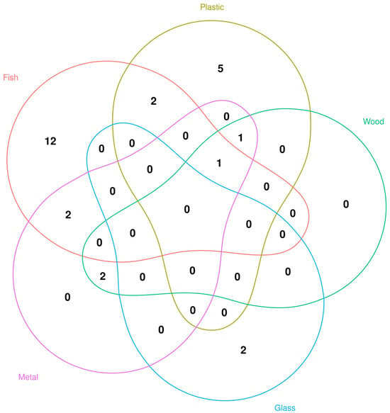
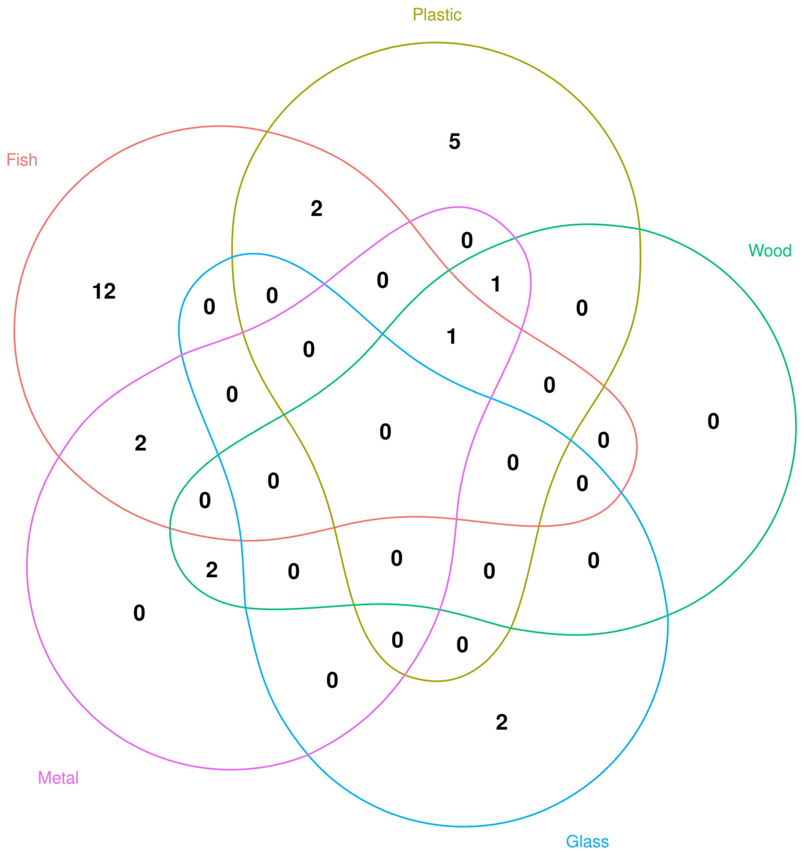
Bacteria identified by MALDI-TOF MS from various surfaces of the fishing vessel/fishing gear in spring and fall are listed in Table 1 and Table 2, comprising bacteria isolated as high-confidence species identification, low-confidence species identification and unreliable species identification. Considering only strains with high-confidence genus identification, bacteria from fish were generally not observed on other surfaces of the fishing vessel/fishing gear. Only 8 genera were shared between fish and some other surfaces, while 12 species were identified exclusively from fish, 5 species exclusively from plastic, and 2 exclusively from glass (Figure 1). In total, 374 bacterial strains were retrieved and identified from fish, 97 from the plastic surfaces of a vessel or fishing gear, 3 from glass, 26 from metal and 16 from wood surfaces, irrespective of genus or species confidence score.
Table 1.
Representative bacteria identified by MALDI-TOF MS from various surfaces of the fishing vessel/fishing gear in spring. Indicated are the most frequent occurrence sites for the isolated bacteria, as well as the second-best match identification results of tested bacteria, from the Bruker Daltonics database.
Table 2.
Representative bacteria identified by MALDI-TOF MS from various surfaces of the fishing vessel/fishing gear in fall. Indicated are the most frequent occurrence sites for the isolated bacteria, as well as the second-best match identification results of tested bacteria, from the Bruker Daltonics database.
Figure 1.
Venn diagram of bacteria isolated as high-confidence genus from fish (sardines and anchovies) and various surfaces of a fishing purse-seine vessel.
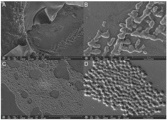
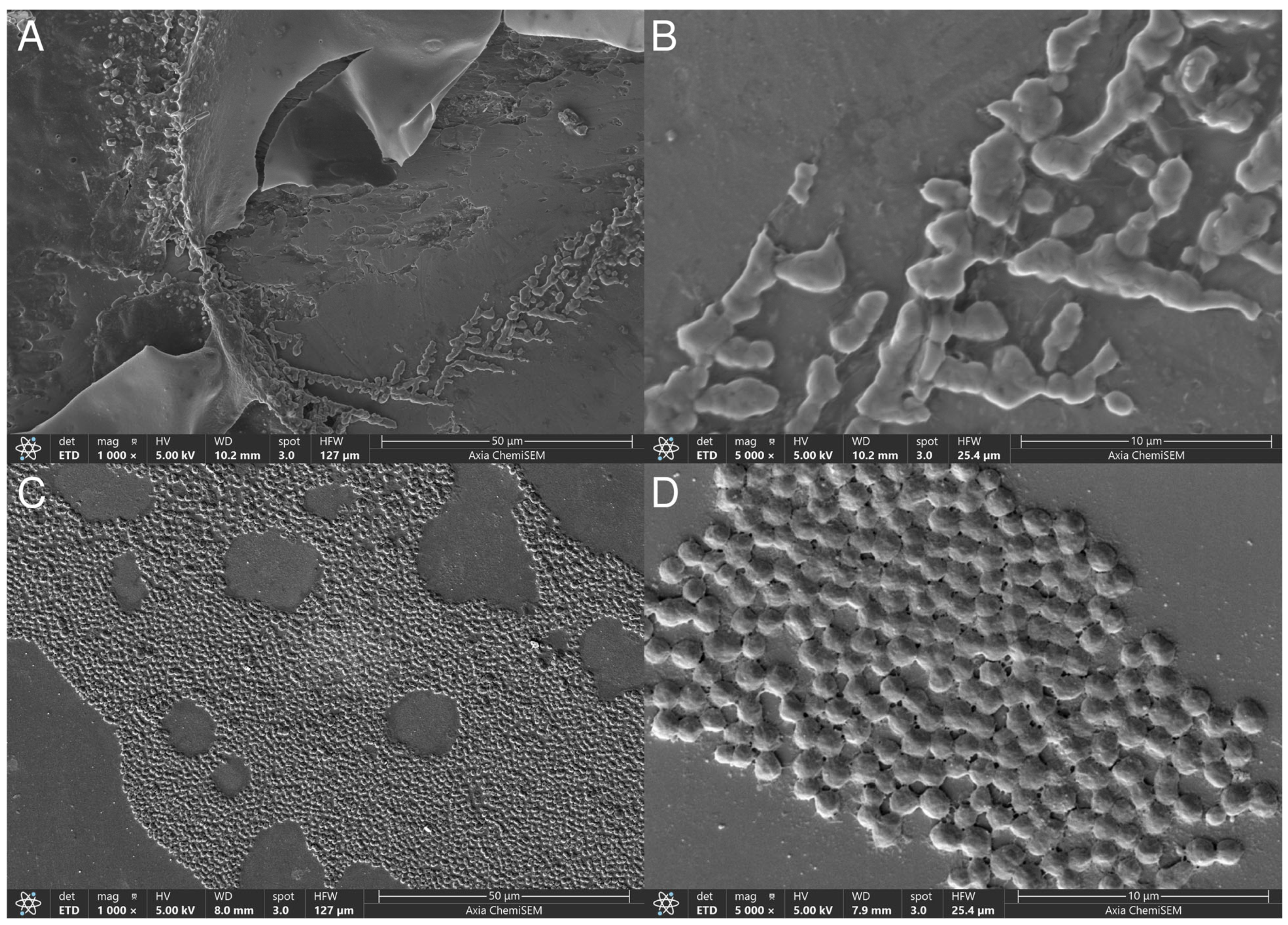
From 10 different species selected for biofilm testing, the overall adhesion on various immersed objects regardless of media was the strongest for P. gessardii, followed by P. damselae and A. veronii. P. gessardii indiscriminately formed biofilms on all immersed materials in all media. Of the immersed objects, irrespective of media, galvanized steel was found to be the best material for biofilm adhesion, followed by glass and polypropylene. However, of all objects, stainless steel had the most biofilm formations when immersed in ZMB, followed by glass, polypropylene, and galvanized steel. Biofilm formation on immersed objects, thus, varied between the tested strains and according to the medium and immersed material (Table 3), and SEM imaging confirmed the presence of lawns of biofilm (Figure 2). B. megaterium, E. mexicanum, P. fragi, K. oceani, Psychrobacter spp., R. endophytica and V. harveyi adhered poorly and were considered as non-biofilm formers or weak biofilm formers on the tested immersed objects in two media, with a few exceptions on galvanized and stainless steel, mostly in ZMB.
Table 3.
Biofilm formation * of 10 bacteria on submerged and air–liquid interfaces of different substrates at 22 °C in Zobell marine broth (MB) and tryptic soy broth (TSB) supplemented with 1.5% (w/v) NaCl.
Figure 2.
Direct visualization of Photobacterium damselae subsp. damselae biofilm using scanning electron microscopy of stainless-steel (A,B) and glass (C,D) surfaces. (A) Low magnification showing material surface irregularities with single and confluent bacterial cells. (B) High magnification of subplot A to show confluence of individual cells into biofilm matrix. (C) Low magnification showing gradual colonization of the surface by bacterial biofilm. (D) High magnification of subplot C to show confluence of individual cells into biofilm matrix.
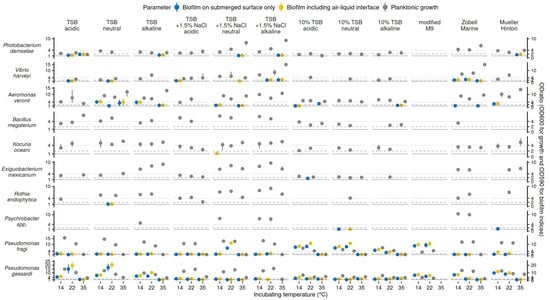
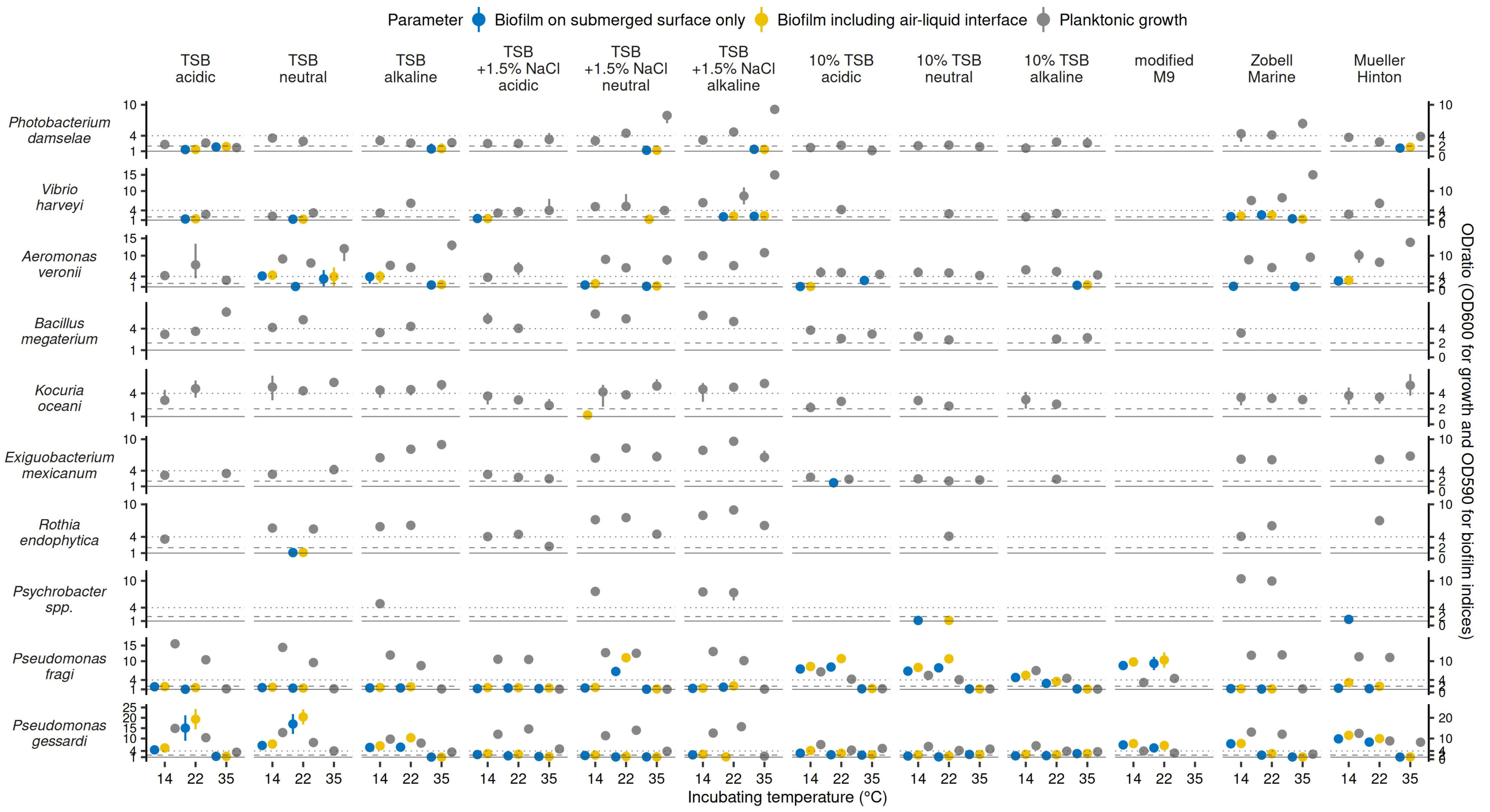
As biofilm formation may vary between media, we further tested 12 different culture media for the effects on planktonic growth and biofilm formation at three incubation temperatures in microtiter plates. Quantitative data results for planktonic growth and biofilm indices are displayed in Figure 3, and a summary per bacterial species is presented in Table 4. It is apparent, for most of the 10 tested strains and 12 media, that planktonic bacterial growth was not consistently associated with their biofilm formation potential. Microtiter experiments showed that varying media broths, incubation temperatures, and pH levels had a large impact on all selected bacterial species. Temperature and pH levels evaluated a range of conditions occurring in the environment, where isolates were sourced and media broths covered nutritious general media with various salt contents, media with limited nutrients, and with depletion of nutrients. These combinations would simulate many environmental conditions, and most important to note is that no species is able to grow in all of these conditions. V. harveyi, P. damselae, K. oceani, R. endophytica, and Psychrobacter spp. showed a wide distribution in results from no growth to strong growth (Table 4).
Figure 3.
Results of planktonic growth and biofilm production on submerged surface only and with air-liquid interface included using polystyrene microtiter plates at 3 incubation temperatures in 12 different culture media of which tryptic soy broth (TSB) has 3 formulations each at 3 pH levels (6.3, 7.3, 8.3) for 10 bacterial species. Parameters are color-coded and only significant results of growth and formed biofilm have been plotted (ODratio being significantly higher than 1 by one-tailed one-sample T test, p < 0.05). Dots show average of biological replicates and line extend to minimal and maximal values observed (if line not observed, the variation is small and is obscured by the dot). ODratios allow quantification by following cutoffs (≤1 no growth/biofilm, solid cutoff line; >1 and ≤2 mild growth/biofilm, dashed line; >2 and ≤4 moderate growth/biofilm; >4 strong growth/biofilm, dotted line).
Table 4.
Quantitative results as overall proportion (%) of categorical results for planktonic growth and two biofilm surface indices tested for 10 bacterial species at 3 temperatures (14, 22, and 35 °C) in 12 different culture conditions (tryptic soy broth (TSB) in three formulations: TSB, TSB + 1.5% NaCl and 10% TSB with each tested at 3 levels of pH (6.3, 7.3, and 8.3), Zobell marine broth, modified M9 minimal, and Mueller-Hinton broth media) in microtiter plates.
Species with the highest capacity to grow in all variations, in descending order, were E. mexicanum, P. gessardi, B. megaterium and A. veronii, which all showed no growth in <15% of all cases and also had the highest proportion of results with strong growth, although with considerable variation in mild and moderate growth results (Table 4). For A. veronii, K. oceani, P. damselae, and V. harveyi, there was an exception with the modified M9 medium, for all three incubation temperatures, where most of the bacterial growth and biofilm production ranged around the same, low values. On the other hand, the results of biofilm formation showed different patterns for these species. Pseudomonas spp. were shown as the strongest biofilm producers, and P. gessardi even formed biofilm where planktonic growth was absent. E. mexicanum and B. megaterium in more than half of cases did not form a biofilm and, together with K. oceani and R. endophytica, had the least capacity for biofilm formation overall. Pathogenic species like A. veronii, V. harveyi, and P. damselae showed results mostly between these opposing ends. In general, the results of biofilm formation on the two surfaces closely agreed, demonstrating the same affinity for the production of either biofilm on the submerged surfaces or biofilm at the air–liquid interface, independent of the medium type, which synchronously varied according to the temperature. More frequent formation of biofilm with air–liquid interface included than on submerged surface only was only observed with Pseudomonas spp. and E. mexicanum relative to others.
We observed that strong biofilm producers were only P. aeruginosa, P. fragi, P. gessardii, A. veronii and R. endophytica. However, their biofilm production was strongly related to the medium type and incubation temperature. For example, A. veronii formed strong biofilms only in the TSB neutral medium at 14 °C, while it formed moderate biofilms in the same medium at 35 °C. P. aeruginosa was the strongest biofilm producer overall, with a high ODratio of up to 25, and measurable at all three incubation temperatures in acidic, neutral and alkaline TSB media supplemented with 1.5% NaCl. However, its biofilms were strong at 14 and 35 °C in unsupplemented acidic, neutral and alkaline TSB media but low to moderate at 22 °C. In some media, it produced strong biofilms only at 35 °C (10% TSB neutral and 10% TSB alkaline) or only at 14 °C (ZMB). Its biofilm production potential differed from the other two tested pseudomonads, as P. fragi favored lower incubation temperatures for biofilm production. It produced strong biofilms at 14 and 22 °C in 10% TSB acidic, 10% TSB neutral and modified M9 media. P. fragi also formed a strong biofilm on neutral TSB medium supplemented with 1.5% NaCl at 22 °C and only in 10% TSB alkaline at 14 °C. No biofilm was produced at 35 °C, just as in the case of P. gessardii. P. gessardii formed strong biofilms at 14 and 22 °C in acidic, neutral, and alkaline TSB, ZMB, modified M9 and MHB. R. endophytica also produced strong biofilms at 14 and 22 °C, but only in MHB.
Multivariate analysis of growth and biofilm formation on two surfaces agreed on the significant effects of all culture conditions and variables. For all three indices, all main fixed effects and all two-way and three-way interactions were significant and could not be reduced from the full model. For both biofilm indices, different days of testing and plate effects were shown to be significant random effects, but for planktonic growth, different days were not significant (p = 0.12). However, the magnitude of these random effects is much smaller compared to the main fixed effects as it additionally explained only ~1–2% of variance, while more than 75% of variance was explained by the latter (Table 5).
Table 5.
Results of three separate linear mixed effects regression models for planktonic growth and two biofilm indices testing media broth, temperature, pH, and bacterial species as fixed effects and different days and plates as random effects.
Numerous interactions were shown to be significant: for growth, 52.9% (117/221) of interactions were significant, and even more for biofilms with 77.4% (171/221) significant interactions on the submerged surface and 72.4% (160/221) when including the air–liquid interface. Concerning the estimated marginal means, while MZB and TSB supplemented with 1.5% NaCl were media with the highest and 10% TSB and mod. M9 with the least growth, MHB and TSB were the media with the highest biofilm formation, and MZB and TSB supplemented with 1.5% NaCl had the lowest biofilm formation. Therefore, mod. M9 with limited sources and 10% TSB with depleted sources showed better biofilm production than support for planktonic growth (Table 5). On the other hand, all three indices showed the same effect of temperature, which was of no significant difference between 14 and 22 °C, and both had significantly higher results than at 35 °C for growth and biofilm indices. However, pH showed the best growth in alkaline media, and all three levels of pH were significantly different, while biofilm formation was higher in neutral and acidic media, which were not significantly different but both significantly higher than in alkaline media. For bacterial species, Pseudomonas spp. were the strongest biofilm producers, followed by pathogens A. veronii, V. harveyi, and P. damselae and environmental Psychrobacter spp. On the other hand, B. megaterium, K. oceani, and E. mexicanum had the lowest capacity for biofilm formation, which occurred only in particular instances.
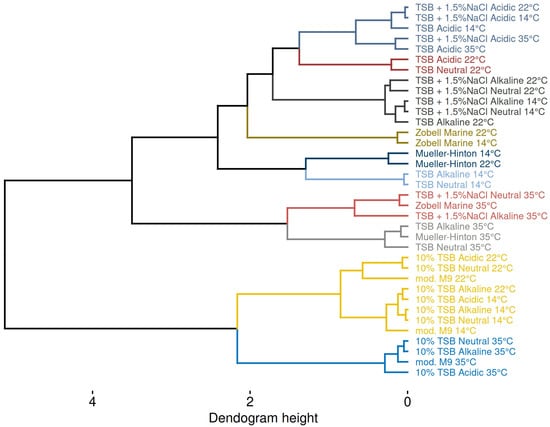
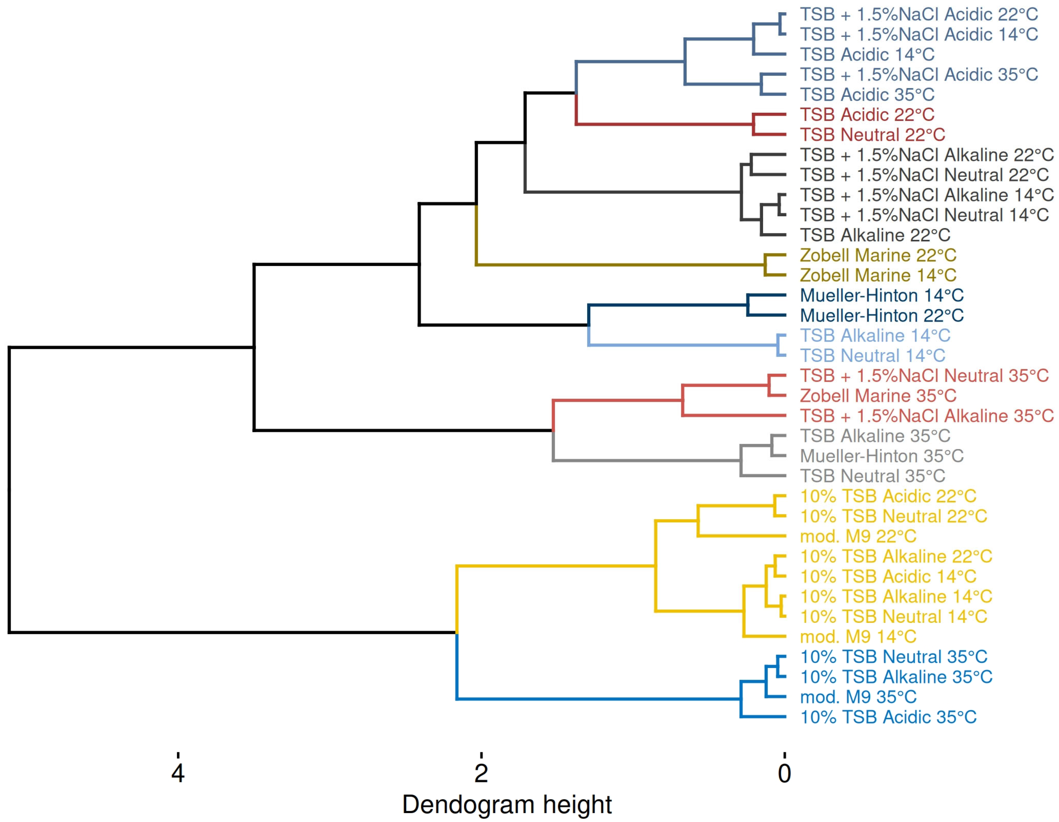
The results of PCA agreed with the modeling results, showing the largest separation by media broths, followed by incubation temperature, and the least influential separation by pH levels. Moreover, significant differences between media broths, temperatures, and pH levels observed with modeling were also evident through hierarchical clustering on principal components (Figure 4). That is, the similarity of low nutrient media mod. M9 and 10%TSB, then of TSB + 1.5% NaCl and ZMB, and of MHB and TSB is evident. Likewise, the grouping of lower incubation temperatures relative to 35 °C within media is consistent, as is the grouping of neutral and alkaline pH levels relative to acidic pH.
Figure 4.
Dendrogram showing hierarchical agglomerative clustering on principal components of growth and biofilm formation data of 10 selected bacterial species tested in 12 different media (some with 3 pH levels) at 3 incubation temperatures. Clustering was performed using Ward’s method and Euclidean distances with color-coded clusters (optimal number according to the largest relative reduction in inertia).
4. Discussion
We investigated the typical surfaces of an operating fishing vessel for bacterial growth from its plastic, wooden, glass and steel areas and compared them to the bacteria retrieved from hauled fish gill tissues. Their biofilm formation was observed in the air–liquid interfaces and in submerged surfaces, such as on the immersed areas of plastic, metal and glass objects, in relation to various incubation temperatures and media types in a comprehensive manner rarely explored. By large, the results of in vitro experiments did not align with field data, since many strains showed the ability to form a biofilm on surfaces from which they were not isolated. However, these discrepancies may be explained by the presence of bacteria below detectable levels for culture or insufficient sample surface area due to scattered and infrequent distribution on the surface, overgrowth on agar, or die-off due to dynamic changes in environmental and nutrient conditions.
The fish mucosal tissues, including gill, skin and intestines, serve as physical barriers for water-borne bacteria and are critical components of their immune response [66]. We expected to find complementary bacterial species on fish and particularly on plastic surfaces, which have direct contact with fish freshly hauled from the sea (fishing nets and storage containers), but some species we identified from plastics only and not from any other surface. Indeed, in an outdoor adhesion experiment in ambient seawater involving polypropylene, polyethylene, polyethylene terephthalate, and expanded polystyrene as the major substrates of marine plastic debris, Jeong et al. [67] confirmed that bacteria colonizing plastic surfaces and seawater differ but that potentially pathogenic bacteria exhibit high adhesion specificity on plastic, which differs according to the plastic material (polystyrene, polypropylene, polyethylene or other). Similar to their work, we observed vibrios on plastic surfaces of fishing nets but also on fish gills, particularly V. alginolyticus, V. gigantis and V. pelagius. V. harveyi, which we investigated as a vibrio representative for biofilm formation, did not show affinity to form a biofilm on PP objects immersed in the broths, neither on polystyrene surfaces nor at the air–liquid interfaces of the 12 investigated broths at any temperature. However, it did form biofilms on the galvanized steel surface, similarly to Desulfovibrio sp. (previously V. desulfuricans), which not only formed a stable biofilm on galvanized steel but also showed a corrosive effect [68].
Stainless steel, on the other hand, is the material that fish-cooling chambers on vessels are made of, having inert surfaces which do not resist bacterial adhesion, although its surface roughness influences the attachment of bacterial cells [69]. V. parahaemolyticus was found to be able to attach to both rough and smooth stainless-steel surfaces, as their flagella and pili assist in grappling and overcoming the hydrodynamic force near the surfaces in a liquid environment [70]. In this work, P. damselae, P. fragi, P. gessardii, Psychrobacter spp. and R. endophytica showed high affinity for stainless-steel attachment. There are no literature confirmations for P. damselae forming biofilms on stainless steel, although it is a known biofilm producer in marine systems, thus having a reservoir other than fish carriers [71,72]. Conversely, pseudomonads are largely distributed bacteria on stainless-steel surfaces, particularly in the food production and processing environment, able to form extracellular polymeric substance-rich biofilms that interact with other bacteria. Biofilms on such surfaces can act as a source of pathogenic and spoilage bacteria as well as their products, which may contribute to a more rapid onset of stored fish spoilage [73]. Amongst them, P. fragi is one of the most frequently identified pseudomonads, with the potential to form biofilms on abiotic surfaces under aerobic and modified atmosphere conditions and at low temperatures [73,74]. P. gessardii was also demonstrated as a stainless-steel biofilm producer [75], but less is known of its affinity for sea-related surfaces and the spoilage potential of fish [76]. Interestingly, pseudomonads and Psychrobacter spp. were previously present in 70% of all biofilms on stainless-steel surfaces related to meat processing [77], while Psychrobacter spp. in this work formed biofilms on stainless steel and glass but not on other tested surfaces. Psychrotrophic Psychrobacter spp. are common spoilage bacteria of fish and meat [78], while R. endophytica is an emerging spoiler of pelagic fish [79], with no previous associations with biofilm formation in marine environments.
We established that the overall adhesion on various immersed objects regardless of media was the strongest for P. gessardii, followed by P. damselae and A. veronii, having high adhesion specificity on galvanized steel, stainless steel, glass and polypropylene, but, among them, only P. gessardii also had high adhesion capability on polystyrene. That is valuable information, since all three bacterial strains are known pathogens. P. damselae is a marine fish pathogen, giving rise to septicemia, with only a few signs in the acute phase [80], but is also an opportunistic pathogen for humans who suffer wounds and cuts in the marine environment [81]. A. veronii is a more aggressive zoonotic agent. It may cause skin and soft tissue infections upon exposure to aquatic environments, although severe infections in healthy people caused by A. veronii are relatively rare [82]. However, in immunocompromised patients, it is a grave pathogen, which may lead to serious outcomes [83]. P. gessardii is a crayfish pathogen [84] and a novel human pathogen leading to corneal ulcers and loss of vision [85]. There are several mechanisms which make pathogens in surface biofilms more likely to cause infection than in the planktonic form. One is the seeding dispersal of numerous bacterial cells; another involves phenotypically heterogeneous pathogens within the biofilm to clonally expand within biofilm; and the third relates to high cell densities within the biofilm, which control quorum-sensing networks [86]. In any case, the formation of biofilms facilitates pathogens to survive both in the environment and in the host.
In this study, of all tested bacteria, strong biofilm producers on polystyrene were P. aeruginosa, P. fragi, and P. gessardii. A. veronii and R. endophytica formed strong biofilms only on 1 of 12 tested growth media. Biofilm productions of all bacteria tested were related to the medium type and the incubation temperature. Pseudomonas aeruginosa, the strongest biofilm producer of all, showed the highest affinity for biofilms in the TSB medium, whether acidic, neutral, alkaline, or acidic, neutral, and alkaline with 1.5% NaCl supplementation, with slight variations according to the medium. Although it formed biofilms mostly at 14 and 35 °C, their rates were the highest at 35 °C. P. aeruginosa is an opportunistic human pathogen, causing a variety of infections [87], and its biofilms were previously shown to exhibit metabolic switching at temperatures comparable to those encountered on human skin (33 to 37 °C) [37]. Thus, metabolic adjustments appropriate to temperature settings are key to its pathogenic success [88]. To date, P. aeruginosa is one of the most investigated bacteria regarding biofilm formation. Some of the stages described during its surface adhesion, such as reversible attachment, monolayer, and mature biofilm, have often been embraced as a universal model for biofilm development. However, in closely related species and even in various strains of P. aeruginosa, they manifest differences [89,90]. Often, they are temperature-dependent, inducing ranges of behaviors regarding temperature fluctuations along thermal gradients. It is not only temperature but the medium type that influences P. aeruginosa to adopt different planktonic/biofilm lifestyles. The current knowledge on the bacterium is mostly derived from studies using rich growth media [91]. However, it is known that the bacterium can also survive for an extended period in nutrient-poor water. Since its biofilm production relates to its pathogenic potential, the growth medium can reveal important data. Although the M9-glutamate enabled biofilm formation of P. aeruginosa in the work of [91], the modified M9 as a minimal medium in this work allowed only weak to no biofilms. However, the ZMB, mimicking seawater in its mineral, nutrient, salt and nitrogenous compounds, facilitated its strong biofilm formation only at 14 °C.
P. fragi favored lower incubation temperatures for biofilm production on polystyrene, forming strong biofilms at 14 and 22 °C in 10% TSB acidic, 10% TSB neutral and modified M9 media. It was also previously found to form biofilms on polystyrene [26]. Interestingly, P. fragi was found to demonstrate stronger biofilm-forming ability at 30 °C than at lower temperatures in TSB or MRS broths [92], as opposed to our results. Motile, flagellated bacteria such as pseudomonads tend to co-aggregate at the air–liquid interface [93], which we confirmed in this study, having higher ODratios than on submerged surfaces. While all the pseudomonads tested were able to form biofilms on polystyrene in 5–9 out of 12 broth conditions, microbial biofilm formation was more favorable within the nutrient-rich media but varied according to the incubation temperature and Pseudomonas species.
5. Conclusions
This study is one of the first works on bacteria retrieved from various fishing vessel surfaces and pelagic fish catches that comprehensively evaluated the variability in biofilm formation between different substrates, media and cultivation conditions using a panel of bacteria. These parameters usually vary between laboratories/studies due to a lack of standardized protocols, limiting the comparability and generalization of findings. This study shows how constricted testing conditions e.g., by adhering only to in-house practices, may lead to the underestimation of bacterial biofilm-forming capacity for a variety of bacterial species. We showed that the biofilm assay has good reproducibility, with replicates and plate effects being negligible compared to the effects of incubation temperature, broth media, and pH on the ability to form biofilms on various surfaces of diverse abiotic materials, covering a wide range of environmental conditions. These findings demonstrate various species-dependent triggers for biofilm production and the large variability therein. To that end, we emphasize the need for the standardization and optimization of protocols for marine biofilm tests regarding the variability of conditions for crystal violet assay. The tests should preferably be conducted on both nutrient-rich and nutrient-poor media, at various incubation temperatures, and on a variety of substrate surfaces, in order to obtain reliable and comparable results and not to miss “hidden” traits. However, it is important to note that the results of in vitro tests need not necessarily correspond to the results from field investigations. From the perspective of hygiene and safety, it is worth noting that all pathogenic species were able to form a biofilm in vitro on all types of materials from which they were never isolated in the field where they were observed only in fish samples. Therefore, substrates, nutritional, common physical and chemical factors do not fully explain the presence and persistence (or absence) of bacteria on various surfaces. These are of particular concern for microbial ecology studies, biomedical and food hygiene and safety as well as material development. To date, there is no single method or a unified framework that would provide consistent results for different bacteria or for a single species that would be appropriate for various application interests and purposes. This work is, thus, conceived as a guiding principle for testing the biofilm formation of (marine/fish/pathogenic) bacteria on various surfaces. It also demonstrates that bacterial biofilms formed on fishing boat surfaces harbor bacterial communities, to some extent aligned with those isolated from anchovy and sardine catches, underscoring these surfaces as possible reservoirs that facilitate bacterial transfer and potential cross-contamination within the fishing environment.
Author Contributions
Conceptualization N.T.P.; methodology N.T.P., K.B., S.P.K., B.B., M.B. and G.B.; data curation K.B., S.P.K., I.S.-P. and R.Č.-R.; formal analysis K.B., S.P.K., M.B. and G.B.; investigation N.T.P., K.B., S.P.K., B.B., M.B., G.B., I.S.-P. and R.Č.-R.; validation N.T.P. and K.B.; writing, original draft preparation N.T.P. and K.B.; visualization K.B.; writing, review and editing N.T.P. and K.B.; supervision, resources, project administration, and funding acquisition I.S.-P. and R.Č.-R. All authors have read and agreed to the published version of the manuscript.
Funding
This research was funded by the EU Operational Programme for Maritime Affairs and Fisheries of the Republic of Croatia—Partnership between Science and Fisheries; and Center of Research Excellence for Marine Bioprospecting—BioProCro, a project co-financed by the Croatian Government and the European Union through the European Regional Development Fund—the Competitiveness and Cohesion Operational Programme (PK.1.1.02): Sustainable bioprospecting of organisms from the Adriatic Sea for innovative natural products. The funders had no role in study design, data collection and analysis, decision to publish, or preparation of the manuscript.
Institutional Review Board Statement
Ethical review and approval were waived for this study due to the fact that all experiments were conducted as per the guidelines of the Institutional Animal Ethics Committee of the Ruđer Bošković Institute, Zagreb, Croatia. Furthermore, fish used in this study were captured for commercial purposes. Therefore, use of these animals in research does not require ethical approval.
Data Availability Statement
The original contributions presented in this study are included in the article. Further inquiries can be directed to the corresponding author.
Acknowledgments
The authors would like to thank the fishermen crew for help with field work as well as Marija Mirosavljević and Luka Pavić for assistance with SEM images.
Conflicts of Interest
The authors declare no conflicts of interest.
References
- Kreve, S.; Reis, A.C.D. Bacterial Adhesion to Biomaterials: What Regulates This Attachment? A Review. Jpn. Dent. Sci. Rev. 2021, 57, 85–96. [Google Scholar] [CrossRef] [Scilit]
- Bharti, A.; Velmourougane, K.; Prasanna, R. Phototrophic Biofilms: Diversity, Ecology and Applications. J. Appl. Phycol. 2017, 29, 2729–2744. [Google Scholar] [CrossRef] [Scilit]
- Costa, A.M.; Mergulhão, F.J.; Briandet, R.; Azevedo, N.F. It Is All about Location: How to Pinpoint Microorganisms and Their Functions in Multispecies Biofilms. Future Microbiol. 2017, 12, 987–999. [Google Scholar] [CrossRef] [Scilit] [PubMed]
- Vasudevan, R. Biofilms: Microbial Cities of Scientific Significance. J. Microbiol. Exp. 2014, 1, 00014. [Google Scholar] [CrossRef] [Scilit]
- Wahl, M.; Goecke, F.; Labes, A.; Dobretsov, S.; Weinberger, F. The Second Skin: Ecological Role of Epibiotic Biofilms on Marine Organisms. Front. Microbiol. 2012, 3, 292. [Google Scholar] [CrossRef] [Scilit]
- De Carvalho, C.C.C.R. Marine Biofilms: A Successful Microbial Strategy With Economic Implications. Front. Mar. Sci. 2018, 5, 126. [Google Scholar] [CrossRef] [Scilit]
- Wahl, M. Marine Epibiosis. I. Fouling and Antifouling: Some Basic Aspects. Mar. Ecol. Prog. Ser. 1989, 58, 175–189. [Google Scholar] [CrossRef] [Scilit]
- Donlan, R.M. Biofilm Formation: A Clinically Relevant Microbiological Process. Clin. Infect. Dis. 2001, 33, 1387–1392. [Google Scholar] [CrossRef] [Scilit]
- Ortega-Morales, B.O.; Chan-Bacab, M.J.; De La Rosa-García, S.D.C.; Camacho-Chab, J.C. Valuable Processes and Products from Marine Intertidal Microbial Communities. Curr. Opin. Biotechnol. 2010, 21, 346–352. [Google Scholar] [CrossRef] [Scilit]
- Weiskerger, C.J.; Brandão, J.; Ahmed, W.; Aslan, A.; Avolio, L.; Badgley, B.D.; Boehm, A.B.; Edge, T.A.; Fleisher, J.M.; Heaney, C.D.; et al. Impacts of a Changing Earth on Microbial Dynamics and Human Health Risks in the Continuum between Beach Water and Sand. Water Res. 2019, 162, 456–470. [Google Scholar] [CrossRef] [Scilit]
- De Carvalho, C.; Caramujo, M. Lipids of Prokaryotic Origin at the Base of Marine Food Webs. Mar. Drugs 2012, 10, 2698–2714. [Google Scholar] [CrossRef] [Scilit]
- Svanevik, C.S.; Roiha, I.S.; Levsen, A.; Lunestad, B.T. Microbiological Assessment along the Fish Production Chain of the Norwegian Pelagic Fisheries Sector – Results from a Spot Sampling Programme. Food Microbiol. 2015, 51, 144–153. [Google Scholar] [CrossRef] [Scilit] [PubMed]
- Topić Popović, N.; Bojanić, K.; Kazazić, S.P.; Bujak, M.; Babić, S.; Bignami, G.; Čož-Rakovac, R.; Matulić, D.; Strunjak-Perović, I. Cultivable Bacterial Communities from Purse-Seined Small Pelagic Fish, Fishing Nets and Storage Tanks. Mar. Biol. 2024, 171, 201. [Google Scholar] [CrossRef] [Scilit]
- Topić Popović, N.; Kazazić, S.P.; Bojanić, K.; Strunjak-Perović, I.; Čož-Rakovac, R. Sample Preparation and Culture Condition Effects on MALDI-TOF MS Identification of Bacteria: A Review. Mass Spectrom. Rev. 2023, 42, 1589–1603. [Google Scholar] [CrossRef] [Scilit] [PubMed]
- Stepanović, S.; Vuković, D.; Hola, V.; Bonaventura, G.D.; Djukić, S.; Ćirković, I.; Ruzicka, F. Quantification of Biofilm in Microtiter Plates: Overview of Testing Conditions and Practical Recommendations for Assessment of Biofilm Production by Staphylococci. Apmis 2007, 115, 891–899. [Google Scholar] [CrossRef] [Scilit] [PubMed]
- Narciso-da-Rocha, C.; Vaz-Moreira, I.; Svensson-Stadler, L.; Moore, E.R.B.; Manaia, C.M. Diversity and Antibiotic Resistance of Acinetobacter Spp. in Water from the Source to the Tap. Appl. Microbiol. Biotechnol. 2013, 97, 329–340. [Google Scholar] [CrossRef] [Scilit]
- Choi, J.-Y.; Kim, Y.; Ko, E.A.; Park, Y.K.; Jheong, W.-H.; Ko, G.; Ko, K.S. Acinetobacter Species Isolates from a Range of Environments: Species Survey and Observations of Antimicrobial Resistance. Diagn. Microbiol. Infect. Dis. 2012, 74, 177–180. [Google Scholar] [CrossRef] [Scilit]
- Li, X.; Tang, Y.; Lu, X. Insight into Identification of Acinetobacter Species by Matrix-Assisted Laser Desorption/Ionization Time of Flight Mass Spectrometry (MALDI-TOF MS) in the Clinical Laboratory. J. Am. Soc. Mass Spectrom. 2018, 29, 1546–1553. [Google Scholar] [CrossRef] [Scilit]
- Bizet, J.; Bizet, C. Strains of Alcaligenes Faecalis from Clinical Material. J. Infect. 1997, 35, 167–169. [Google Scholar] [CrossRef] [Scilit]
- Marathe, S.K.; Vashistht, M.A.; Prashanth, A.; Parveen, N.; Chakraborty, S.; Nair, S.S. Isolation, Partial Purification, Biochemical Characterization and Detergent Compatibility of Alkaline Protease Produced by Bacillus Subtilis, Alcaligenes Faecalis and Pseudomonas Aeruginosa Obtained from Sea Water Samples. J. Genet. Eng. Biotechnol. 2018, 16, 39–46. [Google Scholar] [CrossRef] [Scilit]
- Khan, W.U.; Yasin, N.A.; Ahmad, S.R.; Ali, A.; Ahmed, S.; Ahmad, A. Role of Ni-Tolerant Bacillus Spp. and Althea Rosea L. in the Phytoremediation of Ni-Contaminated Soils. Int. J. Phytoremed. 2017, 19, 470–477. [Google Scholar] [CrossRef] [Scilit]
- Scheldeman, P.; Rodríguez-Díaz, M.; Goris, J.; Pil, A.; De Clerck, E.; Herman, L.; De Vos, P.; Logan, N.A.; Heyndrickx, M. Bacillus Farraginis Sp. Nov., Bacillus Fortis Sp. Nov. and Bacillus Fordii Sp. Nov., Isolated at Dairy Farms. Int. J. Syst. Evol. Microbiol. 2004, 54, 1355–1364. [Google Scholar] [CrossRef] [Scilit] [PubMed]
- Balzer, M.; Witt, N.; Flemming, H.-C.; Wingender, J. Faecal Indicator Bacteria in River Biofilms. Water Sci. Technol. 2010, 61, 1105–1111. [Google Scholar] [CrossRef] [Scilit] [PubMed]
- Dzeing-Ella, A.; Szwebel, T.A.; Loubinoux, J.; Coignard, S.; Bouvet, A.; Le Jeunne, C.; Aslangul, E. Infective Endocarditis Due to Citrobacter Koseri in an Immunocompetent Adult. J. Clin. Microbiol. 2009, 47, 4185–4186. [Google Scholar] [CrossRef] [Scilit] [PubMed]
- Saimmai, A.; Udomsilp, S.; Maneerat, S. Production and Characterization of Biosurfactant from Marine Bacterium Inquilinus Limosus KB3 Grown on Low-Cost Raw Materials. Ann. Microbiol. 2013, 63, 1327–1339. [Google Scholar] [CrossRef] [Scilit]
- McHugh, K.E.; Rhoads, D.D.; Wilson, D.A.; Highland, K.B.; Richter, S.S.; Procop, G.W. Inquilinus Limosus in Pulmonary Disease: Case Report and Review of the Literature. Diagn. Microbiol. Infect. Dis. 2016, 86, 446–449. [Google Scholar] [CrossRef] [Scilit]
- Son, S.; Oh, J.-D.; Lee, S.H.; Shin, D.; Kim, Y. Comparative Genomics of Canine Lactobacillus Reuteri Reveals Adaptation to a Shared Environment with Humans. Genes Genom 2020, 42, 1107–1116. [Google Scholar] [CrossRef] [Scilit]
- Keren, Y.; Keshet, D.; Eidelman, M.; Geffen, Y.; Raz-Pasteur, A.; Hussein, K. Is Leclercia Adecarboxylata a New and Unfamiliar Marine Pathogen? J. Clin. Microbiol. 2014, 52, 1775–1776. [Google Scholar] [CrossRef] [Scilit]
- Grünzweil, O.M.; Palmer, L.; Cabal, A.; Szostak, M.P.; Ruppitsch, W.; Kornschober, C.; Korus, M.; Misic, D.; Bernreiter-Hofer, T.; Korath, A.D.J.; et al. Presence of β-Lactamase-Producing Enterobacterales and Salmonella Isolates in Marine Mammals. IJMS 2021, 22, 5905. [Google Scholar] [CrossRef] [Scilit]
- Kämpfer, P.; Glaeser, S.P.; Packroff, G.; Behringer, K.; Exner, M.; Chakraborty, T.; Schmithausen, R.M.; Doijad, S. Lelliottia Aquatilis Sp. Nov., Isolated from Drinking Water. Int. J. Syst. Evol. Microbiol. 2018, 68, 2454–2461. [Google Scholar] [CrossRef] [Scilit]
- Mounier, J.; Coton, M.; Irlinger, F.; Landaud, S.; Bonnarme, P. Smear-Ripened Cheeses. In Cheese; Elsevier: Amsterdam, The Netherlands, 2017; pp. 955–996. ISBN 978-0-12-417012-4. [Google Scholar]
- Vences, A.; Abushattal, S.; Matanza, X.M.; Dubert, J.; Uzun, E.; Ogut, H.; Osorio, C.R. Highly Transferable pAQU-Related Plasmids Encoding Multidrug Resistance Are Widespread in the Human and Fish Pathogen Photobacterium Damselae Subsp. Damselae in Aquaculture Areas in the Black Sea. Microb. Ecol. 2020, 80, 507–518. [Google Scholar] [CrossRef] [Scilit] [PubMed]
- Berthe, F.; Michel, C.; Bernardet, J. Identification of Pseudomonas Anguilliseptica Isolated from Several Fish Species in France. Dis. Aquat. Org. 1995, 21, 151–155. [Google Scholar] [CrossRef] [Scilit]
- Wang, G.; Li, M.; Ma, F.; Wang, H.; Xu, X.; Zhou, G. Physicochemical Properties of Pseudomonas Fragi Isolates Response to Modified Atmosphere Packaging. FEMS Microbiol. Lett. 2017, 364. [Google Scholar] [CrossRef] [Scilit] [PubMed]
- Raio, A.; Puopolo, G. Pseudomonas Chlororaphis Metabolites as Biocontrol Promoters of Plant Health and Improved Crop Yield. World J. Microbiol. Biotechnol. 2021, 37, 99. [Google Scholar] [CrossRef] [Scilit]
- Crovadore, J.; Calmin, G.; Chablais, R.; Cochard, B.; Schulz, T.; Lefort, F. Whole-Genome Sequence of Pseudomonas Graminis Strain UASWS1507, a Potential Biological Control Agent and Biofertilizer Isolated in Switzerland. Genome Announc. 2016, 4, e01096-16. [Google Scholar] [CrossRef] [Scilit]
- Kačániová, M.; Klūga, A.; Kántor, A.; Medo, J.; Žiarovská, J.; Puchalski, C.; Terentjeva, M. Comparison of MALDI-TOF MS Biotyper and 16S rDNA Sequencing for the Identification of Pseudomonas Species Isolated from Fish. Microb. Pathog. 2019, 132, 313–318. [Google Scholar] [CrossRef] [Scilit]
- Barros, J.; Becerra, J.; González, C.; Martínez, M. Antibacterial Metabolites Synthesized by Psychrotrophic Bacteria Isolated from Cold-Freshwater Environments. Folia Microbiol. 2013, 58, 127–133. [Google Scholar] [CrossRef] [Scilit]
- Abd El-Aziz, F.E.-Z.A.; Bashandy, S.R. Dose-Dependent Effects of Pseudomonas Trivialis Rhizobacteria and Synergistic Growth Stimulation Effect with Earthworms on the Common Radish. Rhizosphere 2019, 10, 100156. [Google Scholar] [CrossRef] [Scilit]
- Kim, Y.; Kim, H.; Beuchat, L.R.; Ryu, J.-H. Inhibition of Escherichia coli O157:H7 on Stainless Steel Using Pseudomonas veronii Biofilms. Lett. Appl. Microbiol. 2018, 66, 394–399. [Google Scholar] [CrossRef] [Scilit]
- Encyclopedia of Food Microbiology; Robinson, R.K., Batt, C.A., Patel, P.D., Eds.; Academic Press: San Diego, 2000; ISBN 978-0-12-227070-3. [Google Scholar]
- Yoon, J.-H.; Yeo, S.-H.; Oh, T.-K.; Park, Y.-H. Psychrobacter Alimentarius Sp. Nov., Isolated from Squid Jeotgal, a Traditional Korean Fermented Seafood. Int. J. Syst. Evol. Microbiol. 2005, 55, 171–176. [Google Scholar] [CrossRef] [Scilit]
- Lee, J.; Kwon, M.; Yang, J.Y.; Woo, J.; Lee, H.K.; Hong, S.G.; Kim, O.-S. Complete Genome Sequence of Psychrobacter Alimentarius PAMC 27889, a Psychrophile Isolated from an Antarctic Rock Sample. Genome Announc. 2016, 4, e00704-16. [Google Scholar] [CrossRef] [Scilit]
- Rubin, L.G. Other Gram-Negative Coccobacilli. In Principles and Practice of Pediatric Infectious Diseases; Elsevier: Amsterdam, The Netherlands, 2018; pp. 967–969.e1. ISBN 978-0-323-40181-4. [Google Scholar]
- Romanenko, L.A.; Lysenko, A.M.; Rohde, M.; Mikhailov, V.V.; Stackebrandt, E. Psychrobacter Maritimus Sp. Nov. and Psychrobacter Arenosus Sp. Nov., Isolated from Coastal Sea Ice and Sediments of the Sea of Japan. Int. J. Syst. Evol. Microbiol. 2004, 54, 1741–1745. [Google Scholar] [CrossRef] [Scilit] [PubMed]
- Yoon, J.-H.; Lee, C.-H.; Yeo, S.-H.; Oh, T.-K. Psychrobacter Aquimaris Sp. Nov. and Psychrobacter Namhaensis Sp. Nov., Isolated from Sea Water of the South Sea in Korea. Int. J. Syst. Evol. Microbiol. 2005, 55, 1007–1013. [Google Scholar] [CrossRef] [Scilit] [PubMed]
- Shivaji, S.; Reddy, G.S.N.; Suresh, K.; Gupta, P.; Chintalapati, S.; Schumann, P.; Stackebrandt, E.; Matsumoto, G.I. Psychrobacter Vallis Sp. Nov. and Psychrobacter Aquaticus Sp. Nov., from Antarctica. Int. J. Syst. Evol. Microbiol. 2005, 55, 757–762. [Google Scholar] [CrossRef] [Scilit] [PubMed]
- Sahuquillo-Arce, J.M.; Ibáñez-Martínez, E.; Hernández-Cabezas, A.; Ruiz-Gaitán, A.; Falomir-Salcedo, P.; Menéndez, R.; López-Hontangas, J.L. Influence of Environmental Conditions and Pollution on the Incidence of Streptococcus Pneumoniae Infections. ERJ Open Res. 2017, 3, 00014–02017. [Google Scholar] [CrossRef] [Scilit]
- Mathew, B.T.; Torky, Y.; Amin, A.; Mourad, A.-H.I.; Ayyash, M.M.; El-Keblawy, A.; Hilal-Alnaqbi, A.; AbuQamar, S.F.; El-Tarabily, K.A. Halotolerant Marine Rhizosphere-Competent Actinobacteria Promote Salicornia Bigelovii Growth and Seed Production Using Seawater Irrigation. Front. Microbiol. 2020, 11, 552. [Google Scholar] [CrossRef] [Scilit]
- Borre, I.; Sonnenschein, E.C. Draft Genome Sequences of Nine Environmental Bacterial Isolates Colonizing Plastic. Microbiol. Resour. Announc. 2021, 10, e01485-20. [Google Scholar] [CrossRef] [Scilit]
- Kim, K.I.; Won, K.-M.; Lee, E.S.; Cho, M.; Jung, S.H.; Kim, M.S. Detection of Vibrio and Ten Vibrio Species in Cage-Cultured Fish by Multiplex Polymerase Chain Reaction Using House-Keeping Genes. Aquaculture 2019, 506, 417–423. [Google Scholar] [CrossRef] [Scilit]
- Ngalimat, M.S.; Yahaya, R.S.R.; Baharudin, M.M.A.; Yaminudin, S.M.; Karim, M.; Ahmad, S.A.; Sabri, S. A Review on the Biotechnological Applications of the Operational Group Bacillus Amyloliquefaciens. Microorganisms 2021, 9, 614. [Google Scholar] [CrossRef] [Scilit]
- Massilamany, C.; Mohammed, A.; Loy, J.D.; Purvis, T.; Krishnan, B.; Basavalingappa, R.H.; Kelley, C.M.; Guda, C.; Barletta, R.G.; Moriyama, E.N.; et al. Whole Genomic Sequence Analysis of Bacillus Infantis: Defining the Genetic Blueprint of Strain NRRL B-14911, an Emerging Cardiopathogenic Microbe. BMC Genom. 2016, 17, 511. [Google Scholar] [CrossRef] [Scilit]
- Vary, P.S.; Biedendieck, R.; Fuerch, T.; Meinhardt, F.; Rohde, M.; Deckwer, W.-D.; Jahn, D. Bacillus Megaterium—from Simple Soil Bacterium to Industrial Protein Production Host. Appl. Microbiol. Biotechnol. 2007, 76, 957–967. [Google Scholar] [CrossRef] [Scilit] [PubMed]
- Dobrzyński, J.; Jakubowska, Z.; Dybek, B. Potential of Bacillus Pumilus to Directly Promote Plant Growth. Front. Microbiol. 2022, 13, 1069053. [Google Scholar] [CrossRef] [Scilit] [PubMed]
- López-Cortés, A.; Schumann, P.; Pukall, R.; Stackebrandt, E. Exiguobacterium Mexicanum Sp. Nov. and Exiguobacterium Artemiae Sp. Nov., Isolated from the Brine Shrimp Artemia Franciscana. Syst. Appl. Microbiol. 2006, 29, 183–190. [Google Scholar] [CrossRef] [Scilit] [PubMed]
- Zhang, L.; Xi, L.; Ruan, J.; Huang, Y. Kocuria Oceani Sp. Nov., Isolated from a Deep-Sea Hydrothermal Plume. Int. J. Syst. Evol. Microbiol. 2017, 67, 164–169. [Google Scholar] [CrossRef] [Scilit]
- Sun, L.-N.; Pan, D.-D.; Wu, X.-W.; Yang, E.-D.; Hua, R.-M.; Li, Q.X. Leucobacter Triazinivorans Sp. Nov., a s-Triazine Herbicide Prometryn-Degrading Bacterium Isolated from Sludge. Int. J. Syst. Evol. Microbiol. 2018, 68, 204–210. [Google Scholar] [CrossRef] [Scilit]
- Datta, S.; Manna, S.; Roy, D. Attachment of Extracellular Metabolic Products of Lysinibacillus sp. DRG3 on Sand Surface under Variable Flow Velocities and Bioprocesses. J. Environ. Eng. 2022, 148, 04022069. [Google Scholar] [CrossRef] [Scilit]
- Guisado, I.M.; Purswani, J.; González-López, J.; Pozo, C. Paenibacillus Etheri Sp. Nov., Able to Grow on Media Supplemented with Methyl Tert-Butyl Ether (MTBE) and Isolated from Hydrocarbon-Contaminated Soil. Int. J. Syst. Evol. Microbiol. 2016, 66, 862–867. [Google Scholar] [CrossRef] [Scilit]
- Da Silva, F.K.L.; De Sa Alexandre, A.R.; Casas, A.A.; Ribeiro, M.C.; De Souza, K.M.C.; Soares, E.S.; Dos Santos Junior, S.R.; Vieira, J.D.G.; Amaral, A.C. Increased Production of Chitinase by a Paenibacillus Illinoisensis Isolated from Brazilian Coastal Soil When Immobilized in Alginate Beads. Folia Microbiol. 2022, 67, 935–945. [Google Scholar] [CrossRef] [Scilit]
- Decewicz, P.; Dziewit, L.; Golec, P.; Kozlowska, P.; Bartosik, D.; Radlinska, M. Characterization of the Virome of Paracoccus spp. (Alphaproteobacteria) by Combined in Silico and in Vivo Approaches. Sci. Rep. 2019, 9, 7899. [Google Scholar] [CrossRef] [Scilit]
- Bozal, N.; Montes, M.J.; Tudela, E.; Guinea, J. Characterization of Several Psychrobacter Strains Isolated from Antarctic Environments and Description of Psychrobacter Luti Sp. Nov. and Psychrobacter Fozii Sp. Nov. Int. J. Syst. Evol. Microbiol. 2003, 53, 1093–1100. [Google Scholar] [CrossRef] [Scilit]
- Elbahnasawy, M.A.; Shehabeldine, A.M.; Khattab, A.M.; Amin, B.H.; Hashem, A.H. Green Biosynthesis of Silver Nanoparticles Using Novel Endophytic Rothia Endophytica: Characterization and Anticandidal Activity. J. Drug Deliv. Sci. Technol. 2021, 62, 102401. [Google Scholar] [CrossRef] [Scilit]
- Prasad, M.M.; Seenayya, G. The Impact of Salinicoccus Roseus and Heat Treatment of Salt on the Shelf Life of Cured Sciaenids (Johnius Dussumieri Dussumieri’s Croaker). J. Aquat. Food Prod. Technol. 2008, 17, 253–265. [Google Scholar] [CrossRef] [Scilit]
- Minich, J.J.; Poore, G.D.; Jantawongsri, K.; Johnston, C.; Bowie, K.; Bowman, J.; Knight, R.; Nowak, B.; Allen, E.E. Microbial Ecology of Atlantic Salmon (Salmo Salar) Hatcheries: Impacts of the Built Environment on Fish Mucosal Microbiota. Appl. Env. Microbiol. 2020, 86, e00411-20. [Google Scholar] [CrossRef] [Scilit] [PubMed]
- Jeong, G.; Kim, H.-J.; Kim, K.E.; Kim, Y.J.; Lee, T.-K.; Shim, W.J.; Jung, S.W. Selective Attachment of Prokaryotes and Emergence of Potentially Pathogenic Prokaryotes on Four Plastic Surfaces: Adhesion Study in a Natural Marine Environment. Mar. Pollut. Bull. 2023, 193, 115149. [Google Scholar] [CrossRef] [Scilit]
- Ilhan-Sungur, E.; Unsal-Istek, T.; Cansever, N. Microbiologically Influenced Corrosion of Galvanized Steel by Desulfovibrio sp. and Desulfosporosinus sp. in the Presence of Ag–Cu Ions. Mater. Chem. Phys. 2015, 162, 839–851. [Google Scholar] [CrossRef] [Scilit]
- Hong, I.T.; Koo, C.H. Antibacterial Properties, Corrosion Resistance and Mechanical Properties of Cu-Modified SUS 304 Stainless Steel. Mater. Sci. Eng. 2005, 393, 213–222. [Google Scholar] [CrossRef] [Scilit]
- Tantratian, S.; Srimangkornkaew, N.; Prakitchaiwattana, C.; Sanguandeekul, R. Effect of Different Stainless Steel Surfaces on the Formation and Control of Vibrio Parahaemolyticus Biofilm. LWT 2022, 166, 113788. [Google Scholar] [CrossRef] [Scilit]
- King, R.K.; Flick, G.J.; Pierson, D.; Smith, S.A.; Boardman, G.D.; Coale, C.W. Identification of Bacterial Pathogens in Biofilms of Recirculating Aquaculture Systems. J. Aquat. Food Prod. Technol. 2004, 13, 125–133. [Google Scholar] [CrossRef] [Scilit]
- Mougin, J.; Roquigny, R.; Flahaut, C.; Bonnin-Jusserand, M.; Grard, T.; Le Bris, C. Abundance and Spatial Patterns over Time of Vibrionaceae and Vibrio Harveyi in Water and Biofilm from a Seabass Aquaculture Facility. Aquaculture 2021, 542, 736862. [Google Scholar] [CrossRef] [Scilit]
- Sterniša, M.; Gradišar Centa, U.; Drnovšek, A.; Remškar, M.; Smole Možina, S. Pseudomonas Fragi Biofilm on Stainless Steel (at Low Temperatures) Affects the Survival of Campylobacter Jejuni and Listeria Monocytogenes and Their Control by a Polymer Molybdenum Oxide Nanocomposite Coating. Int. J. Food Microbiol. 2023, 394, 110159. [Google Scholar] [CrossRef] [Scilit]
- Wang, G.-Y.; Ma, F.; Wang, H.-H.; Xu, X.-L.; Zhou, G. Characterization of Extracellular Polymeric Substances Produced by Pseudomonas Fragi Under Air and Modified Atmosphere Packaging: Characterization of EPS from P. fragi. J. Food Sci. 2017, 82, 2151–2157. [Google Scholar] [CrossRef] [Scilit] [PubMed]
- Belov, S.V.; Danileiko, Y.K.; Egorov, A.B.; Lukanin, V.I.; Semenova, A.A.; Lisitsyn, A.B.; Revutskaya, N.M.; Nasonova, V.V.; Yushina, Y.K.; Tolordava, E.R.; et al. Sterilizer of Knives in the Meat Industry, Working by Activating Aqueous Solutions with Glow Discharge Plasma. Processes 2022, 10, 1536. [Google Scholar] [CrossRef] [Scilit]
- Silbande, A.; Cornet, J.; Cardinal, M.; Chevalier, F.; Rochefort, K.; Smith-Ravin, J.; Adenet, S.; Leroi, F. Characterization of the Spoilage Potential of Pure and Mixed Cultures of Bacterial Species Isolated from Tropical Yellowfin Tuna (Thunnus Albacares). J. Appl. Microbiol. 2018, 124, 559–571. [Google Scholar] [CrossRef] [Scilit] [PubMed]
- Wagner, E.M.; Fischel, K.; Rammer, N.; Beer, C.; Palmetzhofer, A.L.; Conrady, B.; Roch, F.-F.; Hanson, B.T.; Wagner, M.; Rychli, K. Bacteria of Eleven Different Species Isolated from Biofilms in a Meat Processing Environment Have Diverse Biofilm Forming Abilities. Int. J. Food Microbiol. 2021, 349, 109232. [Google Scholar] [CrossRef] [Scilit]
- Møretrø, T.; Langsrud, S. Residential Bacteria on Surfaces in the Food Industry and Their Implications for Food Safety and Quality: Residential Bacteria in Food Industry. Compr. Rev. Food Sci. Food Saf. 2017, 16, 1022–1041. [Google Scholar] [CrossRef] [Scilit]
- Al-Harbi, A.H.; Al-Asous, A.I. Bacterial Community of Salted Bluespot Mullet (Valamugil Seheli). LWT 2019, 107, 208–213. [Google Scholar] [CrossRef] [Scilit]
- Pečur Kazazić, S.; Topić Popović, N.; Strunjak-Perović, I.; Florio, D.; Fioravanti, M.; Babić, S.; Čož-Rakovac, R. Fish Photobacteriosis-The Importance of Rapid and Accurate Identification of Photobacterium Damselae subsp. Piscicida. J. Fish Dis. 2019, 42, 1201–1209. [Google Scholar] [CrossRef] [Scilit]
- Ge, L.; Gao, Y.; Wang, K.; Liu, Q.; Cui, P.; Dong, Q. Analysis of Clinical Characteristics, Pathogen Infection, and Drug Sensitivity of Marine Injury Patients: A Cross-Sectional Study. Medicine 2022, 101, e29943. [Google Scholar] [CrossRef] [Scilit]
- Li, L.; Huang, J.; Xu, L.; Wang, G.; Xiao, S.; Xia, Z.; Qin, Q.; Li, Y.; Ji, S. Severe Skin and Soft Tissue Infection in the Left Upper Limb Caused by Aeromonas Veronii: A Case Report. J. Med. Case Rep. 2023, 17, 29. [Google Scholar] [CrossRef] [Scilit]
- Alatorre-Fernández, C.P.; Cornejo-Juárez, P.; Velázquez-Acosta, C.; Volkow-Fernández, P. Bacteremia Caused by Aeromonas Species in Patients with Cancer: Clinical Manifestations and Outcomes. J. Infect. Dev. Ctries 2023, 17, 359–366. [Google Scholar] [CrossRef] [Scilit]
- Korkut, G.G.; Söderhäll, I.; Söderhäll, K.; Noonin, C. The Effect of Temperature on Bacteria-Host Interactions in the Freshwater Crayfish, Pacifastacus Leniusculus. J. Invertebr. Pathol. 2018, 157, 67–73. [Google Scholar] [CrossRef] [Scilit]
- Deepika, J. Pseudomonas Gessardii—A Novel Pathogenic Bacterium Associated with the Cases of Corneal Ulcers and Producing Virulent Pyoverdine Pigment. J. Appl. Biol. Biotech. 2022, 10, 68–73. [Google Scholar] [CrossRef] [Scilit]
- Hall-Stoodley, L.; Stoodley, P. Biofilm Formation and Dispersal and the Transmission of Human Pathogens. Trends Microbiol. 2005, 13, 7–10. [Google Scholar] [CrossRef] [Scilit] [PubMed]
- Wu, D.C.; Chan, W.W.; Metelitsa, A.I.; Fiorillo, L.; Lin, A.N. Pseudomonas Skin Infection: Clinical Features, Epidemiology, and Management. Am. J. Clin. Dermatol. 2011, 12, 157–169. [Google Scholar] [CrossRef] [Scilit] [PubMed]
- Kahl, L.J.; Eckartt, K.N.; Morales, D.K.; Price-Whelan, A.; Dietrich, L.E.P. Light/Dark and Temperature Cycling Modulate Metabolic Electron Flow in Pseudomonas Aeruginosa Biofilms. mBio 2022, 13, e01407-22. [Google Scholar] [CrossRef] [Scilit] [PubMed]
- Espinosa-Urgel, M.; Ramos-González, M.I. Becoming Settlers: Elements and Mechanisms for Surface Colonization by Pseudomonas Putida. Environ. Microbiol. 2023, 1462–2920.16385. [Google Scholar] [CrossRef] [Scilit]
- Fazli, M.; Almblad, H.; Rybtke, M.L.; Givskov, M.; Eberl, L.; Tolker-Nielsen, T. Regulation of Biofilm Formation in P Seudomonas and B Urkholderia Species: Regulation of Biofilm Formation. Env. Microbiol. 2014, 16, 1961–1981. [Google Scholar] [CrossRef] [Scilit]
- Mendis, N.; Lin, Y.R.; Faucher, S.P. Comparison of Virulence Properties of Pseudomonas Aeruginosa Exposed to Water and Grown in Rich Broth. Can. J. Microbiol. 2014, 60, 777–781. [Google Scholar] [CrossRef] [Scilit]
- Ning, Z.; Xue, B.; Wang, H. Evaluation of the Adhesive Potential of Bacteria Isolated from Meat-Related Sources. Appl. Sci. 2021, 11, 10652. [Google Scholar] [CrossRef] [Scilit]
- Magana, M.; Sereti, C.; Ioannidis, A.; Mitchell, C.A.; Ball, A.R.; Magiorkinis, E.; Chatzipanagiotou, S.; Hamblin, M.R.; Hadjifrangiskou, M.; Tegos, G.P. Options and Limitations in Clinical Investigation of Bacterial Biofilms. Clin. Microbiol. Rev. 2018, 31, e00084-16. [Google Scholar] [CrossRef] [Scilit]

Disclaimer/Publisher’s Note: The statements, opinions and data contained in all publications are solely those of the individual author(s) and contributor(s) and not of MDPI and/or the editor(s). MDPI and/or the editor(s) disclaim responsibility for any injury to people or property resulting from any ideas, methods, instructions or products referred to in the content. |
© 2026 by the authors. Licensee MDPI, Basel, Switzerland. This article is an open access article distributed under the terms and conditions of the Creative Commons Attribution (CC BY) license.