


Abstract
Accurate segmentation of oral mucosal lesions remains challenging because lesion regions often resemble surrounding mucosa, with subtle color differences, weak texture contrast, unclear boundaries, and distractors such as saliva, folds, reflections, and uneven illumination. This study proposes RIVER-NET to improve oral mucosal lesion delineation in complex intraoral images. RIVER-NET is a U-shaped multi-branch progressive aggregation network integrating three task-oriented modules. The Multi-Scale Residual Unit (MSRU) captures multi-scale mucosal texture changes and irregular lesion morphology before spatial downsampling. Dynamic Gated Context Fusion (DGCF) enables gated encoder–decoder context interaction to suppress lesion-like distractors while preserving lesion details. Multi-scale Alignment and Attentive Fusion (MAAF) aligns and attentively aggregates multi-level features to reduce semantic–spatial misalignment and enhance boundary recovery. On the Oral Pathological Mucosa (OPM) dataset, RIVER-NET achieved a Dice score of 75.59% and an IoU of 0.632, outperforming U-Net by 30.88 percentage points in Dice and nnU-Net by 5.23 percentage points. Component-wise and parameter-matched replacement results were consistent with complementary effects of MSRU, DGCF, and MAAF under the current experimental setting. On the independent public ISIC dataset, on which models were trained and evaluated under five-fold cross-validation, RIVER-NET achieved 94.62 ± 2.18% Dice and 0.900 ± 0.039 IoU. Without fine-tuning on the external Autooral dataset, it achieved the highest Dice (0.5547) and specificity (0.9324). RIVER-NET showed promising segmentation performance on the evaluated datasets. These findings demonstrate algorithm-level feasibility but do not establish readiness for clinical use.
1. Introduction
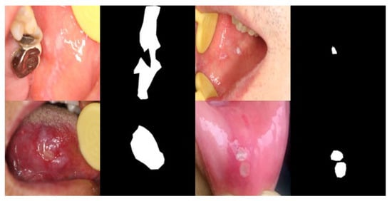
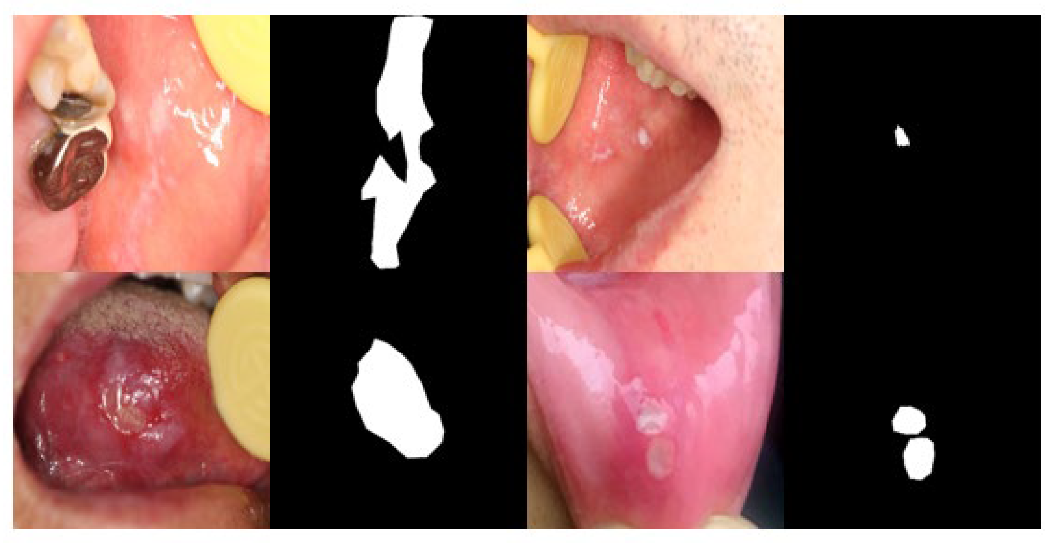
Oral mucosal diseases refer to pathological changes occurring in the mucosal tissues lining the oral cavity. Certain oral mucosal lesions, known as oral potentially malignant disorders, may undergo malignant transformation [1]. Therefore, early diagnosis and timely treatment are essential for preventing oral cancer. In current clinical practice, oral mucosal diseases are mainly diagnosed through visual inspection by clinicians, which is often time-consuming and inconvenient for patients. Moreover, because mucosal lesions may closely resemble normal oral tissues, accurate manual diagnosis remains challenging. To reduce the clinical workload of doctors and improve diagnostic efficiency, automatic segmentation of oral mucosal lesions is highly desirable. Although deep learning has been widely applied in medical image segmentation, its use in oral disease analysis remains relatively limited. Representative intraoral images and their corresponding binary lesion masks are shown in Figure 1, illustrating challenges such as blurred boundaries, small lesions, and specular reflections.
Figure 1.
Examples of oral mucosal lesion samples and their corresponding segmentation annotations. (Top-left) blurred margins with strong specular reflections; (top-right) small lesion; (bottom-left) ambiguous boundaries; (bottom-right) small lesion with specular reflections. In the binary segmentation masks, black denotes the background and white denotes the lesion regions.
Deep learning has promoted automatic medical image segmentation; however, oral mucosal lesion segmentation remains challenging due to the subtle appearance differences between lesions and surrounding mucosa. Existing segmentation models often produce three major types of unsatisfactory results: missed segmentation of small or low-contrast lesion regions, inaccurate or incomplete boundary delineation, and false-positive responses to non-lesion structures such as specular highlights, saliva, mucosal folds, and normal mucosal textures. These limitations indicate that oral mucosal lesion segmentation requires stronger representation learning, contextual modeling, and refinement. Motivated by these observations, we propose RIVER-NET, a multi-branch progressive aggregation network built upon a U-shaped encoder–decoder [2] architecture for precise oral mucosal lesion segmentation in intraoral color images. At a high level, RIVER-NET combines intra-stage multi-scale modeling, gated encoder–decoder interaction, and aligned hierarchical fusion; the detailed operating principles are presented in Section 2.2, Section 2.3 and Section 2.4.
Deep learning has been increasingly applied to oral disease analysis and has achieved promising results. Warin et al. [3] investigated deep learning algorithms for the automatic classification and detection of oral cancer from photographic images. Azam et al. [4] developed a CNN-based segmentation system for upper aerodigestive tract cancer using white-light and narrow-band imaging endoscopic images. In addition, Qiu et al. [5] proposed a dental-arch-prior-assisted method for 3D tooth instance segmentation, while Mărginean et al. [6] developed CariSeg for teeth and carious lesion segmentation in panoramic X-ray images. For oral mucosal diseases, some studies further explored segmentation- and recognition-oriented deep learning frameworks on oral mucosal disease datasets [7,8]. These studies demonstrated the potential of deep learning for automatic lesion localization, segmentation, and disease analysis, thereby reducing the burden of manual diagnosis and annotation. However, most existing approaches were originally designed for oral cancer, teeth, caries, or general medical image analysis tasks, and therefore did not fully account for the unique characteristics of oral mucosal lesions. In particular, oral mucosal lesions often exhibit subtle appearance variations, low visual contrast, and diverse morphological patterns, making discriminative encoding particularly challenging. Consequently, inconspicuous lesion regions may be insufficiently represented, resulting in missed segmentation or incomplete lesion delineation.
The visual similarity between oral mucosal lesions and surrounding tissues makes accurate lesion segmentation particularly challenging. Similar problems have also been widely investigated in natural image analysis, especially in camouflaged object detection and segmentation. Fan et al. [9] introduced camouflaged object detection and proposed SINet to identify objects that are visually embedded in their surroundings. Zhou et al. [10] investigated unified salient and camouflaged object detection to improve the perception of targets with different visual saliency levels. Mei et al. [11] developed PFNet with distraction mining to enhance camouflaged object segmentation under confusing background interference. Furthermore, boundary-guided and uncertainty-guided methods have been explored to distinguish targets from visually similar surroundings. Sun et al. [12] proposed a boundary-guided network that utilized edge semantics to improve camouflaged object detection, while Yang et al. [13] introduced uncertainty-guided transformer reasoning to enhance feature discrimination in ambiguous regions. Yu et al. [14] incorporated multi-stage visual cues for challenging small-organ segmentation, whereas Huang et al. [15] proposed a feature shrinkage pyramid network to progressively aggregate subtle cues for camouflaged object detection. More recently, Niu et al. [16] explored weakly supervised camouflaged object detection by modeling the mutual interaction between region and edge cues. Although these methods demonstrate the importance of contextual modeling, boundary perception, uncertainty estimation, and multi-scale representation, accurately preserving lesion boundaries and maintaining consistent lesion representations remain difficult when lesion-to-mucosa transitions are gradual. Unlike organs with well-defined anatomical boundaries, oral mucosal lesions often exhibit ambiguous margins and progressive changes in color, texture, and surface appearance, causing boundary pixels to share highly similar visual characteristics with adjacent normal tissues. Consequently, segmentation models may generate fragmented lesion regions, boundary discontinuities, or inaccurate estimations of lesion extent. In addition, repeated feature extraction, downsampling, and information propagation across network stages may weaken fine-grained lesion cues while introducing interference from visually similar non-lesion structures, further affecting segmentation reliability and robustness.
These considerations motivate the design of RIVER-NET, which aims to address subtle texture variations, gradual lesion-to-mucosa transitions, scale-varying morphology, and intraoral distractors in intraoral images through progressive multi-branch feature aggregation.
Multi-scale context extraction, attention gating, and pyramid fusion are established design families. RIVER-NET combines task-oriented variants of these families within one progressive framework; detailed architectural definitions are provided in Section 2.2, Section 2.3 and Section 2.4, and their relative behavior under the current experimental protocol is examined through the ablation and parameter-matched replacement studies.
The main contributions of this paper are summarized as follows:
- (1)
- We introduce RIVER-NET, a progressive aggregation framework tailored for fine-grained oral mucosal lesion segmentation in intraoral color images.
- (2)
- We introduce MSRU as an intra-stage multi-scale residual block used in both encoder and decoder pathways.
- (3)
- We introduce DGCF as a gated encoder–decoder interaction module for controlled context exchange.
- (4)
- We introduce MAAF as an alignment-and-attention module for hierarchical feature fusion.
2. Materials and Methods
2.1. Network Architecture
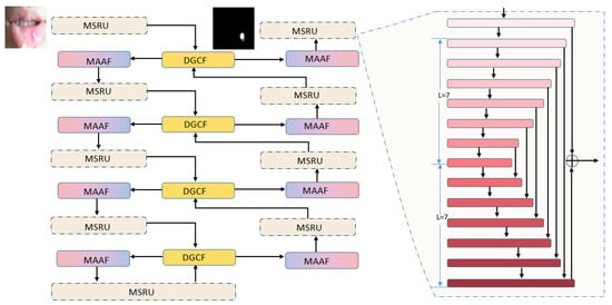
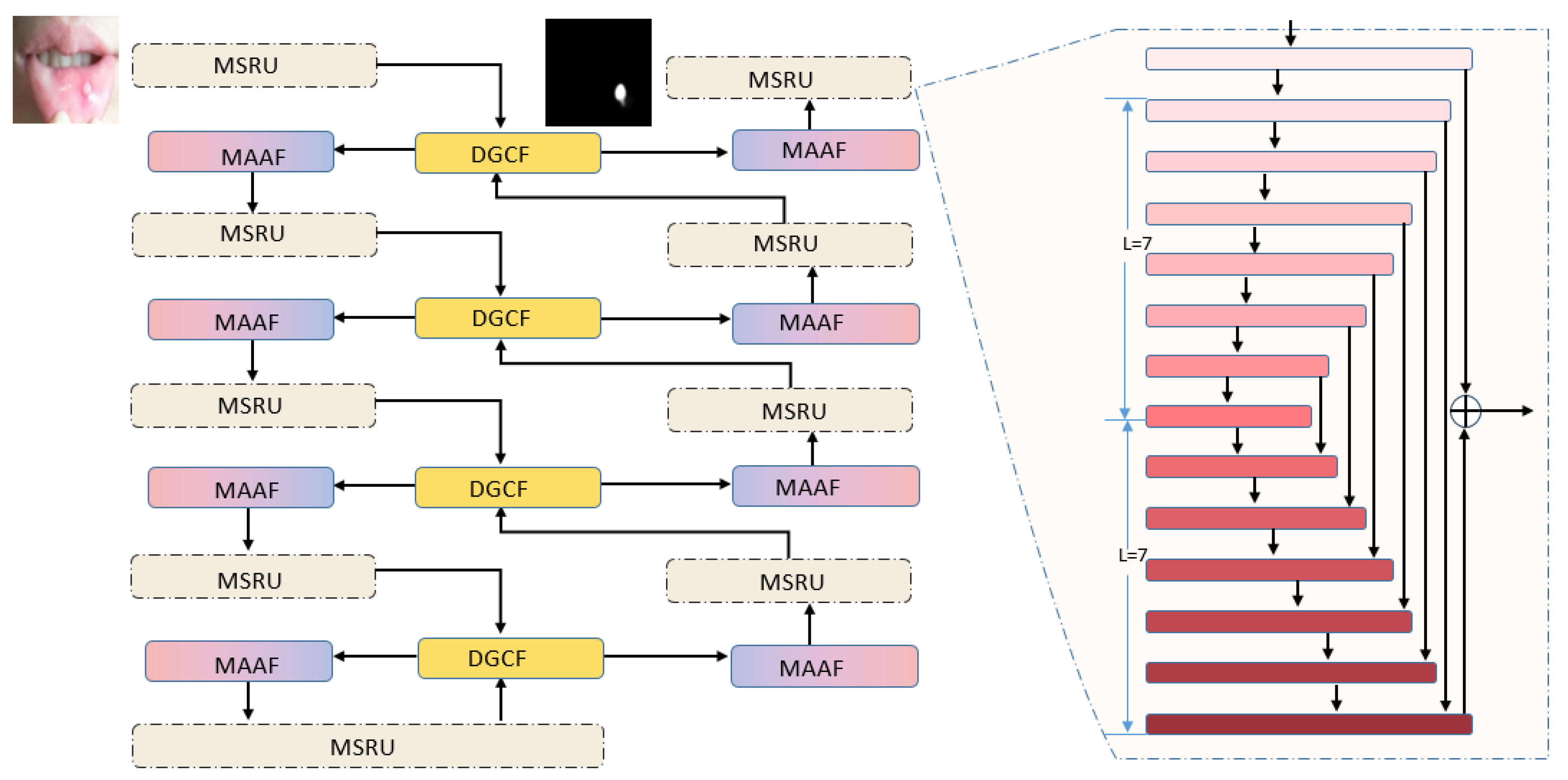
As illustrated in Figure 2, the input is an intraoral image I ∈ R3×H×W. RIVER-NET follows an encoder–decoder architecture, but differs from conventional U-shaped networks by introducing intra-stage multi-scale feature extraction, dynamic encoder–decoder interaction, and multi-level attentive aggregation within the overall framework. At each resolution level of both the encoder and decoder, the conventional convolutional block is replaced with an MSRU-based residual modeling block. In the encoder, MSRU extracts multi-scale lesion representations before cross-stage downsampling. In the decoder, MSRU refines the reconstructed features after DGCF-guided interaction and progressive spatial recovery. The encoder feature generated at the stage is denoted as , which provides same-level structural and appearance information for the decoding process.
Figure 2.
Overall architecture of RIVER-NET. The original image is first processed by MSRU. The module outputs enhanced multi-scale feature maps, which are then fed into the DGCF to obtain context-enhanced feature maps. These maps are subsequently transmitted to MAAF for scale alignment and attention refinement of multi-layer features, yielding enhanced fused hierarchical cues. In the decoder, the DGCF generates adaptive weights based on the joint response of deep decoding features and peer encoding features, selectively boosting lesion-related semantic information and suppressing background noise. The detailed MSRU structure is embedded in the framework. The first layer adopts the basic MSRU structure, while the second, third, fourth and fifth layers are equipped with scale branches of L = 6, L = 5, L = 4, and L = 4, respectively. Through this framework, RIVER-NET combines MSRU-based multi-scale encoding, DGCF-based controlled skip interaction, and MAAF-based hierarchical feature refinement to obtain discriminative and structurally complete representations for oral mucosal lesion segmentation. Colored boxes distinguish the network modules and feature-processing stages; arrows indicate the direction of feature flow.
The decoder starts from the deepest representation and progressively restores the spatial resolution. At the decoding stage, the DGCF receives the same-level encoder feature and the deeper decoder feature as inputs. On the encoder side, DGCF generates the contextual encoder-side feature , which contains refined mucosal structural cues, local texture patterns, and weak boundary responses derived from . The set of encoder-side features is then forwarded to the encoder-side MAAF branch, where hierarchical features are coordinated and selectively refined to strengthen lesion-aware representation during feature extraction. By modeling their contextual relationship and generating adaptive gated responses, DGCF produces the updated decoder-side feature . This representation is used for progressive decoding and is also delivered to the decoder-side MAAF branch, which integrates decoder features from different stages to improve spatial recovery and boundary reconstruction.
2.2. Multi-Scale Residual Unit(MSRU)
To better capture oral mucosal lesions with ambiguous margins and irregular morphology, and to recover fine spatial details during decoding, MSRU is adopted as an intra-stage multi-scale residual modeling block in both the encoder and decoder. Unlike conventional single-stream convolutional blocks, MSRU embeds a compact intra-stage encoder—decoder structure within each resolution level, enabling multi-scale contextual modeling without changing the overall stage resolution. This design progressively enlarges the receptive field via downsampling or dilated encoding, then recovers fine spatial details through corresponding decoding, explicitly separating context aggregation and detail recovery within the same stage. Such separation prevents the blending of coarse semantic cues and fine boundary responses that occurs in simple feature summation, which is particularly important for oral mucosal lesions with weak texture contrast and gradual boundaries that are easily smoothed by repeated cross-stage downsampling. The basic transformation unit consists of a 3 × 3 convolution followed by batch normalization [17] and ReLU activation, and is used for local feature transformation throughout the module.
This design is motivated by the observation that oral mucosal lesion boundaries are often gradual, with weak color contrast and subtle texture changes that are easily lost during repeated downsampling.
In the decoder, MSRU plays a complementary role in feature reconstruction. After decoder features are progressively restored to higher spatial resolutions, decoder-side MSRU further refines the reconstructed representations within the current stage. It helps integrate coarse semantic responses with local structural details, recover gradual lesion boundaries, and reduce coarse reconstruction artifacts caused by direct upsampling. Therefore, encoder-side MSRU focuses on preserving lesion-related cues during feature extraction, whereas decoder-side MSRU focuses on refining lesion structures during spatial recovery.
Given an input feature map at the stage of stream , where denotes the encoder or decoder stream, MSRU first projects it into a local representation and then performs hierarchical intra-stage encoding and decoding:
where denotes the intra-stage multi-scale encoding path and denotes the corresponding decoding path with same-scale feature fusion. Through progressive downsampling or dilated convolution [18] inside the unit, MSRU enlarges the effective receptive field while preserving lesion-related local responses.
Finally, a residual connection is used to combine the multi-scale representation with the original local feature:
where denotes the encoder or decoder stream. is the output of MSRU in the corresponding stream, and denotes a convolutional fusion function. The residual formulation is designed to retain local information while introducing multi-scale context. Shallow high-resolution stages use a deeper intra-stage path, whereas deeper low-resolution stages use dilated convolutions to expand the receptive field without additional spatial compression. These operations are intended to coordinate local texture cues and regional lesion morphology during feature extraction and reconstruction.
2.3. Dynamic Gated Context Fusion(DGCF)
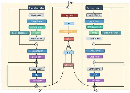
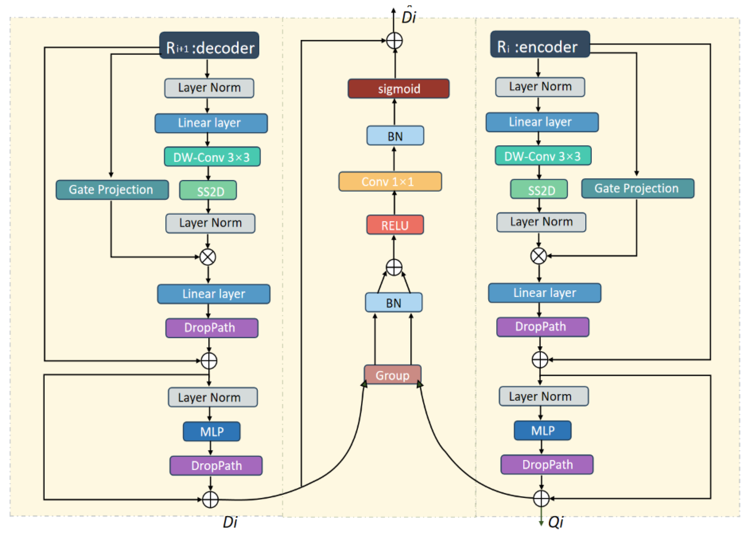
Oral mucosal lesion segmentation is easily affected by lesion-like distractors. In intraoral images, saliva, specular reflections, mucosal folds, and normal mucosal textures may generate edge responses similar to true lesion boundaries, especially in shallow encoder features. Conventional skip connections transmit encoder features to the decoder in an almost indiscriminate manner. Although this helps recover spatial details, it may also introduce noisy boundary responses and background interference. The structure of DGCF is illustrated in Figure 3.
Figure 3.
Structure of the Dynamic Gated Context Fusion (DGCF). Features transmitted from the (i + 1)-th decoder layer are processed by the left branch, with one part output as and the other fed into the central gating unit. Features delivered from the encoder layer are processed by the right branch, generating output and feeding residual information into the gating unit. The final fused feature is exported as . For visual simplicity, the centering operation and the subsequent element-wise residual modulation are omitted from the schematic and are mathematically specified in Equation (4). Equation (4) formally defines the centered gate and the resulting residual modulation applied to the decoder feature. Colored boxes distinguish the processing operations in the decoder, encoder, and gating branches; arrows indicate feature flow.
To address this problem, we propose the DGCF module. The key idea of DGCF is to establish an adaptive interaction between encoder-side structural details and decoder-side semantic context. Instead of directly concatenating the two features, DGCF allows the decoder to determine, according to its current semantic state, which encoder details should be retained and which ambiguous responses should be suppressed.
Before gated fusion, both encoder and decoder features are refined by a context refinement block consisting of depthwise convolution, state-space propagation [19,20], and MLP. This block enhances both local spatial patterns and long-range contextual dependencies, which is important for distinguishing true lesion-related mucosal changes from visually similar non-lesion structures. Standard convolutions alone capture local spatial patterns but struggle to model such long-range dependencies without excessive depth or large kernels. In intraoral images, distinguishing true lesions from lesion-like distractors (e.g., normal mucosal folds) often requires reasoning over spatially distant but semantically related regions. State-space propagation [19,20] addresses this by linearly encoding long-range contextual dependencies with computational efficiency comparable to convolution, making it suitable for maintaining regional consistency in high-resolution intraoral images without the quadratic cost of self-attention.
Let and denote the refined encoder-side and decoder-side features at the stage, respectively. Specifically, contains encoder-side structural and appearance cues, while contains decoder-side semantic information after contextual refinement. Based on their joint response, DGCF generates an adaptive gating map:
where denotes channel-wise concatenation, and are branch-specific projection functions, is the gate generation function, and denotes the Sigmoid activation. The generated gate is conditioned on both encoder-side structural responses and decoder-side semantic cues.
The decoder feature is then recalibrated by the dynamic gate:
To enable bidirectional feature recalibration, the sigmoid gate is centered around 0.5 by defining . The signed gate is then applied to the decoder feature through residual modulation. Consequently, responses with are attenuated, responses with are enhanced, and preserves the original decoder feature. The resulting scaling factor ranges from 0 to 2, allowing DGCF to suppress context-inconsistent responses while emphasizing lesion-consistent activations, where denotes element-wise multiplication. The encoder-side feature is used to guide gate generation rather than being directly concatenated or fully transmitted to the decoder output. DGCF is therefore intended to support regional consistency and boundary stability; its empirical contribution is interpreted only within the ablation settings reported in Section 3.2.
2.4. Multi-Scale Alignment and Attentive Fusion(MAAF)
After MSRU and DGCF, the network obtains features from different hierarchical levels. Although these features have been enhanced by intra-stage multi-scale modeling and gated encoder–decoder interaction, they remain heterogeneous in spatial resolution, receptive field, and semantic granularity. Shallow features provide more precise spatial localization and boundary details, but their semantic abstraction is relatively limited. Deep features contain stronger lesion-level semantic information, but their spatial resolution is lower, which may lead to coarse lesion contours or incomplete recovery of small lesion regions. Therefore, directly aggregating multi-level features after MSRU and DGCF may still suffer from scale inconsistency and semantic-spatial mismatch, limiting the completeness and structural accuracy of lesion segmentation.
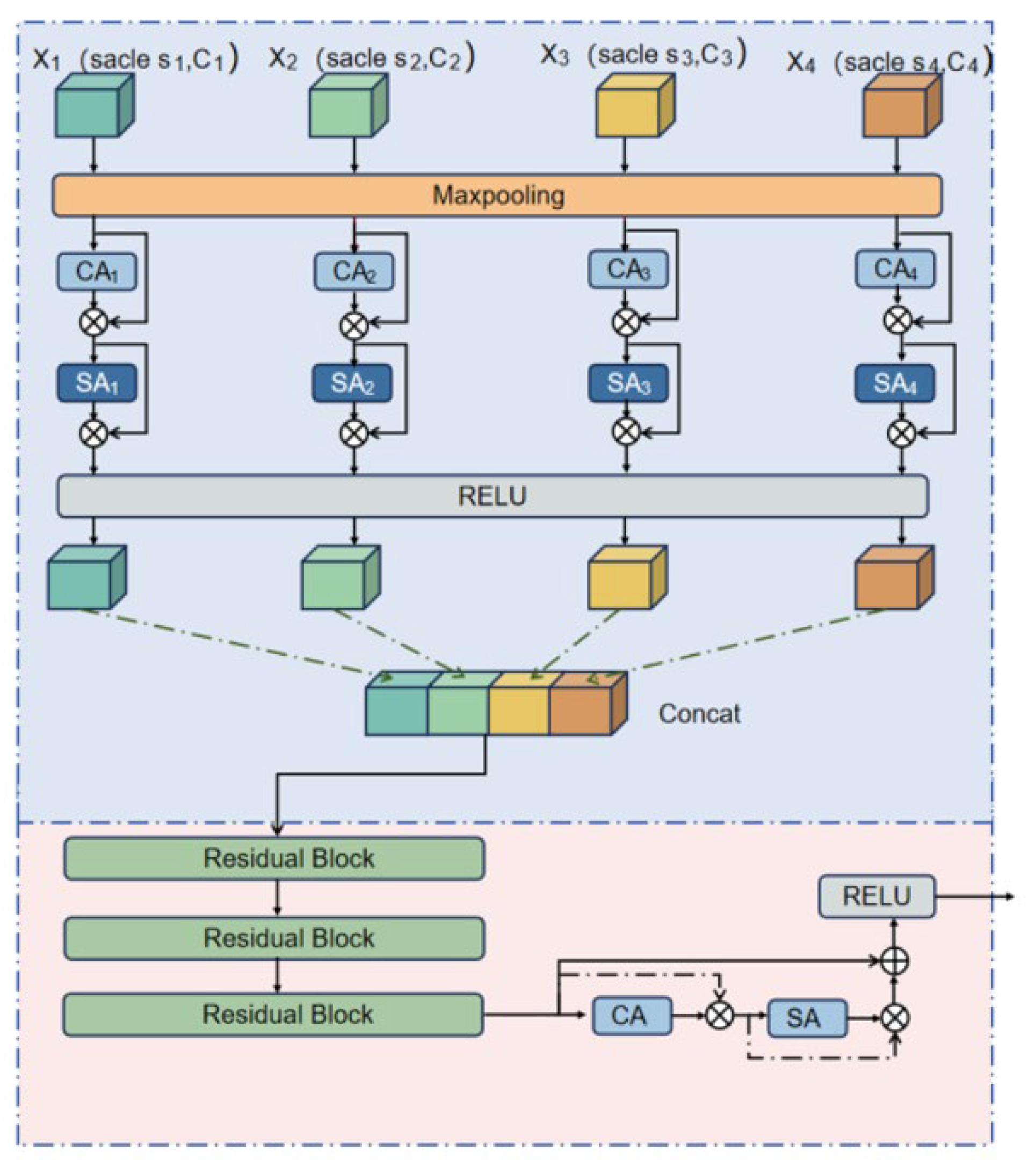
To address this remaining cross-level fusion problem, we propose the MAAF module. The purpose of MAAF is not to serve as the primary mechanism for suppressing local intraoral distractors, which are mainly handled during controlled encoder–decoder interaction in DGCF. Instead, MAAF focuses on integrating shallow boundary-sensitive features and deep semantic features in a unified feature space. Specifically, MAAF first aligns multi-level features to a common resolution using bilinear interpolation or downsampling, and then applies channel and spatial attention to selectively enhance complementary lesion-related cues across different levels. In intraoral images, shallow features contain high-resolution boundary cues but also carry noisy responses from specular reflections and saliva, while deep features encode lesion-level semantics but suffer from coarse spatial localization. Channel attention recalibrates the contribution of each feature channel across hierarchical levels, suppressing channels dominated by background distractors and amplifying those consistently activated by mucosal lesions. Spatial attention further highlights lesion-relevant spatial locations, especially near ambiguous boundaries where shallow and deep features disagree. The sequential application of CA and SA thus mimics a ‘what-to-focus-on-then-where-to-look’ strategy, which is particularly beneficial for oral mucosal lesions with weak texture contrast and gradual margins. In this way, MAAF improves the coordination between shallow structural details and deep semantic representations, contributing to more complete lesion regions and more continuous boundary reconstruction.
In RIVER-NET, MAAF is applied to both the encoder-side and decoder-side feature streams. For the encoder-side branch, the input features are the DGCF-derived encoder-side contextual features , which contain local structural information and weak lesion-related boundary responses from different encoding stages. MAAF aligns and aggregates these features to strengthen lesion-aware representation during feature extraction. For the decoder-side branch, the input features are the DGCF-enhanced decoder features , which contain progressively reconstructed semantic representations after adaptive gated recalibration. MAAF integrates these decoder-side hierarchical features to refine lesion reconstruction during spatial recovery. For simplicity and unified formulation, these features are denoted as in the following description. That is, may refer to in the encoder-side MAAF or in the decoder-side MAAF.
Given a set of multi-level input features , MAAF first performs spatial alignment and channel projection:
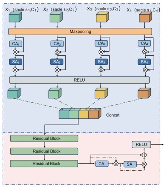
where denotes the scale alignment operation and denotes the channel projection function. It should be noted that the number of input features is not fixed for all MAAF branches. As shown in Figure 4, MAAF can receive features from different scales, such as and denotes the number of input features available to the current MAAF branch. Specifically, for the encoder-side MAAF at the stage, the available inputs consist of the feature at the current encoder stage and the features from all shallower encoder stages. For the decoder-side MAAF at the stage, the available inputs consist of the feature at the current decoder stage and the features from all deeper decoder stages. Therefore, varies with the network stage and represents the number of hierarchical features aggregated by the current MAAF branch.
Figure 4.
Structure of the MAAF. Multi-level features are first aligned to a unified scale and refined by channel attention (CA [21]) and spatial attention (SA [22]). The refined features are then concatenated and fused through residual blocks. Colored elements distinguish feature-processing operations and feature representations; arrows indicate the direction of feature flow.
The aligned features are then concatenated and fused:
where denotes residual fusion, and and denote channel attention and spatial attention, respectively. This design allows MAAF to combine the strengths of different feature levels. Shallow features provide fine spatial details for boundary localization, whereas deep features provide robust lesion-level semantics for region completeness. For the encoder-side stream, MAAF aggregates multiple features to strengthen lesion-aware representation during feature extraction. For the decoder-side stream, MAAF aggregates multiple.
features to refine lesion reconstruction during decoding. The alignment and channel–spatial recalibration operations are intended to coordinate shallow spatial detail with deeper semantic information. MAAF is therefore designed to support lesion completeness and boundary reconstruction; the magnitude and specificity of this contribution remain limited to the comparisons reported in the current experiments.
2.5. Dataset
This retrospective methodological study used a de-identified dataset of intraoral color photographs collected during routine clinical practice at Shanghai Ninth People’s Hospital, Shanghai Jiao Tong University School of Medicine. The dataset comprised 1051 RGB images and corresponding binary segmentation masks from 997 patients with 997 independent lesions. Some lesions were photographed from multiple viewing angles, resulting in more images than patients and lesions. This corpus is hereafter referred to as the Oral Pathological Mucosa (OPM) dataset.
Because the dataset had been de-identified before the present analysis, patient-level demographic and clinical variables, including age, sex, medical history, and other individual characteristics, were not retained in the research dataset and therefore could not be reported. In addition, detailed clinical diagnostic categories and their distributions were not consistently available in the retrospective research records. Consequently, reliable subtype-specific statistics could not be reconstructed, and all annotated lesions were treated as a single foreground class for binary segmentation. No analyses stratified by clinical lesion subtype were performed.
The images were retrospectively obtained during routine clinical care rather than acquired according to a prospectively standardized research imaging protocol. Detailed acquisition metadata, including the imaging device and model, illumination settings, camera-to-lesion distance, viewing angle, and other acquisition parameters, were not consistently recorded or retained in the historical dataset. Therefore, these acquisition conditions could not be reliably reported or retrospectively standardized. Nevertheless, the dataset reflects the variability encountered in routine intraoral photography, including differences in viewing angle, illumination, specular reflection, lesion size, and lesion-to-mucosa contrast.
Two medically trained annotators—a chief physician from the Department of Dermatology at Shanghai Ninth People’s Hospital and a medical doctoral candidate—independently delineated the lesion boundaries in all 1051 images using Labelme version 3.16. The annotators reviewed discrepant regions jointly. Boundary disagreements that remained unresolved after joint review were adjudicated by a senior clinical expert who is a chief physician in the Department of Oral Mucosal Diseases at Shanghai Ninth People’s Hospital. The resulting consensus masks served as the reference standard and were finalized before data partitioning.
The complete dataset of 1051 images was evaluated using patient- and lesion-grouped five-fold cross-validation. Images from the same patient or the same lesion, including multiple views of a lesion, were assigned to the same fold and did not cross folds. In each run, four folds were used for training and the remaining fold was used for validation. Five fold-specific training runs were performed (one per held-out fold), and the reported metrics represent the mean ± standard deviation across the five validation folds. During preprocessing, all images and masks were uniformly resized to 256 × 256 pixels to meet the model input requirement and to ensure consistent input settings across different methods. The original JPG images were converted to lossless PNG format to avoid further lossy recompression, and the masks were processed as single-channel binary images with pixel values of 0 and 1.
For generalization assessment, two additional public datasets were used. The public Autooral oral-ulcer dataset, which contains 420 images from 80 clinical cases, served as a secondary external test set [23]. Models trained on the private OPM dataset were evaluated on Autooral without fine-tuning or threshold adjustment, which serves as the no-fine-tuning external generalization test. The independent public ISIC skin-lesion segmentation dataset [24] was used as a separate training-and-evaluation benchmark; RIVER-NET and the baselines were trained and evaluated on ISIC under its own five-fold cross-validation protocol to assess cross-dataset competitiveness rather than zero-shot cross-domain transfer.
2.6. Evaluation Metrics
The evaluation metrics adopted in this paper include Dice coefficient, Intersection over Union (IoU), Accuracy, Precision, and Specificity. Among them, Dice and IoU directly measure the overlap between the predicted segmentation and the ground truth, where Dice gives additional weight to the agreement of foreground regions. Accuracy reflects the overall classification correctness at the pixel level. Precision quantifies the proportion of true positives among all predicted positives. Specificity measures the ability to correctly identify background pixels. For Accuracy, Precision, and Specificity, the predicted probability map is binarized at a threshold of 0.5 before computation. For the OPM dataset, Dice, IoU, Accuracy, Precision, and Specificity were computed for each image and then macro-averaged within each validation fold. The training phase employs a weighted sum of Dice loss and binary cross-entropy (BCE) loss as the supervisory signal. The Dice loss, first introduced for volumetric medical image segmentation [25], focuses on region-level overlap measurement with inherent robustness to foreground–background imbalance. Let the network output be and the ground truth annotation be Y. The total loss is defined as:
For the external Autooral evaluation, Sensitivity, the 95th-percentile Hausdorff distance (HD95), and the average symmetric surface distance (ASSD) were additionally reported. Sensitivity measures the proportion of lesion pixels correctly identified, whereas HD95 and ASSD quantify boundary-distance errors in pixels, with lower values indicating better boundary agreement. All Autooral metrics were calculated for each image and then macro-averaged across the evaluation set. Because several images belong to the same clinical case, per-image macro-averaging gives each image equal weight; results are reported for this fixed external test set without fold-wise variability.
where the BCE loss provides per-pixel gradient signals beneficial for boundary detail optimization. The two are complementary in optimization objectives. In our experiments,, i.e., equal-weight joint supervision.
Statistical significance was assessed using two-sided paired t-tests on patient/lesion-level aggregated Dice scores. Per-image Dice scores were first aggregated to the 997 patient/lesion-level units (997 independent lesions), and RIVER-NET was paired with each baseline on the same 997 aggregates. Holm correction was applied across the eight comparisons, and Cohen’s d_z was reported as the paired effect size; all adjusted p values were below 0.001.
2.7. Implementation Details
RIVER-NET was implemented based on PyTorch version 2.1.0. In each patient- and lesion-grouped fold, four folds were used for training and the remaining fold was used for validation. All comparison models were trained and evaluated using the same five-fold partitions, preprocessing protocol, and input configuration to ensure fair comparison. The Adam optimizer [26] was used with an initial learning rate of , a batch size of 8, and a total of 200 training epochs. During each training run, validation Dice was used as the monitoring metric for model selection, and the checkpoint with the best performance was retained for evaluation on that fold’s validation subset. Data augmentation employed only random horizontal flipping and color jittering, which simulate common viewpoint variations and illumination differences during intraoral image acquisition. No pretrained weights were used.
Several representative medical image segmentation models were included as comparison baselines, including U-Net, nnU-Net [27], TransAttUNet [28], H-vmUNet [29], U-KAN [30], CSWin-UNet [31], EGE-UNet [32], and Mk-UNet [33]. Attention- or Transformer-enhanced segmentation models, including TransAttUNet and CSWin-UNet, were also compared. Recent lightweight or emerging segmentation models, including EGE-UNet and Mk-UNet, were further included to evaluate the competitiveness of RIVER-NET against efficient medical segmentation architectures. H-vmUNet integrates high-order vision Mamba blocks into a U-shaped architecture, following the recent use of Mamba/state-space models in medical image segmentation [34,35,36]. These models cover both conventional encoder–decoder architectures and recent attention-based or lightweight segmentation networks. All models were trained using the same experimental protocol, and their quantitative and visual results are reported in the following sections.
For the external Autooral evaluation, the same trained model weights, input resizing, binarization threshold (0.5), and preprocessing settings were applied to all methods. No model was retrained or fine-tuned on Autooral, and no dataset-specific threshold adjustment was performed. The ISIC comparison used five-fold cross-validation, with mean ± standard deviation reported across folds.
Computational profiling was performed on an NVIDIA GeForce RTX 3080 10 GB GPU using a batch size of 1 and FP32 precision. All models were evaluated at an input resolution of 256 × 256 under identical settings. Inference throughput was measured after 50 warm-up iterations over 200 timed iterations and independently repeated three times; data loading, preprocessing, and post-processing were excluded. FLOPs were calculated using THOP, with FLOPs defined as twice the number of multiply–accumulate operations (FLOPs = 2 × MACs). Peak GPU memory was recorded under the same inference settings.
Loss-function sensitivity analysis. To evaluate the robustness of RIVER-NET to the loss configuration, we compared the original equal-weight Dice–BCE loss with two alternative Dice/BCE weight ratios (0.25/0.75 and 0.75/0.25), 0.5 Dice + 0.5 Focal Loss with γ = 2, and the original Dice–BCE loss augmented with 0.1 Hausdorff Distance Loss. All configurations were evaluated using the same patient- and lesion-grouped five-fold partitions and identical training settings. Dice, IoU, HD95, and Boundary IoU were reported as mean ± standard deviation across the five folds.
All module-level, component-wise, and replacement-based ablation experiments were conducted using the same predefined patient- and lesion-grouped split containing 812 training images and 239 validation images. All variants used the same preprocessing, data augmentation, loss function, optimizer, learning-rate schedule, number of training epochs, model-selection criterion, and evaluation threshold. For replacement-based ablations, only the target module was replaced, while the remaining network structure and training protocol were unchanged. Channel widths were adjusted so that the replacement variants differed from the complete model by no more than 0.2% in parameter count and 0.32% in FLOPs.
3. Results
3.1. Comparison with State-of-the-Art Methods
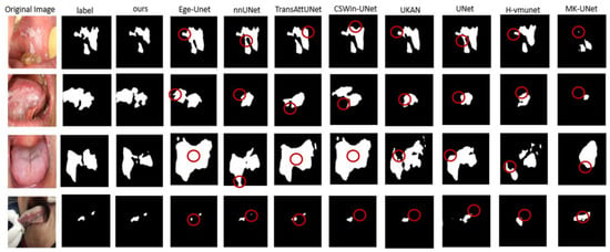
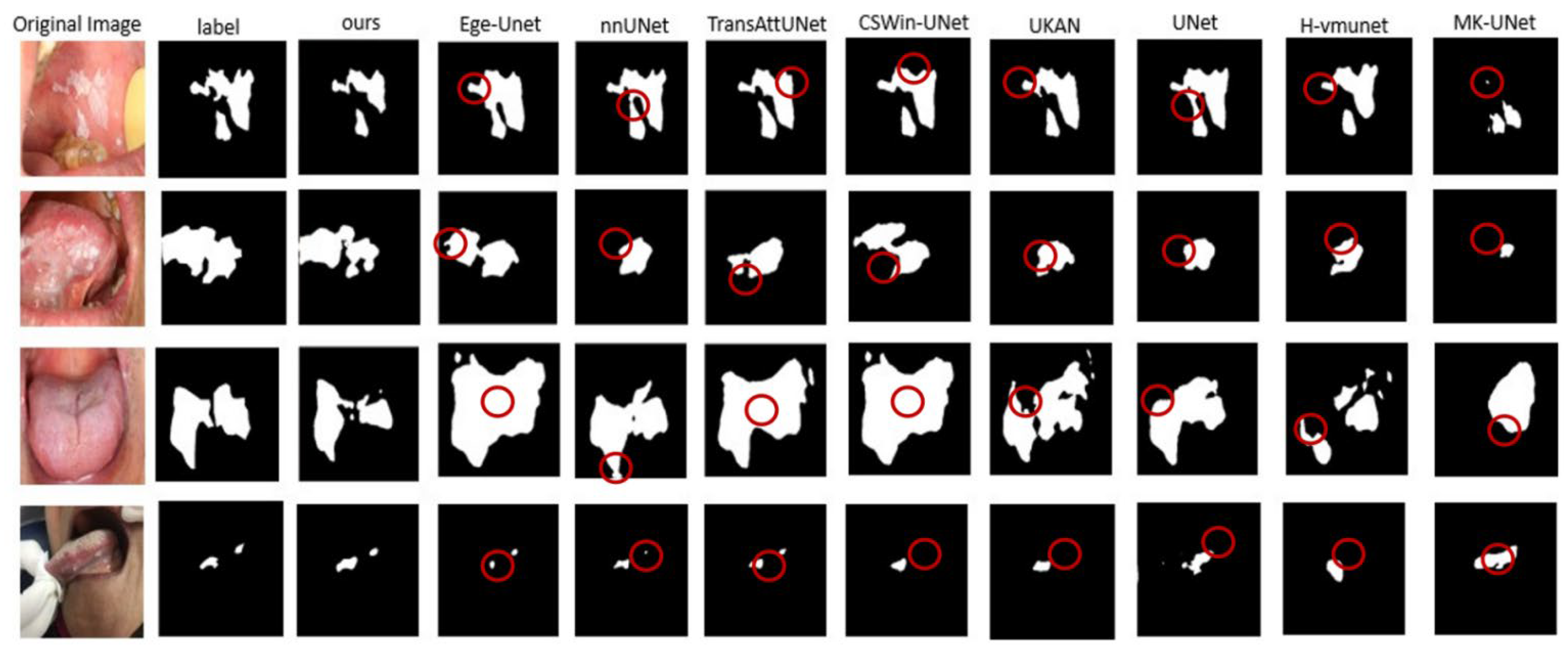
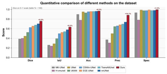
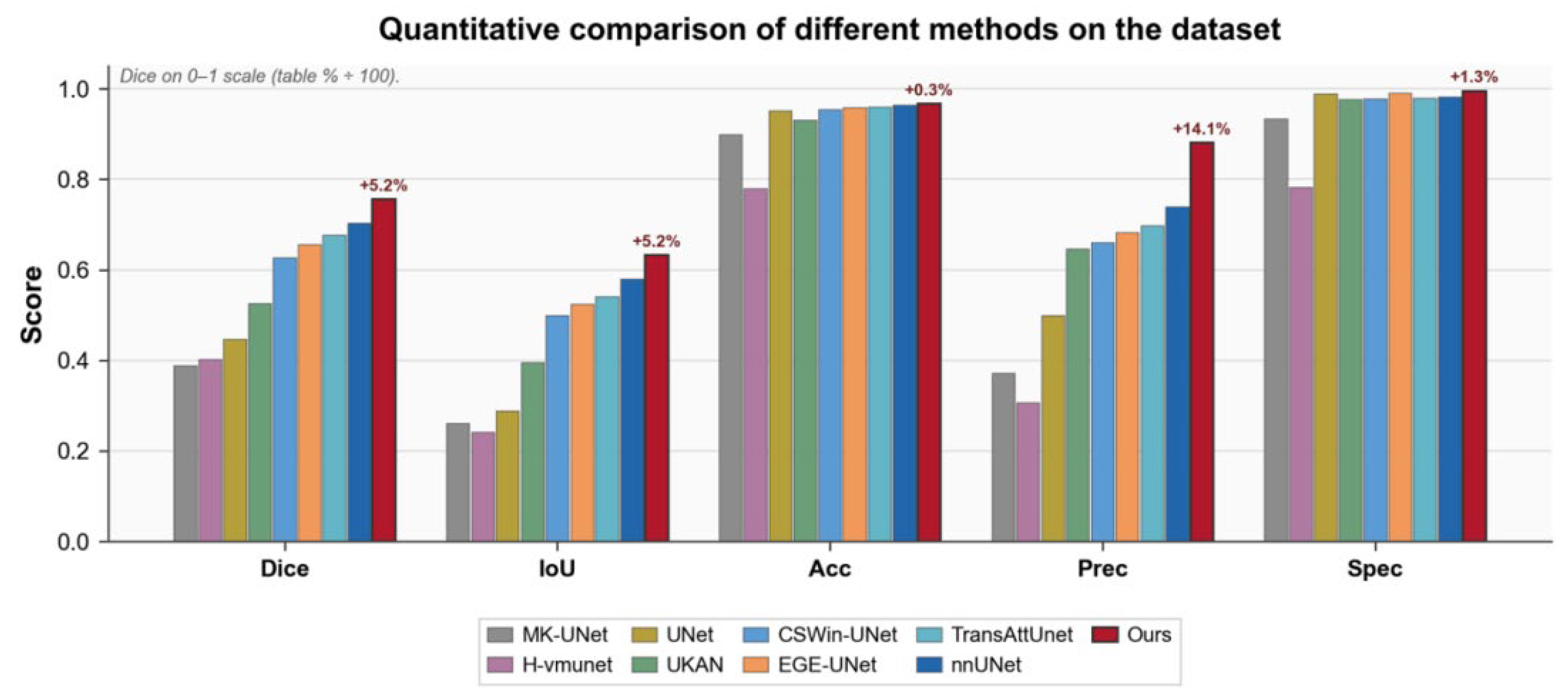
To validate the effectiveness of the proposed method, this paper systematically compares RIVER-NET with multiple comparable baseline methods, as summarized in Table 1. Among all compared methods, RIVER-NET achieves the optimal segmentation performance, with a Dice score of 75.59% and an IoU of 0.632, outperforming U-Net by 30.88 percentage points and nnU-Net by 5.23 percentage points in Dice. The visual comparison in Figure 5 further demonstrates that under challenging scenarios such as blurred boundaries, strong specular reflections, and small lesions, our method achieves more accurate lesion delineation with more complete regions and clearer boundary localization. Figure 6 provides a graphical summary of the quantitative comparison. All quantitative results in Table 1 were calculated from the five validation folds and are reported as the mean ± standard deviation across folds. RIVER-NET significantly outperformed all eight baselines after Holm correction (all adjusted p < 0.001; Table 2). Compared with nnU-Net, the strongest baseline, the mean paired Dice improvement was 5.23 percentage points (95% CI, 4.86–5.60; t(996) = 27.976; adjusted p < 0.001; Cohen’s d_z = 0.886).
Table 1.
Quantitative comparison of different methods on the OPM dataset using patient- and lesion-grouped five-fold cross-validation (1051 images; input size, 256 × 256). Methods are ranked by Dice. Dice is reported as a percentage (%); Acc/Prec/Spec are evaluated at a threshold of 0.5. Per-image Dice, IoU, Acc, Prec, and Spec were computed for each image and macro-averaged within each validation fold; values are the mean ± standard deviation of these fold-wise macro-averages across the five validation folds.
Figure 5.
Visual comparison of segmentation results. Columns 1–3: original oral images, ground-truth labels, and our results. Columns 4–11: EGE-UNet, nnU-Net, TransAttUNet, CSWin-UNet, U-KAN, U-Net, H-vmUNet, and Mk-UNet. Row-wise challenging characteristics: Row 1, blurred boundaries; Rows 2–3, strong specular reflections with blurred boundaries; Row 4, small lesion. Our method achieves more accurate lesion delineation with more complete regions and clearer boundary localization, especially under these conditions. Red circles highlight local segmentation differences or errors.
Figure 6.
Quantitative comparison of different methods on the OPM dataset.
Table 2.
Paired t-test results for patient/lesion-level aggregated Dice comparisons between RIVER-NET and the baseline methods. Mean differences are RIVER-NET minus baseline; p values are two-sided, with Holm adjustment across eight comparisons.
3.2. Module-Level, Component-Wise, and Replacement-Based Ablation Studies
All ablation variants were evaluated once using the same predefined patient- and lesion-grouped split of 812 training and 239 validation images. As shown in Table 3a, removing MSRU, DGCF, and MAAF was associated with Dice decreases from 75.46% to 69.87%, 71.35%, and 74.08%, respectively. At the component level, removing the 11 local residual shortcuts yielded a Dice score of 70.80%; removing cross-scale aggregation or channel–spatial attention from MAAF yielded 74.92% and 74.86%; and replacing the DGCF gate with direct summation yielded 72.00%. Within this single-run fixed-split analysis, these observations are consistent with the intended functions of the corresponding components, but they do not independently establish causal mechanisms.
Table 3.
(a) Module-level and component-wise ablation study using a fixed split (812 training; 239 validation images). Values are single-run estimates without fold-wise standard deviations. “Partial” indicates that the module was retained but the specified internal component was removed or simplified. (b) Replacement-based ablation under closely matched parameter and FLOP budgets. All variants used the same fixed split and training protocol as Table 3a.
Under closely matched parameter and FLOP budgets (Table 3b), replacing DGCF with an Attention Gate [37], MSRU with ASPP [38], and MAAF with FPN [39] yielded Dice scores of 70.30%, 73.49%, and 72.02%, respectively, compared with 75.46% for RIVER-NET. These comparisons reduce model capacity as a simple explanation for the observed differences; however, they do not exclude optimization effects, interactions among modules, or other architecture-specific factors. The results should therefore be interpreted as comparative evidence within the current experimental setting rather than definitive proof of the superiority of any individual mechanism.
3.3. Loss-Function Sensitivity Analysis
Table 4 summarizes the sensitivity of RIVER-NET to different loss configurations. The original 0.5 Dice + 0.5 BCE loss achieved the best numerical performance across all evaluated metrics (Dice, 75.59 ± 3.58%; IoU, 0.632 ± 0.032; HD95, 30.5 ± 5.1 pixels; Boundary IoU, 0.557 ± 0.030). Changing the Dice/BCE ratio to 0.25/0.75 or 0.75/0.25 resulted in Dice scores of 74.72 ± 4.07% and 75.12 ± 3.28%, respectively, indicating limited sensitivity to moderate weight changes. Replacing BCE with Focal Loss or adding 0.1 Hausdorff Distance Loss did not improve the boundary-related metrics. Therefore, the equal-weight Dice–BCE loss was retained because it provided the most favorable balance between region-overlap accuracy and boundary delineation.
Table 4.
Sensitivity analysis of loss weighting and alternative loss functions.
3.4. Independent Public-Dataset and External Validation
On the independent public ISIC dataset (Table 5), RIVER-NET and the baselines were trained and evaluated under five-fold cross-validation on this separate public lesion-segmentation dataset. RIVER-NET achieved the highest Dice score (94.62 ± 2.18%) and IoU (0.900 ± 0.039), indicating competitive performance on an independent public dataset whose imaging and acquisition characteristics differ from those of the OPM dataset. Because all models were retrained on ISIC, these results demonstrate cross-dataset competitiveness rather than zero-shot cross-domain generalization.
Table 5.
Quantitative comparison on the ISIC skin-lesion segmentation dataset using five-fold cross-validation. Values are mean ± standard deviation across folds.
On the independent Autooral dataset (Table 6), RIVER-NET achieved the highest Dice score (0.5547) and specificity (0.9324). Thus, RIVER-NET showed the strongest Dice overlap and background specificity.
Table 6.
Direct external evaluation on the public Autooral dataset without fine-tuning. Metrics were calculated per image and macro-averaged; higher is better except for HD95 and ASSD.
3.5. Computational Complexity and Inference Efficiency
Table 7 summarizes the computational complexity and inference efficiency of all compared methods under the same profiling settings. RIVER-NET achieved the highest Dice score (75.59%) while maintaining a moderate model size of 17.263 M parameters, which was 58.3% smaller than nnU-Net, 44.4% smaller than U-Net, and 33.5% smaller than TransAttUNet. Compared with U-Net and TransAttUNet, RIVER-NET reduced FLOPs by 82.7% and 89.3%, respectively, while increasing inference throughput by 29.6% and 86.3%. It achieved a network-only inference throughput of 84.0 images per second at a batch size of 1 under the stated GPU setting (NVIDIA GeForce RTX 3080 10 GB; data loading, preprocessing, and post-processing excluded), indicating that the additional MSRU, DGCF, and MAAF modules preserved real-time inference capability despite the multi-branch design. Although dedicated lightweight architectures such as EGE-UNet and Mk-UNet required fewer computational resources, their Dice scores were 9.98 and 36.71 percentage points lower than that of RIVER-NET, respectively. Overall, these results show that RIVER-NET provides a favorable balance between segmentation accuracy, model size, and inference efficiency among the compared high-performance architectures.
Table 7.
Computational complexity and inference efficiency of the compared methods. All models were profiled with 256 × 256 inputs, batch size 1, and FP32 precision on an NVIDIA GeForce RTX 3080 10 GB GPU. FPS excludes data loading, preprocessing, and post-processing; FLOPs = 2 × MACs.
4. Discussion
Many existing studies mainly focus on image-level detection, classification, or general oral lesion analysis, whereas fine-grained pixel-level segmentation of oral mucosal lesions remains challenging because lesion regions often resemble surrounding normal mucosa. RIVER-NET was evaluated as a task-oriented combination of multi-scale modeling, gated feature interaction, and hierarchical fusion. In the current experiments, the complete model achieved higher segmentation metrics than the evaluated ablation variants. The largest single-module Dice decrease occurred after removing MSRU; this association is consistent with a useful role for intra-stage multi-scale representation in this dataset, but it should not be interpreted as isolating a causal mechanism because the module ablations used a single predefined split.
The loss-function sensitivity analysis further showed that RIVER-NET maintained relatively stable Dice performance under moderate changes in the Dice/BCE weighting, suggesting that its performance was not dependent on a narrowly tuned loss ratio. Neither Focal Loss nor Hausdorff Distance Loss yielded additional boundary benefits in this dataset, whereas the original equal-weight Dice–BCE loss provided the most stable balance between lesion-region completeness and boundary localization.
The extended ablation results provide comparative, hypothesis-generating evidence about the three modules under the current training protocol. Removing the local residual shortcuts was associated with a 4.66-percentage-point Dice decrease, while the parameter-matched ASPP replacement was 1.97 points below the complete model. Replacing the DGCF gate with direct summation and replacing the complete DGCF with an Attention Gate were associated with decreases of 3.46 and 5.16 points, respectively. For MAAF, removing cross-scale aggregation or channel–spatial attention was associated with decreases of 0.54 and 0.60 points, and the parameter-matched FPN replacement was 3.44 points below the complete model. These patterns are consistent with the proposed design rationale, but they neither prove that the named mechanisms alone caused the observed differences nor exclude module interactions, training variability, or other confounding factors.
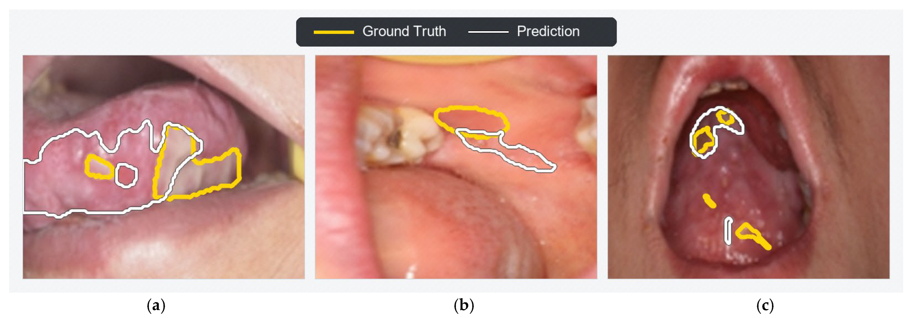
To examine residual errors and provide output-level qualitative interpretability, we visualized three representative low-performing cases using overlaid reference and prediction contours (Figure 7). In Figure 7a (3_10.png; Dice, 0.331; IoU, 0.198), weak lesion-to-mucosa contrast and gradual boundary transitions were associated with substantial prediction extension into adjacent visually similar mucosa, indicating limited boundary discrimination. In Figure 7b (6_0219.png; Dice, 0.199; IoU, 0.110), the predicted region was spatially displaced from the small reference lesion toward a nearby visually salient structure, indicating a localization error under competing local appearance cues. In Figure 7c (6_0631.png; Dice, 0.496; IoU, 0.330), the prediction overlapped the dominant lesion component but missed several small, spatially disconnected components, indicating incomplete recovery of fragmented lesions. The contour overlays therefore make the sources of disagreement directly observable at the output level and identify three residual error patterns—boundary overextension, target displacement, and omission of disconnected components. These qualitative observations describe model behavior in selected difficult cases and do not by themselves establish the internal mechanism responsible for each error.
Figure 7.
Representative failure cases with overlaid lesion contours. The panels show (a) 3_10.png (Dice, 0.331; IoU, 0.198), (b) 6_0219.png (Dice, 0.199; IoU, 0.110), and (c) 6_0631.png (Dice, 0.496; IoU, 0.330). Yellow and white contours denote the ground-truth and RIVER-NET-predicted lesion boundaries, respectively. The three cases illustrate boundary overextension under low lesion-to-mucosa contrast and gradual transitions, spatial displacement of a small lesion near competing local structures, and incomplete recovery of spatially disconnected lesion components.
From a clinical perspective, accurate segmentation of oral mucosal lesions may eventually support lesion-extent assessment, follow-up comparison, and computer-aided screening. However, the present study validates algorithm-level feasibility only. The primary dataset was retrospective and single-center, detailed clinical and acquisition metadata were limited, and the public-dataset experiments do not constitute prospective multi-center clinical validation. Accordingly, RIVER-NET has not undergone multi-center clinical validation, is not ready for real-world clinical deployment, and cannot be directly used for clinical diagnosis, treatment planning, or decision-making. Fixed-resolution resizing may also reduce fine boundary and surface details, and the binary-segmentation setting does not distinguish clinical subtypes, dysplasia grades, or malignant-transformation risk. Future work should include prospective multi-center studies across acquisition devices and patient populations, independent clinical testing, and integration of richer clinical information before any consideration of clinical use.
Future multimodal approaches could combine conventional RGB imaging with optical techniques providing additional tissue contrast, such as hyperspectral imaging. HSI simultaneously captures spatial and spectral information and has been investigated for tissue characterization and disease detection [40], and for image-guided surgery [41]. In oral and head-and-neck applications, hyperspectral signatures have enabled discrimination among tissue types [42] and delineation of tumor margins [43]. Moreover, spectral measurements may reflect histopathological tissue alterations [44] and perfusion-related characteristics [45], potentially supporting both lesion delineation and biological characterization.
5. Conclusions
In this study, we propose an oral mucosal lesion segmentation framework integrating MSRU, DGCF, and MAAF. Across the evaluated datasets and experimental settings, RIVER-NET achieved promising segmentation results, while the component-wise and parameter-matched comparisons were consistent with—rather than definitive proof of—the intended complementary roles of the three modules. The ISIC evaluation, in which models were retrained on this independent public dataset, demonstrates cross-dataset competitiveness, whereas the Autooral evaluation, performed without fine-tuning, provides preliminary algorithm-level evidence of external generalization. Nevertheless, the model has not undergone prospective multi-center clinical validation, is not ready for clinical deployment, and cannot be directly applied in clinical practice. Validation across institutions, acquisition devices, and patient populations is required before broader clinical translation can be considered.
Author Contributions
Conceptualization, R.L.; methodology, R.L.; software, R.L. and Y.T.; validation, W.Z.; formal analysis, C.M.; resources, X.S. and Z.S.; data curation, W.Z. and X.S.; writing—review and editing, Y.T. and W.Z.; supervision, Z.S. All authors have read and agreed to the published version of the manuscript.
Funding
This work was supported by the National Natural Science Foundation of China under Grant No. 62273231 and the Shaanxi Province Outstanding Youth Science Foundation Project under Grant No. 2025JC-JCQN-074. This work was also supported by the Shanghai Municipal Health Commission 2025 Traditional Chinese Medicine Key Disease Construction Project (ZDBZ-202623).
Institutional Review Board Statement
The study was conducted in accordance with the Declaration of Helsinki and approved by the Ethics Committee of the Ninth People’s Hospital Affiliated to Shanghai Jiao Tong University School of Medicine (SH9H-2022-TK337-1, 28 February 2022).
Informed Consent Statement
Informed consent was obtained from all subjects involved in the study or their legal guardians.
Data Availability Statement
The OPM dataset is not publicly available because of patient privacy and ethical restrictions. Access may be requested from the corresponding author and is subject to a reasonable research request and the necessary ethical approvals. The ISIC 2018 dataset is publicly available at https://challenge.isic-archive.com/data/#2018 (accessed on 21 October 2026). The Autooral dataset is available through the official project repository at https://github.com/wurenkai/HF-UNet-and-Autooral-dataset (accessed on 21 October 2026).
Conflicts of Interest
The authors declare no conflict of interest.
References
- Villa, A.; Lodolo, M.; Ha, P. Oncological outcomes of patients with oral potentially malignant disorders. JAMA Otolaryngol. Head Neck Surg. 2025, 151, 65–71. [Google Scholar] [CrossRef] [Scilit] [PubMed]
- Ronneberger, O.; Fischer, P.; Brox, T. U-Net: Convolutional networks for biomedical image segmentation. In Medical Image Computing and Computer-Assisted Intervention (MICCAI); LNCS 9351; Springer: Cham, Switzerland, 2015; pp. 234–241. [Google Scholar] [CrossRef] [Scilit]
- Warin, K.; Limprasert, W.; Suebnukarn, S.; Jinaporntham, S.; Jantana, P. Automatic classification and detection of oral cancer in photographic images using deep learning algorithms. J. Oral Pathol. Med. 2021, 50, 911–918. [Google Scholar] [CrossRef] [Scilit] [PubMed]
- Azam, M.A.; Sampieri, C.; Ioppi, A.; Benzi, P.; Giordano, G.G.; De Vecchi, M.; Campagnari, V.; Li, S.; Guastini, L.; Paderno, A.; et al. Videomics of the upper aero-digestive tract cancer: Deep learning applied to white light and narrow band imaging for automatic segmentation of endoscopic images. Front. Oncol. 2022, 12, 900451. [Google Scholar] [CrossRef] [Scilit] [PubMed]
- Qiu, L.; Ye, C.; Chen, P.; Liu, Y.; Han, X.; Cui, S. DArch: Dental arch prior-assisted 3D tooth instance segmentation with weak annotations. In Proceedings of the IEEE/CVF Conference on Computer Vision and Pattern Recognition, New Orleans, LA, USA, 18–24 June 2022; pp. 20752–20761. [Google Scholar]
- Mărginean, A.C.; Mureșanu, S.; Hedeșiu, M.; Dioșan, L. Teeth segmentation and carious lesions segmentation in panoramic X-ray images using CariSeg, a networks’ ensemble. Heliyon 2024, 10, e30836. [Google Scholar] [CrossRef] [Scilit] [PubMed]
- Zhang, R.; Lu, M.; Zhang, J.; Chen, X.; Zhu, F.; Tian, X.; Chen, Y.; Cao, Y. Research and application of deep learning models with multi-scale feature fusion for lesion segmentation in oral mucosal diseases. Bioengineering 2024, 11, 1107. [Google Scholar] [CrossRef] [Scilit] [PubMed]
- Wang, C.; Zhang, Q.; Zhu, L.; Hou, J. Performance evaluation and clinical application exploration of a ViT-CNN ensemble model for multiclass oral mucosal disease classification: A pilot retrospective analysis based on public datasets. BMJ Open 2025, 15, e102741. [Google Scholar] [CrossRef] [Scilit] [PubMed]
- Fan, D.P.; Ji, G.P.; Sun, G.; Cheng, M.; Shen, J.; Shao, L. Camouflaged object detection. In Proceedings of the IEEE/CVF Conference on Computer Vision and Pattern Recognition, Seattle, WA, USA, 13–19 June 2020; pp. 2777–2787. [Google Scholar]
- Zhou, Z.; Li, Y.; Zhong, C.; Huang, J.; Pei, J.; Li, H.; Tang, H. Rethinking Detecting Salient and Camouflaged Objects in Unconstrained Scenes. In Proceedings of the IEEE/CVF International Conference on Computer Vision, Honolulu, HI, USA, 19–25 October 2025; pp. 22372–22382. [Google Scholar]
- Mei, H.; Ji, G.P.; Wei, Z.; Yang, X.; Wei, X.; Fan, D.-P. Camouflaged object segmentation with distraction mining. In Proceedings of the IEEE/CVF Conference on Computer Vision and Pattern Recognition, Nashville, TN, USA, 20–25 June 2021; pp. 8772–8781. [Google Scholar]
- Sun, Y.; Wang, S.; Chen, C.; Xiang, T.-Z. Boundary-guided camouflaged object detection. In Proceedings of the Thirty-First International Joint Conference on Artificial Intelligence (IJCAI), Vienna, Austria, 23–29 July 2022; pp. 1335–1341. [Google Scholar] [CrossRef] [Scilit] [PubMed]
- Yang, F.; Zhai, Q.; Li, X.; Huang, R.; Luo, A.; Cheng, H.; Fan, D.-P. Uncertainty-guided transformer reasoning for camouflaged object detection. In Proceedings of the IEEE/CVF International Conference on Computer Vision, Montreal, QC, Canada, 10–17 October 2021; pp. 4146–4155. [Google Scholar]
- Yu, Q.; Xie, L.; Wang, Y.; Zhou, Y.; Fishman, E.K.; Yuille, A.L. Recurrent saliency transformation network: Incorporating multi-stage visual cues for small organ segmentation. In Proceedings of the IEEE Conference on Computer Vision and Pattern Recognition, Salt Lake City, UT, USA, 18–22 June 2018; pp. 8280–8289. [Google Scholar]
- Huang, Z.; Dai, H.; Xiang, T.Z.; Wang, S.; Chen, H.-X.; Qin, J.; Xiong, H. Feature shrinkage pyramid for camouflaged object detection with transformers. In Proceedings of the IEEE/CVF Conference on Computer Vision and Pattern Recognition, Vancouver, BC, Canada, 17–24 June 2023; pp. 5557–5566. [Google Scholar]
- Niu, Y.; Yang, L.; Xu, R.; Li, Y.; Chen, Y. MINet: Weakly-supervised camouflaged object detection through mutual interaction between region and edge cues. In Proceedings of the 32nd ACM International Conference on Multimedia, Melbourne, Australia, 28 October–1 November 2024; pp. 6316–6325. [Google Scholar]
- Ioffe, S.; Szegedy, C. Batch normalization: Accelerating deep network training by reducing internal covariate shift. In Proceedings of the 32nd International Conference on Machine Learning (ICML), PMLR 37, Lille, France, 6–11 July 2015; pp. 448–456. [Google Scholar]
- Yu, F.; Koltun, V. Multi-scale context aggregation by dilated convolutions. arXiv 2015, arXiv:1511.07122. [Google Scholar]
- Gu, A.; Dao, T. Mamba: Linear-time sequence modeling with selective state spaces. arXiv 2023, arXiv:2312.00752. [Google Scholar]
- Liu, Y.; Tian, Y.; Zhao, Y.; Yu, H.; Xie, L.; Wang, Y.; Ye, Q.; Jiao, J.; Liu, Y. VMamba: Visual state space model. Adv. Neural Inf. Process. Syst. 2024, 37, 103031–103063. [Google Scholar] [CrossRef] [Scilit]
- Hu, J.; Shen, L.; Sun, G. Squeeze-and-excitation networks. In Proceedings of the IEEE Conference on Computer Vision and Pattern Recognition, Salt Lake City, UT, USA, 18–23 June 2018; pp. 7132–7141. [Google Scholar] [CrossRef] [Scilit]
- Woo, S.; Park, J.; Lee, J.Y.; Kweon, I.S. CBAM: Convolutional block attention module. In Proceedings of the European Conference on Computer Vision (ECCV); LNCS 11211; Springer: Cham, Switzerland, 2018; pp. 3–19. [Google Scholar] [CrossRef] [Scilit]
- Jiang, C.; Wu, R.; Liu, Y.; Wang, Y.; Chang, Q.; Liang, P.; Fan, Y. A high-order focus interaction model and oral ulcer dataset for oral ulcer segmentation. Sci. Rep. 2024, 14, 20085. [Google Scholar] [CrossRef] [Scilit] [PubMed]
- Codella, N.C.F.; Rotemberg, V.; Tschandl, P.; Celebi, M.E.; Dusza, S.; Gutman, D.; Helba, B.; Kalloo, A.; Liopyris, K.; Marchetti, M.; et al. Skin lesion analysis toward melanoma detection 2018: A challenge hosted by the International Skin Imaging Collaboration (ISIC). arXiv 2019, arXiv:1902.03368. [Google Scholar]
- Milletari, F.; Navab, N.; Ahmadi, S.A. V-Net: Fully convolutional neural networks for volumetric medical image segmentation. In 2016 Fourth International Conference on 3D Vision (3DV); IEEE: Stanford, CA, USA, 2016; pp. 565–571. [Google Scholar] [CrossRef] [Scilit]
- Kingma, D.P.; Ba, J. Adam: A method for stochastic optimization. arXiv 2014, arXiv:1412.6980. [Google Scholar]
- Isensee, F.; Jaeger, P.F.; Kohl, S.A.A.; Petersen, J.; Maier-Hein, K.H. nnU-Net: A self-configuring method for deep learning-based biomedical image segmentation. Nat. Methods 2021, 18, 203–211. [Google Scholar] [CrossRef] [Scilit] [PubMed]
- Chen, B.; Liu, Y.; Zhang, Z.; Lu, G.; Kong, A.W.K. TransAttUNet: Multi-level attention-guided U-Net with transformer for medical image segmentation. IEEE Trans. Emerg. Top. Comput. Intell. 2023, 8, 55–68. [Google Scholar] [CrossRef] [Scilit]
- Wu, R.; Liu, Y.; Liang, P.; Chang, Q. H-vmUNet: High-order vision Mamba U-Net for medical image segmentation. Neurocomputing 2025, 624, 129447. [Google Scholar] [CrossRef] [Scilit]
- Li, C.; Liu, X.; Li, W.; Wang, C.; Liu, H.; Liu, Y.; Chen, Z.; Yuan, Y. U-KAN makes strong backbone for medical image segmentation and generation. Proc. AAAI Conf. Artif. Intell. 2025, 39, 4652–4660. [Google Scholar] [CrossRef] [Scilit]
- Liu, X.; Gao, P.; Yu, T.; Wang, F.; Yuan, R.-Y. CSWin-UNet: Transformer U-Net with cross-shaped windows for medical image segmentation. Inf. Fusion 2025, 113, 102634. [Google Scholar] [CrossRef] [Scilit]
- Ruan, J.; Xie, M.; Gao, J.; Liu, T.; Fu, Y. EGE-UNet: An efficient group-enhanced U-Net for skin lesion segmentation. In Medical Image Computing and Computer-Assisted Intervention (MICCAI); Springer: Cham, Switzerland, 2023. [Google Scholar]
- Rahman, M.M.; Marculescu, R. MK-UNet: Multi-Kernel Lightweight CNN for Medical Image Segmentation. In Proceedings of the IEEE/CVF International Conference on Computer Vision Workshops (ICCVW), CVAMD, Honolulu, HI, USA, 19–20 October 2025; pp. 1053–1062. [Google Scholar]
- Liu, J.; Yang, H.; Zhou, H.-Y.; Xi, Y.; Yu, L.; Li, C.; Liang, Y.; Shi, G.; Yu, Y.; Zhang, S.; et al. Swin-UMamba: Mamba-based U-Net with ImageNet-based pretraining. In Medical Image Computing and Computer-Assisted Intervention (MICCAI); LNCS 15009; Springer: Cham, Switzerland, 2024; pp. 615–625. [Google Scholar] [CrossRef] [Scilit]
- Xing, Z.; Ye, T.; Yang, Y.; Cai, D.; Gai, B.; Wu, X.-J.; Gao, F.; Zhu, L. SegMamba-V2: Long-Range Sequential Modeling Mamba for General 3-D Medical Image Segmentation. IEEE Trans. Med. Imaging 2026, 45, 4–15. [Google Scholar] [CrossRef] [Scilit] [PubMed]
- Ma, J.; Li, F.; Wang, B. U-Mamba: Enhancing long-range dependency for biomedical image segmentation. arXiv 2024, arXiv:2401.04722. [Google Scholar]
- Oktay, O.; Schlemper, J.; Le Folgoc, L.; Lee, M.; Heinrich, M.; Misawa, K.; Mori, K.; McDonagh, S.; Hammerla, N.Y.; Kainz, B.; et al. Attention U-Net: Learning where to look for the pancreas. In Proceedings of the 1st Conference on Medical Imaging with Deep Learning (MIDL), Amsterdam, The Netherlands, 4–6 July 2018. [Google Scholar]
- Chen, L.C.; Papandreou, G.; Schroff, F.; Adam, H. Rethinking atrous convolution for semantic image segmentation. arXiv 2017, arXiv:1706.05587. [Google Scholar]
- Lin, T.Y.; Dollár, P.; Girshick, R.; He, K.; Hariharan, B.; Belongie, S. Feature pyramid networks for object detection. In Proceedings of the IEEE Conference on Computer Vision and Pattern Recognition, Honolulu, HI, USA, 21–26 July 2017; pp. 2117–2125. [Google Scholar]
- Tran, M.H.; Ma, L.; Yuan, M.; Fei, B. Medical hyperspectral imaging: An updated review of technology advancements and biomedical applications. J. Biomed. Opt. 2026, 31, 030901. [Google Scholar] [CrossRef] [Scilit] [PubMed]
- Composto, A.; Privitera, L.; Riva, M.; Ardini, B.; Manzoni, C.; Riva, M.; Aquilina, K.; Valentini, G.; Giuliani, S. Hyperspectral imaging for tumor resection guidance in surgery: A systematic review of preclinical and clinical studies. J. Biomed. Opt. 2025, 30, S23909. [Google Scholar] [CrossRef] [Scilit] [PubMed]
- Thiem, D.G.E.; Römer, P.; Gielisch, M.; Al-Nawas, B.; Schlüter, M.; Plaß, B.; Kämmerer, P.W. Hyperspectral imaging and artificial intelligence to detect oral malignancy—Part 1: Automated tissue classification of oral muscle, fat and mucosa using a light-weight 6-layer deep neural network. Head Face Med. 2021, 17, 38. [Google Scholar] [CrossRef] [Scilit] [PubMed]
- Halicek, M.; Dormer, J.D.; Little, J.V.; Chen, A.Y.; Myers, L.; Sumer, B.D.; Fei, B. Hyperspectral imaging of head and neck squamous cell carcinoma for cancer margin detection in surgical specimens from 102 patients using deep learning. Cancers 2019, 11, 1367. [Google Scholar] [CrossRef] [Scilit] [PubMed]
- Lu, G.; Wang, D.; Qin, X.; Muller, S.; Little, J.V.; Wang, X.; Chen, A.Y.; Chen, G.; Fei, B. Histopathology feature mining and association with hyperspectral imaging for the detection of squamous neoplasia. Sci. Rep. 2019, 9, 17863. [Google Scholar] [CrossRef] [Scilit] [PubMed]
- Hren, R.; Sersa, G.; Simoncic, U.; Milanic, M. Imaging perfusion changes in oncological clinical applications by hyperspectral imaging: A literature review. Radiol. Oncol. 2022, 56, 420–429. [Google Scholar] [CrossRef] [Scilit] [PubMed]

Disclaimer/Publisher’s Note: The statements, opinions and data contained in all publications are solely those of the individual author(s) and contributor(s) and not of MDPI and/or the editor(s). MDPI and/or the editor(s) disclaim responsibility for any injury to people or property resulting from any ideas, methods, instructions or products referred to in the content. |
© 2026 by the authors. Licensee MDPI, Basel, Switzerland. This article is an open access article distributed under the terms and conditions of the Creative Commons Attribution (CC BY) license.