


Highlights
What are the main findings?
- •
- Through iTRAQ-based quantitative proteomic screening, a critical gene associated with Verticillium wilt resistance was identified in Solanum torvum.
- •
- Its resistance function was subsequently verified using virus-induced gene silencing (VIGS) and transgenic overexpression assays.
What are the implications of the main findings?
- •
- These findings suggest that StoAOS1 positively regulates Verticillium wilt resistance in Solanum torvum, likely through a JA-dependent defense pathway.
- •
- This study provides a candidate gene resource for improving Verticillium wilt resistance in eggplant.
Abstract
Eggplant (Solanum melongena L.) is an important solanaceous vegetable crop cultivated worldwide. Verticillium wilt, caused by Verticillium dahliae, severely restricts eggplant growth and yield, while most cultivated eggplant varieties show only limited resistance to this disease. In contrast, Solanum torvum, a wild relative of eggplant, exhibits strong natural resistance to Verticillium wilt. The molecular mechanisms underlying the contrasting responses of cultivated eggplant and S. torvum to V. dahliae infection remain poorly understood. In this study, high-throughput iTRAQ-based quantitative proteomics was used to analyze root protein profiles of S. torvum after V. dahliae inoculation. A differentially expressed protein, StoAOS1, was identified in S. torvum and was strongly induced by V. dahliae infection. StoAOS1 encodes a key enzyme in the jasmonic acid (JA) biosynthetic pathway. Further analysis showed that exogenous methyl jasmonate (MeJA) treatment markedly induced StoAOS1 expression. Virus-induced gene silencing of StoAOS1 significantly compromised Verticillium wilt resistance in S. torvum, accompanied by reduced acid-insoluble lignin accumulation and decreased transcript levels of the defense-related marker genes StoPDF1.2, StoVSP2, and StoThi2.1. Conversely, transient overexpression of StoAOS1 in Nicotiana benthamiana enhanced resistance to V. dahliae. Together, these findings suggest that StoAOS1 positively regulates Verticillium wilt resistance in S. torvum, likely through a JA-dependent defense pathway.
1. Introduction
Eggplant (Solanum melongena L.) is a solanaceous vegetable crop of considerable economic significance, and its cultivation now extends to diverse regions across the globe. One of the most serious biotic constraints affecting eggplant production is Verticillium wilt, a disease caused predominantly by the soil-inhabiting vascular pathogen Verticillium dahliae Kleb. [1,2]. Upon entering the root system, the fungus colonizes the xylem vessels and disperses systemically via the transpiration stream, eventually giving rise to vascular discoloration, foliar chlorosis, wilting, defoliation, and, under conditions of severe infection, eventual plant death [1,2]. Because V. dahliae produces microsclerotia that can persist in soil for extended periods and maintains an unusually broad host range, the pathogen often remains viable within infested fields for many years, and once it becomes established, Verticillium wilt proves exceptionally difficult to manage through conventional control measures [1,2]. Relative to cultivated eggplants, which generally exhibit only limited resistance to this disease, its wild relative Solanum torvum displays strong innate resistance and has consequently been employed widely as a resistant rootstock or as donor germplasm in eggplant improvement programs [3,4]. A detailed understanding of the molecular basis underlying this resistance in S. torvum is therefore of substantial importance for the eventual development of resistant eggplant cultivars through breeding.
At present, the precise mechanisms by which eggplant mounts resistance against Verticillium wilt remain incompletely resolved. Accumulated evidence suggests that the resistant phenotype is underpinned by both preformed physical or chemical barriers and inducible defense responses that are activated following pathogen challenge. Constitutively present antimicrobial metabolites, including alkaloids and various phenolic compounds, may serve to restrict fungal proliferation during the earliest stages of root invasion, whereas induced reactions—among them cell-wall reinforcement, lignin deposition, maintenance of reactive oxygen species (ROS) homeostasis, accumulation of pathogenesis-related proteins, and hormone-mediated signaling cascades—collectively act to contain the pathogen within the vascular tissue and limit its systemic spread [4,5]. Several genes derived from S. torvum have previously been implicated in resistance to V. dahliae, including StoL13a [6], StoNPR1 [7], StoCYP77A2 [8], StoWRKY6 [9], StoMYB41 [10], and StoERF109 [11]. Nevertheless, these genetic determinants account for only a fraction of the overall resistance phenotype, and the key regulatory proteins, along with the downstream defense networks they orchestrate, remain to be more fully characterized if we are to understand how S. torvum achieves its notably high level of resistance.
Phytohormone signaling plays a central regulatory role in plant immunity, and the pathways governed by salicylic acid (SA), jasmonic acid (JA), and ethylene (ET) are recognized as major nodes through which defense responses are modulated [12,13]. Although SA has traditionally been linked to defense against biotrophic pathogens, whereas JA and ET are more commonly associated with resistance to necrotrophic pathogens and herbivorous insects, the response to vascular pathogens such as V. dahliae tends to be considerably more complex and may well require the coordinated engagement of multiple hormonal pathways [5,12,13]. In this context, JA signaling has increasingly drawn attention as a critical component of the plant’s defensive arsenal against V. dahliae, and its contribution is now the subject of growing investigative interest. In cotton, for example, modulation of JA biosynthesis and signaling affects host resistance to Verticillium wilt, suggesting that JA-associated pathways may contribute to the regulation of vascular disease resistance [14,15,16].
Proteome-level analysis provides a powerful approach for identifying defense-associated proteins that may not be fully predicted from transcript abundance alone. Isobaric tags for relative and absolute quantification (iTRAQ)-based quantitative proteomics enables comparative analysis of protein accumulation across multiple biological samples with high sensitivity and reproducibility, and has been widely used to investigate plant responses to biotic stress [17,18,19]. In plant–pathogen interaction studies, iTRAQ-based proteomics has been useful for identifying proteins involved in hormone signaling, cell-wall modification, secondary metabolism, oxidative stress responses, and disease resistance. In Gossypium thurberi roots challenged with V. dahliae, iTRAQ-based proteomic profiling has previously uncovered a substantial number of differentially accumulated proteins that map to SA signaling, phenylpropanoid metabolism, lignin biosynthetic routes, and general defense responses [19]. By contrast, comparable proteomic data for eggplant and its wild relatives in response to this vascular pathogen remain scarce. Given this gap, quantitative proteomic analysis using the iTRAQ platform may offer a productive avenue for pinpointing candidate regulatory factors that contribute to the pronounced Verticillium wilt resistance observed in S. torvum.
Within the suite of hormone pathways implicated in defense, the JA biosynthetic route merits particular attention in the context of stress-triggered immune activation. A key enzymatic component of this route is allene oxide synthase (AOS), which belongs to the CYP74 branch of the cytochrome P450 superfamily and catalyzes a critical step in the octadecanoid pathway leading to JA production [20,21]. Its biologically active derivatives. AOS-encoding genes are broadly conserved across the plant kingdom and are often upregulated in response to mechanical wounding, insect herbivory, exogenous JA application, and pathogen attack [22,23]. Loss-of-function studies have demonstrated that disrupting AOS compromises wound-triggered JA accumulation in Arabidopsis [24], whereas inducible overexpression of the rice (Oryza sativa) OsAOS2 gene leads to elevated endogenous JA levels, upregulation of pathogenesis-related transcripts, and improved resistance against fungal infection [25]. In the same species, both OsAOS1 and OsAOS2 have been shown to participate in herbivore-induced JA biosynthesis and associated defensive responses [26]. Despite these findings, whether AOS genes play a comparable role in S. torvum resistance to Verticillium wilt has not yet been experimentally addressed.
Current studies on disease resistance in eggplant have rarely investigated hormone responses to V. dahliae infection, and experimental molecular evidence remains limited. To explore the molecular determinants of Verticillium wilt resistance in S. torvum, we established an infection system using this highly resistant wild relative and applied iTRAQ-based quantitative proteomic profiling to root tissues collected following V. dahliae inoculation. The resulting dataset of differentially accumulated proteins was then subjected to a series of downstream analyses, including functional categorization, pathway overrepresentation testing, RT-qPCR-based transcript validation, and construction of defense-related interaction networks. From the proteomic screening, proteins involved in jasmonic acid biosynthesis and signal transduction pathways were significantly enriched. Among these proteins, AOS exhibited the most pronounced expression change. We therefore hypothesize that this pathway may mediate the resistance response of Solanum torvum against Verticillium wilt, and the AOS gene may act as a key regulatory gene. Finally, a V. dahliae-responsive AOS homolog, designated StoAOS1, was identified in S. torvum. The expression behavior of this gene was subsequently examined under both pathogen challenge and phytohormone treatments, and its physiological relevance was assessed through virus-induced gene silencing in S. torvum alongside transient heterologous overexpression in Nicotiana benthamiana. Collectively, our findings indicate that StoAOS1 acts as a positive modulator of Verticillium wilt resistance, most probably via a JA-linked defense pathway, and thus represents a promising candidate gene for future efforts aimed at enhancing Verticillium wilt tolerance in cultivated eggplant.
2. Materials and Methods
2.1. Plant Cultivation and Growth Condition
Seeds of S. torvum, eggplant, and N. benthamiana were germinated and grown following protocols described in earlier reports [10,11,27]. Seedlings of S. torvum and N. benthamiana were maintained in a growth chamber under controlled conditions set to 25 °C, 60% relative humidity, and a 16 h light/8 h dark photoperiod.
2.2. V. dahliae Cultivation and Inoculation
The procedures for culturing V. dahliae and preparing inoculum were adopted from previously established methods [11,28]. Fungal stocks were first revived on solid potato dextrose agar (PDA) at 25 °C. Agar plugs containing mycelia were then transferred to liquid Czapek Dox Broth Medium (Cat# MM1010-250G) and incubated on a rotary shaker at 23 °C and 200 rpm for 5 days. Spores were harvested by passing the liquid culture through eight layers of sterile gauze, and the resulting suspension was adjusted to a final concentration of 1 × 10⁷ spores/mL.
For inoculation, S. torvum seedlings at the four-true-leaf stage, selected for uniformity of growth, were carefully uprooted from the substrate and their roots were rinsed under running tap water. The cleaned root systems were then submerged in the V. dahliae spore suspension for 1 h, after which the seedlings were transferred to Hoagland nutrient solution for a 2-day incubation period. Root tissues from inoculated plants were harvested and immediately frozen in liquid nitrogen for subsequent RNA extraction. For phenotype evaluation, the root systems of S. torvum seedlings were gently wounded before being irrigated with 15 mL of the spore suspension. Following inoculation, the treated plants were returned to the growth chamber maintained at 25 °C, 60% relative humidity, and a 16 h light/8 h dark cycle.
2.3. Protein Extraction
Total protein isolation from S. torvum tissues was carried out using a modified phenol extraction protocol established in a previous study [29]. Fresh frozen tissue samples were thoroughly ground into fine powder with liquid nitrogen, and crude proteins were precipitated using pre-chilled 10% trichloroacetic acid (TCA)-acetone solution at −20 °C for 60 min. After centrifugation, the resulting pellets were rinsed with ice-cold acetone and incubated again at low temperature to eliminate soluble impurities. Dried protein precipitates were resuspended in phenol extraction buffer and fully homogenized, followed by incubation with pH 7.5 Tris-HCl-balanced phenol solution. After stratification via centrifugation, the upper phenolic phase containing target proteins was collected. Proteins were further precipitated with pre-cooled 0.1 M ammonium acetate–methanol mixture at −20 °C for 1 h. Residual organic solvents were removed through sequential washing with ice-cold methanol and acetone. Purified protein powder was dissolved in lysis buffer, and high-speed centrifugation was applied to obtain clear supernatant. The final total protein samples were stored at −80 °C for subsequent quantification and quantitative proteomic analysis.
2.4. Protein Quality Validation
Protein concentration was quantified via the Bradford colorimetric assay [30]. This method relies on the specific binding interaction between Coomassie Brilliant Blue dye and protein molecules. The free dye exhibits maximum absorbance at 488 nm, while the protein–dye complex produces a stable absorption peak at 595 nm, with absorbance values positively correlated with protein concentration. The binding reaction reaches equilibrium rapidly within 2 min and remains stable for 1 h at room temperature. For protein integrity assessment, 10 μg of protein from each sample was separated by 12% SDS-PAGE. Gel fixation, staining and decolorization procedures were performed following the optimized protocol reported by [31]. After thorough rinsing to remove background staining, gels were scanned with an ImageScanner (Uppsala, Sweden) at 300 dpi grayscale resolution to evaluate protein integrity, purity and sample consistency.
2.5. Protein Reduction, Alkylation and Enzymatic Digestion
Protein digestion was performed using the filter-aided sample preparation (FASP) approach to improve digestion efficiency. A total of 100 μg quantified protein per sample was precipitated with pre-chilled acetone and lyophilized under vacuum conditions. The lyophilized protein pellets were fully dissolved in iTRAQ dissolution buffer, subjected to reduction treatment with specific reagents at 60 °C for 1 h, and then alkylated with cysteine-blocking reagents at room temperature for 10 min to prevent disulfide bond reformation. The treated protein solution was transferred to a 10 kDa ultrafiltration tube for repeated desalting and buffer replacement via high-speed centrifugation. After complete buffer exchange, sequencing-grade trypsin was added for overnight enzymatic hydrolysis at 37 °C. Digested peptide fragments were harvested by centrifugation, and the ultrafiltration membrane was eluted repeatedly to recover residual peptides. All collected eluates were combined uniformly for subsequent labeling procedures.
2.6. iTRAQ Peptide Labeling
Peptide labeling was conducted in strict accordance with the official specifications of the iTRAQ reagent kit. Frozen reagents were restored to room temperature and centrifuged briefly for complete collection before being fully dissolved in isopropanol. A total of 50 μL peptide solution containing 100 μg digested products was mixed with corresponding iTRAQ labeling reagents and incubated at ambient temperature for 2 h to complete stable covalent modification. Ultrapure water was added to terminate the labeling reaction. All individually labeled peptide samples were pooled, vortexed thoroughly and centrifuged. The mixed sample was finally vacuum-lyophilized to prepare for chromatographic separation.
2.7. Strong Cation Exchange (SCX) Chromatography Fractionation
Lyophilized peptide mixtures were redissolved in SCX Buffer A and fractionated using an Agilent 1200 HPLC (Santa Clara, CA, USA) system equipped with a Michrom chromatographic column. Dual ultraviolet detection wavelengths of 215 nm and 280 nm were adopted, with a constant flow rate of 0.3 mL/min throughout elution. Peptide fractions were collected based on programmed elution time: one fraction was collected within the first 5 min, one fraction was acquired every 4 min from 6 min to 44 min, and the final eluent from 45 min to 50 min was collected as a single fraction, generating 12 independent fractions in total. All fractionated samples were fully lyophilized for subsequent LC-MS/MS detection.
2.8. LC-MS/MS Analysis Based on TripleTOF Mass Spectrometry
Lyophilized SCX fraction samples were redissolved in Nano-RPLC Buffer A for mass spectrometry detection. Peptide separation was implemented on an Eksigent nanoLC-Ultra™ 2D system coupled with a TripleTOF 5600 mass spectrometer (AB SCIEX, Framingham, MA, USA). Prepared samples were loaded onto a C18 pre-column at a flow rate of 2 μL/min and desalted continuously for 10 min. A linear gradient elution program was applied on the C18 analytical column, where the proportion of mobile phase B was elevated from 5% to 35% within 70 min to achieve effective peptide separation.
Mass spectral data were acquired in information-dependent analysis (IDA) mode with a NanoSpray III ion source. Key instrumental parameters were set as follows: spray voltage at 2.5 kV, curtain gas pressure at 30 PSI, nebulizer gas pressure at 5 PSI, and ion source heating temperature at 150 °C. The full-scan TOF-MS acquisition time was set to 250 ms per spectrum. In each fixed 2.5 s scanning cycle, up to 35 secondary spectra were automatically collected from precursor ions with 2+ to 5+ charge states and signal intensity over 150 counts per second. A dynamic exclusion duration of 18 s was configured to avoid repeated scanning of identical peptide ions, which matched the chromatographic peak characteristics and optimized overall detection coverage.
2.9. Database Search and Protein Identification
All raw MS/MS spectra were processed and analyzed using Protein Pilot v5.0 (AB SCIEX, Framingham, MA, USA) equipped with the Paragon algorithm. Spectral matching was performed against a custom Solanum torvum protein database constructed from self-built transcriptome data. A decoy database combined with the PSPEP module was utilized to calculate the false discovery rate (FDR) and eliminate false positive matches. Protein identification and relative quantification were accomplished by comparing experimental tandem mass spectra with theoretical spectral profiles predicted from the transcriptome-derived database.
2.10. Exogenous MeJA Treatment
For exogenous MeJA treatment, S. torvum seedlings at the 4–6 leaf stage were sprayed with 100 μM MeJA every 6 h. Treated leaves were harvested at 0, 1, 3, 6, and 12 h after treatment.
2.11. Plant Total RNA Extraction, Synthesis of cDNA, and RT-qPCR
Total RNA extraction, cDNA synthesis, and RT-qPCR were carried out according to protocols detailed in previous work [10,11]. Tissue samples were pulverized to a fine powder using a high-throughput plant tissue grinder in the presence of liquid nitrogen. The ground material was lysed with TRIpure Reagent lysis buffer (RN0102, Aidlab Biotechnologies Co., Ltd., Beijing, China), and phase separation was achieved by adding chloroform. RNA was precipitated with isopropanol, washed with 75% ethanol, and then collected by centrifugation. Messenger RNA from the total RNA pool was reverse-transcribed into cDNA using the HiScript III RT SuperMix for qPCR (+gDNA wiper) (R323-01, Vazyme, Nanjing, China).
For quantification of target gene transcripts, RT-qPCR was performed with ChamQ Universal SYBR qPCR Master Mix (Q711-02, Vazyme, Nanjing, China) in a reaction mixture containing 1 μL cDNA, 0.2 μL each of forward and reverse primers, 5 μL SYBR mix, and 3.6 μL ddH2O. The reaction procedure was as follows: pre-denaturation at 95 °C for 30 s; cyclic reaction at 95 °C for 10 s and 60 °C for 30 s, for 40 cycles; and melting curve analysis at 95 °C for 15 s, 60 °C for 60 s, and 95 °C for 15 s. StoActin was used as the reference gene to normalize the transcript levels of target genes. Three biological replicates were used. The 2−ΔΔCT method was used to calculate the relative expression levels of target genes [29]. The primer sequences are listed in Table S4.
2.12. Vector Construction
For the VIGS assay, a 300 bp specific DNA fragment of StoAOS1 was amplified by PCR using specific primer pairs and then cloned into the EcoR I site of the destination vector TRV2, which was digested with the DNA restriction endonuclease QuickCut™ EcoR I (1611, Takara, Kyoto, Japan), to generate the recombinant vector TRV2:StoAOS1. For overexpression of StoAOS1 in N. benthamiana, the full-length coding sequence of StoAOS1 without the stop codon was amplified by PCR and cloned into the Kpn I and Xba I sites of the pCAMBIA1300-GFP vector digested with the DNA restriction endonucleases QuickCut™ Kpn I (1618, Takara, Kyoto, Japan) and QuickCut™ Xba I (1634, Takara, Kyoto, Japan), generating the 35S:StoAOS1-GFP vector.
2.13. Agrobacterium Tumefaciens Cultivation and VIGS Assay
TRV1, TRV2:00, TRV2:StoPDS, or TRV2:StoAOS1 was transformed into A. tumefaciens strain GV3101 competent cells and then cultured in liquid Luria–Bertani medium containing 50 μg/mL kanamycin and 50 μg/mL rifampicin at 28 °C and 200 rpm for 8 h on a shaker. GV3101 cells were harvested by centrifugation at 25 °C and 8000 rpm for 10 min and then resuspended in infiltration buffer (10 mM MES, 10 mM MgCl2, and 200 μM acetosyringone, pH 5.4) to an optical density at 600 nm (OD600) of 0.8. Cells carrying the TRV1 plasmid were mixed with cells harboring the TRV2:00, TRV2:StoPDS, or TRV2:StoAOS1 construct at a 1:1 ratio. The mixtures were gently incubated on a shaker at 28 °C and 60 rpm for 3 h and then infiltrated into the cotyledons of S. torvum seedlings. The infiltrated seedlings were placed in an illuminated incubator at 20 °C in the dark for 48 h. Following dark treatment, seedlings were placed back into the growth chamber under the same environmental conditions (25 °C, 60% relative humidity, 16 h light/8 h dark) and allowed to grow for approximately 3 weeks.
2.14. Measurement of Fungal Biomass
Fungal biomass in plant tissues was assessed using a previously described method [28]. StoAOS1-silenced and control seedlings that had been inoculated with V. dahliae were sampled at 0, 1, 3, and 7 days post-inoculation, and total DNA was extracted from the harvested tissues. The relative abundance of the V. dahliae ITS gene, serving as an indicator of fungal colonization, was measured by RT-qPCR and normalized against the S. torvum StoActin reference gene.
2.15. Fungal Recovery Assay
A fungal re-isolation assay was performed following the protocol of a prior study [10]. At 7 days after inoculation, stem segments from both StoAOS1-silenced and control plants were collected, surface-sterilized, and placed onto the surface of solid PDA medium. The plates were then incubated in a biochemical incubator at 25 °C for 5 days to allow fungal outgrowth.
2.16. Detection of Acid-Insoluble Lignin Content
Acid-insoluble lignin content was determined according to procedures described in earlier work [30,31].
2.17. Genetic Transformation of N. benthamiana
Genetic transformation of N. benthamiana was carried out as described previously [32].
2.18. Data Analysis
All statistical analyses were performed using SPSS 26.0 software (IBM, Armonk, NY, USA). Prior to statistical comparison, the Shapiro–Wilk test was used to verify the normal distribution of experimental data, and Levene’s test was applied to evaluate the homogeneity of variances. Student’s t-test was adopted for pairwise comparisons between two groups only when the data satisfied both normality and homogeneity of variance. All quantitative data are presented as the mean ± standard deviation (SD) from at least three independent biological replicates.
3. Results
3.1. Proteomic Profiling of S. torvum in Response to V. dahliae Infection
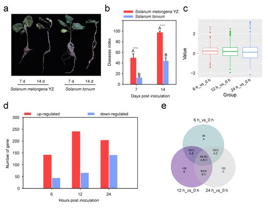
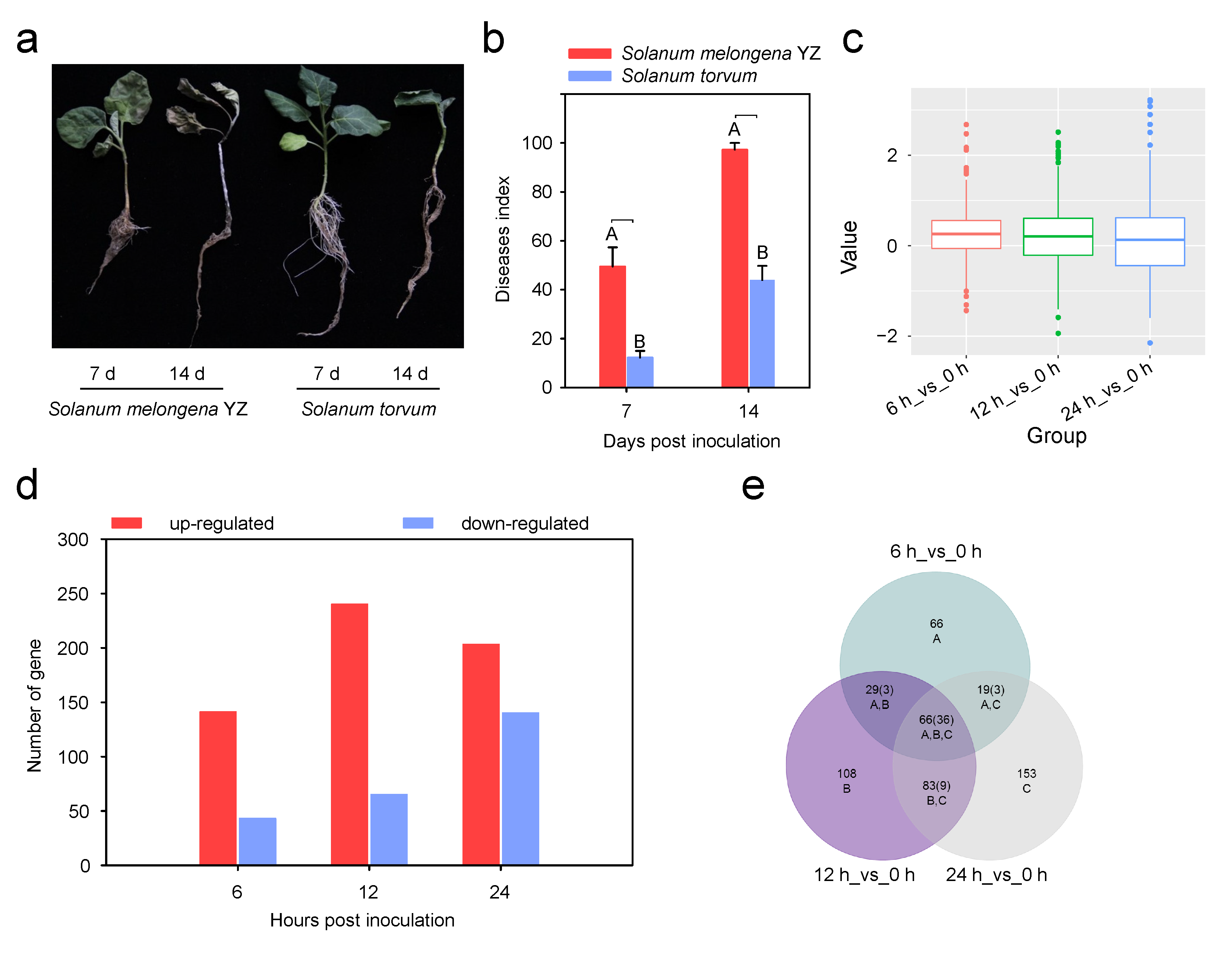
To characterize the resistance phenotype of S. torvum against V. dahliae, seedlings of this wild relative and the susceptible cultivar YZ were inoculated with a preparation containing both fungal spores and crude toxin. By 7 days post-inoculation (dpi), the YZ plants had developed pronounced Verticillium wilt symptoms, including chlorosis and wilting of leaves, whereas the S. torvum plants remained largely symptom-free. At 14 dpi, the YZ plants had undergone complete wilting and mortality, with visible mycelial growth on the stem surface; in marked contrast, the S. torvum plants not only survived but also continued to produce newly emerging leaves (Figure 1a). Correspondingly, the disease index recorded for YZ at 7 dpi was substantially higher than that for S. torvum, with values of 49.2 and 16.1, respectively. By 14 dpi, this index had risen in both genotypes, reaching 98.6 in YZ and 43.5 in S. torvum (Figure 1b). These observations confirm that S. torvum possesses markedly stronger Verticillium wilt resistance than the susceptible YZ line.
Figure 1.
Phenotypic identification and proteomic analysis of S. torvum roots. (a) Phenotypic assessment of S. melongena YZ and S. torvum inoculated with V. dahliae at 7 and 14 days. (b) Disease index of S. melongena and S. torvum inoculated with V. dahliae at 7 and 14 days. All experiments were performed with three biological replicates, and the results are presented as means ± standard deviation. Statistical analyses were conducted using Student’s t-test, and different uppercase letters indicate extremely significant differences between groups (p < 0.01). (c) Principal component analysis of proteome sequencing data. (d) Analysis of the number of DAPs in roots of S. torvum inoculated with V. dahliae at 6, 12, and 24 h post-inoculation. (e) Venn analysis of DAPs in roots of S. torvum inoculated with V. dahliae at 6, 12, and 24 h post-inoculation.
To explore the molecular events underlying this resistance at the protein level, iTRAQ-based quantitative proteomic profiling was conducted on root tissues harvested from S. torvum following V. dahliae inoculation. Principal component analysis (PCA) of the proteomic dataset revealed distinct clustering patterns among samples, indicating that the data were reproducible and of high quality (Figure 1c). Applying selection criteria of an Unused score greater than 1.3 and at least two matched peptides per protein, we identified a total of 2374 high-confidence proteins. Using a fold-change threshold of ≥1.5 to define differential abundance, we detected 832 differentially abundant proteins (DAPs) across the three post-inoculation time points examined. At 6 h post-inoculation (hpi), 184 DAPs were identified, of which 141 showed up-regulation and 43 showed down-regulation. At 12 hpi, the number of DAPs increased to 305, with 240 up-regulated and 65 down-regulated. At 24 hpi, 343 DAPs were detected, comprising 203 up-regulated and 140 down-regulated proteins (Figure 1d). The number of DAPs increased progressively with infection time, and up-regulated proteins accounted for the majority of DAPs at each time point, suggesting that V. dahliae infection induced broad activation of protein accumulation in S. torvum roots.
A Venn diagram was used to compare the DAPs identified at different time points after inoculation. In total, 95 DAPs were shared between 6 and 12 hpi, 85 were shared between 6 and 24 hpi, and 149 were shared between 12 and 24 hpi. In addition, 66 DAPs were common to all three time points (Figure 1e). The largest overlap was observed between 12 and 24 hpi, suggesting that partially shared defense-related metabolic pathways may be activated during the middle and later stages of the early infection response in S. torvum.
3.2. GO and KEGG Enrichment Analyses of DAPs
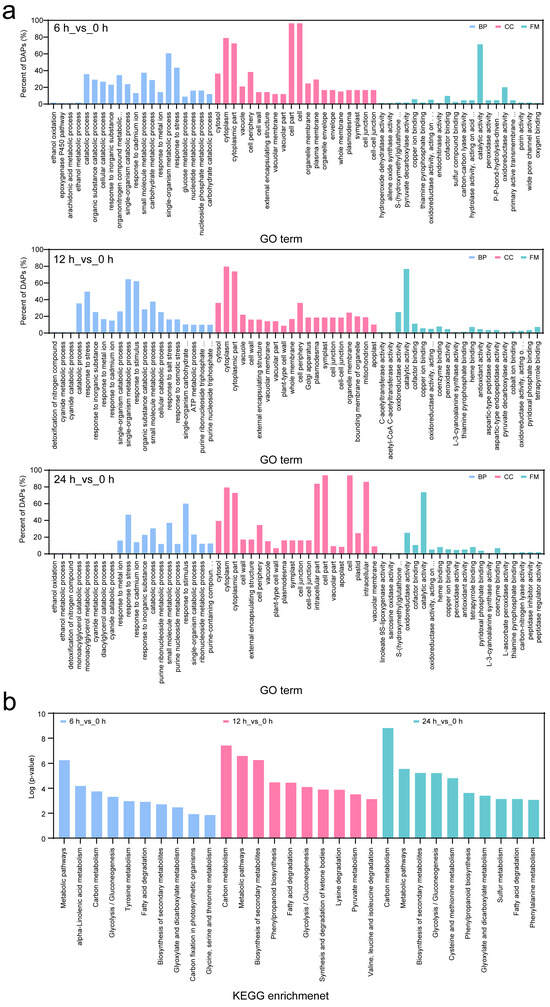
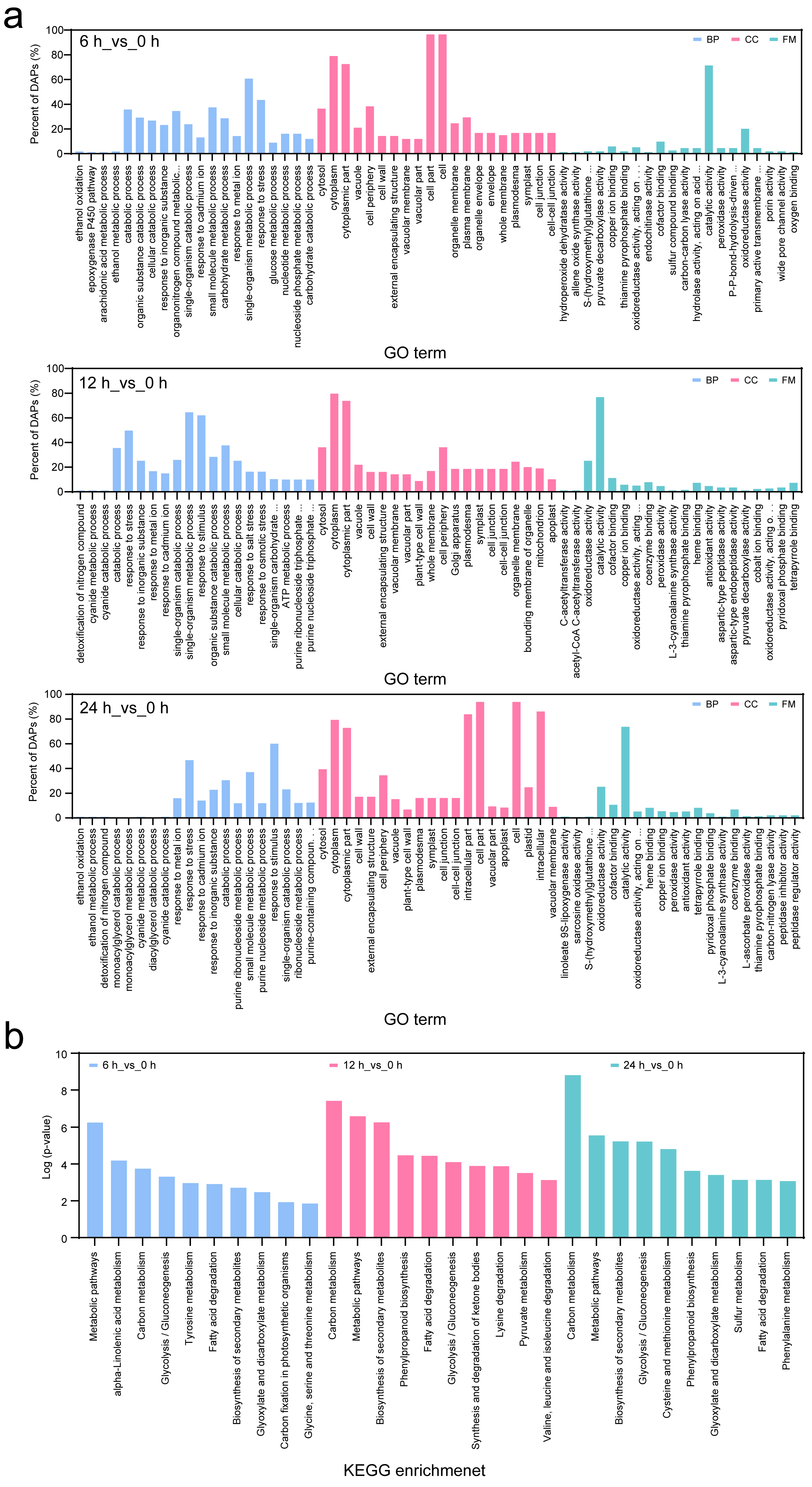
Functional annotation of the DAPs identified in S. torvum roots after V. dahliae inoculation was pursued through Gene Ontology (GO) enrichment analysis. The significantly enriched GO terms fell into the three standard categories of biological process, cellular component, and molecular function. Across the three time points, the numbers of significantly enriched GO terms were 489 at 6 hpi, 814 at 12 hpi, and 924 at 24 hpi, indicating that an expanding array of functional categories was recruited into the defense response as infection progressed. Nevertheless, the specific categories that showed significant enrichment, as well as the number of terms within each category, varied considerably among the three time points. Nine categories were consistently enriched across all three time points, including “catabolic process,” “cellular catabolic process,” “response to stress,” and “small molecule metabolic process.” In contrast, other categories—such as “epoxygenase P450 pathway,” “ATP metabolic process,” and “single-organism catabolic process”—were significantly enriched only at specific stages of infection. The top 20 significantly enriched GO terms at 6, 12, and 24 hpi were further analyzed (Figure 2a, Table S2). These enriched terms suggested that V. dahliae infection induced broad changes in defense-related biological processes in S. torvum roots.
Figure 2.
GO and KEGG enrichment analyses of DAPs. (a) GO analysis of DAPs in roots of S. torvum inoculated with V. dahliae at 6, 12, and 24 h post-inoculation. (b) KEGG enrichment analysis of DAPs in roots of S. torvum inoculated with V. dahliae at 6, 12, and 24 h post-inoculation.
A large number of DAPs were associated with phytohormone biosynthesis and signal transduction. These included 18 proteins related to salicylic acid biosynthesis and signaling, 27 proteins associated with JA biosynthesis and signaling, and 15 proteins involved in ethylene biosynthesis and signaling. In addition, two proteins related to brassinosteroid biosynthesis were identified. These results indicate that multiple hormone pathways may participate in the defense response of S. torvum against V. dahliae. Proteins associated with structural defense and secondary metabolism were also highly represented among the DAPs. In particular, two proteins involved in chitin biosynthesis, 11 proteins related to lignin biosynthesis, one protein associated with phytoalexin biosynthesis, and 11 proteins involved in cell-wall biosynthesis were identified. Several DAPs were also annotated as being involved in responses to fungal stimulus, regulation of programmed cell death, and ROS metabolism (Table S1). These findings suggest that cell-wall reinforcement, secondary metabolite production, oxidative stress regulation, and hormone-mediated signaling may act together to support Verticillium wilt resistance in S. torvum.
KEGG pathway enrichment analysis was subsequently performed to gain further insight into the metabolic and signaling routes represented by the DAPs. At 6 hpi, the 184 DAPs were distributed across 73 KEGG pathways, with eight of these showing significant enrichment. At 12 hpi, 305 DAPs mapped to 88 pathways, among which 14 were significantly enriched. At 24 hpi, 343 DAPs were assigned to 87 pathways, with 19 pathways reaching statistical significance (Figure 2b, Table S3). The progressive increase in the number of significantly enriched pathways over the course of infection suggests that the proteomic response in S. torvum roots became increasingly multifaceted during the early phase of V. dahliae colonization. Among the pathways identified, phenylpropanoid biosynthesis stood out as being strongly activated following pathogen inoculation (Figure 2b). This particular pathway is intimately connected with the deposition of physical and chemical barriers that impede pathogen ingress and also contributes to both local and systemic signaling components of the defense response. A total of 22 DAPs related to phenylpropanoid metabolism showed significant changes in abundance. Among them, PER12 was up-regulated at multiple time points after V. dahliae inoculation, suggesting an enhanced peroxidase-mediated oxidative response. Lignin biosynthesis-related proteins, such as CYP73A5, were also differentially accumulated. These results suggest that peroxidase activity and lignin deposition may contribute to the resistance response of S. torvum to V. dahliae infection.
3.3. Identification and Expression Validation of Candidate P450 Family Genes
Further analysis of the DAP dataset identified 10 cytochrome P450 family proteins that were significantly differentially accumulated after V. dahliae inoculation. Functional annotation indicated that these P450 proteins may be involved in multiple defense-related processes. Among them, StoCYP82C4 and StoCYP19-2 were associated with plant disease resistance; StoCYP72A7, StoCYP72A9, and StoCYP74A/StoAOS1 were related to hormone responses; StoCYP84A1, StoCYP73A5, and StoCYP707A3 were associated with lignin biosynthesis; StoCYP71B34 was linked to stress responses; and StoCYP51G1 was related to sterol biosynthesis. To validate the proteomic results, the transcript levels of the 10 candidate P450 genes were examined by RT-qPCR after V. dahliae inoculation. Among these genes, eight showed expression patterns that were consistent between transcript abundance and protein accumulation, and the highest relative expression was observed for StoCYP74A/StoAOS1 (Figure S1), supporting the reliability of the proteomic data. One gene could not be directly compared because it was specifically detected in eggplant, whereas another gene showed an inconsistent pattern between transcript and protein levels. This discrepancy may reflect post-transcriptional regulation, differences in protein stability, or a temporal delay between mRNA accumulation and protein abundance. Overall, these results suggest that P450 gene family members are broadly involved in the response of S. torvum to V. dahliae infection and may contribute to Verticillium wilt resistance through hormone regulation, lignin biosynthesis, stress responses, and secondary metabolism.
3.4. Sequence Analysis of StoAOS1
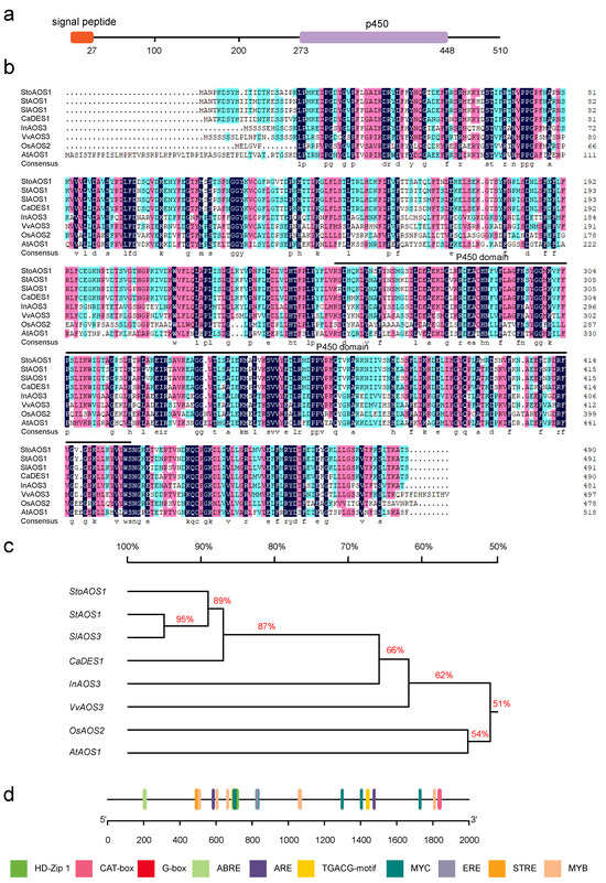
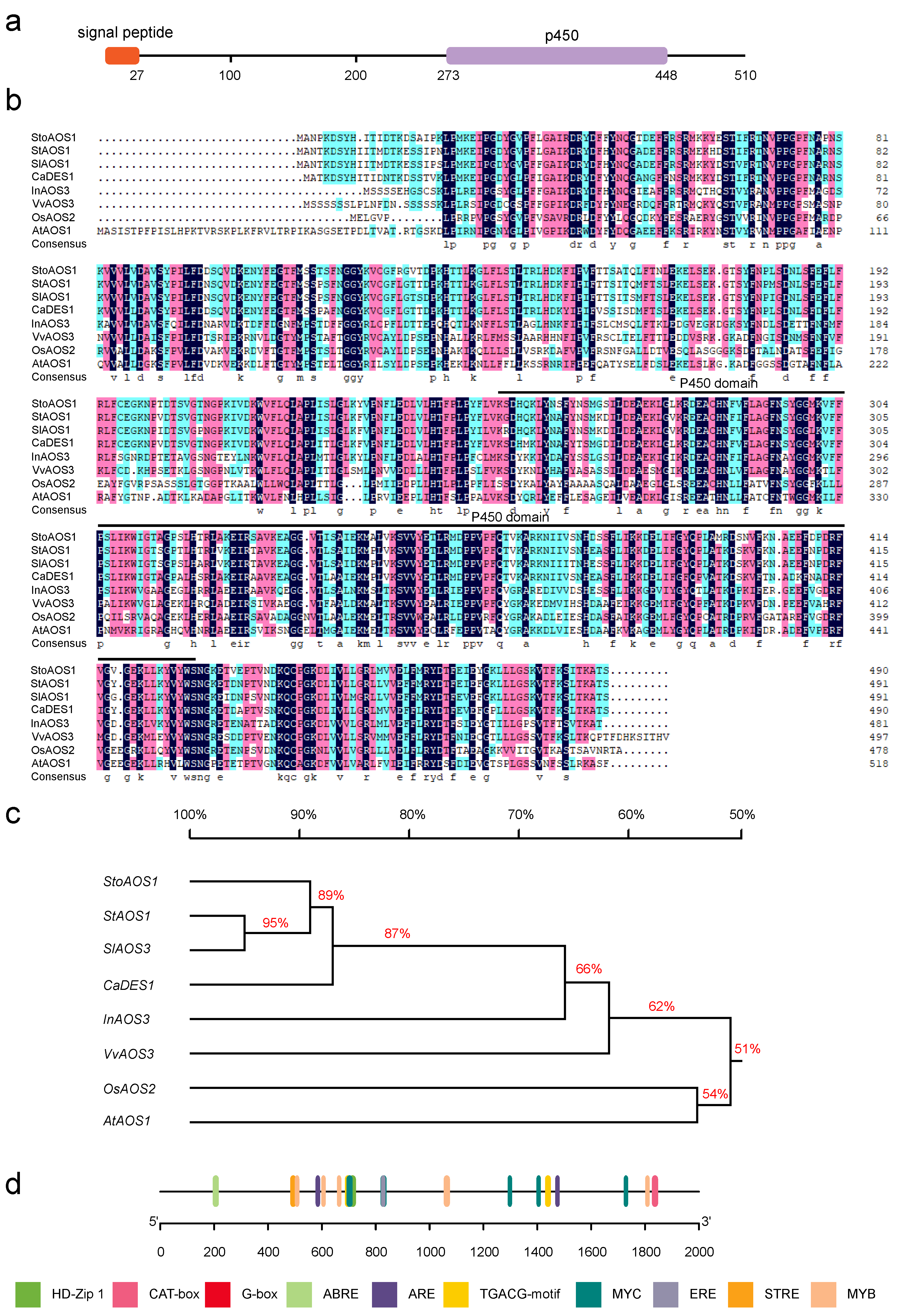
The coding sequence of StoAOS1 was determined to be 1533 bp in length, encoding a predicted protein of 510 amino acids. To examine the domain architecture of this protein, the deduced amino acid sequence was submitted to SMART analysis (https://smart.embl.de/, accessed on 12 May 2026). The results revealed the presence of a canonical cytochrome P450 conserved domain within StoAOS1 (Figure 3a,b), placing this protein within the CYP74A/AOS subfamily of the cytochrome P450 superfamily. To further assess the sequence characteristics of StoAOS1, multiple sequence alignment was carried out using AOS protein sequences derived from a range of plant species. This alignment showed that StoAOS1 exhibits the highest degree of sequence identity with StAOS1 from potato (Solanum tuberosum) (Figure 3b). A phylogenetic tree was then constructed to evaluate the evolutionary placement of StoAOS1 relative to its homologs from other selected species. The resulting phylogeny indicated that StoAOS1 is most closely related to potato StAOS1 and tomato (Solanum lycopersicum) SlAOS3 (Figure 3c), pointing to a close evolutionary relationship among AOS proteins from solanaceous taxa.
Figure 3.
Sequence analysis of StoAOS1. (a) Conserved domain analysis of StoAOS1. (b) Multiple alignment of the amino acid sequence of StoAOS1 with homologous proteins, including StAOS1 in S. tuberosum, SlAOS1 in S. lycopersicum, CaDES1 in pepper, InAOS3 in Ipomoea nil, VvAOS3 in Vitis vinifera, OsAOS2 in rice, and AtAOS1 in A. thaliana. For identity shading, green corresponds to 50–75%, red to 75–100%, and black to 100% identity. (c) Phylogenetic tree analysis. (d) Prediction of cis-elements in the StoAOS1 promoter. The main cis-elements in the StoAOS1 promoter included HD-Zip 1, CAT-box, G-box, ABRE, ARE, TGACG-motif, MYC, ERE, STRE, and MYB.
In addition, the 2000 bp promoter region upstream of the StoAOS1 coding sequence was subjected to cis-acting element prediction using the PlantCARE database (http://bioinformatics.psb.ugent.be/webtools/plantcare/html/, accessed on 15 June 2026). In addition to elements responsive to light, the promoter was found to harbor multiple cis-elements associated with phytohormone signaling, transcription factor binding, stress responses, and various aspects of plant growth and development. Among the hormone-responsive elements identified were ABRE, ERE, and TGACG-motif elements. Transcription factor-binding motifs included G-box, MYB, and MYC elements, while stress-responsive motifs such as ARE and STRE were also detected. Growth- and development-related elements, such as HD-Zip 1 and CAT-box, were present in the promoter region (Figure 3d). These results suggest that StoAOS1 may be regulated by multiple endogenous and environmental signals, particularly hormone- and stress-related pathways.
3.5. MeJA Induces the Expression of StoAOS1 in S. torvum
Previous studies have shown that AOS-mediated JA biosynthesis contributes to plant defense against fungal pathogens [25]. To determine whether StoAOS1 is involved in the JA-mediated signaling pathway in S. torvum, RT-qPCR was performed to examine the transcript levels of StoAOS1 in leaves after exogenous MeJA treatment. Samples were collected at 0, 1, 3, 6, and 12 h after treatment. The results showed that MeJA treatment significantly induced StoAOS1 expression as early as 1 h after application. The transcript level of StoAOS1 continued to increase and reached its highest level at 6 h, followed by a decline at 12 h. StoAOS1 expression at 12 h nevertheless remained significantly higher than that in the ddH2O-treated control plants (Figure S2). Overall, StoAOS1 expression showed a transient induction pattern, increasing during the early response to MeJA and then decreasing at the later stage. These results indicate that StoAOS1 is responsive to exogenous MeJA and may participate in JA-mediated signaling in S. torvum.
3.6. Silencing of StoAOS1 Decreases Resistance of S. torvum to Verticillium Wilt
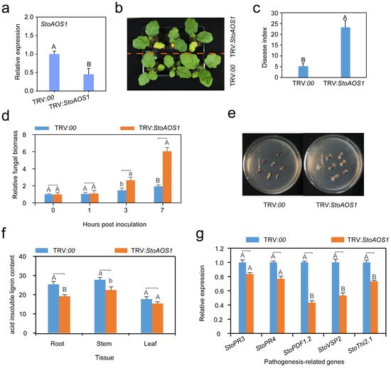
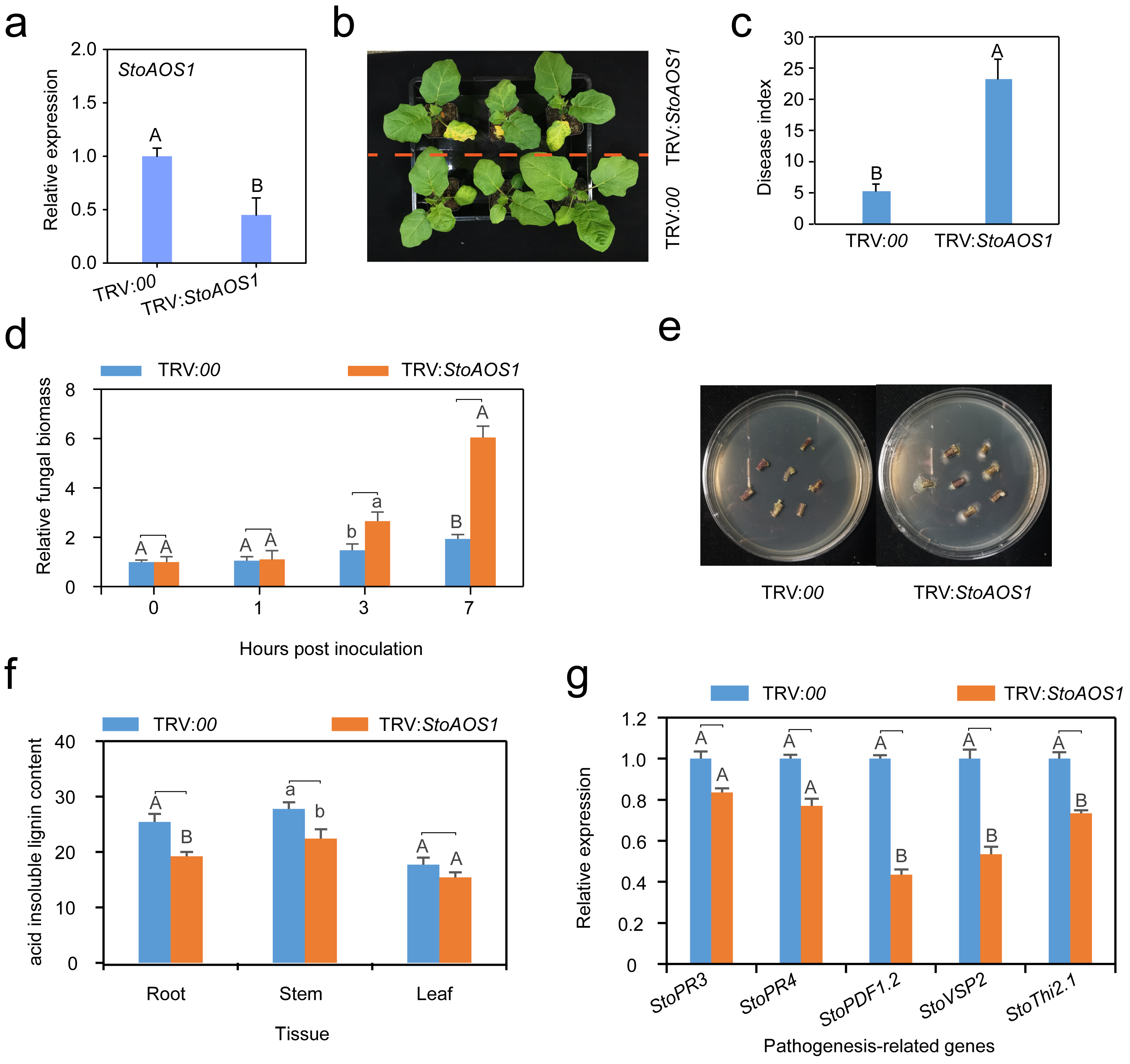
To further investigate the role of StoAOS1 in Verticillium wilt resistance, virus-induced gene silencing (VIGS) was used to suppress StoAOS1 expression in S. torvum. The silencing efficiency was first examined by RT-qPCR. Compared with the control plants (TRV:00), StoAOS1-silenced plants (TRV:StoAOS1) showed a significantly lower transcript level of StoAOS1 in the roots, indicating that StoAOS1 was successfully silenced (Figure 4a). The StoAOS1-silenced and control plants were then subjected to root wounding followed by V. dahliae inoculation through root irrigation. At 9 dpi, the leaves of StoAOS1-silenced plants showed obvious chlorosis, whereas no visible symptoms were observed in the control plants (Figure 4b). Consistently, the disease index of StoAOS1-silenced plants was significantly higher than that of the controls (Figure 4c). These results indicate that silencing StoAOS1 reduced the resistance of S. torvum to Verticillium wilt.
Figure 4.
Silencing of StoAOS1 decreases the tolerance of S. torvum to Verticillium wilt. (a) Detection of StoAOS1 silencing efficiency. (b) Phenotypic assessment of StoAOS1-silenced and control plants inoculated with V. dahliae at 9 days. (c) Disease index analysis of StoAOS1-silenced and control plants inoculated with V. dahliae at 9 days. (d) Measurement of fungal biomass. Fungal biomass in StoAOS1-silenced and control plants was measured at 0, 1, 3, and 7 dpi by RT-qPCR. (e) Fungal recovery assay. Stems collected from StoAOS1-silenced and control plants inoculated with V. dahliae at 14 dpi were placed on solid PDA medium. (f) Detection of acid-insoluble lignin content in roots, stems, and leaves of S. torvum inoculated with V. dahliae. (g) Analysis of relative transcript levels of defense-related genes. The relative transcript levels of defense-related genes, including StoPR3, StoPR4, StoPDF1.2, StoVSP2, and StoThi2.1, were detected in roots of StoAOS1-silenced and control S. torvum inoculated with V. dahliae at 24 h post-inoculation. In (a,c,d,f,g), all experiments were performed with three biological replicates, and the results are presented as means ± standard deviation. Statistical analyses were conducted using Student’s t-test, and different uppercase letters indicate extremely significant differences between groups (p < 0.01), and different lowercase letters indicate significant differences between groups (p < 0.05).
To further evaluate pathogen colonization, fungal biomass in the stems of StoAOS1-silenced and control plants was quantified by RT-qPCR at 0, 1, 3, and 7 dpi. Fungal biomass increased gradually with infection time. At 3 dpi, StoAOS1-silenced plants accumulated significantly higher fungal biomass than control plants. This difference became more pronounced at 7 dpi, when fungal biomass in StoAOS1-silenced plants was significantly higher than that in the controls (Figure 4d). These results suggest that silencing StoAOS1 promoted the proliferation of V. dahliae in S. torvum. Pathogen recovery assays further supported this result. At 14 dpi, stem segments from StoAOS1-silenced and control plants were placed on potato dextrose agar (PDA) medium. Rapid hyphal growth was observed from stem segments of StoAOS1-silenced plants, whereas no regenerated hyphae were detected from control stem segments (Figure 4e). This result was consistent with the fungal biomass analysis. Lignin content was also measured in roots, stems, and leaves. The lignin levels in all three tissues were lower in StoAOS1-silenced plants than in control plants, with significant reductions observed in roots and stems (Figure 4f). These results suggest that StoAOS1 may contribute to lignin accumulation in S. torvum, thereby strengthening structural defense against pathogen infection.
At 12 h after V. dahliae inoculation, transcript levels of several defense-related marker genes were assessed by RT-qPCR in both control and StoAOS1-silenced plants. Compared with the control group, the silenced lines exhibited significantly reduced transcript accumulation for StoPDF1.2, StoVSP2, and StoThi2.1, whereas the expression levels of StoPR3 and StoPR4 remained largely unchanged (Figure 4g). These findings imply that the defense pathway modulated by StoAOS1 may operate through StoPDF1.2, StoVSP2, and StoThi2.1 and may be distinct from the route involving StoPR3 and StoPR4. Taken together, the data support a model in which StoAOS1 functions as a positive regulator of Verticillium wilt resistance in S. torvum.
3.7. Overexpression of StoAOS1 Enhances Resistance to V. dahliae in N. benthamiana
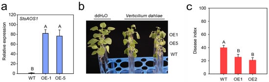
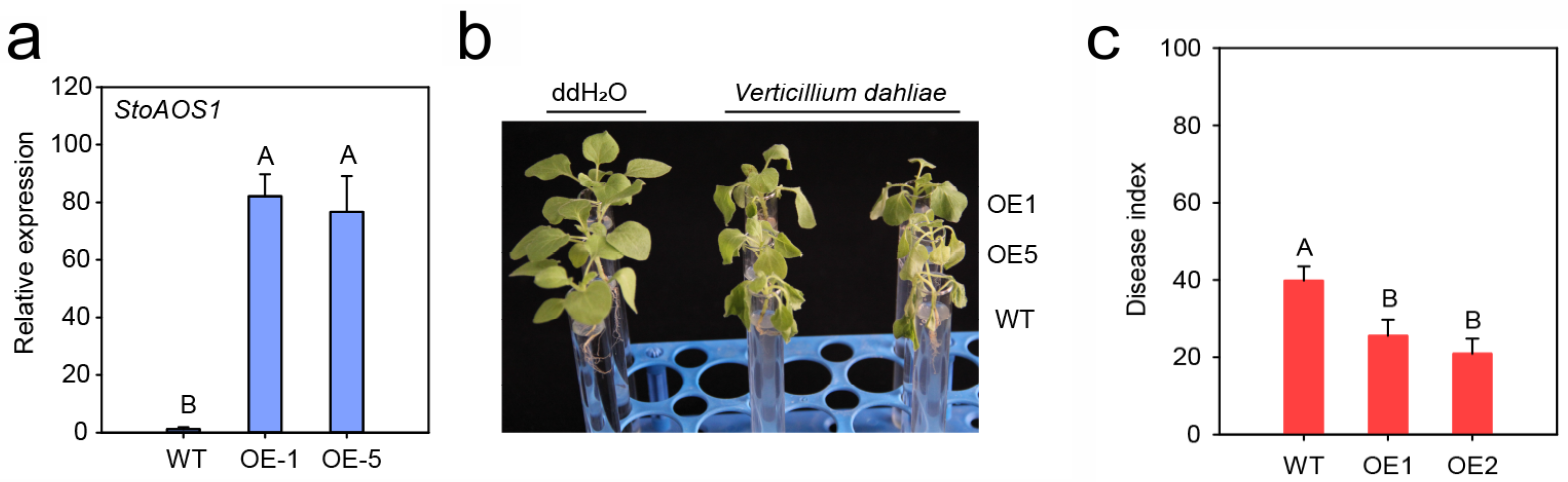
To provide additional functional validation of StoAOS1, we heterologously overexpressed this gene in Nicotiana benthamiana. RT-qPCR analysis confirmed that the transcript level of StoAOS1 was markedly elevated in the OE-1 and OE-5 overexpression lines relative to wild-type (WT) plants, indicating successful transgene expression (Figure 5a). WT plants and the two overexpression lines were subsequently inoculated with V. dahliae. At 6 dpi, the OE-1 and OE-5 plants displayed milder leaf wilting symptoms compared with the WT (Figure 5b). Consistent with this visual assessment, the disease index recorded for the overexpression lines was significantly lower than that for the WT (Figure 5c). These results collectively indicate that ectopic expression of StoAOS1 confers enhanced resistance to V. dahliae in N. benthamiana.
Figure 5.
Overexpression of StoAOS1 enhances tolerance of N. benthamiana to Verticillium wilt. (a) Analysis of relative expression levels of StoAOS1 in WT, OE-1, and OE-5 plants. (b) Phenotypic assessment of WT, OE-1, and OE-5 plants inoculated with V. dahliae at 6 dpi. (c) Disease index analysis of WT, OE-1, and OE-5 plants inoculated with V. dahliae at 6 dpi. In (b,c), all experiments were performed with three biological replicates, and the results are presented as means ± standard deviation. Statistical analyses were conducted using Student’s t-test, and different uppercase letters indicate extremely significant differences between groups (p < 0.01).
4. Discussion
Verticillium wilt is classified as a typical vascular disease, and achieving effective resistance against it depends on both rapid detection of the invading pathogen and the subsequent orchestrated induction of multiple layers of host defense [33,34]. In the work reported here, application of iTRAQ-based quantitative proteomic profiling to S. torvum roots following V. dahliae challenge revealed widespread alterations in protein abundance. A substantial subset of the DAPs identified could be assigned to phytohormone biosynthetic routes and associated signal transduction cascades, encompassing proteins that function in the SA, JA, ET, and brassinosteroid pathways. These observations point to the conclusion that resistance in S. torvum against V. dahliae is not governed by any single signaling module; rather, it appears to be under the control of a coordinated hormone-dependent defense network. SA, JA, and ET are established as central regulators of plant immunity, and their respective signaling pathways are known to engage in context-dependent crosstalk during the course of plant–pathogen interactions [12,13]. Canonical models often link SA primarily with resistance to biotrophic pathogens and with systemic acquired resistance [35,36], while JA and ET are more frequently associated with defense against necrotrophic pathogens and with induced systemic resistance. Nevertheless, the response to vascular pathogens like V. dahliae tends to be considerably more intricate and may well entail the integrated action of several hormonal routes [37]. In the present context, the observed induction of StoAOS1—a protein involved in JA biosynthesis—suggests that JA-associated signaling likely makes a significant contribution to the early defensive response mounted by S. torvum against V. dahliae.
Beyond hormone-related proteins, the DAP dataset also included a number of candidates implicated in cell-wall biosynthesis, lignin deposition, chitin biosynthetic processes, phytoalexin accumulation, ROS homeostasis, regulation of programmed cell death, and responses to fungal elicitation. Taken together, these results indicate that structural reinforcement of cell walls and the activation of secondary metabolic routes constitute important facets of the overall defense strategy employed by S. torvum in response to pathogen challenge. The phenylpropanoid pathway was strongly activated after pathogen inoculation [38]. This pathway provides precursors for lignin biosynthesis and contributes to the formation of physical and chemical barriers that restrict pathogen invasion and systemic spread [39,40]. In this study, phenylpropanoid-related proteins, including PER12 and CYP73A5, were differentially accumulated after V. dahliae inoculation, suggesting that peroxidase activity and lignin deposition may contribute to the restriction of fungal colonization. Similar roles for lignin biosynthesis and cell-wall reinforcement have been reported in other plant—V. dahliae interaction systems, particularly in cotton, where lignin-related genes enhance resistance by strengthening cell walls and limiting pathogen expansion [41,42]. Among the candidate proteins identified in the proteomic analysis, StoAOS1 attracted particular attention because of its association with JA biosynthesis. As a key enzyme operating within the octadecanoid route of JA biosynthesis, AOS is assigned to the CYP74 branch of the cytochrome P450 family [43]. The reaction catalyzed by this enzyme converts fatty acid hydroperoxides—themselves products of lipoxygenase activity—into allene oxide intermediates that are inherently unstable; these are then transformed through subsequent enzymatic steps into 12-oxo-phytodienoic acid and eventually into JA and its biologically active derivatives [21,43]. Given the well-established involvement of JA in plant defensive responses, AOS-encoding genes have been subjected to functional characterization across a range of plant species. In rice, for instance, overexpression of OsAOS2 leads to elevated endogenous JA pools, upregulation of pathogenesis-related transcripts, and improved resistance to fungal pathogens [25]. Such observations lend support to the notion that AOS-mediated JA biosynthesis constitutes a significant component of plant immunity against fungal invaders.
In the current work, StoAOS1 emerged from the proteomic screening as a V. dahliae-inducible gene belonging to the P450 family in S. torvum. Sequence analysis revealed that StoAOS1 carries a conserved P450 domain and displays close phylogenetic affinity with AOS proteins from other solanaceous species, thereby supporting its assignment to the CYP74A/AOS subfamily. Promoter prediction further uncovered a range of cis-acting regulatory motifs associated with hormonal signaling, transcription factor binding, stress responses, and developmental processes—specifically, ABRE, ERE, TGACG-motif, MYB, MYC, ARE, and STRE elements. These promoter features are in good agreement with the expression behavior observed for StoAOS1, which proved responsive to both V. dahliae challenge and exogenous MeJA treatment. Upon MeJA application, transcript accumulation of StoAOS1 rose rapidly, reached a peak at 6 h, and subsequently declined by 12 h while still remaining above the control level. This transient induction profile is suggestive of a role for StoAOS1 in JA signaling, possibly involving feedback regulation of the JA biosynthetic pathway. Furthermore, following V. dahliae inoculation, StoAOS1 was markedly upregulated in S. torvum, a pattern consistent with earlier observations that AOS genes can be activated by both pathogen attack and JA treatment. In rice, OsAOS2 is known to respond to Magnaporthe grisea infection [25], and exogenous MeJA has been shown to trigger expression of JA-biosynthetic genes, including various AOS homologs [44]. The earlier engagement of JA-associated defenses may, at least in part, account for the superior resistance of S. torvum, as it could serve to restrict pathogen proliferation during the critical early phases of infection.
Functional assays provided further evidence that StoAOS1 acts as a positive regulator of Verticillium wilt resistance. Suppression of StoAOS1 expression through virus-induced gene silencing in S. torvum resulted in a marked attenuation of resistance to V. dahliae, as manifested by more pronounced leaf chlorosis, an elevated disease index, increased fungal biomass within host tissues, and enhanced recovery of the pathogen from infected stem segments. These observations indicate that StoAOS1 is necessary for the full expression of resistance in S. torvum against Verticillium wilt. Measurements of acid-insoluble lignin content revealed reductions in roots, stems, and leaves of StoAOS1-silenced plants, with statistically significant decreases recorded in roots and stems. Given that V. dahliae enters through the root system and disseminates via the vascular network, lignin deposition in these particular tissues is likely to play a critical role in impeding pathogen movement [1,42]. Diminished lignification in StoAOS1-silenced plants may therefore compromise cell-wall-based physical barriers and permit more extensive fungal colonization. This interpretation finds support from previous work in cotton, where knockdown of lignin biosynthetic genes such as GhLAC4 and GhCADs attenuated defense-induced lignification and compromised resistance to V. dahliae [42,45]. StoAOS1 may thus contribute to Verticillium wilt resistance not solely through JA signaling per se, but also via downstream defensive outputs including lignin accumulation. The expression profiles of defense-related marker genes lend additional weight to this conclusion. Following V. dahliae inoculation, transcript levels of StoPDF1.2, StoVSP2, and StoThi2.1 were significantly reduced in StoAOS1-silenced plants, whereas those of StoPR3 and StoPR4 remained largely unaltered. Given that PDF1.2 and VSP2 are commonly regarded as markers of JA-dependent defense responses [46], these data suggest that StoAOS1 may govern a specific subset of JA-responsive genes in S. torvum. The lack of substantial changes in StoPR3 and StoPR4 further implies that not all defense-related routes are equally reliant upon StoAOS1 function. Hence, StoAOS1 appears to operate through a particular JA-associated branch of the defense network rather than functioning as a global regulator of all pathogenesis-related transcripts.
The positive regulatory role of StoAOS1 received additional confirmation from heterologous overexpression experiments in N. benthamiana. Relative to wild-type controls, transgenic lines overexpressing StoAOS1 exhibited milder wilting symptoms and lower disease indices upon V. dahliae challenge. Collectively, these data support a positive role for StoAOS1 in the defensive response of S. torvum against Verticillium wilt. In the present study, we characterized the function of StoAOS1 via heterologous overexpression in Nicotiana benthamiana. It should be noted that further verification using stable transgenic materials of eggplant or Solanum torvum is still required in subsequent research to consolidate our conclusions.
5. Conclusions
In the present investigation, iTRAQ-based quantitative proteomic profiling was employed to characterize the response of S. torvum to V. dahliae infection. The proteomic data revealed activation of multiple defense-related pathways during the early stages of infection, encompassing phytohormone signaling, phenylpropanoid metabolism, lignin biosynthetic routes, cell-wall modification, and oxidative stress responses. Among the DAPs identified, StoAOS1—a CYP74A/AOS subfamily member involved in JA biosynthesis—emerged as a candidate positive regulator of Verticillium wilt resistance. The strong induction of StoAOS1 by both V. dahliae and MeJA treatment points to its participation in JA-associated defense signaling. Functional analyses demonstrated that silencing StoAOS1 in S. torvum compromised Verticillium wilt resistance, promoted fungal colonization, diminished acid-insoluble lignin accumulation, and suppressed expression of several defense-associated genes. Conversely, heterologous overexpression of StoAOS1 in N. benthamiana conferred enhanced resistance to V. dahliae. Taken together, these results suggest that StoAOS1 positively regulates Verticillium wilt resistance, likely by modulating JA-mediated defense responses and lignin-associated structural defense. This study provides candidate gene resources for improving Verticillium wilt resistance in eggplant. Our findings offer valid evidence to unravel the molecular mechanism governing Verticillium wilt resistance in eggplant and lay a solid molecular foundation for molecular breeding and the development of disease-resistant eggplant varieties.
Supplementary Materials
The following supporting information can be downloaded at https://www.mdpi.com/article/10.3390/horticulturae12080949/s1, Table S1. Statistics of pathogen defense-related proteins in S. torvum roots after inoculation. Table S2. GO analysis of DAPs in roots of S. torvum inoculated with V. dahliae at 6, 12, and 24 h post-inoculation. Table S3. KEGG enrichment analysis of DAPs in roots of S. torvum inoculated with V. dahliae at 6, 12, and 24 h post-inoculation. Table S4. Primer pair sequences used in this study. Figure S1. Transcript expression analysis of candidate P450 genes. The transcript levels of candidate P450 genes, including StoCYP82C4, StoCYP19-2, StoCYP72A7, StoCYP72A9, StoCYP74A/StoAOS1, StoCYP84A1, StoCYP73A5, StoCYP707A3, StoCYP71B34, and StoCYP51G1, were detected in roots of S. torvum inoculated with V. dahliae at 0, 6, 12, and 24 h post-inoculation by RT-qPCR. All experiments were performed with three biological replicates, and the results are presented as means ± standard deviation. Statistical analyses were conducted using Student’s t-test, and different uppercase letters indicate extremely significant differences between groups (p < 0.01). Figure S2. Transcript expression analysis of StoAOS1 after exogenous MeJA treatment. Transcript levels of StoAOS1 were detected by RT-qPCR in leaves of S. torvum sprayed with exogenous 100 μM MeJA at 0, 1, 3, 6, and 12 h after treatment. All experiments were performed with three biological replicates, and the results are presented as means ± standard deviation. Statistical analyses were conducted using Student’s t-test, and different uppercase letters indicate extremely significant differences between groups (p < 0.01).
Author Contributions
Conceptualization, Y.Z., L.L. and X.Y.; methodology, Y.Z.; software, Y.Z.; validation, Y.Z.; formal analysis, Y.Z.; investigation, Y.Z.; resources, Y.Z.; data curation, X.Y.; writing—original draft preparation, Y.Z.; writing—review and editing, Y.Z. and L.S.; visualization, Y.Z.; supervision, Y.Z.; project administration, L.L. and X.Y.; funding acquisition, X.Y. All authors have read and agreed to the published version of the manuscript.
Funding
This research was funded by the National Natural Science Foundation of China (Grant number:31972395). The project Title: Study on the Molecular Mechanism of StCYP74A Gene Regulating Verticillium Wilt Resistance in Eggplant. The project duration is from 2020 to 2023.
Data Availability Statement
The original contributions presented in this study are included in the article/Supplementary Material. Further inquiries can be directed to the corresponding authors.
Conflicts of Interest
The authors declare no conflicts of interest.
References
- Fradin, E.F.; Thomma, B.P. Physiology and molecular aspects of Verticillium wilt diseases caused by V. dahliae and V. albo-atrum. Mol. Plant Pathol. 2006, 7, 71–86. [Google Scholar] [CrossRef] [Scilit] [PubMed]
- Klosterman, S.J.; Atallah, Z.K.; Vallad, G.E.; Subbarao, K.V. Diversity, pathogenicity, and management of verticillium species. Annu. Rev. Phytopathol. 2009, 47, 39–62. [Google Scholar] [CrossRef] [Scilit] [PubMed]
- Bletsos, F.; Thanassoulopoulos, C.; Roupakias, D. Effect of Grafting on Growth, Yield, and Verticillium Wilt of Eggplant. Hortscience A Publ. Am. Soc. Hortic. Sci. 2003, 38, 183–186. [Google Scholar] [CrossRef] [Scilit]
- Song, R.; Li, J.; Xie, C.; Jian, W.; Yang, X. An Overview of the Molecular Genetics of Plant Resistance to the Verticillium Wilt Pathogen Verticillium dahliae. Int. J. Mol. Sci. 2020, 21, 1120. [Google Scholar] [CrossRef] [Scilit] [PubMed]
- Fradin, E.F.; Zhang, Z.; Juarez Ayala, J.C.; Castroverde, C.D.; Nazar, R.N.; Robb, J.; Liu, C.M.; Thomma, B.P. Genetic dissection of Verticillium wilt resistance mediated by tomato Ve1. Plant Physiol. 2009, 150, 320–332. [Google Scholar] [CrossRef] [Scilit] [PubMed]
- Yang, L.; Xie, C.; Li, W.; Zhang, R.; Jue, D.; Yang, Q. Expression of a wild eggplant ribosomal protein L13a in potato enhances resistance to Verticillium dahliae. Plant Cell Tissue Organ Cult. 2013, 115, 329–340. [Google Scholar] [CrossRef] [Scilit]
- Jue, D.-W.; Yang, L.; Shi, C.; Chen, M.; Yang, Q. Cloning and characterization of a Solanum torvum NPR1 gene involved in regulating plant resistance to Verticillium dahliae. Acta Physiol. Plant. 2014, 36, 2999–3011. [Google Scholar] [CrossRef] [Scilit]
- Yang, L.; Shi, C.; Mu, X.; Liu, C.; Shi, K.; Zhu, W.; Yang, Q. Cloning and expression of a wild eggplant cytochrome P450 gene, StoCYP77A2, involved in plant resistance to Verticillium dahliae. Plant Biotechnol. Rep. 2015, 9, 167–177. [Google Scholar] [CrossRef] [Scilit]
- Zhang, Y.; Shen, L.; Li, L.; Yang, X. The Solanum torvum Transcription Factor StoWRKY6 Mediates Resistance against Verticillium Wilt. Agronomy 2022, 12, 1977. [Google Scholar] [CrossRef] [Scilit]
- Shen, L.; Yang, S.; Zhao, E.; Xia, X.; Yang, X. StoMYB41 positively regulates the Solanum torvum response to Verticillium dahliae in an ABA dependent manner. Int. J. Biol. Macromol. 2024, 263, 130072. [Google Scholar] [CrossRef] [Scilit] [PubMed]
- Feng, C.; Dong, Y.L.; Shen, L. Genome-wide analysis of AP2/ERF gene family and functional characterization of StoERF109 in Solanum torvum response to Verticillium dahliae infection. Planta 2025, 262, 1. [Google Scholar] [CrossRef] [Scilit] [PubMed]
- Bari, R.; Jones, J.D. Role of plant hormones in plant defence responses. Plant Mol. Biol. 2009, 69, 473–488. [Google Scholar] [PubMed]
- Pieterse, C.M.; Van der Does, D.; Zamioudis, C.; Leon-Reyes, A.; Van Wees, S.C. Hormonal modulation of plant immunity. Annu. Rev. Cell Dev. Biol. 2012, 28, 489–521. [Google Scholar] [CrossRef] [Scilit] [PubMed]
- Hu, Q.; Zhu, L.; Zhang, X.; Guan, Q.; Xiao, S.; Min, L.; Zhang, X. GhCPK33 Negatively Regulates Defense against Verticillium dahliae by Phosphorylating GhOPR3. Plant Physiol. 2018, 178, 876–889. [Google Scholar] [CrossRef] [Scilit] [PubMed]
- Zhu, Y.; Hu, X.; Wang, P.; Gao, L.; Pei, Y.; Ge, Z.; Ge, X.; Li, F.; Hou, Y. GhPLP2 Positively Regulates Cotton Resistance to Verticillium Wilt by Modulating Fatty Acid Accumulation and Jasmonic Acid Signaling Pathway. Front. Plant Sci. 2021, 12, 749630. [Google Scholar] [CrossRef] [Scilit] [PubMed]
- Liu, F.; Cai, S.; Ma, Z.; Yue, H.; Xing, L.; Wang, Y.; Feng, S.; Wang, L.; Dai, L.; Wan, H.; et al. RVE2, a new regulatory factor in jasmonic acid pathway, orchestrates resistance to Verticillium wilt. Plant Biotechnol. J. 2023, 21, 2507–2524. [Google Scholar] [CrossRef] [Scilit] [PubMed]
- Ross, P.L.; Huang, Y.N.; Marchese, J.N.; Williamson, B.; Parker, K.; Hattan, S.; Khainovski, N.; Pillai, S.; Dey, S.; Daniels, S.; et al. Multiplexed protein quantitation in Saccharomyces cerevisiae using amine-reactive isobaric tagging reagents. Mol. Cell. Proteom. 2004, 3, 1154–1169. [Google Scholar] [CrossRef] [Scilit] [PubMed]
- Liu, Y.; Lu, S.; Liu, K.; Wang, S.; Huang, L.; Guo, L. Proteomics: A powerful tool to study plant responses to biotic stress. Plant Methods 2019, 15, 135. [Google Scholar] [CrossRef] [Scilit] [PubMed]
- Fang, W.; Xie, D.; Zhu, H.; Li, W.; Xu, Z.; Yang, L.; Li, Z.; Sun, L.; Wang, J.; Nie, L.; et al. Comparative Proteomic Analysis of Gossypium thurberi in Response to Verticillium dahliae Inoculation. Int. J. Mol. Sci. 2015, 16, 25121–25140. [Google Scholar] [CrossRef] [Scilit] [PubMed]
- Laudert, D.; Pfannschmidt, U.; Lottspeich, F.; Hollander-Czytko, H.; Weiler, E.W. Cloning, molecular and functional characterization of Arabidopsis thaliana allene oxide synthase (CYP 74), the first enzyme of the octadecanoid pathway to jasmonates. Plant Mol. Biol. 1996, 31, 323–335. [Google Scholar] [CrossRef] [Scilit] [PubMed]
- Farmer, E.E.; Goossens, A. Jasmonates: What ALLENE OXIDE SYNTHASE does for plants. J. Exp. Bot. 2019, 70, 3373–3378. [Google Scholar] [CrossRef] [Scilit] [PubMed]
- Song, W.C.; Funk, C.D.; Brash, A.R. Molecular cloning of an allene oxide synthase: A cytochrome P450 specialized for the metabolism of fatty acid hydroperoxides. Proc. Natl. Acad. Sci. USA 1993, 90, 8519–8523. [Google Scholar] [CrossRef] [Scilit] [PubMed]
- Schaller, A.; Stintzi, A. Enzymes in jasmonate biosynthesis—Structure, function, regulation. Phytochemistry 2009, 70, 1532–1538. [Google Scholar] [CrossRef] [Scilit] [PubMed]
- Park, J.H.; Halitschke, R.; Kim, H.B.; Baldwin, I.T.; Feldmann, K.A.; Feyereisen, R. A knock-out mutation in allene oxide synthase results in male sterility and defective wound signal transduction in Arabidopsis due to a block in jasmonic acid biosynthesis. Plant J. 2002, 31, 1–12. [Google Scholar] [CrossRef] [Scilit] [PubMed]
- Mei, C.; Qi, M.; Sheng, G.; Yang, Y. Inducible overexpression of a rice allene oxide synthase gene increases the endogenous jasmonic acid level, PR gene expression, and host resistance to fungal infection. Mol. Plant Microbe Interact. 2006, 19, 1127–1137. [Google Scholar] [CrossRef] [Scilit] [PubMed]
- Zeng, J.; Zhang, T.; Huangfu, J.; Li, R.; Lou, Y. Both Allene Oxide Synthases Genes Are Involved in the Biosynthesis of Herbivore-Induced Jasmonic Acid and Herbivore Resistance in Rice. Plants 2021, 10, 442. [Google Scholar] [CrossRef] [Scilit] [PubMed]
- Shen, L.; Xia, X.; Zhang, L.; Yang, S.; Yang, X. SmWRKY11 acts as a positive regulator in eggplant response to salt stress. Plant Physiol. Biochem. 2023, 205, 108209. [Google Scholar] [CrossRef] [Scilit] [PubMed]
- Cheng, H.Q.; Han, L.B.; Yang, C.L.; Wu, X.M.; Zhong, N.Q.; Wu, J.H.; Wang, F.X.; Wang, H.Y.; Xia, G.X. The cotton MYB108 forms a positive feedback regulation loop with CML11 and participates in the defense response against Verticillium dahliae infection. J. Exp. Bot. 2016, 67, 1935–1950. [Google Scholar] [CrossRef] [Scilit] [PubMed]
- Livak, K.J.; Schmittgen, T.D. Analysis of Relative Gene Expression Data Using Real-Time Quantitative PCR and the 2−ΔΔCT Method. Methods 2001, 25, 402–408. [Google Scholar] [CrossRef] [Scilit] [PubMed]
- Zhang, Y.; Wu, L.; Wang, X.; Chen, B.; Zhao, J.; Cui, J.; Li, Z.; Yang, J.; Wu, L.; Wu, J.; et al. The cotton laccase gene GhLAC15 enhances Verticillium wilt resistance via an increase in defence-induced lignification and lignin components in the cell walls of plants. Mol. Plant Pathol. 2019, 20, 309–322. [Google Scholar] [CrossRef] [Scilit] [PubMed]
- Xu, L.; Zhu, L.; Tu, L.; Liu, L.; Yuan, D.; Jin, L.; Long, L.; Zhang, X. Lignin metabolism has a central role in the resistance of cotton to the wilt fungus Verticillium dahliae as revealed by RNA-Seq-dependent transcriptional analysis and histochemistry. J. Exp. Bot. 2011, 62, 5607–5621. [Google Scholar] [CrossRef] [Scilit] [PubMed]
- Niu, Y.; Tian, D.; Xiao, R.; Li, L.; Zhu, Y.; Zheng, M.; Zhu, Y.; Chen, H.; Zheng, H.; Su, H. PcWRKY33 enhances Fusarium oxysporum resistance in Polygonatum Cyrtonema Hua via salicylic acid-mediated defense activation. BMC Plant Biol. 2025, 26, 106. [Google Scholar] [CrossRef] [Scilit] [PubMed]
- Yu, X.; Stiller, W.N.; Conaty, W.C.; Egan, L.M. Fungal foe: Exploring cotton’s physiological responses to Verticillium wilt. Front. Plant Sci. 2026, 17, 1818796. [Google Scholar] [CrossRef] [Scilit] [PubMed]
- Le, D.P.; Trapero, C.; Nguyen, C.P.T.; Tran, T.T.; Gardiner, D.; Chen, A. Verticillium Wilt of Cotton: Identification and Detection of the Causal Pathogen and Its Control. Plants 2026, 15, 239. [Google Scholar] [CrossRef] [Scilit] [PubMed]
- Liu, Y.; Wu, M.; Li, X.; Zhang, Y. Conserved and divergent: Salicylic acid biosynthesis and signaling pathways across the plant kingdom. Mol. Plant 2026, 19, 587–605. [Google Scholar] [CrossRef] [Scilit] [PubMed]
- Mengiste, T.; Liao, C.J. Contrasting Mechanisms of Defense Against Biotrophic and Necrotrophic Pathogens, 20 Years Later: What Has Changed? Annu. Rev. Phytopathol. 2025, 63, 279–308. [Google Scholar] [CrossRef] [Scilit] [PubMed]
- Liu, Z.; Zhang, Y.; Zhou, Y.; Wang, B.; Long, J.; Liao, M.; Fan, X.; Kang, W.; Liu, B.; Sun, X. Synergistic defense in cotton: Lignin-mediated barriers and JA/ET signaling pathways against Verticillium wilt. Plant Sci. 2026, 364, 112943. [Google Scholar] [CrossRef] [Scilit] [PubMed]
- Yao, T.; Feng, K.; Xie, M.; Barros, J.; Tschaplinski, T.J.; Tuskan, G.A.; Muchero, W.; Chen, J.G. Phylogenetic Occurrence of the Phenylpropanoid Pathway and Lignin Biosynthesis in Plants. Front. Plant Sci. 2021, 12, 704697. [Google Scholar] [CrossRef] [Scilit] [PubMed]
- Lee, M.H.; Jeon, H.S.; Kim, S.H.; Chung, J.H.; Roppolo, D.; Lee, H.J.; Cho, H.J.; Tobimatsu, Y.; Ralph, J.; Park, O.K. Lignin-based barrier restricts pathogens to the infection site and confers resistance in plants. EMBO J. 2019, 38, e101948. [Google Scholar] [CrossRef] [Scilit] [PubMed]
- Xiao, S.; Ming, Y.; Hu, Q.; Ye, Z.; Si, H.; Liu, S.; Zhang, X.; Wang, W.; Yu, Y.; Kong, J.; et al. GhWRKY41 forms a positive feedback regulation loop and increases cotton defence response against Verticillium dahliae by regulating phenylpropanoid metabolism. Plant Biotechnol. J. 2023, 21, 961–978. [Google Scholar] [CrossRef] [Scilit] [PubMed]
- Liu, Q.; Luo, L.; Zheng, L. Lignins: Biosynthesis and Biological Functions in Plants. Int. J. Mol. Sci. 2018, 19, 335. [Google Scholar] [CrossRef] [Scilit] [PubMed]
- Wei, T.; Tang, Y.; Jia, P.; Zeng, Y.; Wang, B.; Wu, P.; Quan, Y.; Chen, A.; Li, Y.; Wu, J. A Cotton Lignin Biosynthesis Gene, GhLAC4, Fine-Tuned by ghr-miR397 Modulates Plant Resistance Against Verticillium dahliae. Front. Plant Sci. 2021, 12, 743795. [Google Scholar] [CrossRef] [Scilit] [PubMed]
- Wasternack, C.; Hause, B. Jasmonates: Biosynthesis, perception, signal transduction and action in plant stress response, growth and development. An update to the 2007 review in Annals of Botany. Ann. Bot. 2013, 111, 1021–1058. [Google Scholar] [CrossRef] [Scilit] [PubMed]
- Wei, Q.; Lan, K.; Liu, Y.; Chen, R.; Hu, T.; Zhao, S.; Yin, X.; Xie, T. Transcriptome analysis reveals regulation mechanism of methyl jasmonate-induced terpenes biosynthesis in Curcuma wenyujin. PLoS ONE 2022, 17, e0270309. [Google Scholar] [CrossRef] [Scilit] [PubMed]
- Li, H.; Zhang, S.; Zhao, Y.; Zhao, X.; Xie, W.; Guo, Y.; Wang, Y.; Li, K.; Guo, J.; Zhu, Q.H.; et al. Identification and Characterization of Cinnamyl Alcohol Dehydrogenase Encoding Genes Involved in Lignin Biosynthesis and Resistance to Verticillium dahliae in Upland Cotton (Gossypium hirsutum L.). Front. Plant Sci. 2022, 13, 840397. [Google Scholar] [CrossRef] [Scilit] [PubMed]
- Leon-Reyes, A.; Van der Does, D.; De Lange, E.S.; Delker, C.; Wasternack, C.; Van Wees, S.C.; Ritsema, T.; Pieterse, C.M. Salicylate-mediated suppression of jasmonate-responsive gene expression in Arabidopsis is targeted downstream of the jasmonate biosynthesis pathway. Planta 2010, 232, 1423–1432. [Google Scholar] [CrossRef] [Scilit] [PubMed]
Disclaimer/Publisher’s Note: The statements, opinions and data contained in all publications are solely those of the individual author(s) and contributor(s) and not of MDPI and/or the editor(s). MDPI and/or the editor(s) disclaim responsibility for any injury to people or property resulting from any ideas, methods, instructions or products referred to in the content. |
© 2026 by the authors. Licensee MDPI, Basel, Switzerland. This article is an open access article distributed under the terms and conditions of the Creative Commons Attribution (CC BY) license.