


Abstract
Bacteriophages (phages) are common and diverse viruses that specifically infect bacteria. Although their potential to suppress bacterial pathogens was recognized a century ago, their broader use remained limited for decades. Today, renewed interest in phages is rapidly expanding beyond medical use into agriculture, where they are being explored as environmentally friendly tools for managing bacterial plant diseases. Despite growing interest, our understanding of phage biology and genetics remains limited. This review focuses on phages that specifically infect Xanthomonas campestris pv. campestris (Xcc), a bacterial pathogen that seriously challenges the production of commercially valuable crops such as cabbage and broccoli. Phages could provide a much-needed addition to the current management practices that often fail to provide consistent results, especially when environmental conditions favor disease development. Here we summarize the currently available knowledge on Xcc phages, including their morphology, growth parameters, and stability under various environmental conditions, genomic features and basic genetic characteristics. Given recent changes in phage taxonomy, we also outline the newly adopted genome-based classification system, which has led to the reclassification of all officially recognized Xcc phages. A summary of practical applications provides encouraging results and paves the way for future research on phages of various plant pathogenic bacteria and their potential commercial use.
1. Introduction
Bacteriophages (phages) are the most abundant organisms on the planet. The number of these ubiquitous organisms is estimated at 1031, an abundance that exceeds the total of all other living organisms combined [1]. Phages are predicted to outnumber their bacterial hosts in various microbial ecosystems by up to 100-fold. Their impact on different environments is significant, as they (i) control the population of an immense number of bacterial hosts daily and (ii) contribute to microbial evolution through horizontal gene transfer [2]. The first attempt to control bacterial plant disease using bacteriophages was made by Mallmann and Hemstreet in 1924 [3]. They used a filtrate of decomposed cabbage to inhibit the growth of Xanthomonas campestris pv. campestris (Xcc) [4]. Despite numerous efforts in the following years to employ phages for the control of phytopathogenic bacteria, success was limited, especially in field trials. The poor understanding of the phage biology likely contributed to failed trials as the focus was only on the lytic activity of the phages while neglecting their other important characteristics. With the extreme efficiency of conventional chemical treatments, interest in phage applications became of minor importance [5]. However, the emergence of strains resistant to copper and antibiotics has renewed interest in phages as potential tools for managing bacterial plant diseases [4]. In addition to their lytic life cycle, key factors such as host range, stability, and replication rate are now critical considerations in phage applications for disease control [5].
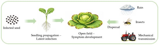
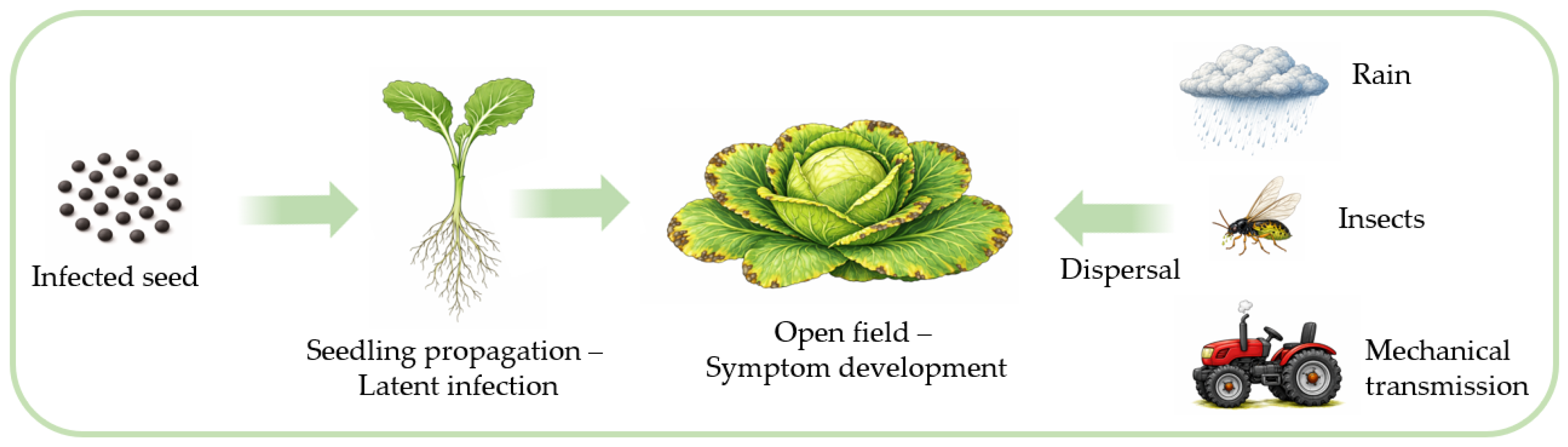
Xcc is a seed-borne pathogen that spreads through the vascular system of a plant. It is an etiological agent of black rot of brassicas, one of the most important diseases of cabbage and other economically important brassicas (broccoli, Brussels sprouts, kale, cauliflower, and kohlrabi). Once infection is present in the field, it can easily be transmitted over short distances (Figure 1). Traditionally, this disease has been a major issue in tropical regions. However, due to climate change and the global presence of Xcc, an increase in the prevalence and severity of the disease is anticipated in regions where it was of minor concern just a decade ago [6]. Growers are implementing different disease management strategies such as the use of non-infected seeds, resistant varieties and chemical control. With an increasing demand for more sustainable methods to control bacterial diseases, the use of chemicals and antibiotics poses significant risks, including the development of resistance and environmental hazards. Various approaches are being investigated that either inhibit the growth of Xcc or promote plant health [7].
Figure 1.
Infection pathway of Xanthomonas campestris pv. campestris.
As natural enemies of bacteria, phages occur wherever their host is present and lyse them with high specificity, typically infecting a limited number of strains of the same bacterial species. In contrast to many chemical control agents, which may accumulate in the environment, phages are rapidly degraded, especially in the absence of the host [8]. Numerous phages infecting different phytopathogenic bacteria have been identified and tested in a variety of applications [9]. Promising results were also obtained with phages infecting different species of Xanthomonas [10].
A considerable number of Xcc phages have been isolated to date from various geographic regions and environmental sources, including soil and infected plant material. Most Xcc phages originate from locations where black rot disease had been previously confirmed (Table S1). While partial characterization has been performed for the majority of these phages, the level of detail varies considerably. Some phages have been only briefly described, primarily in terms of host range and morphology [11,12], whereas others have been the subject of comprehensive studies that also included full genome sequencing, proteomic analysis, or infection kinetics [13,14,15,16]. These detailed characterizations have provided important insights into their taxonomy, biology, genetic features and potential applications in biocontrol.
In this paper we present a comprehensive review of phages infecting Xcc, a major pathogen of cabbage and other brassicas. It covers the challenging aspect of phage classification and major changes in the recent taxonomy, as well as phage morphology, key physiological characteristics, stability under environmental conditions, lifestyle, and genetic diversity. The review concludes with a summary of practical applications, emphasizing the potential of Xcc phages for managing black rot of brassicas.
2. From Morphology-Based to Genome-Based Taxonomy
2.1. Tailed Phages
Historically, tailed phages were classified based on their morphology, and this classification was later formalized by the International Committee on Nomenclature of Viruses (ICNV) in the 1970s and by International Committee on Taxonomy of Viruses (ICTV) in the 1980s. This classification, which included the well-known families of Myoviridae, Podoviridae, and Siphoviridae, was based on tail morphology and remained the golden standard for over four decades [17]. Specifically, Myoviridae were identified by long contractile tails, Siphoviridae by long, non-contractile tails, and Podoviridae by short tails [18]. The introduction of the order Caudovirales in 1998 unified all tailed phages under a single taxonomic category [17,19]. Genera classified within the three aforementioned families of the order Caudovirales were further differentiated mostly based on genome organization and DNA sequence similarity [20]. At that time there were no officially recognized phages infecting Xcc [21].
However, advancements in sequencing technologies soon revealed that (i) morphology-based taxonomy does not reflect evolutionary relationships and (ii) the families within the order Caudovirales are polyphyletic [22]. Consequently, in 2021, both the order Caudovirales and its three constituent families were abolished, automatically assigning all tailed phages to the class Caudoviricetes, which was established in 2019 (Figure 2). This allowed tailed phage families to be placed into separate orders that better reflect their genomic diversity and evolutionary relationships [22]. At the time of writing, the class Caudoviricetes consists of 11 orders and numerous families and genera that remain unclassified [21]. The classification criteria for the class Caudoviricetes are rank-specific, with phages being classified as the same species and as the same genus if they exhibit at least 95% and 70% nucleotide identity of their entire genomes, respectively. Additionally, when phylogenetic analysis on signature genes is performed, members of the same genus should cluster into a well-supported single clade in the phylogenetic tree [17]. Although morphological descriptors like “podovirus,” “myovirus,” and “siphovirus” can still be used informally, they no longer carry formal taxonomic significance following the 2022 ICTV ratification [22].
Figure 2.
Historical transition in the taxonomy of tailed phages.
Prior to the major taxonomic revision in 2022, three tailed Xcc phages were officially recognized by the ICTV: Xanthomonas phage phil7 (family Siphoviridae) (Taxonomy Proposal 2013.028a, approved) and Xanthomonas virus XcP1 and Xanthomonas virus Carpasina (genus Carpasinavirus, family Myoviridae) (Taxonomy Proposal 2019.070B, approved). Following the adoption of the binomial species nomenclature, their names were subsequently updated (Taxonomy Proposal 2021.010B, approved). After the implementation of the genome-based classification in 2021, six additional tailed Xcc phages were officially recognized by the ICTV. Four of these phages were assigned to the newly established genus Foxunavirus (Taxonomy Proposal 2021.031B, approved), while a single Xcc phage was recognized as the sole representative of the genus Foxquatrovirus (Taxonomy proposal 2021.030B, approved). Most recently, an Xcc phage was assigned to the previously established genus Carpasinavirus (Taxonomy Proposal 2024.020B, approved). A detailed overview is provided in Table 1.
Table 1.
Xcc phages officially approved by ICTV [22].
2.2. Filamentous Phages
Filamentous phages of Xcc (Table 1) are classified in the family Inoviridae (order Tubulavirales and class Faserviricetes), which comprises phages with diverse structures and lifestyles [23]. These phages have a simple, flexible rod-shaped structure and contain single-stranded DNA genomes ranging from approximately 5.5 to 10.6 kb [24]. Although the family Inoviridae was established and ratified in 1978 (Taxonomy Proposal Ratification_1978, approved), it was not until 2019 that the family was assigned to higher taxonomic ranks (order Tubulavirales and class Faserviricetes) by the ICTV (Taxonomy Proposal, 2019.005G, approved). The genus Lophivirus is a recently established genus in the family Inoviridae comprising four filamentous phages (Taxonomy Proposal 2021.085B, approved). All of them infect species within the genus Xanthomonas, two of which, Lophivirus Lf-UK and Lophivirus Lf2, specifically infect Xcc [25].
3. Virus Morphology
3.1. Tailed Phages
A review of phages with known morphology published in 2007 revealed that approximately 96% of phages display a tailed morphology [26]. This ancient and highly diverse group of viruses exhibits significant variation in both physical dimensions and structure [27]. Despite this diversity, all of them have a capsid or head, composed of repeating protein units arranged in a similar manner [28]. The most profound differences among them are observed in tail structure, which served as a key criterion in the old morphology-based classification [22], as discussed in the previous section.
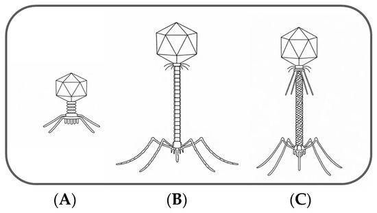
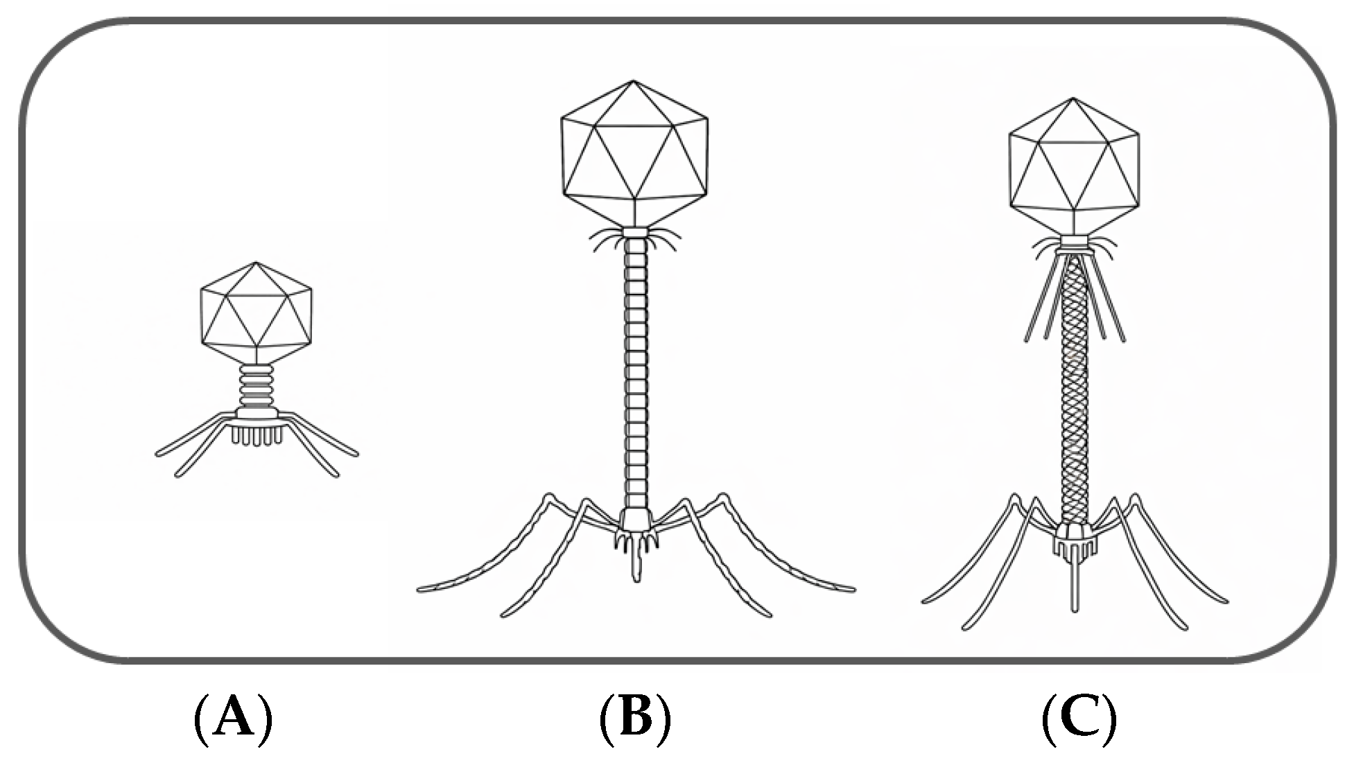
Among the three morphotypes of tailed phages (Figure 3), podoviruses have a relatively short, non-contractile and non-flexible tail. The structure of the podoviral tail appears to be the simplest. Greater structural complexity is observed in the other two morphotypes. Siphoviruses possess a long, thin, non-contractile and often flexible tail, containing a tail tube. Myoviruses exhibit the most complex structure, with a tail tube surrounded by a contractile sheath. Myoviruses also tend to be larger, with thick tails that show considerable variation in length but are more uniform in width [20,27,28].
Figure 3.
Representative schematics of the three major morphotypes of tailed phages are shown: (A) podovirus with a short, non-contractile tail; (B) siphovirus with a long, non-contractile and flexible tail; and (C) myovirus with a long, contractile tail surrounded by a sheath.
The first morphologically characterized Xcc phages were described as sperm-shaped with a flexible tail [29,30], a typical feature of siphoviruses. Various Xcc phages with a siphoviral morphotype were later described by other authors [11,12,14,31,32,33,34,35].
Phage S from Xcc was one of the first with known myoviral morphology [36]. Although the authors do not indicate that it is a myovirus, its description as a tadpole-like phage with a cylindrical tail, supported by the available electron micrograph, suggests that it most likely belongs to this morphotype [36]. The rest of the identified Xcc myoviruses were unambiguously characterized as such, primarily based on clear descriptions of the tail structure [12,13,15,37,38,39,40,41].
The podoviral morphotype is the least common among tailed phages [27], including those infecting Xcc, where only two phages with this morphology have been reported [16,37].
So far, the morphology of 52 tailed Xcc phages has been examined (Table S2). The siphoviral morphotype has been reported as the most common among tailed phages, and a similar trend was observed across the entire Xanthomonas genus [26]. We observed that only about one third of the tailed Xcc phages with identified structure exhibit siphoviral morphology, whereas the majority has myoviral morphology (Table S2).
3.2. Filamentous Phages
Filamentous phages of the family Inoviridae are characterized by long, thin, and flexible virions [28]. To date, only three filamentous Xcc phages have been morphologically described (Table S2). The first such phage reported in Xcc was φLf, which for decades remained the only known representative [42]. Two additional filamentous Xcc phages were only recently characterized [25] and approved by the ICTV (Taxonomy Proposal 2021.085B, approved).
4. Host Range
Host range defines which bacterial genera, species, and strains a phage can lyse [43] or, conversely, which bacteria support phage multiplication [44]. It is a key physiological characteristic and an important factor when considering phages as biological control agents. For accurate host range determination, a single phage isolate should be used, since mixtures of phages may appear to have a broader host range and lead to misleading conclusions [45]. Host range is most commonly assessed by spot testing, in which a phage suspension is applied to a bacterial lawn and lysis is evaluated the following day [44]. Ideally, large and well-characterized strain collections are used for determination of host range, but in practice, assays often rely on pathogenic isolates gathered for specific purposes [43].
Most research of Xcc phages has studied their host range, revealing diverse patterns: from phages that infect only a single strain to those capable of lysing other X. campestris pathovars and even different Xanthomonas species (Table S1). In some cases, Xcc phages have also been tested for infectivity against unrelated phytopathogenic bacteria such as Erwinia, Agrobacterium, and Pseudomonas, and plant growth-promoting bacteria (PGPB), but none of those were found to support phage multiplication [14,29,39,46].
Host range can also be determined using efficiency of plating (EOP), in which serial dilutions of phage suspension are spotted onto bacterial lawn within the same plate. This approach allows for quantitative comparison of plaque formation across susceptible hosts and reflects differences in phage multiplication efficiency, which is strongly influenced by the individual host [43,44]. EOP analysis performed on Xcc phages revealed generally higher efficiency on the bacterial strain used for phage isolation, whereas multiplication with other susceptible strains was reduced [46]. These findings indicate that EOP offers a more precise view of host range, and it has been suggested that this method should be incorporated into phage selection for phage therapy in medicine [47].
When a phage is tested against multiple strains within the susceptible bacterial genus, this approach is referred to as phage typing. The obtained host range pattern can be used not only to differentiate bacterial strains but also to distinguish among the phages [48,49]. Phage typing has been applied in various studies both to differentiate newly isolated phages and Xcc strains [46,50] and to select phages that collectively exhibit the broadest host range [12,51].
5. Phage Growth Parameters
Phage growth involves a series of sequential steps including attachment to the host cell, nucleic acid uptake, synthesis and assembly of phage particles, and release of progeny virions [52]. Some stages of phage growth can be quantitatively described using growth parameters such as adsorption rate, latent period, and burst size [53].
5.1. Adsorption Rate
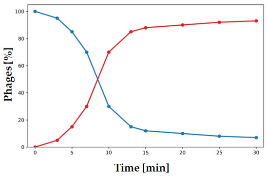
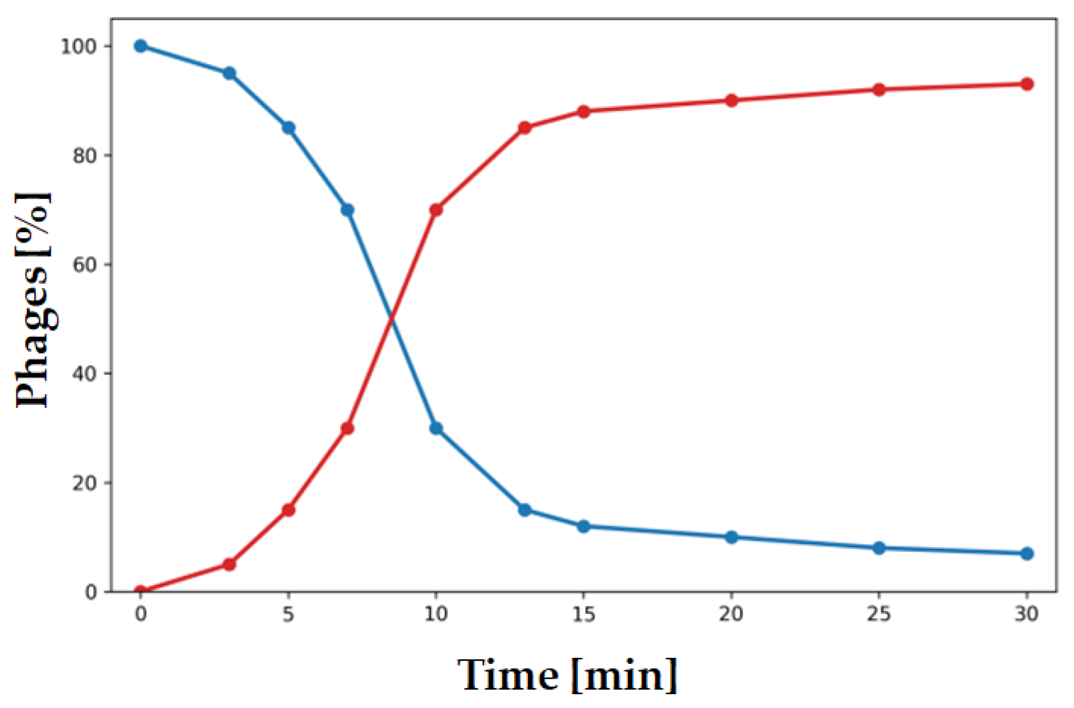
Adsorption begins with the release of virions from the lysed host cell, followed by their extracellular search for a susceptible bacterium and attachment to the bacterial surface, where irreversible binding marks the completion of adsorption [52,54,55]. For most Xcc phages, adsorption has not been measured; where reported, values are expressed either as the percentage of phages adsorbed to the host cell over time (Figure 4) or as the adsorption rate constant (Table S3). The latter is a parameter that describes the rate at which phage particles irreversibly attach to host bacteria under defined conditions [54]. Although many factors are known to influence phage adsorption [55], only phage concentration and different levels of xanthan in susceptible strains have been evaluated for Xcc phages so far [38,40].
Figure 4.
Phage adsorption is presented as the decrease in free phage particles in the supernatant (blue) over time or as an increase in adsorbed phages on the host over time (red).
5.2. Latent Period and Burst Size
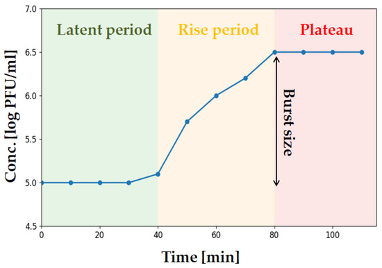
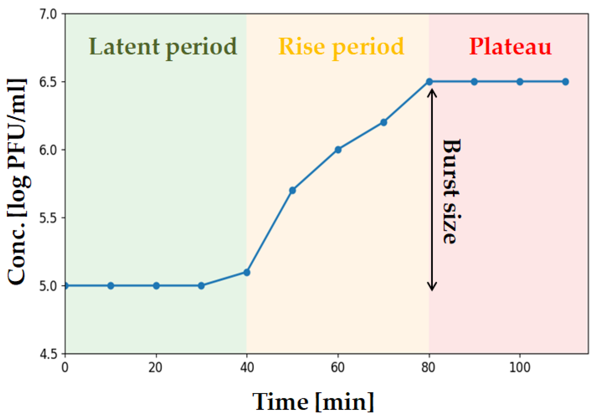
A one-step growth experiment includes adsorption, infection, and release of phages. It is a fundamental method in the characterization of isolated phages and it allows for the determination of key parameters such as the latent period and average burst size [53,56] (Figure 5). After adsorption, a stable phase known as the latent period is observed, during which the number of phages remains constant. This phase is followed by a rapid increase in phage numbers as infected bacterial cells undergo lysis and release newly synthesized progeny phages [57]. This so-called rise period begins at the end of the latent phase and continues until all infected bacterial cell have been lysed [53]. Burst size refers to the number of progeny virions produced per infected cell and is typically calculated as the ratio between the number of phages after the rise and the number of phage-infected bacteria before the start of the rise period [53,58].
Figure 5.
The plot illustrates consecutive phases of phage infection including the latent period, rise period, and plateau phase, during which the phage titer stabilizes. Burst size is subsequently determined as the ratio between phage titers before and after the rise period.
Among Xcc phages with reported one-step growth experiment data, latent periods ranged from about 30 to 210 min, while burst sizes varied from as few as 4 to nearly 100 progeny per cell. Where measured, the rise period lasted approximately 30 to 120 min (Table S3).
Overall, among the phage growth parameters, adsorption rate, latent period, and burst size represent the most important descriptors of the phage multiplication cycle and are particularly relevant when evaluating phages for phage therapy in plant disease management. In this context, phages with higher adsorption rates, shorter latent periods, and larger burst sizes are generally considered more desirable [53,59], although these traits must be weighed alongside other factors such as host range and stability [4].
6. Stability of Phages Under Various Physical and Chemical Conditions
Phage stability is influenced by various chemical and physical factors. Evaluating phage stability under these factors deepens insights into phage biology and provides important criteria for their characterization [27]. The genome of tailed phages is enclosed within a hollow protein capsid that protects the viral DNA from harsh environmental conditions. Virion survival depends primarily on capsid stability and its ability to withstand physicochemical stresses such as temperature fluctuations, extreme pH, ultraviolet (UV) radiation, and desiccation [60,61]. Elevated temperatures and extreme pH may directly compromise capsid integrity while also destabilizing other structural and functional virion elements required for infection. Overall infectivity is thereby probably limited by the most sensitive essential element [62]. Resistance to UV irradiation is largely determined by capsid stability, as the capsid shields the enclosed DNA from direct damage [63].
While temperature is the most commonly tested, assessing additional factors offers a more comprehensive view of phage stability and helps refine future strategies for application. Table 2 summarizes the main findings, including responses to temperature, pH, organic solvents, UV irradiation, and pesticides.
Table 2.
Stability of Xcc phages under different physical and chemical conditions tested in vitro.
6.1. Stability of Xcc Phages In Vitro
Tailed Xcc phages were generally stable at or near a neutral pH, showing reduced stability under more acidic and alkaline conditions. They also differed in their thermal tolerance, with reduced infectivity at 50 °C and complete inactivation at 76 °C (Table 2). The filamentous phage φLf retained full infectivity at 80 °C but was more sensitive to organic solvents [42] compared to tailed phages [15,16,31]. Ultraviolet (UV-C) radiation completely inactivated phages Murka and PBR31 [15,16], whereas the damaging effect of UV-B radiation was reduced by adding various UV-absorbing compounds [64].
For better and cost-effective disease management, Xcc phages could be used in combination with other plant protection products. Compatibility of phage DB1’ with various pesticides appeared feasible, as most products did not affect phage stability significantly. However, the copper-based product had a pronounced negative effect on phage stability [12], as was also reported for phages of other phytopathogenic bacteria [65,66]. This is particularly relevant since copper is among the most widely used pesticides for the control of bacterial plant diseases, especially in integrated and organic production systems [67].
6.2. Stability of Phages In Vivo
Phage stability and persistence should be assessed not only in the laboratory but also outside controlled environments, such as in greenhouses and open fields. Phages are usually applied at the rhizosphere or the phyllosphere [68]. In the phyllosphere, phages are exposed to fluctuating temperatures and pH, UV light, desiccation, chemical pesticides and surfactants [69]. Among all environmental factors, UV-A and UV-B light have the most destructive effect on phage survival [70].
The persistence of phage FoX2 was monitored on the leaves of young healthy cabbage plants grown in greenhouse conditions. After 48 h, no viable phages were detected. Various factors were probably contributing to the complete inactivation, but the absence of the host was considered the main reason [13]. In another study, Xcc phages persisted at higher concentrations when the host was present on the leaves of A. thaliana [71]. Following successful results from in vitro studies, several UV protectants were evaluated in greenhouse experiments. After application onto the leaves of cabbage seedlings, all treatments exhibited a sharp decline in phage titers within the first 30 min, most likely due to the desiccation shock. Differences among formulations became apparent during the following days, with skimmed milk and riboflavin maintaining higher phage concentrations than gum arabic and the control [64].
The phyllosphere represents an extremely hostile environment for phage survival after application. The direct effect of UV light has been demonstrated for phages infecting related Xanthomonas species. The survival of phages on the leaves of tomato throughout the day was strongly influenced by application timing, with evening treatments resulting in improved survival compared to morning and daytime applications, when exposure to intense UV radiation was greater. Protective formulations mitigated UV-induced inactivation and also reduced the effect of temperatures during subsequent days of exposure [65]. Failure to maintain viable phages at sufficiently high concentrations on leaf surfaces may result in only limited suppression of bacterial populations [72], emphasizing that preventing significant reductions in phage titers under field conditions can substantially improve biocontrol efficacy.
7. Lifestyle of Xcc Phages
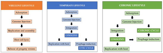
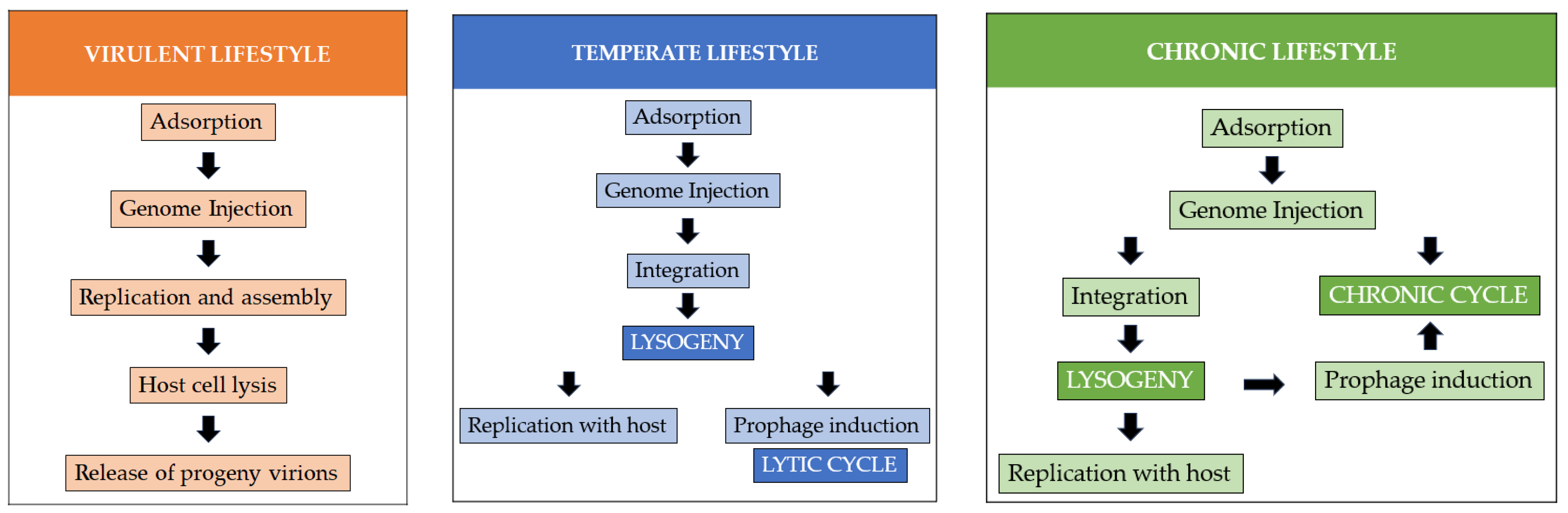
Determination of lifestyle is an important part of phage characterization. Tailed phages can be either virulent or temperate, whereas filamentous phages display chronic infections [24,73] (Figure 6). Virulent or strictly lytic phages kill their host by replicating and lysing the host cell and releasing progeny virions [73]. In contrast, temperate phages are able to integrate their genome into the host chromosome, forming a stable relationship with the bacterium known as lysogeny. The integrated phage genome, known as a prophage, is co-replicated with the host DNA [74]. Bacteria carrying a prophage can become immune to subsequent phage attacks, particularly by phages of the same type. Temperate phages frequently encode virulence factors that may increase the host pathogenicity [75] and enhance host fitness [1]. Filamentous phages (family Inoviridae) are chronically released from the infected cell without killing the host, many of which are capable of integration into the host genome [76].
Figure 6.
Schematic overview of bacteriophage lifestyles identified among Xcc phages, highlighting the key stages in each lifestyle. The virulent lifestyle is shown in orange, the temperate lifestyle in blue, and the chronic lifestyle in green.
Plaque morphology often reflects phage lifestyle; clear plaques are generally produced by strictly lytic phages, whereas turbid plaques usually indicate lysogeny [77]. The turbidity of plaques arises from the growth of surviving lysogenized cells within the lysis zone [78]. The majority of known Xcc phages form clear plaques, indicating a predominantly virulent lifestyle. However, several phages produce turbid plaques, suggesting a temperate lifestyle (Table S4).
Certain conditions can induce a prophage to excise from the host chromosome and enter the lytic cycle [1,79]. Induction is typically triggered by physical or chemical stimuli that damage host DNA [53]. For example, mitomycin C was used to induce phage P165/125, confirming its temperate lifestyle [30].
One of the most reliable ways to determine the phage lifestyle is the identification of lysogeny-associated genes such as integrases that mediate the integration of phage DNA into the host genome [80]. The absence of such genes in most Xcc phages supports their predicted virulent lifestyle (Table S4). A discrepancy was observed in the case of the phage Seregon, which formed turbid plaques even though genome analysis did not reveal any integrase or other lysogeny-associated genes. The authors acknowledged this phenomenon and recommended further investigation [14]. On the other hand, the genome of the phage Murka contains a gene fragment corresponding to only 10–20% of a typical phage integrase, making its functional role in lysogeny unlikely [15].
All three known filamentous phages (φLf, φLf-UK and φLf2) are capable of lysogeny: for φLf, integration was experimentally demonstrated [81], while for φLf-UK andφ, it was confirmed by PCR amplification of integration-specific sites [25].
8. Basic Genomic Characteristics of Xcc Phages
Phage genomes are relatively short, rich in coding regions, and rarely contain long repetitive elements [82]. They commonly encode proteins involved in structural assembly, such as capsid and tail proteins, DNA replication, and metabolic support. Genes responsible for integration and excision are characteristics of temperate phages [80]. Open reading frames (ORFs), defined as potential protein-coding regions [83], are commonly reported in publications describing genome characteristics. Other metadata typically include core genomic descriptors such as genome length and GC content, while additional information on taxonomic assignment, host strain, virion morphology and source of isolation can further improve comparative analysis of phages [84]. To date, 25 complete genomes of Xcc phages are available in the NCBI database, comprising 21 tailed and 4 filamentous phages. They show considerable diversity in genome length (~6–200 kbp), GC content (34.8–67.4%), number of ORFs (11–313), and taxonomic classification, with 19 phages classified at the genus level and 6 currently unassigned (Table 3). Current ICTV criteria for phage taxonomic classification are based on genome comparison and phylogenetic analyses [85]; therefore, a well-characterized phage genome is essential. Adequate genome characterization has been performed for various Xcc phages and some of them show high similarity to officially recognized Xcc phages, yet to be approved by ICTV.
8.1. Tailed Xcc Phages
The phages Seregon and AhaSv display strong genomic similarity, both in size (~55.5 kbp) and GC content (63.2%). Their nucleotide sequence similarity is 90.6%, with 62 similar proteins sharing between 61.08% and 100% amino acid identity. Cohesive ends were identified in both genomes, supporting circularization upon infection. The high degree of genomic and proteomic similarity, along with shared structural features, places these phages within the genus Salvovirus [14,35].
The genus Carpasinavirus comprises four Xcc phages that share similar genome sizes (~61 kbp) and moderately low GC content (52.0–52.5%). These phages contain between 81 and 99 predicted ORFs and exhibit a modular genome organization typical of lytic tailed phages. All four show high sequence similarity, with FoX6 and FoX7 considered two isolates of the same species [13,39].
Phages classified under the genus Foxunavirus show high genomic similarity, with only minor variation in genome size (~44 kbp), GC content (59.5–59.6%), and predicted ORF number across isolates. Representatives of this genus share genomic similarity that ranges from 85% to 94% [15,33,34].
Based on the high nucleotide sequence similarity (95%), the phage HXX_Dennis and two related non-Xcc phages cluster together as isolates of the same species. On the basis of these characteristics, the authors proposed establishing of a new genus, tentatively named Nordvirus [86]. Since each isolate of the newly proposed genus infects a different bacterial species, this raises the question of whether these phages exhibit a broader, potentially polyvalent host range.
Among other Xcc phages, phage X1 stands out with the longest genome at 200 kbp and the highest number of predicted ORFs (313), while SU1 has the lowest GC content among Xcc phages (34.8%) (Table 3).
8.2. Filamentous Xcc Phages
Compared to tailed phages, filamentous Xcc phages have much shorter genomes, encode fewer ORFs, and produce a smaller set of proteins (Table 3). The genus Lophivirus includes three highly similar members (φLf, φLf2, and φLf-UK), with φLf-UK and φLf-2 sharing 98.7% and 85.8% nucleotide identity with φLf, respectively [25]. Phage fSU1 diverges from Lophivirus members and remains unclassified. It has nearly the same GC content as Lophivirus phages but a genome almost twice as long (Table 3).
Table 3.
Genomic features of Xcc phage classification on NCBI.
9. Biocontrol Applications of Xcc Phages
Phages that meet specific criteria could be implemented as biocontrol agents. In addition to a strictly lytic lifestyle, other characteristics are considered during the selection process, some of which are discussed in this manuscript. Ideally, a phage should rapidly lyse its host and produce a high number of progeny virions. It should also be capable of infecting a broad range of target bacterial strains while remaining specific enough not to disrupt the surrounding microbiome [1,102].
The planting material of cabbage and other brassicas is grown in close proximity under humid conditions before being transplanted to the field. Such conditions facilitate pathogen spread, especially if the seeds are already infected [103]. Infections often remain latent in the early stages and can later lead to disease outbreaks in the field [6]. As Xcc is primarily transmitted through infected seeds [104], phages could be employed as seed decontaminants [104]. Several chemical and physical treatments have been shown to significantly reduce surface contamination of cabbage seeds [104], although internal Xcc infection may persist despite such treatments [105]. Seed coating with phages has been shown to reduce contamination on the seed surface, leading to improved germination and reduced disease development [106]. Notably, phage seed coatings significantly reduced bacterial populations originating from internal infections, with differences becoming evident not in the seeds themselves but during early plant growth [107]. These findings suggest that phage-based seed coatings do not directly reduce bacteria within the seeds but instead provide protection during the early stages following germination.
Xcc was the first phytopathogenic bacterium for which phage-mediated inhibition of growth was experimentally demonstrated. As early as 1924, Mallmann and Helmstreet [3] showed that filtered extracts from decomposed cabbage could inhibit in vitro growth of Xcc. However, following this early observation, phage-based control of Xcc phages received little attention for nearly a century. More recently, in vitro studies confirmed the potential of Xcc phages, demonstrating effective suppression of bacterial growth in a planktonic state [14,33] as well as interference with biofilm formation either when applied alone or in combination with complementary agents [40,108]. Additionally, several studies began exploring different application strategies, which are discussed further on.
Artificially infected seeds of cabbage were treated with phages and bacterial load was monitored in the seed extract. The number of bacterial cells was significantly reduced in the extract from seeds treated with phages compared to the untreated control [51]. A similar reduction was observed by Holtappels et al. [13] in both naturally and artificially infected cauliflower seeds. In both studies, seed treatment with phages resulted in a marked reduction in disease symptoms in seedlings [13,51]. Additionally, growth parameters of cauliflower seedlings were evaluated, with untreated controls showing reduced root and shoot growth [13].
Several studies investigated the use of spray applications on cruciferous plants in controlled and open field conditions. In controlled conditions, disease incidence in young plants treated with phages was lower than in untreated plants [13,40,41,109]. In the study of Orynbaev et al. [64], phages even outperformed the used commercially available copper-based product. In the field trials, which were strongly influenced by environmental conditions, Xcc phages were able to reduce the disease severity in three separate experiments in which they used a cocktail of two phages [13]. Other application strategies, such as soaking the whole seedling in phage suspension and irrigation-based treatment, were also used in Xcc control experiments [13,71]. Two irrigation-based experiments were performed by Holtappels et al. [13], where one phage successfully suppressed the disease development while the other one did not.
Together, these studies demonstrate that Xcc phages can effectively reduce disease incidence through multiple application strategies. For biocontrol to succeed, however, phages must encounter their host before being inactivated by environmental stressors [110]. While several organic compounds have been shown to enhance persistence of Xcc phages [64], the timing of phage application has also been found to be important. In particular, late-afternoon or evening treatments have been shown to prolong phage persistence on the leaf surface [65,111]. In addition to these considerations, environmental biosafety has also been addressed and was specifically assessed for phage FoX4 [112].
On the other hand, phages should not be viewed as a standalone approach but rather as one of the complementary tools implemented within integrated disease management strategies [113]. PGPB, particularly members of the genus Bacillus, are well-known for their beneficial effects on plant health [114]. Various species have been reported to exhibit antagonistic activity against Xcc, either through direct competition or the production of antibacterial metabolites [115,116]. The combination of phages with non-pathogenic strains [109], biostimulants such as PGPB, and the use of tolerant or resistant cultivars may further reduce pathogen population densities and lower overall disease pressure, contributing to more stable and sustainable management of black rot.
10. Conclusions
Over the past decade, there has been a notable increase in the discovery of phages that specifically infect Xcc. In the latest Master Species List (MSL) published by the ICTV, eleven Xcc phages have been officially recognized. Additional fully sequenced genomes of several Xcc phages have been deposited in internationally acknowledged databases, potentially supporting future taxonomic proposals. Obtaining complete genomic data is essential for the reliable determination of phage lifestyle, a key factor in selecting phages for subsequent applications. However, the biocontrol potential of individual phages also heavily depends on a thorough understanding of their biology and stability. In line with this perspective, several of these characteristics are highlighted and discussed throughout this review.
The high specificity of phages is a major advantage of phage-based biocontrol, allowing for targeted suppression of plant pathogenic bacteria with minimal impact on beneficial microbiomes. However, this unique characteristic can also limit their effectiveness, as not all strains of a target species are susceptible to a given phage. Fortunately, the host range of the phage-based applications can be broadened by using phage mixtures or cocktails, which can also help reduce the likelihood of resistance development. Once a phage is comprehensively characterized, the processes for obtaining larger amounts of high-titer phage preparations are relatively easy and inexpensive. Moreover, commercial-scale manufacturing services are now available to support agricultural applications.
Phages offer a promising, environmentally friendly alternative for controlling bacterial plant diseases because of their unique mode of action. This is particularly relevant due to growing demand for sustainable strategies that reduce reliance on chemical treatments. Comprehensive characterization of phages is essential to ensure their effective and safe application. Although phages are sensitive to variety of environmental factors, such as UV light, temperature, and desiccation, formulation additives and optimized application strategies can enhance persistence and viability under harsh field conditions. However, for successful biocontrol, it is equally important to understand the pathogen population, particularly the prevalence of aggressive or locally dominant strains, to ensure susceptibility to the selected phages. Future research should focus on optimizing phage formulations, understanding interactions with the plant microbiome, evaluating field efficacy, and developing standardized protocols for phage application. By implementing these approaches, phage-based strategies can become a reliable and sustainable component of plant disease management.
Supplementary Materials
The following supporting information can be downloaded at: https://www.mdpi.com/article/10.3390/horticulturae12030288/s1, Table S1: Previously reported Xcc phages, their country/region of origin and source of isolation, and host range; Table S2: Morphological characteristics of Xcc phages; Table S3: Reported growth parameters of Xcc phages; Table S4: Summary of plaque morphology and/or other available analyses relevant to lifestyle determination. Phage lifestyle is reported from the publication or inferred from plaque morphology and/or other available analyses in the publication.
Author Contributions
Conceptualization: A.B., I.M.P. and J.L.; writing—original draft preparation and editing, A.B.; writing—review and editing, I.M.P. and J.L.; supervision, I.M.P.; visualization: A.B. All authors have read and agreed to the published version of the manuscript.
Funding
The research was supported by The Administration of the Republic of Slovenia for Food Safety, Veterinary and Plant Protection (C2337-25-000024) and the Slovenian Research and Innovation Agency (grant Nos. P4-0431 and P4-0072).
Data Availability Statement
No new data were created or analyzed in this study.
Acknowledgments
We would like to acknowledge the use of AI tool ChatGPT (GPT-5.2) for language refinement and the generation of images included in the figures.
Conflicts of Interest
The authors declare no conflicts of interest. The funders had no role in the preparation of the manuscript.
References
- Buttimer, C.; McAuliffe, O.; Ross, R.P.; Hill, C.; O’Mahony, J.; Coffey, A. Bacteriophages and bacterial plant diseases. Front. Microbiol. 2017, 8, 34. [Google Scholar] [CrossRef] [Scilit]
- Cobian Güemes, A.G.; Youle, M.; Cantu, V.A.; Felts, B.; Nulton, J.; Rohwer, F. Viruses as winners in the game of life. Annu. Rev. Virol. 2016, 29, 197–214. [Google Scholar] [CrossRef] [Scilit]
- Mallman, W.L.; Helmstreet, C. Isolation of inhibitory substances from plants. J. Agric. Res. 1924, 28, 599–602. [Google Scholar]
- Jones, J.B.; Jackson, L.E.; Balogh, B.; Obradovic, A.; Iriarte, F.B.; Momol, M.T. Bacteriophages for plant disease control. Annu. Rev. Phytopathol. 2007, 45, 245–262. [Google Scholar] [CrossRef] [Scilit]
- Jones, J.B.; Svircev, A.M.; Obradović, A.Ž. Crop use of bacteriophages. In Bacteriophages: Biology, Technology, Therapy; Harper, D.R., Abedon, S.T., Burrowes, B.H., McConville, M.L., Eds.; Springer: Cham, Switzerland, 2021; pp. 839–856. [Google Scholar] [CrossRef] [Scilit]
- Vicente, J.G.; Holub, E.B. Xanthomonas campestris pv. campestris (cause of black rot of crucifers) in the genomic era is still a worldwide threat to brassica crops. Mol. Plant Pathol. 2013, 14, 2–18. [Google Scholar] [CrossRef] [Scilit] [PubMed]
- Liu, Z.; Wang, H.; Wang, J.; Lv, J.; Xie, B.; Lou, S.; Wang, S.; Zhang, B.; Li, Z.; Yue, Z.; et al. Physical, chemical, and biological control of black rot of brassicaceae vegetables: A review. Front. Microbiol. 2022, 13, 1023826. [Google Scholar] [CrossRef] [Scilit] [PubMed]
- Halawa, E.M. Challenges of bacteriophages application in controlling bacterial plant diseases and how to overcome them. J. Genet. Eng. Biotechnol. 2023, 21, 98. [Google Scholar] [CrossRef] [Scilit] [PubMed]
- Choudhary, M.; Bankole, I.A.; McDuffee, S.T.; Parajuli, A.; Poudel, M.; Balogh, B.; Paret, M.L.; Jones, J.B. Bacteriophages as Agents for Plant Disease Control: Where Are We After a Century? Viruses 2025, 17, 1033. [Google Scholar] [CrossRef] [Scilit]
- Nakayinga, R.; Makumi, A.; Tumuhaise, V.; Tinzaara, W. Xanthomonas bacteriophages: A review of their biology and biocontrol applications in agriculture. BMC Microbiol. 2021, 21, 291. [Google Scholar] [CrossRef] [Scilit]
- Renu, M.S.; Singh, U.B.; Sahu, U.; Nagrale, D.T.; Sahu, P.K. Characterization of lytic bacteriophage XCC9SH3 infecting Xanthomonas campestris pv. campestris. J. Plant Pathol. 2017, 99, 233–238. [Google Scholar]
- Orynbayev, A.T.; Kabanova, A.P.; Obraztsova, E.A.; Ignatov, A.N.; Miroshnikov, K.A.; Dzhalilov, F.S. Isolation of bacteriophages of Xanthomonas campestris pv. campestris and their use for protection of cabbage from black rot. Izv. Timiryazev Agric. Acad. 2019, 2, 35–48. [Google Scholar] [CrossRef]
- Holtappels, D.; Boulé, J.; Adriaenssens, E.M.; Satin, B.; Gorris, M.T.; Lavigne, R.; Lefeber, T.; Gilbert, M.; Pirnay, J.-P.; Maes, M.; et al. The potential of bacteriophages to control Xanthomonas campestris pv. campestris: From isolation to field application. Microb. Biotechnol. 2022, 15, 1762–1782. [Google Scholar] [CrossRef] [Scilit] [PubMed]
- Erdrich, S.H.; Sharma, V.; Schurr, U.; Arsova, B.; Frunzke, J. Isolation of Novel Xanthomonas Phages Infecting the Plant Pathogens X. Translucens X. campestris. Viruses 2022, 14, 1449. [Google Scholar] [CrossRef] [Scilit] [PubMed]
- Evseev, P.V.; Tarakanov, R.I.; Vo, H.T.N.; Suzina, N.E.; Vasilyeva, A.A.; Ignatov, A.N.; Miroshnikov, K.A.; Dzhalilov, F.S.-U. Characterisation of New Foxunavirus Phage Murka with the Potential of Xanthomonas campestris pv. campestris Control. Viruses 2024, 16, 198. [Google Scholar] [CrossRef] [Scilit] [PubMed]
- Tarakanov, R.I.; Evseev, P.V.; Vo, H.T.N.; Troshin, K.S.; Gutnik, D.I.; Ignatov, A.N.; Toshchakov, S.V.; Miroshnikov, K.A.; Jafarov, I.H.; Dzhalilov, F.S.-U. Xanthomonas Phage PBR31: Classifying the Unclassifiable. Viruses 2024, 16, 406. [Google Scholar] [CrossRef] [Scilit]
- Turner, D.; Kropinski, A.M.; Adriaenssens, E.M. A Roadmap for Genome-Based Phage Taxonomy. Viruses 2021, 13, 506. [Google Scholar] [CrossRef] [Scilit]
- Ackermann, H.-W. Tailed phages: The order Caudovirales. Adv. Virus Res. 1998, 51, 135–201. [Google Scholar] [CrossRef] [Scilit]
- Pringle, C.R. Virus Taxonomy—San Diego 1998. Arch. Virol. 1998, 143, 1449–1459. [Google Scholar] [CrossRef] [Scilit]
- ICTV 9th Report. Available online: https://ictv.global/report_9th (accessed on 5 January 2026).
- Current ICTV Taxonomy Release. Available online: https://ictv.global/taxonomy (accessed on 5 January 2026).
- Turner, D.; Shkoporov, A.N.; Lood, C.; Millard, A.D.; Dutilh, B.E.; Alfenas-Zerbini, P.; van Zyl, L.J.; Aziz, R.K.; Oksanen, H.M.; Poranen, M.M.; et al. Abolishment of morphology-based taxa and change to binomial species names: 2022 taxonomy update of the ICTV bacterial viruses subcommittee. Arch. Virol. 2023, 168, 74. [Google Scholar] [CrossRef] [Scilit]
- Hay, I.D.; Lithgow, T. Filamentous phages: Masters of a microbial sharing economy. EMBO Rep. 2019, 20, 24. [Google Scholar] [CrossRef] [Scilit]
- Knezevic, P.; Adriaenssens, E.M.; ICTV Report Consortium. ICTV Virus Taxonomy Profile: Inoviridae. J. Gen. Virol. 2021, 102, 2. [Google Scholar] [CrossRef] [Scilit] [PubMed]
- Yeh, T.-Y.; Feehley, P.J.; Feehley, M.C.; Ooi, V.Y.; Wu, P.-C.; Hsieh, F.; Chiu, S.S.; Su, Y.-C.; Lewis, M.S.; Contreras, G.P. The packaging signal of Xanthomonas integrative filamentous phages. Virology 2024, 600, 110279. [Google Scholar] [CrossRef] [Scilit] [PubMed]
- Ackermann, H.-W. 5500 Phages examined in the electron microscope. Arch. Virol. 2007, 152, 227–243. [Google Scholar] [CrossRef] [Scilit] [PubMed]
- Ackermann, H.-W. Phage Classification and Characterization. In Bacteriophages: Methods and Protocols, Volume 1: Isolation, Characterization, and Interactions; Clokie, M.R.J., Kropinski, A.M., Eds.; Humana Press: Totowa, NJ, USA, 2009; pp. 127–140. [Google Scholar] [CrossRef] [Scilit]
- Sanz-Gaitero, M.; Seoane-Blanco, M.; van Raaij, M.J. Structure and Function of Bacteriophages. In Bacteriophages Biology, Technology, Therapy; Harper, D.R., Abedon, S.T., Burrowes, B.H., McConville, M.L., Eds.; Springer: Cham, Switzerland, 2021; pp. 19–91. [Google Scholar] [CrossRef] [Scilit]
- Sutton, M.D.; Katznelson, H.; Quadling, C. A bacteriophage that attacks numerous phytopathogenic Xanthomonas species. Can. J. Microbiol. 1958, 4, 493–497. [Google Scholar] [CrossRef] [Scilit]
- Sutton, M.D.; Quadling, C. Lysogeny in a strain of Xanthomonas campestris. Can. J. Microbiol. 1963, 9, 821–828. [Google Scholar] [CrossRef] [Scilit]
- Su, M.-J.; Lai, M.-C.; Weng, S.-F.; Tseng, Y.-H. Characterization of phage φL7 and transfection of Xanthomonas campestris pv. campestris by the phage DNA. Bot. Bull. Acad. Sin. 1990, 31, 197–203. [Google Scholar]
- Lee, C.-N.; Lin, J.-W.; Weng, S.-F.; Tseng, Y.-H. Genomic characterization of the intron-containing T7-like phage phiL7 of Xanthomonas campestris. Appl. Environ. Microbiol. 2009, 75, 7825–7835. [Google Scholar] [CrossRef] [Scilit]
- Holtappels, D.; Fortuna, K.J.; Vallino, M.; Lavigne, R.; Wagemans, J. Isolation, characterization and genome analysis of an orphan phage FoX4 of the new Foxquatrovirus genus. BMC Microbiol. 2022, 22, 304. [Google Scholar] [CrossRef] [Scilit]
- Neoralová, M.; Brázdová, Š.; Eichmeier, A.; Petrzik, K. Complete genome sequence of Xanthomonas phage M29, a new member of Foxunavirus isolated in the Czech Republic. Virus Genes 2023, 59, 874–877. [Google Scholar] [CrossRef] [Scilit]
- Ni, A.; Wu, Q.; Fang, Z.; Xiang, L.; Liu, Q.; Tan, L.; Weng, Q. Genome analysis and classification of Xanthomonas bacteriophage AhaSv, a new member of the genus Salvovirus. Arch. Virol. 2024, 169, 117. [Google Scholar] [CrossRef] [Scilit]
- Watanabe, M.; Naito, K.; Kaneko, K.; Nabasama, H.; Hosokawa, D. Some properties of Xanthomonas campestris pv. campestris phage. Jpn. J. Phytopathol. 1980, 46, 517–525. [Google Scholar] [CrossRef] [Scilit]
- Liew, K.W.; Alvarez, A.M. Biological and morphological characterization of Xanthomonas campestris bacteriophages. Phytopathology 1981, 71, 269–275. [Google Scholar] [CrossRef] [Scilit]
- Weiss, B.D.; Capage, M.A.; Kessel, M.; Benson, S.A. Isolation and characterization of a generalized transducing phage for Xanthomonas campestris pv. campestris. J. Bacteriol. 1994, 176, 3354–3362. [Google Scholar] [CrossRef] [Scilit] [PubMed]
- da Silva, P.F.; da Silva Xavier, A.; Prieto Bruckner, F.; Reis de Rezende, R.; Pereira Vidigal, P.M.; Alfenas-Zerbini, P. Biological and molecular characterization of a bacteriophage infecting Xanthomonas campestris pv. campestris, isolated from brassica fields. Arch. Virol. 2019, 164, 1857–1862. [Google Scholar] [CrossRef] [Scilit]
- Papaianni, M.; Paris, D.; Woo, S.L.; Fulgione, A.; Rigano, M.M.; Parrilli, E.; Tutino, M.L.; Marra, R.; Manganiello, G.; Casillo, A.; et al. Plant dynamic metabolic response to bacteriophage treatment after Xanthomonas campestris pv. campestris infection. Front. Microbiol. 2020, 11, 732. [Google Scholar] [CrossRef] [Scilit]
- Vique, G.; Mendoza-Barberá, E.; Ramos-Barbero, M.D.; Blanco-Picazo, P.; Sala-Comorera, L.; Quirós, P.; Atares, S.; Salaet, I.; Muniesa, M.; Rodríguez-Rubio, L. Efficacy of Erwinia amylovora and Xanthomonas campestris pv. campestris phages to control fire blight and black rot in vivo. Microbiol. Spectr. 2025, 13, E0028025. [Google Scholar] [CrossRef] [Scilit]
- Tseng, Y.-H.; Lo, M.-C.; Lin, K.-C.; Pan, C.-C.; Chang, R.-Y. Characterization of filamentous bacteriophage phi Lf from Xanthomonas campestris pv. campestris. J. Gen. Virol. 1990, 71, 1881–1884. [Google Scholar] [CrossRef] [Scilit]
- Kutter, E. Phage host range and efficiency of plating. In Bacteriophages: Methods in Molecular Biology; Clokie, M.R., Kropinski, A.M., Eds.; Humana Press: Totowa, NJ, USA, 2009; Volume 501, pp. 141–149. [Google Scholar] [CrossRef] [Scilit]
- Glonti, T.; Pirnay, J.-P. In vitro techniques and measurements of phage characteristics that are important for phage therapy success. Viruses 2022, 14, 1490. [Google Scholar] [CrossRef] [Scilit]
- Hyman, P.; Abedon, S.T. Bacteriophage host range and bacterial resistance. In Advances in Applied Microbiology; Laskin, A.I., Sariaslani, S., Gadd, G.M., Eds.; Academic Press (Elsevier): London, UK, 2010; Volume 70, pp. 217–248. [Google Scholar] [CrossRef] [Scilit]
- Liew, K.W. Phage typing and lysotype distribution of Xanthomonas campestris. Phytopathology 1981, 71, 274–276. [Google Scholar] [CrossRef] [Scilit]
- Mirzaei, M.K.; Nilsson, A.S. Correction: Isolation of phages for phage therapy: A comparison of spot tests and efficiency of plating analyses for determination of host range and efficacy. PLoS ONE 2015, 10, E0127606. [Google Scholar] [CrossRef] [Scilit]
- Chirakadze, I.; Perets, A.; Ahmed, R. Phage typing. In Bacteriophages: Methods in Molecular Biology; Clokie, M.R., Kropinski, A.M., Eds.; Humana Press: Totowa, NJ, USA, 2009; Volume 502, pp. 293–305. [Google Scholar] [CrossRef] [Scilit]
- Brown, N.; Cox, C. Bacteriophage use in molecular biology and biotechnology. In Bacteriophages; Harper, D.R., Abedon, S.T., Burrowes, B.H., McConville, M.L., Eds.; Springer: Cham, Switzerland, 2021. [Google Scholar] [CrossRef] [Scilit]
- Inoue, Y.; Matsuura, T.; Azegami, K. Phage-typing of Xanthomonas campestris pv. campestris, the pathogen of bacterial black rot of crucifers, isolated in Japan, and distribution of the types. Annu. Rep. Kanto-Tosan Plant Prot. Soc. 2015, 62, 34–36. [Google Scholar] [CrossRef]
- Ha, V.T.N.; Dzhalilov, F.S.-U.; Ignatov, A.N. Bacteriophages of Xanthomonas campestris pv. campestris as agents for control of black rot of cabbage. Izv. Timiryazev Agric. Acad. 2015, 6, 28–36. [Google Scholar]
- Hyman, P.; Abedon, S.T. Practical methods for determining phage growth parameters. In Bacteriophages: Methods in Molecular Biology; Clokie, M.R., Kropinski, A.M., Eds.; Humana Press: Totowa, NJ, USA, 2009; Volume 501, pp. 175–202. [Google Scholar] [CrossRef] [Scilit]
- Dennehy, J.; Abedon, S.T. Phage infection and lysis. In Bacteriophages: Biology, Technology, Therapy; Harper, D.R., Abedon, S.T., Burrowes, B.H., McConville, M.L., Eds.; Springer: Cham, Switzerland, 2021; pp. 341–383. [Google Scholar] [CrossRef] [Scilit]
- Dennehy, J.; Abedon, S.T. Adsorption: Phage acquisition of bacteria. In Bacteriophages: Biology, Technology, Therapy; Harper, D.R., Abedon, S.T., Burrowes, B.H., McConville, M.L., Eds.; Springer: Cham, Switzerland, 2021; pp. 93–117. [Google Scholar] [CrossRef] [Scilit]
- Abedon, S.T. Bacteriophage adsorption: Likelihood of virion encounter with bacteria and other factors affecting rates. Antibiotics 2023, 12, 723. [Google Scholar] [CrossRef] [Scilit] [PubMed]
- Kropinski, A.M. Practical advice on the one-step growth curve. In Bacteriophages: Methods in Molecular Biology; Clokie, M., Kropinski, A., Lavigne, R., Eds.; Humana Press: New York, NY, USA, 2018; Volume 1681, pp. 41–47. [Google Scholar] [CrossRef] [Scilit]
- Carlson, K. Appendix: Working with bacteriophages: Common techniques and methodological approaches. In Bacteriophages: Biology and Applications, 1st ed.; Kutter, E., Sulakvelidze, A., Eds.; CRC Press: Boca Raton, FL, USA, 2004. [Google Scholar]
- Guttman, B.; Raya, R.; Kutter, E. Basic phage biology. In Bacteriophages: Biology and Applications, 1st ed.; Kutter, E., Sulakvelidze, A., Eds.; CRC Press: Boca Raton, FL, USA, 2004. [Google Scholar]
- Okabe, N.; Goto, M. Bacteriophages of plant pathogens. Annu. Rev. Phytopathol. 1963, 1, 397–418. [Google Scholar] [CrossRef] [Scilit]
- Stone, N.P.; Demo, G.; Agnello, E.; Kelch, B.A. Principles for enhancing virus capsid capacity and stability from a thermophilic virus capsid structure. Nat. Commun. 2019, 10, 4471. [Google Scholar] [CrossRef] [Scilit]
- Mateu, M.G. Assembly, stability and dynamics of virus capsids. Arch. Biochem. Biophys. 2013, 531, 65–79. [Google Scholar] [CrossRef] [Scilit]
- Duarte, J.; Pereira, C.; Calado, R.; Almeida, A. Phage Stability Research: A Systematic Review Bridging Methodological Gaps and Paving the Way for Industrial and Clinical Applications. PHAGE 2025, 6, 189–200. [Google Scholar] [CrossRef] [Scilit]
- De Plano, L.M.; Franco, D.; Rizzo, M.G.; Zammuto, V.; Gugliandolo, C.; Silipigni, L.; Torrisi, L.; Guglielmino, S.P.P. Role of Phage Capsid in the Resistance to UV-C Radiations. Int. J. Mol. Sci. 2021, 22, 3408. [Google Scholar] [CrossRef] [Scilit]
- Orynbayev, A.T.; Dzhalilov, F.S.U.; Ignatov, A.N. Improved efficacy of formulated bacteriophage in control of black rot caused by Xanthomonas campestris pv. campestris on cabbage seedlings. Arch. Phytopathol. Plant Prot. 2020, 53, 379–394. [Google Scholar] [CrossRef] [Scilit]
- Iriarte, F.B.; Balogh, B.; Momol, M.T.; Smith, L.M.; Wilson, M.; Jones, J.B. Factors affecting survival of bacteriophage on tomato leaf surfaces. Appl. Environ. Microbiol. 2007, 73, 1704–1711. [Google Scholar] [CrossRef] [Scilit]
- Gašić, K.; Kuzmanović, N.; Ivanović, M.; Prokić, A.; Šević, M.; Obradović, A. Complete genome of the Xanthomonas euvesicatoria specific bacteriophage Kφ1, its survival and potential in control of pepper bacterial spot. Front. Microbiol. 2018, 9, 2021. [Google Scholar] [CrossRef] [Scilit] [PubMed]
- La Torre, A.; Iovino, V.; Caradonia, F. Copper in plant protection: Current situation and prospects. Phytopathol. Mediterr. 2018, 57, 201–236. [Google Scholar] [CrossRef] [Scilit]
- Gill, J.; Abedon, S.T. Bacteriophage Ecology and Plants. APSnet Feature Artic. 2003, 1103, 1–17. [Google Scholar] [CrossRef] [Scilit]
- Balogh, B.; Jones, J.B.; Iriarte, F.B.; Momol, M.T. Phage therapy for plant disease control. Curr. Pharm. Biotechnol. 2010, 11, 48–57. [Google Scholar] [CrossRef] [Scilit]
- Nawaz, A.; Zafar, S.; Shahzadi, M.; Bukhari, S.M.A.U.S.; Khan, N.; Shah, A.A.; Badshah, M.; Khan, S. Bacteriophages: An overview of the control strategies against phytopathogens. Egypt. J. Biol. Pest Control 2023, 33, 108. [Google Scholar] [CrossRef] [Scilit]
- Erdrich, S.H.; Župunski, M.; Schurr, U.; Grossmann, G.; Frunzke, J.; Arsova, B. Phage biocontrol reduces the burden on plant immunity through suppression of bacterial virulence. bioRxiv 2025. [Google Scholar] [CrossRef] [Scilit]
- Jones, J.B.; Vallad, G.E.; Iriarte, F.B.; Obradović, A.; Wernsing, M.H.; Jackson, L.E.; Balogh, B.; Hong, J.C.; Momol, M.T. Considerations for using bacteriophages for plant disease control. Bacteriophage 2012, 2, 208–214. [Google Scholar] [CrossRef] [Scilit]
- Gill, J.J.; Hyman, P. Phage choice, isolation, and preparation for phage therapy. Curr. Pharm. Biotechnol. 2010, 11, 2–14. [Google Scholar] [CrossRef] [Scilit]
- Łoś, J.; Zielińska, S.; Krajewska, A.; Zalewska, M.; Małachowska, A.; Kwaśnicka, K.; Łoś, M. Temperate phages, prophages, and lysogeny. In Bacteriophages: Biology, Technology, Therapy; Harper, D.R., Abedon, S.T., Burrowes, B.H., McConville, M.L., Eds.; Springer: Cham, Switzerland, 2021; pp. 119–150. [Google Scholar] [CrossRef] [Scilit]
- Dąbrowska, K.; Górski, A.; Abedon, S.T. Bacteriophage pharmacology and immunology. In Bacteriophages: Biology, Technology, Therapy; Harper, D.R., Abedon, S.T., Burrowes, B.H., McConville, M.L., Eds.; Springer: Cham, Switzerland, 2018; pp. 295–341. [Google Scholar] [CrossRef] [Scilit]
- Rakonjac, J. Filamentous bacteriophages: Biology and applications. Encycl. Life Sci. 2022, 3, 1–15. [Google Scholar] [CrossRef] [Scilit]
- van Charante, F.; Holtappels, D.; Blasdel, B.; Burrowes, B. Isolation of bacteriophages. In Bacteriophages: Biology, Technology, Therapy; Harper, D.R., Abedon, S.T., Burrowes, B.H., McConville, M.L., Eds.; Springer: Cham, Switzerland, 2021; pp. 433–464. [Google Scholar] [CrossRef] [Scilit]
- Harper, D.R. Selection of disease targets for phage therapy. In Bacteriophages: Biology, Technology, Therapy; Harper, D.R., Abedon, S.T., Burrowes, B.H., McConville, M.L., Eds.; Springer: Cham, Switzerland, 2020; pp. 1–22. [Google Scholar] [CrossRef] [Scilit]
- Schneider, C.L. Bacteriophage-mediated horizontal gene transfer: Transduction. In Bacteriophages: Biology, Technology, Therapy; Harper, D.R., Abedon, S.T., Burrowes, B.H., McConville, M.L., Eds.; Springer: Cham, Switzerland, 2021; pp. 151–191. [Google Scholar] [CrossRef] [Scilit]
- Grigson, S.R.; Giles, S.K.; Edwards, R.A.; Papudeshi, B. Knowing and naming: Phage annotation and nomenclature for phage therapy. Clin. Infect. Dis. 2023, 77, 352–359. [Google Scholar] [CrossRef] [Scilit]
- Fu, J.F.; Chang, R.Y.; Tseng, Y.H. Construction of stable lactose-utilizing Xanthomonas campestris by chromosomal integration of cloned lac genes using filamentous phage φLf DNA. Appl. Microbiol. Biotechnol. 1992, 37, 225–229. [Google Scholar] [CrossRef] [Scilit]
- Russell, D.A. Sequencing, Assembling, and Finishing Complete Bacteriophage Genomes. In Bacteriophages; Clokie, M., Kropinski, A., Lavigne, R., Eds.; Methods in Molecular Biology; Humana Press: New York, NY, USA, 2018; Volume 1681, pp. 109–125. [Google Scholar] [CrossRef] [Scilit]
- Grigson, S.R.; Bouras, G.; Dutilh, B.E.; Olson, R.D.; Edwards, R.A. Computational function prediction of bacteria and phage proteins. Microbiol. Mol. Biol. Rev. 2025, 89, e00022-25. [Google Scholar] [CrossRef] [Scilit] [PubMed]
- Aziz, R.K.; Ackermann, H.-W.; Petty, N.K.; Kropinski, A.M. Essential Steps in Characterizing Bacteriophages: Biology, Taxonomy, and Genome Analysis. In Bacteriophages; Clokie, M., Kropinski, A., Lavigne, R., Eds.; Methods in Molecular Biology; Humana Press: New York, NY, USA, 2018; Volume 1681, pp. 197–215. [Google Scholar] [CrossRef] [Scilit]
- Valencia-Toxqui, G.; Ramsey, J. How to introduce a new bacteriophage on the block: A short guide to phage classification. J. Virol. 2024, 98, e01821-23. [Google Scholar] [CrossRef] [Scilit] [PubMed]
- Jdeed, G.; Morozova, V.; Kozlova, Y.; Tikunov, A.; Ushakova, T.; Bardasheva, A.; Manakhov, A.; Mitina, M.; Zhirakovskaya, E.; Tikunova, N. StM171, a Stenotrophomonas maltophilia bacteriophage that affects sensitivity to antibiotics in host bacteria and their biofilm formation. Viruses 2023, 15, 2455. [Google Scholar] [CrossRef] [Scilit]
- Xanthomonas Phage Carpasina, Complete Genome. Available online: https://www.ncbi.nlm.nih.gov/nuccore/NC_047962.1 (accessed on 16 January 2026).
- Xanthomonas Phage FoX1, Complete Genome. Available online: https://www.ncbi.nlm.nih.gov/nuccore/NC_055835.1 (accessed on 16 January 2026).
- Xanthomonas Phage FoX2, Complete Genome. Available online: https://www.ncbi.nlm.nih.gov/nuccore/NC_055836.1 (accessed on 16 January 2026).
- Xanthomonas Phage FoX3, Complete Genome. Available online: https://www.ncbi.nlm.nih.gov/nuccore/NC_055837.1 (accessed on 16 January 2026).
- Xanthomonas Phage FoX5, Complete Genome. Available online: https://www.ncbi.nlm.nih.gov/nuccore/NC_055838.1 (accessed on 16 January 2026).
- Xanthomonas Phage FoX4, Complete Genome. Available online: https://www.ncbi.nlm.nih.gov/nuccore/NC_055839.1 (accessed on 16 January 2026).
- Xanthomonas Phage phiL7, Complete Genome. Available online: https://www.ncbi.nlm.nih.gov/nuccore/NC_012742.1 (accessed on 16 January 2026).
- Xanthomonas Phage X1, Complete Genome. Available online: https://www.ncbi.nlm.nih.gov/nuccore/OQ427096.1 (accessed on 16 January 2026).
- Xanthomonas Phage SU1, Complete Genome. Available online: https://www.ncbi.nlm.nih.gov/nuccore/OR842880.1 (accessed on 16 January 2026).
- Xanthomonas Phage HXX_Dennis, Complete Genome. Available online: https://www.ncbi.nlm.nih.gov/nuccore/ON711490.1 (accessed on 16 January 2026).
- Xanthomonas Phage vB_Xcc_GYRb1, Complete Genome. Available online: https://www.ncbi.nlm.nih.gov/nuccore/PQ569993.1 (accessed on 16 January 2026).
- Xanthomonas Phage Valrath, Complete Genome. Available online: https://www.ncbi.nlm.nih.gov/nuccore/PV037726.1 (accessed on 16 January 2026).
- Xanthomonas Phage phiLF, Complete Genome. Available online: https://www.ncbi.nlm.nih.gov/nuccore/NC_073754.1 (accessed on 16 January 2026).
- Xanthomonas Phage phiLf2, Complete Genome. Available online: https://www.ncbi.nlm.nih.gov/nuccore/NC_073753.1 (accessed on 16 January 2026).
- Xanthomonas Phage fSU1, Complete Genome. Available online: https://www.ncbi.nlm.nih.gov/nuccore/PQ067315.1 (accessed on 16 January 2026).
- Villalpando-Aguilar, J.L.; Matos-Pech, G.; López-Rosas, I.; Castelan-Sánchez, H.G.; Alatorre-Cobos, F. Phage Therapy for Crops: Concepts, Experimental and Bioinformatics Approaches to Direct Its Application. Int. J. Mol. Sci. 2023, 24, 325. [Google Scholar] [CrossRef] [Scilit]
- Holtappels, D.; Fortuna, K.; Lavigne, R.; Wagemans, J. The future of phage biocontrol in integrated plant protection for sustainable crop production. Curr. Opin. Biotechnol. 2021, 68, 60–71. [Google Scholar] [CrossRef] [Scilit]
- Sanna, M.; Gilardi, G.; Gullino, M.L.; Mezzalama, M. Evaluation of physical and chemical disinfection methods of Brassica oleracea seeds naturally contaminated with Xanthomonas campestris pv. campestris. J. Plant Dis. Prot. 2022, 129, 1145–1152. [Google Scholar] [CrossRef] [Scilit]
- van der Wolf, J.M.; van der Zouwen, P.S.; van der Heijden, L. Flower infection of Brassica oleracea with Xanthomonas campestris pv. campestris results in high levels of seed infection. Eur. J. Plant Pathol. 2013, 136, 103–111. [Google Scholar] [CrossRef] [Scilit]
- Rahimi-Midani, A.; Kim, J.-O.; Kim, J.H.; Lim, J.; Ryu, J.-G.; Kim, M.-K.; Choi, T.-J. Potential use of newly isolated bacteriophage as a biocontrol against Acidovorax citrulli. Arch. Microbiol. 2020, 202, 377–389. [Google Scholar] [CrossRef] [Scilit]
- Kimmelshue, C.; Goggi, A.S.; Cademartiri, R. The use of biological seed coatings based on bacteriophages and polymers against Clavibacter michiganensis subsp. nebraskensis in maize seeds. Sci. Rep. 2019, 9, 17950. [Google Scholar] [CrossRef] [Scilit]
- Papaiani, M.; Ricciardelli, A.; Fulgione, A.; d’Errico, G.; Zoina, A.; Lorito, M.; Woo, S.L.; Vinale, F.; Capparelli, R. Antibiofilm activity of a Trichoderma metabolite against Xanthomonas campestris pv. campestris, alone and in association with a phage. Microorganisms 2020, 8, 620. [Google Scholar] [CrossRef] [Scilit]
- Nagai, H.; Miyake, N.; Kato, S.; Maekawa, D.; Inoue, Y.; Takikawa, Y. Improved control of black rot of broccoli caused by Xanthomonas campestris pv. campestris using a bacteriophage and a nonpathogenic Xanthomonas sp. strain. J. Gen. Plant Pathol. 2017, 83, 373–381. [Google Scholar] [CrossRef] [Scilit]
- Goodridge, L.D. Bacteriophage biocontrol of plant pathogens: Fact or fiction? Trends Biotechnol. 2004, 22, 384–385. [Google Scholar] [CrossRef] [Scilit] [PubMed]
- Balogh, B.; Jones, J.B.; Momol, M.T.; Olson, S.M.; Obradovic, A.; King, P.; Jackson, L.E. Improved efficacy of newly formulated bacteriophages for management of bacterial spot on tomato. Plant Dis. 2003, 87, 949–954. [Google Scholar] [CrossRef] [Scilit] [PubMed]
- Fortuna, K.J.; Szoboszlay, M.; Holtappels, D.; Lavigne, R.; Tebbe, C.T.; Wagemans, J. Assessing the environmental biosafety of phage-based biocontrol applications. Biol. Control 2023, 187, 105375. [Google Scholar] [CrossRef] [Scilit]
- Pertics, B.Z.; Király, L.; Bozsó, Z.; Krüszelyi, D.; Kolozsváriné Nagy, J.; Künstler, A.; Samu, F.; Schwarczinger, I. Phage Therapy in Plant Disease Management: 110 Years of History, Current Challenges, and Future Trends. Plants 2026, 15, 368. [Google Scholar] [CrossRef] [Scilit]
- Tsotetsi, T.; Nephali, L.; Malebe, M.; Tugizimana, F. Bacillus for Plant Growth Promotion and Stress Resilience: What Have We Learned? Plants 2022, 11, 2482. [Google Scholar] [CrossRef] [Scilit]
- da Silva, R.S.; Moutinho, B.L.; dos Santos, D.R.; Vasconcelo-Rodrigues, I.S.; Talamini, V.; Fernandes, M.F.; Fernandes, R.P.M. Using antagonistic soil bacteria and their cell-free filtrates to control the black rot pathogen Xanthomonas campestris pv. campestris. J. Phytopathol. 2018, 166, 494–501. [Google Scholar] [CrossRef] [Scilit]
- Monteiro, L.; Mariano, R.L.R.; Souto-Maior, A.M. Antagonism of Bacillus spp. against Xanthomonas campestris pv. campestris. Braz. Arch. Biol. Technol. 2005, 48, 23–29. [Google Scholar] [CrossRef] [Scilit]



Disclaimer/Publisher’s Note: The statements, opinions and data contained in all publications are solely those of the individual author(s) and contributor(s) and not of MDPI and/or the editor(s). MDPI and/or the editor(s) disclaim responsibility for any injury to people or property resulting from any ideas, methods, instructions or products referred to in the content. |
© 2026 by the authors. Licensee MDPI, Basel, Switzerland. This article is an open access article distributed under the terms and conditions of the Creative Commons Attribution (CC BY) license.