


Abstract
Salicylic acid (SA) is a key signaling molecule regulating secondary metabolism and stress responses in plants, but its preharvest role as a low-cost elicitor in cannabis remains underexplored. This study evaluated the effects of preharvest foliar SA application at different concentrations and application intervals on pigments, antioxidants, and cannabinoids in Cannabis sativa L. leaves and inflorescences. In leaves, moderate SA (0.1 M) significantly enhanced total phenolic content, total flavonoid content, and antioxidant activity (%DPPH inhibition), while higher concentrations suppressed these responses, reflecting a regulated metabolic trade-off rather than irreversible tissue damage. A significant interaction between SA concentration and preharvest time was observed for chlorophyll a (p < 0.01), whereas chlorophyll b and total chlorophyll were not significantly influenced by the interaction. In inflorescences, short-term application of 0.1 M SA (1 h preharvest) maximized phenolics, flavonoids, antioxidant capacity, and pigment accumulation, whereas the untreated controls showed the lowest levels. Cannabinoids exhibited distinct responses: Δ9-tetrahydrocannabinol (Δ9-THC), total tetrahydrocannabinol (Total THC), and tetrahydrocannabinolic acid (THCA) peaked at 0.1 M SA applied 1 h preharvest, while cannabidiol (CBD) was less concentration-dependent, with maximum accumulation observed at 1.0 M SA applied 24 h preharvest. Preharvest SA elicitation strongly modulated cannabis secondary metabolism. Short-term application of moderate SA promoted total phenolic, total flavonoid, antioxidant, pigment, and THC-group cannabinoid accumulation, while CBD displayed broader tolerance to concentration and application timing. These findings highlighted the potential of SA as a preharvest elicitor to improve cannabis phytochemical quality.
1. Introduction
Cannabis (Cannabis sativa L.) is now increasingly recognized as an economic crop and medicinal plant of high pharmacological relevance, attracting substantial attention in Thailand due to its versatile applications in medicine, nutraceuticals, cosmetics, and agriculture [1]. In Thailand, the leaves and inflorescences are the primary tissues used for value-added products, given their high phytochemical richness and commercial demand [2]. The species produces a wide spectrum of phytochemicals, particularly secondary metabolites such as cannabinoids, terpenes, flavonoids, and phenolics, which confer diverse bioactivities, including anti-inflammatory, antioxidant, analgesic, antispasmodic, and antimicrobial properties [3,4]. These attributes contribute to applications in pharmaceuticals, cosmetics, food, and agricultural industries. Cannabis is a high-value medicinal crop. The U.S. cannabis industry was valued at USD 45 billion in 2025 [5], while the global legal cannabis market is projected to be worth USD 102.2 billion by 2030 [6]. Secondary metabolites are not necessary for plant growth, but they serve important functions in how plants defend themselves and cope with stress by neutralizing harmful reactive oxygen species (ROS), shielding the plant from UV damage, reinforcing cell walls, and acting as chemical defenses against pathogens and herbivores [7]. Plant stress can be broadly categorized into biotic stress (e.g., insects, weeds, and pathogens) and abiotic stress (e.g., drought, temperature extremes, nutrient imbalance, and heavy metals) [8]. Such stresses lead to the overproduction of ROS, including O2− and H2O2, which induce oxidative damage to proteins, membranes, and organelles [9]. To mitigate this, plants accumulate secondary metabolites with high antioxidant capacity as an adaptive response to reduce oxidative damage, thereby restoring cellular homeostasis [10]. However, strategies to regulate phytochemical accumulation immediately before harvest remain insufficiently characterized.
Elicitor-based strategies have recently been applied to enhance secondary metabolite production in plants. Elicitors can be broadly classified as biotic (e.g., chitin, flagellin, microbial peptides) or abiotic (e.g., salicylic acid (SA), jasmonic acid (JA), calcium ions (Ca2+), heavy metals, UV light, salt stress) [11,12]. These agents trigger signaling cascades beginning with perception by pattern recognition receptors (PRRs), followed by Ca2+ influx, ROS bursts, MAPK activation, and transcriptional induction of key biosynthetic pathways such as the phenylpropanoid and terpenoid routes [13]. SA, a pivotal plant signaling molecule and one of the most effective abiotic elicitors for secondary metabolite induction [14], is synthesized via the isochorismate and phenylalanine ammonia-lyase pathways and functions as a central regulator of defense-related secondary metabolism. Activated NPR1 translocates to the nucleus and interacts with TGA and WRKY transcription factors, promoting transcription of defense-related genes [15]. This results in increased production of phenolics, flavonoids, and cannabinoids.
SA has been applied to plants via diverse methods, including addition to culture media for callus or hairy root cultures [16], seed soaking [17], and direct tissue treatments in Murashige and Skoog (MS) medium-based systems [18]. However, foliar spraying is by far the most widely adopted approach in both experimental and agronomic contexts, due to its proven effectiveness in enhancing growth, stress tolerance, and secondary metabolite accumulation across multiple crops. SA foliar sprays influence metabolite accumulation in different organs.
Turmeric rhizomes (Curcuma longa L.) treated with 10-3 M SA showed increased Demethoxycurcumin (104.09 μg/mL) and curcumin (338.83 mg/g μg/mL) [19], while the application of 75 ppm SA in carrot (Daucus carota subsp. sativus) enhanced β-carotene content to 7.14 mg/100 g FW [20]. In leafy vegetables, lettuce (Lactuca sativa L.) sprayed with 100 μM SA accumulated folate up to 138.2 μg/100 g FW [21], and holy basil (Ocimum tenuiflorum L.) treated with 1.0 mM SA displayed substantial increases in eugenol (17,829.5 μg/g DW), total phenolics (444.1 mg GAE/g), flavonoids (382.7 mg QE/g), and antioxidant activity [22]. Similarly, in ornamental flowers, foliar SA enhanced pigments, such as anthocyanins in chrysanthemum (Dendranthema indicum L.) (0.1 mM; from 0.45 to 0.68 mg/g DW) [23] and carotenoids in marigold (Tagetes erecta L.) (0.5 mM; from 1.21 to 1.72 mg/g DW) [24]. The timing of SA application also plays a decisive role in metabolite induction. Beyond long-term regulatory effects, foliar-applied SA triggers early signaling within hours, activating heterotrimeric G-proteins, Ca2+ influx, ROS bursts, and MAPK cascades [25]. These cascades rapidly upregulate genes in the phenylpropanoid pathway, including PAL and CHS, within 1–6 h of treatment [26]. Consequently, measurable increases in phenolics, flavonoids, and pigments have been reported within 6–24 h of SA spraying [27,28], demonstrating its capacity as a fast-acting elicitor.
SA foliar application in C. sativa received increasing attention. Mirzamohammad et al. [29] reported that 1 M SA elevated total phenolic content (TPC) (18.6 to 27.4 mg GAE/g DW) and total flavonoid content (TFC) (42.7 to 63.5 mg QE/g DW). Likewise, Tebdoie et al. [30] observed that 1 M SA applied 24 h before harvest enhanced TPC from 19.3 to 28.8 mg GAE/g DW and total chlorophyll from 1.56 to 2.37 mg/g DW compared with non-treated plants, underscoring its role in preharvest elicitation strategies for cannabis improvement.
Based on this evidence, the leaves and inflorescences of cannabis represent the primary tissues of interest for enhancing phytochemical quality. In this study, we hypothesized that moderate SA concentrations applied shortly before harvest would enhance antioxidant and cannabinoid accumulation, whereas excessive concentration or prolonged exposure would suppress pigment stability. The effects of SA concentration and timing of preharvest application on the accumulation of phenolics, flavonoids, antioxidant activity, pigments, and cannabinoids in C. sativa leaves and inflorescences were investigated. Our findings will benefit cannabis growers by providing a cost-effective strategy to enhance phytochemical content in leaves and inflorescences through the use of low SA concentrations and short-term preharvest application.
2. Materials and Methods
2.1. Plant Material and Cultivation
Cuttings of C. sativa were obtained from six-month-old mother plants maintained in a controlled greenhouse. Uniform shoots (10–15 cm in height, 5–6 nodes) were rooted for three weeks in trays filled with a peat moss: perlite substrate (80:20, v/v). Successfully rooted cuttings were transplanted into 10 × 20 inch planting bags containing a biologically active soilless mix composed of coconut coir, perlite, vermiculite, and vermicompost (3:1:1:1), adapted from Tebdoie et al. [31]. Plants were spaced 1.2 m within rows and 2.0 m between rows to ensure adequate airflow and homogeneous growth. Standard Thailand cannabis good agricultural and collection practices (GACP), including fertigation, pruning, and integrated pest management were consistently applied throughout the experiment.
2.2. Greenhouse Conditions
The experiments were conducted in a temperature-controlled greenhouse set at 25–28 °C during the day and 20–22 °C at night, with relative humidity maintained at 50–60% [32]. Natural daylight was supplemented with light emitting diode (LED) lamps when photosynthetic photon flux density (PPFD) fell below 400 µmol m−2 s−1 [33]. The plants were irrigated through a drip fertigation system with nutrient solution adjusted to an electrical conductivity (EC) of 1.6–1.8 dS m−1 and pH 5.8–6.0. Environmental conditions and plant physiological status were monitored daily to maintain uniform growth and minimize stress [34].
2.3. Preparation of SA Solutions
A 2 M stock solution of SA (Sigma-Aldrich, Burlington, MA, USA) was prepared by dissolving 276.24 g of crystalline SA in 95% ethanol with constant stirring. The solution was brought to a final volume of 1 L with sterile distilled water. Working concentrations 0, 0.01, 0.1, 0.5, and 1 M were prepared by serial dilution with sterile distilled water. Control plants received distilled water without SA.
2.4. Experimental Design
2.4.1. Experiment 1—Effect of Preharvest Foliar Application of SA on Bioactive Compounds in Cannabis Leaves
A 3 × 4 factorial experiment was arranged in a completely randomized design (CRD), with factor A = SA concentrations (0, 0.1, 0.5 M) and factor B = preharvest times (1, 6, 12, and 24 h before harvest). Each treatment consisted of three replicates, and the experiment was conducted twice in time (biological repeats). Fully expanded, healthy Cannabis sativa L. ‘Hang Kra Rog’ leaves were sampled from the upper canopy of three randomly selected plants per replicate, pooled, and treated as a single analytical unit to minimize intra-plot variation. Following harvest, the leaf material was oven-dried at 40 °C for 48 h using a forced-air drying oven (UF55, Memmert GmbH + Co. KG, Schwabach, Germany) before biochemical analyses using a microplate reader (SpectraMax i3x; Molecular Devices, San Jose, CA, USA). Based on the outcomes of Experiment 1 and consistent findings from earlier studies, Experiment 2 was designed and conducted using a different cultivar, selected in response to market demand.
2.4.2. Experiment 2—Effect of Preharvest SA Foliar Application on Bioactive Compounds in Cannabis Inflorescences
A 5 × 4 factorial was arranged under a completely randomized design (CRD) with factor A = SA concentrations (0, 0.01, 0.1, 0.5, 1.0 M) and factor B = preharvest times (1, 6, 12, and 24 h before harvest). Each treatment had three replications of Cannabis sativa L. ‘Killer A5 Haze’. The curing step was included to simulate commercial postharvest handling and to stabilize the cannabinoid and phenolic profiles before analysis. Inflorescences were harvested at the designated preharvest intervals, trimmed, and cured for 14 days at 24–25 °C and 45–50% RH prior to analysis. Biochemical parameters were quantified using a microplate reader (SpectraMax i3x; Molecular Devices, San Jose, CA, USA). Cannabinoid analysis was conducted using a high-performance liquid chromatography system (HPLC; 1260 Infinity II LC, Agilent Technologies, Santa Clara, CA, USA) equipped with a quaternary pump (G7111B), autosampler (G7129A), thermostatted column compartment (G7116A), and diode array detector (G7117C). Separation was achieved on a reversed-phase C18 column (ZORBAX Eclipse Plus C18, 4.6 mm × 150 mm, 5 µm; Agilent Technologies, Santa Clara, CA, USA).
2.5. Sample Preparation and Extraction
After curing, representative samples of dried inflorescences and leaves were collected from the upper, middle, and lower portions of the canopy. The samples were oven-dried at 40 °C for 3 h to achieve uniform moisture content (UF55, Memmert GmbH + Co. KG, Schwabach, Germany), then ground into a fine powder using a laboratory mill. For metabolite extraction, 0.30 g of inflorescence powder and 0.12 g of leaf powder were each mixed with 10 mL of 95% ethanol and subjected to ultrasonic-assisted extraction at 40 °C for 30 min (CPX2800H, Branson Ultrasonics Corporation, Danbury, CT, USA). Extractions were performed in triplicate, pooled to a final volume of 30 mL, filtered through Whatman No. 1 filter paper, and stored at 4 °C until analysis. Ethanol was used as a blank control.
2.6. Determination of TPC
TPC was determined using the Folin–Ciocalteu method [35]. Briefly, 67 μL of plant extract was mixed with 500 μL of 10% (v/v) Folin–Ciocalteu reagent. After 5 min, 500 μL of 6% (w/v) sodium carbonate solution was added. The mixture was incubated at room temperature in the dark for 90 min, and absorbance was measured at 765 nm using a microplate reader (SpectraMax i3x; Molecular Devices, San Jose, CA, USA). The TPC was calculated from a gallic acid calibration curve and expressed as milligrams of gallic acid equivalent per gram dry weight (mg GAE/g DW).
2.7. Determination of TFC
TFC was quantified following the aluminum chloride colorimetric method with modifications [36]. A 25 μL aliquot of plant extract was combined with 475 μL of distilled water, followed by 250 μL of 2% (w/v) AlCl3 solution. The mixture was incubated for 30 min in the dark at room temperature, with absorbance recorded at 415 nm. The TFC was calculated from a quercetin standard curve and expressed as milligrams of quercetin equivalent per gram dry weight (mg QE/g DW).
2.8. Determination of Pigments
To determine chlorophyll a, chlorophyll b, total chlorophyll, and carotenoids the absorbance of the solution was measured at three specific wavelengths of 666 nm, 653 nm, and 470 nm using a microplate reader (SpectraMax i3x; Molecular Devices, San Jose, CA, USA), and these values were compared to those obtained from an ethanol control in this study. The content of chlorophyll a, chlorophyll b, total chlorophyll, and carotenoids for each extract was determined using the following formulas [37]:
Chlorophyll a = 15.65 A666 − 7.340 A653
Chlorophyll b = 27.05 A653 − 11.21 A666
Total chlorophyll = Chl a + Chl b
Carotenoids = [1000 A470 − 2.86 (Chl a) − 85.9 (Chl b)]/245
2.9. Determination of Antioxidant Activity (DPPH Assay)
The antioxidant capacity was assessed by the DPPH method following Khattak et al. [38] using 100 μL of plant extract solution diluted with 95% ethanol 1:10. The mixture was well mixed with the extract, poured into a microcentrifuge tube, and 900 μL of DPPH solution was added. The solution was mixed and rested in a dark environment at room temperature for 15 min, and the absorbance was measured at 517 nm using a microplate reader (SpectraMax i3x; Molecular Devices, San Jose, CA, USA). The same procedure was followed for the control but instead of the plant extract, 100 μL of 95% methanol was used.
2.10. Quantification of Cannabinoids
Cannabinoids, including cannabidiol (CBD), tetrahydrocannabinolic acid (THCA), Δ9-tetrahydrocannabinol (Δ9-THC), and total THC were quantified using HPLC adapted from the validated method of Thongchin et al. [39]. Ethanolic extracts were diluted (1:10, v/v) with 95% ethanol, passed through 20 μm nylon syringe filters, and injected in triplicate. Quantification was performed using certified analytical standards of CBD, THCA, and Δ9-THC (Cayman Chemical, USA). Calibration curves were generated individually for each standard compound. Total THC was calculated according to the formula:
Total THC = Δ9-THC + 0.877 × THCA.
2.11. Statistical Analysis
Data were analyzed using Statistix v.8 software. A two-way analysis of variance (ANOVA) was performed for a factorial experiment arranged in a completely randomized design (CRD) to evaluate the effects of SA concentration, preharvest interval, and their interaction. Means were compared by Tukey’s HSD test at p ≤ 0.01. Results were expressed as mean ± standard deviation.
3. Results
3.1. Effect of Preharvest Foliar Application of SA on Bioactive Compounds in Cannabis Leaves
3.1.1. Effect of SA Concentrations on Chlorophyll Content in Cannabis Leaves
Preharvest SA concentration significantly affected chlorophyll a content in cannabis leaves (p < 0.01; Figure 1A). In contrast, chlorophyll b and total chlorophyll were not significantly influenced by SA concentration (p > 0.05; Figure 1B,C), although decreasing numerical trends were observed with increasing SA levels.
Figure 1.
Influence of preharvest foliar application of salicylic acid (SA) at different concentrations (0, 0.1, and 0.5 M) and preharvest sampling times (1, 6, 12, and 24 h) on (A) chlorophyll a, (B) chlorophyll b, and (C) total chlorophyll content in cannabis leaves. Data are expressed as mean ± SD (n = 3). Different letters indicate significant differences among treatment combinations according to Tukey’s HSD test (p ≤ 0.01) for chlorophyll a only. No significant interaction was detected for chlorophyll b or total chlorophyll.
Chlorophyll a content declined progressively as SA concentration increased. Control plants (0 M SA) exhibited the highest mean chlorophyll a value (1.284 mg/g DW), whereas plants treated with 0.5 M SA showed the lowest value (0.708 mg/g DW). Although chlorophyll b and total chlorophyll showed similar downward tendencies, these differences were not statistically significant.
3.1.2. Effect of SA Concentrations on TPC and TFC, and Antioxidant Activity in Cannabis Leaves
Results demonstrated that foliar application of SA significantly affected TPC (Figure 2A), TFC (Figure 2B), and antioxidant activity (Figure 2C) in cannabis leaves (p < 0.01). The highest values were observed at 0.1 M SA, yielding 21.477 mg GAE/g DW for TPC, 59.109 mg QE/g DW for TFC, and 13.770% DPPH inhibition. By contrast, the untreated control plants (0 M SA) exhibited lower levels (TPC: 17.490 mg GAE/g DW, TFC: 52.904 mg QE/g DW, DPPH inhibition: 10.240%). At 0.5 M SA, all the parameters declined again, showing no significant difference from the control. TPC, TFC, and DPPH inhibition were highest at 0.1 M SA and declined at 0.5 M.
Figure 2.
Effects of preharvest foliar application of salicylic acid (SA) at different concentrations (0, 0.1, and 0.5 M) and preharvest sampling times (1, 6, 12, and 24 h) on (A) total phenolic content (TPC; mg GAE/g DW), (B) total flavonoid content (TFC; mg QE/g DW), and (C) antioxidant activity (DPPH radical scavenging activity, %inhibition) in cannabis leaves. Data are expressed as mean ± SD (n = 3). Different letters indicate significant differences among treatment combinations according to Tukey’s HSD test (p ≤ 0.01).
3.1.3. Effect of Preharvest SA Application Times on TPC in Cannabis Leaves
Preharvest foliar application times of SA significantly influenced phenolic accumulation in cannabis leaves (p < 0.01). TPC increased in a time-dependent manner, reaching its maximum at 12 h (19.838 mg GAE/g DW). Prolonged exposure (24 h) did not differ significantly from the 12 h treatment, whereas the lowest value was observed at 1 h (15.415 mg GAE/g DW) (Figure 2A).
3.1.4. Effect of Preharvest SA Application Times on TFC in Cannabis Leaves
SA preharvest application times also had a significant effect on flavonoid accumulation (p < 0.01). TFC peaked at 1 h (57.475 mg QE/g DW), with values not significantly different from 1 h and 6 h. By contrast, a decline was recorded at 24 h (49.956 mg QE/g DW), indicating reduced accumulation under prolonged exposure (Figure 2B).
3.1.5. Effect of Preharvest SA Application Times on Antioxidant Activity in Cannabis Leaves
Antioxidant activity, measured as DPPH scavenging, varied significantly with SA application times (p < 0.01). The highest inhibition occurred at 24 h (13.568%) but did not differ significantly from the 6 h and 12 h treatments. The lowest activity was observed at 1 h (8.489%) (Figure 2C).
3.1.6. Effect of Preharvest SA Application Times on Chlorophyll Content in Cannabis Leaves
SA preharvest application times significantly influenced chlorophyll a (Figure 1A), chlorophyll b (Figure 1B), and total chlorophyll (Figure 1C) contents in cannabis leaves (p < 0.01). Chlorophyll levels declined progressively with increasing duration after treatment. The highest values were detected at 1 h before harvest (chlorophyll a: 1.152 mg/g DW, chlorophyll b: 1.582 mg/g DW, total chlorophyll: 2.775 mg/g DW) and were not statistically different from the 6 h treatment. By contrast, the lowest pigment levels were recorded at 24 h (0.808, 1.295, and 2.287 mg/g DW, respectively), showing a significant reduction compared with both 1 h and 6 h treatments.
3.1.7. Interaction of SA Concentrations and Preharvest Application Times on TPC and TFC, and Antioxidant Activity in Cannabis Leaves
A significant interaction between SA concentrations and preharvest times was observed for TPC, TFC, and antioxidant activity (p < 0.05). The highest TPC (27.717 mg GAE/g DW) occurred with 0.1 M SA applied 12 h before harvest, whereas the lowest value (13.348 mg GAE/g DW) (Figure 2A) was recorded under 0.5 M SA at 1 h. Similarly, maximum TFC was obtained with 0.1 M SA applied 6 h preharvest (65.222 mg QE/g DW), with no significant differences among the 1, 6, and 12 h treatments at the same concentration. By contrast, untreated plants assessed at 24 h displayed the lowest flavonoid level (44.250 mg QE/g DW) (Figure 2B).
Antioxidant activity followed a comparable pattern, reaching a peak under 0.1 M SA at 24 h preharvest (22.262%), while 0.5 M SA at the same timing yielded the lowest inhibition (Figure 2C).
3.1.8. Interaction of SA Concentrations and Preharvest Application Times on Photosynthetic Pigments in Cannabis Leaves
The interaction between SA concentration and preharvest application time was significant for chlorophyll a (p < 0.01), but not for chlorophyll b or total chlorophyll (p > 0.05). Therefore, the combined effects of SA concentration and timing are interpreted primarily for chlorophyll a, whereas chlorophyll b and total chlorophyll are discussed cautiously due to the absence of a significant interaction.
For chlorophyll a, untreated plants (0 M SA) sampled 1 h before harvest exhibited the highest value (1.594 mg/g DW), which was significantly greater than values observed under 0.5 M SA applied at 12 and 24 h preharvest (Figure 1A). Chlorophyll a content declined progressively with increasing SA concentration and longer exposure duration.
In contrast, although numerical variation among treatment combinations was observed for chlorophyll b and total chlorophyll, the interaction term was not statistically significant. Apparent reductions under higher SA concentrations and extended exposure times should therefore be interpreted as trends rather than statistically supported differences (Figure 1B,C).
The lowest pigment levels were recorded under 0.5 M SA applied 24 h before harvest (chlorophyll a: 0.664 mg/g DW; chlorophyll b: 1.251 mg/g DW; total chlorophyll: 1.940 mg/g DW). Overall, increasing SA concentration combined with prolonged exposure tended to reduce pigment accumulation, with chlorophyll a being the most responsive parameter.
3.2. Effect of Preharvest SA Foliar Application on Bioactive Compounds in Cannabis Inflorescences
3.2.1. Effects of SA Concentrations on Secondary Metabolites, Photosynthetic Pigments, and Antioxidant Activity in Cannabis Inflorescences
Application of SA significantly altered secondary metabolite accumulation and antioxidant activity in cannabis inflorescences (p < 0.01). TPC was lowest in untreated plants (35.975 mg GAE/g DW) and peaked at 0.1 M SA (57.278 mg GAE/g DW), with no significant differences compared to 0.5 M (53.009 mg GAE/g DW) and 1 M (54.281 mg GAE/g DW) (Table 1). Similarly, TFC and pigment accumulation were maximized under 0.1 M SA, with values of 84.688 mg QE/g DW (Table 2), chlorophyll a: 1.478 mg/g DW, chlorophyll b: 2.811 mg/g DW, and total chlorophyll: 4.288 mg/g DW (Figure 3). Both higher concentrations (0.5–1 M) and untreated controls showed reduced flavonoid and pigment levels.
Table 1.
Influence of preharvest salicylic acid application on total phenolic contents (TPC) in cannabis inflorescences.
Table 2.
Influence of preharvest salicylic acid application on total flavonoid contents (TFC) in cannabis inflorescences.
Figure 3.
Effects of preharvest foliar application of salicylic acid (SA) at different concentrations (0.00, 0.01, 0.10, 0.50, and 1.00 M) and preharvest sampling times (1, 6, 12, and 24 h) on (A) chlorophyll a, (B) chlorophyll b, (C) total chlorophyll, and (D) carotenoid content (mg/g DW) in cannabis inflorescences. Data are expressed as mean ± SD (n = 3). Different letters indicate significant differences among treatment combinations according to Tukey’s HSD test (p ≤ 0.01).
Antioxidant activity, measured by DPPH radical scavenging, also varied significantly among treatments. The highest inhibition (29.160%) was obtained with 0.1 M SA, comparable to 0.01 M (28.351%) and 0.5 M (26.697%). The lowest activity was recorded in untreated plants (19.266%) (Table 3). These results confirmed that a moderate SA concentration (0.1 M) enhances TPC, TFC, and pigment accumulation, as well as antioxidant activity, whereas excessive concentration or the absence of SA reduce these responses.
Table 3.
Influence of preharvest salicylic acid application on percentage of DPPH inhibition content in cannabis inflorescences.
3.2.2. Effect of SA Concentrations on Carotenoids Content in Cannabis Inflorescences
Carotenoid accumulation in cannabis inflorescences was significantly influenced by SA concentrations (p < 0.01). The highest carotenoid level was obtained at 0.1 M SA (0.488 mg/g DW), which was not significantly different from the untreated control (0.341 mg/g DW). The lowest carotenoid accumulation was observed at 0.01 M SA (0.182 mg/g DW), suggesting that low elicitor concentrations suppress pigment production (Figure 3D).
3.2.3. Effect of SA Concentrations on Cannabinoid Accumulation in Cannabis Inflorescences
Significant differences in cannabinoid composition were observed among SA concentrations (p < 0.01). THCA content peaked at 4.565% under 0.1 M SA, compared with 3.552% in the untreated control (Table 4), while values under 0.01 M and 1 M treatments were statistically similar. Likewise, Δ9-THC and total THC reached maximum levels under 0.1 M SA (10.960% and 15.444%, respectively), whereas the lowest concentrations were detected in untreated plants (7.663% and 11.216%). CBD exhibited a distinct response, with the highest value observed at 0.01 M SA (0.239%), though levels across 0.01–1 M were not statistically different. The lowest CBD content occurred in untreated sample (0.178%) (Table 5), indicate that moderate SA application (0.1 M) preferentially enhances THC-group cannabinoid biosynthesis, whereas CBD appears less sensitive to concentration variation but was minimized in the absence of SA treatment.
Table 4.
Influence of preharvest salicylic acid application on tetrahydrocannabinolic acid (THCA) content in cannabis inflorescences.
Table 5.
Influence of preharvest salicylic acid application on cannabidiol (CBD) content in cannabis inflorescences.
3.2.4. Effect of Preharvest SA Application Times on TPC, TFC and Antioxidant Activity in Cannabis Inflorescences
Preharvest application times of SA significantly affected TPC in cannabis inflorescences (p < 0.01). The highest value was observed at 24 h preharvest (51.332 mg GAE/g DW), which did not differ significantly from the 1 h (50.712 mg GAE/g DW) and 6 h (52.130 mg GAE/g DW) treatments. A modest reduction was noted at 12 h (46.830 mg GAE/g DW) (Table 1).
TFC did not vary significantly among the treatments. The maximum TFC was recorded at 6 h (68.817 mg QE/g DW), followed by 12 h (66.867 mg QE/g DW) and 24 h (62.563 mg QE/g DW), while the lowest value occurred at 1 h (62.150 mg QE/g DW) (Table 2).
Antioxidant activity, measured by DPPH radical scavenging, also differed significantly across timings (p < 0.01). The highest inhibition (28.642%) was obtained at 1 h preharvest, significantly higher than the 6 h treatment (21.790%). Values at 12 h (26.827%) and 24 h (25.712%) were intermediate and not significantly different from 1 h (Table 3).
3.2.5. Effect of Preharvest SA Application Times on Pigment Accumulation in Cannabis Inflorescences
Preharvest SA application time significantly influenced pigment accumulation in cannabis inflorescences (p < 0.01). As shown in Figure 3, chlorophyll b content peaked at 6 h (2.535 mg/g DW). However, this value was not significantly different from 1 h (2.133 mg/g DW). Both time points were significantly higher than at 12 h (1.610 mg/g DW) and 24 h (1.687 mg/g DW). Chlorophyll a displayed a similar pattern, with the highest value at 6 h (1.423 mg/g DW) and the lowest at 12 h (0.815 mg/g DW).
Total chlorophyll reached its maximum at 6 h (3.958 mg/g DW), while the lowest level was recorded at 12 h (2.425 mg/g DW), and not significantly different from 24 h (2.622 mg/g DW). Carotenoid content also followed this trend, peaking at 6 h (0.519 mg/g DW) and declining to the minimum at 24 h (0.142 mg/g DW). These findings suggest that ~6 h preharvest was optimal for pigment retention, whereas prolonged exposure (12–24 h) reduces chlorophyll and carotenoid contents.
3.2.6. Effect of Preharvest SA Application Times on Cannabinoid Accumulation in Cannabis Inflorescences
Cannabinoid profiles were influenced by preharvest times of SA application (p < 0.01). THCA levels were not significantly affected, although a gradual increase was observed with longer preharvest intervals, ranging from 3.966% at 1 h to 4.254% at 24 h (Table 5). The highest Δ9-THC concentration occurred at 24 h (10.627%), whereas the lowest was recorded at 6 h (8.587%) (Table 6). Similarly, total THC peaked at 24 h (14.824%) and was not significantly different from the 1 h treatment (13.781%) but significantly higher than the 6 h treatment (12.582%) (Table 7).
Table 6.
Influence of preharvest salicylic acid application on tetrahydrocannabinol (Δ9-THC) content in cannabis inflorescences.
Table 7.
Influence of preharvest salicylic acid application on total tetrahydrocannabinol (Total THC) content in cannabis inflorescences.
CBD accumulation also varied significantly (p < 0.01). The maximum value was obtained at 24 h (0.233%), statistically similar to 1 h (0.222%) and 12 h (0.215%), but significantly greater than 6 h (0.191%) (Table 4). These results suggested that longer preharvest intervals, particularly 24 h, favored the accumulation of Δ9-THC, total THC, and CBD, whereas THCA remains relatively stable across timings.
3.2.7. Interaction of SA Concentrations and Preharvest Application Times on TPC, TFC, and Antioxidant Activity in Cannabis Inflorescences
The interaction of SA concentrations and preharvest times significantly affected TPC, TFC, and antioxidant responses in cannabis inflorescences (p < 0.01). The highest TPC (83.072 mg GAE/g DW) (Table 1) and TFC (128.367 mg QE/g DW) (Table 2) contents were obtained with 0.1 M SA at 1 h preharvest, whereas the lowest levels occurred in untreated plants (15.530 mg GAE/g DW and 10.167 mg QE/g DW, respectively). A clear trend indicated that short-term elicitation (1–6 h) enhanced phenolic and flavonoid accumulation more effectively than prolonged intervals (12–24 h), particularly under 0.1 M SA.
Antioxidant activity, measured as DPPH scavenging, followed a similar pattern. The highest inhibition (38.587%) was recorded under 0.1 M SA at 1 h preharvest, whereas the untreated control displayed the lowest inhibition (13.576%) (Table 3). These results suggest that short-term preharvest elicitation with SA enhances phenolic and flavonoid biosynthesis, as well as antioxidant potential in cannabis inflorescences, while extended intervals are less effective.
3.2.8. Interaction of SA Concentrations and Preharvest Application Times on Pigment Accumulation in Cannabis Inflorescences
Pigment accumulation in cannabis inflorescences, including chlorophylls and carotenoids, was significantly affected by the interaction of SA concentrations and times (p < 0.01). The highest levels were observed with 0.1 M SA applied 1 h preharvest, yielding chlorophyll a: 2.814 mg/g DW, chlorophyll b: 5.420 mg/g DW, total chlorophyll: 8.235 mg/g DW, and carotenoids: 0.986 mg/g DW. By contrast, the untreated control (0 M SA, 1 h) exhibited the lowest pigment levels (chlorophyll a: 0.392 mg/g DW, chlorophyll b: 0.535 mg/g DW, total chlorophyll: 0.927 mg/g DW, and carotenoids: 0.023 mg/g DW) (Figure 3). These findings highlight that short-term preharvest elicitation with moderate SA concentration (0.1 M, 1 h) maximizes pigment accumulation, whereas the absence of elicitation results in substantial reductions in chlorophyll and carotenoid levels.
3.2.9. Interaction of SA Concentrations and Preharvest Application Times on Cannabinoid Accumulation in Cannabis Inflorescences
Cannabinoid accumulation in cannabis inflorescences was significantly influenced by both SA concentrations and preharvest times (p < 0.01). The HPLC chromatogram (Figure 4) showed clear separation of CBD, Δ9-THC, and THCA at retention times of 4.779, 8.142, and 11.271 min, respectively. Quantitative analysis indicated that total THC was the predominant cannabinoid (25.488%), followed by Δ9-THC (18.093%), THCA (7.737%), and CBD (0.282%) obtained with 0.1 M SA applied 1 h before harvest, while the untreated control (0 M SA, 1 h) showed the lowest levels at 4.436%, 2.826%, 1.435%, and 0.085% respectively (Table 4, Table 5 and Table 6).
Figure 4.
HPLC chromatogram of cannabinoids; CBD (1), Δ9-THC (2) and THCA (3) in cannabis inflorescences from 0.1 M salicylic acid 1 h preharvest treatment.
These results demonstrate that optimized SA elicitation promotes THC biosynthesis. CBD accumulation also varied significantly with treatments (p < 0.01). The highest CBD concentration was recorded under 1.0 M SA at 24 h preharvest (0.300%), whereas the lowest level occurred in the untreated control (0.085%) (Table 4). Maximum accumulation was observed at higher concentrations and longer intervals, but CBD levels under 0.01–0.5 M SA at 1 h (0.283–0.282%) were not significantly different.
These results indicated that preharvest SA elicitation at moderate concentration (0.1 M, 1 h) was optimal for THC-group cannabinoids, while CBD displayed a broader tolerance to concentration and timing, with higher accumulation favored by extended exposure.
4. Discussion
4.1. Experiment 1: Effect of Preharvest Foliar Application of Salicylic Acid (SA) on Bioactive Compounds in Cannabis Leaves
4.1.1. Effect of SA Concentrations on TPC, TFC, and Antioxidant Activity in Cannabis Leaves
In this study, foliar preharvest application of 0.1 M SA significantly promoted the accumulation of secondary metabolites in C. sativa leaves. The treatment yielded the highest TPC (21.477 mg GAE/g DW), TFC (59.109 mg QE/g DW), and antioxidant activity measured by DPPH inhibition (13.770%) compared with the untreated leaves. These results confirm that SA functions as an effective elicitor of secondary metabolism in C. sativa leaves.
Salicylic acid acts as a stress-associated signal that rapidly redirects metabolic flux toward secondary metabolism through ROS- and PAL-dependent pathways [40]. Reviews and cannabis-focused studies describe these SA-driven adjustments as shifting resource allocation toward secondary metabolism under elicited or stress-like conditions [41].
This carbon reallocation favors phenolic and flavonoid biosynthesis at the expense of growth-related processes [42]. SA alters cellular energy homeostasis and carbohydrate utilization. This shift prioritizes secondary metabolite biosynthesis under elicitation. Conceptually and empirically [43], SA-elicited treatments have been shown across species to increase phenolic/flavonoid pools and antioxidant capacity, consistent with a redistribution of metabolic fluxes [44].
The effective concentration of SA for enhancing bioactive compound biosynthesis differs among plant species. Treating Orostachys cartilaginous with 100 μM SA significantly increased flavonoids and phenolics [45], while the application of SA at 300 μM in Thevetia peruviana increased the content of flavonoid and phenolic compounds [46]. Sharifi et al. [47] also demonstrated that exogenous SA (20 mg/mL) increased TPC (438.75 mg GAE/g DW), TFC, and DPPH scavenging capacity in Ruta graveolens L.
4.1.2. Effect of SA Concentrations on Chlorophyll Content in Cannabis Leaves
This study revealed that exogenous SA influenced chlorophyll metabolism in C. sativa leaves. A clear dose-dependent trend was observed, with application and increasing SA concentrations leading to reduced levels of chlorophyll a, chlorophyll b, and total chlorophyll. The most pronounced decline occurred at 0.5 M SA (0.967, 1.328, and 2.113 mg/g DW, respectively), compared with the control treatment (0 M SA; 1.284, 1.502, and 2.889 mg/g DW). These results indicated that excessive SA exposure induces a controlled reduction in chlorophyll content, reflecting a strategic reallocation of resources rather than irreversible damage. The effect of exogenous SA on growth depends on its concentration and plant species [48] And it also depends on endogenous SA status. In Agastache rugosa, applying 400 μmol mol−1 SA resulted in the highest increase in chlorophyll a, chlorophyll b, and total chlorophyll [49].
At the mechanistic level, high doses of SA are reported to stimulate abscisic acid (ABA) accumulation and promote the overproduction of ROS, often surpassing the plant’s antioxidant buffering capacity [50]. ABA-driven stomatal closure further intensifies photorespiration and rubisco oxygenation, thereby reducing carbon fixation and promoting the formation of additional ROS [51,52]. Imbalances in essential micronutrients, particularly Mg and Fe, may aggravate oxidative stress. These elements are directly linked to chlorophyll structure and electron transport [53,54]. The resulting oxidative stress destabilizes chlorophyll–protein complexes and enhances lipid peroxidation, leading to chloroplast membrane deterioration and pigment loss, thus lowering chlorophyll content and leaf greenness after SA application. Similarly, Upadhyaya et al. [55] linked H2O2 accumulation in rice with concurrent reductions in chlorophyll, carotenoids, and proteins. Our findings are consistent with these reports, as SA-treated C. sativa leaves exhibited visible yellowing and reduced chlorophyll levels.
4.1.3. Effect of Preharvest SA Application Times on Secondary Metabolites, Photosynthetic Pigments, and Antioxidant Activity in Cannabis Leaves
Our results demonstrated that pre-harvest foliar application of SA at different time points exerted a significant influence on the accumulation of TPC, TFC, and antioxidant activity in C. sativa leaves. Application 1 h before harvest produced the highest TFC (57.475 mg QE/g DW), although this increase was statistically comparable to the 6 h and 12 h treatments. By contrast, TPC peaked at 12 h (19.838 mg GAE/g DW), while antioxidant activity was highest at 24 h (13.568% DPPH inhibition). These patterns indicate that SA elicitation is transient and dependent on application timing. Foliar application of SA 1 h before harvest maximized the accumulation of TFC (57.475 mg QE/g DW) and maintained higher levels of chlorophyll a (1.152 mg/g DW), chlorophyll b (1.582 mg/g DW), and total chlorophyll (2.775 mg/g DW) in C. sativa leaves.
Moustakas et al. [56] attributed the reduction in chlorophyll and β-carotene contents, photosynthetic efficiency, stomatal activity, and carboxylating enzymes in tomato leaves to the inhibitory influence of SA, suggesting a regulatory role of SA in photosynthetic reactions. Similarly, SA alters chlorophyll and carotenoid levels and affects enzymes such as rubisco and carbonic anhydrase [57]. High SA concentrations inhibit the net photosynthetic rate (Pn), decrease Rubisco activity, and reduce chlorophyll content in several species including cowpea, wheat, and Arabidopsis [58,59]. Such SA-induced pigment loss may serve as a protective mechanism that limits photoinhibition and PSII photodamage rather than reflecting injury. In barley seedlings treated with 0.5 mM SA for 24 h in the dark followed by 6 h under light, photosynthesis and transpiration declined by ≈25% relative to controls, demonstrating the rapid inhibitory action of SA on photosynthetic metabolism [60].
By contrast, despite the decline in chlorophyll, the accumulation of secondary metabolites such as phenolics, flavonoids, and terpenoids increased after SA exposure [61]. This inverse trend represents an adaptive response where plants compensate for stress-induced pigment loss by redirecting energy and carbon flux toward the synthesis of antioxidant and protective compounds. These metabolites scavenge ROS and alleviate oxidative damage contributing to chlorophyll degradation [62]. Hence, reduced pigment levels may coincide with enhanced secondary metabolism, serving as a compensatory mechanism to maintain redox balance and photosynthetic integrity under SA-induced stress.
Supporting evidence from other studies aligns with this interpretation: Tebdoie et al. [30] showed that SA treatment applied 24 h before harvest significantly enhanced secondary metabolite production in C. sativa, leading to the highest antioxidant activity, Gacnik et al. [28] observed a rapid anthocyanin increase in apple fruit within 3 h of SA exposure, Dehghan et al. [63] reported early activation of phenylpropanoid metabolism in safflower, Heidarian and Roshandel [64] found that SA simultaneously induced oxidative stress and antioxidant responses in Phaseolus vulgaris L., emphasizing its dual role as both signal and stressor depending on dose and timing. The duration and magnitude of PAL activation also depend on the elicitor type, plant species, and developmental stage. For example, in tomato seedlings, photoinduction of PAL activity follows a rapid time course, reaching a maximum at 4 h before declining sharply, consistent with a transient but intense activation of the phenylpropanoid pathway [65].
4.1.4. Interaction of SA Concentration and Preharvest Application Timing on Photosynthetic Pigments in Cannabis Leaves
This study demonstrated that preharvest foliar application of SA altered chlorophyll metabolism in C. sativa. Increasing concentrations of SA (0.1–0.5 M) progressively reduced chlorophyll a, chlorophyll b, and total chlorophyll, with the highest decline at 0.5 M (0.708, 1.328, and 2.113 mg/g DW, respectively). By contrast, untreated leaves (0 M SA) maintained the highest pigment levels (1.284, 1.502, and 2.889 mg/g DW, respectively). A comparable pattern was observed in the preharvest application times experiment, where prolonged exposure (24 h before harvest) resulted in the most severe pigment depletion. These observations indicate that both concentrations and preharvest times are key factors driving pigment depletion and critical determinants of SA phytotoxicity [66].
The mechanistic basis for this decline is linked to hormonal and redox perturbations. Elevated SA stimulates ABA biosynthesis [67], restricting stomatal conductance and reducing CO2 availability. This physiological shift favors Rubisco oxygenation and photorespiration, thereby intensifying ROS formation [68]. Accumulated ROS, particularly H2O2 and O2−, destabilize chlorophyll–protein complexes, disrupt thylakoid structure, and initiate lipid peroxidation, ultimately accelerating pigment degradation [69,70]. Prior evidence supports this interpretation: Houot et al. [71] reported that elevated H2O2 in tobacco reduced chlorophyll and increased cell death, while Upadhyaya et al. [55] demonstrated that rice subjected to oxidative stress exhibited declines in chlorophyll, carotenoids, and proteins.
Our results are consistent with previous studies. Moharekar et al. [57] observed SA-driven chlorophyll loss in wheat and moong seedlings, while Kim et al. [72] found that 1 mM SA reduced chlorophyll fluorescence in rice within five days. Likewise, Heidarian and Roshandel [64] showed that SA in P. vulgaris L. simultaneously activated oxidative stress and antioxidant responses, highlighting its dual role as a signaling molecule at moderate levels but a stress inducer under excessive exposure. These findings suggested that while SA has utility in modulating secondary metabolism, concentrations beyond the plant’s detoxification threshold or extended preharvest application can compromise photosynthetic stability through ROS overaccumulation and pigment loss.
4.1.5. Interaction of SA Concentrations and Preharvest Application Times on TPC and TFC, and Antioxidant Activity in Cannabis Leaves
This study revealed that preharvest foliar application of SA significantly influenced the accumulation of total phenolics, total flavonoids, and antioxidant activity in C. sativa leaves. Among the concentrations tested, 0.1 M SA produced the highest levels of these compounds, A time-course analysis with 0.1 M SA further showed that flavonoid content peaked at 6 h before harvest (not significantly different from 1 and 12 h), phenolic content reached a maximum at 12 h, and antioxidant capacity was highest at 24 h. These results indicate that appropriately timed SA application functions as a potent elicitor, driving secondary metabolism in a temporally regulated manner.
The observed enhancement of phenolic and flavonoid pools can be attributed to stimulation of the phenylpropanoid pathway. Phenolic derivatives such as cinnamoyl quinic acids, dihydrofurochromones, and flavonol O-glycosides have been reported to accumulate following SA exposure [73]. At the enzymatic level, SA activates phenylalanine ammonia-lyase (PAL), which converts phenylalanine into cinnamic acid, a key precursor for phenolic biosynthesis [74]. SA induces chalcone synthase (CHS), the gateway enzyme of flavonoid metabolism, facilitating the rapid build-up of flavonoids. Transcript-level evidence supports this interpretation [75,76]. Han et al. [76] demonstrated that ZmCHS expression in maize increased within 6 h of SA treatment, while Shaki et al. [77] observed early induction of CtCHS in safflower within 3 h. These transcriptional responses are closely linked to the accelerated flavonoid accumulation reported here [78].
Our findings also concurred with previous reports on other species. Ramabulana et al. [78] showed that Bidens pilosa leaves treated with 0.5 mM MeSA for 12 h exhibited increased flavonoid content. Similarly, Feng et al. [79] reported that maize exposed to 0.5 mM SA had significantly higher TPC than untreated controls, while Liu et al. [80] demonstrated that 12 h of SA treatment reduced oxidative stress markers (MDA, H2O2, O2−) while enhancing PAL activity, illustrating the dual role of SA as both a pro-oxidant signal and an antioxidant inducer. These results reinforce the conclusion that optimal SA concentration and exposure time enhance phenolic and flavonoid biosynthesis while simultaneously improving antioxidant capacity in C. sativa leaves.
4.2. Experiment 2. Effect of Preharvest SA Foliar Application on Bioactive Compounds in Cannabis Inflorescences
4.2.1. Effects of SA Concentrations on Secondary Metabolites, Photosynthetic Pigments, Cannabinoid and Antioxidant Activity in Cannabis Inflorescences
Preharvest application of SA at 0.1 M was most effective in stimulating the accumulation of multiple bioactive compounds in C. sativa, including TPC, TFC, antioxidant activity, chlorophyll a, chlorophyll b, total chlorophyll, carotenoids, total pigments, and cannabinoids (THCA, Δ9-THC, and total THC), compared with the untreated controls. Interestingly, CBD levels did not differ from those obtained with the lower concentration (0.01 M), suggesting compound-specific sensitivity to SA. These findings highlight the potential of SA as a potent elicitor of both primary and secondary metabolism, whereas higher concentrations (≥0.5 M) reduced metabolite accumulation.
During photosynthesis, plants generate ATP and NADPH as the primary sources of cellular energy [81]. Under normal conditions, these energy molecules are mainly utilized for growth-related processes, such as cell division and carbohydrate synthesis [82]. However, following SA application, plants experience a reorganization of both cellular energy and hormonal signaling networks [83]. Energy that is normally directed toward anabolic growth pathways is redirected to defense-related metabolism. This metabolic shift supports the synthesis of protective and antioxidant compounds rather than structural biomolecules [16].
SA-driven signaling also interacts with ABA, JA, and ethylene (ET), thereby amplifying stress-responsive pathways and antioxidant defenses [84]. Such hormonal crosstalk enhances ROS-mediated signaling and primes the plant’s internal defense machinery [85]. As a result, ATP and NADPH generated by photosynthesis are increasingly diverted toward the biosynthesis of phenolic, flavonoid, and terpenoid compounds—key metabolites that contribute to protection against oxidative damage and improved stress tolerance [49]. These coordinated adjustments in energy flow and hormonal balance prioritize the biosynthesis of secondary metabolites, while temporarily downregulating primary growth processes.
Comparative evidence from other species reinforces this conclusion. In grapevine (Vitis vinifera L.), 100 mg/L SA elevated phenolic content and reduced IC50 values for antioxidant activity [86]. SA has also been implicated in cannabinoid regulation in cannabis, through the highest phenolic, flavonoids, carotenoids, chlorophyll, and antioxidant capacity were obtained in 1 M in C. sativa [87].
Tebdoie et al. [30] found that preharvest foliar application of 1 M SA increased secondary metabolite accumulation in cannabis inflorescences, with TFC significantly enhanced to 67.228 mg QE/g DW. This result aligned with Mirzamohammad et al. [29], who reported that 1 mM SA treatment from the vegetative stage through harvest increased TFC to 11.28 mg QE/g DW. Conversely, this study demonstrated that a lower SA concentration (0.1 mM) applied preharvest produced greater stimulation of secondary metabolite accumulation, with TFC increasing from 49.079 to 84.688 mg QE/g DW.
4.2.2. Effect of Preharvest SA Application Times on Secondary Metabolites, Photosynthetic Pigments, Cannabinoid, and Antioxidant Activity in Cannabis Inflorescences
This study demonstrates that the timing of SA application has a decisive influence on secondary metabolic responses in C. sativa, shaping the type and abundance of accumulated compounds. Short-term exposure (1–6 h pre-harvest) promoted higher levels of TFC, TPC, chlorophyll, and cannabinoids compared with longer intervals, indicating that the plant responds to SA through a transient burst of metabolic activation followed by attenuation [62].
The accumulation of secondary metabolites in plants often increases when they are exposed to both biotic and abiotic stresses. These stresses can be monitored through changes in stress-related enzyme activities such as SOD, CAT, POD, and APX [88], which rise in response to oxidative stress and correlate with enhanced secondary metabolite production [89]. Stress conditions also activate internal signaling cascades mediated by phytohormones including SA, JA, ABA [90], and ET [91], which collectively stimulate the biosynthesis of defense-related compounds [92].
For instance, SA treatment in maize for 3–72 h significantly increased peroxidase, polyphenol oxidase, catalase, and superoxide dismutase activities, accompanied by higher levels of DIMBOA and total phenolics [93]. Similarly, in Nicotiana tabacum L., SA treatment for 2–12 h enhanced the promoter activities of CsCBDAS and CsPT1, as evidenced by increased GUS reporter activity [94]. These genes encode key enzymes involved in CBD and THC biosynthesis [95]. These findings suggest that SA acts as a rapid signaling molecule that activates both stress-responsive enzymes and secondary metabolite biosynthetic pathways through coordinated changes in cellular signaling and carbon flux redistribution.
These responses concurred with findings in other crops. In watercress, NaSA treatment 24 h pre-harvest increased chlorophyll and pigment accumulation [96]. In apple fruit, SA application 6 h prior to harvest maximized phenolic accumulation [28], while in grapevine, 100 mg/L SA increased phenolic content and reduced IC50 values for antioxidant activity [86]. These cross-species comparisons confirm that short-term SA elicitation is generally more effective than prolonged exposure in enhancing phytochemical accumulation.
Following SA application, rapid activation of internal signaling in cannabis inflorescences resulted in time-dependent pigment accumulation between 1 and 24 h. Chlorophyll a, chlorophyll b, total chlorophyll, and carotenoids were maximized at 6 h preharvest. Conversely, Tebdoie et al. [30] reported that chlorophylls and carotenoids in cannabis inflorescences decreased at 36 h after 1 M SA treatment but recovered and increased at 42 h preharvest. These findings indicated that SA rapidly elicited pigment accumulation in cannabis within hours, with pigment levels subsequently fluctuating depending on postharvest treatment duration.
4.2.3. Interaction of SA Concentrations and Preharvest Application Timing on TPC, TFC and Antioxidant Activity in Cannabis Inflorescences
This study demonstrates that preharvest inflorescence application of SA at 0.1 M for 1 h significantly enhanced the accumulation of TPC, TFC, and antioxidant activity (DPPH scavenging) in C. sativa inflorescence compared with the untreated controls, whereas higher concentrations (0.5–1 M) resulted in marked reductions in these metabolites. This concentration-dependent pattern suggests a narrow threshold where SA acts as a potent elicitor at moderate levels but becomes phytotoxic when applied excessively.
At the mechanistic level, SA is perceived via the NPR1–TGA transcriptional module, which activates genes in the shikimate–phenylpropanoid pathway, particularly PAL and CHS, the entry points for phenolic and flavonoid biosynthesis [26,97]. SA induces Ca2+–ROS signaling waves and MAPK cascades (e.g., MPK3/6) that contribute to early transcriptional reprogramming and rapid metabolite production within hours [25,98]. The resulting accumulation of phenolics and flavonoids enhances ROS scavenging (O2−, H2O2), stabilizes chlorophyll–protein complexes, and reduces lipid peroxidation, thereby maintaining chlorophyll and total pigment levels [70].
These observations consistent with findings in other crops. In tea (Camellia sinensis var. assamica), MeSA treatment 24 h before harvest increased flavonoids through PAL activation [99]. In apple (Malus domestica L.), SA applied within 6–24 h elevated flavonols and anthocyanins in association with PAL/CHS induction [28]. Similarly, in grapevine (Vitis vinifera L.), SA at 100 mg/L enhanced TPC and decreased IC50 of antioxidant activity [100].
These results highlighted that short-term, moderate SA application serves dual functions: (1) stimulating the biosynthesis of bioactive secondary metabolites, and (2) safeguarding photosynthetic pigments against oxidative stress. By contrast, excessive SA exposure (≥0.5 M) overwhelms antioxidant defenses, resulting in metabolic downregulation and phytotoxicity [101]. These findings underscore the importance of optimizing both SA concentration and application timing to maximize phytochemical quality while avoiding adverse effects in C. sativa and other economically important crops.
4.2.4. Interaction of SA Concentration and Preharvest Application Timing on Pigment Accumulation in Cannabis Inflorescences
Our findings indicate that preharvest inflorescence application of SA at 0.1 M, applied 1 h before harvest, maximized pigment accumulation in C. sativa inflorescence, including chlorophyll a, chlorophyll b, total chlorophyll, carotenoids. By contrast, higher concentrations (0.5–1 M) significantly reduced pigment levels, underscoring that short-term, moderate SA application is optimal for maintaining pigment stability and preventing oxidative stress-related degradation.
Mechanistically, SA functions as a signal molecule perceived through the NPR1–TGA module and secondary messengers such as Ca2+ and ROS, which induce the expression of antioxidant enzyme genes including CAT, APX, and SOD. The resulting activation reduces ROS accumulation in chloroplasts (e.g., H2O2 and O2•−) [102], thereby preserving chlorophyll–protein complexes and suppressing lipid peroxidation, which sustain photosynthetic efficiency [10].
These results aligned with previous studies in diverse species, including maize [103] and wheat [104], where SA similarly protected chlorophyll and enhanced antioxidant enzyme activity. Comparable effects have also been reported in watercress, where NaSA applied 24 h preharvest maximized pigment accumulation [96]. Rapid responses within 3–6 h are particularly important, as plants exhibit sharp increases in SOD, CAT, and APX activities during this early phase [105].
Conversely, excessive SA exposure (≥0.5 M) led to ROS accumulation beyond the detoxification capacity of the antioxidant system, causing structural damage to chloroplasts and pigment degradation—clear indicators of phytotoxicity [106,107]. These results emphasize that both SA concentration and application timing are critical determinants of pigment preservation and physiological quality in C. sativa and other economically important crops.
4.2.5. Interaction of SA Concentration and Preharvest Application Timing on Cannabinoid Accumulation in Cannabis Inflorescences
This present study demonstrates that preharvest application of SA significantly enhances cannabinoid accumulation in C. sativa inflorescence, with 0.1 M applied 1 h before harvest yielding the highest levels of total THC, Δ9-THC, and THCA. By contrast, higher concentrations (0.5–1 M) reduced metabolite levels, suggesting that elicitation was only effective within a narrow dose–time window. Notably, CBD content did not differ significantly across treatments, consistent with differential pathway sensitivity to SA.
Mechanistically, SA functions as a signaling molecule, perceived via the NPR1–TGA module and propagated through Ca2+–ROS waves and MAPK cascades (e.g., MPK3/6), which activate early transcriptional responses [25]. This cascade induces genes in the shikimate–phenylpropanoid pathway (PAL, CHS) as well as cannabinoid-related prenyltransferases (CsPT1, CsPT4), which catalyze the biosynthesis of cannabigerolic acid (CBGA), the universal precursor for THCA and CBDA [108]. Subsequent accumulation of THCA and CBDA is therefore linked to SA-mediated enhancement of CBGA pools. Conversion to THC and CBD occurs via non-enzymatic decarboxylation, which may be triggered by heat or aging, rather than directly by SA. Earlier reports describe chemical transformations such as acid-catalyzed rearrangement of CBD to THC [107], these reactions occur under laboratory conditions, not in plant. Thus, the observed increase in THC levels should be interpreted as the outcome of elicitor-induced transcriptional and enzymatic activity rather than direct acid-mediated conversion [108].
Comparable findings have been reported in other systems, where SA at short-term, moderate doses rapidly induces secondary metabolic fluxes. In cannabis, previous transcriptomic studies confirmed that SA (0.3 mM, 3–6 h) upregulated CsPT1 and CsPT4, leading to enhanced CBGA biosynthesis [109].
These results provide clear evidence that SA acts as an elicitor of cannabinoid biosynthesis in C. sativa, particularly under short-term exposure at optimal concentrations. The data support a multi-level mechanism involving (i) signal perception via Ca2+–ROS–MAPK cascades, (ii) transcriptional induction of PAL, CHS, and CsPT genes, and (iii) enhanced physiological defenses, all converging to promote cannabinoid and associated metabolite accumulation. Our results demonstrated that both SA concentration and preharvest interval strongly influenced bioactive compound elicitation in cannabis inflorescences. Metabolite accumulation followed a non-linear temporal pattern, with an initial decline after application followed by gradual recovery, rather than a monotonic increase. This result aligned with Tebdoie et al. [30], who reported that foliar application of 1 M SA at 24, 36, and 42 h preharvest increased TPC, TFC, chlorophylls, and carotenoids at 24 and 42 h, but all parameters decreased at 36 h. Similarly, Mirzamohammad et al. [29] showed that SA concentration impacted secondary metabolite accumulation in cannabis when applied from the seedling stage, but longer exposure was required for maximal response. By contrast, our short-term preharvest treatment with 0.1 mM SA applied 1 h before harvest produced the highest enhancement in TPC, TFC, chlorophyll, carotenoids, THCA, Δ9-THC, and total THC (83.072 mg GAE/g DW, 128.367 mg QE/g DW, 8.235 mg/g DW, 0.984 mg/g DW, 7.737%, 18.093%, and 25.488%, respectively), surpassing levels obtained from 24 h preharvest application.
4.3. Implications
Our findings demonstrate that preharvest foliar application of SA is an effective strategy to enhance cannabis phytochemical quality. A moderate concentration (0.1 M) applied 1–12 h before harvest maximized phenolics, flavonoids, pigments, and THC-related cannabinoids, whereas higher concentrations or prolonged exposure reduced these benefits. Short-term SA elicitation was sufficient to activate stress signaling and phenylpropanoid biosynthesis without imposing high metabolic costs, while sustained or high-dose treatments may divert resources from secondary metabolism or induce feedback inhibition [110,111]. From a horticultural perspective, optimized SA application represents a practical and cost-effective approach to increase metabolite yield and product value; however, validation across cultivars and environmental conditions is necessary before commercial implementation.
5. Conclusions
In cannabis leaves, preharvest foliar application of SA significantly affected secondary metabolite accumulation and chlorophyll a stability. A moderate concentration (0.1 M) maximized TPC, TFC, and antioxidant activity, whereas higher concentrations (0.5–1.0 M) reduced these parameters to levels comparable with or below the control. Pigment analysis indicated a significant interaction effect for chlorophyll a, while chlorophyll b and total chlorophyll showed only numerical decreases without statistical significance. Increasing SA concentration and prolonged exposure tended to reduce pigment levels, with chlorophyll a being the most responsive parameter. Time-course experiments demonstrated that phenolic and flavonoid accumulation was enhanced at short-to-mid intervals (6–12 h), while antioxidant activity peaked at 24 h.
In cannabis inflorescences, both SA concentration and timing strongly influenced bioactive compounds, pigments, and cannabinoids. A moderate SA concentration (0.1 M) applied 1 h before harvest produced the highest TPC, TFC, pigments (chlorophylls and carotenoids), and antioxidant activity, whereas untreated controls exhibited the lowest levels. Longer intervals (12–24 h) reduced pigment contents, with 6 h identified as optimal for chlorophyll and carotenoid retention. Cannabinoids responded differentially: THC-related compounds (THCA, Δ9-THC, and total THC) were maximized under 0.1 M SA at 1 h preharvest, while CBD accumulation peaked at a higher concentration and longer interval (1.0 M, 24 h). These findings demonstrated that short-term preharvest elicitation with moderate SA favors phenolic, flavonoid, antioxidant, pigment, and THC biosynthesis, whereas CBD exhibits a broader and more tolerant response.
Overall, the interaction between SA concentration and preharvest timing significantly affected chlorophyll a, whereas chlorophyll b and total chlorophyll were comparatively stable and not significantly influenced by the interaction. Short-term preharvest elicitation with moderate SA represents an effective horticultural strategy to enhance phytochemical quality and selectively influence cannabinoid composition in cannabis. Future studies should evaluate genotype-dependent responses, environmental interactions, and underlying regulatory mechanisms to support the development of standardized preharvest elicitation protocols for commercial production.
Author Contributions
Conceptualization, N.M., C.T., P.K., P.S., B.T., A.C., C.Y., R.D. and S.P.; methodology, N.M., C.T., P.K., P.S., B.T., A.C., C.Y., R.D. and S.P.; validation, N.M., C.T., P.K., P.S., B.T., A.C., C.Y., R.D. and S.P.; formal analysis, N.M., C.T., P.K., P.S., B.T., A.C., C.Y., R.D. and S.P.; investigation, N.M., C.T., P.K., P.S., B.T., A.C., C.Y., R.D. and S.P.; data curation, N.M., C.T., P.K., P.S., B.T., A.C., C.Y., R.D. and S.P.; writing—original draft preparation, N.M., C.T., P.K., P.S., B.T., A.C., C.Y., R.D. and S.P.; writing—review and editing, N.M., C.T., P.K., P.S., B.T., A.C., C.Y., R.D. and S.P. All authors have read and agreed to the published version of the manuscript.
Funding
This research is part of the project entitled “The use of cannabis as feed additive for ruminants” (Grant No. RE-KRIS/FF69/39), funded by King Mongkut’s Institute of Technology Ladkrabang (KMITL) has received funding support from the NSRF.
Data Availability Statement
The original contributions presented in this study are included in the article. Further inquiries can be directed to the corresponding authors.
Acknowledgments
The authors gratefully acknowledge the Education Nine Co., Ltd., and The Mountain House, Chiang Rai, Thailand, for providing the cannabis plants, cultivation site and greenhouse facilities. The experimental facilities were also provided by the Department of Animal Production Technology and Fisheries, Office of Administrative Interdisciplinary Program on Agricultural Technology, School of Agricultural Technology, King Mongkut’s Institute of Technology Ladkrabang, Bangkok, Thailand, and the Thailand Institute of Scientific and Technological Research (TISTR).
Conflicts of Interest
The authors declare no conflicts of interest. The Education Nine Co., Ltd., and The Mountain House, Chiang Rai, Thailand, provided only cannabis plants, cultivation site and greenhouse facilities and had no role in the research process or publication decision.
References
- Kalayasiri, R.; Boonthae, S. Trends of Cannabis Use and Related Harms Before and After Legalization for Recreational Purpose in a Developing Country in Asia. BMC Public Health 2023, 23, 911. [Google Scholar] [CrossRef] [Scilit]
- Iftikhar, A.; Zafar, U.; Ahmed, W.; Shabbir, M.A.; Sameen, A.; Sahar, A.; Bhat, Z.F.; Kowalczewski, P.Ł.; Jarzębski, M.; Aadil, R.M. Applications of Cannabis sativa L. in Food and Its Therapeutic Potential: From a Prohibited Drug to a Nutritional Supplement. Molecules 2021, 26, 7699. [Google Scholar] [CrossRef] [Scilit]
- Andre, C.M.; Hausman, J.F.; Guerriero, G. Cannabis sativa L.: The Plant of the Thousand and One Molecules. Front. Plant Sci. 2016, 7, 19. [Google Scholar] [CrossRef] [Scilit] [PubMed]
- Russo, E.B. Taming THC: Potential Cannabis Synergy and Phytocannabinoid-Terpenoid Entourage Effects. Br. J. Pharmacol. 2011, 163, 1344–1364. [Google Scholar] [CrossRef] [Scilit] [PubMed]
- Flowhub. 2025 Marijuana Industry Statistics & Data Insights; 2025 Marijuana Industry Statistics & Data Insights: 2025. Available online: https://www.flowhub.com/cannabis-industry-statistics (accessed on 29 August 2025).
- Grand View Research. Global Legal Marijuana Market Size & Outlook, 2023–2030; Projected market value US$ 102,245.4 million in 2030 (CAGR 25.4% from 2024); Grand View Researchz; Grand View Research: San Francisco, CA, USA, 2023; Available online: https://www.grandviewresearch.com/horizon/outlook/legal-marijuana-market-size/global (accessed on 25 June 2025).
- Isah, T. Stress and Defense Responses in Plant Secondary Metabolites Production. Biol. Res. 2019, 52, 39. [Google Scholar] [CrossRef] [Scilit] [PubMed]
- Mittler, R. ROS Are Good. Trends Plant Sci. 2017, 22, 11–19. [Google Scholar] [CrossRef] [Scilit]
- Gill, S.S.; Tuteja, N. Reactive Oxygen Species and Antioxidant Machinery in Abiotic Stress Tolerance in Crop Plants. Plant Physiol Biochem. 2010, 48, 909–930. [Google Scholar] [CrossRef] [Scilit]
- Hu, S.; Zhu, X.; Ding, Y.; Zhang, J.; Nie, C.; Chen, Y. Jasmonic Acid Signaling in Environmental Stress Adaptation of Horticulture Crops. Adv. Plant Sci. Environ. 2024, 1, 13–22. Available online: https://bgapress.com/apse/index.php/ojs/article/view/3 (accessed on 25 June 2025).
- Mujahid, A.; Muhammad, H.M.D. Insights Into Different Mitigation Approaches for Abiotic Stress in Horticultural Plants. Adv. Plant Sci. Environ. 2024, 1, 3–12. Available online: https://bgapress.com/apse/index.php/ojs/article/view/2 (accessed on 25 June 2025).
- Razzaq, A.; Zafar, M.M.; Ali, A.; Ihsan, L.; Qadir, F.; Khan, M.N.; Zhang, Y.; Gao, L.; Cong, H.; Iqbal, R.; et al. Elicitormediated enhancement of secondary metabolites in plant species: A review. Front. Plant Sci. 2025, 16, 1706600. [Google Scholar] [CrossRef] [Scilit]
- Thakur, M.; Bhattacharya, S.; Khosla, P.K.; Puri, S. Improving Production of Plant Secondary Metabolites through Biotic and Abiotic Elicitation. J. Appl. Res. Med. Aromat. Plants 2019, 12, 1–12. [Google Scholar] [CrossRef] [Scilit]
- Dempsey, D.A.; Klessig, D.F. SOS—Too Many Signals for Systemic Acquired Resistance. Trends Plant Sci. 2012, 17, 538–545. [Google Scholar] [CrossRef] [Scilit] [PubMed]
- Wu, Y.; Zhang, D.; Chu, J.Y.; Boyle, P.; Wang, Y.; Brindle, I.D.; De Luca, V.; Després, C. The Arabidopsis NPR1 Protein Is a Receptor for the Plant Defense Hormone Salicylic Acid. Cell Rep. 2012, 1, 639–647. [Google Scholar] [CrossRef] [Scilit] [PubMed]
- Benavides, K.; Sánchez-Kopper, A.; Jiménez-Quesada, K.; Perez, R.; Garro-Monge, G. Evaluation of Salicylic Acid and Methyl Jasmonate as Elicitors in Phyllanthus acuminatus L. Hairy Roots by Non-Targeted Analysis Using High-Resolution Mass Spectrometry. Moleculers 2023, 29, 80. [Google Scholar] [CrossRef] [Scilit]
- Elshahawy, I.E.; El-Wahed, M.S. Efficacy of Plant Resistance Elicitor Salicylic Acid (SA) against Magnaporthiopsis maydis and Its Exogenous Application for Controlling Late Wilt Disease in Maize. Cereal Res. Commun. 2025, 53, 1545–1558. [Google Scholar] [CrossRef] [Scilit]
- Grzyb, M.; Kalandyk, A.; Mikuła, A. Effect of TIBA, Fluridone and Salicylic Acid on Somatic Embryogenesis and Endogenous Hormone and Sugar Contents in the Tree Fern Cyathea delgadii L. Acta Physiol. Plant 2018, 40, 2653. [Google Scholar] [CrossRef] [Scilit]
- El Sherif, F.; Alkuwayti, M.A.; Khattab, S. Foliar Spraying of Salicylic Acid Enhances Growth, Yield, and Curcuminoid Bio-synthesis Gene Expression as Well as Curcuminoid Accumulation in Curcuma longa. Horticulturae 2022, 8, 417. [Google Scholar] [CrossRef] [Scilit]
- Narolia, S.L.; Choudhary, M.; Ujjainiya, P.; Garhwal, O.P.; Yadav, D.K.; Bairwa, S.K. Effect of Irrigation Intervals and Salicylic Acid on Quality Attributes of Carrot (Daucus carota L.). Plant Arch. 2024, 24, 993–998. [Google Scholar] [CrossRef] [Scilit]
- Al-Khafaji, M.H.H.; Al-jubouri, D.H. Upgrading Growth, Yield, and Folate Levels of Lettuce via Salicylic Acid and Spirulina, Vermicompost Aqueous Extracts. Iraqi J. Agric. Sci. 2023, 54, 235–241. [Google Scholar] [CrossRef] [Scilit]
- Rithichai, P.; Jirakiattikul, Y.; Nambuddee, R.; Itharat, A. Effect of Salicylic Acid Foliar Application on Bioactive Compounds and Antioxidant Activity in Holy Basil (Ocimum sanctum L.). Int. J. Agron. 2024, 2024, 8159886. [Google Scholar] [CrossRef] [Scilit]
- Mekapogu, M.; Vasamsetti, B.M.K.; Kwon, O.-K.; Ahn, M.-S.; Lim, S.-H.; Jung, J.-A. Anthocyanins in Floral Colors: Biosynthesis and Regulation in Chrysanthemum Flowers. Int. J. Mol. Sci. 2020, 21, 6537. [Google Scholar] [CrossRef] [Scilit]
- Pacheco, A.C.; Cabral, C.d.S.; Fermino, E.S.S.; Alemah, C.C. Salicylic Acid-Induced Changes to Growth, Flowering and Flavonoids Production in Marigold Plants. J. Med. Plants Res. 2013, 7, 3158–3163. [Google Scholar]
- Clayton, S.A.; MacDonald, L.; Kurowska-Stolarska, M.; Clark, A.R. Mitochondria as Key Players in the Pathogenesis and Treatment of Rheumatoid Arthritis. Front. Immunol. 2021, 12, 673916. [Google Scholar]
- Lai, Y.; Renna, L.; Yarema, J.; Ruberti, C.; He, S.Y.; Brandizzi, F. Salicylic Acid-Independent Role of NPR1 is Required for Protection from Proteotoxic Stress in the Plant Endoplasmic Reticulum. Proc. Natl. Acad. Sci. USA 2018, 115, 5203–5212. [Google Scholar] [CrossRef] [Scilit]
- Kuai, X.; MacLeod, B.J.; Després, C. Integrating data on the Arabidopsis NPR1/NPR3/NPR4 salicylic acid receptors; A differ-entiating argument. Front. Plant Sci. 2015, 6, 235. [Google Scholar] [CrossRef] [Scilit]
- Gacnik, S.; Veberič, R.; Hudina, M.; Marinovic, S.; Halbwirth, H.; Mikulič-Petkovšek, M. Salicylic and Methyl Salicylic Acid Affect Quality and Phenolic Profile of Apple Fruits Three Weeks before the Harvest. Plants 2021, 10, 1807. [Google Scholar] [CrossRef] [Scilit]
- Mirzamohammad, E.; Alirezalu, A.; Alirezalu, K.; Norozi, A.; Ansari, A. Improvement of the Antioxidant Activity, Phytochemicals, and Cannabinoid Compounds of Cannabis sativa L. by Salicylic Acid Elicitor. Food Sci. Nutr. 2021, 9, 6873–6881. [Google Scholar] [CrossRef] [Scilit]
- Tebdoie, C.; Deewatthanawong, R.; Kongchinda, P.; Montri, N. Influence of Exogenous Salicylic Acid on Phytochemical Improvement and Antioxidant Activity in Cannabis sativa L. Int. J. Agric. Technol. 2024, 20, 365–380. [Google Scholar]
- Tebdoie, C.; Khana, A.; Pajareeyanont, S.; Deewatthanawong, R.; Kongchinda, P.; Montri, N. Optimizing Simplified Growing Media to Enhance Cannabis Cultivation. Int. J. Agric. Technol. 2025, 20, 65–78. [Google Scholar] [CrossRef] [Scilit]
- The BudGrower. Grow Guide: A Complete and Comprehensive Guide to Successfully Growing at Home. The BudGrower. 2022. Available online: https://thebudgrower.com/wp-content/uploads/2022/05/Grow-Guide.pdf (accessed on 9 February 2026).
- Schimelpfenig, G. LED Lighting for Cannabis Cultivation and Controlled Environment Agriculture; Resource Innovation Institute: Portland, OR, USA, 2019; Available online: https://resourceinnovation.org/wp-content/uploads/2021/04/RII-LED-BPG.pdf (accessed on 9 February 2026).
- DryGair Energies Ltd. Cannabis Greenhouses with the DryGair Solution: Case Study; DryGair Energies Ltd.: Bitan Aharon, Israel, 2020; Available online: https://drygair.com/wp-content/uploads/2020/02/Cannabis-Case-Study-A4.pdf (accessed on 9 February 2026).
- Velioglu, Y.S.; Mazza, G.; Gao, L.; Oomah, B.D. Antioxidant Activity and Total Phenolics in Selected Fruits, Vegetables, and Grain Products. J. Agric. Food Chem. 1998, 46, 4113–4117. [Google Scholar] [CrossRef] [Scilit]
- Li, G.; Yu, S.; Zhou, Y.-H.; Chen, Q.-F. Spectrophotometric Determination of Flavonoids Content in Leaves of Fagopyrum cymosum Complex. Asian J. Chem. 2013, 25, 7575–7578. [Google Scholar] [CrossRef] [Scilit]
- Lichtenthaler, H.K.; Wellburn, A.R. Determinations of Total Carotenoids and Chlorophylls and of Leaf Extracts in Different Solvents. Biochem. Soc. Trans. 1983, 11, 591–592. [Google Scholar] [CrossRef] [Scilit]
- Khattak, K.F. Nutrient Composition, Phenolic Content and Free Radical Scavenging Activity of Some Uncommon Vegetables of Pakistan. Pak. J. Pharm. Sci. 2011, 24, 277–283. [Google Scholar]
- Thongchin, T.; Thiemthieprat, P.; Ruengkhet, S.; Marsud, S.; Ontong, S.; Chaisomboonpan, S.; Shuayprom, A. Development of Analytical Methods and Validation of Cannabinoid Quantification in Cannabis sativa L. Flowers by Ultra High Performance Liquid Chromatography. Thai J. Pharm. Sci. 2024, 22, 123–135. [Google Scholar]
- Vlot, A.C.; Dempsey, D.A.; Klessig, D.F. Salicylic Acid, a Multifaceted Hormone to Combat Disease. Annu. Rev. Phytopathol. 2009, 47, 177–206. [Google Scholar] [CrossRef] [Scilit] [PubMed]
- Bano, K.; Kumar, B.; Alyemeni, M.N.; Ahmad, P. Exogenously Sourced Salicylic Acid Imparts Resilience towards Arsenic Stress by Modulating Photosynthesis, Antioxidant Potential and Arsenic Sequestration in Brassica napus Plants. Antioxidants 2022, 11, 2010. [Google Scholar] [CrossRef] [Scilit]
- Austen, N.; Walker, H.J.; Lake, J.A.; Phoenix, G.K.; Cameron, D.D. The Regulation of Plant Secondary Metabolism in Response to Abiotic Stress: Interactions between Heat Shock and Elevated CO2. Front. Plant Sci. 2019, 10, 1463. [Google Scholar] [CrossRef] [Scilit]
- Dong, C.J.; Wang, X.L.; Shang, Q.M. Salicylic Acid Regulates Sugar Metabolism That Confers Tolerance to Salinity Stress in Cucumber Seedlings. Sci. Hortic. 2011, 129, 629–636. [Google Scholar] [CrossRef] [Scilit]
- Humbal, A.; Pathak, B. Influence of Exogenous Elicitors on the Production of Secondary Metabolite in Plants: A Review (“VSI: Secondary Metabolites”). Plant Stress. 2023, 8, 100166. [Google Scholar] [CrossRef] [Scilit]
- Wen, T.; Hao, Y.J.; An, X.L.; Sun, H.D.; Li, Y.R.; Chen, X.; Piao, X.C.; Lian, M.L. Improvement of bioactive compound accumulation in cell cultures of Orostachys cartilaginous A. Bor. through elicitation with salicylic acid and effect of cell extract on bioactive activity. Ind. Crops Prod. 2019, 139, 111570. [Google Scholar] [CrossRef] [Scilit]
- Mendoza, D.; Cuaspud, O.; Arias, J.P.; Ruiz, O.; Arias, M. Effect of Salicylic Acid and Methyl Jasmonate on the Production of Phenolic Compounds in Plant Cell Suspension Cultures of Thevetia peruviana. Biotechnol. Rep. 2018, 19, e00273. [Google Scholar] [CrossRef] [Scilit]
- Sharifi, Y.; Omran, V.; Tavabe Ghavami, T.S.; Gharakhili, A.; Ebrahimzadeh, M. Effect of Salicylic Acid on Phenols and Flavonoids Content and DPPH Scavenging Activity in Cell Suspension Culture of Iranian Sodab (Ruta graveolens L.). Tabari Biomed. Stud. Res. J. 2020, 1, 1–6. [Google Scholar] [CrossRef] [Scilit]
- Li, A.; Sun, X.; Liu, L. Action of salicylic acid on plant growth. Front. Plant Sci. 2022, 13, 878076. [Google Scholar] [CrossRef] [Scilit] [PubMed]
- Phong Lam, V.; Loi, D.N.; Shin, J.; Mi, L.K.; Park, J. Optimization of Salicylic Acid Concentrations for Increasing Antioxidant Enzymes and Bioactive Compounds of Agastache rugosa in a Plant Factory. PLoS ONE 2024, 19, e0306340. [Google Scholar] [CrossRef] [Scilit] [PubMed]
- Horváth, E.; Csiszár, J.; Gallé, Á.; Poór, P.; Szepesi, Á.; Tari, I. Hardening with Salicylic Acid Induces Concentration-Dependent Changes in Abscisic Acid Biosynthesis of Tomato under Salt Stress. J. Plant Physiol. 2015, 183, 54–63. [Google Scholar] [CrossRef] [Scilit]
- Mittler, R. Oxidative Stress, Antioxidants and Stress Tolerance. Trends Plant Sci. 2002, 7, 405–410. [Google Scholar] [CrossRef] [Scilit]
- Ganesan, V.; Thomas, G. Salicylic Acid Response in Rice: Influence of Salicylic Acid on H2O2 Accumulation and Oxidative Stress. Plant Sci. 2001, 160, 1095–1106. [Google Scholar] [CrossRef] [Scilit]
- Ishfaq, M.; Wang, Y.; Yan, M.; Wang, Z.; Wu, L.; Li, C.; Li, X. Physiological Essence of Magnesium in Plants and Its Widespread Deficiency in the Farming System of China. Front. Plant Sci. 2022, 13, 802274. [Google Scholar] [CrossRef] [Scilit]
- Li, J.; Cao, X.; Jia, X.; Liu, L.; Cao, H.; Qin, W.; Li, M. Iron Deficiency Leads to Chlorosis through Impacting Chlorophyll Synthesis and Nitrogen Metabolism in Areca catechu L. Front. Plant Sci. 2022, 12, 710093. [Google Scholar] [CrossRef] [Scilit]
- Upadhyaya, H.; Khan, M.H.; Panda, S.K. Hydrogen Peroxide Induces Oxidative Stress in Detached Leaves of Oryza sativa L. Gen. Appl. Plant Physiol. 2007, 33, 83–95. [Google Scholar]
- Moustakas, M.; Sperdouli, I.; Adamakis, I.D.S.; Moustaka, J.; İşgören, S.; Şaş, B. Harnessing the Role of Foliar Applied Salicylic Acid in Decreasing Chlorophyll Content to Reassess Photosystem II Photoprotection in Crop Plants. Int. J. Mol. Sci. 2022, 23, 7038. [Google Scholar] [CrossRef] [Scilit] [PubMed]
- Moharekar, S.; Lokhande, S.; Hara, T.; Tanaka, R.; Tanaka, A.; Chavan, P.D. Effect of Salicylic Acid on Chlorophyll and Carotenoid Contents of Wheat and Moong Seedlings. Photosynthetica 2003, 41, 315–317. [Google Scholar] [CrossRef] [Scilit]
- Rao, M.V.; Paliyath, G.; Ormrod, D.P.; Murr, D.P.; Watkins, C.B. Influence of Salicylic Acid on H2O2 Production, Oxidative Stress, and H2O2-Metabolizing Enzymes. Plant Physiol. 1997, 115, 137–149. [Google Scholar] [CrossRef] [Scilit] [PubMed]
- Chandra, A.; Bhatt, R.K. Biochemical and Physiological Response to Salicylic Acid in Relation to Systemic Acquired Resistance. Photosynthetica 1998, 35, 255–258. [Google Scholar] [CrossRef] [Scilit]
- Pancheva, T.V.; Popova, L.P.; Uzunova, A.M. Effect of Salicylic Acid on Growth and Photosynthesis in Barley Plants. J. Plant Physiol. 1996, 149, 57–63. [Google Scholar] [CrossRef] [Scilit]
- Moustakas, M.; Sperdouli, I.; Moustaka, J.; Şaş, B.; İşgören, S.; Morales, F. Mechanistic Insights on Salicylic Acid-Mediated Enhancement of Photosystem II Function in Oregano Seedlings Subjected to Moderate Drought Stress. Plants 2023, 12, 518. [Google Scholar] [CrossRef] [Scilit]
- Rivas-San Vicente, M.; Plasencia, J. Salicylic Acid beyond Defence: Its Role in Plant Growth and Development. J. Exp. Bot. 2011, 62, 3321–3338. [Google Scholar] [CrossRef] [Scilit]
- Dehghan, S.; Sadeghi, M.; Pöppel, A.; Fischer, R.; Lakes-Harlan, R.; Kavousi, H.R.; Vilcinskas, A.; Rahnamaeian, M. Differential Inductions of Phenylalanine Ammonia-Lyase and Chalcone Synthase during Wounding, Salicylic Acid Treatment, and Salinity Stress in Safflower (Carthamus tinctorius L.). Biosci. Rep. 2014, 34, e00114. [Google Scholar] [CrossRef] [Scilit]
- Heidarian, F.; Roshandel, P. Salicylic Acid Improves Tolerance against Salt Stress through Boosting Antioxidant Defense System in Black Bean. Int. J. Hortic. Sci. Technol. 2021, 8, 175–189. [Google Scholar] [CrossRef] [Scilit]
- Sreelakshmi, Y.; Sharma, R. Differential regulation of phenylalanine ammonia-lyase activity and protein level by light in tomato seedlings. Plant Physiol. Biochem. 2008, 46, 444–451. [Google Scholar] [CrossRef] [Scilit]
- Barwal, S.K.; Goutam, C.; Chauhan, C.; Vimala, Y.; Alyemeni, M.N.; Ahmad, P.; Siddique, K.H.M. Salicylic Acid Alleviates Salt-induced Phytotoxicity by Modulating Physiochemical Attributes and Upregulating the AsA-GSH Cycle and Glyoxalase System in Capsicum annuum L. Seedlings. S. Afr. J. Bot. 2023, 161, 222–234. [Google Scholar] [CrossRef] [Scilit]
- Zhang, Y.; Fu, X.; Feng, Y.; Zhang, X.; Bi, H.; Ai, X. Abscisic Acid Mediates Salicylic Acid-Induced Chilling Tolerance of Grafted Cucumber by Activating H2O2 Biosynthesis and Accumulation. Int. J. Mol. Sci. 2022, 23, 16057. [Google Scholar] [CrossRef] [Scilit] [PubMed]
- Chaves, M.M.; Flexas, J.; Pinheiro, C. Photosynthesis under Drought and Salt Stress: Regulation Mechanisms from Whole Plant to Cell. Ann. Bot. 2009, 103, 551–560. [Google Scholar] [CrossRef] [Scilit] [PubMed]
- Mittler, R.; Vanderauwera, S.; Gollery, M.; Van Breusegem, F. Reactive Oxygen Gene Network of Plants. Trends Plant Sci. 2004, 9, 490–498. [Google Scholar] [CrossRef] [Scilit]
- Mattos, L.M.; Moretti, C.L. Oxidative Stress in Plants Under Drought Conditions and the Role of Different Enzymes. Enzy. Eng. 2016, 5, 1. [Google Scholar] [CrossRef]
- Houot, V.; Etienne, P.; Petitot, A.S.; Barbier, S.; Blein, J.P.; Suty, L. Hydrogen peroxide induces programmed cell death features in cultured tobacco BY-2 cells, in a dose-dependent manner. FEBS Lett. 2001, 511, 118–122. [Google Scholar] [CrossRef] [Scilit]
- Kim, Y.H.; Mun, B.G.; Khan, A.L.; Waqas, M.; Kim, H.H.; Shahzad, R.; Imran, M.; Yun, B.W.; Lee, I.J. Regulation of Reactive Oxygen and Nitrogen Species by Salicylic Acid in Rice Plants under Salinity Stress Conditions. PLoS ONE 2018, 13, e0192650. [Google Scholar] [CrossRef] [Scilit]
- Lachowicz-Wiśniewska, S.; Ochmian, I.; Oszmiański, J.; Wiśniewski, R.; Bernatek, M.; Rubiński, P.; De Vita, D. Polyphenolic Composition, Mineral Profile, and Biological Activities in Different Organs of Alpine Woundwort-Insights into Antioxidant and Enzyme Inhibitory Potential. Pharmaceuticals 2025, 18, 674. [Google Scholar] [CrossRef] [Scilit]
- Wu, X.; Zhu, S.; He, L.; Cheng, G.; Li, T.; Meng, W.; Wen, F. Phenylalanine Ammonia-Lyase: A Core Regulator of Plant Carbon Metabolic Flux Redistribution—From Molecular Mechanisms and Growth Modulation to Stress Adaptability. Plants 2025, 14, 3811. [Google Scholar] [CrossRef] [Scilit]
- Dao, T.T.H.; Linthorst, H.J.M.; Verpoorte, R. Chalcone Synthase and Its Functions in Plant Resistance. Phytochem. Rev. 2011, 10, 397–412. [Google Scholar] [CrossRef] [Scilit]
- Han, Y.; Ding, T.; Su, B.; Jiang, H. Genome-Wide Identification, Characterization and Expression Analysis of the Chalcone Synthase Family in Maize. Int. J. Mol. Sci. 2016, 17, 161. [Google Scholar] [CrossRef] [Scilit]
- Shaki, F.; Maboud, H.E.; Niknam, V. Central Role of Salicylic Acid in Resistance of Safflower (Carthamus tinctorius L.) Against Salinity. J. Plant Interact. 2017, 12, 414–420. [Google Scholar] [CrossRef] [Scilit]
- Ramabulana, A.T.; Steenkamp, P.A.; Madala, N.E. Profiling of Altered Metabolomic States in Bidens pilosa Leaves in Response to Treatment by Methyl Jasmonate and Methyl Salicylate. Plants 2020, 9, 1275. [Google Scholar] [CrossRef] [Scilit]
- Feng, Y.; Wang, X.; Du, T.; Shu, Y.; Tan, F.; Wang, J. Effects of Salicylic Acid Concentration and Post-Treatment Time on the Direct and Systemic Chemical Defense Responses in Maize (Zea mays L.) Following Exogenous Foliar Application. Molecules 2022, 27, 6917. [Google Scholar] [CrossRef] [Scilit]
- La, V.H.; Tran, D.H.; Han, V.C.; Nguyen, T.D.; Duong, V.C.; Nguyen, V.H.; Tran, A.T.; Nguyen, T.H.G.; Ngo, X.B. Drought Stress-responsive Abscisic Acid and Salicylic Acid Crosstalk with the Phenylpropanoid Pathway in Soybean Seeds. Physiologia plantarum 2023, 175, 14056. [Google Scholar] [CrossRef] [Scilit]
- Alberts, B.; Bray, D.; Lewis, J. Molecular Biology of the Cell, 3rd ed.; Garland Science: New York, NY, USA, 1994. Available online: https://www.ncbi.nlm.nih.gov/books/NBK20684/ (accessed on 25 June 2025).
- Yang, J.; Duan, G.; Li, C.; Liu, L.; Han, G.; Zhang, Y.; Wang, C. The Crosstalks Between Jasmonic Acid and Other Plant Hormone Signaling Highlight the Involvement of Jasmonic Acid as a Core Component in Plant Response to Biotic and Abiotic Stresses. Front. Plant Sci. 2019, 10, 1349. [Google Scholar] [CrossRef] [Scilit]
- Hu, Y.R.; Wang, Y.; Chen, Y.J.; Chai, Q.Q.; Dong, H.Z.; Shen, J.W.; Qi, Y.C.; Wang, F.Q.; Wen, Q. Salicylic Acid Enhances Heat Stress Resistance of Pleurotus ostreatus L. Through Metabolic Rearrangement. Antioxidants 2022, 11, 968. [Google Scholar] [CrossRef] [Scilit]
- Li, N.; Han, X.; Feng, D.; Yuan, D.; Huang, L.J. Signaling Crosstalk between Salicylic Acid and Ethylene/Jasmonate in Plant Defense: Do We Understand What They Are Whispering? Int. J. Mol. Sci. 2019, 20, 671. [Google Scholar] [CrossRef] [Scilit]
- Mittler, R.; Zandalinas, S.I.; Fichman, Y.; van Breusegem, F. Reactive Oxygen Species Signalling in Plant Stress Responses. Nat. Rev. Mol. Cell Biol. 2022, 23, 663–679. [Google Scholar] [CrossRef] [Scilit]
- Habibi, G. Impact of Salicylic Acid on Phenolic Metabolism and Antioxidant Activity in Four Grape Cultivars during Cold Storage. Int. J. Hortic. Sci. Technol. 2017, 4, 217–228. [Google Scholar] [CrossRef] [Scilit]
- Wang, Y.; Wang, D.; Li, S.; Zhai, Y.; Zhao, Y.; Zhong, F.Y.; Wang, Z.G.; Zhao, M. Methyl jasmonate and salicylic acid enhance the total flavonoid, phenolics, and cannabidiol contents of Cannabis sativa L. adventitious roots. Plant Cell Tiss Organ Cult. 2024, 159, 49. [Google Scholar] [CrossRef] [Scilit]
- Song, Y.L.; Dong, Y.J.; Kong, J.; Tian, X.Y.; Bai, X.Y.; Xu, L.L. Effects of Root Addition and Foliar Application of Nitric Oxide and Salicylic Acid in Alleviating Iron Deficiency-Induced Chlorosis of Peanut Seedlings. J. Plant Nutr. 2017, 40, 63–81. [Google Scholar] [CrossRef] [Scilit]
- Radwan, D.E.M.; Fayez, K.A.; Mahmoud, S.Y.; Lu, G.Q. Modifications of Antioxidant Activity and Protein Composition of Bean Leaf Due to Bean Yellow Mosaic Virus Infection and Salicylic Acid Treatments. Acta Physiol. Plant. 2010, 32, 891–904. [Google Scholar] [CrossRef] [Scilit]
- Mansouri, H.; Asrar, Z.; Szopa, J. Effects of ABA on Primary Terpenoids and Δ9-Tetrahydrocannabinol in Cannabis sativa L. at Flowering Stage. Plant Growth Regul. 2009, 58, 269–277. [Google Scholar] [CrossRef] [Scilit]
- Mansouri, H.; Asrar, Z. Effects of Abscisic Acid on Content and Biosynthesis of Terpenoids in Cannabis sativa L. at Vegetative Stage. Biol. Plant. 2012, 56, 153–156. [Google Scholar] [CrossRef]
- Hasanuzzaman, M.; Nahar, K.; Rahman, A.; Mahmud, J.A.; Alharby, H.F.; Fujita, M. Exogenous Glutathione Attenuates Lead-Induced Oxidative Stress in Wheat by Improving Antioxidant Defense and Physiological Mechanisms. J. Plant Interact. 2018, 13, 203–212. [Google Scholar] [CrossRef] [Scilit]
- Feng, Y.; Wang, X.; Du, T.; Shu, Y.; Tan, F.; Wang, J. Effects of Exogenous Salicylic Acid Application to Aboveground Part on the Defense Responses in Bt (Bacillus thuringiensis) and Non-Bt Corn (Zea mays L.) Seedlings. Plants 2022, 11, 2162. [Google Scholar] [CrossRef] [Scilit]
- Fayaz, M.; Angmo, T.; Katoch, K. The Promoter Regions of CBDAS and PT Genes of Cannabinoid Biosynthesis in Cannabis sativa L. Respond to Phytohormones and Stress-Related Signals. Planta 2025, 261, 135. [Google Scholar] [CrossRef] [Scilit]
- Apicella, P.V.; Sands, L.B.; Ma, Y.; Berkowitz, G.A. Delineating Genetic Regulation of Cannabinoid Biosynthesis during Female Flower Development in Cannabis sativa L. Plant Direct 2022, 6, e412. [Google Scholar] [CrossRef] [Scilit]
- Szczykutowicz, M.; Dziurka, M.; Blažević, I.; Đulović, A.; Apola, A.; Ekiert, H.; Szopa, A. Impacts of Elicitors on Metabolite Production and on Antioxidant Potential and Tyrosinase Inhibition in Watercress Microshoot Cultures. Appl. Microbiol. Biotechnol. 2022, 106, 619–633. [Google Scholar] [CrossRef] [Scilit]
- Vogt, T. Phenylpropanoid Biosynthesis. Mol. Plant. 2010, 3, 2–20. [Google Scholar] [CrossRef] [Scilit]
- Lin, W.; Huang, D.; Shi, X.; Deng, B.; Ren, Y. Plant Mitogen-Activated Protein Kinase Cascades in Environmental Stresses. Int. J. Mol. Sci. 2021, 22, 1543. [Google Scholar] [CrossRef] [Scilit] [PubMed]
- Li, X.; Wei, Y.; Zhang, L. Methyl Salicylate Enhances Flavonoid Biosynthesis in Tea (Camellia sinensis L.). Plant Physiol. Biochem. 2019, 139, 24–31. [Google Scholar]
- Blanch, G.P.; Gómez-Jiménez, M.C.; Ruiz del Castillo, M.L. Exogenous Salicylic Acid Improves Phenolic Content and Antioxidant Activity in Table Grapes. Plant Foods Hum. 2020, 75, 177–183. [Google Scholar] [CrossRef] [Scilit] [PubMed]
- Khan, M.I.R.; Fatma, M.; Per, T.S.; Anjum, N.A.; Khan, N.A. Salicylic Acid-Induced Abiotic Stress Tolerance and Underlying Mechanisms in Plants. Front. Plant Sci. 2015, 6, 462. [Google Scholar] [CrossRef] [Scilit]
- Arif, Y.; Singh, P.; Siddiqui, H.; Bajguz, A.; Hayat, S. Salicylic Acid in Relation to Other Phytohormones in Plant: A Study Towards Physiology and Signal Transduction under Challenging Environment. Environ. Exp. Bot. 2020, 175, 104040. [Google Scholar] [CrossRef] [Scilit]
- Gunes, A.; Inal, A.; Alpaslan, M.; Eraslan, F.; Bagci, E.G.; Cicek, N. Salicylic Acid Induced Changes on Some Physiological Parameters Symptomatic for Oxidative Stress and Mineral Nutrition in Maize (Zea mays L.) grown under salinity. J. Plant Physiol. 2007, 164, 728–736. [Google Scholar] [CrossRef] [Scilit]
- Kang, G.; Li, G.; Xu, W.; Peng, X.; Han, Q.; Zhu, Y.; Guo, T. Proteomic Analysis of Salicylic Acid-Induced Salt Tolerance in Wheat. Biochim. Biophys. Acta 2012, 1824, 1324–1333. [Google Scholar] [CrossRef] [Scilit]
- Gharib, F.A. Effect of Salicylic Acid on the Growth, Metabolic Activities and Oil Content of Basil and Marjoram. Int. J. Agric. Biol. 2007, 9, 294–301. [Google Scholar]
- Livingston, S.J.; Quilichini, T.D.; Booth, J.K.; Wong, D.C.J.; Rensing, K.H.; Laflamme-Yonkman, J.; Castellarin, S.D.; Bohlmann, J.; Page, J.E.; Samuels, A.L. Cannabis Glandular Trichomes Alter Morphology and Metabolite Content during Flower Maturation. Plant J. 2020, 101, 37–56. [Google Scholar] [CrossRef] [Scilit]
- Hanuš, L.O.; Meyer, S.M.; Muñoz, E.; Taglialatela-Scafati, O.; Appendino, G. Phytocannabinoids: A Unified Critical Inventory. Nat. Prod. Rep. 2016, 33, 1357–1392. [Google Scholar] [CrossRef] [Scilit]
- Merrick, J.; Lane, B.; Sebree, T.; Yaksh, T.; O’Neill, C.; Banks, S.L. Identification of Psychoactive Degradants of Cannabidiol in Simulated Gastric and Physiological Fluid. Cannabis Cannabinoid Res. 2016, 1, 102–112. [Google Scholar] [CrossRef] [Scilit]
- Sands, L.B.; Haiden, S.R.; Ma, Y. Hormonal Control of Promoter Activities of Cannabis sativa L. Prenyltransferase 1 and 4 and Salicylic Acid Mediated Regulation of Cannabinoid Biosynthesis. Sci Rep. 2023, 13, 8620. [Google Scholar] [CrossRef] [Scilit]
- Decsi, K.; Ahmed, M.; Abdul-Hamid, D.; Tóth, Z. The Role of Salicylic Acid in Activating Plant Stress Responses—Results of the Past Decade and Future Perspectives. Inte. J. Molec. Sci. 2025, 26, 4447. [Google Scholar] [CrossRef] [Scilit]
- Kováčik, J.; Grúz, J.; Bačkor, M.; Strnad, M.; Repčák, M. Salicylic Acid-Induced Changes to Growth and Phenolic Metabolism in Matricaria Chamomilla plants. Plant Cell Rep. 2009, 28, 135–143. [Google Scholar] [CrossRef] [Scilit]
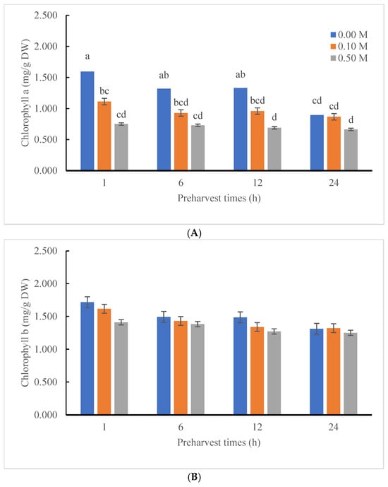
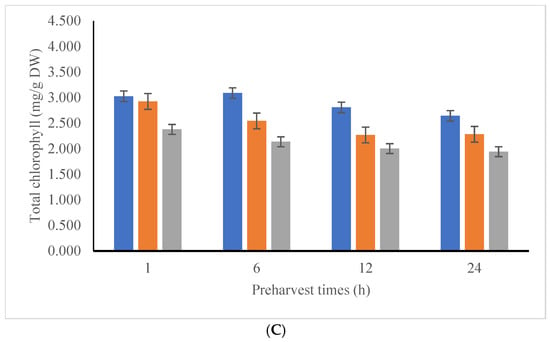
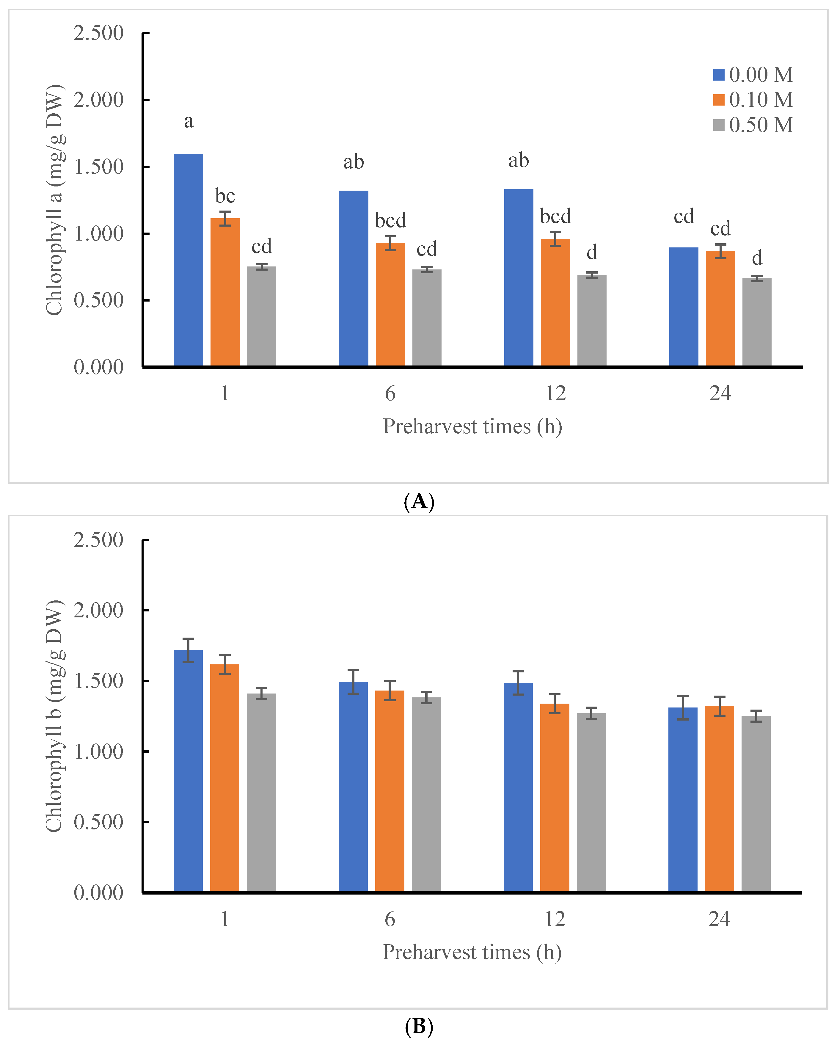
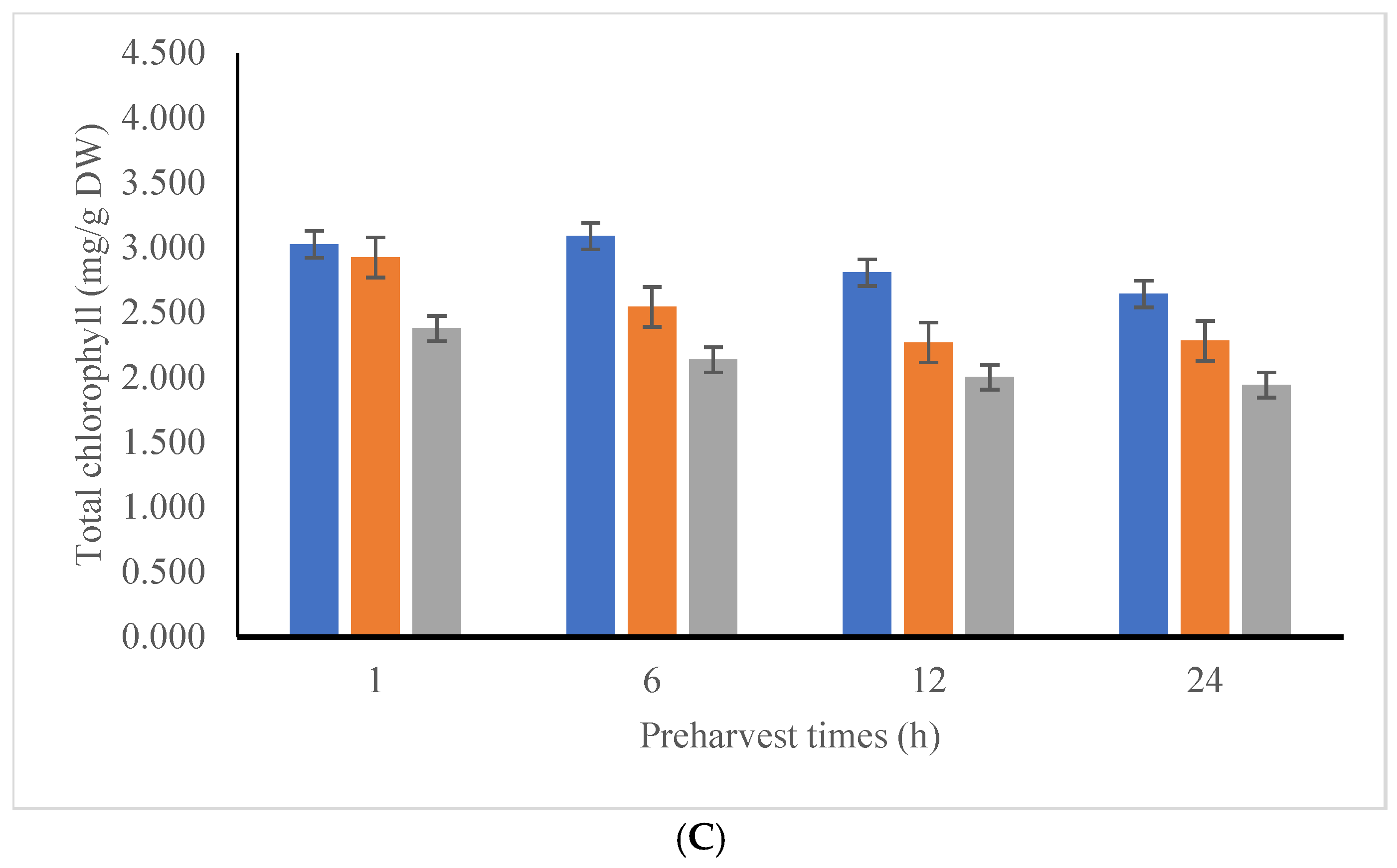
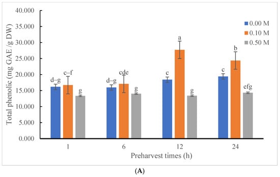
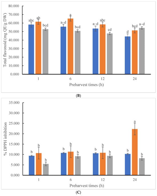
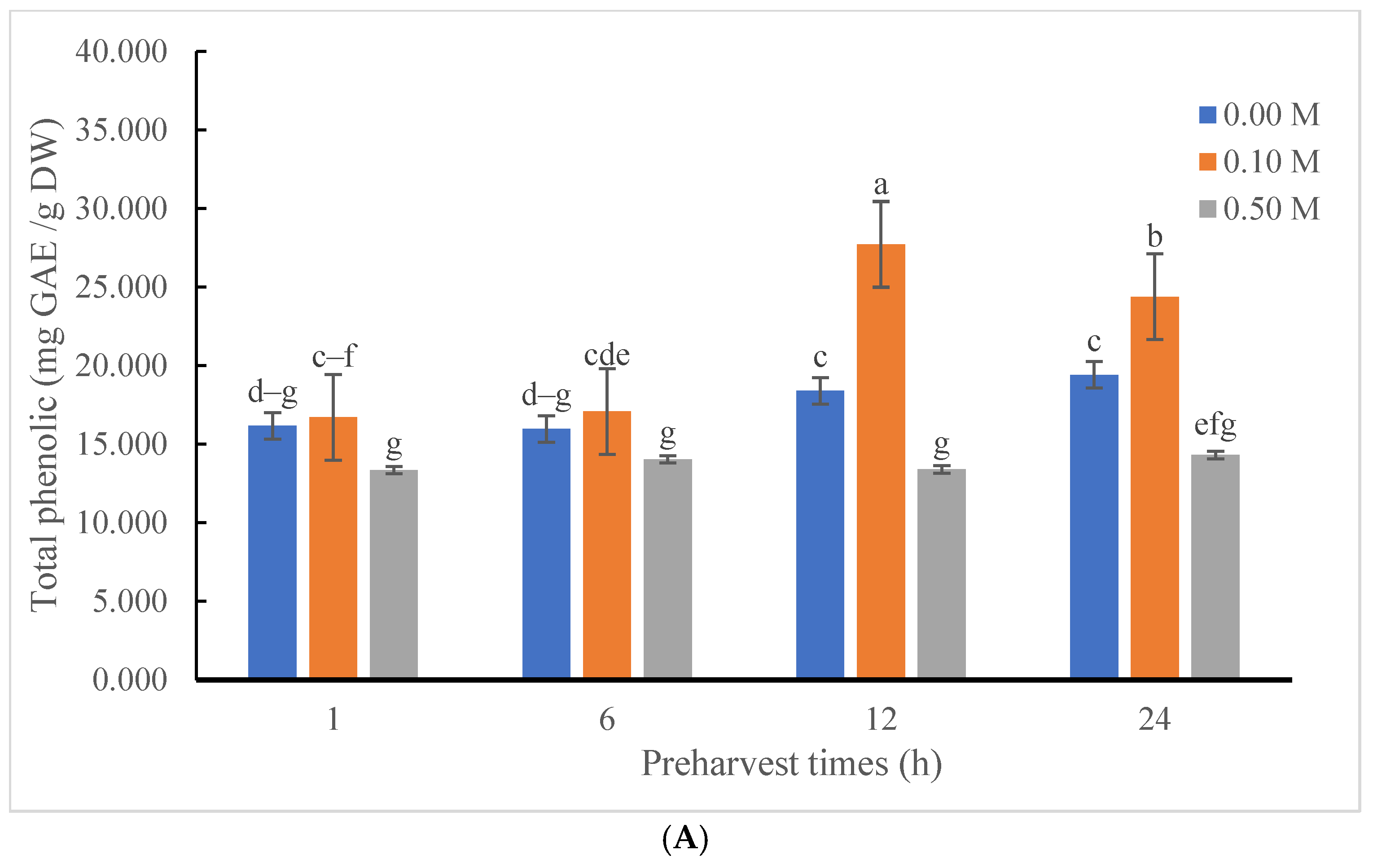
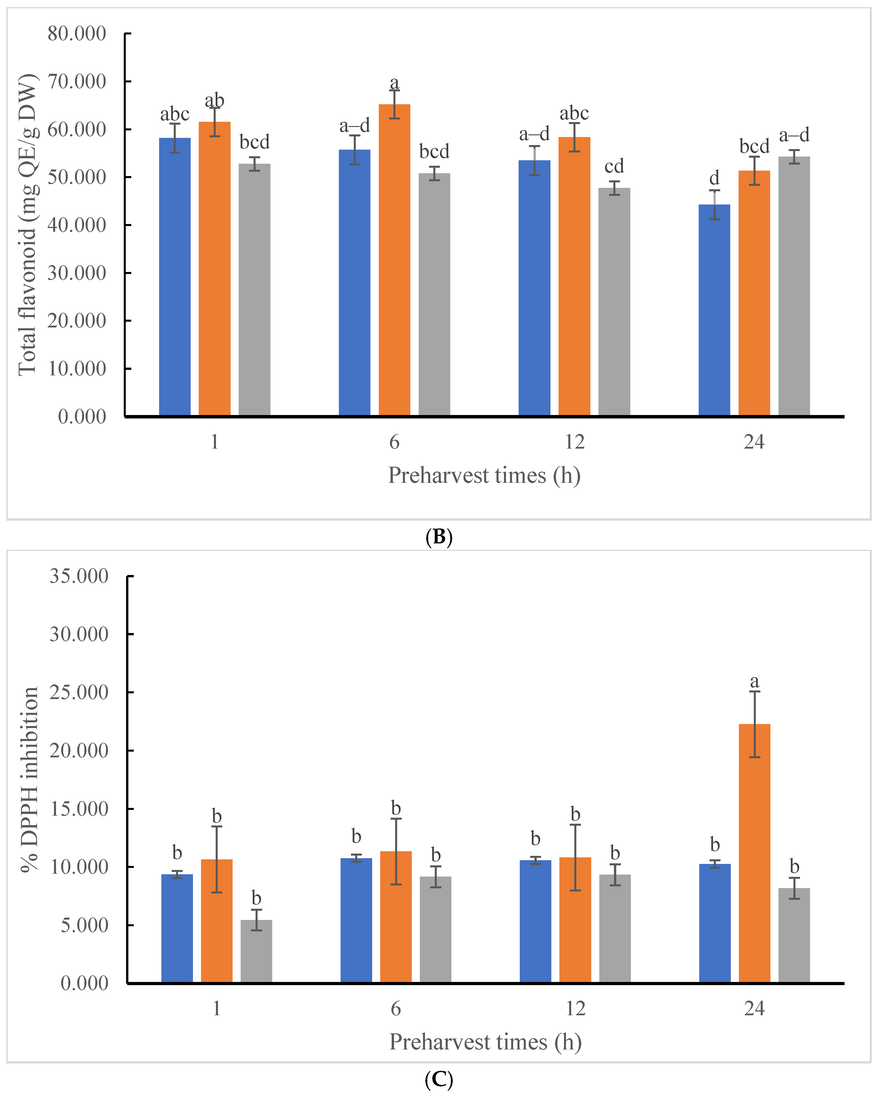
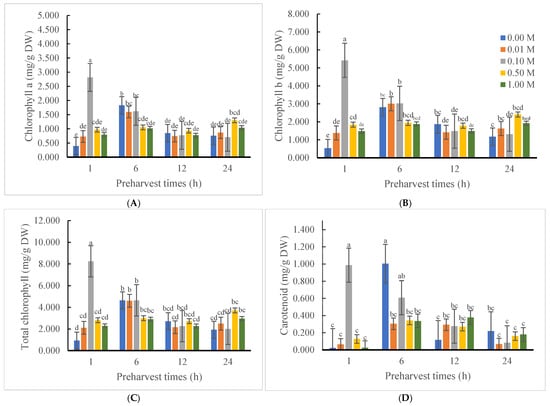
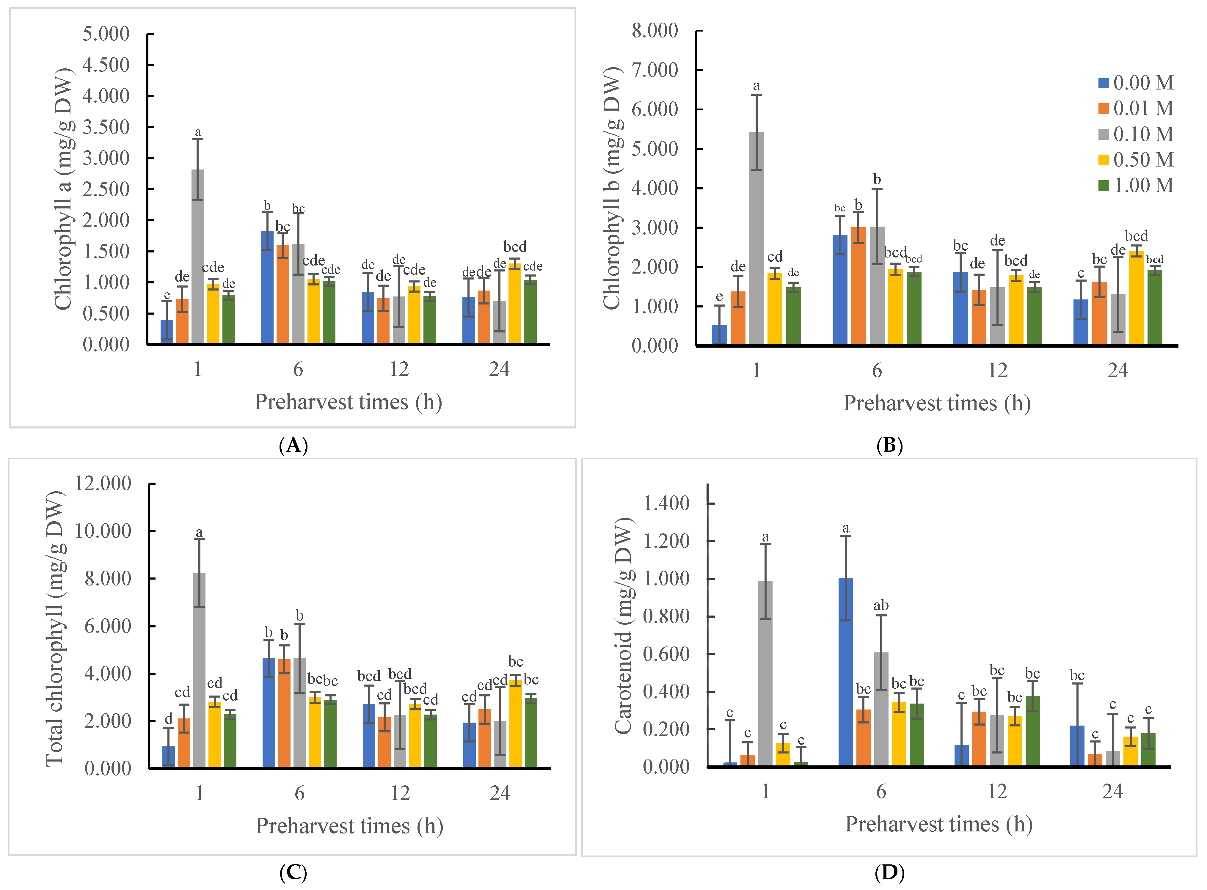
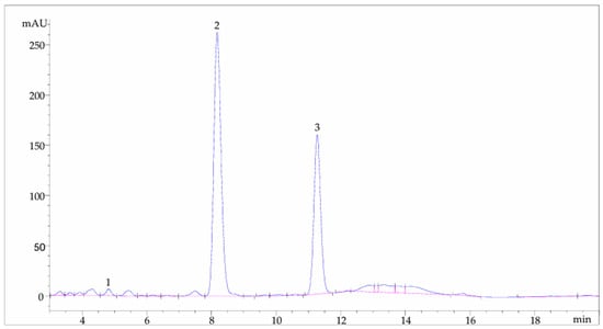
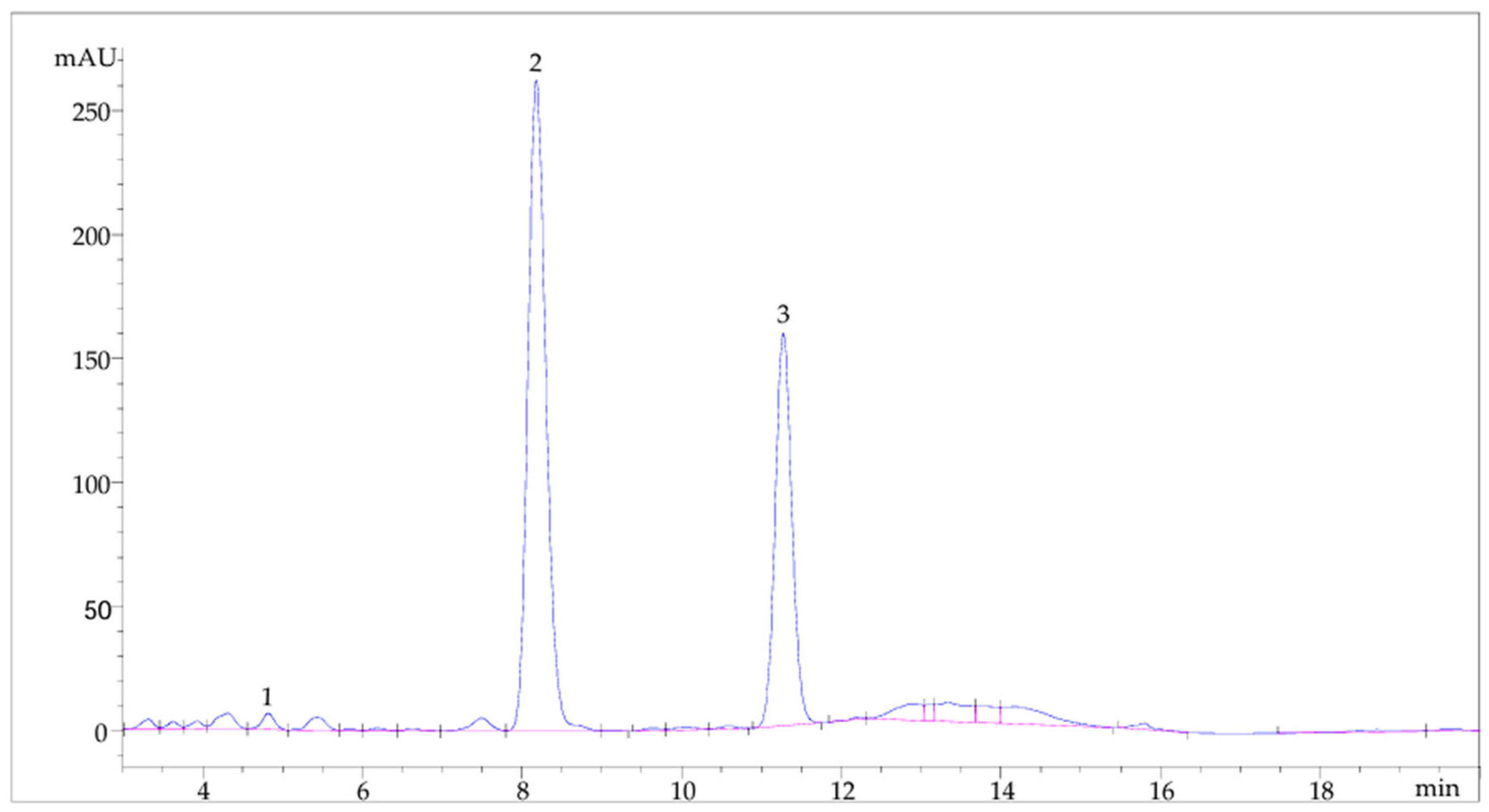

Disclaimer/Publisher’s Note: The statements, opinions and data contained in all publications are solely those of the individual author(s) and contributor(s) and not of MDPI and/or the editor(s). MDPI and/or the editor(s) disclaim responsibility for any injury to people or property resulting from any ideas, methods, instructions or products referred to in the content. |
© 2026 by the authors. Licensee MDPI, Basel, Switzerland. This article is an open access article distributed under the terms and conditions of the Creative Commons Attribution (CC BY) license.