


Abstract
‘Korlaxiangli’ pear occupies a pivotal position in the pear industry of Xinjiang, with both its cultivation area and total output ranking first in the region. However, ‘Korlaxiangli’ is vulnerable to freezing injury and has suffered frequent frost damage in recent years. A cold-tolerant superior individual plant was identified via preliminary field surveys and designated as cold-tolerant superior line 15-25-15 (abbreviated as ‘HY15’). To facilitate the large-scale application of this germplasm, tissue culture technology was adopted for its rapid propagation. Using spring-collected young shoots and autumn-collected dormant buds as explants, the optimal proliferation medium was determined to be DKW supplemented with 1.0 mg/L of 6-BA and 0.5 mg/L of NAA, while the optimal rooting medium was identified as 1/2 MS containing 2.5 mg/L of NAA and 15 g/L of sucrose. Transcriptome analysis revealed eight candidate genes potentially associated with shoot proliferation, among which ARF3 and ARR12 might be associated with high proliferation efficiency. This study provides a theoretical foundation and practical technical support for the conservation, genetic improvement, and establishment of efficient propagation systems of elite pear germplasm resources.
1. Introduction
Pear is one of the most important fruit tree species. ‘Korlaxiangli’ (Pyrus sinkiangensis) pear is the leading cultivated pear variety in Xinjiang. P. sinkiangensis has been reported to originate from the hybridization between Asian and European pear species, thus possessing both Asian and European pear lineages [1,2]. In recent years, the Bayingolin Mongol Autonomous Prefecture in Xinjiang has been consecutively hit by extreme low temperatures in winter, which have caused severe freezing injury to ‘Korlaxiangli’ pear trees, leading to weakened tree vigor, reduced yield, and deteriorated fruit quality [3]. During preliminary field surveys, we identified a freezing-tolerant elite individual plant of ‘Korlaxiangli’ pear in an orchard, which was designated as cold-tolerant line 15-25-15 (abbreviated as ‘HY15’). This line can remain free from freezing damage even in areas subjected to extreme low-temperature stress. In addition, it exhibits a certain level of resistance to canker disease, which is associated with tree vigor decline induced by freezing injury. Therefore, establishing an efficient propagation protocol for this promising cold-tolerant pear genotype provides the foundation for future cold tolerance validation studies.
Tissue culture refers to a biotechnology that involves inoculating explants (stems, leaves, etc.) onto artificial media containing carbon sources and plant growth regulators (phytohormones) [4]. Under controlled conditions of temperature, light, and humidity, these explants are cultured and eventually develop into complete plantlets [5]. Tissue culture is not restricted by seasonal changes or natural environmental conditions, enabling efficient and year-round propagation. It can rapidly produce large quantities of high-quality regenerated plantlets [6], thereby laying a solid foundation for the breeding of germplasm resources. In recent years, extensive research has been reported on the establishment of tissue culture systems for different pear varieties. A stable and efficient tissue culture and rapid propagation system was established for P. communis [7]. A tissue culture and rapid propagation system for ‘Yuluxiang’ pear was developed with optimal proliferation and rooting media [8]. Researchers have systematically established and optimized tissue culture proliferation systems by focusing on phytohormone combination regulation and culture condition optimization [9,10].
As reported in several studies, tissue culture approaches are commonly employed to improve cold tolerance in horticultural plants under controlled culture conditions [11,12]. Nevertheless, in woody plants, maintaining trait stability during propagation is a challenge that directly affects the reliability of clonal propagation and the success of breeding programs [13]. Unlike herbaceous species, woody plants often exhibit prolonged juvenile phases and complex genetic backgrounds, making them more susceptible to genetic and epigenetic changes under tissue culture conditions [14]. Somaclonal variation, which refers to spontaneous genetic, epigenetic, and phenotypic alterations in plants regenerated from tissue cultures, is particularly prevalent in woody species and can arise from factors such as explant type, culture duration, hormone concentrations, and repeated subculturing [15,16]. Although somaclonal variation may occasionally generate useful traits, it more frequently leads to undesirable characteristics such as altered growth habits, reduced vigor, abnormal morphology, and unstable yield performance. Consequently, minimizing somaclonal variation and ensuring genetic fidelity have become critical objectives in the micropropagation of forest trees, fruit trees, and ornamental woody species. Recent studies have focused on optimizing culture conditions and applying molecular markers to detect and monitor variation [17], providing new strategies to enhance the stability of woody plant materials in tissue culture.
Shoot proliferation is a core process in plant growth and development, which is synergistically regulated by multiple genes and signaling pathways. Extensive studies have been conducted on proliferation-related genes in model plants such as Arabidopsis thaliana and Solanum lycopersicum [18,19]. Genes belonging to the CDK (Cyclin-Dependent Kinase) family, such as CDKA;1 and CDKB1;1, act as key kinases that regulate the G1/S and G2/M phase transitions of the cell cycle, respectively. In contrast, cyclin genes including Cyclin D3;1 and CyclinA2;3 activate CDK activity through binding, thereby promoting cell cycle progression [20,21]. Meanwhile, genes of the KRP (Kip-Related Protein) family, such as KRP2 and SIM, exert negative regulatory effects to prevent excessive cell proliferation [22,23]. Genes involved in phytohormone signaling pathways play important mediating roles in this process. For instance, the auxin response factor ARF5 and the core cytokinin signaling component ARR1 maintain the proliferative capacity of root apical and shoot apical meristems by activating the expression of downstream cell-cycle-related genes [24]. Epigenetic regulatory genes and meristem maintenance genes are also indispensable. The negative feedback loop formed by WUS (WUSCHEL) and CLV3 (CLAVATA3) precisely balances the proliferation and differentiation of shoot apical stem cells, while the STM (SHOOT MERISTEMLESS) gene maintains proliferation potential by inhibiting cell differentiation [25]. Collectively, these genes ensure normal plant proliferation by regulating the rate of cell division and maintaining meristem activity.
To accelerate the proliferation of ‘HY15’, this study focuses on establishing a tissue culture and rapid propagation system for this line. Specifically, we aimed to address two main questions: (1) identifying the medium formulation that maximizes the proliferation of ‘HY15’ pear, and (2) determining the gene expression changes correlated with high proliferation capacity. By screening optimal proliferation and rooting media, we aimed to achieve rapid propagation of this germplasm, which has potential application value. Additionally, we sampled the basal parts of tissue-cultured stem segments with varying proliferation coefficients to analyze gene expression patterns, thereby identifying genes closely associated with shoot proliferation. The findings of this study are expected to provide theoretical guidance for future transgenic modification of pear cultivars.
2. Materials and Methods
2.1. Plant Materials
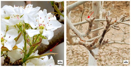
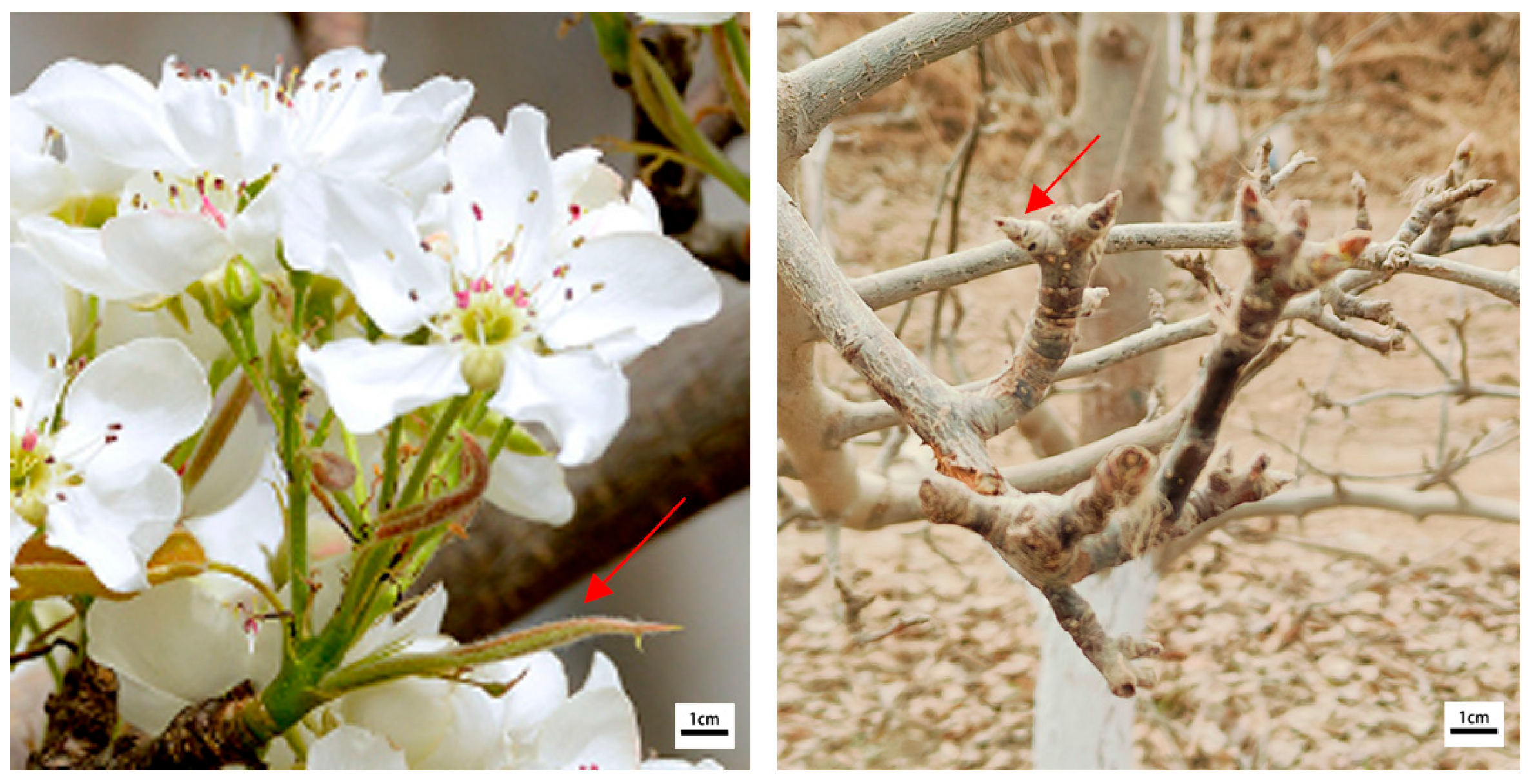
P. sinkiangensis ‘HY15’ was used as the experimental material. Plant samples were collected from the experimental base of the Institute of Agricultural Sciences, affiliated with the Second Division of Xinjiang Production and Construction Corps. Long-term continuous observation revealed that this germplasm exhibits strong freezing resistance. To facilitate year-round sampling, spring young shoots and autumn dormant buds were selected separately for propagation (Figure 1). In spring 2024, semi-lignified branches were collected, with all leaves removed. The branches were gently brushed along the direction of growth with a soft brush dipped in a liquid detergent solution, followed by rinsing under running tap water for 2 h. The branches were then cut into 2–3 cm segments, with one young shoot retained per segment. Similarly, for dormant buds on branches collected in autumn 2024, the surfaces were thoroughly scrubbed with a soft brush dipped in soapy water and then rinsed under running tap water for 3 h.
Figure 1.
The young shoots in spring and the dormant buds in autumn. Young shoots refer to the tender shoots emerging from mixed flower buds of pear trees. Dormant buds refer to swollen buds in the dormant period. The arrows indicate the sampling positions.
2.2. Screening of Sterilization Methods
To investigate the effects of different sterilization durations on tissue culture seedlings, we designed combinations of distinct sterilants and treatment times for the sterilization of young shoots and dormant buds. For young shoots, explants were first sterilized with 75% ethanol for 30 s, followed by sterilization with 0.1% mercuric chloride solution for 4, 6, and 8 min, respectively. Finally, they were rinsed three times with sterile water. Given the thick scale coverings of dormant buds, the sterilization duration was appropriately extended. Specifically, the dormant buds were first sterilized with 75% ethanol for 30 and 45 s, respectively, and then treated with 0.1% mercuric chloride solution for 6, 8, and 10 min, respectively, before being rinsed three times with sterile water as the final step. The explants were blotted dry to remove surface moisture prior to inoculation onto the initial culture medium. After 30 days of culture, the browning rate, contamination rate, and survival rate of the explants were assessed. Each treatment consisted of 3 biological replicates. Each replicate consisted of 10 explants.
2.3. Screening of Primary Culture Media and Subculture Media
Three media (MS, 1/2 MS, and DKW; produced by Coolaber, Beijing, China) were used, and their complete compositions are as follows: MS medium contains macronutrients (NH4NO3 1650, KNO3 1900, CaCl2·2H2O 440, MgSO4·7H2O 370, KH2PO4 170 mg·L−1), micronutrients (KI 0.83, H3BO3 6.2, MnSO4·4H2O 22.3, ZnSO4·7H2O 8.6, Na2MoO4·2H2O 0.25, CuSO4·5H2O 0.025, CoCl2·6H2O 0.025 mg·L−1), iron salt (Na2-EDTA 37.3, FeSO4·7H2O 27.8 mg·L−1), vitamins (Thiamine-HCl 0.1, Pyridoxine-HCl 0.5, Nicotinic acid 0.5, Glycine 2.0 mg·L−1), sucrose (30 g·L−1) and agar (8 g·L−1), with pH adjusted to 5.8; 1/2 MS medium is characterized by half-strength macronutrients of MS medium, while maintaining full-strength micronutrients, iron salt, vitamins, sucrose and agar (pH of 5.8); and DKW medium includes macronutrients (NH4NO3 1400, KNO3 1900, Ca(NO3)2·4H2O 1680, MgSO4·7H2O 370, KH2PO4 200, CaCl2·2H2O 96 mg·L−1), the same micronutrients and iron salt as MS medium, vitamins (Thiamine-HCl 10.0, Pyridoxine-HCl 1.0, Nicotinic acid 1.0, Myo-inositol 100.0 mg·L−1), sucrose (30 g·L−1) and agar (8 g·L−1), and its pH is adjusted to 5.6.
The primary culture medium was prepared using 1/2 MS, supplemented with different concentrations of 6-BA, IBA, and GA3, and the pH value of the medium was adjusted to 5.8. The surface-sterilized young shoots and dormant buds were inoculated onto the medium. Each treatment consisted of 3 biological replicates. Each replicate consisted of 10 explants. The culture conditions were set as follows: a temperature of (25 ± 1) °C, light intensity of 2000 lx, and photoperiod of 16 h light/8 h dark. After 30 days of culture, the germination status and browning rate of explants were investigated.
For subculture propagation, a three-factor and three-level orthogonal experiment was conducted. MS, 1/2 MS, and DKW were used as the basal media, supplemented with different concentrations of 6-BA and NAA. The tissue culture seedlings with a height of approximately 1–1.5 cm, which were obtained from the optimal medium, were selected for inoculation. Each treatment consisted of 3 biological replicates. Each replicate consisted of 15 explants. The culture conditions were maintained as follows: a temperature of (25 ± 1) °C, a light intensity of around 2000 lx, and a photoperiod of 16 h light/8 h dark. After 30 days, the proliferation coefficient, seedling height, and growth status of the tissue culture seedlings were investigated and statistically analyzed.
2.4. Screening of Rooting Culture Media
A three-factor and three-level orthogonal experiment was conducted using 1/2 MS as the basal medium, supplemented with different concentrations of sucrose, NAA, and a two-step rooting protocol. Tissue culture seedlings with normal growth and a height of 3 cm, selected from subculture, were inoculated onto the medium. Each treatment consisted of 3 biological replicates. Each replicate consisted of 10 explants. The culture conditions were set as follows: a temperature of (25 ± 1) °C, light intensity of approximately 2000 lx, and photoperiod of 16 h light/8 h dark. After 30 days, the rooting rate and the number of roots per seedling were investigated and recorded.
2.5. Acclimatization and Transplantation
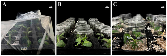
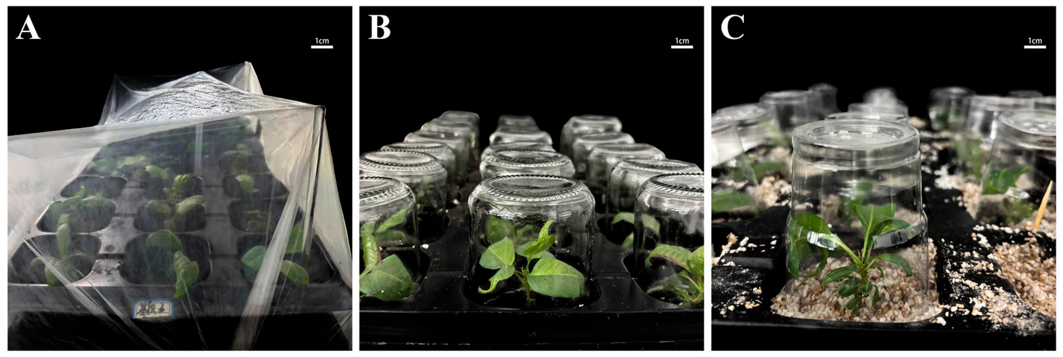
Tissue culture seedlings with robust growth and a root length of 2–3 cm were selected for acclimatization. Seedling hardening and acclimatization were carried out in a greenhouse constructed by Jiamu Horticulture Co., Ltd. (Tiemenguan, China) and the Agricultural Science Research Institute, 2nd Division, XPCC. The greenhouse was equipped with a real-time monitoring system that automatically activated humidification and heating when values fell below the set range, maintaining a temperature of 20–25 °C and a relative humidity of 40–60%. The seedlings were transferred from the tissue culture room to the greenhouse and acclimatized via the following procedure: they were first placed undisturbed for 3 days and then cultured with the bottle caps half-opened (with approximately 1/2 of the bottle mouth exposed) for another 3 days or 5 days, with 15 bottles assigned to each treatment. Data were recorded during the acclimatization period to analyze the effects of different acclimatization methods on the transplant survival rate of ‘HY15’ tissue culture seedlings. Subsequently, the vigorously growing seedlings were transplanted into a substrate consisting of perlite and humus soil at a ratio of 1:3, and different mulching materials were applied for moisture retention. Three treatments were designed as follows: (1) covering with plastic film; (2) covering with glass tissue culture bottles for 7 days, followed by perforated plastic cups for another 7 days; (3) covering with glass tissue culture bottles for 14 days, followed by perforated plastic cups for 7 days. Thirty seedlings were used for each treatment. After 30 days of transplanting, relevant data were recorded, plant growth status was observed, and the transplant survival rate and seedling growth vigor were statistically analyzed. Each treatment consisted of 3 biological replicates. Each replicate consisted of 10 explants.
2.6. Transcriptome Analysis of Shoot Proliferation
During the propagation culture period, two medium formulations with significantly different proliferation coefficients were selected. The total RNA of the basal tissue of stem segments cultured on these two media (E4 and E9) was extracted using the CTAB method. These two media not only differ in proliferation coefficient induction but also represent two distinct culture media (MS and DKW media). There were three biological replicates. Each replicate consisted of 6 basal parts of stem segments. The purity and concentration of RNA were evaluated with a NanoDrop 2000 spectrophotometer (Wilmington, DE, USA). RIN values were all greater than 6.5, and 260/280 ratios ranged from 1.94 to 2.08. cDNA libraries were constructed using the NEBNext Ultra RNA Library Prep Kit (Ipswich, MA, USA). After library construction, Qubit 3.0 Fluorometer was first used for preliminary quantification, with the required concentration reaching ≥1 ng·μL−1. Subsequently, a high-throughput analysis system was employed to detect the insert fragments of the library. Once the insert fragments were consistent with the expected size, quantitative real-time PCR (qPCR) was performed for accurate quantification of the effective library concentration (required effective concentration > 2 nM). Following successful library quality control, paired-end 150 bp (PE150) sequencing was carried out on the Illumina HiSeq™ 4000 Platform (San Diego, CA, USA). Raw sequence data were filtered to remove low-quality reads. The filtered reads were mapped to the pear reference genome (NCBI, GCF_000315295.1) [26] using Hisat2 software (2.2.1), and the gene expression levels were calculated by the fragments per kilobase of transcript per million mapped reads (FPKM) method with RSEM software (1.3.3). Raw counts were analyzed using DESeq2 software (1.46) based on the negative binomial distribution model. Genes/transcripts with differentially expression between groups were screened under the criteria of False Discovery Rate ≤ 0.05 and |Log2FoldChange| ≥ 1. The different expressed genes were annotated by KEGG pathways [27].
2.7. Quantitative Real-Time PCR
Quantitative real-time PCR (qRT-PCR) was performed to verify the gene expression levels. cDNA synthesis was used Takara PrimeScript™ RT Kit (Kusatsu, Shiga, Japan) to eliminate genomic DNA. qRT-PCR was performed with Takara TB Green® Premix (Kusatsu, Shiga, Japan) (20 μL reaction system: 10 μL of Premix, 0.8 μL of each primer, 2 μL of cDNA, 6.4 μL of nuclease-free water) with 40 cycles (95 °C 5 s, 60 °C 30 s) and dissociation curve analysis. Actin and UBI were selected as the reference genes. Twenty genes were selected to qRT-PCR.
2.8. Data Analysis
All data were analyzed using SPSS software (29.0.2). When the number of explants was 30 or 45, they were grouped into experimental replicates with 10 or 15 explants per replicate, respectively, for data collection and statistical analysis. Statistical analysis was performed using one-way analysis of variance (ANOVA) followed by Duncan’s new multiple-range test to examine significant differences in the measured values among different groups. Percentage data (germination rate, browning rate, rooting rate) were subjected to arcsine square root transformation to meet ANOVA assumptions of normality and homoscedasticity, while count data (proliferation coefficient and root number) were subjected to square root transformation prior to analysis. All statistical calculations were conducted with a significance level set at α = 0.05. Data are presented as the means ± standard deviations of three replicates in the tables.
3. Results
3.1. Sterilization Treatment
To obtain axenic seedlings, the effects of different sterilization methods on young shoots and dormant buds were investigated (Figure 1). All explants were first sterilized with 75% ethanol for 30 s, followed by treatment with mercuric chloride. Significant differences in the survival rate of P. sinkiangensis ‘HY15’ young shoots and dormant buds were observed among different sterilization durations (Table 1). In the young shoot groups (A1–A3), with increasing mercuric chloride sterilization time, the contamination rate showed a significant downward trend, decreasing from 60.0% to 3.3%. In contrast, both the survival rate and browning rate increased significantly, with the survival rate rising from 40.0% to 86.7% and the browning rate increasing from 0% to 10%. When the sterilization duration was 8 min, the survival rate of young shoots reached 86.7%, accompanied by relatively low levels of browning and contamination rate. In the dormant bud groups (B1–B3), the browning rate of explants showed an increasing trend while the contamination rate exhibited a decreasing trend with prolonged sterilization time. At a sterilization duration of 8 min, the contamination rate of explants was 40.0% and the survival rate was 60.0%. Comprehensive analysis indicated that the optimal sterilization method for both young shoots and dormant buds of ‘HY15’ was 75% ethanol sterilization for 30 s followed by 0.1% mercuric chloride treatment for 8 min.
Table 1.
Effects of different sterilization treatments on explants of ‘HY15’.
3.2. Primary Culture
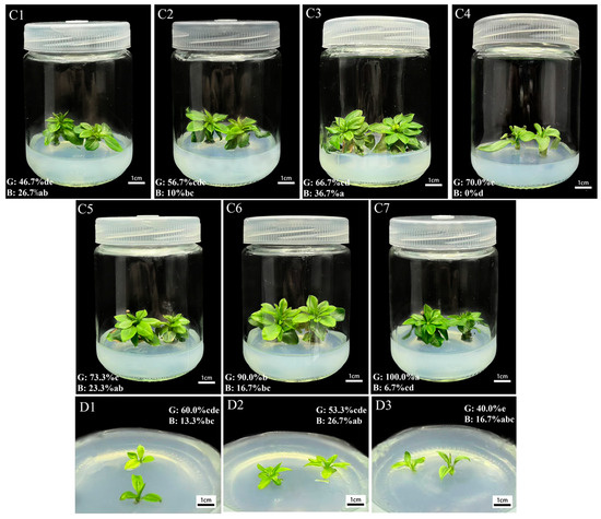
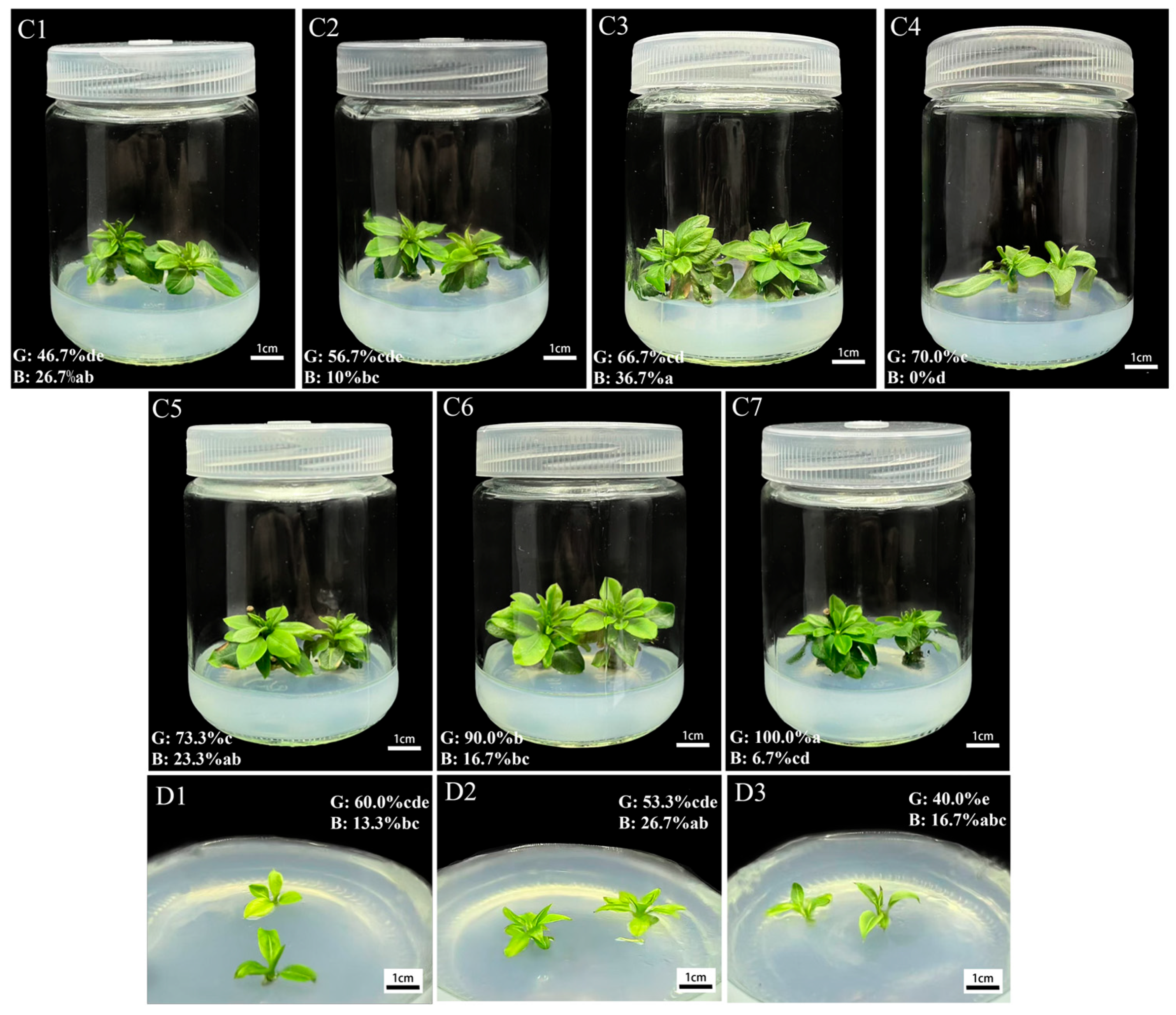
During primary culture, significant differences were observed in the germination rate and browning rate of young shoots and dormant buds of ‘HY15’ among different hormone combinations based on 1/2 MS medium (Table 2). For the young shoot groups (C1–C4) without GA3 supplementation, the germination rate showed a significant upward trend with increasing concentrations of 6-BA and IBA, gradually rising from 46.7% to 70.0%. In the young shoot groups (C5–C7), the germination rate also increased significantly as the concentration of GA3 increased. When the concentration of GA3 reached 0.5 mg/L, the germination rate peaked at 100%, with the browning rate as low as 6.7%. For the dormant bud groups (D1–D3), with increasing concentrations of 6-BA, IBA and GA3, the germination rate first increased and then decreased, while the browning rate showed a continuous downward trend. Treatment D1 exhibited a high germination rate and a low browning rate, but its sprouting speed was relatively slow. Treatment D2 achieved a germination rate of 53.3%, with relatively rapid sprouting, tender green leaves, and robust seedling growth (Figure 2). Treatment D3 resulted in weak growth vigor of the explants. Comprehensive analysis indicated that the optimal induction medium for young shoots of ‘HY15’ was the C7 medium, whereas that for dormant buds was the D2 medium.
Table 2.
Effects of different hormone combinations on primary culture of ‘HY15’.
Figure 2.
Growth performance of young shoots and dormant buds in primary culture with different medium formulations. Different lowercase letters in the lower left corner indicate significant differences (p < 0.05, one-way ANOVA followed by Duncan’s test), where G and B represent germination rate and browning rate, respectively. The labels in the upper left corner correspond to the treatments in Table 2 one-to-one.
3.3. Selection of Subculture Medium
Nine medium formulations were used for the subculture of P. sinkiangensis ‘HY15’. The results showed that all the subculture media could induce the proliferation of ‘HY15’ tissue culture seedlings (Table 3), but significant differences were observed in the proliferation coefficients.
Table 3.
Effects of different medium formulations on subculture proliferation of ‘HY15’.
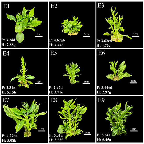
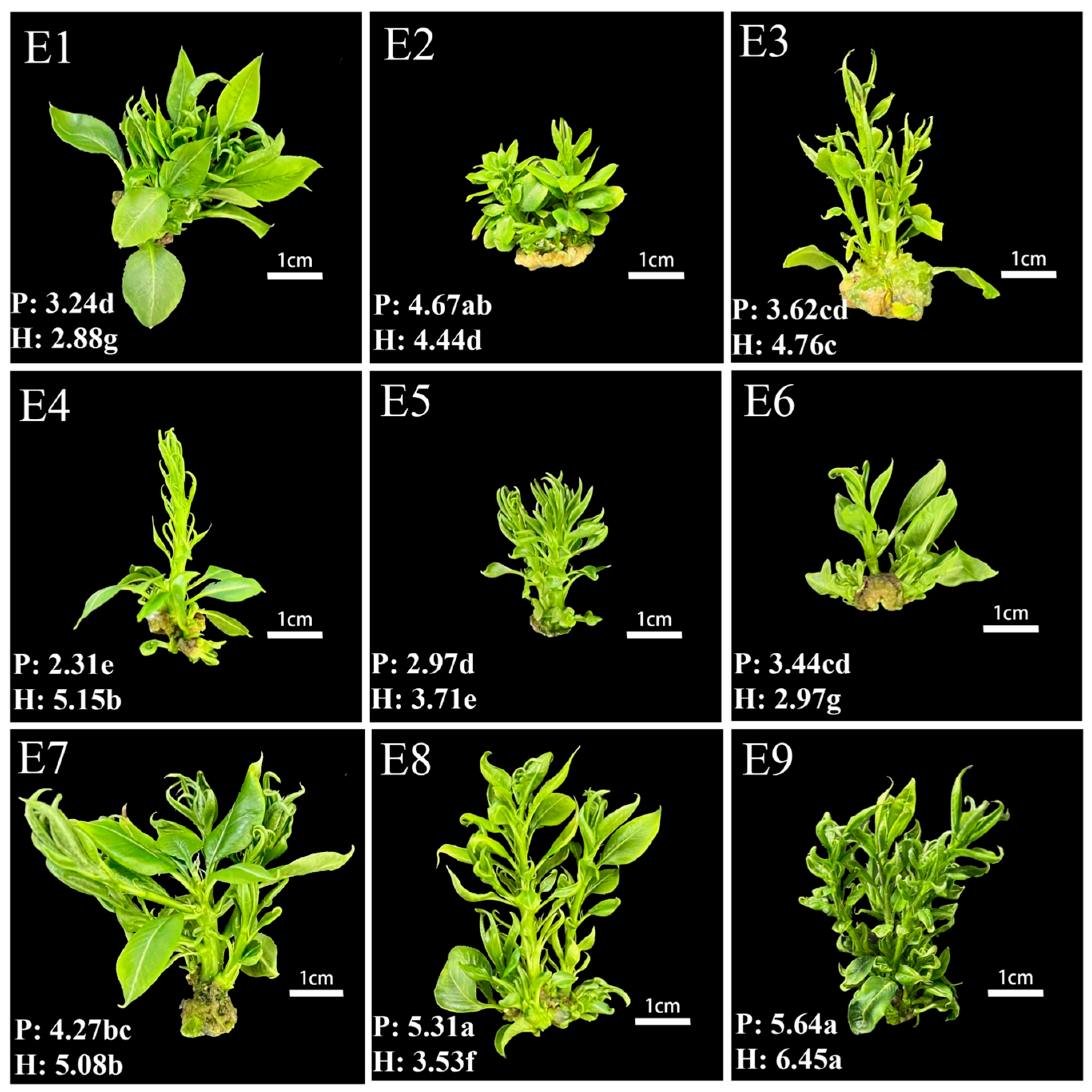
Among the three basal medium types (1/2 MS, MS, and DKW), the DKW medium yielded the highest proliferation coefficient, followed by the 1/2 MS medium, and the MS medium yielded the lowest. The proliferation coefficient of the DKW medium ranged from 4.27 to 5.64, with the plantlets exhibiting robust growth. Specifically, the E9 formulation achieved the highest proliferation coefficient of 5.64, accompanied by large leaf areas, thick and long stem segments, and the highest average seedling height (Figure 3). Although the E8 formulation contained no NAA, there was no significant difference in the proliferation coefficient between the E8 and E9 treatments. However, the seedling height in the E8 treatment was significantly lower than that in the E9 treatment. These results indicated that in the DKW medium, NAA concentration exerted little effect on the explant proliferation coefficient but could promote an increase in seedling height. The E4 formulation had the lowest proliferation coefficient (only 2.31), suggesting weak regeneration and proliferation ability of plantlets under this formulation. In general, the MS medium showed relatively low proliferation coefficients across all treatments, indicating that it was not suitable as a basal medium for the subculture of ‘HY15’.
Figure 3.
Growth performance of subculture proliferation with different medium formulations. Different lowercase letters in the lower left corner indicate significant differences (p < 0.05, one-way ANOVA followed by Duncan’s test), where P and H represent proliferation coefficient and average seedling height, respectively. The labels in the upper left corner correspond to the treatments in Table 3 one-to-one.
3.4. Rooting Response of ‘HY15’ to Different Hormone Combinations
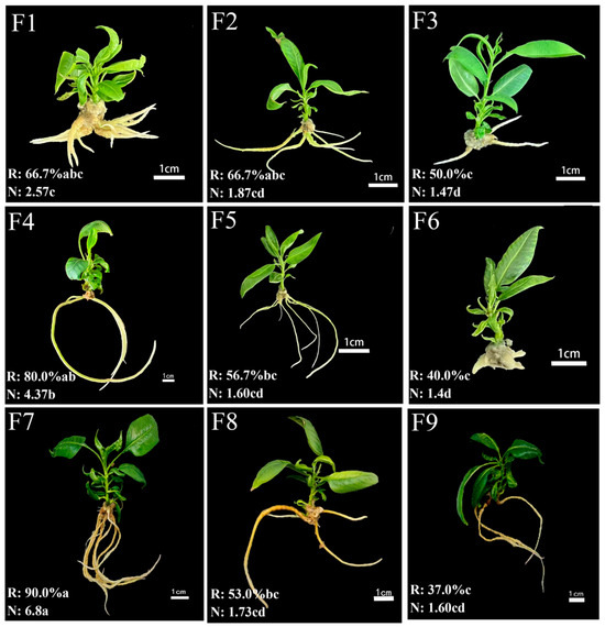
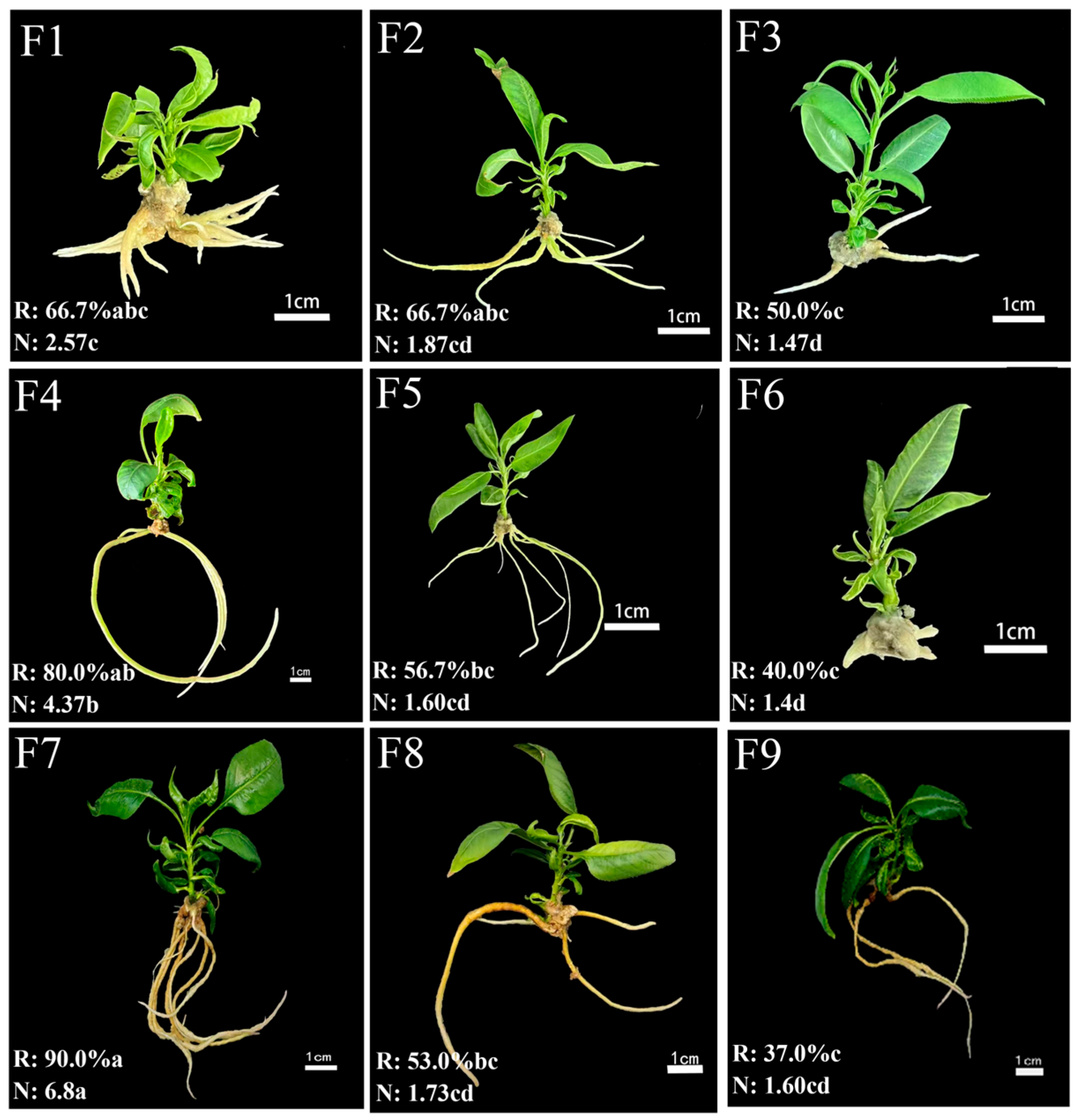
All tested rooting medium formulations were capable of inducing rooting in ‘HY15’ tissue culture seedlings (Table 4), but significant differences in rooting efficacy were observed among different formulations. The rooting rate of the two-step method was significantly higher than that of the one-step method, indicating that NAA stimulation was required in the early stage of rooting. However, after the formation of root primordia, a hormone-free medium was more conducive to the subsequent growth of roots. With other factors kept constant, the rooting rate increased significantly with the rise in NAA concentration in the first-step medium, which indicated that a moderate increase in NAA concentration was conducive to rooting. In addition to NAA, sucrose also exerts an effect on rooting. When the NAA concentration was 2.0 mg/L, increasing the sucrose concentration from 15 g/L to 20 g/L led to a decrease in the rooting rate from 80.0% to 56.7%. When the NAA concentration was 2.5 mg/L, elevating the sucrose concentration from 15 g/L to 20 g/L reduced the rooting rate from 53.0% to 37.0%. These results demonstrated that 15 g/L sucrose was the optimal concentration for promoting the rooting rate. High sucrose concentration (30 g/L) exerted a significant inhibitory effect on the rooting rate. The F7 formulation achieved the highest rooting rate and average number of roots, which were significantly higher than those of other formulations. The rooting rate reached 90.0%, with an average root number of 6.80, and the root system was thick and long (Figure 4). These findings indicated that F7 was the optimal formulation for rooting.
Table 4.
Effects of different hormone combinations on rooting of ‘HY15’.
Figure 4.
Effects of different medium formulations on rooting. Different lowercase letters in the lower left corner indicate significant differences (p < 0.05, one-way ANOVA followed by Duncan’s test), where R and N represent the rooting rate and number of roots, respectively. The labels in the upper left corner correspond to the treatments in Table 4 one-to-one.
3.5. Effects of Different Acclimatization Methods on Survival Rate of ‘HY15’
Significant differences in the survival rate of ‘HY15’ tissue culture seedlings were observed with different acclimatization durations and covering materials (Table 5). First, the culture bottle caps were opened halfway to acclimatize the seedlings to the low-humidity external environment. The survival rate of the G1 treatment (half-cap opening for 3 days) reached 80.0%, whereas with the extension of acclimatization time, the survival rate of the G2 treatment (half-cap opening for 5 days) dropped to only 60.0%. The G1 treatment not only achieved the highest survival rate but also resulted in tender green leaves and vigorous seedling growth. Therefore, half-cap opening for 3 days was deemed the suitable pre-transplantation treatment duration for ‘HY15’. Subsequently, the seedlings were transplanted into the culture soil. Different covering materials also exerted significant effects on the survival rate of ‘HY15’ tissue culture seedlings. Among these treatments, the H2 treatment performed the best, followed by the H3 and H1 treatments. The H2 treatment involved covering the seedlings with glass culture bottles for 7 days, followed by recovering them with perforated plastic cups for another 7 days. This treatment achieved a survival rate of 80.0%, accompanied by tender green leaves and healthy growth. In contrast, the H1 treatment adopted plastic film covering, which led to excessively high humidity in the transplanting environment and thus promoted mold growth. Consequently, the H1 treatment showed the lowest survival rate of 40.0%, with the seedlings exhibiting slight wilting (Figure 5). Although the H3 treatment extended the covering duration with glass bottles, it only achieved a survival rate of 43.3% and caused seedling wilting. Based on the above results, the combination of the G1 pre-treatment and H2 post-treatment could significantly improve the transplant survival rate, with the cumulative survival rate reaching 64%. The propagated HY15 plants are still in the seedling stage, so we have not yet conducted cold tolerance tests. These tests will be performed once the seedlings have matured.
Table 5.
Effects of different acclimatization treatments on survival rate of ‘HY15’.
Figure 5.
Diagram of different covering materials. (A): Plastic film covering; (B): glass bottle covering; (C): perforated plastic cup covering. Note: This is a schematic illustration only and is not intended for statistical comparison.
3.6. Transcriptome Analysis of the Basal Part of ‘HY15’ Stem Segments
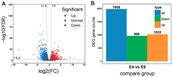
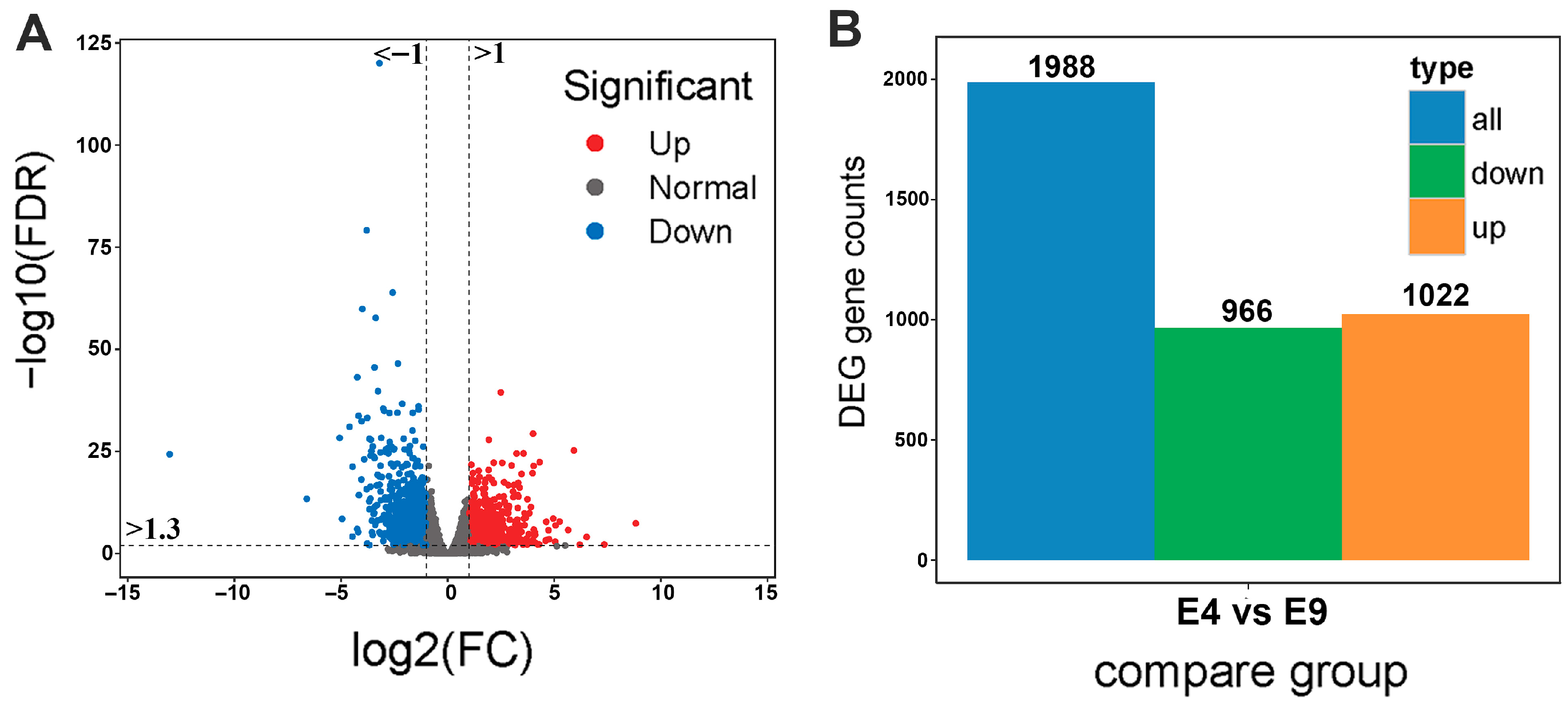
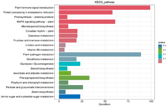
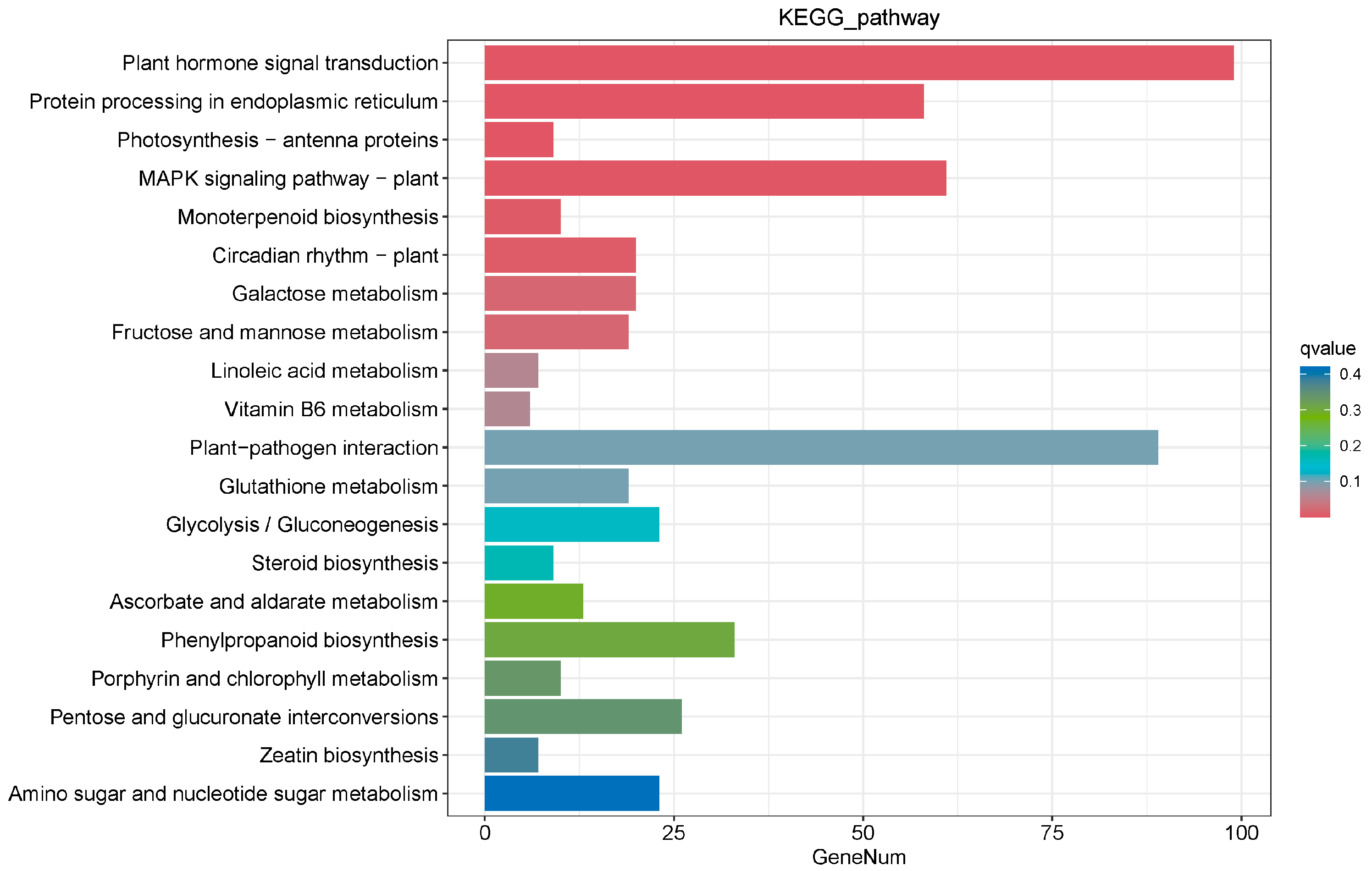
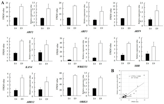
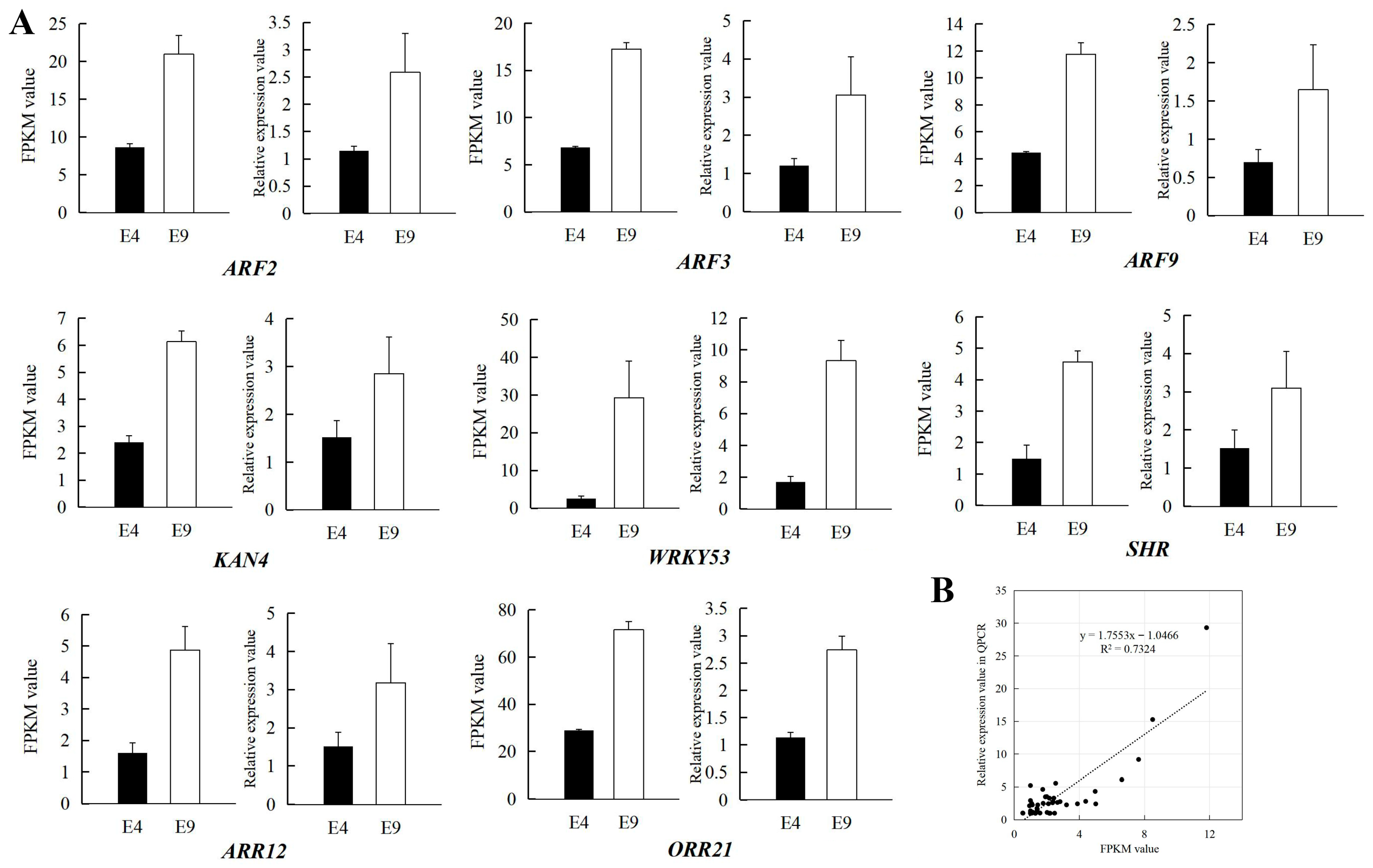
The core of rapid propagation of ‘HY15’ lies in improving the proliferation coefficient. To further investigate the genes associated with the proliferation coefficient of ‘HY15’, transcriptome sequencing was performed on the basal stem segments with different proliferation coefficients cultured on E4 and E9 media. The results showed that a total of 1988 differentially expressed genes (DEGs) were identified (Figure 6A), among which 1022 were up-regulated genes and 966 were down-regulated genes (Figure 6B). KEGG enrichment analysis revealed that 13.52% of the genes were mapped to the plant hormone signal transduction pathway (Figure 7), followed by the plant–pathogen interaction pathway at 12.16% and then the MAPK (mitogen-activated protein kinase) signaling pathway, endoplasmic reticulum protein processing pathway, and starch and sucrose metabolism pathway. The MAPK pathway is an important signaling pathway in plants. Eighty-nine DEGs were enriched in this pathway, and their activations may be related to hormone synthesis and signal transduction during tissue culture seedling proliferation. Eight candidate genes potentially related to proliferation were selected from these metabolic pathways, namely auxin response factor 2 (ARF2), auxin response factor 3 (ARF3), auxin response factor 9 (ARF9), transcription factor KAN4, transcription factor WRKY53, protein SHORT-ROOT (SHR), two-component response regulator ARR12, and two-component response regulator ORR21 (Figure 8A). These genes are closely associated with plant growth and cell division, including three auxin response genes, two transcription factors, one gene related to bud and root development, and the remaining two genes closely linked to cell division. ARR12 and ORR21 showed differential expression correlated with high proliferation rates. Quantitative real-time PCR (qRT-PCR) validation confirmed that the expression levels of twenty genes were consistent with the FPKM values derived from the transcriptome sequencing data (Figure 8A, Figure S1). The coefficient of determination (R2) reached 0.73 (Figure 8B).
Figure 6.
Mining of differentially expressed genes (DEGs) in basal stem segment tissues under E4 and E9 medium formulations. (A): Volcano plot of DEGs, log2(FC) > 1 or <−1, −log10(FDR) > 1.3; (B): number of DEGs.
Figure 7.
KEGG annotation of DEGs. The q-value is the p-value corrected for multiple-hypothesis testing. A smaller q-value indicates a more reliable significance of the enrichment of differentially expressed genes (DEGs) in the corresponding pathway.
Figure 8.
Expression profiles of eight genes related to shoot proliferation. (A): The expression of eight genes in the transcriptome and qPCR. For each gene, the left panel shows the FPKM values from transcriptome sequencing, while the right panel shows the validation results from qPCR. Error bars represent standard error of three biological replicates. (B): Coefficient analysis between the gene expression from the transcriptome and qPCR.
4. Discussion
As an interspecific hybrid of Asian and European pears, P. sinkiangensis ‘Korlaxiangli’ pear inherits the characteristic aromatic flavor from its European pear ancestors, while retaining the crisp-textured flesh trait derived from Asian pear lineages [2]. ‘Korlaxiangli’ pear is highly favored by consumers, and its cold-resistant germplasm ‘HY15’ will become a valuable germplasm resource. In the breeding and promotion of this pear variety, the tissue culture system provides a path for standardized breeding and the utilization of high-quality germplasm resources. Explant sterilization is the first step in plant tissue culture technology, which eliminates bacteria and fungi on the explant surface and ensures the survival rate of tissue-cultured plantlets [28]. In practical application, it is necessary to consider explant type and sampling time, as well as the type, concentration and sterilization duration of the disinfectant used [29]. To enable sampling across multiple seasons without delaying the experimental process, the present study selected young shoots in spring and dormant buds in autumn as test materials. The contamination rate of young shoots was significantly lower than that of dormant buds, which was consistent with previous research results [30]. Selecting newly sprouted shoots in early spring as explants could minimize the contamination rate of tissue culture and maximize the probability of success.
Common basic media used in pear tissue propagation mainly include MS, 1/2 MS, NN69, WPM, B5 and QL [31]. MS medium is one of the most widely utilized basal media in plant tissue culture due to its high salt concentration, which supports robust plant growth across many species [32]. It has been frequently employed in various plant tissue culture applications, including pear [33]. In European pears (such as P. commnuis), MS, B5, DKW and WPM media are mostly used in pear tissue culture [7,34,35]. In Asian pears (such as P. pyrifolia), MS medium is mostly used [36,37]. Owing to divergent genetic backgrounds among different pear species, the optimal proliferation media for the P. sinkiangensis may differ accordingly. In the present study, a systematic comparison was conducted on the subculture proliferation of ‘HY15’ using different basal media (1/2 MS, MS, and DKW). The results showed that the optimal proliferation medium was DKW, with a high proliferation coefficient in P. sinkiangensis. DKW medium is one commonly used medium for the tissue culture of woody plants, and its high nitrogen and potassium ratios are well suited to the growth requirements of woody plants [38]. Its high nitrogen content likely supports protein synthesis required for rapid cell division, while its potassium concentration enhances membrane stability, reducing vitrification commonly observed in pear tissue culture. In addition to basal media, an appropriate hormone ratio also exerts a significant effect on proliferation [39]. Commonly used cytokinins in the tissue culture of plants include 6-BA and BA, while common auxins are IBA, IAA and NAA, and GA3 is the most frequently used gibberellin [40]. In the present study, a certain amount of GA3 was added to the initial culture media for young shoots and dormant buds, resulting in tissue-cultured plantlets with multiple stem branches and robust growth performance. This indicated that the supplementation of an appropriate concentration of gibberellin during the initial culture stage was conducive to the sprouting of buds in tissue culture [41]. In addition, the culture effect was unsatisfactory when only cytokinins but no auxins were added to the proliferation medium. For the screening of subculture proliferation media for ‘HY15’, a combination of 6-BA and NAA was adopted in this study to promote explant sprouting. The optimal growth status was achieved at a 6-BA concentration of 1.0 mg/L and an NAA concentration of 0.5 mg/L, with the pear plantlets producing abundant clustered buds and the proliferation coefficient reaching 5.64. High proliferation efficiency in this study not only facilitates the conservation of ‘HY15’ but also contributes to further enhancement of cold tolerance traits in tissue-cultured plantlets via chilling stress treatment.
Rooting culture generally requires reducing the nutrient concentration of the medium to induce the formation of root primordia and obtain rooted plantlets. Low-salt media such as 1/2 MS are thus the preferred choice in European and Asian pears [42], which we also selected in this study. The optimal sucrose concentration suitable for rooting of ‘HY15’ was screened out as 15 g/L, while high sucrose concentrations were found to be unfavorable for rooting [43]. Common hormones used for rooting in tissue culture include NAA, IBA and IAA, and the application of either one or a combination of two or more of these hormones can promote rooting [44]. P. communis is sensitive to IBA, with a low concentration of 0.2–0.5 mg/L commonly used in rooting media; P. pyrifolia usually requires a combination of IBA and NAA, with a higher total auxin concentration of up to 2 mg/L [9]. This study found that the sole application of NAA (2.5 mg/L) was sufficient to promote rooting of ‘HY15’, with the rooting rate reaching as high as 90.0%. In addition, we found that the two-step rooting method was superior to the one-step rooting method. The addition of auxins could promote the formation of root primordia in the early stage but might inhibit the continuous growth of roots in the later stage. Transferring the plantlets to an auxin-free medium after the observation of root primordia formation was more conducive to root system development.
Tissue culture shoot proliferation is a complex physiological process tightly regulated by hormone signaling pathways and transcription networks [45]. Genes including auxin response factors (ARFs), transcription factor KAN4, transcription factor WRKY53, protein SHORT-ROOT (SHR), and two-component response regulators (ARR12, ORR21) play crucial and interactive roles in shoot proliferation of ‘HY15’. ARF2, ARF3, and ARF9, as core mediators of auxin signaling, exert distinct regulatory effects on shoot proliferation [46]. ARF3 is reported to participate in meristem maintenance by modulating the expression of stem cell-related genes [47], while ARF2 and ARF9 may function as negative regulators to balance auxin responses [48,49]. Their appropriate expression levels are essential to avoid over-suppression of shoot regeneration. ARR12, a positive regulator of cytokinin signaling, promotes cell division in shoot meristems [50], whereas ORR21 may antagonize ARR12 activity to prevent excessive shoot branching, highlighting the importance of cytokinin signal homeostasis [51]. ARF3 can modulate cytokinin biosynthesis and signaling [47]. Cytokinin and auxin signaling regulate shoot meristem development through cross-talk [52], and ARF3 and ARR12 might be associated with shoot proliferation in this study. Transcription factors KAN4 and WRKY53 contribute to shoot proliferation through regulating meristem development and stress responses, respectively. KAN4 maintains the boundary of shoot meristems to ensure ordered shoot initiation [53]. WRKY53 links plant development, senescence, and immune responses [54]. It may play an indirect role in the shoot proliferation process of ‘HY15’. Additionally, SHR, well known for its role in root radial patterning, was recently found to be involved in shoot meristem regulation by modulating auxin–cytokinin gradients [55]. Collectively, these genes form an intricate regulatory network in the shoot proliferation process of ‘HY15’: ARFs and two-component regulators coregulate hormone signals, and KAN4 and SHR maintain meristem integrity. Dissecting their synergistic mechanisms will provide a theoretical basis for optimizing tissue culture protocols to improve shoot proliferation efficiency. This study still lacks functional verification of key proliferation-related genes. Further research is required in the future to clarify the core genes involved in shoot proliferation and their underlying mechanisms of action.
In addition to proliferation-related genes, some DEGs were enriched in the plant–pathogen interaction pathway and the MAPK pathway during shoot proliferation. These two overlapping metabolic pathways, as key signaling pathways in plants, rapidly induce the expression of downstream defense-related genes in response to bacterial infection [56]. In recent years, the MAPK pathway has also been reported to be involved in all physiological processes of plants, including growth and development, yield formation, and biotic stress adaptation. As a result, this pathway has emerged as a key target for crop improvement [57]. In this study, cyclic nucleotide gated channel, mitogen-activated protein kinase 3, mitogen-activated protein kinase kinase 4/5, and WRKY transcription factor 33 were identified to be significantly upregulated during shoot proliferation. This upregulation may be involved in the synthesis and signal transduction of shoot-proliferation-related hormones (e.g., cytokinins and auxins) in tissue-cultured seedlings. The enrichment of plant–pathogen interaction pathways likely reflects the cellular response to the unique stress conditions inherent in tissue culture. The process of explant excision involves mechanical wounding, which is a primary trigger for the expression of defense-related genes such as WRKY transcription factors and pathogenesis-related proteins. Furthermore, the artificial nature of the culture medium, often supplemented with exogenous plant growth regulators like high concentrations of cytokinins, can lead to physiological and hormonal imbalances. These imbalances may perturb normal cellular homeostasis, eliciting a response analogous to biotic stress signaling. This pathway may indirectly support the high rates of cell division necessary for callus formation and shoot regeneration by integrating stress signals.
A limitation of the present study is the lack of cold tolerance validation in the tissue-cultured plantlets. Since the regenerated ‘HY15’ lines were still in the juvenile stage at the time of analysis, physiological assessments of their freezing resistance could not be performed. Consequently, the practical implications and agricultural applicability of these lines under field conditions, particularly in regions prone to low temperatures, remain speculative. In addition, there are some reports indicating that trait instability occurs during the tissue culture process. In Aldrovanda vesiculosa, two clones were observed to produce plants with atypical growth architectures, characterized by shorter shoots, increased lateral branching, and reduced fresh weight [58]. In the new individuals generated from poplar tissue culture, a total of six indicators showed significant changes, including the fresh weight of seedlings, shoot length, root length, leaf length, number of leaves, and number of roots [59]. The combination of slow-growth tissue culture preservation technology and micro-grafting technology in apples can significantly reduce the number of single-nucleotide polymorphisms and insertion–deletion mutations in tissue culture seedlings [60]. Despite these distinct morphological variations, molecular marker analyses could confirm the genetic stability consistent with individuals from natural populations [61,62]. Given the risk of variation, future work should include physiological cold tolerance testing (e.g., electrolyte leakage assays, controlled freeze–thaw cycles) and field validation of acclimatized plantlets in ‘HY15’, as well as systematic evaluation of the genetic stability of target traits and clarification of their applicability for commercial cultivation.
5. Conclusions
In this study, a tissue culture and rapid propagation system was established for ‘HY15’, a selection of ‘Korlaxiangli’ pear. The optimal formulation for subculture proliferation medium was DKW + 1.0 mg/L of 6-BA + 0.5 mg/L of NAA, with the proliferation coefficient reaching as high as 5.64. The optimal rooting medium was 1/2 MS + 2.5 mg/L of NAA + 15 g/L of sucrose. This tissue culture system for ‘HY15’ pear can achieve rapid propagation of seedlings by virtue of high proliferation efficiency. The preservation of cold tolerance traits during tissue culture propagation requires separate validation via physiological and field testing. Transcriptome analysis identified eight genes potentially associated with proliferation, among which ARF3 and ARR12 might be involved in shoot proliferation. In future work, we will perform functional validation of candidate proliferation genes through transgenic approaches and assessment of cold tolerance retention in tissue-cultured plantlets using electrolyte leakage assays and controlled winter exposure tests, in comparison with the original source material.
Supplementary Materials
The following supporting information can be downloaded at https://www.mdpi.com/article/10.3390/horticulturae12020193/s1. Figure S1: An additional twelve genes were selected for validation by quantitative real-time PCR. The expression of twelve genes in the transcriptome and qPCR. For each gene, the left panel shows the FPKM values from transcriptome sequencing, while the right panel shows the validation results from qPCR. Error bars represent the standard error of three biological replicates. All genes showed significant differences between E4 and E9, as determined by the t-test (p < 0.05).
Author Contributions
Conceptualization, S.J. and A.Y.; formal analysis, A.Y. and F.L.; investigation, A.Y.; writing—original draft preparation, S.J. and A.Y.; supervision, W.Y. and J.B. All authors have read and agreed to the published version of the manuscript.
Funding
This work was financed by a grant for ‘Research and demonstration of key technologies for quality improvement and efficiency enhancement in high-density ‘Korlaxiangli’ pear orchard’ (Grant No. 2023GG1204) supported by the Science and Technology Bureau of the Second Division of Xinjiang Production and Construction Corps.
Data Availability Statement
The data presented in this study are openly available through the China National Center for Bioinformation at https://www.cncb.ac.cn/, accessed on 19 October 2025 (reference numbers: BioProject No. PRJCA053951; GSA No. CRR2467616–CRR2467621).
Conflicts of Interest
The authors declare that they have no known competing financial interests or personal relationships that could have appeared to influence the work reported in this paper.
References
- Jiang, S.; Zheng, X.; Yu, P.; Yue, X.; Ahmed, M.; Cai, D.; Teng, Y. Primitive genepools of asian pears and their complex hybrid origins inferred from fluorescent sequence-specific amplification polymorphism (SSAP) markers based on LTR retrotransposons. PLoS ONE 2016, 11, e0149192. [Google Scholar] [CrossRef] [Scilit]
- Sun, M.; Cao, B.; Li, K.; Li, J.; Liu, J.; Xue, C.; Gu, K.; Xu, S.; Li, Y.; Li, Q. Haplotype-resolved, gap-free genome assemblies provide insights into the divergence between Asian and European pears. Nat. Genet. 2025, 57, 2040–2051. [Google Scholar] [CrossRef] [Scilit]
- Zhang, S.; Gu, J. Improvement and Prediction of Cold Freezing Injury Index of Korla Fragrant Pear Trees in Winter. Chin. J. Agrometeorol. 2019, 40, 467. [Google Scholar]
- Loyola-Vargas, V.M.; Ochoa-Alejo, N. An introduction to plant tissue culture: Advances and perspectives. In Plant Cell Culture Protocols; Methods in Molecular Biology; Humana Press: New York, NY, USA, 2018; Volume 1815, pp. 3–13. [Google Scholar]
- Thorpe, T.A. History of plant tissue culture. Mol. Biotechnol. 2007, 37, 169–180. [Google Scholar] [CrossRef] [Scilit] [PubMed]
- Smith, R.H. Plant Tissue Culture: Techniques and Experiments; Academic Press: New York, NY, USA, 2013. [Google Scholar]
- Kaviani, B.; Barandan, A.; Tymoszuk, A.; Kulus, D. Optimization of in vitro propagation of pear (Pyrus communis L.) ‘Pyrodwarf®(S)’ rootstock. Agronomy 2023, 13, 268. [Google Scholar] [CrossRef] [Scilit]
- Cheng, X.H.; Zhao, S.L.; Zhang, Y.; Zhang, Y.X. Study on establishment of tissue culture and rapid propagation system of Pyrus pyrifolia ‘Yulu Xiang’. J. Fruit Sci. 2025, 42, 227–236. (In Chinese) [Google Scholar]
- Song, J.Y.; Bae, J.; Lee, Y.Y.; Han, J.W.; Lee, Y.; Nam, S.H.; Lee, H.; Kim, S.C.; Kim, S.H.; Yun, B.H. High-Efficiency in vitro root induction in pear microshoots (Pyrus spp.). Plants 2024, 13, 1904. [Google Scholar] [CrossRef] [Scilit]
- Xue, C.; Guo, S.; Gu, K.; Xing, M.; Wu, J. Optimization of the regeneration and Agrobacterium-mediated transformation in pear. Hortic. Adv. 2023, 1, 13. [Google Scholar] [CrossRef] [Scilit]
- Chugh, S.; Sharma, S.; Rustagi, A.; Kumari, P.; Agrawal, A.; Kumar, D. Enhancing cold tolerance in horticultural plants using in vitro approaches. In Abiotic Stress-Mediated Sensing and Signaling in Plants: An Omics Perspective; Springer: Singapore, 2018; pp. 225–241. [Google Scholar]
- Bilavčík, A.; Zámečník, J.; Grospietsch, M.; Faltus, M.; Jadrná, P. Dormancy development during cold hardening of in vitro cultured Malus domestica Borkh. plants in relation to their frost resistance and cryotolerance. Trees 2012, 26, 1181–1192. [Google Scholar] [CrossRef] [Scilit]
- Bednarek, P.T.; Orłowska, R. Plant tissue culture environment as a switch-key of (epi) genetic changes. Plant Cell Tissue Organ Cult. 2020, 140, 245–257. [Google Scholar] [CrossRef] [Scilit]
- Us-Camas, R.; Rivera-Solís, G.; Duarte-Aké, F.; De-la-Peña, C. In vitro culture: An epigenetic challenge for plants. Plant Cell Tissue Organ Cult. 2014, 118, 187–201. [Google Scholar] [CrossRef] [Scilit]
- Leva, A.; Petruccelli, R.; Rinaldi, L. Somaclonal variation in tissue culture: A case study with olive. In Recent Advances in Plant In Vitro Culture; IntechOpen: Rijeka, Croatia, 2012. [Google Scholar]
- Joshi, A.; Tripathi, A.; Sharma, N.; Tailor, A. Somaclonal variation for the improvement of tree species. In Biotechnological Approaches for Sustaining Forest Trees and Their Products; Springer: Cham, Switzerland, 2024; pp. 77–102. [Google Scholar]
- Savita; Pati, P.K.; Virk, G.; Nagpal, A. An efficient somatic embryogenesis protocol for Citrus jambhiri and assessment of clonal fidelity of plantlets using RAPD markers. J. Plant Growth Regul. 2015, 34, 309–319. [Google Scholar] [CrossRef] [Scilit]
- Omidbakhshfard, M.A.; Sokolowska, E.M.; Di Vittori, V.; Perez de Souza, L.; Kuhalskaya, A.; Brotman, Y.; Alseekh, S.; Fernie, A.R.; Skirycz, A. Multi-omics analysis of early leaf development in Arabidopsis thaliana. Patterns 2021, 2, 100235. [Google Scholar] [CrossRef] [Scilit] [PubMed]
- da Silveira, W.A.; Fazelinia, H.; Rosenthal, S.B.; Laiakis, E.C.; Kim, M.S.; Meydan, C.; Kidane, Y.; Rathi, K.S.; Smith, S.M.; Stear, B. Comprehensive multi-omics analysis reveals mitochondrial stress as a central biological hub for spaceflight impact. Cell 2020, 183, 1185–1201.e20. [Google Scholar] [CrossRef] [Scilit] [PubMed]
- Shimotohno, A.; Aki, S.S.; Takahashi, N.; Umeda, M. Regulation of the plant cell cycle in response to hormones and the environment. Annu. Rev. Plant Biol. 2021, 72, 273–296. [Google Scholar] [CrossRef] [Scilit]
- Hydbring, P.; Malumbres, M.; Sicinski, P. Non-canonical functions of cell cycle cyclins and cyclin-dependent kinases. Nat. Rev. Mol. Cell Biol. 2016, 17, 280–292. [Google Scholar] [CrossRef] [Scilit]
- Liu, H.; Ma, X.; Han, H.N.; Hao, Y.J.; Zhang, X.S. AtPRMT5 regulates shoot regeneration through mediating histone H4R3 dimethylation on KRPs and pre-mRNA splicing of RKP in Arabidopsis. Mol. Plant 2016, 9, 1634–1646. [Google Scholar] [CrossRef] [Scilit]
- Eshed Williams, L. Genetics of shoot meristem and shoot regeneration. Annu. Rev. Genet. 2021, 55, 661–681. [Google Scholar] [CrossRef] [Scilit]
- Dello Ioio, R.; Nakamura, K.; Moubayidin, L.; Perilli, S.; Taniguchi, M.; Morita, M.T.; Aoyama, T.; Costantino, P.; Sabatini, S. A genetic framework for the control of cell division and differentiation in the root meristem. Science 2008, 322, 1380–1384. [Google Scholar] [CrossRef] [Scilit]
- Agarwal, Y.; Shukla, B.; Manivannan, A.; Soundararajan, P. Paradigm and framework of WUS-CLV feedback loop in stem cell niche for SAM maintenance and cell identity transition. Agronomy 2022, 12, 3132. [Google Scholar] [CrossRef] [Scilit]
- Wu, J.; Wang, Z.; Shi, Z.; Zhang, S.; Ming, R.; Zhu, S.; Khan, M.A.; Tao, S.; Korban, S.S.; Wang, H.; et al. The genome of the pear (Pyrus bretschneideri Rehd.). Genome Res. 2013, 23, 396–408. [Google Scholar] [CrossRef] [Scilit]
- Kanehisa, M.; Furumichi, M.; Tanabe, M.; Sato, Y.; Morishima, K. KEGG: New perspectives on genomes, pathways, diseases and drugs. Nucleic Acids Res. 2017, 45, D353–D361. [Google Scholar] [CrossRef] [Scilit] [PubMed]
- Ranjan, T.; Sahni, S.; Prasad, B.D.; Kumar, R.R.; Rajani, K.; Jha, V.K.; Sharma, V.; Kumar, M.; Kumar, V. Sterilization technique. In Plant Biotechnology; Apple Academic Press: Boca Raton, FL, USA, 2017; Volume 1, pp. 69–86. [Google Scholar]
- Munir, M.; Iqbal, S.; Baloch, J.; Khakwani, A. In vitro explant sterilization and bud initiation studies of four strawberry cultivars. J. Appl. Hortic. 2015, 17, 192–198. [Google Scholar] [CrossRef] [Scilit]
- Sarmast, M.K. In vitro propagation of conifers using mature shoots. J. For. Res. 2018, 29, 565–574. [Google Scholar] [CrossRef] [Scilit]
- Reed, B.M.; Denoma, J.; Wada, S.; Postman, J. Micropropagation of pear (Pyrus sp.). In Methods in Molecular Biology, 2012/11/28 ed.; Humana Press: Totowa, NJ, USA, 2013; Volume 11013, pp. 3–18. [Google Scholar]
- Phillips, G.C.; Garda, M. Plant tissue culture media and practices: An overview. In Vitro Cell. Dev. Biol.-Plant 2019, 55, 242–257. [Google Scholar] [CrossRef] [Scilit]
- Jamshidi, S.; Yadollahi, A.; Arab, M.M.; Soltani, M.; Eftekhari, M.; Shiri, J. High throughput mathematical modeling and multi-objective evolutionary algorithms for plant tissue culture media formulation: Case study of pear rootstocks. PLoS ONE 2020, 15, e0243940. [Google Scholar] [CrossRef] [Scilit]
- Bell, R.L.; Srinivasan, C.; Lomberk, D. Effect of nutrient media on axillary shoot proliferation and preconditioning for adventitious shoot regeneration of pears. In Vitro Cell. Dev. Biol.-Plant 2009, 45, 708–714. [Google Scholar] [CrossRef] [Scilit]
- Ricci, A.; Mezzetti, B.; Navacchi, O.; Sabbadini, S. In vitro shoot regeneration from leaves of Pyrus communis L. rootstock and cultivars. Plant Biotechnol. Rep. 2023, 17, 341–352. [Google Scholar] [CrossRef] [Scilit]
- Haq, Z.; Kaloo, Z. In vitro micro propagation of “sand pear” Pyrus pyrifolia (Burm. f.) Nakai. Front. Agric. China 2010, 4, 358–361. [Google Scholar] [CrossRef] [Scilit]
- Sandeepani, K.; Samanmalee, L.; Weerasinghe, P.; Munasinghe, M. Development of a suitable in vitro regeneration protocol for Rahangala pear (Pyrus pyrifolia L.) in Sri Lanka. Plant Tissue Cult. Biotechnol. 2024, 34, 25–33. [Google Scholar] [CrossRef] [Scilit]
- Ashrafi, E.N.; Vahdati, K.; Ebrahimzadeh, H.; Mirmasoumi, M.; Lotfi, N. Optimization of walnut tissue culture medium using seed composition. Hortic. Environ. Biotechnol. 2009, 50, 148–153. [Google Scholar]
- Fazeli-Nasab, B.; Rahmani, A.F.; KHajeh, H. Effects of culture medium and plant hormones in organogenesis in olive (CV. Kroneiki). J. Plant Bioinform. Biotechnol. 2021, 1, 1–13. [Google Scholar]
- Pasternak, T.P.; Steinmacher, D. Plant growth regulation in cell and tissue culture in vitro. Plants 2024, 13, 327. [Google Scholar] [CrossRef] [Scilit] [PubMed]
- Mehalaine, S.; Menasria, T.; Bouguessa, S.; Yahia, A. In vitro seed germination of some Algerian medicinal plants and the effect of Gibberellic acid (GA3) on breaking dormancy. J. Mater. Environ. Sci. 2017, 8, 2034–2039. [Google Scholar]
- Wang, T.; Li, H.; Zhao, J.; Huang, J.; Zhong, Y.; Xu, Z.; He, F. Exploration of suitable conditions for shoot proliferation and rooting of Quercus robur L. in plant tissue culture technology. Life 2025, 15, 348. [Google Scholar] [CrossRef] [Scilit]
- Zhang, J.; Gao, W.Y.; Wang, J.; Li, X.l. Effects of sucrose concentration and exogenous hormones on growth and periplocin accumulation in adventitious roots of Periploca sepium Bunge. Acta Physiol. Plant. 2012, 34, 1345–1351. [Google Scholar] [CrossRef] [Scilit]
- Chen, H.; Lei, Y.; Sun, J.; Ma, M.; Deng, P.; Quan, J.; Bi, H. Effects of different growth hormones on rooting and endogenous hormone content of two Morus alba L. cuttings. Horticulturae 2023, 9, 552. [Google Scholar] [CrossRef] [Scilit]
- Che, P.; Lall, S.; Nettleton, D.; Howell, S.H. Gene expression programs during shoot, root, and callus development in Arabidopsis tissue culture. Plant Physiol. 2006, 141, 620–637. [Google Scholar] [CrossRef] [Scilit]
- Zhang, K.; Zhang, H.; Pan, Y.; Niu, Y.; Guo, L.; Ma, Y.; Tian, S.; Wei, J.; Wang, C.; Yang, X. Cell- and noncell-autonomous AUXIN RESPONSE FACTOR3 controls meristem proliferation and phyllotactic patterns. Plant Physiol. 2022, 190, 2335–2349. [Google Scholar] [CrossRef] [Scilit]
- Zhang, K.; Wang, R.; Zi, H.; Li, Y.; Cao, X.; Li, D.; Guo, L.; Tong, J.; Pan, Y.; Jiao, Y. AUXIN RESPONSE FACTOR3 regulates floral meristem determinacy by repressing cytokinin biosynthesis and signaling. Plant Cell 2018, 30, 324–346. [Google Scholar] [CrossRef] [Scilit]
- Roodbarkelari, F.; Du, F.; Truernit, E.; Laux, T. ZLL/AGO10 maintains shoot meristem stem cells during Arabidopsis embryogenesis by down-regulating ARF2-mediated auxin response. BMC Biol. 2015, 13, 74. [Google Scholar] [CrossRef] [Scilit] [PubMed]
- Ma, J.; Wang, Y.; Chen, S.; Xu, T. The central role of auxin in orchestrating apical stem cells in plants. Plant Cell Environ. 2025, 48, 5053–5072. [Google Scholar] [CrossRef] [Scilit] [PubMed]
- Dai, X.; Liu, Z.; Qiao, M.; Li, J.; Li, S.; Xiang, F. ARR12 promotes de novo shoot regeneration in Arabidopsis thaliana via activation of WUSCHEL expression. J. Integr. Plant Biol. 2017, 59, 747–758. [Google Scholar] [CrossRef] [Scilit] [PubMed]
- Wei, G.; Yu, W.; Chen, X.; Yun, H.; Wang, T.; Wang, N.; Zhang, T.; He, G. The overexpression of ORR3 negatively regulates the growth of young rice roots by reducing the cell size and the number in the root meristematic zone. Plants 2025, 14, 1627. [Google Scholar] [CrossRef] [Scilit]
- Su, Y.H.; Liu, Y.B.; Zhang, X.S. Auxin–cytokinin interaction regulates meristem development. Mol. Plant 2011, 4, 616–625. [Google Scholar] [CrossRef] [Scilit]
- Caggiano, M.P.; Yu, X.; Bhatia, N.; Larsson, A.; Ram, H.; Ohno, C.K.; Sappl, P.; Meyerowitz, E.M.; Jönsson, H.; Heisler, M.G. Cell type boundaries organize plant development. eLife 2017, 6, e27421. [Google Scholar] [CrossRef] [Scilit]
- Xie, Y.; Huhn, K.; Brandt, R.; Potschin, M.; Bieker, S.; Straub, D.; Doll, J.; Drechsler, T.; Zentgraf, U.; Wenkel, S. REVOLUTA and WRKY53 connect early and late leaf development in Arabidopsis. Development 2014, 141, 4772–4783. [Google Scholar] [CrossRef] [Scilit]
- Li, P.; Cai, Q.; Wang, H.; Li, S.; Cheng, J.; Li, H.; Yu, Q.; Wu, S. Hydrogen peroxide homeostasis provides beneficial micro-environment for SHR-mediated periclinal division in Arabidopsis root. New Phytol. 2020, 228, 1926–1938. [Google Scholar] [CrossRef] [Scilit]
- Zhang, S.; Klessig, D.F. MAPK cascades in plant defense signaling. Trends Plant Sci. 2001, 6, 520–527. [Google Scholar] [CrossRef] [Scilit]
- Manna, M.; Rengasamy, B.; Sinha, A.K. Revisiting the role of MAPK signalling pathway in plants and its manipulation for crop improvement. Plant Cell Environ. 2023, 46, 2277–2295. [Google Scholar] [CrossRef] [Scilit]
- Parzymies, M.; Głębocka, K.; Pogorzelec, M.; Banach-Albińska, B.; Świstowska, A.; Arciszewski, M. Phenotypic and genetic stability of the Aldrovanda vesiculosa L. plants regenerated in tissue culture. Genes 2025, 16, 1003. [Google Scholar] [CrossRef] [Scilit]
- Hussien, E.T.; Ahmed, M.F.; Ahmed, E.Z. Biometric measurements and genetic instability assessment of in vitro micro-shoots culture of Populus alba. Plant Physiol. Rep. 2022, 27, 398–406. [Google Scholar] [CrossRef] [Scilit]
- Lu, X.; Sun, P.; Liu, R.; Wang, C.; Tong, L.; Tahir, M.M.; Ma, X.; Bao, J.; Zhang, D.; Wang, M. In vitro slow-growth conservation, acclimatization, and genetic stability of virus-free apple plants. Hortic. Adv. 2024, 2, 30. [Google Scholar] [CrossRef] [Scilit]
- Qin, Y.; Shin, K.S.; Woo, H.J.; Lim, M.H. Genomic variations of rice regenerants from tissue culture revealed by whole genome re-sequencing. Plant Breed. Biotechnol. 2018, 6, 426–433. [Google Scholar] [CrossRef] [Scilit]
- Liu, Q.; Yang, Y.; Liu, J.; Song, J.; Li, D.; Wang, R.; Wang, R. Establishment of regeneration system of Pyrus and the genetic stability analysis of regenerated population. Plant Cell Tissue Organ Cult. 2023, 152, 215–228. [Google Scholar] [CrossRef] [Scilit]

Disclaimer/Publisher’s Note: The statements, opinions and data contained in all publications are solely those of the individual author(s) and contributor(s) and not of MDPI and/or the editor(s). MDPI and/or the editor(s) disclaim responsibility for any injury to people or property resulting from any ideas, methods, instructions or products referred to in the content. |
© 2026 by the authors. Licensee MDPI, Basel, Switzerland. This article is an open access article distributed under the terms and conditions of the Creative Commons Attribution (CC BY) license.