


Abstract
The flow behavior of fluids can be characterized by rheology and is especially used in the field of polymeric materials. This study focused on characterizing cerebrospinal fluid (CSF) of patients who developed hydrocephalus after subarachnoid hemorrhage (SAH) with rheology. Samples were drawn from an external ventricular drainage (EVD) at four pre-defined time points after the initial hemorrhage. The CSF samples were analyzed using a rotational rheometer with a double gap geometry. In addition to the characterization of viscoelastic parameters, the cumulative storage factor was calculated to determine the interactions in the fluid. In order to investigate the temperature dependence of the CSF properties, the oscillatory measurements were implemented at certain temperatures that simulated specific conditions, such as 5 °C, at which temperature the CSF samples were stored; 35 °C for hypothermic conditions; 37 °C for physiologic conditions; and 40 °C for elevated body temperature. The overall goal was to evaluate whether rheology-based parameters may help in the prediction of shunt dependence for post-hemorrhagic hydrocephalus patients. For this aim, rheological parameters were correlated to certain laboratory parameters, such as erythrocyte and leukocyte count, glucose, lactate, and total protein concentration.
1. Introduction
Rheology has received little scientific attention in the fields of medical science. Some studies have already been conducted on synovia and blood using rheology [1,2,3].
The cerebrospinal fluid (CSF), as a mechanical buffer, has the function to protect the brain as well as the spinal cord, but it also has high importance for metabolic processes. Whenever the circulation of CSF is impaired, hydrocephalus can occur. Hydrocephalus is characterized by an enlargement of the ventricles and increased intracranial pressure. The subtypes can be distinguished by their entities, such as post-hemorrhagic, post-infectious, post-traumatic, and normal pressure hydrocephalus, among others [4,5,6,7]. This study focuses on patients suffering from hydrocephalus after subarachnoid hemorrhage (SAH), which commonly arises from a ruptured aneurysm. The blood in the subarachnoid space can cause dysfunction of CSF circulation and consequently increase intracranial pressure [6].
If acute hydrocephalus develops, patients need CSF diversion by means of, for example, an external ventricular drainage (EVD) to lower the intracranial pressure and drain the CSF for diagnostic analysis [8]. If patients cannot be weaned from the EVD after the acute phase, the CSF circulation of these patients is still not physiological and relies on a permanent CSF diversion, typically a ventriculoperitoneal (VP) shunt [9].
Some studies investigated CSF, showing coherence between either the CSF flow or the ventricle enlargement of hydrocephalus patients with certain surfactant protein concentrations in the CSF [10,11].
In former studies, CSF was analyzed using a viscosimeter or a rheometer, but only rotational tests were performed [12,13,14,15]. Bloomfield et al. [13] suggest classifying CSF as a Newtonian fluid; however, their analysis was limited to only high shear rate ranges. Hollister et al. [14] showed non-Newtonian shear behavior of CSF at low shear rates using a rheometer equipped with a conventional concentric cylinder geometry. They showed that at low shear rates, a shear-thinning effect of CSF is present due to the content of proteins and cellular components. Different from Hollister et al. [14], the present work used a measurement setup that was even more sensitive to low viscosity fluids; rotational measurements and viscoelastic properties were determined to characterize the CSF by using an oscillatory shear flow.
Nowadays, rheometers are much more sensitive as the torque sensor is not ball-bearing but air-bearing; also, a relatively new measuring geometry, the double gap, has been developed. This measuring geometry is a special type of conventional concentric cylinder. Due to the higher contact area between the sample and measuring geometry, a higher torque is achieved and the required sample volume is reduced. The double gap setup is used for samples with a very low viscosity or liquids with components that tend to sediment [16,17,18].
This study investigated the stationary shear behavior of the CSF; therefore, rotational tests were implemented. In addition, amplitude pretests were performed to determine the linear viscoelastic region of the CSF samples and frequency tests were performed to investigate the viscoelastic response [19]. Therefore, curves of the storage modulus (G′), loss modulus (G″), and the complex viscosity over a specific angular frequency range were analyzed. Furthermore, the influence of temperature on the viscoelastic behavior of CSF was investigated using four predefined temperature settings: 5 °C, 35 °C, 37 °C, and 40 °C. These temperatures were selected to reflect clinically relevant conditions, including storage temperature (5 °C), hypothermia (35 °C), physiological body temperature (37 °C), and elevated body temperature (40 °C). The choice also accounts for the frequent occurrence of hyperthermia in patients with SAH and the fact that brain temperature can deviate from core body temperature, typically being slightly higher [20,21]. In the case of a Newtonian liquid, the flow behavior can be described by the power law. In Equation (1), the power-law is defined with the parameters τ for shear stress, η for viscosity, and for shear rate, and n is defined as the power law exponent.
As, generally, the correlation of rheological curves with other material parameters is difficult, the cumulative storage factor was calculated for additional characterization and comparison purposes. This cumulative factor was already introduced in previous work in the field of polymer characterization [22,23]. Contrary to the cumulative loss factor (tan delta/damping behavior), the cumulative storage factor describes the rigidity behavior of a sample, i.e., the extent of interactions between components within the sample. Different cumulative factors were calculated from the data of the measured frequency tests. For the evaluation of the cumulative storage factor, the storage and the loss modulus curves were integrated over the measured angular frequency range, as Equation (2) shows [22,23].
The cumulative complex viscosity is the integral over the complex viscosity curve of a certain angular frequency range [22,23]. This calculation is analogous to that of the cumulative storage factor. The relationship between cumulative storage factor and cumulative complex viscosity enables one to compare the level of physical interactions (rigidity behavior) in the CSF at different temperatures. This work focused on the rheological characterization of CSF, correlating specific laboratory parameters with the calculated cumulative factors to evaluate whether rheological parameters can help in achieving a better understanding of circulation dysfunctions of CSF.
2. Materials and Methods
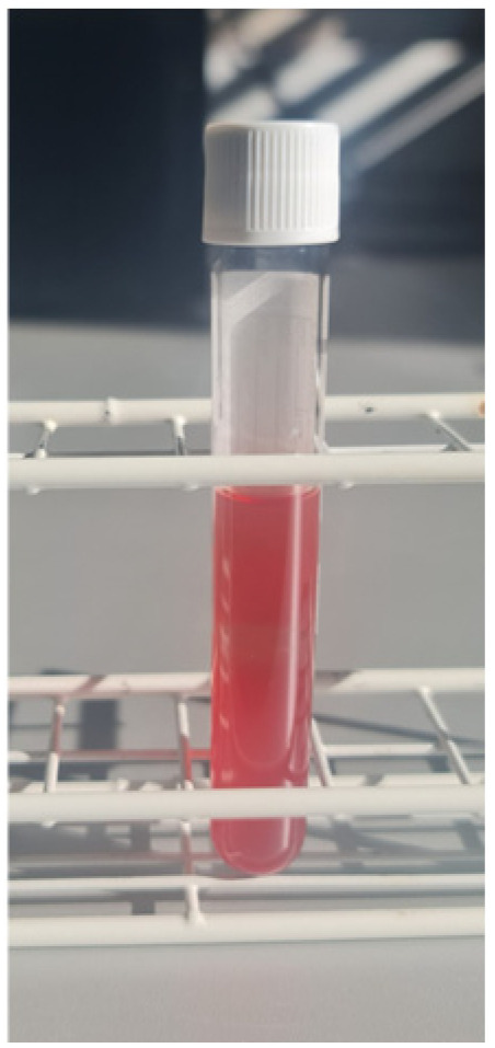
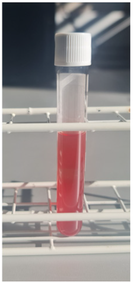
CSF samples of patients after SAH, as can be seen in Figure 1, were drawn from an EVD at the Department of Neurosurgery, Kepler University Hospital (KUK) at pre-defined time points of 0, 5, 10, and 15 days after the initial bleed, with a tolerance of two days. All CSF samples were anonymized by identification numbers (IDs). The numbers represent the patients and the letters correspond to the different time points of drainage (day 0 = A, 5 = B, 10 = C, 15 = D after the SAH). Informed consent was obtained from each participant, their next of kin, or retrospectively in cases where the patient was unable to provide consent. Patients who lacked a next of kin and died prior to the possibility of obtaining retrospective consent were also included in this study. The study protocol was approved by the Ethics Commission of the Faculty of Medicine at Johannes Kepler University Linz (protocol number 1025/2022).
Figure 1.
CSF sample of patient with hydrocephalus after SAH.
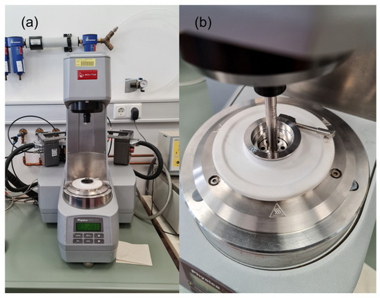
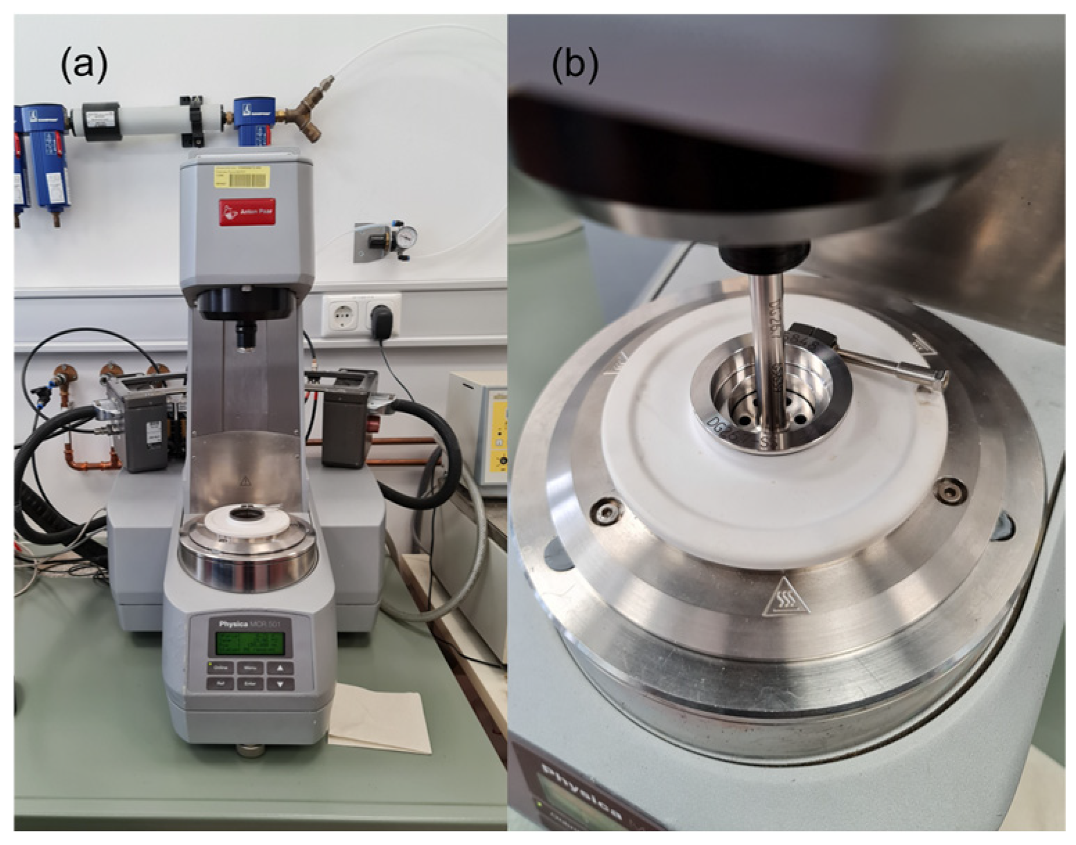
The laboratory parameters of the CSF samples were determined by the Department of Medical and Chemical Laboratory Diagnostics (KUK). The rheological measurements were performed at the Institute of Polymer Science (JKU) with a Physica MCR 501 rheometer (Anton Paar Ltd., Graz, Austria) equipped with a double gap geometry (DG26.7/T200/SS). Both can be seen in Figure 2.
Figure 2.
(a) General assembly of rheometer setup (Anton Paar MCR 501). (b) Double gap geometry in measuring position.
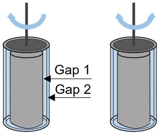
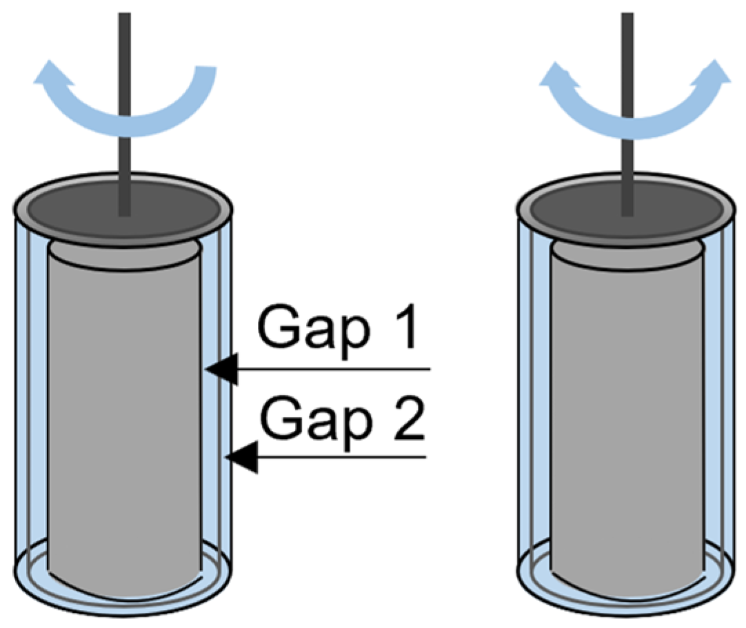
The rheological measurements were performed in two modes: the rotational and the oscillation modes. The motion profile is schematically illustrated in Figure 3. The upper part in the rotational mode turned only in one direction, whereas, for oscillation measurements, the upper part oscillated around the axis in a sinusoidal manner. The rotational tests were performed for a shear rate range of 1 to 700 s−1. The following basic parameters were preset for the oscillation measurements: for the amplitude sweep, a constant angular frequency of 10 rad s−1 was used over the range of 0.01 to 100% deformation, while, for the frequency sweep, constant 10% deformation was set over an angular frequency range of 0.1 rad s−1 to 628 rad s−1. Frequency tests were conducted at specific measurement temperatures to simulate relevant physiological and storage conditions: 5 °C to represent sample storage temperature, 35 °C to mimic mild hypothermia, 37 °C for normal physiological body temperature, and 40 °C to reflect elevated body temperature. Using the storage and loss modulus data from the frequency tests, the cumulative storage factor was calculated and the complex viscosity curve was used for the calculation of the cumulative complex viscosity. Correlations of the cumulative storage factor and the laboratory parameter erythrocyte count were evaluated by using linear regression analysis.
Figure 3.
Schematical double gap measurement geometry. (left) rotational mode: rotation in only one direction. (right) oscillation mode: upper part oscillates in sinus motion.
3. Results
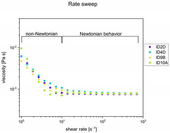
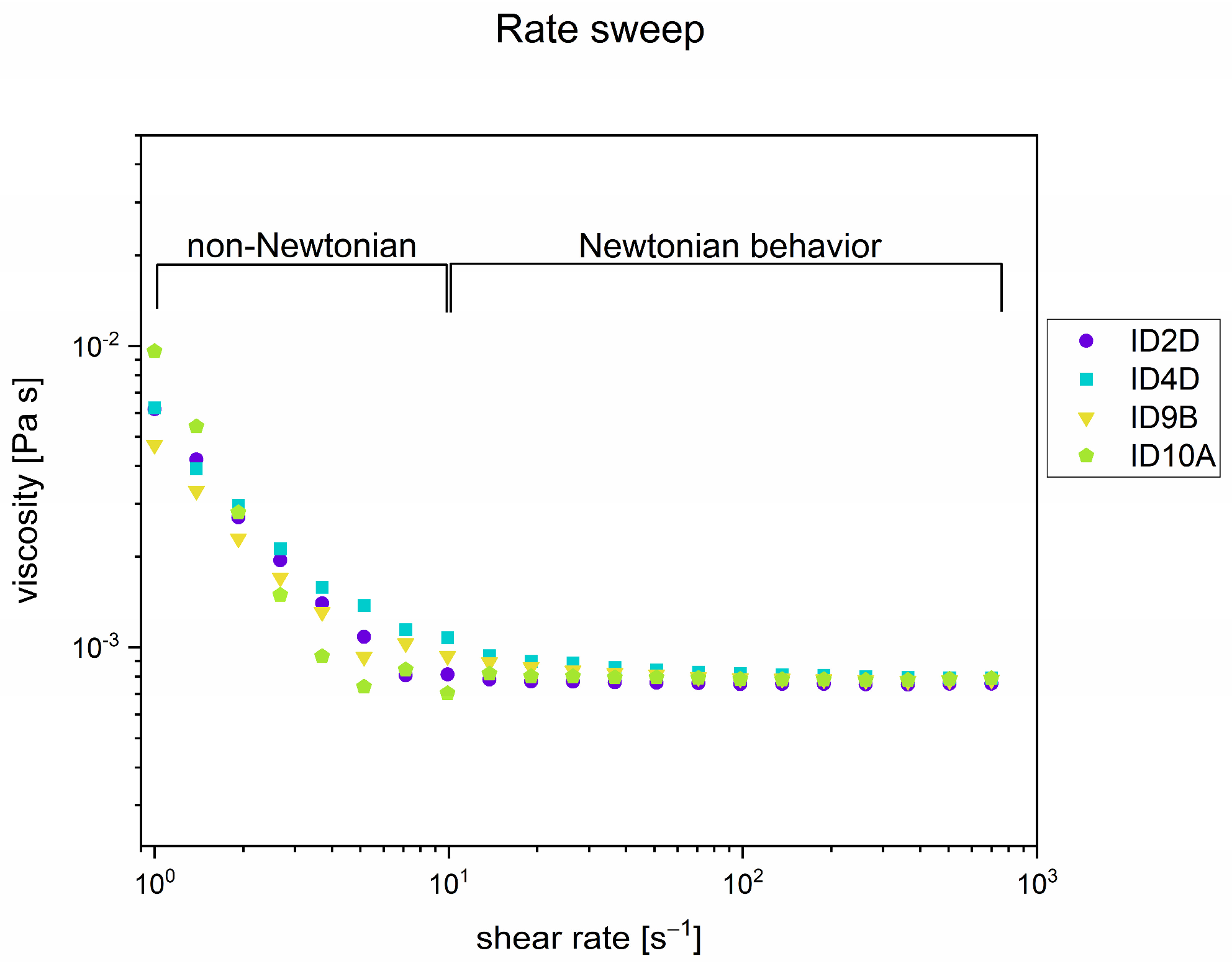
Rotational tests were performed to analyze the stationary shear behavior of the CSF, as well as the viscosity. In Figure 4, viscosity curves of certain samples measured at 37 °C can be seen. The CSF behavior changed with increasing mechanical stress. At low shear rates, the CSF sample acted as a non-Newtonian fluid. This lower shear rate range contained the most information about the sample structure and was also comparable to the shear rates that occur in a shunt flow, in the order of 1 s−1 [14]. In pseudoplastic (shear-thinning) liquids, viscosity decreases with increases in shear rate. This is valid for CSF in the range of lower shear rates due to interactions between the macromolecular components. With increasing shear rate, Newtonian behavior could be observed as, at this point, the original structure was disturbed and all components were oriented in the flow direction. In the range above 10 s−1, the viscosity of CSF was independent of shear rate, indicating a Newtonian liquid, as the macromolecules were already oriented in the flow direction and, therefore, there was no further change in viscosity.
Figure 4.
Rotational test: different samples measured at 37 °C.
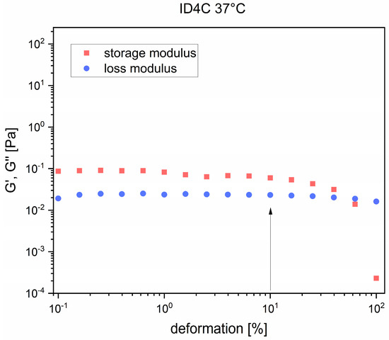
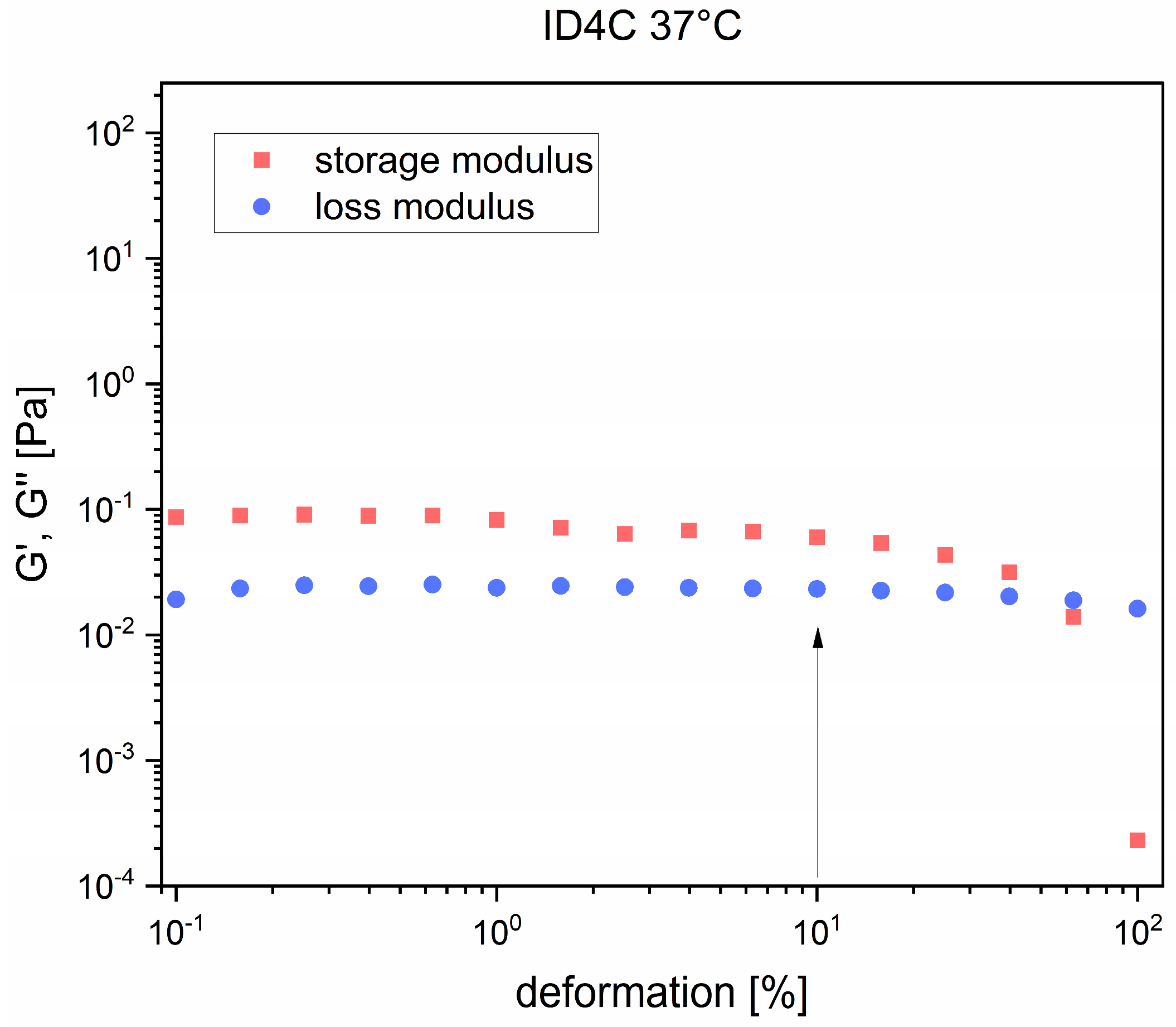
Oscillation measurements were performed to determine the viscoelastic behavior of the CSF samples. Figure 5 shows the amplitude sweep of sample ID4C at 37 °C in order to identify the upper deformation limit of the linear-viscoelastic region of the CSF samples. A strong decrease in values using an amplitude sweep test indicated the end of the region with linear viscoelasticity. The deformation amplitude for the frequency sweeps was chosen according to these results to investigate the viscoelastic behavior of the CSF sample.
Figure 5.
Amplitude sweep: sample ID4C measured at 37 °C. The arrow highlights the deformation parameter which was selected for the subsequent frequency test to ensure measurements remained within the linear viscoelastic region of the CSF.
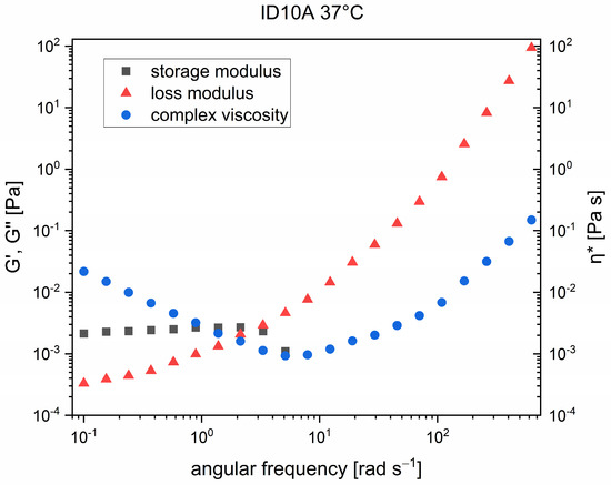
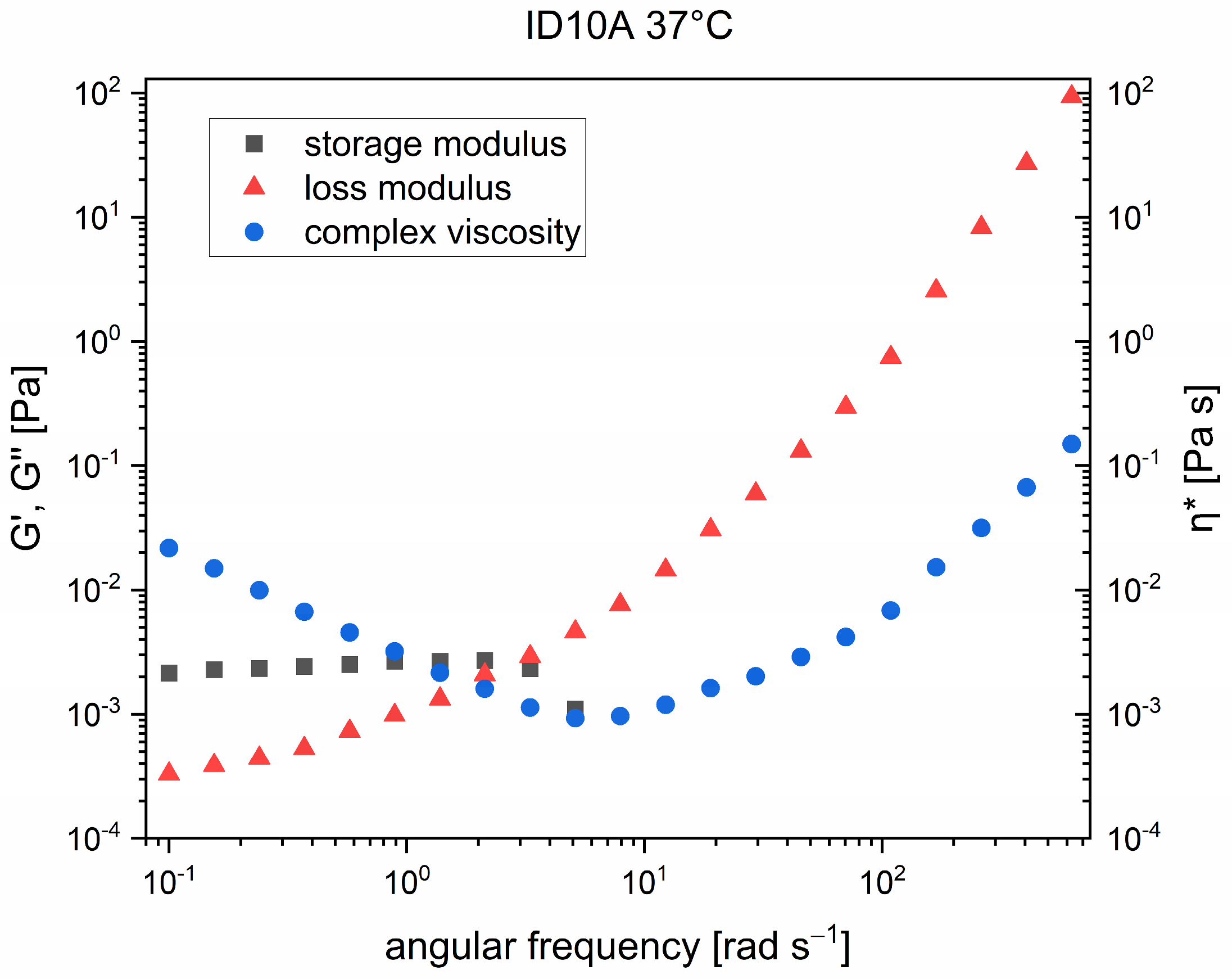
In Figure 6, the frequency test results of sample ID10A measured at 37 °C are shown. The complex viscosity decreased with increasing angular frequency as the CSF was a non-Newtonian liquid. As the storage modulus showed a higher plateau compared to the loss modulus, the elasticity was more dominant. The CSF sample was an aqueous solution that contained macromolecular components, such as different proteins. Considering the whole range of measured angular frequencies, the CSF was not elastic-dominated; therefore, the elastic part at some point was not measurable anymore. At an angular frequency of approximately 8 rad s−1 for the storage modulus, shear-thinning behavior appeared and the complex viscosity values rose again due to the flow dynamic phenomena that occurred. According to the modeling of the frequency measurement in the range of 0.1 to 2.13 rad s−1, the power law exponent for the loss modulus is 0.72911 ± 0.05311 and for the complex viscosity, the power law exponent had a value of (−0.88629 ± 0.00756). Concerning the upcoming secondary flow and shear waves at higher angular frequencies, only the lower angular frequency range was considered for further evaluation.
Figure 6.
Frequency test: sample ID10A at 37 °C.
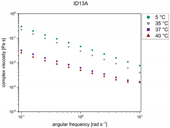
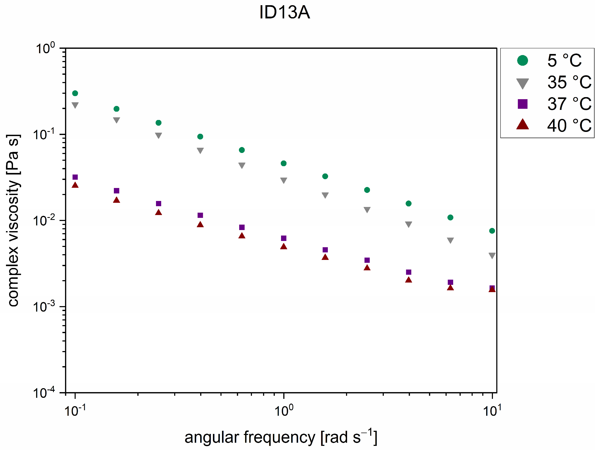
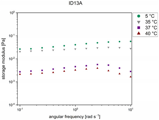
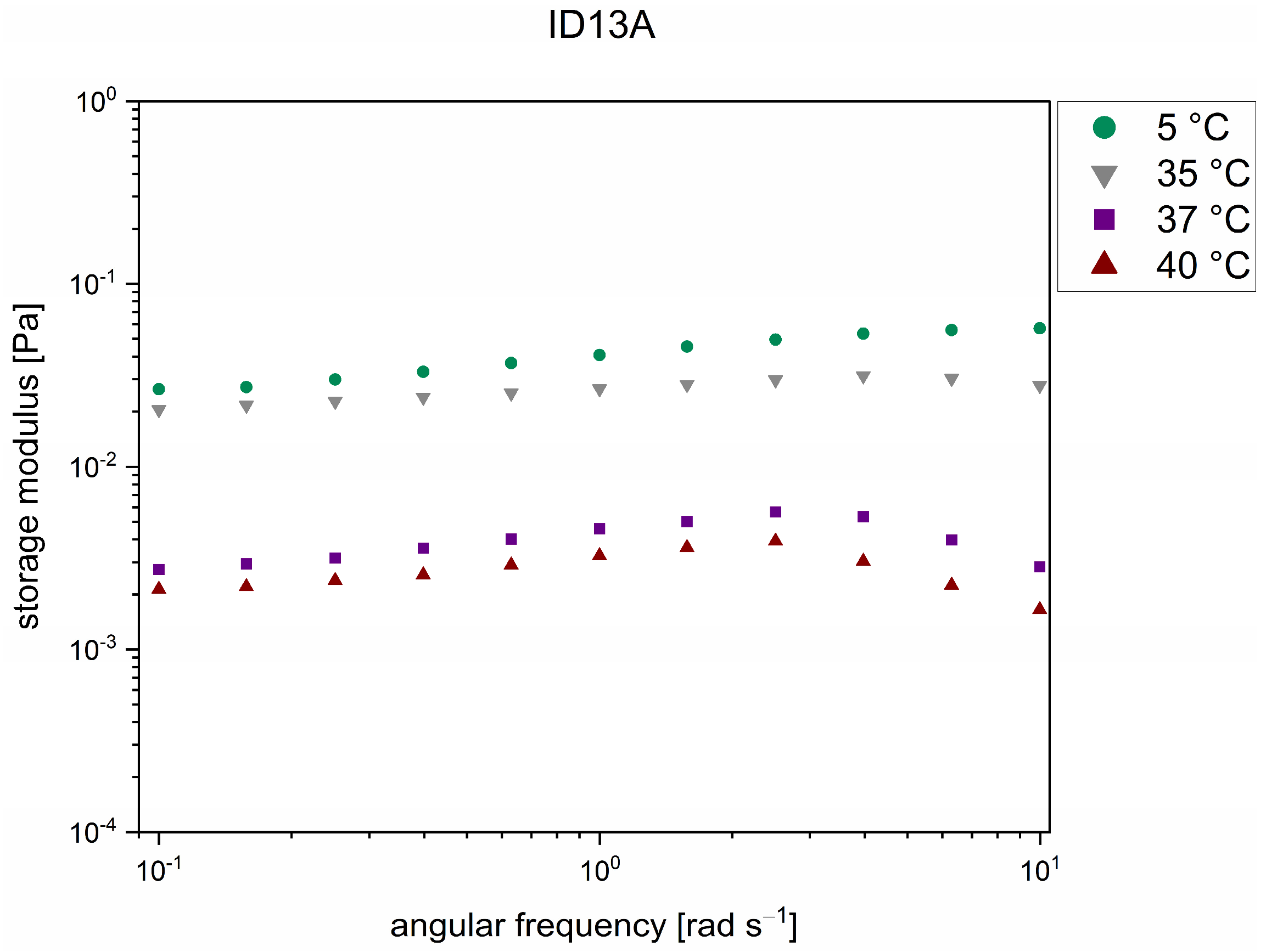
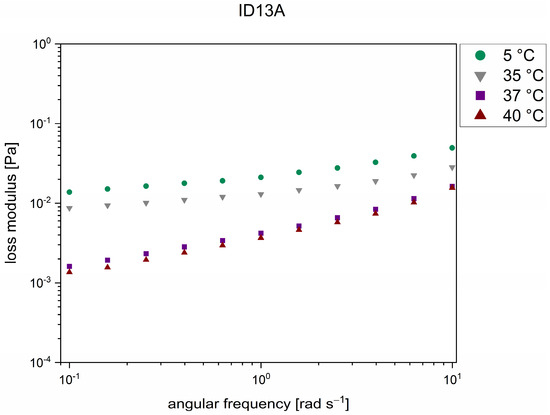
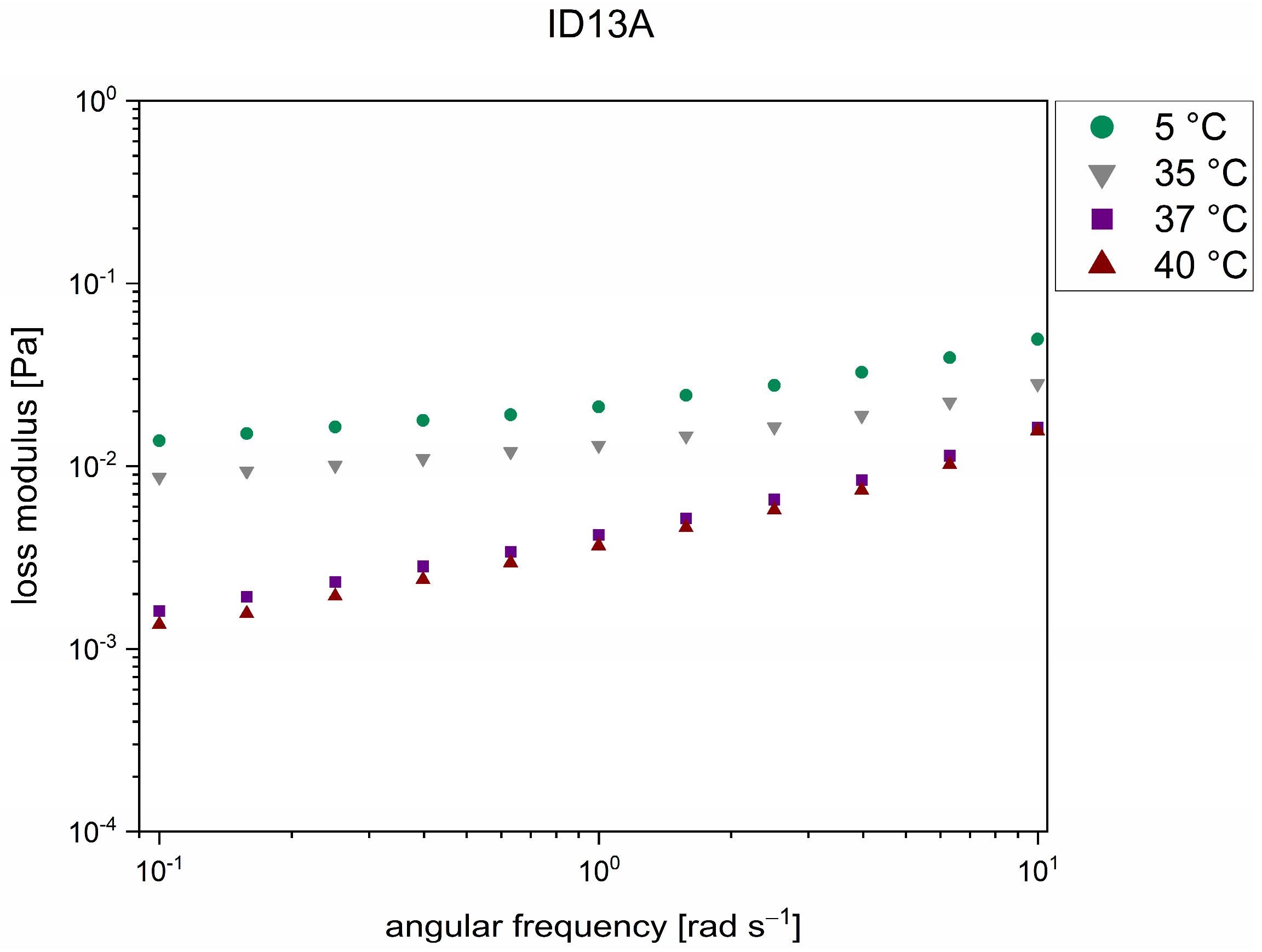
The frequency tests were operated at four different temperatures and the results showed a natural trend. At the lowest temperature, the sample showed the highest viscosity and at the highest temperature, it showed the lowest viscosity, which can be seen in Figure 7. The same tendency occurred for the storage modulus (Figure 8) and the loss modulus (Figure 9). This trend can be generally described by the Brownian motion of particles in a suspension [24]. This trend could not be found for all analyzed samples. This may have been due to the low sensitivity of the rheometer or the time frame between drawing the sample and measuring it. The big gap between the curves of the 35 °C and 37 °C measurements can be associated with a conformation change in the structure of the macromolecular components [25,26]. In fact, the curves measured at the four different temperatures showed a difference in shape, as seen in Figure 7, Figure 8 and Figure 9, which also affirms this assumption. At higher temperatures, the elastic part was in general less dominant and, therefore, the storage modulus decreased at higher temperatures at lower angular frequencies. At lower temperatures, the interactions between macromolecules were, in general, higher and therefore not only the storage modulus but also the loss modulus were influenced in the same way. The power law exponent was also calculated for ID13A at 37 °C based on the loss modulus (0.20339 ± 0.007) and the complex viscosity (−0.72301 ± 0.0130) over the angular frequency range of 0.1 to 2.51 rad s−1.
Figure 7.
Frequency test: complex viscosity of sample ID13A at 4 different temperatures.
Figure 8.
Frequency test: storage modulus of sample ID13A at 4 different temperatures.
Figure 9.
Frequency test: loss modulus of sample ID13A at 4 different temperatures.
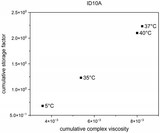
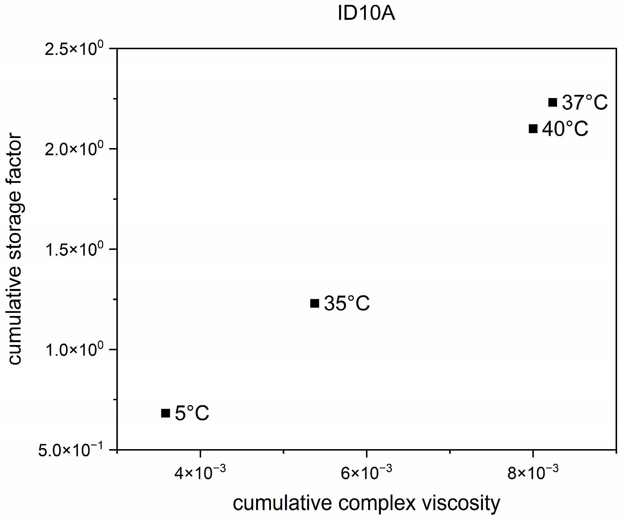
The cumulative storage factor was calculated over the relation of the integral of storage and the loss modulus curves over a certain angular frequency range. Due to upcoming flow dynamic phenomena, a reduced angular frequency range was considered. The same was also calculated for the cumulative complex viscosity. Figure 10 shows the plot of the cumulative storage factor against cumulative complex viscosity. The measurement at 37 °C showed the highest value for the cumulative storage factor. This indicated the greatest interaction between the components in the sample and, therefore, the greatest rigidity behavior. The most important constituents of the CSF were erythrocytes, leukocytes, glucose, lactate, and proteins, which were considered to be responsible for the interactions in the sample.
Figure 10.
Cumulative storage factor of sample ID10A.
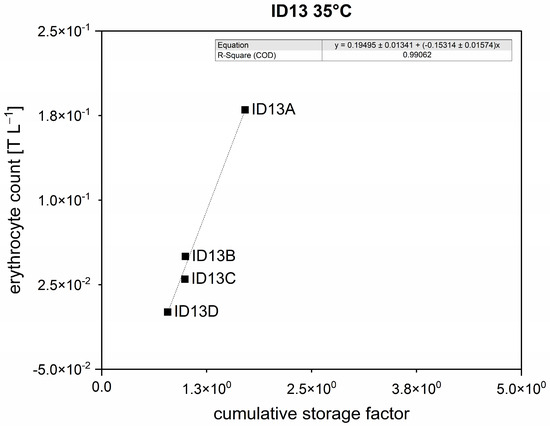
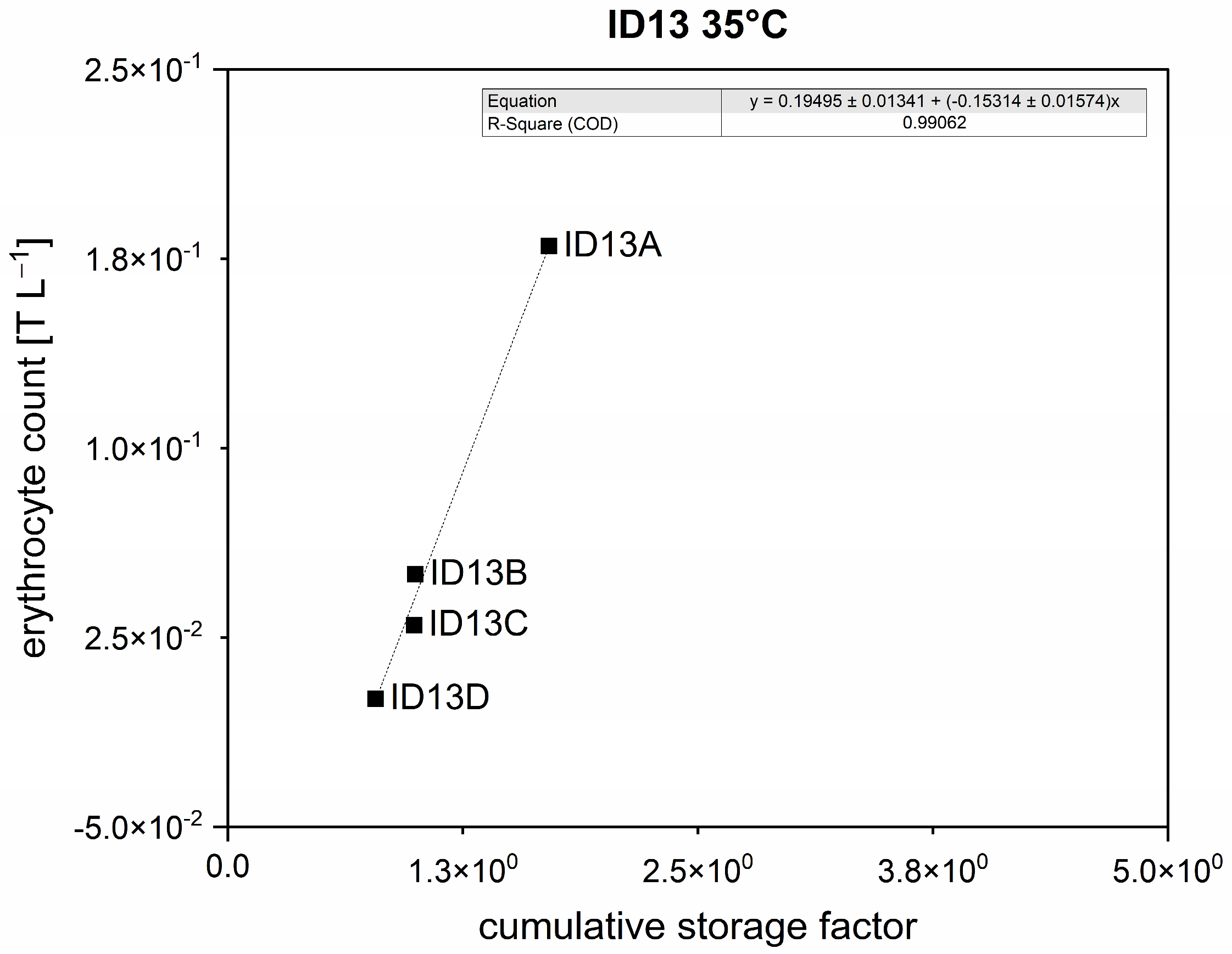
The calculated cumulative factors were correlated with the important diagnostic parameter, erythrocyte count. These correlations were made according to each patient and the time point of sample drainage and separated for each measuring temperature. The correlations were evaluated using linear regression analysis. Figure 11 shows the correlations of ID13 erythrocyte count with the cumulative storage factor. Based on the high linear regression coefficient (R-Square) of 0.99062, a very strong correlation was achieved.
Figure 11.
Correlation of ID13 erythrocyte count and cumulative storage factor with a slope of 0.19495 ± 0.01341, an intercept of (−0.15314) ± 0.01574, and a linear regression coefficient of 0.99062.
4. Discussion
The flow behavior of cerebrospinal fluid samples of patients after subarachnoid hemorrhage was measured and characterized using conventional as well as advanced rheological evaluation and certain cumulative parameters. Rotational tests showed, at low shear rates, a non-Newtonian behavior; for higher shear rates, the CSF acted as a Newtonian fluid. The non-Newtonian behavior of the sample that was present in the shunt application needs to be the focus of ongoing investigations. In order to evaluate the temperature dependence of the CSF properties, oscillation tests were performed at 4 different temperatures (5 °C, 35 °C, 37 °C, and 40 °C) to simulate different situations in the human body. This frequency test displayed a predicted natural trend and showed a non-expected big gap between the 35 °C and 37 °C measurements. Further investigations will focus on this fact and on the reasons for the natural trend not being followed in some cases. Based on the data received from the frequency sweeps, cumulative factors were calculated. The cumulative storage factor indicated that the interacting components were erythrocytes, leukocytes, glucose, lactate, and proteins; these are the most relevant components of CSF. A high cumulative factor suggested high rigidity behavior and, therefore, strong interactions in the sample. High correlations between the important laboratory diagnostic parameter and the cumulative storage factor were obtained and investigated using linear regression analysis. Other studies have investigated the influence of proteins, cells, and glucose on CSF viscosity, but they did not look at the viscoelastic properties using a rheometer equipped with double gap geometry or conduct evaluations with already well-established cumulative factors. Ongoing work will concentrate on statistical evaluations of the correlations in order to reach the overall goal of evaluating whether rheological parameters may help prognosticate the need for ventriculoperitoneal shunt implantation in SAH patients.
5. Conclusions
Preliminary results of this study showed non-Newtonian behavior of CSF in the low shear rate range, the temperature dependency of viscoelastic properties, and the potential of the cumulative storage factor to display interactions of the components in a sample and correlate rheological behavior with important diagnostic parameters, in particular, erythrocyte count. The principal limitation of this study lies in the statistical evaluation. Further investigations will focus on the statistical validation of these correlations and on establishing the cumulative storage factor as a definitive indicator for biological fluids.
Author Contributions
Conceptualization, T.-C.B., E.B., S.H., A.G., T.R., F.R.-N., J.O., H.S. and M.K.; methodology, T.-C.B., E.B., S.H., A.G., T.R., F.R.-N., J.O., H.S. and M.K.; software, T.-C.B.; validation, M.K.; formal analysis, T.-C.B., E.B., T.R., F.R.-N. and J.O.; investigation, T.-C.B., E.B., S.H., A.G., T.R., F.R.-N., J.O., H.S. and M.K.; resources, H.S. and M.K.; data curation, T.-C.B.; writing—original draft preparation, T.-C.B.; writing—review and editing, T.-C.B., E.B., S.H., A.G., T.R., F.R.-N., J.O., H.S. and M.K.; visualization, T.-C.B.; supervision, S.H., A.G., H.S. and M.K.; project administration, T.R., H.S. and M.K.; funding acquisition, T.R., H.S. and M.K. All authors have read and agreed to the published version of the manuscript.
Funding
The authors acknowledge the financial support of the Faculty of Medicine of the Johannes Kepler University Linz through its internal funding program Impetus Nr. I-10-22 and the LIT Institute of Technology of the Johannes Kepler University Linz, project number LIT-2022-11-SEE-123.
Institutional Review Board Statement
This study involving human samples was approved by the Ethics Commission, Faculty of Medicine, Johannes Kepler University Linz, MED Campus I, ADM Building 7th Floor, Krankenhausstaße 5, 4020 Linz (approval No. 1025/2022) on 1 July 2022. This study was conducted in accordance with local legislation and institutional requirements. The participants provided written informed consent to participate in this study.
Data Availability Statement
The raw data supporting the conclusion of this article will be made available by the authors without undue reservation.
Acknowledgments
Supported by Johannes Kepler University Open Access Publishing Fund.
Conflicts of Interest
The authors have no conflicts to disclose.
Abbreviations
The following abbreviations are used in this manuscript:
| CSF | Cerebrospinal fluid |
| SAH | Subarachnoid hemorrhage |
| EVD | External ventricular drainage |
| VP | Ventriculoperitoneal |
| ID | Identification number |
References
- Bradt, E.; Hild, S.; Reichel, E.K.; Kracalik, M. Microrheology of Biofluids. In Book of Abstracts Annual European Rheology Conference, Sorrento, Italy, 17–18 April 2018; European Society of Rheology: Zurich, Switzerland, 2018. [Google Scholar]
- Tomaiuolo, G.; Carciati, A.; Caserta, S.; Guido, S. Blood linear viscoelasticity by small amplitude oscillatory flow. Rheol. Acta 2016, 55, 485–495. [Google Scholar] [CrossRef] [Scilit]
- Windberger, U.; Pöschl, C.; Peters, S.; Huber, J.; van den Hoven, R. Measurement of whole blood of different mammalian species in the oscillating shear field: Influence of erythrocyte aggregation. J. Phys. Conf. Ser. 2017, 790, 12035. [Google Scholar] [CrossRef] [Scilit]
- Barrand, M.; Hladky, S. Mechanisms of fluid movement into, through and out of the brain: Evaluation of the evidence. Fluids Barriers CNS 2014, 11, 26. [Google Scholar] [CrossRef] [Scilit] [PubMed]
- Klebe, D.; McBride, D.; Krafft, P.R.; Flores, J.J.; Tang, J.; Zhang, J.H. Posthemorrhagic hydrocephalus development after germinal matrix hemorrhage: Established mechanisms and proposed pathways. J. Neurosci. Res. 2020, 98, 105–120. [Google Scholar] [CrossRef] [Scilit] [PubMed]
- Mumenthaler, M.; Mattle, H. Fundamentals of Neurology: An Illustrated Guide; Georg Thieme Verlag KG: Stuttgart, Germany, 2006; pp. 83–110. [Google Scholar]
- Ropper, A.H.; Samuels, M.A.; Klein, J.P. Adams & Victor’s Principles of Neurology, 10th ed.; McGraw-Hill Education: New York, NY, USA, 2014; pp. 615–638. [Google Scholar]
- Bitsch, A.; Prange, H.; Meiners, M.; Neumann, P.; Prange, H.; Rathgeber, J.; Reimers, C.D. Neurologische Intensivmedizin: Praxisleitfaden für Neurologische Intensivstationen und Stroke Units, 12th ed.; Prange, H., Bitsch, A., Eds.; Georg Thieme Verlag KG: Stuttgart, Germany, 2004; pp. 40–43. [Google Scholar]
- Fowler, J.B.; De Jesus, O.; Mesfin, F.B. Ventriculoperitoneal Shunt. In StatPearls; StatPearls Publishing: Treasure Island, FL, USA, 2022. [Google Scholar]
- Kazakov, V.N.; Barkalova, E.L.; Levchenko, L.A.; Klimenko, T.M.; Fainerman, V.B.; Miller, R. Dilation rheology as medical diagnostics of human biological liquids. Colloids Surf. A Physicochem. Eng. Asp. 2011, 391, 190–194. [Google Scholar] [CrossRef] [Scilit]
- Schob, S.; Weiß, A.; Dieckow, J.; Richter, C.; Pirlich, M.; Voigt, P.; Surov, A.; Hoffmann, K.-T.; Quaeschling, U.; Preuß, M. Correlations of Ventricular Enlargement with Rheologically Active Surfactant Proteins in Cerebrospinal Fluid. Front. Aging Neurosci. 2017, 8, 324. [Google Scholar] [CrossRef] [Scilit] [PubMed]
- Yoshida, S.; Takeshita, M.; Sugiura, M.; Jimbo, M.; Kitamura, K. Rheology of the cerebrospinal-fluid. Biorheology 1981, 18, 121. [Google Scholar]
- Bloomfield, I.G.; Johnston, I.H.; Bilston, L.E. Effects of proteins, blood cells and glucose on the viscosity of cerebrospinal fluid. Pediatr. Neurosurg. 1998, 28, 246–251. [Google Scholar] [CrossRef] [Scilit] [PubMed]
- Hollister, J.C.P.; Wang, A.C.; Kim, W.; Giza, C.C.; Prins, M.L.; Kavehpour, H.P. Shear thinning behavior of cerebrospinal fluid with elevated protein or cellular concentration. Front. Phys. 2023, 11, 1308136. [Google Scholar] [CrossRef] [Scilit]
- Brydon, H.L.; Hayward, R.; Harkness, W.; Bayston, R. Physical properties of cerebrospinal fluid of relevance to shunt function. 1: The effect of protein upon CSF viscosity. Br. J. Neurosurg. 1995, 9, 639–644. [Google Scholar] [CrossRef] [Scilit] [PubMed]
- Mezger, T.G. Angewandte Reologie, 3rd ed.; Anton Paar GmbH: Graz, Austria, 2017; pp. 19–172. [Google Scholar]
- Schramm, G. A Practical Approach to Rheology and Rheometry, 2nd ed.; Thermo Electron GmbH: Karlsruhe, Germany, 2004; pp. 36–194. [Google Scholar]
- Anselmo, S.P.; Edson, J.S. Polymer degradation of dilute solutions in turbulent drag reducing flows in a cylindrical double gap rheometer device. J. Non-Newton. Fluid Mech. 2012, 179, 9–22. [Google Scholar] [CrossRef] [Scilit]
- Laske, S.; Witschnigg, A.; Mattausch, H.; Kracalik, M.; Pinter, G.; Feuchter, M.; Maier, G.; Holzer, C. Determining the Ageing of Polypropylene Nanocomposites Using Rheological Measurements. Appl. Rheol. 2012, 22, 24590. [Google Scholar] [CrossRef] [Scilit]
- Addis, A.; Gaasch, M.; Schiefecker, A.J.; Kofler, M.; Ianosi, B.; Rass, V.; Lindner, A.; Broessner, G.; Beer, R.; Pfausler, B.; et al. Brain temperature regulation in poor-grade subarachnoid hemorrhage patients—A multimodal neuromonitoring study. J. Cereb. Blood Flow Metab. 2021, 41, 359–368. [Google Scholar] [CrossRef] [Scilit] [PubMed]
- Sung, D.; Risk, B.B.; Wang, K.J.; Allen, J.W.; Fleischer, C.C. Resting-State Brain Temperature: Dynamic Fluctuations in Brain Temperature and the Brain-Body Temperature Gradient. J. Magn. Reson. Imaging 2023, 57, 1222–1228. [Google Scholar] [CrossRef] [Scilit] [PubMed]
- Kracalik, M. Recycled clay/PET nanocomposites evaluated by novel rheological analysis approach. Appl. Clay Sci. 2018, 166, 181–184. [Google Scholar] [CrossRef] [Scilit]
- Kracalik, M. Different Approaches for Calculation of Cumulative Storage Factor as Novel Parameter for Analysis of Reinforcement in Complex Polymer Nanocomposites. In Proceedings of the AIP Conference Proceedings, Zlín, Czech Republic, 30–31 July 2019. [Google Scholar]
- Mao, Y.; Nielsen, P.; Ali, J. Passive and Active Microrheology for Biomedical Systems. Front. Bioeng. Biotechnol. 2022, 10, 916354. [Google Scholar] [CrossRef] [Scilit] [PubMed]
- Kunitsyn, V.G.; Panin, L.E.; Polyakov, L.M. Anomalous change of viscosity and conductivity in blood plasma lipoproteins in the physiological temperature range. Int. J. Quantum Chem. 2001, 81, 348–369. [Google Scholar] [CrossRef]
- Michieletto, D.; Marenda, M. Rheology and Viscoelasticity of Proteins and Nucleic Acids Condensates. JACS Au 2022, 2, 1506–1521. [Google Scholar] [CrossRef] [Scilit] [PubMed]
Disclaimer/Publisher’s Note: The statements, opinions and data contained in all publications are solely those of the individual author(s) and contributor(s) and not of MDPI and/or the editor(s). MDPI and/or the editor(s) disclaim responsibility for any injury to people or property resulting from any ideas, methods, instructions or products referred to in the content. |
© 2026 by the authors. Licensee MDPI, Basel, Switzerland. This article is an open access article distributed under the terms and conditions of the Creative Commons Attribution (CC BY) license.