


Abstract
Morphological and molecular phylogenetic studies of Chlorencoelia in China revealed three novel species, which are described and illustrated as C. biguttulata, C. ellipsoidea, and C. sinensis. Chlorencoelia biguttulata is characterized by greyish green to blackish green hymenium surface, clavate tomentum hyphae on receptacle surface, and inequilateral ellipsoidal ascospores having two large guttules and 8.5–14.5 × 3.5–4.5 μm. Chlorencoelia ellipsoidea differs from the known species by distinctly stipitate apothecia with a dark green hymenium surface and ellipsoidal ascospores 7.2–9.1 × 3.5–4.2 μm. Chlorencoelia sinensis is characterized by substipitate apothecia, blackish receptacle surface, and fusoidal ascospores 9–13.7 × 2.2–3.5 μm. Sequence analyses of the ITS region supported recognition of them as separate species. All three new species occurred on rotten wood in montane forests. Morphological features for species delimitation and diversity of Chlorencoelia are discussed. An identification key to the known species of the genus is provided.
1. Introduction
The genus Chlorencoelia J.R. Dixon was introduced by Dixon in 1975, and typified by C. versiformis (Pers.) J.R. Dixon, which included C. torta (Schwein.) J.R. Dixon as well [1]. The two species were once treated as members of Chlorosplenium Fr. (Chlorospleniaceae) and Chlorociboria Seaver ex C.S. Ramamurthi, Korf & L.R. Batra (Chlorociboriaceae). Thereafter, C. indica (K.S. Thind, E.K. Cash & Pr. Singh) W.Y. Zhuang was transferred into the genus [2]. Subsequently, C. ripakorfii Iturr. & Mardones from Venezuela and C. macrospora F. Ren & W.Y. Zhuang from China were added [3,4]. New members were not reported in the following 10 years (2015–2024). Recently, Patejuk et al. introduced two new species from New Zealand, C. australis P.R. Johnst. & Patejuk and C. northlandica P.R. Johnst. & Patejuk, as well as a new combination, C. olivacea (Rodway) P.R. Johnst. & Patejuk., on the basis of integrative study of DNA sequences and morphology [5]. Currently, the genus comprises eight known species.
Chlorencoelia was originally regarded as a member of the family Helotiaceae [1]. Later, it was placed in the family Hemiphacidiaceae based on phylogenetic analyses of rDNA sequences [6,7,8]. The multi-locus phylogeny revealed that members of Hemiphacidiaceae and the core group of the subfamily Encoelioideae (including Chlorencoelia) belonged to the resurrected family Cenangiaceae [9], which was confirmed by the subsequent studies [10,11,12,13,14].
Chlorencoelia is characterized by shallow cupulate to infundibuliform apothecia, mostly less than 10 mm in diam., sub-stipitate to stipitate, solitary to gregarious; hymenium surface olive-yellow, olive-green to dark green; receptacle surface dark olive-green to blackish; ectal excipulum of hyaline- to brown-celled textura angularis, giving rise to clavate or filamentous tomentum hyphae; medullary excipulum of textura intricata with cells having hyaline to dark brown walls; asci cylindric-clavate, J+, 8-spored; ascospores ellipsoid, cylindric-oblong to allantoid, hyaline, guttulate; paraphyses filiform, septate, with pale yellow to green vacuolar bodies. Shape and size of ascospores, asci, and tomentum hyphae are key characteristics for species delimitation [1,3,4,5].
Chlorencoelia species are saprotrophic on rotten wood of angiosperm and gymnosperm trees. C. versiformis was collected on Betula, Nothofagus, Quercus, Tsuga, and unidentified wood [1]. C. torta was found on Acer, Betula, Fagus, Quercus, Tabebuia, coniferous, and unidentified wood [1]. C. indica was recorded on Cedrus deodara [2]. C. australis grew on Fuscospora solandri, Leptospermum scoparium, Nothofagus, Nothofagaceae, and unidentified wood [5]. C. olivacea occurred on Fuscospora fusca, Fuscospora cliffortioides, Nothofagus, Pterophylla racemosa, Nothofagaceae, and unidentified wood [5]. C. macrospora, C. northlandica, and C. ripakorfii on woody substrate were not clearly documented. The correlations between Chlorencoelia species and their tree substrates were not confirmed.
Three Chlorencoelia species, C. macrospora, C. torta, and C. versiformis, were previously reported from China [4,15,16]. During the diversity surveys of helotialean fungi in recent years, additional specimens of Chlorencoelia were collected. The identities of those collections were determined based on morphological features and DNA sequences. It turns out that three new species were discovered. The previous records of C. torta and C. versiformis in China were based on misidentifications, and should be excluded from the Chinese mycobiota.
2. Materials and Methods
2.1. Morphological Observations
Specimens of Chlorencoelia preserved in the Herbarium Mycologicum Academiae Sinicae (HMAS) were loaned and re-examined. New specimens were collected on decorticated rotten wood from Chongqing City, Xizang, and Yunnan during 2016–2020. Due to the excessive decayed substrate, it is difficult to recognize the tree species. The substrates were simply recorded as decorticated rotten wood. Fresh apothecia were dried at about 50 °C. Macroscopic photographs were taken with a Canon G16 digital camera (Canon Inc., Tokyo, Japan). Macroscopic characteristics were recorded according to the field notes or photographs of fresh apothecia. Dry specimens were observed under a SZX7 stereomicroscope (Olympus Corporation, Tokyo, Japan). Dried apothecia were soaked in distilled water for rehydration, and longitudinal sections were made using a Yidi YD-1508A freezing microtome (Jinhua Yidi Medical Appliance Co., Ltd., Jinhua, China) at a thickness of 15−20 μm. Amyloid reactions of ascus apical rings were examined in Lugol’s solution and Melzer’s reagent with or without pretreatment of 3% KOH solution. Microscopic examinations and measurements were carried out from sections and squash mounts in lactophenol cotton blue solution via an Olympus BH-2 microscope (Olympus Corporation, Tokyo, Japan). A ZEISS Axiocam 305 colour microscope camera (Carl Zeiss AG, Göttingen, Germany) attached to a Zeiss Axioskop 2 Plus microscope (Göttingen, Germany) was used to take microscopic photographs. Newly collected specimens were deposited in HMAS.
2.2. DNA Extraction, Amplification, and Sequencing
Plant Genomic DNA Kit (TIANGEN Biotech. Co., Beijing, China) was used to extract genomic DNA from dried apothecia following the manufacturer’s instructions. The primer pairs used for amplification and sequencing are ITS1&ITS4 or ITS5&ITS4 for internal transcribed spacers region of the ribosomal DNA (ITS) [17], LR0R/LR5 for the D1/D2 region of large subunit ribosomal DNA (28S) [18,19], RPB1-Af/RPB1-Cr for RNA polymerase II largest subunit gene (RPB1) [20,21], fRPB2-5f/fRPB2-7cr for RNA polymerase II second largest subunit gene (RPB2) [22], and EF1-983F/EF1-1567R for translation elongation factor 1-alpha gene (TEF1) [23]. Applied Biosystems 2720 thermocycler (Foster City, CA, USA) was used for PCR reactions following the components and cycling parameters adopted by Zheng & Zhuang 2022 [24]. The PCR products were purified and sequenced by Beijing Tianyi Huiyuan Bioscience and Technology, Beijing, China.
2.3. Sequence Assembly, Alignment and Phylogenetic Analyses
Forward and reverse sequences generated were assembled with BioEdit 7.0.5.3 [25]. Newly generated sequences were submitted to GenBank and provided as Supplementary Files (S1–S5) as some sequence accession numbers are still pending. Other sequences used for phylogenetic analyses were downloaded from GenBank. Hymenoscyphus fraxineus (T. Kowalski) Baral, Queloz & Hosoya and H. fructigenus (Bull.) Gray were chosen as the outgroup taxa. The accession numbers and related information of the ITS sequences used in phylogenetic analyses are presented in Table 1. Sequences were aligned and optimized through BioEdit 7.0.5.3 [25]. GenBank accession numbers of the newly generated RPB1, RPB2, and TEF1 sequences are provided following the voucher specimen numbers for future phylogenetic consideration.
Table 1.
List of taxa with information on origin, voucher/strain, substrate, and GenBank accession numbers of ITS sequences used in phylogenetic analyses (numbers in bold indicating newly generated sequences).
Bayesian inference (BI) analysis was performed with MrBayes 3.1.2 [26], and Maximum likelihood (ML) analysis was conducted using RAxML 8.0 [27]. The best-fit substitution model for the ITS matrix was calculated based on the Akaike Information Criterion (AIC) with MrModeltest 2.3 [28]. For BI analysis, two parallel runs were conducted. Each run contained four simultaneous Markov Chain Monte Carlo (MCMC) chains. MCMC chains were run for 2 million generations with trees sampled at 100-generation intervals. The first 25% of the trees were discarded as burn-in, and the remaining trees were used to calculate the posterior probabilities (PP) of the majority rule consensus tree. ML analysis was run in RAxML 8.0 using a GTRGAMMA model and tested with 1000 nonparametric bootstrap replicates [27]. The phylogenetic trees were visualized in FigTree 1.4.4 [29].
3. Results
3.1. Phylogenetic Analyses
All the ITS sequences labelled as Chlorencoelia in the GenBank database were downloaded and evaluated. Except for some wrongly identified ones [e.g., an endophytic fungal strain (CMON55, ITS JQ754001) of common bean (Phaseolus vulgaris) leaves and two endophytic strains (FeC10, ITS MW447089 and FeC18, ITS MW447090)] and those shorter than 300 bp, others joined the phylogenetic analyses together with the newly generated ones.
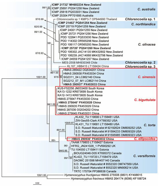
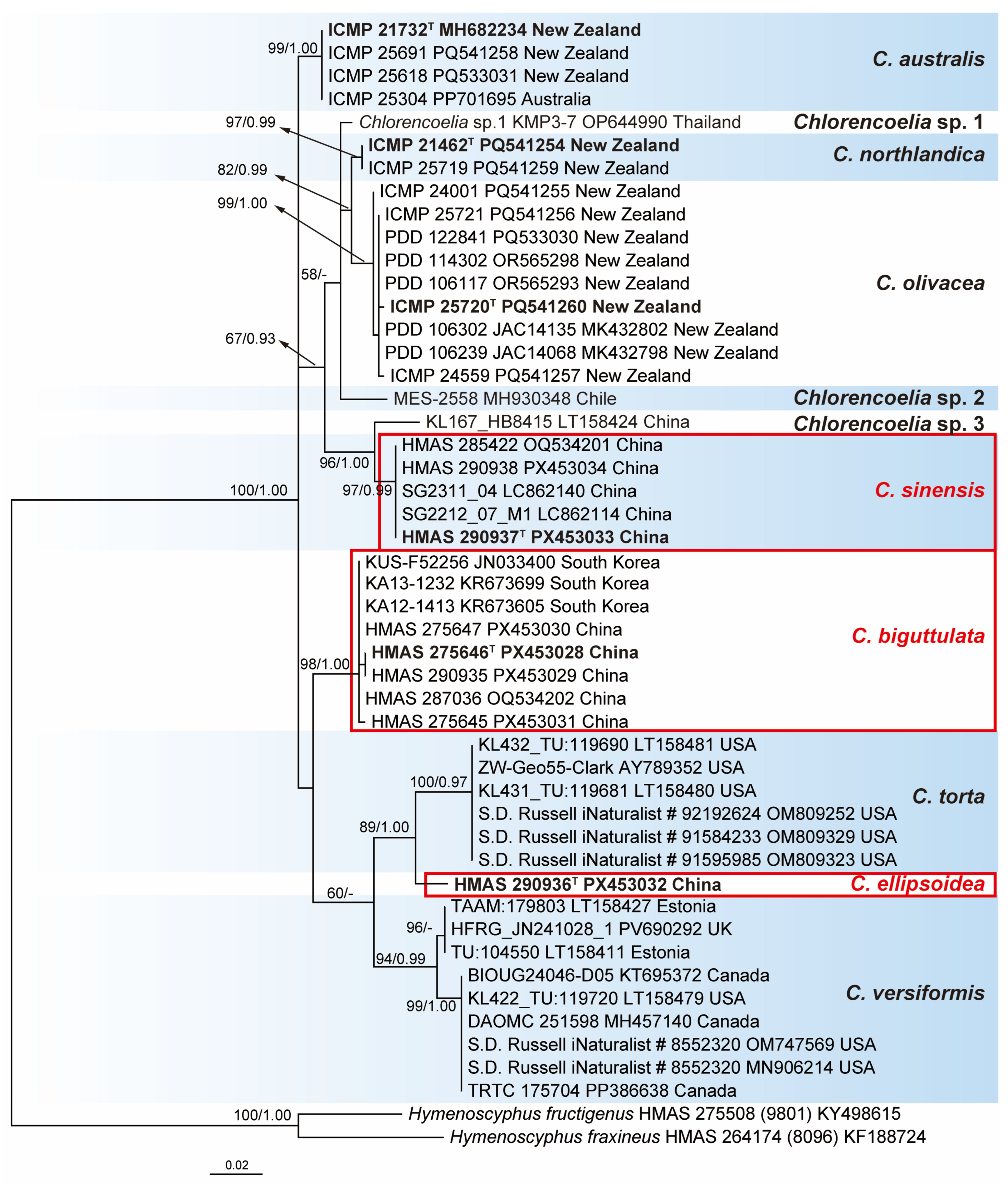
The ITS dataset consisted of 49 sequences representing 11 taxa of Chlorencoelia with two Hymenoscyphus species as outgroup taxa. The alignment consisted of 526 characters, eliminating poorly aligned regions, including 373 constant ones, 52 variable ones, and 101 parsimony-informative ones. SYM + G was selected as the best-fit model for BI analysis. The topologies of the ML and BI trees are similar, and only the phylogram of the ML tree is shown in Figure 1.
Figure 1.
Maximum likelihood phylogenetic tree of Chlorencoelia inferred from ITS sequences. Bootstrap support values ≥ 50% (left) of ML and posterior probability values ≥ 0.90 (right) of BI analysis are shown at nodes. Holotype specimens or ex-type strains are shown in bold and marked with superscript letter “T”. New species are encircled by red boxes. The origins of the materials are indicated at the end of each sample.
All the sampled sequences of Chlorencoelia formed a well-supported monophyletic group (100% MLBP and 1.00 BIPP). Eleven species-level clades are recognized within the genus, which correspond to the eight known species (including the proposed three new species) and three undescribed taxa. The type locality of C. torta is the USA [1]. Sequences of C. torta derived from North American materials were aggregated together to form a separate clade and thus considered as reliable representatives of the species. The type locality of C. versiformis is uncertain and was presumed to be Germany [1], and sequences under that name derived from European (Estonian and UK) materials were represented as an independent species. As for C. versiformis, two subclades were recognized, one comprising the European samples and the other from North America. Sequences of C. australis, C. northlandica, and C. olivacea from New Zealand, as well as those of the to-be-described new species, clustered as six well-supported clades and recognized at species rank. The remaining three terminal lineages, “Chlorencoelia sp.1 KMP3-7 OP644990” (as C. torta in GenBank) from Thailand, “Chlorencoelia sp.2 MES-2558 MH930348” from Chile (as cf. Chlorencoelia sp. in GenBank), and “Chlorencoelia sp.3 KL167_HB8415 LT158424” (as C. torta in GenBank) from China, each having a single sequence, are presumably putative new species in view of the tree topology (Figure 1).
3.2. Taxonomy
3.2.1. Chlorencoelia biguttulata H.D. Zheng & W.Y. Zhuang, sp. nov. (Figure 2a–c and Figure 3)
Fungal Names—FN 573049.
Etymology—The specific epithet refers to the two large guttules in ascospores of the fungus.
Holotype—China, Xizang, Nyingchi, Lulang, on decorticated rotten wood, 23 September 2016, Huan-Di Zheng, Zhi-He Yu, Zhao-Qing Zeng et al. 11236 (HMAS 275646; ITS: PX453028, 28S: PX630171, RPB1: PX557843, RPB2: PX557850, TEF1: PX528999).
Chinese name—双油滴绿散孢盘菌, shuāng yóu dī lǜ sǎn bāo pán jūn.
Figure 2.
Apothecia of Chlorencoelia spp. on woody substrates ((a–c,e,f,k). dry apothecia; (d,g–j). fresh apothecia). (a–c) C. biguttulata, (a,b) HMAS 275646, holotype; (c) HMAS 275645, paratype; (d–f) C. ellipsoidea, HMAS 290936, holotype; (g–k) C. sinensis, HMAS 290937, holotype. Scale bars: (a–c,e,f,h–j) = 2 mm; (d,k) = 5 mm.
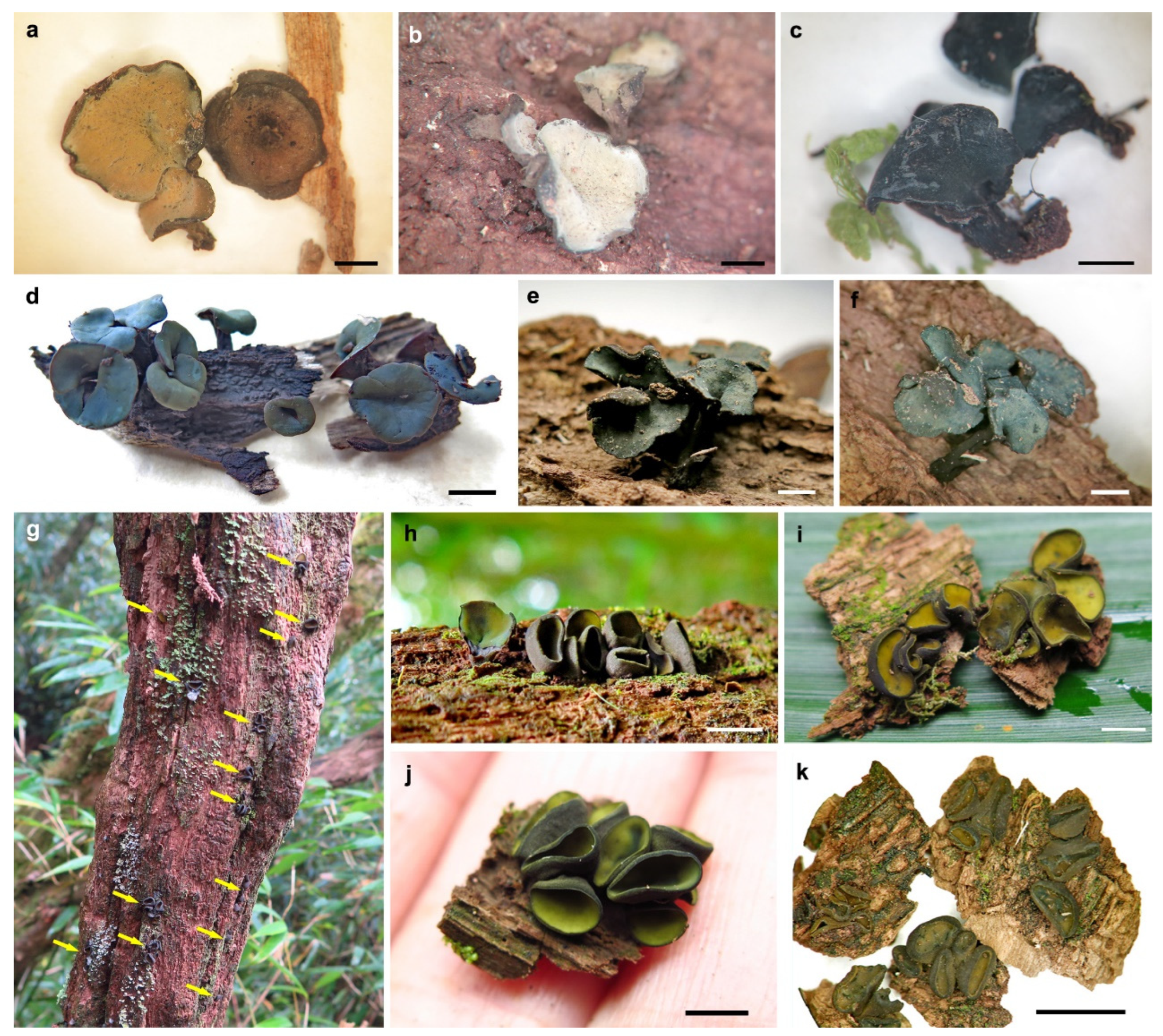
Figure 3.
Microscopic features of Chlorencoelia biguttulata (HMAS 275646, holotype). (a) Longitudinal section of apothecium (partial); (b,c) excipular structure of flank; (d–f) asci; (g,h) crozier at ascus base; (i–p) ascospores; (q–t) IKI reaction of ascus apical ring without KOH pretreatment. Scale bars: (a) = 100 μm; (b–t) = 10 μm. Mounting media: (a–p) lactophenol cotton blue; (q–t) Melzer’s reagent.
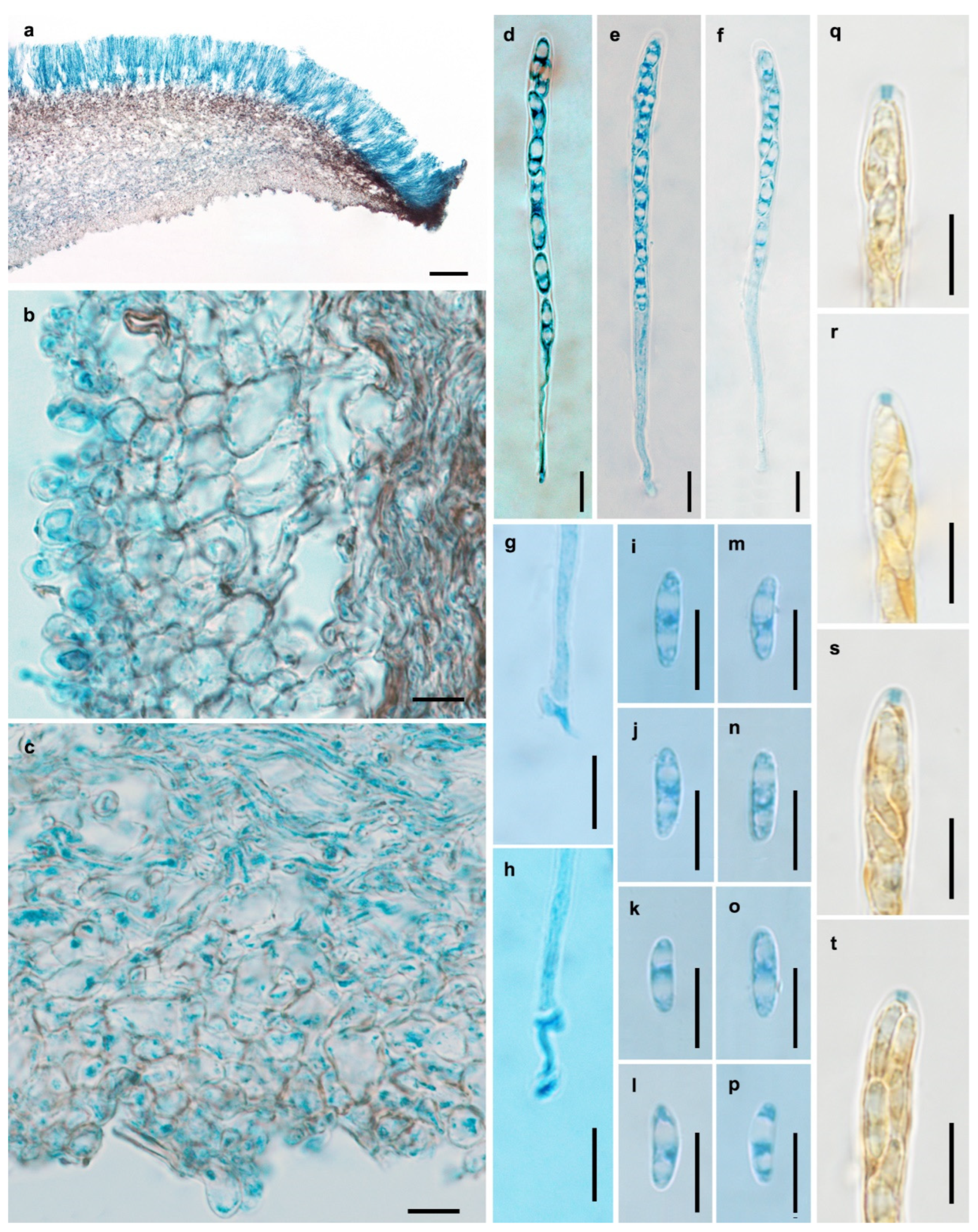
Description—Apothecia solitary, gregarious to caespitose, shallowly infundibuliform, with central to slightly eccentrical stipes, 3–13 mm in diam.; hymenium surface greyish green to blackish green when fresh, yellowish brown to greyish brown after drying; receptacle surface brown; stipe concolorous with the receptacle. Ectal excipulum of textura angularis, 20–55 μm thick; cells light brown to brown, nearly isodiametric or somewhat irregular, becoming smaller towards the surface, 3.5–15 μm in diam., some up to 22 × 14 μm, thin- to slightly thick-walled, giving rise to tomentum hyphae oriented nearly perpendicularly to the flank surface; tomentum hyphae clavate, 8–22 × 5–14 μm, walls up to 3.5 μm thick. Medullary excipulum of textura intricata, 30–860 μm thick; hyphae light brown to brown, 3–6 μm wide. Subhymenium not distinguishable. Hymenium 110–180 μm thick. Asci arising from croziers, 8-spored, cylindric-clavate, apex rounded, apical rings J+ in Melzer’s reagent and Lugol’s solution without KOH pretreatment, visible as two blue lines, 102–132 × 5.5–7.5 μm. Ascospores oblong ellipsoidal, somewhat inequilateral, with one side slightly flattened, non-septate, thin-walled, hyaline, smooth, with two large guttules and some small ones, filling most parts of the ascospores, irregularly biseriate, 8.5–14.5 × 3.5–4.5 μm. Paraphyses filiform, septate, simple, hyaline, 2.5–3.5 μm wide at apex and 2–3 μm wide below, equaling to or slightly exceeding the asci by 5–10 μm.
Paratypes—China, Xizang, Nyingchi, Lulang, on decorticated rotten wood, 23 September 2016, Huan-Di Zheng, Zhi-He Yu, Zhao-Qing Zeng et al. 11237 (HMAS 290935, ITS: PX453029, 28S: PX630172, RPB1: PX557844, RPB2: PX557851, TEF1: PX529000); ibid., 11238 (HMAS 275647, ITS: PX453030, 28S: PX630173, RPB1: PX557845, RPB2: PX557852, TEF1: PX529001); ibid., 11243 (HMAS 275645, ITS: PX453031, 28S: PX630174, RPB1: PX557846, RPB2: PX557853, TEF1: PX529002).
Additional specimens examined—China, Anhui, Luan, Shucheng, Wanfoshan National Forest Park, 31.0209° N, 116.3308° E, alt. 1316 m, on decorticated rotten wood, 17 October 2020, Xin Tao DBSAH2020101705 (HMAS 287036, as C. torta); Beijing, Donglingshan, alt. 1100 m, on decorticated rotten wood, 20 August 1998, Zheng Wang & Shuang-Lin Chen 0287 (HMAS 75866, as C. torta); ibid., alt. 1150 m, on decorticated rotten wood, 19 August 1998, Zheng Wang & Xiao-Qing Zhang 0258 (HMAS 75865, as C. torta); Heilongjiang, Yichun, Dailing, Liangshui forest station, alt. 400–500 m, on decorticated rotten wood, 27 August 1996, Zheng Wang & Wen-Ying Zhuang 1282 (HMAS 71905, as C. torta); Hubei, Wufeng, Houhe, alt. 800 m, on decorticated rotten wood, 13 September 2004, Wen-Ying Zhuang & Chao-Yang Liu 5608 (HMAS 266515, as C. torta); Jilin, Dunhua, Huangnihe, Donggou, alt. 350 m, on decorticated rotten wood, 17 August 2000, Wen-Ying Zhuang & Zhi-He Yu 3559 (HMAS 78145, as C. torta); Yunnan, Xishuangbanna, Menghai, Mangao, on decorticated rotten wood, 20 October 1998, Wen-Ying Zhuang & Zhi-He Yu 3192 (HMAS 266513, as C. torta); Yunnan, Xishuangbanna, Mengla, on decorticated rotten wood, 18 October 1998, Wen-Ying Zhuang & Zhi-He Yu 3136 (HMAS 266514, as C. torta).
Known distribution—China (Anhui, Beijing, Heilongjiang, Hubei, Jilin, Xizang, Yunnan), South Korea.
Notes—Chlorencoelia biguttulata is morphologically similar to C. torta in features of excipular structure, asci, and ascospores, but they are phylogenetically distinct. Meanwhile, the two species can be differentiated by hymenium surface colour, which is olive yellow to olive green in C. torta and greyish green to blackish green in C. biguttulata [1]. This is the most commonly seen Chlorencoelia species in China. Specimens of C. biguttulata from China were misidentified as either C. torta or C. versiformis [15], but they were proven to be divergent from the authoritative materials of the two species in ITS sequence (Figure 1). Therefore, C. torta or C. versiformis ought to be removed from the Chinese species checklist. Three ITS sequences (JN033400, KR673605, and KR673699) derived from the South Korean materials were also wrongly identified as C. torta, for which C. biguttulata is the correct name.
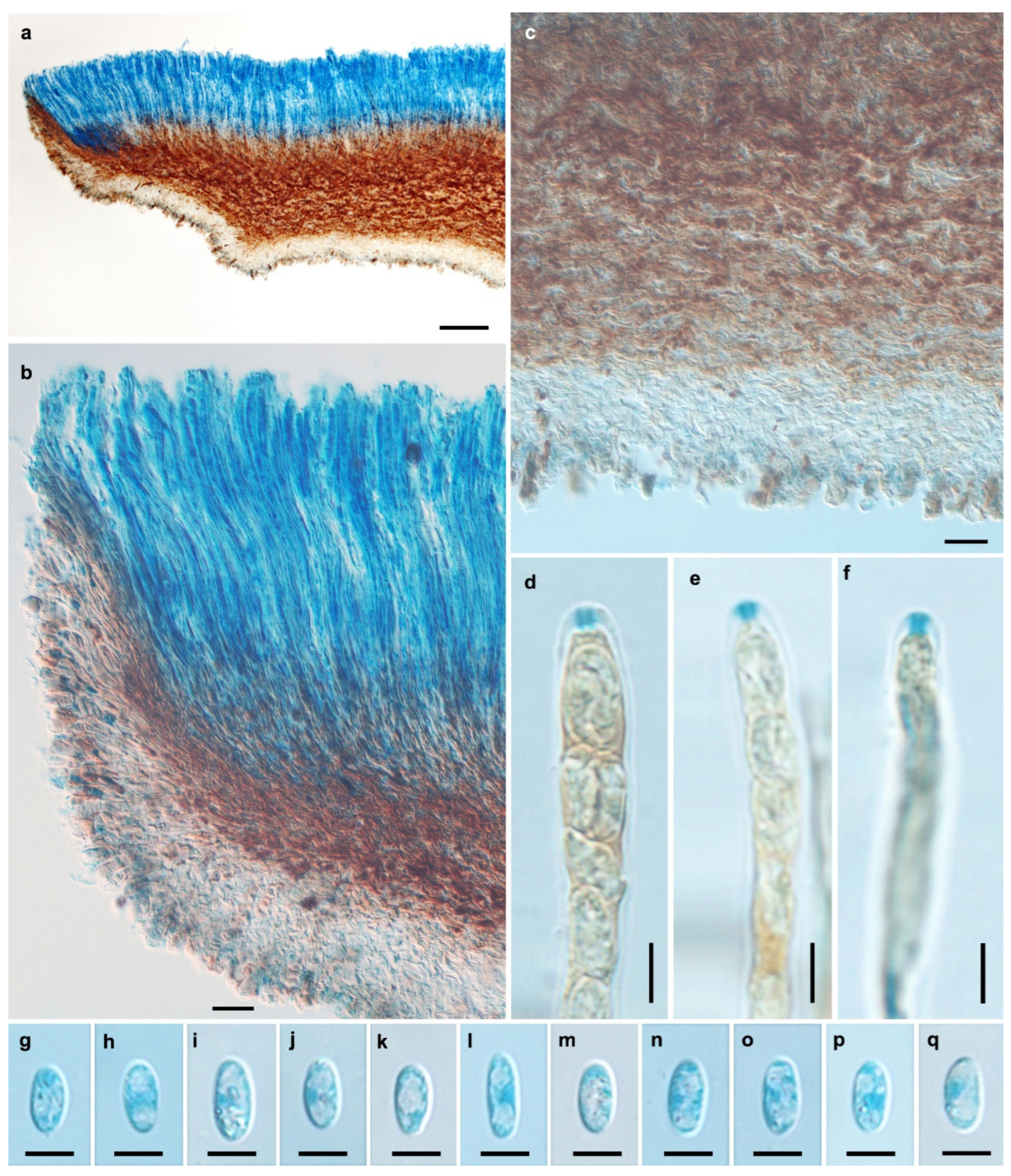
3.2.2. Chlorencoelia ellipsoidea H.D. Zheng & W.Y. Zhuang, sp. nov. (Figure 2d–f and Figure 4)
Fungal Names—FN 573050.
Etymology—The specific epithet refers to the ellipsoid ascospores of the fungus.
Figure 4.
Microscopic features of Chlorencoelia ellipsoidea (HMAS 290936, holotype). (a) Longitudinal section of apothecium (partial); (b) hymenium and excipular structure of margin and flank; (c) excipular structure of flank; (d–f) IKI reaction of ascus apical ring without KOH pretreatment; (g–q) ascospores. Scale bars: (a) = 100 μm; (b,c) = 20 μm; (d–q) = 5 μm. Mounting media: (a–c,g–q) Lactophenol cotton blue; (d–f) Melzer’s reagent.
Holotype—China, Yunnan, Gaoligong Mountains, Baihualing, Jinchanghe, on decorticated rotten wood, 17 September 2017, Huan-Di Zheng, Xin-Cun Wang, Yu-Bo Zhang & Yi Zhang 11442 (HMAS 290936, ITS: PX453032, 28S: PX630175, RPB1: PX557847, RPB2: PX557854, TEF1: PX529003).
Chinese Name—椭孢绿散孢盘菌, tuǒ bāo lǜ sǎn bāo pán jūn.
Description—Apothecia solitary, gregarious to caespitose, shallowly infundibuliform, distinctly stipitate, 7–12 mm in diam.; hymenium surface atrovirens, dark green to blue green when fresh; receptacle surface brown, somewhat furfuraceous; stipe concolorous with the receptacle. Ectal excipulum of textura angularis, 40–105 μm thick; cells hyaline to light brown, 19–30 × 9–16.5 μm, some nearly isodiametric and 11–16.5 μm in diam., thin- to slightly thick-walled, giving rise to tomentum hyphae oriented nearly perpendicularly to the flank surface; tomentum hyphae clavate, 11–22 × 7–12 μm, walls 2–4 μm thick. Medullary excipulum of textura intricata, tightly arranged, 50–260 μm thick; hyphae brown, 2.5–5 μm wide. Subhymenium not distinguishable. Hymenium 205–220 μm thick. Asci arising from simple septa, 8-spored, cylindric-clavate, apex rounded, apical rings J+ in Melzer’s reagent and Lugol’s solution without KOH pretreatment, visible as two blue lines, 115–126 × 5.5–6 μm. Ascospores broad ellipsoidal, non-septate, thin-walled, hyaline, smooth, with 1–2 large guttules and some small ones, filling most part of the ascospores, uniseriate, 7.2–9.1 × 3.5–4.2 μm. Paraphyses filiform, septate, simple, hyaline, 3–3.5 μm wide at apex and 2–2.5 μm wide below, equaling to or slightly exceeding the asci by 5–10 μm.
Known distribution—China (Yunnan).
Notes—Chlorencoelia ellipsoidea is distinct from other members of the genus by dark green hymenium surface, distinct stipe and broad ellipsoidal ascospores. Phylogenetically, it is a sister species of C. torta, which is different in smaller apothecia, olive-yellow to olive-green hymenium surface, larger, biguttulate, and subcylindric-ellipsoidal ascospores [1].
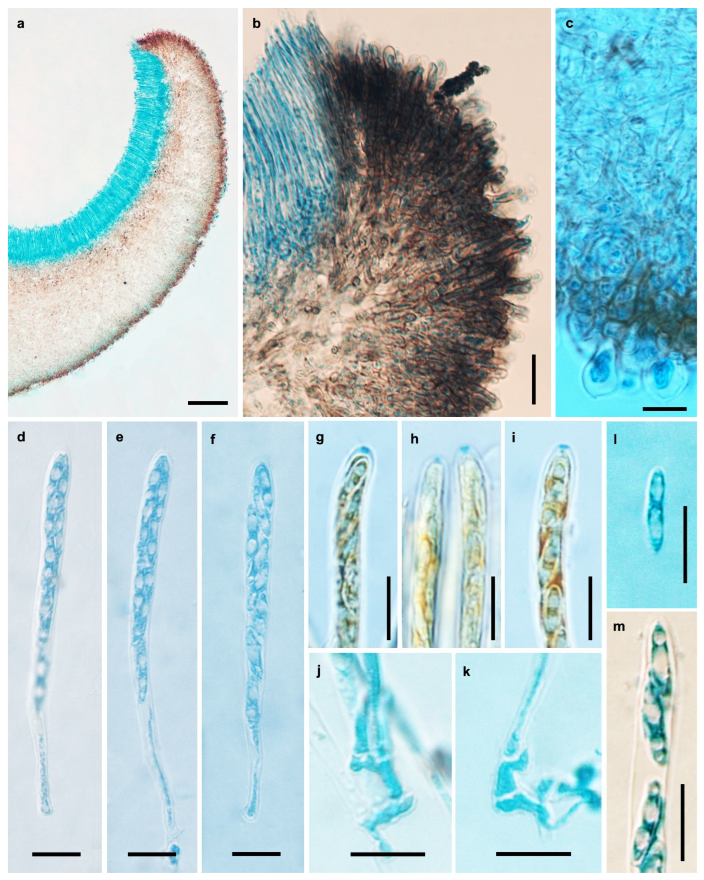
3.2.3. Chlorencoelia sinensis H.D. Zheng & W.Y. Zhuang, sp. nov. (Figure 2g–k and Figure 5)
Fungal Names—FN 573051.
Etymology—The specific epithet refers to the known geographic range of the fungus.
Holotype—China, Chongqing, Jinfo Mountain, on decorticated rotten wood, 29.0275° N, 107.1869° E, alt. 2130 m, 26 October 2020, Huan-Di Zheng, Zhao-Qing Zeng, Xin-Cun Wang & Chang Liu 12699 (HMAS 290937, ITS: PX453033, 28S: PX630176, RPB1: PX557848, RPB2: PX557855, TEF1: PX529004).
Chinese Name—中国绿散孢盘菌, zhōng guó lǜ sǎn bāo pán jūn.
Description—Apothecia solitary, gregarious to caespitose, occasionally 2–3 forming on a common base, discoid to shallowly cupulate, short-stipitate to sub-stipitate, 2–7 mm in diam.; hymenium surface yellowish green when fresh, greyish green after drying; receptacle surface nearly black; stipe concolorous with the receptacle. Ectal excipulum of textura angularis, 30–70 μm thick; oriented nearly perpendicularly to the receptacle surface, cells 5.5–11 μm in diam., walls brown, giving rise to filamentous (margin to upper portion) to clavate (flank to lower portion) tomentum hyphae, 6–19 × 5–11 μm, thick-walled, walls 1.5–5 μm thick. Medullary excipulum of textura intricata, 80–680 μm thick; hyphae brownish, 3–4.5 μm wide. Subhymenium not distinguishable. Hymenium 95–110 μm thick. Asci arising from croziers, 8-spored, cylindric-clavate, apex rounded, narrowed gradually into a short stalk, apical rings J+ in Melzer’s reagent and Lugol’s solution without KOH pretreatment, visible as two blue lines, 75–100 × 5.2–6 μm. Ascospores fusoidal, anterior end round, posterior end pointed, slightly flattened on one side, aseptate, thin-walled, hyaline, smooth, with two large guttules and some small ones, filling most part of the ascospores, irregularly biseriate, 9–13.7 × 2.2–3.5 μm. Paraphyses filiform, septate, simple, hyaline, 2.5–3.5 μm wide at apex and 2–3 μm wide below, equaling to or slightly exceeding the asci.
Paratype—China, Yunnan, Shangri La, Jiantang County, Potatso National Park, 27.8275° N, 99.9579° E, alt. 3670 m, on decorticated rotten wood, 30 September 2020, Xiang-Hua Wang 9258 (HMAS 290938, ITS: PX453034, 28S: PX630177, RPB1: PX557849).
Additional specimen examined—China, Anhui, Anqing, Yuexi, Miaodaoshan National Forest Park, 30.4953° N, 116.0528° E, alt. 622 m, on decorticated rotten wood, 19 October 2021, Lei Yao DBSAH20211019100 (HMAS 285422, as C. torta).
Known distribution—China (Anhui, Chongqing, Taiwan, Yunnan).
Figure 5.
Microscopic features of Chlorencoelia sinensis (HMAS 290937, holotype). (a) Longitudinal section of apothecium (partial); (b) excipular structure of margin and flank; (c) excipular structure of flank; (d–f) asci; (g–i) IKI reaction of ascus apical ring without KOH pretreatment; (j,k) croziers at ascus bases; (l) ascospore; (m) ascospores in ascus. Scale bars: (a) = 100 μm; (b) = 20 μm; (c–m) = 10 μm. Mounting media: (a–f,j–m) Lactophenol cotton blue; (g–i). Melzer’s reagent.
Notes—Chlorencoelia sinensis is similar to C. torta in excipular structure and resembles C. versiformis in ascospore morphology [1], but it is distinct in the combination of substipitate apothecia, blackish receptacle surface and fusoidal ascospores. Collection of the species from Anhui Province (HMAS 285422) and ITS sequences generated from materials from Taiwan Province (LC862140, LC862114) were previously misidentified as C. torta. Phylogenetically, C. sinensis exhibits distant relationships with C. torta and C. versiformis, while it is relatively close to C. olivacea, C. northlandica and the three undescribed species (Figure 1). C. olivacea is different in dark green or bluish hymenium surface and pale brown excipular cells. C. northlandica can be distinguished by dark green to grey-blue hymenium surface and broader ascospores (9.5–13 × 3–4.5 μm).
3.2.4. Comparison of Morphological Characteristics Among Chlorencoelia Species
To give a straightforward comparison and facilitate recognition of Chlorencoelia species, the key morphological taxonomic characteristics are summarized in Table 2.
Table 2.
Comparisons of morphological characteristics of Chlorencoelia species.
3.2.5. Key to the Known Species of Chlorencoelia
| 1. Ascospores 20–40 × 4.5–6 μm, cylindric-fusoid | C. macrospora |
| 1. Ascospores less than 20 μm long | 2 |
| 2. Ascospores pyriform, 5–8 × 1.5–3 μm | C. ripakorfii |
| 2. Ascospores of other shapes, mostly longer than 8 μm | 3 |
| 3. Tomentum hyphae filamentous | C. versiformis |
| 3. Tomentum hyphae clavate | 4 |
| 4. Ascospores eguttulate, ellipsoidal, 0–1 septate, 7.5–9.7 × 2.2–3.7 μm | C. indica |
| 4. Ascospores guttulate | 5 |
| 5. Ascospores fusoidal, 9–13.7 × 2.2–3.5 μm | C. sinensis |
| 5. Ascospores ellipsoidal | 6 |
| 6. Ascospores broad-ellipsoidal, length/width less than 2.2, 7.2–9.1 × 3.5–4.2 μm, medullary excipulum brown | C. ellipsoidea |
| 6. Ascospores ellipsoidal, length/width more than 2.2 | 7 |
| 7. Ascospores ellipsoidal, 8.5–12 × 2.5–3.5 μm | C. olivacea |
| 7. Ascospores oblong-ellipsoidal | 8 |
| 8. Tomentum hyphae barely differentiated, ascospores 9.5–13 × 3–4.5 μm | C. northlandica |
| 8. Tomentum hyphae well-differentiated | 9 |
| 9. Tomentum hyphae slightly thick-walled, ascospores relatively large, (9.5–)11–16.5 × (2.5–)3.5–5 μm | C. australis |
| 9. Tomentum hyphae distinctly thick-walled, ascospores relatively small | 10 |
| 10. Hymenium surface olive yellow to olive green when fresh, ascospores (5.6–)9–11(–12) × 2–4 μm | C. torta |
| 10. Hymenium surface greyish green to blackish green when fresh, ascospores 8.5–14.5 × 3.5–4.5 μm | C. biguttulata |
4. Discussion
Upon the establishment of Chlorencoelia, taxonomy of the genus, for a long time, was mainly based on morphological features of tomentum hyphae, ascus, and ascospores. Owing to the small apothecia and inconspicuous coloration, members of Chlorencoelia are not easily determined in the field; thus limited attention was paid to the genus and taxonomy of the genus was not well documented. Moreover, due to the morphological features of different species being very similar or overlapping, it is difficult to draw clear boundaries among species, which results in the existence of cryptic species. According to morphological traits, a considerable number of specimens from different parts of the world were identified as C. torta or C. versiformis, the two commonly known species. Nowadays, integrative analysis of molecular and morphological data facilitates accurate species identification. For example, C. australis and C. northlandica were separated from C. torta or C. versiformis based on DNA sequencing and morphological analyses of specimens from New Zealand [5]. Similar results can be found in the present study; C. biguttulata and C. sinensis were distinguished from C. torta or C. versiformis according to the combined data. As the number of species found increases, it becomes difficult to distinguish Chlorencoelia species merely by morphology, especially the species having clavate tomentum hyphae and ellipsoidal ascospores (C. australis, C. biguttulata, C. northlandica, C. olivacea and C. torta). Molecular inference plays an important role in accurate species recognition in addition to morphological observations.
As for DNA barcodes for species identification, only ITS sequences are available for most Chlorencoelia species, and the fragment has been proven to be efficient. It is unfortunate that ITS sequences of C. indica, C. macrospora, and C. ripakorfii are not available, and we cannot locate their phylogenetic positions (Figure 1). We have acquired the 28S, RPB1, RPB2, and TEF1 sequences of the new species; however, these DNA markers are not available for other species. Multi-gene phylogenetic analysis surely awaits the accumulation of more DNA fragments of the group.
In subsequent research, mycologists worldwide need to pay more attention to Chlorencoelia, carry out field surveys across a broader geographical range, cover diverse vegetation types, collect specimens from various substrates, isolate pure cultures, check morphological features in detail, conduct multi-locus phylogenetic analyses, and undertake systematic comparative studies. In doing so, an increasing number of novel species will be uncovered, the ecology and substrate preference of the genus will be better understood, and a comprehensive infrageneric framework of Chlorencoelia will be fully resolved in the future.
Supplementary Materials
The following supporting information can be downloaded at: https://www.mdpi.com/article/10.3390/jof11120855/s1, Supplementary File S1: Newly generated ITS sequences; Supplementary File S2: Newly generated 28S sequences; Supplementary File S3: Newly generated RPB1 sequences; Supplementary File S4: Newly generated RPB2 sequences; Supplementary File S5: Newly generated TEF1 sequences.
Author Contributions
Conceptualization, H.-D.Z. and W.-Y.Z.; methodology, H.-D.Z.; software, H.-D.Z.; validation, H.-D.Z. and W.-Y.Z.; formal analysis, H.-D.Z.; investigation, H.-D.Z.; writing—original draft preparation, H.-D.Z.; writing—review and editing, H.-D.Z. and W.-Y.Z.; visualization, H.-D.Z.; supervision, W.-Y.Z. All authors have read and agreed to the published version of the manuscript.
Funding
This study was supported by the National Key Research and Development Program of China (2022YFC2303000), the Taxonomic Scientist Position of the Chinese Academy of Sciences (CAS-TAX-24-020), and the National Natural Science Foundation of China (31770019).
Institutional Review Board Statement
Not applicable.
Informed Consent Statement
Not applicable.
Data Availability Statement
The newly generated sequences were deposited in GenBank; accession numbers are included in the article (https://www.ncbi.nlm.nih.gov/genbank, accessed on 9 October 2025 and 11 November 2025; Supplementary Files S1–S5). Names of the new species were formally registered in the global data repository Fungal Names (https://nmdc.cn/fungalnames, accessed on 10 October 2025). The original contributions presented in this study are included in the article. Further inquiries can be directed to the corresponding author.
Acknowledgments
The authors would like to thank Xiang-Hua Wang (Kunming Institute of Botany, Chinese Academy of Sciences) for sending a valuable specimen. All co-collectors of the specimens are acknowledged for their help and cooperation during fieldwork. We are also grateful to reviewers for their invaluable suggestions and comments.
Conflicts of Interest
The authors declare no conflicts of interest.
Correction Statement
This article has been republished with a minor correction to resolve spelling errors. This change does not affect the scientific content of the article.
References
- Dixon, J.R. Chlorosplenium and its segregates II. The genera Chlorociboria and Chlorencoelia. Mycotaxon 1975, 1, 193–237. [Google Scholar] [CrossRef] [Scilit]
- Zhuang, W.-Y. Studies on some discomycete genera with an ionomidotic reaction: Ionomidotis, Poloniodiscus, Cordierites, Phyllomyces, and Ameghiniella. Mycotaxon 1988, 31, 261–298. [Google Scholar] [CrossRef] [Scilit]
- Iturriaga, T.; Mardones, M. A new species of Chlorencoelia from Parque Nacional El Avila, Venezuela. Mycosystema 2013, 32, 457–461. [Google Scholar]
- Ren, F.; Zhuang, W.-Y. A new species of the genus Chlorencoelia (Helotiales) from China. Mycoscience 2014, 55, 227–230. [Google Scholar] [CrossRef] [Scilit]
- Patejuk, K.; Johnston, P.R.; Park, D.; Padamsee, M. Chlorencoelia (Leotiomycetes, Helotiales, Cenangiaceae) in New Zealand. MycoKeys 2025, 119, 351–368. [Google Scholar] [CrossRef] [Scilit] [PubMed]
- Wang, Z.; Binder, M.; Schoch, C.L.; Johnston, P.R.; Spatafora, J.W.; Hibbett, D.S. Evolution of helotialean fungi (Leotiomycetes, Pezizomycotina): A nuclear rDNA phylogeny. Mol. Phylogenet. Evol. 2006, 41, 295–312. [Google Scholar] [CrossRef] [Scilit] [PubMed]
- Wang, Z.; Johnston, P.R.; Takamatsu, S.; Spatafora, J.W.; Hibbett, D.S. Toward a phylogenetic classification of the Leotiomycetes based on rDNA data. Mycologia 2006, 98, 1065–1075. [Google Scholar] [CrossRef] [Scilit]
- Wang, Z.; Johnston, P.R.; Yang, Z.L.; Townsend, J.P. Evolution of reproductive morphology in leaf endophytes. PLoS ONE 2009, 4, e4246. [Google Scholar] [CrossRef] [Scilit]
- Pärtel, K.; Baral, H.-O.; Tamm, H.; Põldmaa, K. Evidence for the polyphyly of Encoelia and Encoelioideae with reconsideration of respective families in Leotiomycetes. Fungal Divers 2017, 82, 183–219. [Google Scholar] [CrossRef] [Scilit]
- Johnston, P.R.; Quijada, L.; Smith, C.A.; Baral, H.O.; Hosoya, T.; Baschien, C.; Partel, K.; Zhuang, W.Y.; Haelewaters, D.; Park, D.; et al. A multigene phylogeny toward a new phylogenetic classification of Leotiomycetes. IMA Fungus 2019, 10, 1. [Google Scholar] [CrossRef] [Scilit]
- Voglmayr, H.; Jaklitsch, W.M.; Tello, S. Mycosphaerangium and Neomelanconium (Cenangiaceae) are closest relatives: Phylogenetic relationships, morphology and a new species. Mycol. Prog. 2020, 19, 1329–1352. [Google Scholar] [CrossRef] [Scilit]
- Wijayawardene, N.N.; Hyde, K.D.; Al-Ani, L.K.T.; Tedersoo, L.; Haelewaters, D.; Rajeshkumar, K.C.; Zhao, R.L.; Aptroot, A.; Leontyev, D.V.; Saxena, R.K.; et al. Outline of Fungi and fungus-like taxa. Mycosphere 2020, 11, 1060–1456. [Google Scholar] [CrossRef] [Scilit]
- Gomez-Zapata, P.A.; Haelewaters, D.; Quijada, L.; Pfister, D.H.; Aime, M.C. Notes on Trochila (Ascomycota, Leotiomycetes), with new species and combinations. MycoKeys 2021, 78, 21–47. [Google Scholar] [CrossRef] [Scilit]
- Ribes, M.A.; Escobio, V.; Negrin, R.; Baral, H.O.; Pfister, D.H.; Quijada, L. Wanted on Agave americana! Hymenobolus agaves, an overlooked introduced pathogen in the western palearctic region. Fungal. Syst. Evol. 2021, 8, 129–142. [Google Scholar] [CrossRef] [Scilit] [PubMed]
- Zhuang, W.-Y.; Zheng, H.-D.; Ren, F.; Song, X. Flora Fungorum Sinicorum Volume 56 Helotiaceae; Science Press: Beijing, China, 2018; p. 231. [Google Scholar]
- Zhuang, W.-Y.; Zheng, H.-D.; Zeng, Z.-Q. Species Catalogue of China, Volumen 3 Fungi, Cup-Fungi; Science Press: Beijing, China, 2018; p. 142. [Google Scholar]
- White, T.J.; Bruns, T.; Lee, S.; Taylor, J. Amplification and direct sequencing of fungal ribosomal RNA genes for phylogenetics. In PCR Protocols: A Guide to Methods and Applications; Innis, M.A., Gelfand, D.H., Snisky, J.J., White, T.J., Eds.; Academic Press: London, UK, 1990; pp. 315–322. [Google Scholar] [CrossRef] [Scilit]
- Moncalvo, J.-M.; Wang, H.-H.; Hseu, R.-S. Phylogenetic relationships in Ganoderma inferred from the internal transcribed spacers and 25S ribosomal DNA sequences. Mycologia 1995, 87, 223–238. [Google Scholar] [CrossRef] [Scilit]
- Vilgalys, R.; Hester, M. Rapid genetic identification and mapping of enzymatically amplified ribosomal DNA from several Cryptococcus species. J. Bacteriol. 1990, 172, 4238–4246. [Google Scholar] [CrossRef] [Scilit]
- Stiller, J.W.; Hall, B.D. The origin of red algae: Implications for plastid evolution. Proc. Natl. Acad. Sci. USA 1997, 94, 4520–4525. [Google Scholar] [CrossRef] [Scilit]
- Matheny, P.B.; Liu, Y.J.; Ammirati, J.F.; Hall, B.D. Using RPB1 sequences to improve phylogenetic inference among mushrooms (Inocybe, Agaricales). Am. J. Bot. 2002, 89, 688–698. [Google Scholar] [CrossRef] [Scilit] [PubMed]
- Liu, Y.J.; Whelen, S.; Hall, B.D. Phylogenetic relationships among Ascomycetes: Evidence from an RNA polymerse II subunit. Mol. Biol. Evol. 1999, 16, 1799–1808. [Google Scholar] [CrossRef] [Scilit]
- Rehner, S.A.; Buckley, E. A Beauveria phylogeny inferred from nuclear ITS and EF1-alpha sequences: Evidence for cryptic diversification and links to Cordyceps teleomorphs. Mycologia 2005, 97, 84–98. [Google Scholar] [CrossRef] [Scilit]
- Zheng, H.-D.; Zhuang, W.-Y. Additions to the knowledge of the genus Pezicula (Dermateaceae, Helotiales, Ascomycota) in China. Biology 2022, 11, 1386. [Google Scholar] [CrossRef] [Scilit]
- Hall, T.A. BioEdit: A user-friendly biological sequence alignment editor and analysis program for Windows 95/98/NT. Nucleic Acids Symp. Ser. 1999, 41, 95–98. [Google Scholar]
- Ronquist, F.; Huelsenbeck, J.P. MrBayes 3: Bayesian phylogenetic inference under mixed models. Bioinformatics 2003, 19, 1572–1574. [Google Scholar] [CrossRef] [Scilit]
- Stamatakis, A. RAxML version 8: A tool for phylogenetic analysis and post-analysis of large phylogenies. Bioinformatics 2014, 30, 1312–1313. [Google Scholar] [CrossRef] [Scilit] [PubMed]
- Nylander, J.A.A. MrModeltest, v2. Program distributed by the author. Evolutionary Biology Centre, Uppsala University: Uppsala, Sweden, 2004.
- Rambaut, A. FigTree, v1.4.4; Institute of Evolutionary Biology, University of Edinburgh: Edinburgh, UK, 2018. Available online: http://tree.bio.ed.ac.uk/software/figtree (accessed on 22 May 2019).
- Thind, K.S.; Cash, E.K.; Singh, P. The Helotiales of the Mussoorie Hills—II. Mycologia 1959, 51, 833–839. [Google Scholar] [CrossRef] [Scilit]

Disclaimer/Publisher’s Note: The statements, opinions and data contained in all publications are solely those of the individual author(s) and contributor(s) and not of MDPI and/or the editor(s). MDPI and/or the editor(s) disclaim responsibility for any injury to people or property resulting from any ideas, methods, instructions or products referred to in the content. |
© 2025 by the authors. Licensee MDPI, Basel, Switzerland. This article is an open access article distributed under the terms and conditions of the Creative Commons Attribution (CC BY) license (https://creativecommons.org/licenses/by/4.0/).