


Abstract
Wild Polygala crotalarioides is an ethnomedicinal plant traditionally used for neurological health. Endophytic fungi associated with medicinal plants are increasingly recognized as sources of bioactive natural products; however, their diversity and biological activities in wild P. crotalarioides remain poorly understood. We used high-throughput sequencing and culture-dependent methods to explored the diversity, community composition, and bioactivities of endophytic fungi associated with wild P. crotalarioides. Culturable isolates were identified via ITS sequencing and further screened for AChE-inhibitory and antioxidant activities. Our results revealed that a total of 818 fungal OTUs were detected, revealing pronounced tissue-specific community differentiation. Stem tissues harbored the highest fungal diversity, richness, and number of unique OTUs. Ascomycota and Basidiomycota dominated fungal communities across all tissues, while distinct biomarker taxa were enriched in different organs. Functional annotation indicated marked tissue-dependent variation in trophic composition. Among 108 culturable isolates, Penicillium sp. PRP024 and Trichoderma sp. PRP008 exhibited the strongest AChE inhibitory activities (>80%), whereas Fusarium sp. PSP053, Diaporthe sp. PSM027 and Exserohilum sp. PSP042 showed notable antioxidant activities. Wild P. crotalarioides hosts highly diverse and tissue-specific endophytic fungal communities. Several isolates exhibited promising acetylcholinesterase inhibitory and antioxidant activities, highlighting medicinal plant-associated endophytic fungi as potential sources of antioxidative natural products.
1. Introduction
Medicinal plants are indispensable reservoirs of bioactive substances and serve as core materials in global traditional medicine, providing abundant compounds for modern pharmaceutical and healthcare product development [1,2,3]. Endophytic fungi ubiquitously colonize the internal tissues of healthy host plants and form stable symbiotic relationships with their hosts [4,5]. Accumulating ethnopharmacological and phytochemical evidence confirms that endophytic fungi derived from medicinal herbs can synthesize secondary metabolites structurally and functionally similar to host plant components, possessing diverse pharmacological properties [6,7]. Accordingly, endophytic fungi have emerged as a promising and untapped microbial resource for exploring novel natural bioactive compounds from traditional medicinal plants.
The Polygala (Polygalaceae), consisting of approximately 500 species worldwide, with over 39 species distributed in China [8], is a well-recognized medicinal plant group in traditional folk medicine [8,9], with prominent effects on neuroprotection, antioxidation, memory improvement and anti-inflammation [10,11]. In the past three to five years, increasing studies have focused on the phytochemical profiles, pharmacological mechanisms and quality evaluation of Polygala species. Multiple bioactive constituents, including triterpene saponins, xanthones, oligosaccharide esters, flavonoids and coumarins, have been extensively identified from Polygala plants. Among them, tenuifolin, polygalaxanthone III and 3,6′-disinapoyl sucrose are widely adopted as standard quality control markers [8,9]. Modern pharmacological studies have further demonstrated that Polygala extracts and monomeric compounds exert remarkable neuroprotective effects against Alzheimer’s disease (AD) by inhibiting Aβ aggregation, suppressing tau hyperphosphorylation, relieving oxidative stress, and regulating cholinergic and neuroinflammatory pathways [12,13]. Given the critical roles of oxidative stress and cholinergic dysfunction in AD progression, acetylcholinesterase (AChE) has emerged as a key therapeutic target, and natural antioxidants are considered promising intervention candidates [14,15,16]. However, currently available AChE inhibitors, such as donepezil, rivastigmine, and galantamine, are frequently associated with gastrointestinal, cardiovascular, and neurological adverse effects, which may reduce patient adherence and restrict long-term therapeutic efficacy [16,17]. Consequently, screening for natural AChE inhibitors from plant endophytes has become a research priority for developing safe and novel neuroprotective agents. Given the inherent antioxidant and neuroprotective properties of Polygala plants, their symbiotic endophytic fungi are considered excellent candidates for mining bioactive metabolites with AChE-inhibitory and antioxidant activities. Nevertheless, existing studies on P. crotalarioides are merely confined to cultivation techniques and preliminary phytochemical analysis. Notably, the community composition, tissue distribution and temporal variation in endophytic fungi associated with wild P. crotalarioides across different plant tissues and growth stages remain unexplored.
In addition to pharmacological studies, recent research has also explored the genetic diversity, resource conservation, and sustainable utilization of Polygala medicinal materials [18,19]. Polygala crotalarioides, a perennial herb endemic to Southwest China, is locally used as a traditional medicinal plant [20]. Our research team has previously explored the artificial cultivation and basic biological characteristics of this species, providing a foundation for its sustainable resource utilization [21]. Nevertheless, existing studies on P. crotalarioides remain confined to cultivation techniques and preliminary phytochemical analysis; the community composition, tissue distribution, and temporal variation in endophytic fungi associated with wild P. crotalarioides across different tissues and growth stages remain entirely unexplored.
The integration of culture-independent high-throughput sequencing (HTS) and traditional culture-dependent isolation has been widely adopted as a complementary approach for comprehensively exploring the diversity and bioresource potential of endophytic fungi [22,23]. HTS enables comprehensive characterization of the diversity and community structure of plant endophytic fungi, whereas culture-dependent isolation provides living fungal strains for downstream bioactivity screening, metabolite discovery, and pharmacological validation [21]. To date, studies on wild P. crotalarioides have mainly focused on the characterization of endophytic fungal community diversity, whereas investigations integrating spatiotemporal community dynamics with the bioactivity evaluation of cultivable endophytic fungi remain limited.
Based on the above findings and our previous studies on cultivated P. crotalarioides [21], two key questions remain unresolved: (i) how endophytic fungal communities are distributed among different tissues of wild P. crotalarioides and whether tissue-specific differences exist; and (ii) whether cultivable endophytic fungi from wild P. crotalarioides possess antioxidant and AChE inhibitory activities. Addressing these questions is important for understanding the endophytic fungal resources associated with this ethnomedicinal plant and their potential functional significance.
Therefore, the present study integrated HTS and culture-dependent isolation to systematically explore the endophytic fungi inhabiting the leaves, stems, and roots of wild P. crotalarioides. Community richness, diversity, and dominant fungal taxa were characterized across different plant tissues, and the isolated fungal strains were further evaluated for DPPH and ABTS radical scavenging activities as well as AChE inhibitory activity in vitro. This study aimed to reveal the endophytic fungal community characteristics of wild P. crotalarioides and identify cultivable fungal resources with potential antioxidant and AChE inhibitory activities, thereby providing a basis for future studies on the functional exploitation of endophytic fungi associated with medicinal Polygala species.
2. Materials and Methods
2.1. Sample Collection
Wild Polygala crotalarioides plants were sampled in May 2022 from Shangzhai Natural Village, Cangyuan County, Yunnan, China (23°08′48.16″ N, 99°14′46.31″ E). Three independent 3 × 3 m plots (≥150 m apart) were established, and five healthy plants were randomly selected per plot. Root, stem, leaf, and flower tissues were collected separately and pooled by organ type within each plot to generate one composite sample per plot, yielding three biological replicates per tissue. Fresh samples were transported to the laboratory under sterile, low-temperature conditions and processed within 24 h. A portion of each sample was used directly for high-throughput sequencing, and the remainder was processed aseptically for endophytic fungal isolation. Sample codes followed the format: R (root), S (stem), L (leaf), and F (flower).
2.2. Sample Processing
Roots and aerial tissues were separated using sterile scissors, and surface soil was removed by rinsing with sterile water. Samples were surface-sterilized by sequential immersion in 75% ethanol (2 min), 2.5% sodium hypochlorite (NaClO) solution containing 0.1% Tween 80 (5 min), and 75% ethanol (30 s), followed by three rinses with sterile water and drying with sterile filter paper [22]. The effectiveness of sterilization was verified by spreading 100 μL of the final rinse solution on PDA medium and incubating at 28 °C for 24 h; absence of colony formation confirmed successful surface disinfection [21]. Sterilized tissues were cut into 0.5 cm fragments under aseptic conditions. Intact lesion-free samples (3 g) were weighed, transferred into sterile 50 mL tubes, and stored at −80 °C until DNA extraction.
2.3. High-Throughput Sequencing Analysis
2.3.1. DNA Extraction and PCR Amplification
All samples’ total DNA was extracted from plant tissues using the E.Z.N.A.® Plant DNA Kit (Omega Bio-Tek, Norcross, GA, USA) following the manufacturer’s protocol. Extraction blanks (sterile deionized water) were included to monitor cross-contamination. DNA integrity was assessed by 1% agarose gel electrophoresis, and DNA concentration and purity were measured using a NanoDrop 2000 spectrophotometer (Thermo Fisher Scientific, Waltham, MA, USA).
Fungal ITS fragments were amplified using primers ITS1F (5′-CTTGGTCATTTAGAGGAAGTAA-3′) and ITS2R (5′-GCTGCGTTCTTCATCGATGC-3′) [24] (Thermo Fisher Scientific, Waltham, MA, USA). Each 20 µL reaction contained 4 µL of 5× FastPfu buffer, 2 µL of 2.5 mM dNTPs, 0.8 µL of each primer (5 µM), 0.4 µL of FastPfu DNA polymerase (TransGen Biotech, Beijing, China), and 10 ng of template DNA. Thermal cycling consisted of initial denaturation at 95 °C for 3 min; 27 cycles of 95 °C for 30 s, 55 °C for 30 s, and 72 °C for 45 s; and a final extension at 72 °C for 10 min. Negative controls (sterile water replacing template DNA) were included in each batch. Amplicons were verified by 2% agarose gel electrophoresis, and paired-end sequencing was performed on the Illumina NextSeq 2000 platform at Majorbio Bio-Pharm Technology Co., Ltd. (Shanghai, China). Raw reads were deposited in the NCBI Sequence Read Archive (SRA) under BioProject accession PRJNA1207743, with sample accessions ranging from SRR31976288 to SRR31976299.
2.3.2. Sequence Processing and OTU Clustering
Raw sequencing data were quality-filtered using fastp v0.20.0 [25]. with a sliding window (50 bp) quality threshold of Q20; reads shorter than 50 bp or containing ambiguous bases were discarded. High-quality paired-end reads were assembled using FLASH v1.2.7 [26] with default parameters (minimum overlap: 10 bp; maximum mismatch ratio: 0.2). Valid sequences were demultiplexed according to barcode sequences, allowing up to two primer mismatches. Clean reads were clustered into operational taxonomic units (OTUs) at a 97% similarity threshold using UPARSE v7.1 [27], with the threshold adopted following standard practice for fungal ITS metabarcoding. Non-fungal sequences (chloroplast and mitochondrial origin) were excluded. Taxonomic annotation was performed against the UNITE fungal ITS database (v8.0) [28] using the RDP Classifier (v2.14) with a 70% bootstrap confidence threshold [29]. To minimize biases from uneven sequencing depth, the OTU table was rarefied to the minimum valid sequence count per sample.
2.3.3. Statistical Analysis
α- Diversity indices (Shannon, Simpson, Chao1, and ACE) were calculated using mothur v1.48.0 [30]. Normality was assessed using the Shapiro–Wilk test. For non-normally distributed data, group differences were evaluated using the Kruskal–Wallis test followed by Dunn’s post hoc test. β-Diversity was assessed via principal coordinate analysis (PCoA) based on Bray–Curtis distances. Community structure differences were statistically tested using ANOSIM and PERMANOVA (999 permutations). Linear discriminant analysis effect size (LEfSe) was employed to identify stage- and tissue-specific biomarkers (LDA score ≥ 2.5, p < 0.05). Fungal ecological guilds were assigned using FUNGuild v1.1 [31]. All bioinformatic and statistical analyses were performed in R v4.5.2 using the vegan v2.6-4, ggplot2 v3.4.4, VennDiagram v1.7.3, pheatmap, and UpSetR v1.4.0 packages [32]. Statistical significance was set at p < 0.05, with p < 0.01 considered highly significant.
2.4. Isolation and Characterization of Culturable Endophytic Fungi
Surface-sterilized tissues were cut into 3–5 mm fragments and placed on PDA medium (5–8 fragments per plate). Plates were incubated at 28 °C for at least 10 days; emerging fungal colonies were subcultured onto fresh PDA and preliminarily classified based on colony morphology [33]. Owing to limited flower tissue availability, the number of isolates obtained from these samples was restricted. Purified strains were preserved in 50% (v/v) glycerol at −80 °C.
Fungal genomic DNA was extracted using a modified CTAB method [34] with reference to the Omega fungal DNA extraction kit protocol. DNA quality and concentration were assessed using a NanoDrop spectrophotometer, retaining samples with DNA concentration ≥ 100 ng/25 μL. The ITS region was amplified with primers ITS1 and ITS4 [35]. The 60 μL PCR system contained 42.6 μL sterile ddH2O, 6 μL 10× buffer, 1.2 μL 2 mmol dNTPs, 1.5 μL of each 10 pmol primer, 1.2 μL 5 U Taq polymerase and 6 μL DNA template. Amplification conditions were set as initial pre-denaturation at 94 °C for 5 min; 35 cycles of 94 °C for 90 s, 53 °C for 90 s and 72 °C for 2 min; followed by final extension at 72 °C for 10 min. Amplicons were verified by 1% agarose gel electrophoresis and sent to Kunming Shuoqing Biotechnology Co., Ltd. (Kunming, China) for Sanger sequencing. Obtained sequences were compared against the GenBank database via BLAST 2.15. Isolates exhibiting sequence similarity below 95% were tentatively considered unidentified species; those with ≥95% similarity were assigned to genus level, and sequences with ≥97% identity were tentatively classified to species level. All sequences have been deposited in GenBank under accession numbers PV175970–PV176077.
2.5. Evaluation of Biological Activity
2.5.1. Strain Fermentation and Extract Preparation
For metabolite production, solid-state fermentation was performed in glass culture vessels (5.7 cm internal diameter × 9.2 cm height), each containing 75 g of commercially available white rice and 75 mL of distilled water, with 1 mL of distilled water replaced by trace element solution to maintain a final liquid volume of 75 mL and a strict 1:1 (w/v) ratio of rice to liquid. The trace element solution contained, per liter: MgSO4·7H2O 0.01 g, CuSO4·5H2O 0.005 g, and FeSO4·7H2O 0.01 g. The vessels were sealed with breathable membrane closures to allow gas exchange while maintaining sterility. Following autoclaving at 121 °C for 20 min and cooling to room temperature, each vessel was inoculated with five agar plugs (5 mm diameter) taken from the actively growing margin of 7-day-old PDA cultures. Inoculated cultures were incubated at 28 °C in the dark for 28 days without agitation. After fermentation, 150 mL of methanol was added to each vessel, and the mixture was ultrasonicated for 1 h at room temperature; this extraction was repeated three times. The combined methanolic extracts from each sample were filtered through No. 1 filter paper and concentrated under reduced pressure using a rotary evaporator at 40 °C. The resulting crude extracts were dried at 50–55 °C to constant weight and stored at 4 °C until further analysis. Extraction yield was calculated as:
(dry weight of crude extract/initial rice medium weight) × 100%.
For conspecific endophytic fungal isolates recovered from wild P. crotalarioides, a single representative strain per species was selected for subsequent bioactivity assays. All crude fungal extracts were preliminarily screened at 2000 μg/mL to assess acetylcholinesterase (AChE) inhibitory activity and antioxidant potential. Isolates exhibiting strong dual bioactivities underwent further quantitative analysis to calculate their half-maximal inhibitory concentration (IC50) values. A total of 21 fungal strains were subjected to fermentation extraction and activity screening, with the corresponding strain information and extraction rates listed in Table S1.
2.5.2. AChE Inhibitory Activity Assay
Acetylcholinesterase (AChE) inhibitory activity was measured using a modified Ellman colorimetric method [36]. Fermented rice extracts were dissolved in Dimethyl sulfoxide (DMSO, Sinopharm Chemical Reagent Co., Ltd., Shanghai, China) and serially diluted with water to concentrations of 40, 20, 15, 10, 5 and 2.5 mg/mL, then filtered through 0.45 μm membranes. The final DMSO proportion in reaction solution was controlled at 0.10%, with 2.0% DMSO contained in stock samples. Galantamine (Sigma-Aldrich, St. Louis, MO, USA) solution (1 mg/mL) served as positive control, 2.0% DMSO as negative control, and blank culture medium as blank control. The reaction mixture was prepared in 96-well plates (Corning Inc., Corning, NY, USA) by sequentially adding 110 μL pH 8.0 phosphate buffer (Sinopharm Chemical Reagent Co., Ltd., Shanghai, China), 10 μL sample solution and 40 μL 0.1 U/mL AChE solution (Shanghai Yuanye Bio-Technology Co., Ltd., Shanghai, China). After 20 min incubation at 37 °C, 20 μL 6.25 mmol/L 5,5’-Dithiobis(2-nitrobenzoic acid) (DTNB, Sigma-Aldrich, St. Louis, MO, USA) and 20 μL 6.25 mmol/L Acetylthiocholine iodide (ATChI, Sigma-Aldrich, St. Louis, MO, USA) were supplemented. Absorbance at 405 nm was detected within one hour using an ELISA reader (Tecan Trading AG, Männedorf, Switzerland). All assays were performed in triplicate. AChE inhibition rate was calculated as:
where ANC and AS represent absorbance of the negative control and test sample, respectively. The IC50 was computed with GraphPad Prism v8.0.2 (GraphPad Software, San Diego, CA, USA), and raw data were sorted in Microsoft Excel.
Inhibition rate (%) = (ANC − AS)/ANC × 100%.
2.5.3. DPPH Radical Scavenging Activity Assay
(2,2′-diphenyl-1-picrylhydrazyl) (DPPH) radical scavenging activity was determined using a modified DPPH assay according to Akinsanya, M.A. et al. [37]. DPPH powder (Sigma-Aldrich, St. Louis, MO, USA) was dissolved in methanol to prepare a 0.2 mM working solution. Test samples and the positive control (ascorbic acid, vitamin C; Sigma-Aldrich, St. Louis, MO, USA) were serially diluted with methanol to concentrations of 0.25–4 mg/mL and 0.1–1.0 mg/mL, respectively. A 100 μL aliquot of each sample solution was mixed with 100 μL of DPPH working solution in 96-well microplates. After incubation in the dark at room temperature for 30 min, the absorbance of the reaction mixture was measured at 515 nm using a microplate reader. The DPPH radical scavenging rate was calculated using the following formula:
where AC is the absorbance of the blank control (methanol and DPPH solution without test samples or positive controls), and AS is the absorbance of reaction systems supplemented with samples or vitamin C standards. Raw data were analyzed using Microsoft Excel 2021, graphs were plotted with Origin 2024, and IC50 values were calculated via GraphPad Prism v8.0.2.
Scavenging rate (%) = (AC − AS)/AC × 100%.
2.5.4. ABTS Radical Scavenging Activity Assay
The 2,2’-azinobis(3-ethylbenzothiazoline-6-sulfonicacid) (ABTS) radical scavenging activity was measured in this assay [38,39]. ABTS (Sigma-Aldrich, St. Louis, MO, USA) radical cation was generated by mixing ABTS stock solution with 2.45 μM potassium persulfate (Sinopharm Chemical Reagent Co., Ltd., Shanghai, China), and the mixture was incubated in darkness at room temperature for 12–16 h before use. The prepared solution was diluted with methanol to obtain an absorbance value of 0.700 ± 0.02 at 734 nm, followed by equilibration at 30 °C. Fresh solution was newly prepared for each independent test. Rice fermentation extracts were dissolved in methanol and serially diluted to concentrations of 40, 20, 15, 10, 5 and 2.5 mg/mL. Vitamin C was adopted as a positive control with concentrations ranging from 0.1 to 1.0 mg/mL. For the reaction setup, 10 μL of the sample solution was blended with 190 μL of the solution. After 7 min of incubation, absorbance was measured at 734 nm. The radical scavenging rate was calculated as follows:
where AC refers to the absorbance of the blank group containing ABTS radical and methanol, and AS represents the absorbance of the sample or control reaction system. Raw data were analyzed via Microsoft Excel 2021, graphs were plotted using Origin 2024, and IC50 values were calculated with GraphPad Prism 8.0.2.
Scavenging rate (%) = (AC − AS)/AC × 100%.
3. Results
3.1. Fungal ITS Gene Sequencing Data
After filtering low-quality and duplicate reads, 538,528 high-quality sequences were retained, with an average length of 243 bp. All samples were normalized to the minimum read count of 31,294 for downstream analysis, and detailed sequencing information is presented in Table S2. Sequences were clustered into operational taxonomic units (OTUs) at a 97% similarity threshold. After removing sequences affiliated with chloroplasts and mitochondria, 818 non-redundant OTUs were obtained in total (Table S3). The rarefaction curves of samples from different plant organs gradually plateaued with increasing sequencing depth (Figure S1). The Good’s coverage of all samples exceeded 99%, indicating that the sequencing adequately captured the endophytic fungal communities of wild Polygala crotalarioides.
3.2. α-Diversity of Fungal Communities Across Tissues
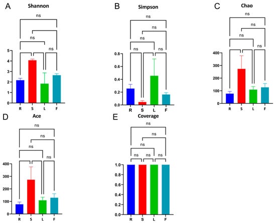
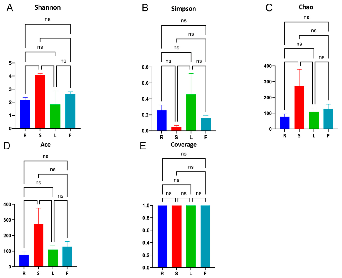
We compared the alpha diversity of endophytic fungi in different tissues of wild P. crotalarioides (Figure 1). Alpha diversity differed significantly among tissues (p < 0.05). Stems had the highest Shannon index and the lowest Simpson index, indicating markedly higher fungal diversity than roots, leaves and flowers. Meanwhile, stems exhibited the maximum ACE and Chao values for community richness, with significant differences observed relative to the other three tissues (p < 0.05). Collectively, stems possessed the greatest fungal diversity and richness, followed by flowers with moderate diversity, roots with intermediate indices, and leaves with the lowest diversity and richness.
Figure 1.
Fungal diversity and richness indices of endophytic communities in different tissues of wild Polygala crotalarioides. (A) Shannon diversity index, (B) Simpson diversity index, (C) Chao richness index, (D) Ace richness index, and (E) Good’s coverage index. Bars represent the mean ± standard deviation (SD) of three biological replicates. Different asterisks indicate significant differences between groups (Kruskal–Wallis test, ns, not significant, p > 0.05). R, root; S, stem; L, leaf; F, flower.
3.3. Composition of Endophytic Fungal Communities in Wild Polygala crotalarioides
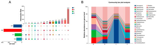
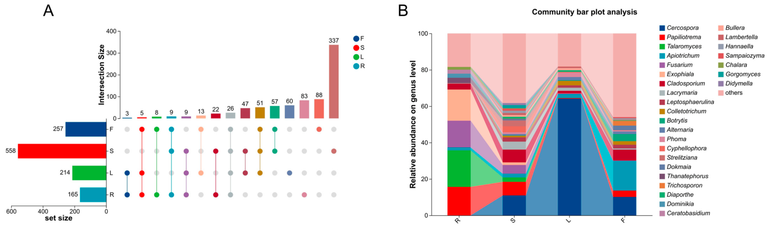
OTU profiling revealed pronounced tissue-specific structuring of endophytic fungal communities in wild P. crotalarioides. Stems harbored the highest overall fungal diversity and richness, and also exhibited the largest number of tissue-specific OTUs. Only a minimal proportion of OTUs was shared among all organs, indicating a highly restricted core mycobiome. These results suggest low inter-organ microbial overlap (Figure 2A).
Figure 2.
Endophytic fungal community composition and shared taxa in different tissues of wild Polygala crotalarioides. (A) UpSet plot showing the number of shared and unique fungal OTUs among the root (R), stem (S), leaf (L), and flower (F) tissues. The horizontal bars represent the total number of OTUs in each tissue, and the vertical bars represent the size of the intersection. colored dots represent individual tissue groups: root (R, cyan), stem (S, red), leaf (L, green), flower (F, dark-blue). Colored connecting-lines link selected dots to indicate combinations of tissue groups, and each vertical bar above denotes the number of shared fungal OTUs within that intersection set. (B) Stacked bar plot showing the relative abundance of fungal genera in each tissue. The “others” category includes genera with relative abundance < 1% in all samples.
Taxonomic annotation of representative OTU sequences assigned 818 OTUs to 9 phyla, 31 classes, 80 orders, 180 families, and 288 genera (Table S3). At the phylum level, wild P. crotalarioides endophytic fungi were mainly affiliated with Ascomycota and Basidiomycota across all tissues, respectively. The relative abundance of Ascomycota varied markedly among tissues: 65.15% in roots, 71.76% in stems, 85.04% in leaves and 44.22% in flowers. For Basidiomycota, the corresponding values were 26.36%, 24.59%, 8.07% and 26.47%. Minor phyla and unidentified fungal groups were detected in all tissues, and the relative abundance of other phyla was below the detectable threshold (Figures S2 and S3).
Genus-level analysis showed that the composition of dominant endophytic fungal genera varied distinctly across roots, stems, leaves and flowers. Cercospora was the absolute dominant genus in leaves. Roots were mainly dominated by Talaromyces, Exophiala, Papiliotrema and Fusarium. Stems harboured high abundances of Cercospora and Papiliotrema, whereas flowers were predominated by Apiotrichum and Cercospora. Most other genera had extremely low relative abundances in all tissues (Figure 2B).
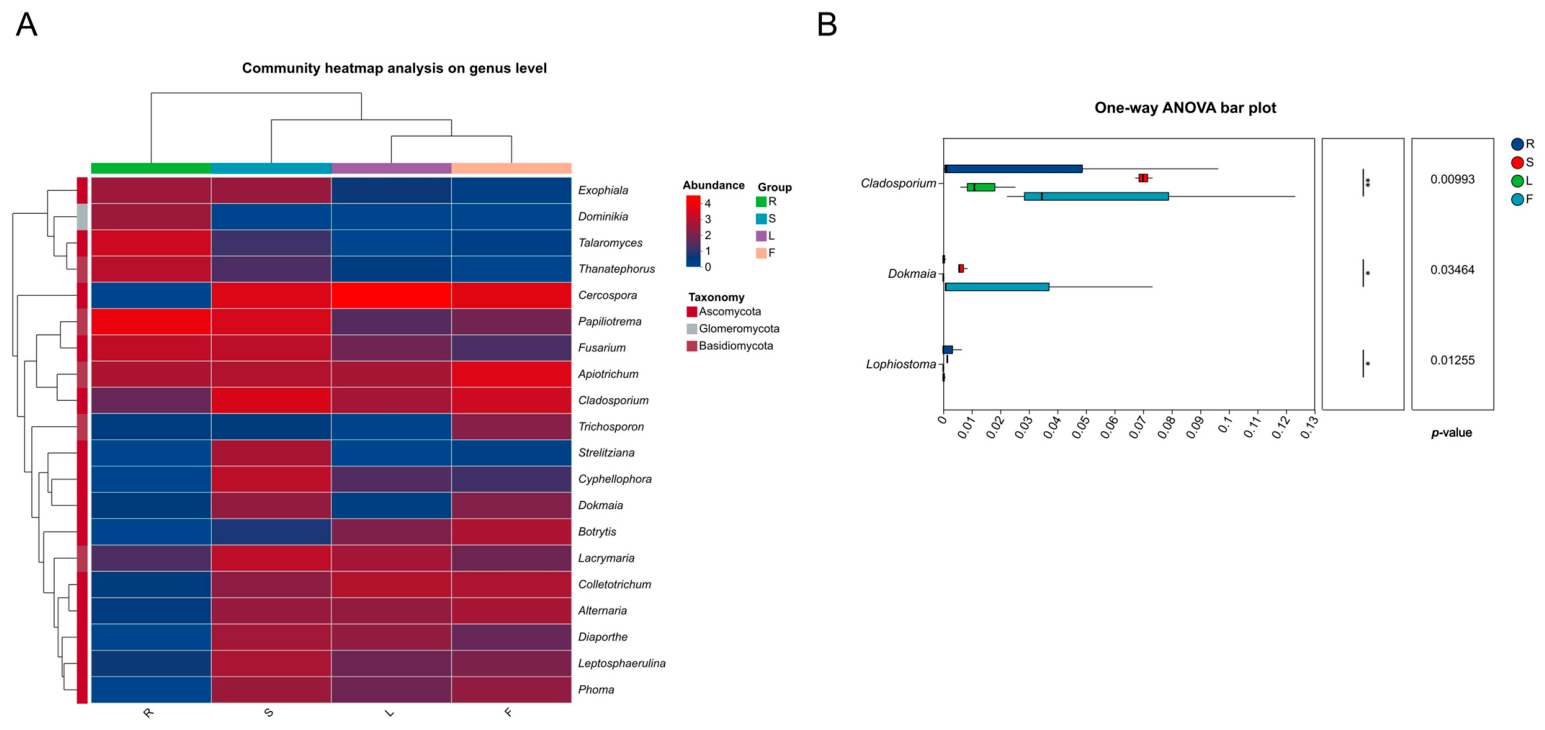
Horizontal cluster analysis at the genus level showed that endophytic fungal communities from leaves, stems, and flowers clustered together, indicating high compositional similarity among these three organs. By contrast, root communities formed a separate clade (Figure 3A). One-way ANOVA detected significant differences in the relative abundances of dominant genera across the four organs (p < 0.05). Specifically, Cladosporium, Dokmaia and Lophiostoma differed significantly among organs (p < 0.05). The relative abundance of Cladosporium presented a clear gradient across organs, while Dokmaia and Lophiostoma showed uneven distribution among different plant organs (Figure 3B).
Figure 3.
Genus-level abundance and differential analysis of endophytic fungi in wild Polygala crotalarioides. (A) Heatmap showing relative abundance of fungal genera across root (R), stem (S), leaf (L), and flower (F) tissues; color gradient indicates abundance from low (blue) to high (red). (B) One-way ANOVA bar plot displaying genera with significant abundance differences among tissues, with mean proportion (%) on the horizontal axis, standard deviation error bars, and significance levels (* p < 0.05; ** p < 0.01) indicated.
3.4. β-Diversity of Endophytic Fungal Communities
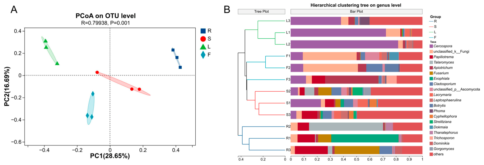
PCoA and hierarchical clustering analyses consistently demonstrated striking tissue-specific separation of endophytic fungal communities in wild P. crotalarioides. OTU-based PCoA ordination exhibited complete spatial segregation of root, stem, leaf and flower samples, and ANOSIM verified significant community dissimilarity across compartments (R = 0.78704, p = 0.001) (Figure 4A). Hierarchical clustering on genus-level profiles fully partitioned replicates by tissue type, with each cluster displaying unique dominant fungal genera (Figure 4B).
Figure 4.
(A) Community dissimilarity analysis of endophytic fungal communities from four tissue compartments of wild Polygala crotalarioides. Axes represent the first (PC1, 28.05%) and second (PC2, 16.90%) principal coordinates, with values in parentheses indicating the percentage of explained variation. different colors and symbols represent four tissue groups: dark-blue squares for root (R), red circles for stem (S), green triangles for leaf (L), and cyan diamonds for flower (F). Proximity between points indicates similarity in community composition. ANOSIM results: R = 0.79938, p = 0.001. (B) Genus-level hierarchical clustering tree combined with stacked bar charts showing genus relative abundance of each replicate.
3.5. Analysis of Differentially Enriched Taxa of Fungal Communities Across Different Tissues
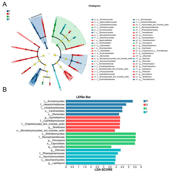
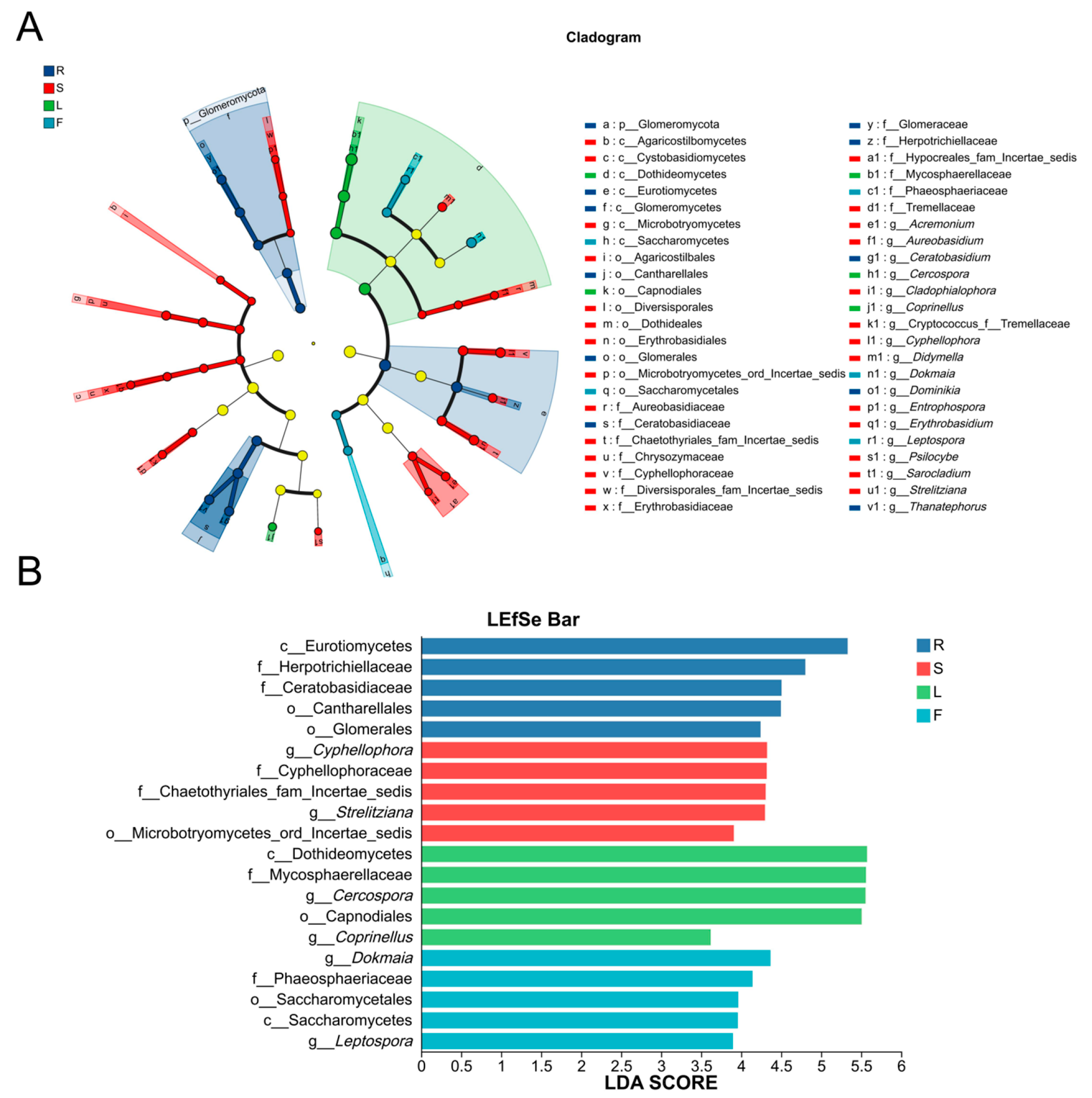
LEfSe analysis was conducted to screen tissue-specific biomarker fungi across four tissues (root, stem, leaf and flower) of wild P. crotalarioides. The results confirmed significant tissue-specific differentiation of endophytic fungal taxa, with distinct differential fungal populations enriched in roots, stems, and leaves, while no significantly differential fungal taxa were detected in flowers (Figure 5A). At multiple taxonomic levels, fungal communities exhibited differential enrichment patterns among various tissues. Root tissues harbored fungal biomarkers with high LDA scores, mainly including the class Eurotiomycetes, the order Cantharellales, the family Ceratobasidiaceae, and the genus Thanatephorus. At the class level, Eurotiomycetes and Cystobasidiomycetes were specifically abundant in root tissues. In stem tissues, multiple fungal taxa were significantly enriched, covering the orders Diaporthales and Hypocreales, the families Cyphellophoraceae, Trichomeriaceae, Didymosphaeriaceae and Diaporthaceae, as well as the genera Cyphellophora, Articulospora and Diaporthe. Leaf tissues possessed a unique assemblage of differential fungal biomarkers. The phylum Ascomycota, the class Dothideomycetes, the order Capnodiales, the family Mycosphaerellaceae, and the genera Cercospora and Coprinellus presented relatively high LDA scores and dominated the fungal community of leaves. All these tissue-enriched fungal taxa formed independent phylogenetic clades corresponding to specific plant tissues. Overall, the composition of differentially enriched endophytic fungi varied markedly among root, stem, and leaf tissues of wild P. crotalarioides, strongly demonstrating the distinct tissue-specific distribution characteristics of endophytic fungal communities in this plant (Figure 5B).
Figure 5.
LEfSe analysis of tissue-specific biomarker fungi in root (R), stem (S) and leaf (L) compartments of wild Polygala crotalarioides. (A) Phylogenetic cladogram of differential fungal taxa; colors denote the enriched tissue for each taxonomic clade. (B) LDA score bar plot showing the discriminative taxa with LDA threshold > 2.5.
3.6. FUNGuild-Based Trophic Composition of Endophytic Fungi in Wild Polygala crotalarioides
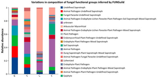
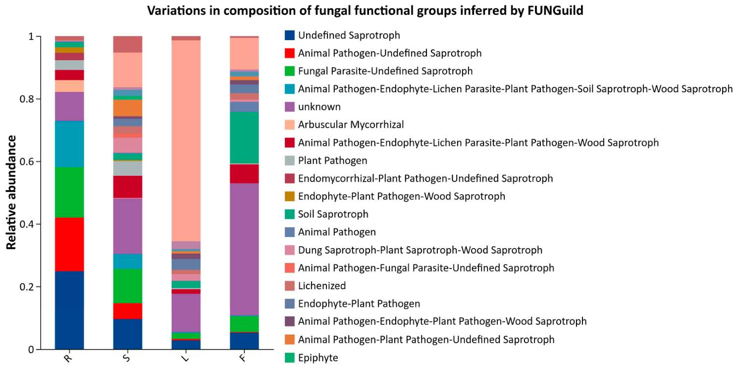
FUNGuild functional annotation demonstrated marked organ-dependent divergence in the trophic composition of endophytic fungi isolated from wild P. crotalarioides. Arbuscular mycorrhizal fungi dominated and peaked in relative abundance within leaf tissues, while undefined saprotrophs and animal-pathogenic fungi constituted the predominant functional fraction in roots. Floral fungal assemblages were chiefly composed of functionally unclassified fungi and soil saprotrophs, and stems accommodated the richest spectrum of trophic groups with relatively uniform abundance distribution across diverse functional guilds. A substantial fraction of unknown and undefined saprotrophic fungi was recovered in all four tested plant organs (Figure 6).
Figure 6.
Fungal functional guild composition predicted via FUNGuild in four tissue compartments (R: root, S: stem, L: leaf, F: flower) of wild Polygala crotalarioides. Stacked bars show the relative abundance of each trophic functional group.
3.7. Isolation and Identification of Culturable Endophytic Fungi in Wild Polygala crotalarioides
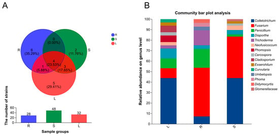
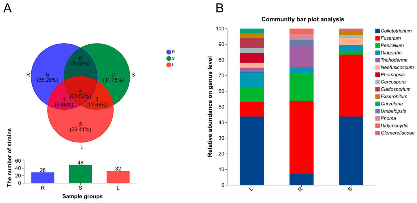
In total, 108 endophytic fungal strains were isolated from the leaves, roots, and stems of wild P. crotalarioides, including 32 strains from leaves, 28 strains from roots, and 48 strains from stems. Based on ITS sequence identification, all isolates were taxonomically classified into 2 phyla, 4 classes, 9 orders, 14 families, and 17 genera. Among the identified fungi, Colletotrichum sp. and Fusarium sp. were isolated from all three tested organs with relatively high isolation frequencies (Table S4). The remaining fungal genera were recovered from only one or two plant organs.
Genus-level comparative analysis and Venn diagram analysis identified unique and shared fungal genera among leaf, root and stem tissues of wild P. crotalarioides. A total of 5, 6 and 2 tissue-specific genera were detected in leaves, roots and stems, respectively. Four genera, Colletotrichum, Diaporthe, Fusarium and Penicillium, were common to all three tissues. Trichoderma was shared exclusively by leaves and roots, while three genera including Cercospora, Exserohilum, and Neofusicoccum were shared between leaves and stems (Figure 7A and Table S5).
Figure 7.
Venn diagram analysis and genus-level distribution of culturable endophytic fungi isolated from different tissues of wild Polygala crotalarioides. (A) Venn diagram illustrating the counts and proportions of unique and shared fungal genera across root (R), stem (S), and leaf (L) tissues. Numbers within each segment represent the number of genera, and percentages denote their proportions relative to the total number of detected genera. (B) Bar plot showing the genus-level relative abundance of fungal isolates pooled from all tissues (leaf, root and stem); each bar represents the percentage of isolates assigned to the corresponding genus.
Fungal community composition exhibited slight differences across different plant tissues at the phylum level. Ascomycota and Mucoromycota were detected in root tissues, whereas only Ascomycota was present in leaf and stem tissues. Ascomycota dominated the fungal communities of roots, stems, and leaves, with relative abundances of 100%, 96.30% and 100%, respectively (Table S5).
Genus-level analysis revealed obvious differences in dominant fungal genera across leaf, root and stem tissues. Leaf tissues harbored 11 fungal genera, while both root and stem tissues contained 8 genera each. The dominant taxa varied greatly among organs: Colletotrichum showed the highest relative abundance in leaves; Fusarium predominated in roots; and Colletotrichum was the most abundant genus in stems (Figure 7B).
3.8. Bioactivity Screening of Endophytic Fungi
3.8.1. Screening of AChE Inhibitory Activity and IC50 Determination
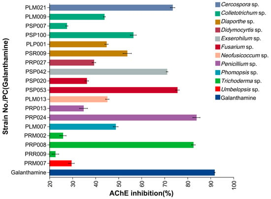
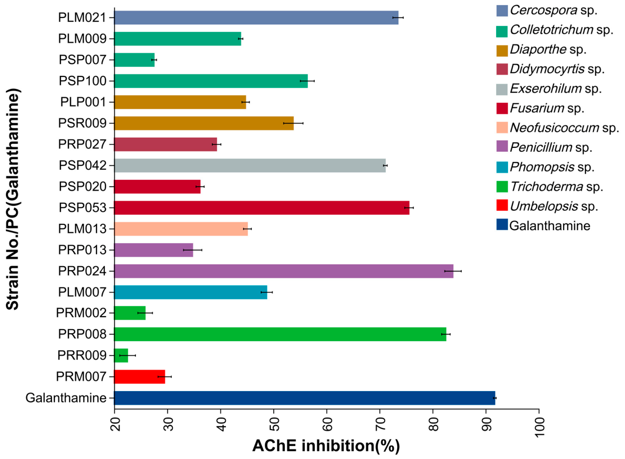
Acetylcholinesterase (AChE) inhibitory activity was screened among endophytic fungi isolated from wild P. crotalarioides. Two strains exhibited the highest AChE inhibitory activity, with inhibition rates exceeding 80%: Penicillium sp. PRP024 (83.87 ± 1.51%) and Trichoderma sp. PRP008 (82.53 ± 0.80%). Five strains showed moderate-to-high activity (>50%), and the remaining 11 strains displayed low to moderate activity (22.55–49.0%). All assays showed high reproducibility, with standard deviations <2%. The IC50 values of seven high-activity strains for AChE inhibition were further determined. The lowest IC50 was recorded for Cercospora sp. PLM021 (418.1 μg/mL, 95% CI: 393.6–449.6 μg/mL), followed by Trichoderma sp. PRP008 (441.3 μg/mL, 95% CI: 441.9–470.2 μg/mL). IC50 values for the other strains ranged from 536.7 μg/mL (Penicillium sp. PRP024) to 827.5 μg/mL (Diaporthe sp. PSR009). The positive control galantamine had an IC50 of 67.5 μg/mL (95% CI: 64.00–71.08 μg/mL). All samples produced narrow 95% confidence intervals (Figure 8 and Table S6).
Figure 8.
AChE inhibitory activity of endophytic fungal extracts from wild Polygala crotalarioides. The horizontal bars represent the mean percentage of AChE inhibition ± standard deviation (SD, n = 3) at a fixed concentration of 2000 μg/mL. Galanthamine (PC) was used as the positive control. The strain names corresponding to each code are listed in the legend.
3.8.2. DPPH Radical Scavenging Activity and IC50 Determination of Endophytic Fungi
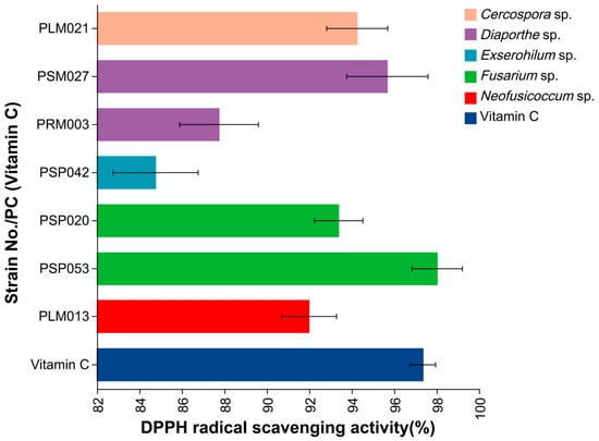
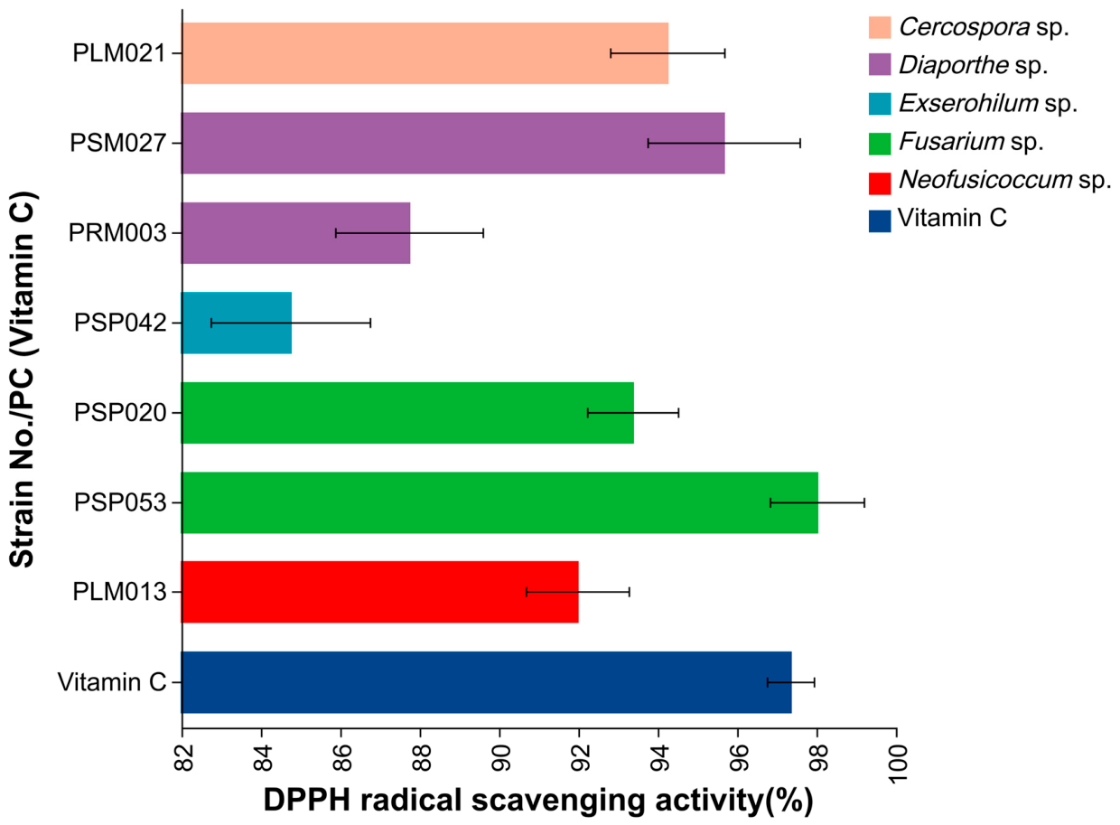
(2,2-diphenyl-1-picrylhydrazyl) (DPPH) radical scavenging activity was also evaluated for these fungal strains. Seven strains showed high activity with scavenging rates ≥ 80%, and Fusarium sp. PSP053 (98.03 ± 1.19%) presented the strongest activity, followed by Diaporthe sp. PSM027 (95.67 ± 1.92%) and Cercospora sp. PLM021 (94.25 ± 1.44%). Standard deviations of all measurements were below 2%. The IC50 values for DPPH scavenging were measured in seven high-activity strains. Exserohilum sp. PSP042 had the lowest IC50 at 258.7 μg/mL (95% CI: 238.8–275.0 μg/mL), followed by Fusarium sp. PSP053 (436.0 μg/mL, 95% CI: 388.4–487.7 μg/mL) and Fusarium sp. PSP020 (466.6 μg/mL, 95% CI: 414.1–514.7 μg/mL). The IC50 values of the rest of the strains ranged from 693.6 μg/mL to 771.5 μg/mL. The positive control vitamin C showed an IC50 of 10.10 μg/mL (95% CI: 8.20–12.36 μg/mL), and all assays had narrow 95% confidence intervals (Figure 9 and Table S7).
Figure 9.
DPPH radical scavenging activity of endophytic fungal extracts from wild Polygala crotalarioides. The horizontal bars represent the mean percentage of DPPH radical scavenging activity ± standard deviation (SD, n = 3) at a concentration of 2000 μg/mL. Vitamin C (PC) was used as the positive control. The strain names corresponding to each code are listed in the legend.
3.8.3. ABTS Radical Scavenging Activity and IC50 Determination of Endophytic Fungi
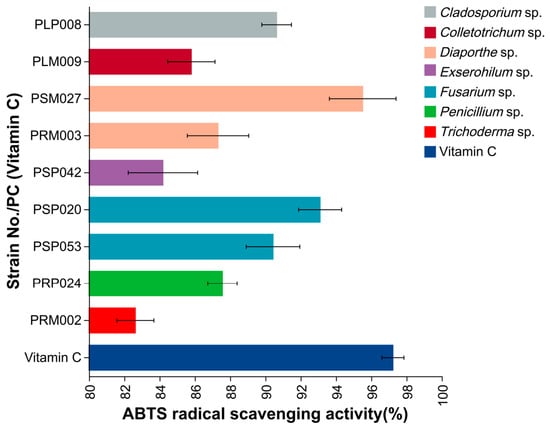
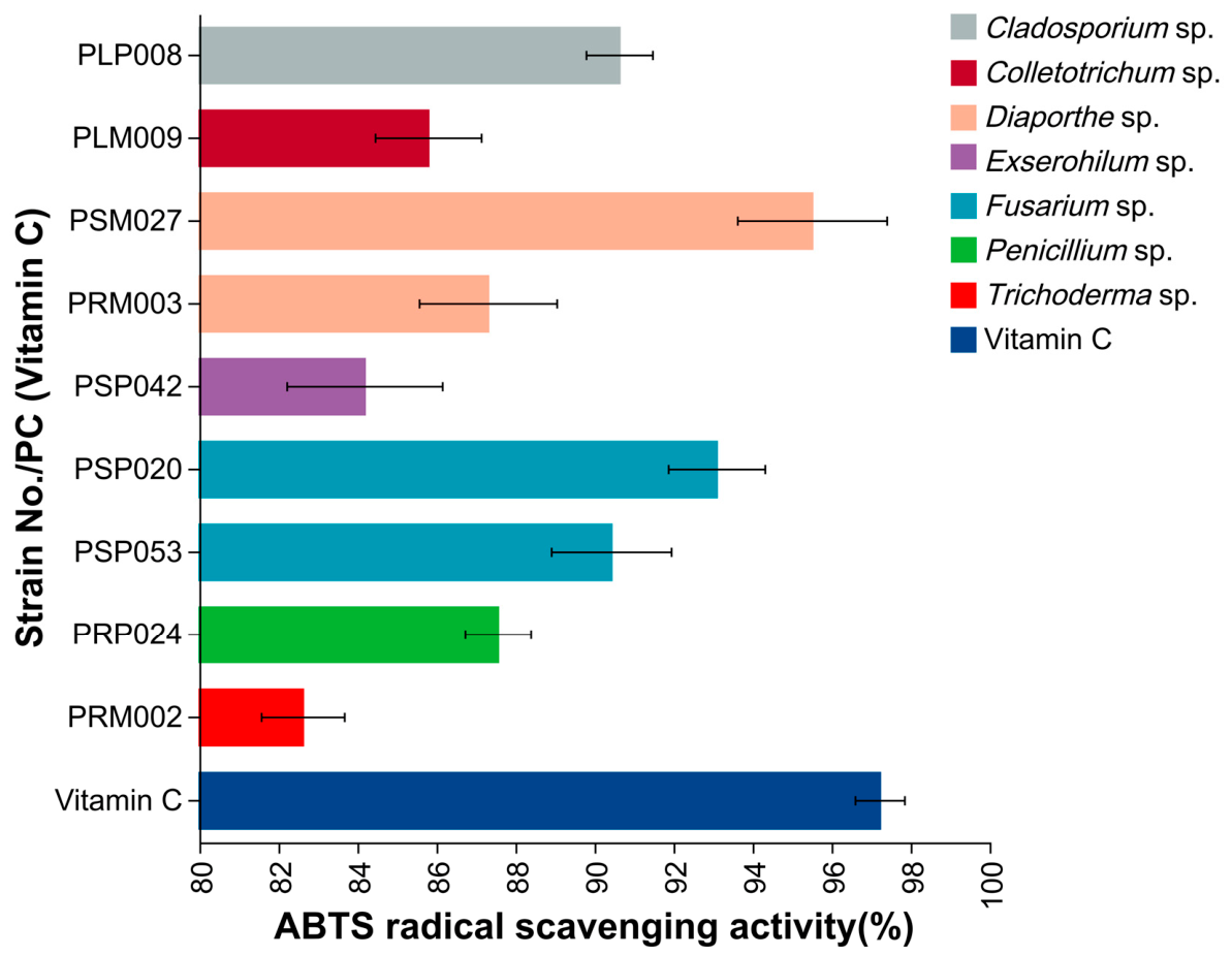
For the 2,2′-azinobis(3-ethylbenzothiazoline-6-sulfonicacid) (ABTS) radical scavenging activity, nine strains displayed high activity with scavenging rates ≥ 80%, among which Diaporthe sp. PSM027 (95.52 ± 1.89%) had the highest activity. All standard deviations were less than 2%. The IC50 values for ABTS scavenging were determined in nine high-activity strains. Fusarium sp. PSP053 yielded the lowest IC50 of 418.7 μg/mL (95% CI: 402.4–434.7 μg/mL), followed by Penicillium sp. PRP024 (423.2 μg/mL, 95% CI: 402.6–443.6 μg/mL). IC50 values of the remaining strains ranged from 585.7 μg/mL to 712.5 μg/mL. The positive control vitamin C had an IC50 of 70.06 μg/mL (95% CI: 42.16–83.80 μg/mL). Narrow 95% confidence intervals were observed across all tests, indicating good reproducibility (Figure 10 and Table S8).
Figure 10.
ABTS radical scavenging activity of endophytic fungal extracts from wild Polygala crotalarioides. The horizontal bars represent the mean percentage of ABTS radical scavenging activity ± standard deviation (SD, n = 3) at a fixed concentration of 2000 μg/mL. Vitamin C (PC) was used as the positive control. The strain names corresponding to each code are listed in the legend.
4. Discussion
This study integrated high-throughput sequencing and culture-dependent approaches to characterize the endophytic fungal communities in different tissues of wild Polygala crotalarioides and to evaluate the antioxidant and AChE-inhibitory activities of cultivable isolates. The recovered strains were predominantly assigned to Penicillium, Fusarium, Diaporthe, and Cercospora, with several isolates displaying both antioxidant and AChE-inhibitory properties. Notably, wild-host-derived endophytes exhibited distinct community composition and bioactive profiles compared to those previously reported from cultivated P. crotalarioides.
α-diversity analysis revealed that stems of P. crotalarioides harbored the highest endophytic fungal richness and diversity among the four tissues examined (root, stem, leaf, and flower), as evidenced by the maximum Shannon, ACE, and Chao1 indices, the lowest Simpson index, and the highest total (558) and unique (337) OTU counts. This tissue-specific distribution is consistent with the widely reported niche differentiation of plant endophytes [40]. Several ecological factors may hypothetically contribute to the observed stem-biased fungal enrichment in wild P. crotalarioides. The stem tissues provide nutrient-transport pathways and heterogeneous carbon niches [41] that may favor fungal colonization. In contrast, leaf tissues are subject to senescence, high ultraviolet radiation, and strong environmental filtering [42,43], which may constrain fungal establishment. The low fungal diversity in flowers may be associated with the accumulation of defensive phytometabolites during anthesis, whereas the relatively lower complexity of root fungal communities could be limited by rhizosphere microbial competition and variable soil conditions. It should be noted, however, that the ecological interpretations proposed above remain hypothetical and require further experimental validation in future work.
Ascomycota predominated across all examined organs, matching widespread distribution patterns of endophytic fungi in terrestrial medicinal plants [44,45]. Variable Ascomycota relative abundances across tissues (85.04% leaf, 71.76% stem, 65.15% root, 44.22% flower) may reflect differential host selective pressure across discrete physiological microhabitats [46], while Basidiomycota mainly accumulated in root and flower fractions, predominantly comprising animal-pathogenic and soil saprotrophs taxa capable of intermittent microbial exchange with surrounding bulk soil [47]. Genus-level compositional variation further pointed toward evident organ preference among core fungal genera: Cercospora prevailed in leaf communities; Talaromyces, Exophiala and Fusarium dominated root mycobiota; floral assemblages were characterised by abundant Apiotrichum alongside numerous unclassified fungal lineages. Integrative PCoA, ANOSIM (R = 0.79938, p = 0.001) and hierarchical clustering suggested leaf fungal communities may be phylogenetically distinct from the clustered root–stem–flower group, implying leaf tissues impose stronger filtering effects on colonising fungi. LEfSe biomarker profiling further illustrated niche partitioning: roots tended to enrich Eurotiomycetes and Thanatephorus taxa potentially associated with belowground nutrient cycling; stems accumulated differential Diaporthales, Diaporthe and Cyphellophora genera previously documented to synthesise bioactive secondary metabolites [48,49].
We retrieved 108 culturable isolates from root, stem and leaf tissues, numerically far fewer than the 818 OTUs resolved via amplicon sequencing. Such quantification mismatch commonly occurs in endophyte surveys, where culture-independent sequencing routinely detects substantially higher diversity than culture-based approaches. This discrepancy is often attributed to cultivation bias, as standard laboratory media may not reproduce critical in planta conditions, including host-derived metabolites, oxygen gradients and physicochemical microenvironments required by many uncultured fungi [50].
In comparison with our previous studies on both cultivated P. crotalarioides [21] and Polygala sibirica [51] from Yunnan, the present study revealed bioactivities in endophytic fungi isolated from wild P. crotalarioides. Our earlier study on P. sibirica isolated only 23 cultivable strains, of which 12 displayed moderate to high AChE inhibitory activity, with Fusarium sp. exhibiting the strongest inhibitory effect. Another previous study isolated 55 endophytic strains from cultivated P. crotalarioides and evaluated their AChE inhibitory activity in vitro. Among these, five strains—Phialophora sp. PCAM010, Diaporthe sp. PCBM027, Fusarium sp. LP41, Fusarium sp. SR60, and Phoma sp. SM81—showed strong activity, with inhibition rates exceeding 50%. By contrast, the present study obtained 108 cultivable fungal strains from wild P. crotalarioides, with several strains exhibiting notable dual bioactivities. Specifically, two isolates displayed superior AChE inhibitory activity exceeding 80%, while five strains achieved over 80% DPPH and ABTS radical-scavenging capacity with favorable IC50 values. Notably, endophytic fungi isolated from wild P. crotalarioides in the present study exhibited higher AChE inhibitory activity than those previously obtained from cultivated P. crotalarioides in our prior work. However, these two studies adopted distinct fermentation strategies: the previous study used liquid PDB medium, whereas the current study applied rice-based solid-state fermentation. Accumulating evidence indicates that solid-state fermentation generally yields higher quantities and greater chemical diversity of fungal secondary metabolites than conventional liquid fermentation systems [52,53]. Accordingly, the observed differences in AChE inhibitory activity may be partially explained by disparities in fermentation modes. Nevertheless, several strains isolated from wild P. crotalarioides still exhibited prominent AChE inhibitory activity under the unified solid-state fermentation system, suggesting their potential for further investigation. Overall, these bioactivity screening results provide a foundational dataset to support subsequent isolation and purification of bioactive compounds.
Our study revealed that several endophytic fungal strains isolated from wild P. crotalarioides exhibited prominent AChE inhibitory and antioxidant activities. These bioactive isolates, primarily assigned to Penicillium, Trichoderma, Fusarium, Diaporthe, and Exserohilum, represent valuable microbial resources harbored by this subtropical medicinal plant. Although bioactivity evaluations were performed using crude fungal extracts and the observed potency was lower than that of positive controls, the consistent AChE inhibitory and radical-scavenging activities still indicate the metabolic potential of these endophytes. Nevertheless, several limitations should be acknowledged. First, all samples were collected from a single site in Cangyuan County; thus, multi-site sampling across diverse altitudinal and climatic gradients in Yunnan is needed to clarify the geographic effects on endophytic community assembly and functional variation. Second, all bioactivity assessments were limited to in vitro screening of crude extracts; further cellular-level assays and in vivo pharmacological studies are required to validate the practical efficacy and pharmaceutical potential of the prioritized strains. Overall, this work provides foundational strain resources and preliminary bioactivity evidence for future exploration of bioactive metabolites from wild P. crotalarioides endophytic fungi.
5. Conclusions
In conclusion, wild Polygala crotalarioides harbors diverse and tissue-specific endophytic fungal communities, with stems representing the primary reservoir of fungal diversity. Several fungal isolates exhibited promising acetylcholinesterase inhibitory and antioxidant activities, indicating that these strains represent candidates for preliminary bioactivity screening to guide further chemical characterization of active metabolites. These results expand our understanding of plant–fungus associations in this medicinal species and provide a basis for future discovery of bioactive natural products from its endophytic fungal resources.
Supplementary Materials
The following supporting information can be downloaded at https://www.mdpi.com/article/10.3390/jof12090669/s1, Figure S1: Rarefaction curves of fungal communities in different tissues of wild P. crotalarioides; The observed species richness (Sobs index) is plotted against the number of sampled reads. R, root; S, stem; L, leaf; F, flower. Figure S2: Stacked bar chart showing the relative abundance of endophytic fungal taxa at the phylum level across four tissue compartments of wild P. crotalarioides. R: root; S: stem; L: leaf; F: flower. Taxa with low relative abundance were pooled into the group “others”. Figure S3: Taxonomic composition of culturable endophytic fungi at the phylum level isolated from different tissues of wild P. crotalarioides. (A) Root (R); (B) Stem (S); (C) Leaf (L); (D) Flower (F). Each pie chart displays the relative abundance of dominant fungal phyla, including Ascomycota, Basidiomycota, Glomeromycota, and other rare phyla. Table S1: Information of representative isolated fungal strains and yields of their crude fermentation extracts. Table S2: Sequence statistics of endophytic fungi from wild P. crotalarioides. Table S3: OTUs of endophytic fungi from wild P. crotalarioides. Table S4: ITS Identification Results of Culturable Endophytic Fungi from Wild P. crotalarioides. Table S5: Common genera of endophytic fungi in different organs of wild P. crotalarioides. Table S6: AChE inhibitory activity and IC50 values of endophytic fungi isolated from wild P. crotalarioides. Table S7: DPPH radical scavenging activity and IC50 values of endophytic fungi isolated from wild P. crotalarioides. Table S8: ABTS radical scavenging activity and IC50 values of endophytic fungi isolated from wild P. crotalarioides.
Author Contributions
Conceptualization, K.S., W.Z. and S.Y.; methodology, K.S., M.X. and X.C.; software, X.C., H.Z. and M. L; validation, M.X., X.C. and M.L.; formal analysis, K.S., X.C. and M.X.; investigation, X.D. and H.Z. resources, W.Z., K.S. and S.Y.; data curation, K.S. and M.L.; writing—original draft preparation, K.S., M.X. and X.C.; writing—review and editing, W.Z. and S.Y.; visualization, M.X. and M.L.; supervision, W.Z. and S.Y.; project administration, W.Z. and S.Y.; funding acquisition, W.Z. and S.Y. All authors have read and agreed to the published version of the manuscript.
Funding
This work was supported by the National Natural Science Foundation of China (grant number: 32160063) and the Science Research Fund Project of the Yunnan Provincial Department of Education (grant number: 2026J1148).
Institutional Review Board Statement
Not applicable.
Informed Consent Statement
Not applicable.
Data Availability Statement
Raw sequencing data generated in this study have been deposited in the NCBI BioProject database under accession number PRJNA1207743, with corresponding SRA accessions ranging from SRR31976288 to SRR31976299. Other datasets used and analysed during the current study are available from the authors on reasonable request.
Conflicts of Interest
The authors declare no conflict of interest.
References
- Heinrich, M.; Appendino, G.; Efferth, T.; Fürst, R.; Izzo, A.A.; Kayser, O.; Pezzuto, J.M.; Viljoen, A. Best practice in research—Overcoming common challenges in phytopharmacological research. J. Ethnopharmacol. 2020, 246, 112230. [Google Scholar] [CrossRef] [Scilit] [PubMed]
- Atanasov, A.G.; Zotchev, S.B.; Dirsch, V.M.; Supuran, C.T. Natural products in drug discovery: Advances and opportunities. Nat. Rev. Drug Discov. 2021, 20, 200–216. [Google Scholar] [CrossRef] [Scilit] [PubMed]
- Roviello, G.N. Nature-Inspired Pathogen and Cancer Protein Covalent Inhibitors: From Plants and Other Natural Sources to Drug Development. Pathogens 2025, 14, 1153. [Google Scholar] [CrossRef] [Scilit] [PubMed]
- Aleynova, O.A.; Kiselev, K.V. Interaction of plants and endophytic microorganisms: Molecular aspects, biological functions, community composition, and practical applications. Plants 2023, 12, 714. [Google Scholar] [CrossRef] [Scilit] [PubMed]
- Waqar, S.; Bhat, A.A.; Khan, A.A. Endophytic fungi: Unravelling plant-endophyte interaction and the multifaceted role of fungal endophytes in stress amelioration. Plant Physiol. Biochem. 2024, 206, 108174. [Google Scholar] [CrossRef] [Scilit] [PubMed]
- Nazir, A.; Puthuveettil, A.R.; Hussain, F.H.N.; Hamed, K.E.; Munawar, N. Endophytic fungi: Nature’s solution for antimicrobial resistance and sustainable agriculture. Front. Microbiol. 2024, 15, 1461504. [Google Scholar] [CrossRef] [Scilit] [PubMed]
- Tsipinana, S.; Husseiny, S.; Alayande, K.A.; Raslan, M.; Amoo, S.; Adeleke, R. Contribution of endophytes towards improving plant bioactive metabolites: A rescue option against red-taping of medicinal plants. Front. Plant Sci. 2023, 14, 1248319. [Google Scholar] [CrossRef] [Scilit] [PubMed]
- Liu, M.; Wang, X.; Gao, D. Polygalae Radix: Review of metabolites, pharmacological activities and toxicology. Front. Pharmacol. 2024, 15, 1420853. [Google Scholar] [CrossRef] [Scilit] [PubMed]
- Tang, H.P.; Zhu, E.L.; Bai, Q.X.; Wang, S.; Wang, Z.B.; Wang, M.; Kuang, H.X. Polygala japonica Houtt.: A comprehensive review on its botany, traditional uses, phytochemistry, pharmacology, and pharmacokinetics. Fitoterapia 2024, 179, 106233. [Google Scholar] [CrossRef] [Scilit] [PubMed]
- Hashem, A.H.; Attia, M.S.; Kandil, E.K.; Fawzi, M.M.; Abdelrahman, A.S.; Khader, M.S.; Khodaira, M.A.; Emam, A.E.; Goma, M.A.; Abdelaziz, A.M. Bioactive compounds and biomedical applications of endophytic fungi: A recent review. Microb. Cell Fact. 2023, 22, 107. [Google Scholar] [CrossRef] [Scilit] [PubMed]
- Zhu, J.; Wang, Z.; Song, L.; Fu, W.; Liu, L. Anti-Alzheimer’s natural products derived from plant endophytic fungi. Molecules 2023, 28, 2259. [Google Scholar] [CrossRef] [Scilit] [PubMed]
- Li, S.; Hou, Z.; Ye, T.; Song, X.; Hu, X.; Chen, J. Saponin components in Polygala tenuifolia as potential candidate drugs for treating dementia. Front. Pharmacol. 2024, 15, 1431894. [Google Scholar] [CrossRef] [Scilit] [PubMed]
- Li, C.; Gao, F.; Qu, Y.; Zhao, P.; Wang, X.; Zhu, G. Tenuifolin in the prevention of Alzheimer’s disease-like phenotypes: Investigation of the mechanisms from the perspectives of calpain system, ferroptosis, and apoptosis. Phytother. Res. 2023, 37, 4621–4638. [Google Scholar] [CrossRef] [Scilit] [PubMed]
- Grabowska, W.; Bijak, M.; Szelenberger, R.; Gorniak, L.; Podogrocki, M.; Harmata, P.; Cichon, N. Acetylcholinesterase as a multifunctional target in amyloid-driven neurodegeneration: From dual-site inhibitors to anti-agregation strategies. Int. J. Mol. Sci. 2025, 26, 8726. [Google Scholar] [CrossRef] [Scilit] [PubMed]
- Sienes Bailo, P.; Llorente Martín, E.; Calmarza, P.; Montolio Breva, S.; Bravo Gómez, A.; Pozo Giráldez, A.; Sánchez-Pascuala Callau, J.J.; Vaquer Santamaría, J.M.; Dayaldasani Khialani, A.; Cerdá Micó, C.; et al. The role of oxidative stress in neurodegenerative diseases and potential antioxidant therapies. Adv. Lab. Med. 2022, 3, 342–360. [Google Scholar] [CrossRef] [Scilit] [PubMed]
- Gao, Y.; Liu, Y.; Li, Y. Safety and efficacy of acetylcholinesterase inhibitors for Alzheimer’s disease: A systematic review and meta-analysis. Adv. Clin. Exp. Med. 2024, 33, 1179–1187. [Google Scholar] [CrossRef] [Scilit] [PubMed]
- Liu, S.; Zhao, M.; Liu, Y.; Yang, X.; Yan, H.; Xu, H.; Wu, Y.; Xu, Y. Comparative efficacy and safety of symptomatic therapy and disease-modifying therapy for Alzheimer’s disease: A systematic review and network meta-analysis. Front. Neurosci. 2025, 19, 1656906. [Google Scholar] [CrossRef] [Scilit] [PubMed]
- Salgotra, R.K.; Chauhan, B.S. Genetic diversity, conservation, and utilization of plant genetic resources. Genes 2023, 14, 174. [Google Scholar] [CrossRef] [Scilit] [PubMed]
- Tian, H.; Wu, C.; Zhang, L.; Guo, S.; Wang, Y.; Pei, S.; Zuo, X.; Wang, Q.; Zhang, Q. Phenotypic diversity analysis of Polygala tenuifolia Willd germplasm resources in China. Genet. Resour. Crop Evol. 2024, 71, 3303–3314. [Google Scholar] [CrossRef] [Scilit]
- Ma, J.; Wang, J.; Li, C.; Liu, J.; Chen, C.; Hua, Y. The complete chloroplast genome characteristics of Polygala crotalarioides Buch.-Ham. ex DC. (Polygalaceae) from Yunnan, China. Mitochondrial DNA Part B Resour. 2021, 6, 2838–2840. [Google Scholar] [CrossRef] [Scilit] [PubMed]
- Shen, K.; Xiong, Y.; Liu, Y.; Fan, X.; Zhu, R.; Hu, Z.; Li, C.; Hua, Y. Community Structure and Diversity of Endophytic Fungi in Cultivated Polygala crotalarioides at Two Different Growth Stages Based on Culture-Independent and Culture-Based Methods. J. Fungi 2024, 10, 195. [Google Scholar] [CrossRef] [Scilit] [PubMed]
- Dos Reis, J.B.A.; Lorenzi, A.S.; do Vale, H.M.M. Methods used for the study of endophytic fungi: A review on methodologies and challenges, and associated tips. Arch. Microbiol. 2022, 204, 675. [Google Scholar] [CrossRef] [Scilit] [PubMed]
- Zheng, H.; Qiao, M.; Xu, J.; Yu, Z. Culture-based and culture-independent assessments of endophytic fungal diversity in aquatic plants in southwest China. Front. Fungal Biol. 2021, 2, 692549. [Google Scholar] [CrossRef] [Scilit] [PubMed]
- Zuo, Y.L.; Hu, Q.N.; Qin, L.; Liu, J.Q.; He, X.L. Species identity and combinations differ in their overall benefits to Astragalus adsurgens plants inoculated with single or multiple endophytic fungi under drought conditions. Front. Plant Sci. 2022, 13, 933738. [Google Scholar] [CrossRef] [Scilit] [PubMed]
- Chen, S.; Zhou, Y.; Chen, Y.; Gu, J. Fastp: An ultra-fast all-in-one FASTQ preprocessor. Bioinformatics 2018, 34, i884–i890. [Google Scholar] [CrossRef] [Scilit] [PubMed]
- Magoč, T.; Salzberg, S.L. FLASH: Fast length adjustment of short reads to improve genome assemblies. Bioinformatics 2011, 27, 2957–2963. [Google Scholar] [CrossRef] [Scilit] [PubMed]
- Edgar, R.C. UPARSE: Highly accurate OTU sequences from microbial amplicon reads. Nat. Methods 2013, 10, 996–998. [Google Scholar] [CrossRef] [Scilit] [PubMed]
- Kõljalg, U.; Nilsson, R.H.; Abarenkov, K.; Tedersoo, L.; Taylor, A.F.; Bahram, M.; Bates, S.T.; Bruns, T.D.; Bengtsson-Palme, J.; Callaghan, T.M.; et al. Towards a unified paradigm for sequence-based identification of fungi. Mol. Ecol. 2013, 22, 5271–5277. [Google Scholar] [CrossRef] [Scilit] [PubMed]
- Wang, Q.; Garrity, G.M.; Tiedje, J.M.; Cole, J.R. Naive Bayesian classifier for rapid assignment of rRNA sequences into the new bacterial taxonomy. Appl. Environ. Microbiol. 2007, 73, 5261–5267. [Google Scholar] [CrossRef] [Scilit] [PubMed]
- Schloss, P.D.; Westcott, S.L.; Ryabin, T.; Hall, J.R.; Hartmann, M.; Hollister, E.B.; Lesniewski, R.A.; Oakley, B.B.; Parks, D.H.; Robinson, C.J.; et al. Introducing mothur: Open-source, platform-independent, community-supported software for describing and comparing microbial communities. Appl. Environ. Microbiol. 2009, 75, 7537–7541. [Google Scholar] [CrossRef] [Scilit] [PubMed]
- Nguyen, N.H.; Song, Z.; Bates, S.T.; Branco, S.; Tedersoo, L.; Menke, J.; Schilling, J.S.; Kennedy, P.G. FUNGuild: An open annotation tool for parsing fungal community datasets by ecological guild. Fungal Ecol. 2016, 20, 241–248. [Google Scholar] [CrossRef] [Scilit]
- Chen, H.; Boutros, P.C. VennDiagram: A package for the generation of highly-customizable Venn and Euler diagrams in R. BMC Bioinform. 2011, 12, 35. [Google Scholar] [CrossRef] [Scilit] [PubMed]
- Sahu, P.K.; Tilgam, J.; Mishra, S.; Hamid, S.; Gupta, A.K.J.; Verma, S.K.; Kharwar, R.N. Surface sterilization for isolation of endophytes: Ensuring what (not) to grow. J. Basic Microbiol. 2022, 62, 647–668. [Google Scholar] [CrossRef] [Scilit] [PubMed]
- Guo, L.D.; Hyde, K.D.; Liew, E.C.Y. Identification of endophytic fungi from Livistona chinensis based on morphology and rDNA sequences. New Phytol. 2000, 147, 617–630. [Google Scholar] [CrossRef] [Scilit] [PubMed]
- Thi Minh Le, T.; Thi Hong Hoang, A.; Thi Bich Le, T.; Thi Bich Vo, T.; Van Quyen, D.; Hoang Chu, H. Isolation of endophytic fungi and screening of Huperzine A-producing fungus from Huperzia serrata in Vietnam. Sci. Rep. 2019, 9, 16152. [Google Scholar] [CrossRef] [Scilit] [PubMed]
- Ellman, G.L.; Courtney, K.D.; Andres, V., Jr.; Feather-Stone, R.M. A new and rapid colorimetric determination of acetylcholinesterase activity. Biochem. Pharmacol. 1961, 7, 88–95. [Google Scholar] [CrossRef] [Scilit] [PubMed]
- Akinsanya, M.A.; Goh, J.K.; Lim, S.P.; Ting, A.S. Diversity, antimicrobial and antioxidant activities of culturable bacterial endophyte communities in Aloe vera. FEMS Microbiol. Lett. 2015, 362, fnv184. [Google Scholar] [CrossRef] [Scilit] [PubMed]
- An, T.; Chen, M.; Zu, Z.; Chen, Q.; Lu, H.; Yue, P.; Gao, X. Untargeted and targeted metabolomics reveal changes in the chemical constituents of instant dark tea during liquid-state fermentation by Eurotium cristatum. Food Res. Int. 2021, 148, 110623. [Google Scholar] [CrossRef] [Scilit] [PubMed]
- Dong, L.; Li, Y.; Xu, J.; Yang, J.; Wei, G.; Shen, L.; Ding, W.; Chen, S. Biofertilizers regulate the soil microbial community and enhance Panax ginseng yields. Chin. Med. 2019, 14, 20. [Google Scholar] [CrossRef] [Scilit] [PubMed]
- Harrison, J.G.; Griffin, E.A. The diversity and distribution of endophytes across biomes, plant phylogeny and host tissues: How far have we come and where do we go from here? Environ. Microbiol. 2020, 22, 2107–2123. [Google Scholar] [CrossRef] [Scilit] [PubMed]
- Fang, K.; Miao, Y.F.; Chen, L.; Zhou, J.; Yang, Z.P.; Dong, X.F.; Zhang, H.B. Tissue-specific and geographical variation in endophytic fungi of Ageratina adenophora and fungal associations with the environment. Front. Microbiol. 2019, 10, 2919. [Google Scholar] [CrossRef] [Scilit] [PubMed]
- Kan, H.; Zhang, S. Leaf status drives shifts in both community composition and trophic mode of foliar endophytic fungi via variation in functional traits in Tibetan alpine meadow. Oecologia 2026, 208, 57. [Google Scholar] [CrossRef] [Scilit] [PubMed]
- Bahnweg, G.; Heller, W.; Stich, S.; Knappe, C.; Betz, G.; Heerdt, C.; Kehr, R.D.; Ernst, D.; Langebartels, C.; Nunn, A.J.; et al. Beech leaf colonization by the endophyte Apiognomonia errabunda dramatically depends on light exposure and climatic conditions. Plant Biol. 2005, 7, 659–669. [Google Scholar] [CrossRef] [Scilit] [PubMed]
- Zheng, H.; Liu, Q.; Di, Y.; Liu, T.; Su, Y.; He, Y.; Chen, J.; Peng, J.; Lee, S.Y.; Hoa, I.T.; et al. Endophytic fungal diversity in Carpesium lipskyi from the Gaoligong Mountains, Yunnan, China. J. Fungi 2025, 11, 704. [Google Scholar] [CrossRef] [Scilit] [PubMed]
- Raimi, A.; Adeleke, R. Bioprospecting of endophytic microorganisms for bioactive compounds of therapeutic importance. Arch. Microbiol. 2021, 203, 1917–1942. [Google Scholar] [CrossRef] [Scilit] [PubMed]
- He, C.; Meng, D.; Li, W.; Li, X.; He, X. Dynamics of endophytic fungal communities associated with cultivated medicinal plants in farmland ecosystem. J. Fungi 2023, 9, 1165. [Google Scholar] [CrossRef] [Scilit] [PubMed]
- Netherway, T.; Bahram, M. Melanized root-associated fungi: Key players in plant-soil systems. Trends Microbiol. 2024, 32, 1190–1199. [Google Scholar] [CrossRef] [Scilit] [PubMed]
- Hilário, S.; Gonçalves, M.F.M. Endophytic Diaporthe as promising leads for the development of biopesticides and biofertilizers for a sustainable agriculture. Microorganisms 2022, 10, 2453. [Google Scholar] [CrossRef] [Scilit] [PubMed]
- Jiang, L.; Ma, Q.; Li, A.; Sun, R.; Tang, G.; Huang, X.; Pu, H. Bioactive secondary metabolites produced by fungi of the genus Diaporthe (Phomopsis): Structures, biological activities, and biosynthesis. Arab. J. Chem. 2023, 16, 105062. [Google Scholar] [CrossRef] [Scilit]
- De la Vega-Camarillo, E.; Quattrone, A.C.; Okumoto, S.; Rajan, N.; Hernández-Rodríguez, C.; Bernal, J.S.; Antony-Babu, S. Bridging the cultivation gap in plant microbiomes: A comparative study of aerial root mucilage microbiome characterization by conventional isolation, prospector high-throughput cultivation, and molecular profiling. MicrobiologyOpen 2026, 15, e70268. [Google Scholar] [CrossRef] [Scilit] [PubMed]
- Shen, K.; Hu, Z.; You, M.; Luo, L.; Zhu, R.; Wu, D.; Hua, Y. Identification and AChE Inhibitory Activity of Culturable Endophytic Fungi Isolated from Polygala sibirica. J. Southwest For. Univ. 2024, 44, 106–115. [Google Scholar]
- Son, S.Y.; Lee, S.; Singh, D.; Lee, N.R.; Lee, D.Y.; Lee, C.H. Comprehensive secondary metabolite profiling toward delineating the solid and submerged-state fermentation of Aspergillus oryzae KCCM 12698. Front. Microbiol. 2018, 9, 1076. [Google Scholar] [CrossRef] [Scilit] [PubMed]
- Shen, L.; Roullier, C.; Porée, F.H.; Gaslonde, T.; Riffault-Valois, L.; Grovel, O.; Ruprich-Robert, G.; Chapeland-Leclerc, F. Complementary strategies to unlock Biosynthesis gene clusters encoding secondary metabolites in the filamentous fungus Podospora anserina. J. Fungi 2022, 9, 9. [Google Scholar] [CrossRef] [Scilit] [PubMed]
Disclaimer/Publisher’s Note: The statements, opinions and data contained in all publications are solely those of the individual author(s) and contributor(s) and not of MDPI and/or the editor(s). MDPI and/or the editor(s) disclaim responsibility for any injury to people or property resulting from any ideas, methods, instructions or products referred to in the content. |
© 2026 by the authors. Licensee MDPI, Basel, Switzerland. This article is an open access article distributed under the terms and conditions of the Creative Commons Attribution (CC BY) license.